

There Is No Role for ERCP in the Setting of Abdominal Pain of
Pancreatobiliary Origin
P.J. PasrichaDivision of Gastroenterology and
HepatologyUniversity of Texas Medical Branch
Galveston. TX
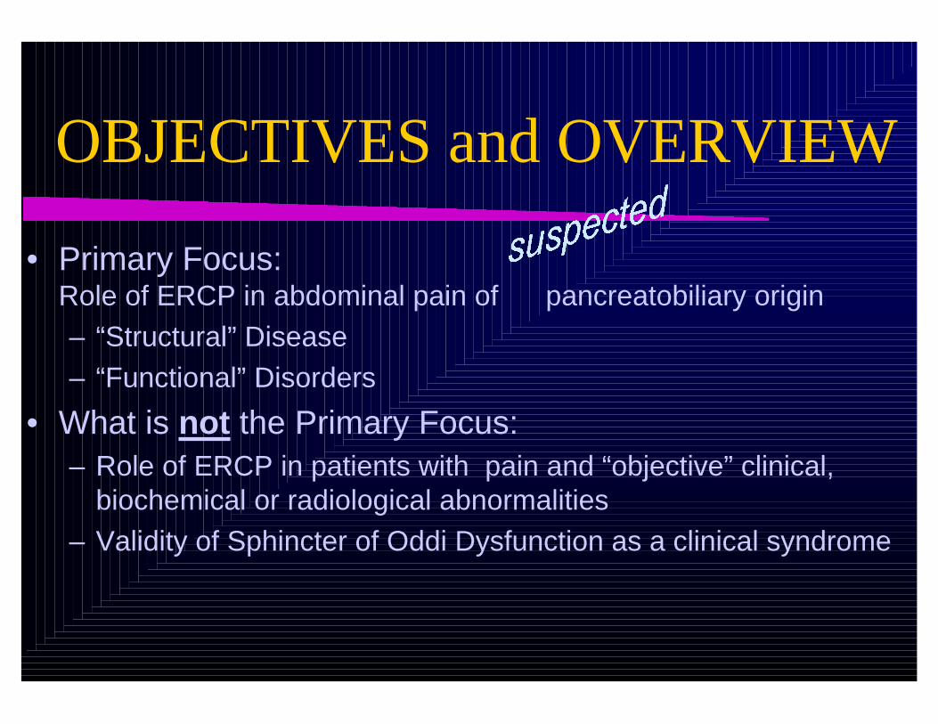
OBJECTIVES and OVERVIEW
• Primary Focus: Role of ERCP in abdominal pain of pancreatobiliary origin– “Structural” Disease– “Functional” Disorders
• What is not the Primary Focus:– Role of ERCP in patients with pain and “objective” clinical,
biochemical or radiological abnormalities– Validity of Sphincter of Oddi Dysfunction as a clinical syndrome

Aims of ERCP in Unexplained Abdominal Pain
• Discover subtle “structural” abnormalities• Diagnose sphincter of Oddi dysfunction• Others
– Bile collection?

ERCP and Pain: Underlying Assumptions
ü The clinical pattern of chronic pain can reliably indicate pancreatic or biliary disease, even in the absence of “objective” findings
ü In the absence of morphological changes, it is important to exclude functional changes in the pancreatobiliary sphincter in these patients
ü These morphological or functional changes correlate with pain and their detection leads to effective treatment

Origin of Chronic Right Upper Quadrant Pain
• 22 consecutive patients with severe chronic RUQ pain
• Average work-up:– 3.5 consultations– 7.3 procedures– 1.7 operations– >20 blood tests
Kingham and Dawson Gut 1985;26:783-788

40 ml
250 ml
Kingham and Dawson Gut 1985;26:783-788

Balloon Distention Sites and Reproduction of Spontaneous RUQ Pain
• Esophagus 0• Duodenum 6• Jejunum 15• Ileum 12• Right colon 9• Left colon 0
21 of 22 at least one site
12 of 22 > one site

Carlson et al (Br J Surg 1992;79:1342-45)• 5000 ERCPs (1976-1989)• 384 patients with post-cholecystectomy *pain• 4 groups:
– Pain only*– Pain and clinical/biochemical abnormality– Pain and Imaging Abnormality– Pain and both clinical/biochemical or imaging abnormality
• *Caveats: – Presumably for gallstone disease– Imaging may not have been done in every patient
Diagnostic Yield of ERCP in Abdominal Pain

528 (76%)33Pain + C/B + Imaging
834 (60%)57Pain + imaging
1576 (54%)140Pain + C/B
9 (CP = 2; “Amp stenosis” = 2)
20 (13%)150Pain Only
OthersCBD stonesN
Diagnostic Yield of ERCP in Abdominal Pain
Carlson et al

Diagnostic Yield of ERCP in Abdominal Pain
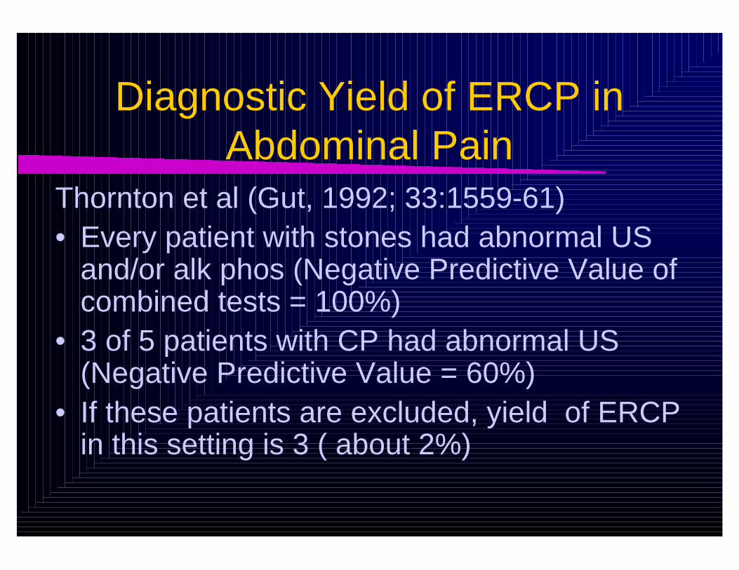
Thornton et al (Gut, 1992; 33:1559-61)• 138/1005 ERCPs between 1989 and 1990 for
evaluation of abdominal pain• 130 patients analyzed• Findings
– Bile stones 10– CP 5– Ca 1TOTAL 16 (12%)
Diagnostic Yield of ERCP in Abdominal Pain
Thornton et al (Gut, 1992; 33:1559-61)• Every patient with stones had abnormal US
and/or alk phos (Negative Predictive Value of combined tests = 100%)
• 3 of 5 patients with CP had abnormal US (Negative Predictive Value = 60%)
• If these patients are excluded, yield of ERCP in this setting is 3 ( about 2%)
Diagnostic Yield of ERCP in Abdominal Pain
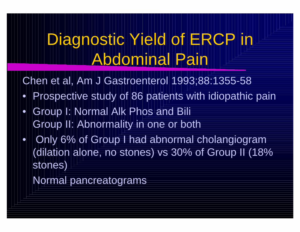
Chen et al, Am J Gastroenterol 1993;88:1355-58• Prospective study of 86 patients with idiopathic pain• Group I: Normal Alk Phos and Bili
Group II: Abnormality in one or both• Only 6% of Group I had abnormal cholangiogram
(dilation alone, no stones) vs 30% of Group II (18% stones)Normal pancreatograms
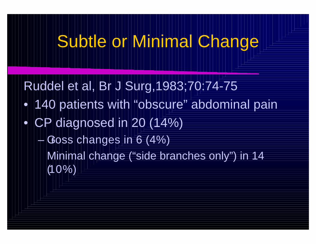
Subtle or Minimal Change
Ruddel et al, Br J Surg,1983;70:74-75• 140 patients with “obscure” abdominal pain• CP diagnosed in 20 (14%)
– Gross changes in 6 (4%)Minimal change (“side branches only”) in 14 (10%)

Subtle or Minimal Change
• Clinical significance of subtle ductographic changes controversial
Ma y be found in elderly or at autopsy in the absence of any evidence for pancreatitis (Anand et al, Gastrointest
1989;35:210; Schmitz--14)
– Of 20 patients with normal secretin-pancreozymin test and abnormal ERCP 17 remained free of any evidence of pancreatitis after a mean follow-up of 84 months (Lankisch et al, Pancreas 1996;12:149-52)
• Conversely, ERCP may miss “true” CP not involving the ducts (Walsh et al, Gut 1992;33:1566-71)

ERCP in “Functional Disorders”
Sphincter of Oddi Dysfunction (Hogan and Geenan)
Type I: • sGOT or > 2 x normal (> twice)• Delayed drainage of contrast (>45 minutes)• Dilated CBD (> 12 mm)
Type II– One or more of the above
Type III– None of the above (pain only)
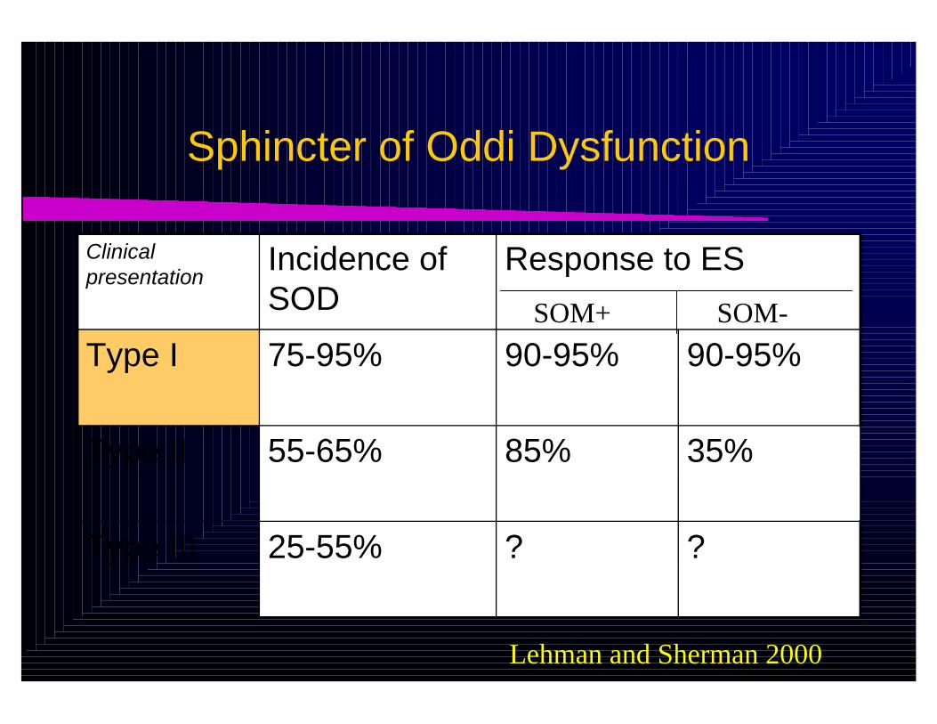
Sphincter of Oddi Dysfunction
?
85%
90-95%
Response to ES
?25-55%Type III
35%55-65%Type II
90-95%75-95%Type I
Incidence of SOD
Clinical presentation
SOM+ SOM-
Lehman and Sherman 2000
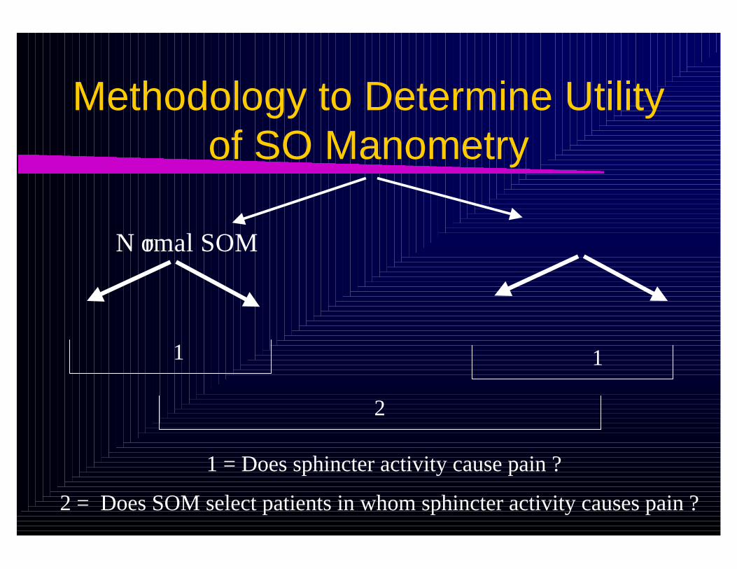
Methodology to Determine Utility of SO Manometry
N ormal SOM
1 1
2
1 = Does sphincter activity cause pain ?
2 = Does SOM select patients in whom sphincter activity causes pain ?

Type II SOD: Randomized before SOM
0
20
40
60
80
100
% good or fair
response
Normal SOM SOM > 40
1 year symptom response to ES
ShamES
Geenen et al. NEJM 1989;320:82-7
3rd Qtr

SOD*: Randomized after SOM
0
20
40
60
80
100
% good or fair
response
Normal SOM SOM > 40
2 year symptom response to ES
ShamES
Toulli et al Gut 2000;46:98-102
3rd Qtr
* 80% type I or II

What about Type III SOD?
Normal SOM Abnormal SOM
ES ES1 1
2
1 = Does sphincter activity cause pain ?
2 = Does SOM select patients in whom sphincter activity causes pain ?
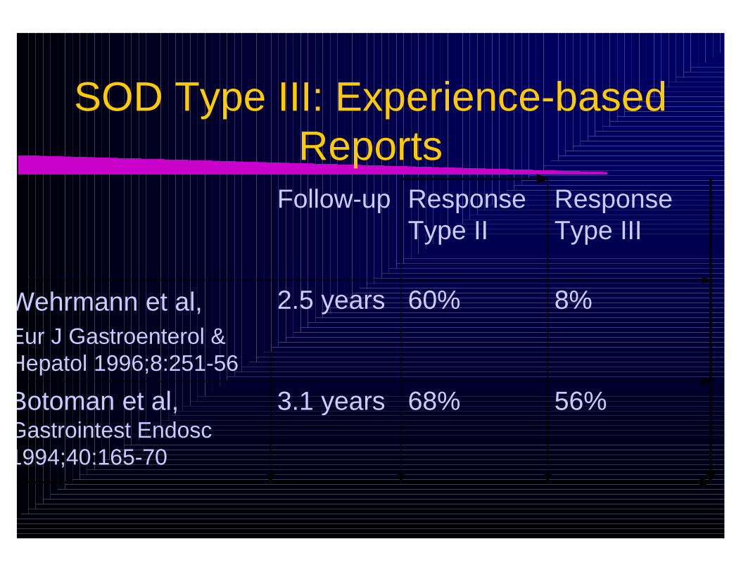
SOD Type III: Experience-based Reports
56%68%3.1 yearsBotoman et al, Gastrointest Endosc 1994;40:165-70
8%60%2.5 yearsWehrmann et al, Eur J Gastroenterol & Hepatol 1996;8:251-56
Response Type III
Response Type II
Follow-up

SOD Type III: pain only
• Assuming that 50% of these patients will have a positive SOM
• Even assuming the “best” response rate of 50%, and a conservative placebo response of 35%, this translates into an NNT of 13

Poor Correlation Between SOM and Response to ES
Two broad explanations• SO Dysfunction is a marker but not a cause for pain
in Type III patients– Overlap with other functional pain syndromes: NCCP, IBS– Similar psychosocial profiles– Visceral hyperalgesia
• SO Dysfunction plays a causative role in a subset of patients in Type III patients, but SOM cannot accurately detect this– Not physiological– Does not provide correlation with pain

Alternatives to SOM
• Imaging Tests: ? More physiological– Fatty Meal Sonography (FMS)
sensitivity: 74%specificity: 100%
– Quantitative Hepatobiliary Scintigraphy (QHBS)sensitivity: 67-100%specificity: 80-100%
• Problems– comparison to “invalid” gold standard (SOM)
True gold standard should be response to ES at 1 year– Limited data on Type III patients

Alternatives to SOM
• Therapeutic Trials: Requirements – A reliable and safe means of lowering SO pressure– Relief of pain implies sphincter at fault: patient may
benefit from ES– If not, ES not necessary
• Possible candidates:– Calcium channel antagonists– BoTox

S OD Type II: Response to
0 5 10 15 20
Painepisodes
Analgesicrequirements
ER visits
Pain score
NifedipinePlacebo
***
***
*
**
* P < 0.05** P < 0.01***P < 0.001
Khuroo et al. Br J Clin Pharmac 1992;33:477-85
0
20
40
60
80
100
% responders
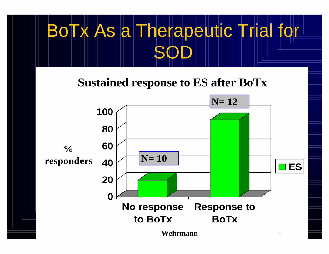
No responseto BoTx
Response toBoTx
Sustained response to ES after BoTx
ESN= 10
N= 12
3rd Qtr
Wehrmann -
BoTx As a Therapeutic Trial for SOD
Summary
• Clinical criteria do not reliably indicate the true site of origin of pain
• “Structural Disease”– In patients with pain only, the yield of ERCP for
gross structural abnormalities such as biliary stones, chronic pancreatitis and cancer is negligible
– Modern imaging (US, spiral CT, MRCP) are able to detect most if not all such cases
–a bno rmalit ies th at ma y be detec ted rema i ns unc lea r

Functional DisordersNo evidence base to support utility of SOM in
– High complication rate and degree of difficulty
– Ob s ession with implicating sphincter distracted

Directions for Research
• Better understanding of minimal change pancreatitis– Ability to acquire pancreatic tissue at ERCP
• Need for Randomized Control Trials in Type III SOD– ES– Tricyclic antidepressants– Cognitive behavioral therapy
• Need to develop more reliable ways to predict SOD as cause of pain– More physiologic imaging with pain response as gold
standard– Therapeutic Trials


