

Copyright is owned by the Author of the thesis. Permission is given for a copy to be downloaded by an individual for the purpose of research and private study only. The thesis may not be reproduced elsewhere without the permission of the Author.

This document is a copy of the Thesis submitted by
JOHN DUNSTAN ATKINSON
codeword , ASSURED,
in partial fulfilment of the requirements of the
Degree of MASTER of AGRICULTURAL SCIENCE
MASSEY COLEGE University of New Zealand
1932.
STUDIES ON THE DI.EBACK OF LAC.EBARKS.
M:vxosporium Hoheria. Il•£•.fil2.•
By, "Assured".

T.ABL~ OF CONTENTS.
Page Introduction 1.
Historical 2.
Distribution 3.
Types of injury prevalent on the hosts 3.
Cultural studies. a. Method of isolation 8. Types 0£ fungi isolated 8. Reactions of fungus to temperature 10. Reactions of fungus to media 12. Description of colony characters 13.
Spore Germination 13.
Inoculation experiments 15.
Methods of transmission and infection 19.
Overwintering 19.
Life history of causal organis:gl. 20.
Control. 21.
Morphology 21.
Comparism of Gleoaporium and Myxosporium. 24.
Diagnosis of M.Hoheria n.f.ap. 25.
SUmmary 27.
Appendix 2 9 •
Literature cited. 31.

STUDIES ON THE DIEBACK OF LACEB.ARKS.
14,yxosporium Hoheria. n.f.sp.
INTROIXJCTION.
The Maori names Houi, Whauwhi and Houhere, or the
settlers terms lacebark, and ribbon-wood, cover several species
of flowering plants belonging to the order Malvalea. These
species, which are all indigenous to New Zealand, fall into the
genera Hoheria, and Plagianthua, Laing and Blackwell (1927) list
the following eight speoiea:
Hoheria do do do
populnea. aexstylosa. angustifolia. Lyallii.
Hoheria glabrata. do Allanii.
Plagianthus divaricatus. do betulinus.
H.Populnea is found chiefly in the Auckland and
North AUOltl.and districts, as a member of the subtropical rain
forest, but Laing has recently recorded its occurrence in
Karamea. H.aexstylosa occurs throughout both islands as a member
of the lowland bush communities. H.angustifolia is typically
a south Island plant found in large numbers on Banks penninsula,
but is also found in the south of the North Island. H.Lyallii
is a deciduous shrub growing in the mountainous districts of the
South Island. H. glabrata belongs to the eubalpine flora, growing
usually in situations where it oen obtain abundant light, e.g.
recent landslips. Cockayne (1928). H.illanii is a small leaved
shrub recorded from the Rakaia gorge, Canterbury.
Ple.gianthus divarioatus belongs to the coatal. flora,
growing as a dense bushy shrub on .salt meadows end round tidal
estur-iea. - P.betulinue ocoure throughout the Dominion and out-
lying islands to the South'• It is the largest of the ribbon-woods,
growing into a canopy tree up to 60ft. in height. {Allan. 1928)

In many parts particularly the southern districts it assumes
a deciduous habit.
2.
Like many other New Zealand trees the lacebarks show a
great differ:ence between their juvenile and mature forms. Thus
P.betulinus in its adolescent stage is a small divaricating shrub
with small leaves only 1/3 - 3/4" iong, while in its mature form
it becomes a tall tree with leaves l - 4 11 long.
Economically the lacebarks are of no great importance.
The Maori used the tough, lace-like inner bark for making mats,
rope and twine, but today these arts have been almost lost. Two
of the wild species (H.populnea and H. sexstylosa) and their
hybrids are widely cultivated in the North Island as ornamental
shrubs and fast growing shelter trees.
Some years ago it was observed that these cultigens
~ere suffering from a form of dieback. Today the condition has
become widespread, and large numbers of trees have been killed
outright, so that the usefulness of these plants is seriously
curtailed. Field observations indicated the presence of a
pathogenic :fungus, and the present work was undertaken in an
attempt to determine the oause of the disease,.
HSITORICAL.
Although no record has been made of a fungus causing
diebaok of laoebarks, it was observed about 1920 that fungous
fruotificatioll:lof the Myxosporium type often appeared on the
bark immediately following the death of twigs and branches.
During the years 1920 - 1923 specimens of lacebark bearing these
acervuli were collected from Waikato, Southland, Canterbury and
Wellington; in each case they were found only on the stems.
Dr. Cunningham, Government Mycologist, New Zealand, identified
this organism as a member of the form-genus Myxosporium, and
later his decision was confirmed by Dr. Butler, Imperial Bureau
of Myoology, Kew. Though it appeared probable that this :fungus
was causing diebaok, no attempt was made to prove its pathogenicity.

' l. v•
Saooordo lists 101 form-species ascribed to the genus
Myxosporium, but of these more than hali are imperfectly described,
and therefore of little value in diagnosis. Only three of those
listed were found on hosts belonging to the same order as the
laoebarka (Malvales). There is no record of a Myxosporium species
ocouring on a species of the :fomily Malvaceae, to which the lacebarks
belong.
A survey of the literature shows that the genus
Myxosporium has received little attention. The few species with
which experiments have been mude have proved to be weak parasites
of secondary importance or saprophytes associated with other
diseases. Miss Gilchrist (19G3), Briton-Jones (1925), and Zeller
(1926), found that M.corticolum attacked only weak or unhealthy
apple trees, and was not a serious pathogen. Day (1928) showed
that M.abietinum (Rostrup) was commonly found in tissues of t h e
Sitka Spruce, and Douglas F'ir, t hat ha d been damaged by frost, but
that it seldom spread beyond the injured areas. Fraulein Beck (1926)
has shown that M.oingulatum is the eause of an anthracnose disease
of privet (Ligustrum vulgare) seedlings, and also that Gnomonia
oingulata (Beck) . is the p erfect stage of this fungus. Other workers
have shown that certain Myxosporium form-species are imperfect forms
of asoomycetoue fungi. Miss Wilson (1928) demonstrated that M.
abietinum is the conidial stage of Dermatea livida (B.et Br.)
DISTRIBUTION.
Specimens of lacebark showing Myxosporium fruotifica
tione have been collected from Waikato, Hawke 1 s Bay, Manawatu,
Wellington, Nelson, Canterbury and Southland. Collections have not
been made from the remaining districts of the Dominion, but as the
disease is known to occur from Waikato to Southland it is probable
that the trouble is general throughout New Zealand·.
TYPES OF INJURY PREVALENT ON THE HOSTS.
Dead .wood on laoebarks in the field mey be grouped
on superficial appearances into four classes:-

l. Small isolated dead twigs.
2. Lesions on branches and trunks not bearing the
acervuli of .M;yxosporium.
3. Lesions on branches and trunks bearing the
acervuli of Myxosporium.
4.
4. Ins ect injury common in the forks of the smaller
branches.
1. Small isolated dead twigs appear to be common to
many different trees, and are found on the three host species
treated in this paper, H. populnea; H.sexstylosa; and p. betulinus.
These twigs may be apparently free from fungous infection, or show
fungous fructificatioll!of various types. Inoculatiom.with fungi
obtained f'rom such twigs have given only negative results, and it
is probable that death was caused by mechanical injury or natural.
agents such as frost, or insufficient light.
2. The only fungous disease recorded among lacebarks
is the rust, Puccinia Plagianthi.(CWiningham 1931):. Excluding
lesions caused by this pathogen, it is more common to f'ind dead
wood, without Ivry:x,osporium fructificationa, on H. sexstylosa than on
H• popu1nea. Mature trees of H • .exatylosa of'ten show long narr.ow
lesions on the trunk and branches. These are slightly depressed,
arul reddish-brown flecked with white, standing out quite clearly
against the darker trunk. Fungous fruotif'ications of varying
types are found on such lesions. Whilst no pycnidia of the Phoma
type have been observed on them, yet on making isolations from
such areas a fungus, which forms pycnidia in culture, is obtained
with fair regularity. Experiments with this fungus will be dealt
with in a later section.
3. Dieback occurs commonly on H. popu1nea and to a lesser
extent on H.sexstylosa, and p. betulinus. The first symptom of
the disease . is the wilting and death of the leaves above the point
of infection. At this point the bark appears normal, but is soft
and spongy to the touch, for the under-lying cells have eollapsed.

5.
Canker formation does not take place, and it is necessary to cut
away the outer bark to find the edge of the lesion. The latter
appears as a light brown zone quite distinct 1rom the greenish
white of healthy tissue. As the lesion beoomes older the infected
wood darkens, in some cases turning almost blue~,
· -t . . .. •
Fig. l.1• H.populnea hybrids showing severe dieback.
(Photo by author)
Shortly after the death of the infected branch small lumps appear
on the bark. These are formed by the developing acervul.1, which
eventually rupture the bark, and push it aside until at maturity it
appears as a wall bounding the central, pulvinate, salmon-pink
spore-mass. The pink acervuli are a characteristic feature of
diebaok, and occur on diseased woody tissue of all ages, but they
have not as yet been found on the leavoa. The pustules are ~ene1~al1y
ell.ipsoid in shape varying in size from 2 x ¼ m.m. to 4 x ½ m.in.,
and usually lie with their long a.xis parallel to the length of the
branob. They may be thickly clustered or widely scattered over
the infected area, and often occur in rows following the grain of
the bark, Usually single, the aoervuli occasionally merge into
one another forming larger irregular musses.

--=-.,:i::.;;:zr~~~,~~f{{fl£Ji;1i{ti~~i1.S:~i-\;c~t~~ •~,. -~f'~W. "'.;;i_-. .. ..A-~~.:.-:..~# • • - "· .. .., -11 •-••~~--• • •·••~ • ~J"-A-.. ...... 41,. ,.,--_.."':..,.fi~: ·, .,:"'£!.r.V_~":.-~~- • ~ i;:swtf='", ,-, _,. -_- -_., ; :~-•~\'"<:t:t~~#_]..}tj,.ft, . :tf6"°::' . -.· --~
)'f,-,. ,;,- • ,:'.lil'..,-.~:" ~"-• ;1•. ,,. : :;:. • ;.,· .,.•., l r'.i,"t :: "i:-~·· •:~ -':X"t'"°;;"'Z~
//i{~~t4~V?HI?l~f:,:;.;t~-~f~~w~,c
Fig. 2. Myxosporium aoervuli on the bark of
H. populnea. (Natural size).
(Photo by H.Drake).
The spores developing from the aoervulus are held together by a
gelatinous matrix, which is hygrosoopio in nature. In wet weather
the spores are extruded from the acervulus, and the gelatinous
material is disolved awey. Some of them may be carried down to
the ground by the water, others left to dry on the bark and be blown
away.
Once the fungus is established it grows rapidly, under
favourable conditions, developing a copious intracellular mycelium.
The hyphae spread in all directions, disorganising the cortex and
cambium and penetrating into the wood vessels by way of the . pits
in their walls. The distruction of the cortex and cambium prevents
further growth of the branch at the point of infection, and· stops
the downward passage of elaborated food materials. By girdling
the branch and blocking the wood vessels with a mass of hyphae, the
fungus outs off supplies of food and water, and the branch rapidly

·, ..
Fig. 3 •. Myxosporiwn hyphae in the wood vessels. x 370.
(original)
4. Apart from fungous attack considerable damage is
caused by insects, particularly a species of beetle. The grubs of
this beetle feed usually in the forks of the smaller branches, often
killing them by eating away all the outer layers of tissue from their
bas~Q. Branches killed by their attacks rapidly become covered
with all manner of fungous growths, but the characteristic, deep,
ireegular wounds are usually sufficient in themselves to account for
the death of the part concerned. The insects have not yet been
found feeding on dead wood, and the Myxosporium does not form
cankers. Therefore if a dead branch has a canker- like wound at its
base, it is reasonably safe to assume that death was caused -by
insects, and not by the diebaok organism •
. It would appear from :field observations that H.populnea
and closely related hybrids are more susceptible to diebaok _ than
H.sexatylosa or p. betulinus. No evidence regarding the incidence
of the disease on other lacebark species is available. Over 300
H.popul.nea cultigens were examined, and of these fully 6CJ% showed
symptoms of dieback. On the other hand, acervuli could not be
found on more thun l<J% of four hundred H.sexstylnea specimens.

tj.
Only a few plants of P.betulinus were examined but here again the
percentage infection was low. H. populnea, however, shows few
lesions other than those o:f dieback, while H.sexstylosa shows many
lesions which do not bear Myxosporium acervuli •
CULTURAL STUDIES.
To obtain purecultures of the causal organism, or
organisms fifty specimens were collected from diseased lacebarks.
In this first series no discrimination was made between the types
of lesion or the species of the host. Isolations were made from
the specimens by the following method:-
The specimen was surface sterilised with 1:1000
acidulated Mecuric chloride and then introduced into the culture
cabinet. With a sterile scalpel the outer layers of bark were cut
aw~ exposing the edges of the lesion. A second scalpel was used
to remove small pieces o:f tissue from the edge of the lesion. Four
such pieces were taken :from each lesion and placed equidistant :from
one another towards the periphery of a petri-dish containing prune
dextrose agar. (see appendix). The dishes were incubated at 21°c.
until the fungus myoelium had grown well clear of the wood, usually
5 - 10 m.m., when a small section of the outer edge of the colony
was transferred to a fresh dish of potato dextrose agar, (see appendix)
and again incubated at 21°c. By this method clean colonies were
oo.tained in the majority of cases.
As a check to these isolations single spore cultures
were made by the poured plate method from Myxosporium spores, taken
from acervuli on the host. By using prune dextrose agar as the
pouring medium it was possible to obtain single spore colonies free -
from contamination. All contaminated cultures were discarded.
The isolants showed a distinct relation to the type of
lesion from which they were obtained, and could be arranged into
three groups:-
1. Isolations from 20 lesions showing pink aoe-rvuli
yielded 12 cultures of Myxosporium, and three of Neotria oinnabarina, +hn -ft.-nn.;~~-- _,,,..._.,,,_"_ 1,..-~-- - .... -J.. .... __ • _ _, ..L._.!1

2. Isolations from l5 lesions showing no constant
fungw.sfructifications gave 12 cultures of a fast growing fungus,
which produced pyenida and spores of the Fhoma type in five days,
and three unidentified cultures.
3. Isolations from the bases of 15 small twigs which
had died back to their parent branches produced several. different
species of fungi. Of these a species of the genus Fusarium and •an
Ascomycete of the family Sphaeriaceae appeared in three,and four
cultures respectively.
The regularity with which the first two types of
lesions produced the fungi mentioned, indicated that these
organisms might be pathogenic. In the third group no single
organism appeared to be regularly associated with the dead twigs.
Thus it seemed likely that the fungi concerned were merely
saprophytes. Inoculations made with this group of fungi gave
only negative results, and in view of these facts, small isolated
dead twigs were set aside as having littl~ if any connection with
the dieback condition.
A second collection of seventy specimens was made
from diseased plants of the species, H.populnea, H.sexstylosa,
and P.betulinus in Nelson, Wellington, Hawke 1 s Bay, and Manawatu.
Small dead twigs were excluded from this aeries. Isolations
were made from each specimen by the meth9d previously described.
The results confi1~1ed those obtained from the first series;
where pink acervull were present the lesion usually yielded the
Myxosporium, occ usionally N.oinnabarina; from the remaining
lesions cultures of the Phoma-like fungus were generally obtained.
ill three of these- fungi were obtained in culture from isolations
made during the winter and spring months " - , showing that they can
normally overwinter by means of an internal myoelium.
The Myxosporium was the only one of the fungi
isolated from lacebarks, that inoculation experiments proved
pa thog~mio. The following tests were carried out with this
fungue 0 to _detennine its physiological. reactions to light,
temperature, and certain synthetic media.

obtained by the poured plate method of single spore isolation,
from spores produced in culture. These pure cultures were used
for inoculations and cultural experiments. It was found that
normal spores were produced in 15-18 days when the fungus was
grown on potato dextrose agar at 21°c. in a dark incubator, and
spore production was similar on autoclaved H.sexstylosa stems.
kept under the same conditions. This behaviour differed from
that reported by Miss Gilchrist (1923) for M.corticolum (Edgert).
She found that this species produced abnormally small spores on
nutrient agar, yet Lewis(l912), working with the same fungus,
records that normal spores were produced on sterilised bean pods.
To determine the effects of light and temperature on
cultures of the Myxosporium the following tests were carried out.
Twelve petri - dishes of potato dextrose agar were sown from a
single pure colony, and three dishes placed under each of the
following conditions:-
1. 30 - s0c. dark refrigerator.
2. 21°c.
3. 30°c.
4. 21°c.
dark incubator.
dark incubator.
glass fronted incubator.
Ricaminationa were made daily and records kept of the
growth rate as measured by the colony diameter.
Brown (1925) has shown that environment has a direct
and most marked effect upon the growth rate of certain strains of
Fusaria. Unfavourable conditions produced staling, one feature
of which was a reduction of sporulation. Spore production
is desirable whether the culture is to be used as an aid to
identification or as a source of inocul.um. , • Therefore the
conditions most suitable for routine cul.tural use would be those
producing a freely sporulating, or non-staling colony. Brown
found that the daily rate of increase in colony diameter was a
convenient measure of staling, and therefore an indication of the
worth of any particular medium. It is reasonable to assume
that the Myxosporium under consideration would react similarly
to changes of environment, and its growth rate has been taken as a
measure of the relative value Gittr any set of condittons.

No measurable differences could be found between
the cultures grown in the dark and those subjected to the
norme.l al.ternationoi night and day. Temperature, however,
had a marked effect upon growth. These results are shown
graphically in Fig. 4.
Fig. 4.
-~
~· __ .3o' c
_:::.- ·- ·- ·- ·- -- - ·- •- ·- - -- - -- - - - - - - -- - - - --
,0.
~- 4 • ,
Growth curves of Myxosporium Hoheria at varying
temperatures.
11.
These cultures to be incubated at 3o0 c. were kept
at 21°c. for two days after sowing to allow growth a good start.
On transfer to the 30°c. incubator the growth rate dropped
rapidly until the colony became dormant, and it remained i,n this
state for five days. , At the end of this time the cultures were
taken baok to the 21°c. incubator, and within two deys the normal
the fungus was killed. Replication of these tests produced
no significant variation in results. As the fungus was apparentl.y
indifferent to light, and gave rapid, non-staling growth at 21°c.,
all later cultural work was carried out in a dark incubator at this
temperature.
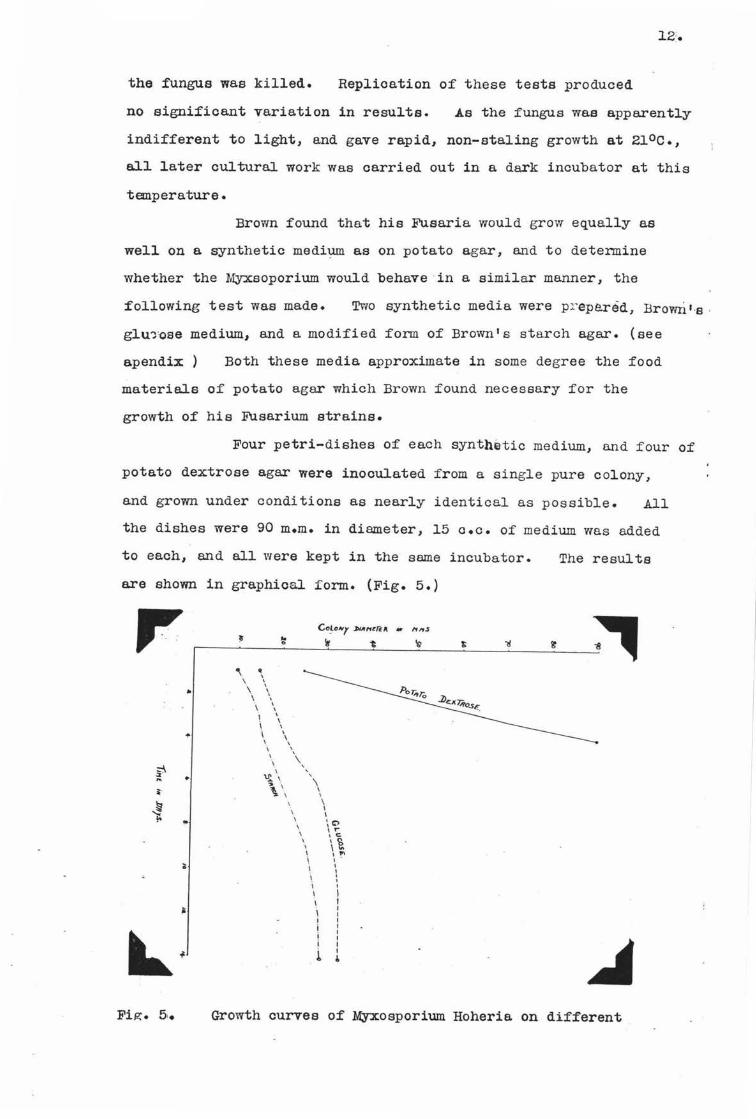
Brown found that his Fusaria would grow equally as
wel.l on a synthetic medi~ as on potato agar, and to detennine
whether the Myxsoporium would behave in a similar manner, the
following test was made. Two synthetic media were pr epe.reid, Browri '-s .
glu~ose medium, and a modified form of Brown's starch agar. (see
apendix) Both these media approximate in some degree the food
materials of potato agar which Brown found necessary for the
growth of his Fusarium strains.
Four petri-dishes of each synthetic medium, and four of
potato dextrose agar were inoculated from a single pure colony,
and grown under conditions as nearly identical as possible. All
the dishes were 90 m.m. in diameter, 15 o.c. of medium was added
to each, and all were kept in the same incubator.
are shown in graphical form. (Fig. 5.) ,,. ..
"'
~ .. .. .. ~ ~ ..
'\ ~ . \ \ \
\ ' \ \
I I
\ \ \ \ \ \
\., \,,, ~ \ \ \ \ \
i i I I i I
\
\ I I ; c;, I ,, c
\ 'a I ... I I
\ ' \ I I I I I I I I
l l
The results
Fig. 5,. Growth curves of Myxosporium Hoheria on different
.L'.J .
Tile curves representing growth on the synthetic media
show staling, and a definite imeriority to potato dextrose agar.
A duplicate series gave similar results. Cellulose in the fonn
of dessicated filter paper was added to each medium in a third
series but produced no change in growth :form.
No saltants appeared when the fungus was grown on
potato dextrose agur, and different batches of this medium gave
colonies that could not be distinguished from one another by any
measurable, morphological, or physiological characters. Tllus t he
potato dextrose agar was superior to the other media used, and
apparently quite satie:factory as a medium :for growing t his fungus.
GROWTH AND APPEA.RA.NCE OF QYLTURES.
Colony characters on potato dextrose a gar at 21°c.
in dark incubator, 90 m.m:. petri-dish, 15 c.c. of medium per dish.
Colony :fast growing, reaching periphery in f ive to
seven days; margin entire. At :first slimy when viewed :from
above; when 25 - 35 m.m. in diameter type of growth changes; and
outer parts show small tu:fts of aerial hyphae. These increase
in size and dens~ty until outer zone is grumose, w~ile central
ring becomes pubescent. Light pink, scattered masses o:f
conidia can be seen when colony is one month old. At this ac-e
the myoelial mass is white from above; :from below, medium ie
coloured light brown, with darker brown spots under spore clumps.
On this mediwn a characteristic odour is produced, bearing a
slight resemblance to the smell o:f esthera :from apples in cool store_.
SPORE GER1UNATION.
Be:fore making inoculation the germination capacity
of the ~osporium spores, and those o:f the Phoma type :fungus,
was teated -by two methods:-
1. Spores were placed in a drop of sterile water hanging
:from ·the cover slip into the cavity o:f a hollow ground slide.

Fig. 6-. One month old colony of M.Hoheria grown on potato
dextrose agar at 21 °c. in the dark. X5/ 6
(Photo by T.Gabriel)
2. Spores were sown on a thin flat film of potato
dextrose agar, also suspended over the cavity of a hollow
ground slide.
21°0.
All the slides vrnre incubated in petri-dishes at
The atmosphere was JJtept saturated by placing wet
filter ·paper in the bottoms of the dishes.
14;.
The germination percentage was lower in water than
on the nutrient medium, but no differences were observed in the
manner of germination. Myxosporiuru spores taken :from acervuli
on the host gave 60 70<fo germination, those from pure cultures
slightly less ( 50%) in 16 hours. The Phoma type spores could
be found only in culture, and these showed a 5°'fo germination in
24 hours. The Myxosporium spore . puts out from one to three
germ tubes. These usually appear towards the ends o:f the spore,
but mey grow out from any portion of the wall.. If on a
nutrient medium the hyphae grow rapidly, become septate, and

15.
INOCULATION EXl?ERIMENTS.
Seedlings for inoculation were collected early in the
year, and grown in pots, away from insects, for three months before
use. As no sign of imection appeared during that period the
plants were assumed to be free from disease and were not sterilised
in any we;y. The collection represented three species of lacebark,
H. populnea, H• sexstylosa, and P. betulinus, together with a
number of horticultural varieties of these.
The inoculum was taken from pure lines of
Myxosporium. Some of the first inoculations were made with
myoelium alone, but these in every case showed negative results.
Later inoculations were made with spores from agar, or sterilised
twig cultures, a certain amount of mycelium being included
incd.dentally. Four methods of inoculation were used; the
details of which are as follows:-
1. With a sterile scalpel a small piece of tissue was
lifted by cutting upwards along tlle stem. From below this
flap a piece of wood was removed, the inoculum placed in the cavity,
and the f'lap pressed back into place. To prevent drying out
the wound was bound with moist cotton wool and f~nally cellophane .•
2. As f'or No. 1. but some of the tissue under the flap
was killed with a hot needle bef'ore inserting the inoculum.
~. Injecting a suspension of spores with a hypedermic
needle, the method adopted by Neill and Brien (1931),.
4. A portion of the stem was killed by ring-barking and
the f'ungus established as a saprophyte on the dead parts.
One, or two uninoculated wounds were made on each
plant to check the results. The results of these inoculations
are shown in tabular form (Table :L.) Seedling plants were
used throughout this series of' tests.
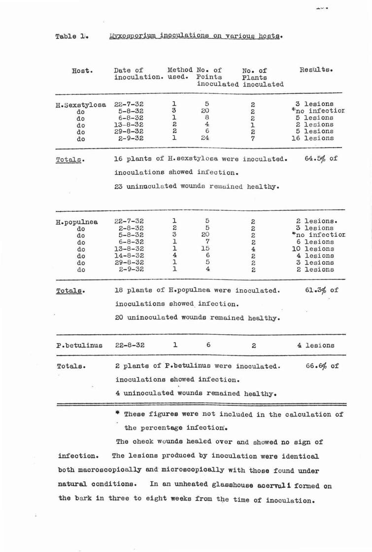
Tabl.e l i.
Host.
H.sexstylosa do do do do do
Totals.
H.populnea do do do do do do do
Total.a.
P.betulinus
Total.a.
infection.
JmQ_s~ori.lllll inoculations on various hosta.
Date of Method No. of No. of inoculation. used. Points Plants
inoculated inoculated
22-7-32 l 5 2 5-8-32 3 20 2 6-8-32 l 8 2
13-8-32 2 4 1 29-8-32 2 6 2 2-9-32 l 24 7
].6 pl.ants of H.sexstylosa were inoculated.
inoculations showed inf'ection.
23 uninuculated wounds remained healthy.
22-7-32 1 5 2 2-8-32 2 5 2 5-8-32 3 20 2 6-8-32 1 7 2
13-8-32 ]. 15 4 14-8-32 4 6 2 29-8-32 1 5 2
2-9-32 l 4 2
18 plants of H.populnea. were inoculated.
inoculations showed infection.
20 uninoculated wounds remained healthy.
22-8-32 ]. 6 2
2 pl.ants of P.betulinus were inoculated.
inoculations showed infection.
4 uninoculated wounds remained healthy.
Results.
3 lesions *no in£ectior.
5 lesions 2 lesions 5 lesions
16 lesions
64.5% of
2 lesions. 3 lesions
..,no infectio:r: 6 lesions
10 lesions 4 lesions 3 lesions 2 lesions
61.3% of
4 lesions
66.~ of
• These figures were not included in the calculation of
the percentage infection.
The check wounds healed over and showed no sign of
The lesions produced by inoculation were identical.
both macroscopioally and microecopioall.y with those found under
natural conditions. In an unheated glasshouse acervul 1 formed on
the bark in three to eight weeks from t~e time of inoculation.

each case cultures of Myxosporiuni were obtained.
Fig.7. H.populnea seedling showing branch killed as a rellUlt of inoculo.tion with M.Hoheria. The cross marks the point of inoculation.
(Photo by T.Gabriel).
From these results it appears that this Myxosporium
is an active parasite on the three species into which it has been
inoculated.
Further inoculations were made with the other fungi
isolated from diseased lacebarks. Ninety inoculations on seedlings
and thirty-two on mature tree in the field were made during the
period· January/ July 1932, with the fungus of the Phoma type.
The methods of inoculation were the same as those mentioned
previously. On seedlings these have failed to show any positive
results, but on old trees the fungus in some cases appears to
have penetrated a short distance into the tissue. It has been
reisolated from the extreme edge of the wound three months from
the cl.ate of inoculation, but some of the original inoculum may
have retained its vitality for this period. Until :further
evidence is obtained this fungus cannot be considered parasitic.

18.
The oause o:f the original oankers has not yet been determined!.
Seventy-:five inoculations were made with the speoies
o:f Sphaeriaoeae; sixty on seedlings and :fi:fteen on mature trees.
No positive results were obtained. In cases where the :fungus was
established as a saprophyte on wood killed by heat it :failed to
spread to living tissue. Twenty-two inoculations made with the
speoiea o:f Fusarium also :failed to show any imection.
o:f these three :fungi have as yet been identified.
None
Both the aecigerous ahd conidial stages o:f Nectaria
connabarina have been collected several times from H.sexatyloaa,
and the :fungus has also been obtained in culture :from the edges
o:f lesions. Although this organism is a weak parasite on a
wide re.nge of hosts (Cunningham 1925), no experimental evidence
oan as yet be brought :forward to show that it is parasitic on
the lace bark.
The swnmarised results of this second aeries of
inoculations are given in Table 2.
Table 2'• Ino_s:jJJ~tions with various fungi.
Fungus Host No. of Total No. Period over Results. Species. plant. plants inocula- which inocu-
treated.tione. lations were made.
Species o:f 11.sexstylosa 30 101 Jan. - July 1932 No Inf'ect-the Phoma ion • . . type. H.populnea 7 21 Jan. - July 1932 No Imect- -
ion. · . Sphaeria- H.aexstylosa 11 45 Mar. - July 1932 No Inf'eot-aoe.ae ion. species. H.pupulnea 8 30 Mar. - July 193~ No In:fect-
ion. Fuse.rium H.aexatylosa 5 22 May 1932 No In:fect-species. ion.
Neotria. H.eexetyloea 4 16 August 1932 No In:fect-oinne.bar- ion. ina.

METHODS OF TRANSMISSION AND INFECTION.
It seemed possible that the gall producing mite,
common on lacebarks, might play a part in spreading the disease,
but field observation failed to show any correlation between
gall formation and dieback imection. No experiments have
been made in relation to dissemination but from the manner in
which the spores are produced, and the widespread in£ection in
the field it seems probable that wind plays a big part in
dispersal.
Inoculation experiments have shown that infection
hyphae from the spores can infect injured tissues. To determine
whether the fungus could penetrate undamaged bark the following
experiments were carried out,-
1. Seedlings kept in a water saturated atmosphere under
a bell jar were sprayed with a susp ension of spores.
2. Spores were ~neared over the surface of the bark,
and covered with moist cotton wool and cellophane.
3. Pieces of l acebark stem bearing pure cultures of the
Myxosporium were tied to healthy br~nches and the whole wrapped
with damp cotton Yiool and cellophane.
Twelv ·3 plants in all were inoculated, :four by each
method. Two direct wound . inoculations were made in ea.ch
plant in order to check the results.
No positive results were obtained from these tests,
though the chec~ inoculations showed 60p infection. Such negative
evidence, though it does not prove that the fungus cannot enter ·
an undamaged sur:faae, at least indicates that wound infection is
the more usual method of entry. Field observations bear out this
indication, :for in the large majority o:f lesions examined some
form of wound was found, from which the fungus appeared to have
spread. Infection was often :found to be abundant where trees
had been cut back; the :fungus apparently entering through the
cut surfaces.
OV ERYUNT.ERIN G.
The conidia of. this Myxosporium have been found to

germination percentage of spores from specimens collected in
June and July was 60 - 70% at the time of collecting, but fell
below 1% by the end of September.
As mentioned previously several form-species
originally assigned to the genus Myxosporium have since been
connected with ascigerous forms. No perfect stage of the form
species under consideration has yet been found. However, the
fungus is apparently able to overwinter without the uid of u
special spore form. The conidia may retain their vitality
through the winter, and isolations ma.de during this period showed
that the mycelium can overwinter within the host tissues.
LIFE HISTORY OF THE CAUSAL ORGANISM.
Dieback of lacebarks is caused by a speoieo of
Myxosporium, a fungus with only one knovm spore stage in its
lifecycle. Conidia produced from acervuli on dead,wood may be
carried by wind or other agency to wounds on fresh hosts. Under
favourable conditions of temperature and moisture the spores
germinate and produce infection hyphae which penetrate the
injured tissue. A copious internal mycelium develops, spreading
rapidly from the point of entry. The hyphae break down the
cortex and cambium, - and penetrate into the xylew ,vessels,
usually entering through the pits in their walls. Blockage of
these vessels prevents the upward passage of water with food
substances in solution, with the result that the branch wilts
wid dies.
After the death of the infected limb the fungus
continues to live saprophytically, and from byphae lying close to
the periphery of the wood, produces stromata. A pallisade layer
of conidiophores grows up from each stroma. These conidiophores
are of unequal length, and each bears apically a single ellipsoid
conidium. The developing acervulus pushes its way through the
now dead oortex, and appears on the surface as a salmon-pink,
. pulvinate pustule• The mature spores break awe;y :from the
oonidiophores, but are held together in a clump by a gelatinous
matrix. This matrix is -hygroscopic, and swells under humid

conditions forcing the spore mass upwards out of the acervulus.
Rain dissolves away the gelatinous material liberating the
spores for dispersal.
CONTROL.
No attempts have been made to control this disease
but a study of the life history of the causal organism suggests
certain remedial measures.
Spreying would obviously be impractical for the
trees are seldom of sufficient value to warrant the expense.
However, as the fungus appears to be largely a wound parasite,
infection oould be kept in check by cutting out,and burning all
diseased branches, sterilising these cuts with acidulated
Mercurio chloride, and finally painting all wounds with coal tar.
MORPHOLOGY.
A number of acervuli taken from natural, and
artificially produced lesions on the host, were sectioned by
the garhl:fih process, and t he sect ions sta ined by the iron-elum,
haematoxylin, and light green comb 1. nation.
Comparison of the sections showed that the acervuli
were constant in general form and structure though varying consid~
erably in size. At the base of the acervulus is a stroma, from
which rises the hymenial layer. The latter is a. closely compacted
palisade of filiform, septate, unbranched, conidiophoree, which
exhibit considerable variation in length •.
,.,
Fig.a. Longitudinal _section of a young acervul.us of M.Hoheria.

Fig. 9 .• Vertical section of the stroma and hymenium X 455.
(c) conidia (p) conidiophores (h) bymenium ( s) atrorna.
(Orieinal)
Conidia are produced by simple abstriction of the
swollen end of the conidiophores. Each conidiophore bears only a
single spore, but in vertical section the spores appear to be in
clumps or rows because of the unequal length of the conidiophores •
.Among the mature spores at the surface of the acervulus are slightly
curved, filiform, vacuolate bodies which are bluntly curved at the
ends. They show continuous variation in size between fairly well
defined limits, and appear to be fragments of the conidiophores
broken off at the septae. Whether they can be used as a diagnostic
feature it is not possible to determine without studying the whole
group.
The conidia themselves exhibit appreciable variation
in length and diameter, and are very largely ellipsoid in shape.
Mature spores of any one acervubus usually exhibit almost the
entire range of variation, This character may be of some value
to distinguish this species from others of the group, but cannot
be used to separate the individual specimens examined from one
another. The colour of the acervulua varies on different
specimens from a dingy white, to a brownish red, the moat cormnon
shade being salmon-pink. Spores from the different colour types
all gave similar colonies and could not be distinguished
morphologically.
As no constant morphological differences could be
found among the specimens examined, it must be concluded that they
were all members of the same form species.
Owing to the large number of allegro. species listed by
Sacoardo under the genus Myxosporium, it was not considered feasible
to construct a morphological key for the species of this genus,
without access to specimens, and a much more complete knowledge of
the group. Although by no means a general rule, it is reasonably
safe to assume that members of one genus of fungi occurring on one
order of host plants will resemble one another more closely, than
members of the same genue on Tiidely separated hosts. Working on
this assumption, a comparison was made between the species under
consideration and the three others recorded from the Malvalea.
One of these M.pubesoens, ie incompletely described, but the
remaining two may be separated from the lacebark organism by
distinct morphologioal differences as shown in the following key:-
A. Spores straight.
1. Conidiophores short, less than 12/- M.Mollerianum
2. Conidiophores long, more than 20~. M. I X I
B. Spores curved. M. fumosum.
Other differences are also recorded, thus, both
M. Mollerianum and M.fumosum had definitely smaller spores than
the lacebark Myxosporium. Further the spores of M. fumosum are
nointed and those of M.Mollerianwn are guttulate while thoeP. nf

the lacebark species are bluntly curved at.-the ends and non-guttulate.
The practice of' classifying by the host plant is conven~
ient but open to serious criticism. However, no description
covering this f'ungus could be f'ound in Saccardo's "Sylloge Fungorum11•
Saccardo in his arrangement of' the fungi divides the
Melanconieae into six sec tions, viz:
Hyalosporae; Scole ::: o - allantosporae; Phaeosporae;
Dieymosporae; Phragmosporae; and Diotyosporae.
The subdivision is based on the shape, colour, and
septation of' the spores, which are morphological features usually
:found to be constant among the fungi. The Hyalsoporae are further
split on the structure of the acervulus, the shape of the spores and
the manner in which they are borne. Thus the members of the
Hyalosporae, which have muticate, s olitary conidia, borne in
acervuli without a setose margin, are sep arated from the remaining
forms and further subdivided into four form-genera:-
Hainesia; Melanstroma; GloeGsporiurn; and Myxosporium.
Hainesia is a small genus characterised by long
filif'orm conidiophores, of'ten bearing small branches in bundles
at the tips and sides. Mele.nstroma, another small genus, is
dif':ferentiated by very short conid~phoree, and the f'act that the
spores are at :first catenulate.
The two remaining genera appear to be similar \
morphologically,, From the published descriptions the only separating
f'eature appears to be a physiological one, namely the relative position
on the host; Gloeosporium is recorded f'rom leaves and f'ruits;
Myxosporium f rom branches and stems. Such a criterion can have no
value in a ~~rphological classification, except perhaps f'or splitting
a species into biotypes, and cannot be used :for separating genera.
Cunningham (1927) has clearly illustrated the conf'usion that may
arise in taxonomy f'rom the inclusion of' physiological characters
f'or anything higher the.n subspecif'ic grouping. Thus the f'ungus
causing dry-rot of' swedes becomes Phoma siliquastrum on ailiquas,
P.Napobraaaioae on bulbs, Phylloaticta Brassicae on leaves, and
Plenodomus lin€,:Qm on mummied bulbs; yet cultural and inoculation

experiments have shown these foI111s to be all members of the one
species, Phoma lingam. Similar discrepancies have been noted in
the genera under consideration.
Zeller (1925) dealing wi t J- , a fungus of the Hyalosporo.o
group found that it produced lessions on the branches and fruit o:f
the apple. Yet he chose to include it in the form-genus
Gloeosporium, rather than Myxosporium, for the sole reason thut
species of the latter genus are supposed to occur only on bark.
From descriptions of the species of the two genera no
constant morphological differences could be found. The spores of
both are of the seme general shape and size. The condiophorea of
either genus exhibit a wide variation in size, though all are of
the bacillar type. Thus M.Pruni-.Mahaleb has conidiophores of
8 - 16 x 4_,µ. ; M.Cytisi of 20 - 30 x 7"·; G.Eucalypti of 50 - GO x
5 ~ - ; and G. obtusipes of 11 - 14 x 2 - 3 .~µ ••
Link erected the genus Myx osporiu.m in 1825, while
Gloeosporium was not proposed until 1849. As t hese two genera
apparently cannot be separated on morphological characters, it
appears that they should be grouped together under the prior name,
.Myxosporium. Whether this contention be substantiated or not,
the species discussed in the present paper will fall into the
genus Myxosporium. Therefore, as neither an ascigerous form nor a
published description of this species ha s been found, the name
Myxosporium Hoheria (n.f.sp.) is proposed.
The following diagnosis has been drawn up from the
material discussed above.
MYXOSPORIUM HOHERIA. n.f.sp.
Acervuli variable in size, from 2 - 4 x t - ½ m.m.
erumpent, arising sue - epidennally.
held together by a gelatinous matrix.
Spore mass salmon-pink a.nd
Conidiophores cylindrical, septate, hyaline, straight
or slightly curved, 20 - 100 x 2 - ;u. arranged in palisade
l.eyer. Spores borne apically. Conidia separate, hyaline,
granular, continuous, ellipsoid, 14 - 23. x 5 - ~• mean 21 x "fµ.

26.
J{nown hosts. Hoheria populnea, H.sexstylosa, and Plagianthus
betulinus.
Known distirbution. New Zeeland.
Waikato, Hawke 1 a Bey, Manewetu, Wellington, Nelson,
Canterbury, and Southland.

SUMMARY.
1. The botanical species included by the term lacebark, and their
economio significance have been briefly outlined.
2. A survey of literature showed that the genus Myxosporium is
not important pathogenically, ani that some 11 species 11 have been
connected vri th their ascigerous :forms·.
3. The dead wood :foun.d on lacebarks has bee1; divided into four types.
4. The symptoms and occurrence o:f these types have been discussed.
5. Isolations showed a definite correlation between the type of
lesion and the fungus isolated.
6. Cultural studies with Myxosporium Hoheria showed that it is
apparently indifferent to light, and grew best at 21°c. on
potato dextrose agar. No J.·mal spores were produced in 15 - 18
days, and the colonies showed no sign of saltation.
7. The spores were found to germinate in 16 hours in water or on
media.
a. Inoculation experiments have demonstrated that M. Hoheria is
the oause of diebaok on H.populnea, H. sexstylosa, and p.
betulinus.
9. Inoculations with other _fungi isolated from lacebarks gave
negative results.
10. Infection appeared to take place only through wounds.
11. It has been shown that the fungus oan overwinter in the
mycelial stage within the ho-st, and the spores may remain
viable for three months.
12. Infected branches died following the blocking of the xylem
vessels, and destruction of the cambium by the fungus. After
the death of the limb the fungus grew aapropbytically forming
acervuJ.i from which fresh conidia were produced.
13. It was suggested that the disease might be checked by cutting
out and burning all infected wood, sterilising these cuts
with acidulated Mercuric chloride, e.nd finally painting with
coal tar.
14. A description of the morphology of :M.Hoheria has been givenr.

15. Morphological comparisons showed that recorded species of
Myxosporium do not correspond with the fungus studied.
16. The genus Gloeosporium appeared to be synonymous with
Myxosporium as no differences could be found in their morpholoeY'•
17. Myxosporium Hoheria n.f.sp. has been proposed as the name for
the fungus causing dieback of lacebark$•

Brown's starch medium with increased asparagin content.
Water 1..gar Glucose Asparagin K:31'04 MgSG4 Potato starch
1,000 a.a·. 15 grs.
2 grs. 2 grs.
L.25 grs. 0.75 grs.
10 grs.
30.
This medium gave results very similar to those obtained
with Brown's glucose agar.

31.
LITERATURE CITED.
Allan, R.H. 1928. New Zealand Trees and Shrubs. pp 55 8Jld 57.
Beok, Olga. 1926. A Disease of Privet Seedlings and Branches, (M. cingulatum or G.cingulata n.ap.) Zeitschr. fur. Pflangerkankl. :xxxvi 3 - 4 PP• 65-71. Paper not seen. Abstract in R.A.M. Vol. 5. P• 493.
Briton-Jones, H.R. 1925. On Diseases Known as Bark Canker and Diaback in Fruit Trees. Jour. Pomol. & Hort. Science. iv. 3-4, pp 162-183.
Brown, w. 1925. studies in the Genus Fusarium.ll. Annala of Botany. Vol. 39. pp 405. -
Cockayne, L.C. 1928. Vegetation of Nevl Zealand.
Cunningham, G.H. 1925:. FuJ1gus Diseases of Fruit Trees in New Zealand, PP• 245 - 25h
1927. Dry-rot of Swedes and Turnips, Its Cause a.rid Control. N.Z.Dept. of Ar;rh Bull. No. 133, 50 PP•
1931•• The Rust Fungi of New Zealand. pp 135.
Day, W.R. 1928. Damage by Late i?rost on Douglas Fir. Sitka spruce and Other Coni:f era. Forestry ii. l. pp. l 9-30'.
Gilcl!r .'. st Grace G. 1923. Bark Canker Disease of Apple Trees. M. cortioolum. Trans. Brit. Myco. Soc. Vol. 8. PP• 230-242.
Laing & Blaokwell,1927. Plants of New Zealand.
Lewis,C.E. 1921. Inoculation Experiments with Fungi Associated with Apple leaf spot and canker. Phytopathology, ii. pp 49.
Neill,J.C.& Brien R.M. 1931. A Method to Obtain Dry-rot Infected swede Seed. N.Z.Jour.Agri'. Vol. x.lii. PP• 433.
Sacoardo, P.A. 1882 - 1920. Sylloge Fungorum. Vol.3.p.722,Vol.lO.p.464,Vol.ll.p.568 1 Vol.14.p. 1013·, Vol .16 •P .1014, Vol .18 .p.459.
Wilson,Mary J.F. 1928. A Disease of the Douglas Fir and Other Conifers. Gard. Chron. lx:x:xiii, 2146. P• 105.
Zeller, s.M. & Childs L. 1925. Perennial Canker of Apple Trees. _ Oregon Agr. College Expr. Stat. Bull.. 217, pp.l7i.
Zell.er,s.M. 1926'. Cankers of Apple and Pear in Oregon and Their Control. Oregon Agr. College Ex:pr.stat. Ciro'. 73-79. PP• ~/:I figs.


