
CERAMICSINTERNATIONAL
Available online at www.sciencedirect.com
0272-8842/$ - se
http://dx.doi.or
nCorrespond
E-mail addr
nanxyvb@gma
Ceramics International 39 (2013) 3921–3929
www.elsevier.com/locate/ceramint
Synthesis of hybrid compounds apatite–alendronate by reactive millingand effects on the structure and morphology of the apatite phase
Nancy Vargas-Becerrila,n, Cristobal Patino-Carachureb, Luis M. Rodriguez-Lorenzoc,d,Lucıa Tellez-Juradoa
aNational Politecnic Institute, E.S.I.Q.I.E. Zacatenco, 07738 Mexico City, MexicobUNACAR Campus III 24115 Ciudad del Carmen, Campeche, Mexico
cBiomaterials Group, ICTP-CSIC, Juan de la Cierva, Madrid 3 (28006), SpaindNetworking Biomedical Research Centre in Bioengineering, Biomaterials and Nanomedicine, CIBER-BBN, Spain
Received 14 May 2012; received in revised form 8 October 2012; accepted 22 October 2012
Available online 5 November 2012
Abstract
The preparation of apatite–alendronate hybrid materials by reactive milling is proposed in this work. Calcium phosphate compounds
of various compositions have been associated to bisphosphonates and found suitable for local application with release kinetics of the
drug compatible with the inhibition of bone resorption. Hybrid compounds have been obtained by reactive milling. The compositions
used were: AP(X-100), Alendronate(X) where X¼7 and X¼15. An interaction between the hydroxyl group of the apatite and the amine
group of alendronate can be identified with FTIR and enables to confirm the formation of the hybrids. The incorporation of the
alendronate hinders the growing of the apatite crystals resulting in smaller coherent domains of diffraction for the apatite phase.
& 2012 Elsevier Ltd and Techna Group S.r.l. All rights reserved.
Keywords: D. Apatite; Alendronate; Hybrids; Reactive milling
1. Introduction
Hydroxyapatite (HAp) functions as a bioactive materialthat directly regulates the behavior of both normal andtransformed cells [1]. For example, HAp has been shownto enhance normal bone formation and to alter growth andexpression profiles of bone metastatic tumors [2]. Also,HAp can bypass a host foreign body response system andintegrate with the surrounding tissues, unlike other artifi-cial materials. The versatile apatite structure accept numer-ous substitutions such as Kþ , Naþ , Mg2þ , OH� or CO3
2þ
(replacing to the Ca2þ , PO43� and/or OH� ions). These
ions and groups may play a role on the reactivity, stabilityand properties of the apatite [3]. Also, hydroxyapatite hasthe ability to form strong chemical bonds with naturalbone and a variety of molecules [4,5]. Bisphosphonates(Bps), which possess a strong affinity to hydroxyapatite
e front matter & 2012 Elsevier Ltd and Techna Group S.r.l. A
g/10.1016/j.ceramint.2012.10.239
ing author. Tel.: þ52 55 538 200 09.
esses: [email protected],
il.com (N. Vargas-Becerril).
under physiological conditions [6] are between thosemolecules with a great medical potential.Bisphosphonates are synthetic pyrophosphate analogs,
which have a P–C–P bridge and two phosphonic acidgroups bonded to the same central carbon [7–9]. There arealso two sides chains in the structure normally referred asR1 and R2. The biological activity and affinity to bone aredefined by this structure. It is generally accepted than theR1 side chain determines the binding to bone mineral andthe cellular effects depend on the R2 side chain [10–12].The bisphosphonates are widely used drugs for the treat-ments in bone disorders, such as osteoporosis, Paget’sdesease or hypercalcemia [10].The Bps are currently administered orally, but their
absorption is very poor (about 1% of administered dose)and only the 20% of the absorbed compound is incorpo-rated into bone. Consequently, other administration routessuch as nasal delivery, subcutaneous or intramuscularinjection have been developed [13,14]. The main goal forthese drugs is their in situ administration, avoiding in thisway the side effects associated with conventional systemic
ll rights reserved.
5 10 15 20 25 30 35 40 45 50 55 60
(004)(222)
(301) (310)
(300)
(210)
(111)
(213)
(202)
(002)
(100)
Inte
nsity
(a.u
.)
2θ (º)
(b)
(a)
(211)
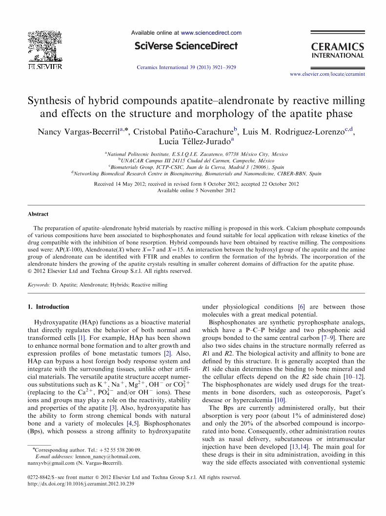
Fig. 1. X-ray diffractograms of apatite obtained by co-precipitation rute:
(a) Ap1 and (b) Ap2.
4000 3600 3200 1800 1600 1400 1200 1000 800
% T
rans
mitt
ance
Wavenumber cm-1
(a)
(b)
961
876
1088
1022
17403566
1645
1229
1217
1370
3320
1452
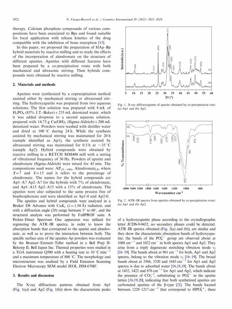
Fig. 2. ATR–IR spectra from apatites obtained by co-precipitation route:
(a) Ap1 and (b) Ap2.
N. Vargas-Becerril et al. / Ceramics International 39 (2013) 3921–39293922
therapy. Calcium phosphate compounds of various com-positions have been associated to Bps and found suitablefor local application with release kinetics of the drugcompatible with the inhibition of bone resorption [15].
In this paper, we proposed the preparation of HAp–Bphybrid materials by reactive milling and to study the effectsof the incorporation of alendronate on the structure ofdifferent apatites. Apatites with different features havebeen prepared by a co-precipitation route with bothmechanical and ultrasonic stirring. Then hybrids com-pounds were obtained by reactive milling.
2. Materials and methods
Apatites were synthesized by a coprecipitation methodassisted either by mechanical stirring or ultrasound stir-ring. The hydroxyapatite was prepared from two aqueoussolutions; The first solution was prepared with 8 mL ofH3PO4 (85% J.T.-Baker)þ235 mL deionized water, whichit was added dropwise to a second aqueous solution,prepared with 14.75 g Ca(OH)2 (Sigma-Aldrich)þ200 mLdeionized water. Powders were washed with distiller waterand dried at 100 1C during 24 h. While the synthesisassisted by mechanical stirring was maintained for 20 h(sample identified as Ap1), the synthesis assisted byultrasound stirring was maintained for 0.5 h at �35 1C(sample Ap2). Hybrid compounds were obtained byreactive milling in a RETCH MM400 mill with a settingof vibrational frequency of 30 Hz. Powders of apatite andalendronate (Sigma-Aldrich) were mixed for 45 min. Thecompositions used were: AP(X�100), Alendronate(X) whereX¼7 and X¼15 and it refers to the percentage ofalendronate. The names for the hybrid compounds areAp1–A7 Ap2–A7 for the hybrids with 7% of alendronate,and Ap1–A15 Ap2–A15 with a 15% of alendronate. Theapatites were also subjected to the same process free ofbisphosphonate and were identified as Ap1-0 and Ap2-0.
The apatites and hybrid compounds were analysed in aBruker D8 Advance with CuKa (l¼1.54 A) radiation, andwith a diffraction angle (2y) range between 51 to 601, and thestructural analysis was performed by FullPROF suite. APerkin–Elmer Spectrum One apparatus was utilized forregistering the ATR–IR spectra, in order to know theabsorption bands that correspond to the apatite and alendro-nate, as well as to prove the interaction between both. Thespecific surface area of the apatites Ap powders was evaluatedby the Brunaer–Emmett–Teller method in a Bell Prep II-Belsorp II, Bell Japan Inc. Thermal properties were studied ina TGA instrument Q500 with a heating rate to 10 1Cmin�1
and a maximum temperature of 800 1C. The morphology andmicrostructure was analized by a Field Emission ScanningElectron Microscopy SEM model JEOL JSM-670IF.
3. Results and discussion
The X-ray diffractions patterns obtained from Ap1(Fig. 1(a)) and Ap2 (Fig. 1(b)) show the characteristic peaks
of a hydroxyapatite phase according to the crystallographicletter JCDS-9-0432, no secondary phases could be detected.ATR–IR spectra obtained (Fig. 2(a) and (b)), are similar andthey show the characteristic absorption bands of hydroxyapa-tite; the bands of the PO4
3� group are observed about at1088 cm�1 and 1022 cm� in both spectra Ap1 and Ap2. Theyarise from a triply degenerate stretching vibration mode n3
[16–19]. The bands about at 961 cm�1 for both, Ap1 and Ap2spectra, belong to the vibration mode n1 [16–19]. The broadbands about at 3566, 3320 and 1645 cm�1 for Ap1 and Ap2spectra is due to adsorbed water [16,18,19]. The bands aboutat 1452, 1422 and 876 cm�1 for Ap1 and Ap2, which indicatethe presence of CO3
�2, substituting to PO43� in the apatite
lattice [16,19,20], indicating that both synthetized apatites arecarbonated apatites of the b-type [21]. The bands locatedbetween 1229–1217 cm�1 that correspond to HPO4
2þ , these
100 200 300 400 500 600 700 800
6
4
2
0
Ap1
Wei
ght l
oss (
%)
Temperature (ºC)
100 200 300 400 500 600 700 800
6
4
2
0
Wei
ght l
oss (
%)
Temperature (ºC)
Ap2
a
b
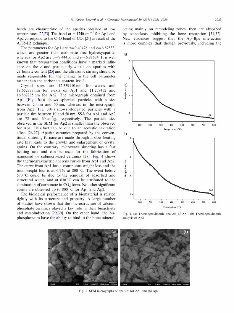
Fig. 4. (a) Thermogravimetric analysis of Ap1. (b) Thermogravimetric
analysis of Ap2.
N. Vargas-Becerril et al. / Ceramics International 39 (2013) 3921–3929 3923
bands are characteristic of the apatites obtained at lowtemperatures [22,23]. The band at �1740 cm�1 for Ap1 andAp2 correspond to the C–O bond of CO2 [24] as result of theATR–IR technique.
The parameters for Ap1 are a¼9.40478 and c¼6.87533,which are greater than carbonate free hydroxyapatite,whereas for Ap2 are a¼9.44456 and c¼6.88634. It is wellknown that preparation conditions have a marked influ-ence on the c and particularly a-axis on apatites withcarbonate content [25] and the ultrasonic stirring should bemade responsible for the change in the cell parameterrather than the carbonate content itself.
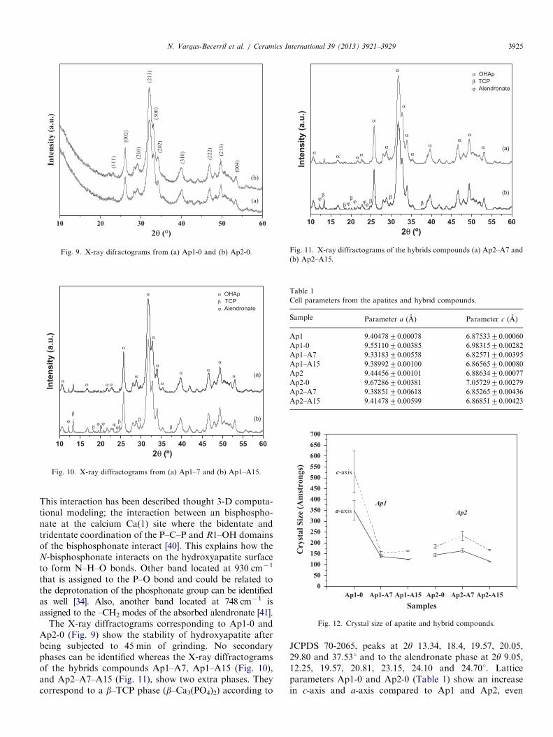
Crystal sizes are 12.339110 nm for a-axis and18.652337 nm for c-axis on Ap1 and 11.257452 and16.862285 nm for Ap2. The micrograph obtained fromAp1 (Fig. 3(a)) shows spherical particles with a sizebetween 20 nm and 50 nm, whereas in the micrographfrom Ap2 (Fig. 3(b)) shows elongated particles with aparticle size between 10 and 50 nm. SSA for Ap1 and Ap2are 72 and 60 cm2/g, respectively. The particle sizeobserved in the SEM for Ap2 is smaller than the observedfor Ap1. This fact can be due to an acoustic cavitationeffect [26,27]. Apatite ceramics prepared by the conven-tional sintering furnace are made through a slow heatingrate that leads to the growth and enlargement of crystalgrains. On the contrary, microwave sintering has a fastheating rate and can be used for the fabrication ofnanosized or submicrosized ceramics [28]. Fig. 4 showsthe thermogravimetric analysis curves from Ap1 and Ap2.The curve from Ap1 has a continuous weight loss and thetotal weight loss is at 6.7% at 800 1C. The event before570 1C could be due to the removal of adsorbed andstructural water, and at 630 1C can be attributed to theelimination of carbonate in CO2 form. No other significantevents are observed up to 800 1C for Ap1 and Ap2.
The biological performance of a biomaterial is relatedtightly with its structure and property. A large numberof studies have shown that the microstructure of calciumphosphate ceramics played a key role in their bioactivityand osteoinduction [29,30]. On the other hand, the bis-phosphonates have the ability to bind to the bone mineral,
Fig. 3. SEM micrographs of ap
acting mainly on remodeling zones, then are absorbedby osteoclasts inhibiting the bone resorption [31,32].New evidences suggest that the Ap–Bps interactionis more complex that though previously, including the
atites (a) Ap1 and (b) Ap2.

4000 3600 3200 1600 1400 1200 1000 80091695
6
1060
1132
1022
1608
2965
867
827
1180
Tra
nsm
ittan
ce (a
.u.)
Wavenumber cm-1
Alendronate3486
1240
Fig. 5. ATR–IR spectrum from alendronateTM.
1800 1600 1400 1200 1000 800
1178
1230
1356 748
8251545
Tra
nsm
ittan
ce (a
.u.)
Wavenumber cm-1
(a)
(b)
875
1019
963
930
1419
1086
14511643
1736
Fig. 7. ATR–IR spectra of hybrid compounds (a) Ap1–A7 and
(b) Ap1–A15.
1800 1600 1400 1200 1000 800
Tran
smitt
ance
(a.u
.)
Wavenumber cm-1
1020
1087
1737
1545
1643
1451
1419 13
56
1230
1178
748
825
875
930
963
(a)
(b)
Fig. 8. ATR–IR spectra of hybrid compounds of (a) Ap2–A7 and
(b) Ap2–A15.
4000 3600 32001800 1600 1400 1200 1000 800
1063
Tra
nsm
ittan
ce (a
.u.)
Wavenumber cm-1
(a)
(b)
10401052
1088
960
1460
1417
874
Fig. 6. ATR–IR spectra from (a) Ap1-0 and (b) Ap2-0.
N. Vargas-Becerril et al. / Ceramics International 39 (2013) 3921–39293924
displacement of inorganic phosphate groups by PO3
groups from the Bp. Thus composition and texturalparameters of the apatite should play a role in thisinteraction [33]. In this paper, a study of the interactionof alendronate with carbonated apatite with differentfeatures follows.
The IR spectrum obtained from the alendronateTM isshown in Fig. 5. The spectrum shows a band at 3486 cm�1
that can be assigned to the N–H group [9]. A C–Hvibration mode can be assigned at 2965 cm�1 [34]. Theband at 1608 cm�1 should correspond to the C¼C doublebond [34]. The bands at 1240, 1180 and 1132 cm�1 areattributed to the P¼O and P–O modes [9,34,35]. Asymmetric P–OH vibration mode is located at 1060 cm�1
and the bands at 1022, 956 and 916 cm�1 belong to theasimmetric vibration of P–OH [35,36]. Also, the asim-metric vibration of the POP group is located at 867 cm�1
and the P–OH vibration mode is located at 827 cm�1 [36].
The ATR–IR spectra from the apatites with/withoutalendronateTM are shown in the next figures. ATR–IRspectra from Ap1-0 and Ap2-0 (Fig. 6), show the char-acteristics bands of the hydroxyapatite; the bands in theregion of 1088 to 1040 cm�1 are due to n3 vibrationalmode of PO4
3� group and the band located about960 cm�1 correspond to the n1 vibrational mode of PO4
3�
group [37,38]. The bands between 1460 to 1417 are dueto the n3 vibrational mode of CO3
2� ion and �874 cm�1
are attributed to the n2 vibrational mode of CO32� ion
[37,39]. Hybrid compounds spectra (Figs. 7 and 8) indicatethe presence of bands that identify the interaction betweenthe apatite and the alendronate. The bands located at1545 and 1356 cm�1 could be assigned to the N=Obond in both spectra, this bond is between the hydroxylgroup of apatite and the amine group of alendronate.
10 20 30 40 50 60
(202)
(300)
(210)
(310) (213)
(004)
(211)
(222)
(002)
Inte
nsity
(a.u
.)
2θ (°)
(a)
(b)
(111)
Fig. 9. X-ray difractograms from (a) Ap1-0 and (b) Ap2-0.
10 15 20 25 30 35 40 45 50 55 60
ϕ ϕϕϕϕβ
β
β
β
ββ
α α
α
α
α
α
α
α
αα
α
α
α
Inte
nsity
(a.u
.)
2θ (º)
(b)
(a)
α OHApβ TCPϕ Alendronate
α
Fig. 10. X-ray diffractograms from (a) Ap1–7 and (b) Ap1–A15.
10 15 20 25 30 35 40 45 50 55 60
α OHApβ TCPϕ Alendronate
Inte
nsity
(a.u
.)
2θ (º)
(a)
(b)
α
ϕβ
α
β ϕβ
ϕ
α α
ϕϕβ
α
α
β
α
α
α
α
β
αα
α
α
Fig. 11. X-ray diffractograms of the hybrids compounds (a) Ap2–A7 and
(b) Ap2–A15.
Table 1
Cell parameters from the apatites and hybrid compounds.
Sample Parameter a (A) Parameter c (A)
Ap1 9.4047870.00078 6.8753370.00060
Ap1-0 9.5511070.00385 6.9831570.00282
Ap1–A7 9.3318370.00558 6.8257170.00395
Ap1–A15 9.3899270.00100 6.8656570.00080
Ap2 9.4445670.00101 6.8863470.00077
Ap2-0 9.6728670.00381 7.0572970.00279
Ap2–A7 9.3885170.00618 6.8526570.00436
Ap2–A15 9.4147870.00599 6.8685170.00423
Ap1-0 Ap1-A7 Ap1-A15 Ap2-0 Ap2-A7 Ap2-A150
50100150200250300350400450500550600650700
Ap1Ap2
Cry
stal
Siz
e (A
mst
rong
s)
Samples
a-axis
c-axis
Fig. 12. Crystal size of apatite and hybrid compounds.
N. Vargas-Becerril et al. / Ceramics International 39 (2013) 3921–3929 3925
This interaction has been described thought 3-D computa-tional modeling; the interaction between an bisphospho-nate at the calcium Ca(1) site where the bidentate andtridentate coordination of the P–C–P and R1–OH domainsof the bisphosphonate interact [40]. This explains how theN-bisphosphonate interacts on the hydroxyapatite surfaceto form N–H–O bonds. Other band located at 930 cm�1
that is assigned to the P–O bond and could be related tothe deprotonation of the phosphonate group can be identifiedas well [34]. Also, another band located at 748 cm�1 isassigned to the –CH2 modes of the absorbed alendronate [41].
The X-ray diffractograms corresponding to Ap1-0 andAp2-0 (Fig. 9) show the stability of hydroxyapatite afterbeing subjected to 45 min of grinding. No secondaryphases can be identified whereas the X-ray diffractogramsof the hybrids compounds Ap1–A7, Ap1–A15 (Fig. 10),and Ap2–A7–A15 (Fig. 11), show two extra phases. Theycorrespond to a b–TCP phase (b–Ca3(PO4)2) according to
JCPDS 70-2065, peaks at 2y 13.34, 18.4, 19.57, 20.05,29.80 and 37.531 and to the alendronate phase at 2y 9.05,12.25, 19.57, 20.81, 23.15, 24.10 and 24.701. Latticeparameters Ap1-0 and Ap2-0 (Table 1) show an increasein c-axis and a-axis compared to Ap1 and Ap2, even
100 200 300 400 500 600 700 800
12
10
8
6
4
2
0
Ap1-A7
Wei
ght l
oss (
%)
Temperature (ºC)
Ap1-A15
100 200 300 400 500 600 700 800
12
10
8
6
4
2
0
Wei
ght l
oss (
%)
Temperature (ºC)
Ap2-A7
Ap2-A15
a
b
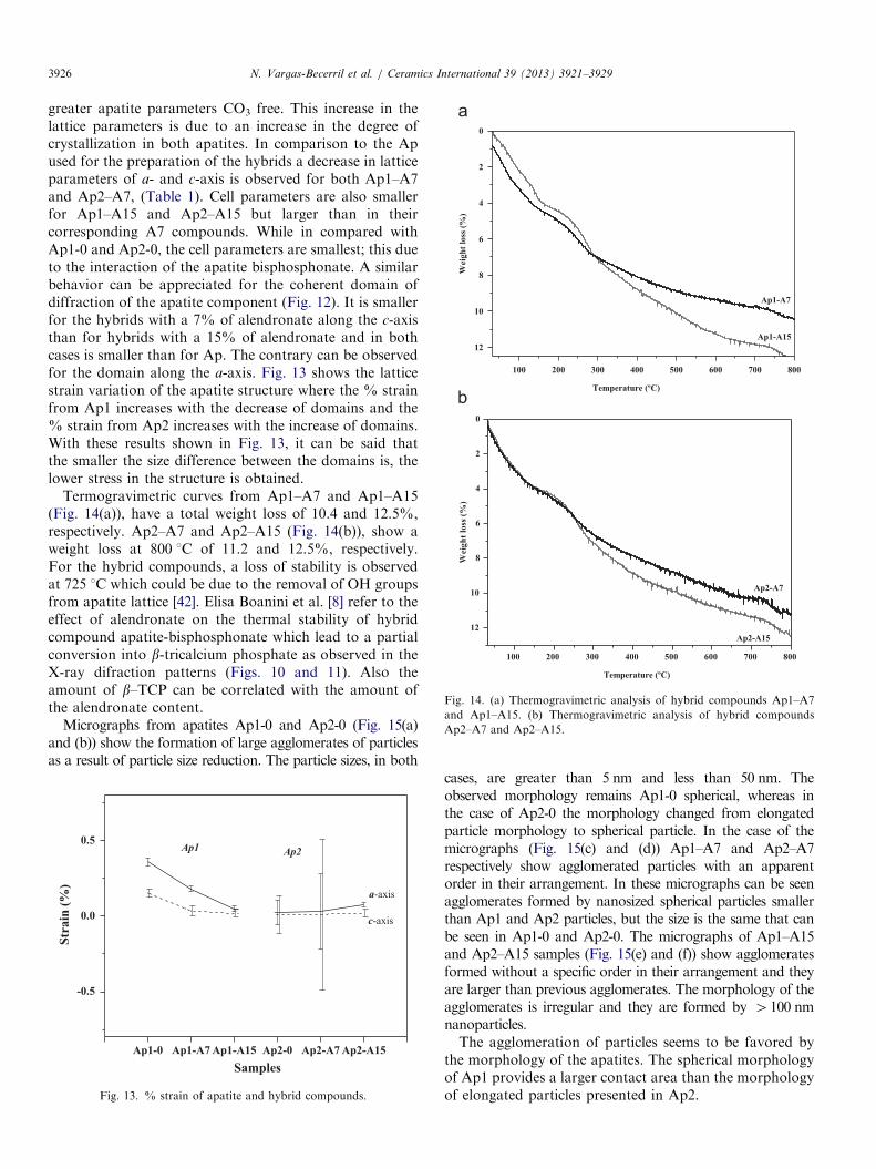
Fig. 14. (a) Thermogravimetric analysis of hybrid compounds Ap1–A7
and Ap1–A15. (b) Thermogravimetric analysis of hybrid compounds
Ap2–A7 and Ap2–A15.
N. Vargas-Becerril et al. / Ceramics International 39 (2013) 3921–39293926
greater apatite parameters CO3 free. This increase in thelattice parameters is due to an increase in the degree ofcrystallization in both apatites. In comparison to the Apused for the preparation of the hybrids a decrease in latticeparameters of a- and c-axis is observed for both Ap1–A7and Ap2–A7, (Table 1). Cell parameters are also smallerfor Ap1–A15 and Ap2–A15 but larger than in theircorresponding A7 compounds. While in compared withAp1-0 and Ap2-0, the cell parameters are smallest; this dueto the interaction of the apatite bisphosphonate. A similarbehavior can be appreciated for the coherent domain ofdiffraction of the apatite component (Fig. 12). It is smallerfor the hybrids with a 7% of alendronate along the c-axisthan for hybrids with a 15% of alendronate and in bothcases is smaller than for Ap. The contrary can be observedfor the domain along the a-axis. Fig. 13 shows the latticestrain variation of the apatite structure where the % strainfrom Ap1 increases with the decrease of domains and the% strain from Ap2 increases with the increase of domains.With these results shown in Fig. 13, it can be said thatthe smaller the size difference between the domains is, thelower stress in the structure is obtained.
Termogravimetric curves from Ap1–A7 and Ap1–A15(Fig. 14(a)), have a total weight loss of 10.4 and 12.5%,respectively. Ap2–A7 and Ap2–A15 (Fig. 14(b)), show aweight loss at 800 1C of 11.2 and 12.5%, respectively.For the hybrid compounds, a loss of stability is observedat 725 1C which could be due to the removal of OH groupsfrom apatite lattice [42]. Elisa Boanini et al. [8] refer to theeffect of alendronate on the thermal stability of hybridcompound apatite-bisphosphonate which lead to a partialconversion into b-tricalcium phosphate as observed in theX-ray difraction patterns (Figs. 10 and 11). Also theamount of b–TCP can be correlated with the amount ofthe alendronate content.
Micrographs from apatites Ap1-0 and Ap2-0 (Fig. 15(a)and (b)) show the formation of large agglomerates of particlesas a result of particle size reduction. The particle sizes, in both
Ap1-0 Ap1-A7 Ap1-A15 Ap2-0 Ap2-A7 Ap2-A15
-0.5
0.0
0.5Ap2Ap1
Str
ain
(%)
Samples
c-axis
a-axis
Fig. 13. % strain of apatite and hybrid compounds.
cases, are greater than 5 nm and less than 50 nm. Theobserved morphology remains Ap1-0 spherical, whereas inthe case of Ap2-0 the morphology changed from elongatedparticle morphology to spherical particle. In the case of themicrographs (Fig. 15(c) and (d)) Ap1–A7 and Ap2–A7respectively show agglomerated particles with an apparentorder in their arrangement. In these micrographs can be seenagglomerates formed by nanosized spherical particles smallerthan Ap1 and Ap2 particles, but the size is the same that canbe seen in Ap1-0 and Ap2-0. The micrographs of Ap1–A15and Ap2–A15 samples (Fig. 15(e) and (f)) show agglomeratesformed without a specific order in their arrangement and theyare larger than previous agglomerates. The morphology of theagglomerates is irregular and they are formed by 4100 nmnanoparticles.The agglomeration of particles seems to be favored by
the morphology of the apatites. The spherical morphologyof Ap1 provides a larger contact area than the morphologyof elongated particles presented in Ap2.
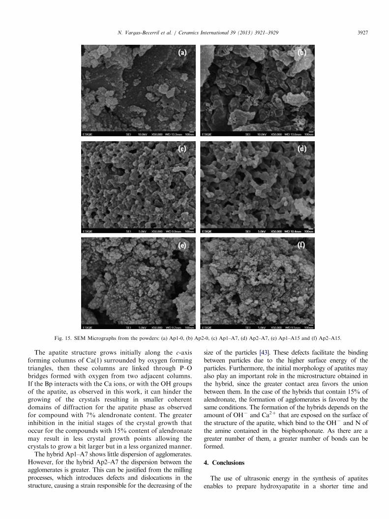
Fig. 15. SEM Micrographs from the powders: (a) Ap1-0, (b) Ap2-0, (c) Ap1–A7, (d) Ap2–A7, (e) Ap1–A15 and (f) Ap2–A15.
N. Vargas-Becerril et al. / Ceramics International 39 (2013) 3921–3929 3927
The apatite structure grows initially along the c-axisforming columns of Ca(1) surrounded by oxygen formingtriangles, then these columns are linked through P–Obridges formed with oxygen from two adjacent columns.If the Bp interacts with the Ca ions, or with the OH groupsof the apatite, as observed in this work, it can hinder thegrowing of the crystals resulting in smaller coherentdomains of diffraction for the apatite phase as observedfor compound with 7% alendronate content. The greaterinhibition in the initial stages of the crystal growth thatoccur for the compounds with 15% content of alendronatemay result in less crystal growth points allowing thecrystals to grow a bit larger but in a less organized manner.
The hybrid Ap1–A7 shows little dispersion of agglomerates.However, for the hybrid Ap2–A7 the dispersion between theagglomerates is greater. This can be justified from the millingprocesses, which introduces defects and dislocations in thestructure, causing a strain responsible for the decreasing of the
size of the particles [43]. These defects facilitate the bindingbetween particles due to the higher surface energy of theparticles. Furthermore, the initial morphology of apatites mayalso play an important role in the microstructure obtained inthe hybrid, since the greater contact area favors the unionbetween them. In the case of the hybrids that contain 15% ofalendronate, the formation of agglomerates is favored by thesame conditions. The formation of the hybrids depends on theamount of OH� and Ca2þ that are exposed on the surface ofthe structure of the apatite, which bind to the OH� and N ofthe amine contained in the bisphosphonate. As there are agreater number of them, a greater number of bonds can beformed.
4. Conclusions
The use of ultrasonic energy in the synthesis of apatitesenables to prepare hydroxyapatite in a shorter time and

N. Vargas-Becerril et al. / Ceramics International 39 (2013) 3921–39293928
produces smaller crystals than conventional stirring. Thereactive milling process introduces defects and dislocations inthe apatite structure and a decrease on the particle size onapatite powders favoring the formation of apatite–alendronatehybrids. The incorporation of the alendronate hinders thegrowing of the apatite crystals resulting in smaller coherentdomains of diffraction for the apatite phase. An interactionbetween the apatite and alendronate can be achieved with theenergy provided by the reactive milling process.
Acknowledgments
The authors thank the financial support from CON-ACyT, the National Politecnic Institute IPN, Mexico City,and the Science and Technology Institute of PolymersITCP, Madrid Spain, for their support and for providingthe necessary tools to do this work through CICYT projectMAT2010-18155.
References
[1] G. Balasundaram, M. Sato, T.J. Webster, Using hydroxyapatite
nanoparticles and decreased crystallinity to promote osteoblast
adhesion similar to functionalizing with RGD, Biomaterials 27 (14)
(2006) 2798–2805.
[2] S.P. Pathi, D.D.W. Lin, J.R. Dorvee, L.A. Estroff, C. Fischbach,
Hydroxyapatite nanoparticle-containing scaffolds for the study of breast
cancer bone metastasis, Biomaterials 32 (22) (2011) 5112–5122.
[3] S. Chakraborty, S. Bag, S. Pal, A.K. Mukherjee, Structural and
microstructural Characterization of bioapatites and synthetic hydro-
xyapatite using X-ray powder diffraction and Fourier transform
infrared techniques, Journal of Applied Crystallography 39 (2006)
385–390.
[4] S.H. Zhu, B.Y. Huang, K.C. Zhou, S.P. Huang, F. Liu, Y.M. Li,
Z.G. Xue, Z.G. Long, Hydroxyapatite nanoparticles as a novel gene
carrier, Journal of Nanoparticle Research 6 (2004) 307–311.
[5] T.M. Sridhar, T.K. Arumugam, S. Rajeswari, M. Subbaiyan, Elec-
trochemical bahaviour of hydroxyapatite-coated stainless steel
implants, Journal of Materials Science Lettters 16 (1997) 1964–1966.
[6] J.E.I. Wright, L. Zha, P. Choi, H. Uludag, Simulating hydroxyapa-
tite binding of bone-seeking bisphosphonate, Biomaterials (2004)
139–148.
[7] E. Maltezou, M. Stylianou, S. Roy, C. Drouza, A.D. Keramidas,
Synthesis, solutions and structural characterization of tetrahydrofur-
anyl-2, 2-bisphosphonic acid disodium salt, Bioinorganic Chemistry
and Applications (2010) 1–7.
[8] E. Boanini, M. Gazzano, K. Rubini, A. Bigi, Composite nanocrystals
provide new insight on alendronate interaction with hydroxyapatite
structure, Advanced Materials 19 (2007) 2499–2502.
[9] L.M. Rodriguez-Lorenzo, B. Vazquez, J. San Roman, Surface
modification of calcium hydroxyfluor carbonate apatites by bispho-
sphonates, Key Engineering Materials 284–286 (2005) 357–360.
[10] T.J. Martin, V. Grill, Bisphosphonates-mechanisms of action,
Australian Prescriber 23 (6) (2000) 130–132.
[11] F.P. Coxon, K. Thompsom, M.J. Rogers, Recent advances in
understanding the mechanism of action of bisphosphonate, Current
Opinion in Pharmacology 6 (2006) 307–312.
[12] H. Fleisch, Development of bisphosphonates, Breast Cancer
Research 4 (2002) 30–34.
[13] A. Ezra, G. Golomb, Administration routes and delivery systems of
bisphosphonates for the treatment of bone resorption, Advanced
Drug Delivery Reviews 42 (2000) 175–195.
[14] S. Patashnik, L. Rabinovich, G. Golomb, Preparation and evalua-
tion of chitosan microspheres containing bisphosphonates, Journal
of Drug Targeting 4 (1997) 371–380.
[15] S. Josse, C. Faucheux, A. Soueidan, G. Grimandi, D. Massiot,
B. Alonso, P. Janvier, S. Laıb, P. Pilet, O. Gauthier, G. Daculsi,
J. Guicheux, B. Bujoli, J.M. Bouler, Novel biomaterials for bispho-
sphonate delivery, Biomaterials 26 (2005) 2073–2080.
[16] C.C. Silva, A.G. Pinhero, M.A.R. Miranda, J.C. Goes,
A.S.B. Sombra, Structural propierties of hydroxyapatite obtained
by mechanosynthesis, Solid State Science 5 (2003) 553–558.
[17] M.A. Martins, C. Santos, M.M. Almeida, M.E.V. Costa, Hydro-
xyapatite micro- and nano-particles: nucleation and growth mechan-
isms in the presence of citrate species, Journal of Colloid and
Interface Science 318 (2008) 210–216.
[18] M.P. Mahabole, R.C. Aiyer, C.V. Ramakrishna, B. Sreedhar,
R.S. Khairnar, Synthesis, characterization and gas sensing property
of hydroxyapatite ceramic, Bulletin of Material Science 28 (6) (2005)
535–545.
[19] M. Sadat-Shojai, Preparation of hydroxyapatite nanoparticles: com-
parison between hydrothermal and solvo-treatment processes and
colloidal stability of produced nanoparticles in a dilute experimental
dental adhesive, Journal of Iranian Chemical Society 6 (2) (2009)
386–392.
[20] M. Mathew, S. Takagi, Structures of biological minerals in dental
research, Journal of Research of the National Institute of Standards
and Technology 106 (2001) 1035–1044.
[21] A. Balamurugan, J. Michel, J. Faure, H. Benhayoune, L. Wortham,
G. Sockalingum, V. Banchet, S. Bouthors, D. Laurent-Maquin,
G. Balossier, Synthesis and structural analysis of sol gel derived
stoichiometric monophasic hydroxyapatite, Ceramic–Silikaty 50 (1)
(2006) 27–31.
[22] S. Lazic, S. Zec, N. mIljevic, S. Milonjic, The effect of temperature on the
properties of hydroxyapatite precipitated from calcium hydroxide and
phosphoric acid, Thermochimic Acta 374 (2001) 13–22.
[23] G. Gafni, D. Septier, M. Goldberg, Effect of chondroitin sulfate and
biglycan on the crystallization of hydroxyapatite under physiological
conditions, Journal of Crystal Growth 205 (1999) 618–623.
[24] /www.unav.es/organica/docencia/espectroscopia_d/y/Infrarrojo.
pdfS April 18, 2012.
[25] J.C. Elliott, Structure and Chemistry of the Apatites and Other
Calcium Orthophosphates, Elsevier, Amsterdam, 1994.
[26] A.B. Martinez, G. Carbajal, R. Torres, L. Tellez, H.E. Esparza,
Production of polyurethane/nano-hydroxyapatite hybrid materials
and microstructural characterization, Int. J. Phys. Sci. 6 (11) (2011)
2731–2743.
[27] M. de Campos, F.A. Muller, A.H.A. Bressiani, J.C. Bressiani,
P. Greil, Sonochemical synthesis of calcium phosphate powders,
Journal of Materials Science: Materials in Medicine 18 (2007)
669–675.
[28] X.D. Zhu, H.J. Zhang, D.X. Li, H.S. Fan, X.D. Zhang, Study on the
enhanced protein adsorption of microwave sintered hydroxyapatite
nanoceramic particles: role of microstructure, Journal of Biomedical
Materials Research Part B 100 (B) (2012) 516–523.
[29] M. Tamai, K. Isama, R. Nakaoka, T. Tsuchiya, Synthesis of a novel
b-tricalcium phosphate/hydroxyapatite biphasic calcium phosphate
containing niobium ions and evaluation of its osteogenic properties,
Journal of Artificial Organs 10 (2007) 22–28.
[30] W. Paul, C.P. Sharma, Development of porous spherical hydro-
xyapatite granules: application towards protein delivery, Journal of
Materials Science: Materials in Medicine 10 (1999) 383–388.
[31] K. Olson, C. Van Poznak, Significance and impact of
bisphosphonate-induced acute phase responses, Journal of Oncology
Pharmacy Practice 13 (2007) 223–229.
[32] M.A. Gunther Sillero, A. de Diego, E. Silles, F. Perez-Zuniga,
A. Sillero, Synthesis of bisphosphonate derivatives of ATP by T4
RNA ligase, FEBS Letters 580 (2006) 5723–5727.
[33] A. Juillard, G. Falgayrac, B. Cortet, M.-H. Vieillard, N. Azaroual,
J.-C. Hornez, G. Penel, Molecular interactions between zoledronic
N. Vargas-Becerril et al. / Ceramics International 39 (2013) 3921–3929 3929
acid and bone: an in vitro Raman microspectroscopic study, Bone 47
(2010) 895–904.
[34] M.A. Bayle, K. Nasr, G. Gregoire, P. Sharrock, Acrylophosphonic
acid reactivity with calcium ions and biological apatite, Dental
Materials 24 (2008) 386–391.
[35] F. Al-Ali, A. Lebugle, I. Rico-Lattes, G. Etemad-Moghadam,
Preparation and characterization of new hybrid organic/inorganic
systems derived from calcium (a-aminoalkyl)-phosphonates and-
phosphonocarboxylates, Journal of Colloid and Interface Science
289 (2005) 504–511.
[36] L.M. Rodriguez-Lorenzo, M. Fernandez, J. Parra, B. Velazquez,
A. Lopez-Bravo, J. San Rom�an, Acrylic injectable and self-curing
formulations for the local release of bisphosphonates in bone tissue,
Journal of Biomedical Materials Research Part B: Applied Bioma-
terials (2007) 596–608.
[37] I. Rehman, W. Bonfield, Characterization of hydroxyapatite and
carbonated apatite byn photo acoustic FTIR spectroscopy, Juornal
of Material Science: Materials in Medicine 8 (1997) 1–4.
[38] Y. Han, X. Wang, S. Li, A simple route to prepare stable hydro-
xyapatite nanoparticles suspension, Journal of Nanoparticle
Research 11 (2009) 1235–1240.
[39] M. Okada, T. Furuzono, Fabrication of high-dispersibility nano-
crystals of calcined hydroxyapatite, Journal of Materials Science 41
(2006) 6134–6137.
[40] R.G.G. Russell, N.B. Watts, F.H. Ebetino, M.G. Rogers, Mechan-
isms of action of bisphosphonates: similarities and differences and
their potential influence on clinical efficacy, Osteoporos International
19 (2008) 733–759.
[41] H. Agougui, A. Aissa, S. Maggi, M. Debbabi, Phosphonate-
hydroxyapatite hybrid compounds prepared by hydrothermal
method, Applied Surface Science 257 (20109) 1377–1382.
[42] X.-F. Xiao, R.-F. Liu, Y.-J. Gao, Hydrothermal preparation of
nanocarboted hydroxyapatite crystallites, Material Science Technol-
ogy 24 (2008) 1199–1203.
[43] C.C. Koch, Top-down synthesis of nanostructured materials:
mechanical and thermal processing methods, Reviews on Advanced
Materials Science 5 (2003) 91–99.


