
Dr. SK Sharm
a
TECHNIQUES OF LOCAL ANAESTHESIA IN ANIMALS
Dr. SK Sharma, Associate Professor (Surgery)
Local anaesthetic techniques are primarily used in cattle, buffalo and equines but
can very well be adopted in other animals depending upon the cooperative nature of the
patient.
Advantages:
• Minimal equipment needed • Minimal systemic effects
Disadvantages:
• Requires cooperative patient with or without significant restraint. • May require sedation.
Classification of Nerve Fibers:
A (myelinated)
• - motor • - touch and pressure • - muscle spindle • - pain
B (myelinated; autonomic – pre-ganglionic sympathetic)
C (non-myelinated; pain & temperature)
Rate of Transmission dependent on:
• Diameter of axon • Presence or absence of myelin sheath • Order of Blockade and Recovery
Sensation lost in the following order:
• Pain • Cold • Warmth • Touch • Joint
Dr. SK Sharm
a
• Pressure
Types of local anesthetic techniques: In animals mainly 4 techniques are used to
produce local anaesthesia. These are as follows:
a) Topical or surface anaesthesia:
This technique is primarily used for the desensitization of the superficial layers of
the skin/mucous membrane e.g. eye, vulvar region, glans penis and for opening of the
abscess. The simplest way to achieve surface analgesia is by application of the ice. The
other methods are:
Using volatile agents: Ethyl chloride, ether etc. These agents evaporate
instantly thereby decreasing the surface temperature and so causing
desensitization of the area. Such method should not be used frequently as may
cause tissue necrosis.
Using local anaesthetic drugs:
a) 2% or 4% lignocaine HCl can be used for the relief of pain in abrasions or
eczematous areas. Soak a piece of cotton or gauge in the local anaesthetic
solution and then put on the affected area for 5 min. The analgesia is seen
for 30-45 min.
b) 4% lignocaine HCl as spray is used for the surface analgesia of mucous
membranes like vulva, glans penis, pharynx, larynx etc.
c) For eyes 4% cocaine or lignocaine can be used.
d) Lignocaine or amethocaine jelly can be used for desensitization of
abrasions, eczematous areas.
e) In horses and cattle intrasynovial injection of 2% lignocaine HCl is used
for the surface anaesthesia of the hygromatous areas at the joints.
Complete asepsis is a must.
b) Infiltration anaesthesia:
In this technique the nerve endings are blocked at the actual site of the operation.
The volume used will depend on the size of the animal and the area to be blocked.
2

Dr. SK Sharm
a
Advantages: Requires no greater skill or the knowledge of the anatomy of the
site.
Disadvantages:
a) Large volumes of anesthetic in the tissues to be incised and sutured. b) Epinephrine in the anesthetic solution may also interfere with the blood
supply and retard healing.
c) If a flank laparotomy is being done local anesthetic must not only be
infiltrated subcutaneously but into the muscles and fascia as well.
Extravascular infiltration Techniques:
a) Linear infiltration: The infiltration of the local anaesthetic is done on the line of
the incision or just parallel to the line of the incision. The amount to be infiltrated
is 1 ml/cm in small animals and 2 ml/cm in case of large animals. The anaesthetic
is to be infiltrated into the subcutaneous area as well as into the muscles. The
technique can be used for any type of the surgical operation like rumenotomy,
cystotomy etc. However in this technique the amount used is quite large; the
anatomy of the site is also disturbed; the healing of the wound is normally
delayed.
b) Inverted ‘L’ or ‘T’ or inverted ‘V’ block: This technique (done for flank
laparotomies) is a nonspecific regional analgesic technique in which all the nerves
entering the surgical field are desensitized from two sides. 2% Lignocaine HCl is
injected into the tissues bordering the dorsocaudal aspect of the last rib and
ventrolateral aspect of the lumbar transverse processes. Advantages include
deposition of the anaesthetic away from the surgical site thus decreasing edema
and haematoma formation from the block. The site anatomy is not disturbed and
therefore there is normal healing of the surgical wound. Disadvantages include
incomplete analgesia and muscle relaxation of the deeper layers of the abdominal
wall. Inverted ‘V’ block technique is primarily used for teat surgery or
management of accidental wounds of the limbs.
3

Dr. SK Sharm
a
Inverted ‘L’ Block Inverted ‘V’ Block ‘T’ Block
c) Field Block and Ring Block: In this technique a wall of the local anaesthetic is
formed around the site of the operation. The advantages and the disadvantages are
the same as for the inverted ‘L’ or ‘T’ block.
Diamond (Field) Block
Intravascular Infiltration techniques:
This includes ‘Retrograde intravenous regional anaesthesia: It is a simple and
safe anaesthetic technique to produce regional anaesthesia of lower limbs. The technique
was developed in 1908.
Methodology:
a) The animal is restrained in the lateral recumbency with affected limb on the upper
side.
b) Tightly apply a rubber tube tourniquet on the upper part (above the elbow or hock
joint) of the limb to stop blood flow.
c) Any prominent superficial vein below the tourniquet is isolated.
d) Inject 2% lignocaine without adrenaline into the vein using 23/24 gaze needle, 10-
25 ml in large animals and 3-10 ml in small animals.
e) The analgesia is achieved within 10 min and remains as long as the tourniquet is
there (maximum 60-90 min).
4
Dr. SK Sharm
a
f) There is complete and uniform analgesia below the tourniquet of all the structures
including the bones.
g) The major disadvantage of this technique is the rupture of muscles (because of
less O2 to the muscles) and persistent lameness if the tourniquet is kept for a very
longer time.
Regional anaesthetic techniques:
This is an advantageous technique in comparison to infiltration techniques
and is achieved by blocking the conduction in sensory nerve(s) innervating the region
where the surgery is to be carried out. The advantages of this technique are as under:
Smaller amount of the local anaesthetic is required.
The anatomy of the site of the operation is not disturbed.
Less toxicity in the body due to small quantity of the local anaesthetic.
Produces uniform analgesia of the site.
There is normal healing of the surgical wound.
Various regional blocks are as under:
c) Peripheral neural blockade: a) Lingual Nerve Block in cattle and buffaloes: Lingual nerve(s) are the sensory
supply to the tongue of cattle and buffaloes. These include lingual branches of
V and IX cranial nerves and chorda tympani of VII. The motor supply is from
hypoglossal nerve. The technique is accomplished only under sedation/narcosis
using tranquillizers or chloral hydrate.
Indications: Lacerations; Ranula; Foreign bodies
Methodology: Open the mouth with an appropriate mouth gag. Hold the tongue
from the base in semi twisted position. 15 cm long 18 gaze needle is passed along
the medial surface of 4th and 5th lower cheek teeth and introduced approximately 1.5
cm caudoventrally through the connective tissue to block the sensory supply by
5

Dr. SK Sharm
a
injecting 10-15 ml of 2% lignocaine HCl. Repeat the procedure on the other side.
To block the hypoglossal nerve, supplying to the styloglossus and hypoglossus
muscles, the needle is inserted in the longitudinal groove between the bellies of
these two muscles, caudoventrally at the base of the tongue (approximately 1.5 cm
deep into the muscle) and then inject 10 ml of 2% lignocaine HCl. The block is
rarely used under the field conditions.
b) Mandibulo-alveolar Block: It is a branch of mandible nerve. The block causes
desensitization of lower jaw, lower lip along with the molar teeth. The
technique is mainly used in horses, cattle and buffaloes and rarely in dogs and
cats.
Indications: Surgical conditions of molar teeth and incisors and body of mandible
in the lower jaw, lower lip.
Methodology: The site of infiltration of the local anaesthetic is the mandibular
foramen. A 15 cm, 18 gaze needle is inserted from the lower angle of the jaw along
the medial surface of the ramus of the mandible. Reach appoint formed by an
imaginary line drawn from the masticatory surface of lower molar teeth bisected by
the perpendicular line drawn from the lateral canthus of the eye (site of mandible
foramen). Inject approximately 20 ml of the 2% lignocaine HCl. Repeat on the
other side.
6

Dr. SK Sharm
a
c) Mental Nerve Block: Mental nerve is the branch of mandible nerve supplying
to lower lip and lower jaw. The block is mostly employed in cattle, buffalo,
horses, dog and cats. The block is indicated for the surgery of lower lip and the
lower jaw.
Methodology: The site of infiltration of the local anaesthetic is the mentle foramen
on the lateral aspect of the ramus of lower jaw just behind the 4th incisor. It is
present in the inter-dental space in equines. A 3-4 cm long 18/20 gaze needle is
taken and bent about half circle. Keep the concave surface of the needle laterally
and blindly insert the needle into the foramen. Inject 5-15 ml of the local
anaesthetic (2% lignocaine HCl). Repeat the procedure on the other side
d) Infraorbital Nerve Block: The Infraorbital nerve emerges from the
Infraorbital foramen just rostral to the facial tuberosity and dorsal to the first
molar tooth. The block is indicated for the surgery of upper lip, incisors and the
gums.
7

Dr. SK Sharm
a
Methodology: The site of the injection is the Infraorbital foramen. A 20 gaze, 3-4
cm long needle is inserted deep into the foramen and 5-15 ml of the local
anaesthetic is injected. Repeat the procedure on the other side.
e) Supraorbital (Frontal) Nerve Block: It is a branch of ophthalmic nerve and
emerges through the supraorbital foramen. It supplies to the upper eyelid and
the forehead. The block is mostly employed in horses, cattle and buffaloes. It is
indicated for operations on the upper eyelid, trephining of the frontal sinus and
management of the wounds of the forehead.
Methodology: The foramen is felt as small depression midway across the supra
orbital process on the ventral line running upward from the median angle of the eye.
A 20 gaze, 2-3 cm long needle is inserted into the foramen and about 5 ml of the
local anaesthetic is injected. In case of the cattle the injection is made above the
median canthus and spread the anaesthetic laterally along the supra orbital process.
f) Cornual Nerve Block: It involves the blocking of the cornual nerve, the
terminal branch of lacrimal supplying to the base of the horn. The block causes
desensitization of the base of the horn along with its corium. Used in bovines,
caprines and ovine.
• Indication: a) Dehorning b) Horn injury
8

Dr. SK Sharm
a
• Injection site (Bovines):
a) The upper third of the temporal ridge, about 2.5 cm below the
base of the horn at a point below the frontal crest, almost
midway along the line joining the center of the orbit and the
base of the horn..
b) The nerve is relative superficial, about 0.7-1 cm deep.
• Anaesthetic: 2% lignocaine HCl 3-5 ml
• Onset of analgesia: Within 10-15 minutes
• Duration of analgesia: Approximately 1-2 hour.
• Variability: In adult cattle few branches of 1st and 2nd cervical nerves
also supply the base of the horn. Therefore a 2nd injection is to be
made about 1 cm behind the 1st injection site bisecting the line
between base of the horn and the base of the ear. If analgesia is still
not seen a ring block around the base of the horn may be necessary.
• Caprines: There are two sites to block the cornual branches of
lacrimal and infratrochlear nerves. A) Behind the root of the supra
9
Dr. SK Sharm
a
orbital process to block the lacrimal branch; B) Close to the dorsomedial
margin of the orbit to block the infratrochlear branch.
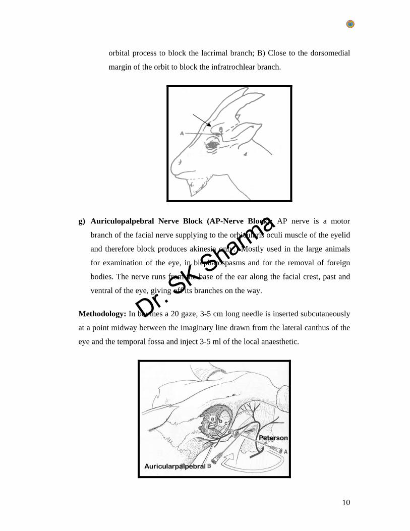
g) Auriculopalpebral Nerve Block (AP-Nerve Block): AP nerve is a motor
branch of the facial nerve supplying to the orbicularis oculi muscle of the eyelid
and therefore block produces akinesia only. Mostly used in the large animals
for examination of the eye, in blepharospasms and for the removal of foreign
bodies. The nerve runs from the base of the ear along the facial crest, past and
ventral of the eye, giving off its branches on the way.
Methodology: In bovines a 20 gaze, 3-5 cm long needle is inserted subcutaneously
at a point midway between the imaginary line drawn from the lateral canthus of the
eye and the temporal fossa and inject 3-5 ml of the local anaesthetic.
10

Dr. SK Sharm
a
h) PETERSON EYE BLOCK: The eye block was developed by Peterson in
1966. In this block the nerves blocked are: a) Mandible branch of V; b)
Maxillary branch of V supplying to the lower eyelid; c) Ophthalmic branch of
V supplying to upper eyelid, 3rd eyelid, median canthus and eyeball; d)
Oculomotor, trochlear and abducens nerves providing the motor supply to the
ocular muscles. The indications of this block are: Extirpation of eye,
enucleation, conjunctival flaps and any type of ocular surgery.
Methodology: The injection is made into the temporal fossa using a 7-11 cm long
18 G needle just above the pterygopalatine fossa in front of the foramina
orbitorotundum. Inject 10-15 ml of 2% lignocaine HCl. When touching the
pterygopalatine fossa, don’t apply excessive pressure to prevent the penetration of
the bony plate.
i) PARAVERTEBRAL BLOCK: This block is used in bovines, yaks and
camels and there is blocking of the spinal nerves emerging from the
intervertebral foramina. The technique is better than the infiltration analgesic
technique because it produces complete and uniform analgesia upto the
peritoneum and also reduces the intra abdominal pressure because high
pressure hampers the surgical maneuvering. The block can be achieved
following two techniques:
PROXIMAL PARAVERTEBRAL BLOCK (Farquharson, Hall or Cambridge Technique)
• Indications: Standing laparotomy surgery such as rumenotomy, caecotomy,
cystotomy, abomasal displacement and management of intestinal obstruction and
volvulus.
• Anatomy and injection site:
In bovine the dorsal aspect of the transverse processes of the last
thoracic (T-13) and first and second lumbar (L-1 and L-2) vertebrae is
the site for needle placement.
11

Dr. SK Sharm
a
The dorsal and ventral never roots of the last thoracic (T13) and 1st and
2nd lumbar spinal nerves emerge from the intervertebral foramina are
desensitized. In yaks T14, L1 and L2 spinal nerves are blocked whereas
in camels T12, L1, L2 and dorsal branches of L3 are blocked.
• Anaesthetic agents: 10-15 ml of 2% lignocaine HCl is injected to each site.
• Onset of Effect:
o Analgesia seen within 10 minutes of injection.
o Analgesia of the skin.
o Decrease in the intra abdominal pressure - due to paralysis of the
paravertebral muscles.
o Skin temperature is increased due to vasodilation (paralysis of cutaneous
vasomotor nerves).
• Duration of analgesia: Approximately 90 minutes.
DISTAL PARAVERTEBRAL ANALGESIA: Mostly used clinically due to ease in
the employment.
• Indications: Same as proximal paravertebral block above
• Anatomy and injection site:
o The dorsal and ventral rami of the spinal nerves T13, L1 and L2 are
desensitized at the distal ends of L-1, L-2 and L-3.
o A 7.5-cm, 18-gauge needle is inserted ventral to the tips of the respective
transverse processes in cows where approximately 10-20 ml of a 2%
lidocaine solution is injected in a fan-shaped infiltration pattern.
o The needle is completely withdrawn and reinserted dorsal to the transverse
process, where the cutaneous branch of the dorsal rami is injected with
about 5 ml of the analgesic.
o The procedure is repeated for the second and third lumbar transverse
processes.
• Anaesthetic: 2% lignocaine HCl.
• Onset of analgesia: Within 10 minutes of injection
12

Dr. SK Sharm
a
• Duration of analgesia: Approximately 90 minutes.
The same technique can be employed in sheep and goats but rarely used.
ADVANTAGES AND DISADVANTAGES OF FOUR COMMON LOCAL ANAESTHETIC TECHNIQUES
TECHNIQUES ADVANTAGES DISADVANTAGES Proximal Paravertebral Block
• Small dose of analgesic • Wide and uniform area of
analgesia and musclerelaxation
• Minimal intra-abdominal pressure
• Increased intestinal toneand motility
• Absence of local analgesicfrom the operative woundmargins
• Technical difficulty • Arching up of the spine
due to paralysis of theback muscles
• Risk of penetrating vitalstructures such as theaorta and thoraciclongitudinal vein on theleft side and the caudalvena cava on the rightside
Distal Paravertebral Block
• The use of more routine size needles, no risk of penetrating a major blood vessel.
• Lack of scoliosis minimal weakness in the pelvic limb
o Larger doses of anesthetic are needed.
o Variation in efficiency exist, particularly if the nerves vary in their anatomical
13
Dr. SK Sharm
a
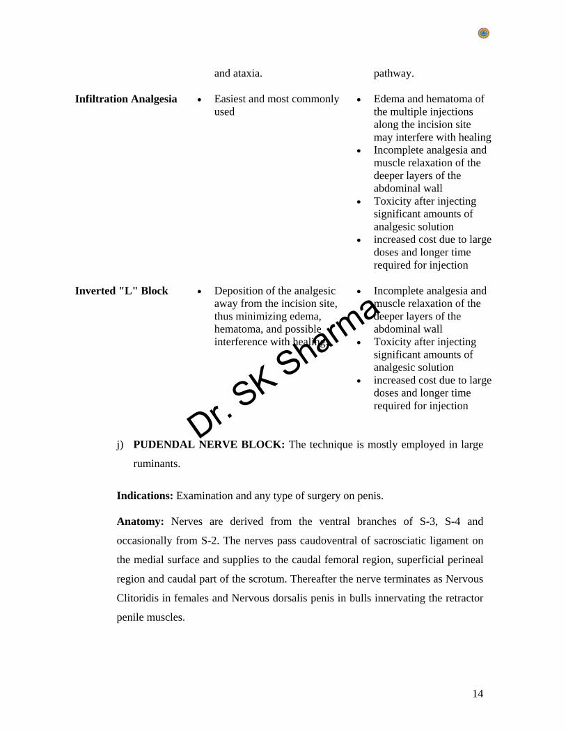
and ataxia. pathway.
Infiltration Analgesia • Easiest and most commonly used
• Edema and hematoma of the multiple injections along the incision site may interfere with healing
• Incomplete analgesia and muscle relaxation of the deeper layers of the abdominal wall
• Toxicity after injecting significant amounts of analgesic solution
• increased cost due to large doses and longer time required for injection
Inverted "L" Block • Deposition of the analgesic away from the incision site, thus minimizing edema, hematoma, and possible interference with healing.
• Incomplete analgesia and muscle relaxation of the deeper layers of the abdominal wall
• Toxicity after injecting significant amounts of analgesic solution
• increased cost due to large doses and longer time required for injection
j) PUDENDAL NERVE BLOCK: The technique is mostly employed in large
ruminants.
Indications: Examination and any type of surgery on penis.
Anatomy: Nerves are derived from the ventral branches of S-3, S-4 and
occasionally from S-2. The nerves pass caudoventral of sacrosciatic ligament on
the medial surface and supplies to the caudal femoral region, superficial perineal
region and caudal part of the scrotum. Thereafter the nerve terminates as Nervous
Clitoridis in females and Nervous dorsalis penis in bulls innervating the retractor
penile muscles.
14

Dr. SK Sharm
a
Technique: The technique was developed by Larson in 1953. Go per rectally and
feel a triangular depression formed medially by the head of the tail, laterally by
the caudal part of the sacrosciatic ligament and ventrally by the ischial tuberosity.
A 20 G, 3-5 cm needle is inserted from the outside to reach the dorso-anterior part
of this triangular depression and infuse 15-20 ml of the local anaesthetic. Repeat
the procedure on the other side to complete the pudendal nerve block. The onset is
seen within 10 min and the effect remains for 1.5-2.0 hr.
K) BRACHIAL PLEXUS BLOCK:
Indication: Brachial plexus block is suitable for analgesia induced by the
surgery on the front limb, within or distal to the elbow.
The technique should be performed in a well sedated or anesthetized
animal.
Species: This block can be used in dogs, cats, small ruminants, calves, and
foals.
Anatomy: The brachial plexus nerves are derived from C-6, C-7, C-8 and
T-1 spinal nerves roots. The nerves arise from the plexus are:
Suprascapular; Subscapular; Anterior thoracic nerve; Posterior thoracic
nerve; Long thoracic nerve; Thoracodorsal nerve; Axillary nerve;
Musculocutaneous nerve; Median nerve; Ulnar nerve and Radial nerve.
Technique: A 7.5-cm, 20-22 gauge spinal needle is inserted medial to the
shoulder joint and directed parallel to the vertebral column toward the
costochondral junction of the Ist rib. In larger size animal, if no blood is
aspirated into the syringe as the needle is withdrawn; approximately 10-15
ml of 2% Lignocaine HCl is injected. Gradual sensation and loss of motor
function occurs within 10-15 minutes leading to characteristic posture like
seen in radial paralysis i.e. elongated limb with knee flexed. Anaesthesia
lasts for approximately 2 hours, and total recovery requires approximately
6 hours.
15

Dr. SK Sharm
a
Brachial plexus block is relatively simple and safe to perform and
produces selective anesthesia and relaxation of the limb and analgesia to
the forelimb.
L) RADIAL NERVE BLOCK:
Indications: Fracture repair, dislocation below elbow and surgery below elbow
joint:
Anatomy: The radial nerve supplies to the dorsal aspect of the fore arm and
manus except in horses where it stops at the carpus.
Technique: The nerve is blocked at a point where it spirals around the humerus
from medial to the lateral aspect. The site of injection is midway between the
olecranon process and the acromion process i.e. upper third of the humerus on the
posterior aspect. Inject 2-4 ml of the local anesthetic using 20 G 3-5 cm long
needle. The effect is seen within 10 min and lasts for about 2 hr.
m) MEDIAN NERVE BLOCK (LARGE ANIMALS): Median nerve supplies
to the median and dorsal surface of the digits. The nerve lies 1-2 cm deep
between the flexor carpi radialis muscle and the radius bone just below the
insertion of the anterior superficial pectoral muscle. The block is indicated for
the median neurectomy.
16

Dr. SK Sharm
a
Technique: The site of the injection is the medial aspect of the elbow joint just
anterior to the medial epicondyle of the humerus. The nerve is covered by skin
and fascia only. Inject 5-10 ml of the local anaesthetic to cause the median nerve
block.
n) ULNAR NERVE BLOCK (LARGE ANIMALS): Ulnar nerve is a sensory
supply to the caudomedial and caudolateral parts of the forearm and lateral
aspect of the manus.
Indications: Surgery on the volar aspect of the foreleg e.g. Tenotmy.
Technique: The site of the injection is about 7-10 cm above the accessory carpel
bone on the volar (posterior) aspect of the limb in the groove between the flexor
carpi ulnaris and ulnaris lateralis muscle. The needle is inserted about 0.5-1.5 cm
deep and 10 ml of the local anaesthetic is administered to achieve the block.
D. CENTRAL NEURAL BLOCKADE
EPIURAL ANAESTHESIA:
When anaesthesia is injected within the canal but outside the duramater it
is called epidural anaesthesia.
When the injection is made in the cerebrospinal fluid, it is termed as the
subarachnoid or intrathecal anaesthesia.
Spinal anaesthesia is mostly subarachnoid anaesthesia in human beings.
In epidural anaesthesia there is desensitization of first sensory nerves
followed by sacral, parasympathetic, sympathetic and motor nerves.
Depending upon the site of injection epidural anaesthesia can be:
i. Caudal Epidural anaesthesia.
ii. Lumbosacral epidural anaesthesia.
iii. Lumber segmental epidural anaesthesia.
Out of these techniques the most commonly followed is the ‘Caudal
Epidural Anaesthesia’
17

Dr. SK Sharm
a
CAUDAL EPIDURAL ANAESTHESIA:
Caudal epidural anaesthesia mostly produces the desensitization of sacral region,
tail, anus, vulva, perineum and caudal aspect of the femoral region. It doesn’t affect the
motor response of the hind limbs.
Indications: Obstetrical operations; Perineal region operations; Management of
dystocia; Tail docking; Episiotomy; Management of ante/post partum prolapse of
vagina or uterus; Prolapse of rectum; Amputation of rectum; Management of recto-
vagina fistula and congenital defects like atrasia ani, atrasia recti etc.
Site of injection: Horse: Ist intercoccygeal space; Cattle, buffalo and camels:
Sacrococcygeal or Ist intercoccygeal space; Sheep and goats: Sacrococcygeal; Dogs
and cats: Sacrococcygeal or Ist intercoccygeal space using 23 G needle or go for the
anterior epidural at Lumbo-sacral space (Most preferred). In buffaloes the sacral ridge
is inclined downwards so the anaesthetic solution may not reach the upper side.
Therefore Sacrococcygeal is the most preferred site for the injection. The structures
penetrated (Ist intercoccygeal space) are skin, superficial fascia, coccygeal fascia, inter
muscular septa, interspinous and laminar fibrous sheet.
Technique: The location is found by elevating and lowering the tail and palpating the
depression and movement between the respective vertebrae. A small volume of
analgesic is injected into the sacrococcygeal (S5-C1) space or first intercoccygeal (C1-
C2) space. The amount of the anaesthetic used varies between 0.2-5 ml for small
animals and between 2-10 ml for the large animals. Before injecting the anaesthetic
withdraw the piston and see that blood should not come out and then inject the
anaesthetic; there should be free movement of the piston. When the quantity of the
anaesthetic is increased it will lead to anterior or high epidural block due to the
spreading of the anaesthetic anteriorly. This is characterized by staggering,
incoordination of movements and normally the animal will sit down.
Anaesthetic agents: 2% lignocaine HCl (approximately 1ml/100kg); Bupivacaine HCl
with adrenaline @ 0.01ml/kg; Xylazine HCl 0.02-0.04 mg/kg+ 0.5% lignocaine
18

Dr. SK Sharm
a
(additional advantage of generalized sedation); 70-90% ethyl alcohol+2% Lignocaine
HCl in equal amounts for longer epidural block; Detomidine HCl @ 0.04 mg/kg
(additional advantage of generalized sedation).
Location of Sacral-Coccygeal junction for the epidural injection in cattle
Onset of analgesia: Maximal analgesia may require 5-20 minutes
Duration of analgesia: Lignocaine: 120 minutes; xylazine: 180-210 minutes;
Bupivacaine: 4-14 hr.
The shaded areas represent the area blocked following a caudal epidural
lignocaine injection
Advantages: Suspended defecation; Suspended straining; Uniform analgesia; Very
good muscle relaxation; doesn’t affect the uterine motility.
19

Dr. SK Sharm
a
Disadvantages: Sometimes effect achieved after long time or no effect; Loss of hind
limb control; Systemic and toxic effects when dose is high; needle placement is
difficult in pigs.
Contraindications: When damage of lumber/sacral vertebrae; Stenosis of vertebral
canal; Infection near the site of injection; Paresis or paralysis of the hind limbs; low
blood pressure; Congenital malformation of the lumbosacral region.
Why sometimes the effect following ‘Caudal Epidural Block’ is delayed or
no effect is seen?
Intrinsic Factors: More spreading, more fast absorption so shorter duration of action.
Depends on the extent of epidural space which is a cylindrical reservoir. This space
depends upon the diameter of the canal, size of structures which are present in the canal
e.g. spinal cord, nerves, meninges etc. and the escape channels for drainage (most
important).
Escape Channels:
• Intervertebral foramina: Some are less patent while in some animals they
are more patent. So if more patent solution escapes into the peritoneal cavity
and therefore no effect is seen or there is less effect.
• Blood supply: If the extradural venous network is more, there is more
drainage of the anaesthetic solution so less effect is seen.
• Lymphatics: These also absorb and remove the anaesthetic solutions and
therefore the effect following epidural anaesthesia varies depending upon
the extent of lymphatics present.
• Fat: More fat, more absorption of the anaesthetic, so less effect as well as
the onset of the action is also delayed.
• Duramater: It is partially permeable to the anaesthetic solutions. May
affect the duration of the analgesia.
20

Dr. SK Sharm
a
• Age: In young animals the space is small, so longer duration of action. In
adult animals the space is maximum, so less duration of analgesia in
comparison to young animals. In old animals the space becomes less due to
more fibrous tissue, so longer duration of action. Also blood flow is less
brisk in old animals which prolongs the duration of action following
epidural anaesthesia.
Extrinsic factors:
• Drug used: Lignocaine is better than procaine; Lignocaine with adrenaline is
still better.
• Technique: When faulty i.e. the placement of the needle is not in the epidural
space, no effect is seen.
.
21


