

The link between covert attention and saccade programming: Evidence from competitive tasks
Anna Klapetek-Dünnweber
Dissertation der Graduate School of Systemic Neurosciences
der Ludwig-Maximilians-Universität München
Munich, 25.7.2016

ii
Supervisor: Prof. Dr. Heiner Deubel
2nd reviewer: Prof. Dr. Paul Taylor
3rd reviewer: Prof. Dr. Alexander Schütz
Defense date: 31.10.2016

iii
Acknowledgments
In the first place, I would like to thank my advisor Heiner Deubel for his supervision,
guidance and support during my work on this thesis. I also thank him for sparking my
interest in the study of eye movements as well as my passion for Mai Tais, and for
creating a truly unique work environment that offered many opportunities for stimulating
scientific discussions as well as for developing real friendships.
I also want to thank Donatas Jonikaitis for being a friend and mentor and for teaching me
how to program experiments and analyze eye movement data.
I am very grateful to Paul Taylor and Bas Neggers, who taught me the technique of TMS
and gave me valuable feedback on my results, and I particularly want to thank Bas for
inviting me to his lab and making my work on TMS of the frontal eye fields possible.
Veronika Petrovych, Jasper Dezwaef, Nina Hanning, and Katharina Wegner also deserve
my thanks for helping me with data collection.
I would also like to express my gratitude to the GRK 1091 and the Graduate School of
Systemic Neuroscience, who funded my work financially, and to Maj-Catherine
Botheroyd-Hoboehm, who helped me with all kinds of administrative matters and
organized great retreats and social activities.
Finally, I want to thank my parents for believing in me and supporting me financially and
emotionally during my studies, my dear husband Jan for always being there for me and
helping me to achieve my goals, and my daughter Lola for brightening up my days and
helping me to keep my feet on the earth.

iv

v
Summary
The present dissertation investigates the link between visuospatial attention, saccade
decisions and saccade programming in the human brain, mainly relying on psychophysical
methods, but also with the help of transcranial magnetic stimulation (TMS).
Several lines of evidence indicate that attention is automatically allocated to the
goals of saccades in preparation (e.g., Deubel & Schneider, 1996; Moore & Fallah, 2004)
and a number of studies of our research group have proven that visual discrimination
performance, as a measure of attention deployment, can be used as an index of target
selection in early saccade planning (Baldauf & Deubel, 2008; Dhawan, Deubel, &
Jonikaitis, 2013; Jonikaitis & Deubel, 2011; Rolfs, Jonikaitis, Deubel, & Cavanagh, 2011).
The studies reported in this thesis also made use of this method and the second study
extends it by showing that visual performance also indexes saccadic decision making.
The first study (Chapter 2.1) examines attentional dynamics in the antisaccade
task. We measured visual discrimination performance at both the cue location (the most
salient visual stimulus) and the antisaccade goal while participants programmed
antisaccades and found evidence for a parallel attentional selection of both locations. The
pre-saccadic visual selection of the antisaccade goal was associated with correct saccade
performance, suggesting that visual and oculomotor selection in the antisaccade task are
mediated by a common attentional process. The analysis of error trials provided evidence
that the antisaccade task may involve the automatic parallel programming of two
competing saccade programs.
The second study (Chapter 2.2) investigates how perceptual selection is
modulated during the course of decisions between two alternative saccade targets, in
a rule-based and in a free choice condition. We tracked visual selection at both possible
saccade targets as well as at saccade-irrelevant locations and observed a parallel selection
of both possible targets, with a clear perceptual advantage at the final saccade goal. This
saccade-related bias was evident both before correct and before incorrect rule-based
responses, which shows that the pattern of perceptual facilitation reflects the ongoing
motor decision.
The third study (Chapter 2.3) studies how TMS of the frontal eye fields (FEF)
affects the coupling between visual selection and saccade programming. We delivered
TMS to three possible scalp locations while participants were performing a dual visual-
saccadic task and found that TMS of the left FEF facilitated endogenous attention to the
right visual hemifield and reduced attention at the goal of leftwards saccades, most likely
through interhemispheric competition. This indicates that endogenous attention and
saccade programming are separable within the FEF.

vi

vii
Table of Contents
Acknowledgments........................................................................................................................iii
Summary..........................................................................................................................................v
1 General Introduction...............................................................................................................1
1.1 Covert and overt attention: Two sides of the same coin?....................................1
1.1.1 The link between covert attention and saccades ...................................1
1.1.2 Differences between endogenous and exogenous attention................3
1.1.3 Saccade decisions and their relation to attention...................................6
1.1.4 Attention and saccade programming in the brain..................................7
1.2 Aims of this thesis....................................................................................................11
2 Cumulative Thesis.................................................................................................................15
2.1 Attention allocation before antisaccades...............................................................15
2.2 Attention reflects saccade decisions.......................................................................45
2.3 TMS of the left frontal eye field biases endogenous attention independent of saccade programming..................................................................77
3 General Discussion..............................................................................................................107
3.1 Summary of findings..............................................................................................105
3.2 Parallel saccade programming...............................................................................106
3.3 Attention, decision making, and saccade programming...................................107
3.4 Endogenous and exogenous attention................................................................112
3.5 Conclusions and future perspectives...................................................................114
References (General Introduction and Discussion).......................................................117
Curriculum Vitae.......................................................................................................................133
List of Publications...................................................................................................................135
Eidesstattliche Erklärung/Affidavit....................................................................................137
Author Contributions…...........................................................................................................139

viii

1
General Introduction
The first part of the Introduction contains a review of the most important theories and
findings concerning the relationship between covert attention, saccade programming and
saccadic decisions. In the second part, I will explain our motivation for the three studies
that constitute this cumulative thesis.
1.1 Covert and overt attention: Two sides of the same coin?
When inspecting a visual scene, humans frequently make rapid goal-directed eye
movements, so-called saccades, with the purpose to bring objects of interest into their
fovea, where visual acuity is highest. However, it is also known that we can allocate visual
attention without making an overt eye movement and that such covert attentional shifts
result in enhanced visual processing at the attended location (Posner, 1980). The relation
between both types of visual orienting has been a matter of scientific debate for the last
three decades and no consensus has been reached to date.
In the following sections, I will first introduce the most important theories
regarding the coupling between covert attention and saccades, including evidence for and
against these views. I will continue by discussing findings regarding the potentially
different relation of exogenous and endogenous orienting to the oculomotor system and
the relation between attention and saccadic decision making. Finally, I will review
evidence on how visual attention, saccade programming and saccadic decisions could be
linked at the level of the brain. I hope to convince the reader that the debate whether
covert and overt attention are linked or independent is obsolete, as they are simply two
different consequences of the same competitive processes.
1.1.1 The link between covert attention and saccade programming
Findings on neural correlates of saccade programming and attention in the superior
colliculus (Goldberg & Wurtz, 1972; Schiller & Koerner, 1971; Schiller & Stryker, 1972;
Wurtz & Mohler, 1976) led to the view that attention might be equivalent to the readiness
to make a motor response (Wurtz & Mohler, 1976). This idea was taken up in the
oculomotor readiness hypothesis (Klein, 1980), which states that covert visual orienting equals
to the programming of an eye movement that is never executed.
2
Klein (1980; Klein & Pontefract, 1994) did not find empirical support for the two main
predictions of their hypothesis (perceptual facilitation at planned saccade goals and
speeded saccade latencies to the attended locations) and hence rejected it. Other authors
also adopted the view that attentional shifts are independent from eye movement
planning and that their relationship is at best functional, in terms that they often share
a common goal (e.g., Remington, 1980; Posner, 1980).
The oculomotor readiness hypothesis was transformed into the very influential
premotor theory of attention (Rizzolatti, Riggio, Dascola, & Umiltà, 1987), which vindicates the
idea that visual attention reflects eye movement programming (on the basis of the
authors’ own behavioral experiments and by revealing weak points in Klein’s
methodology) and extends it to all other effector systems (Rizzolatti, Riggio, & Sheliga,
1994).
Further evidence in favor of a tight link between eye movements and covert visual
attention comes from dual-task studies, in which subjects had to prepare a saccade to
a location in space and make perceptual judgments about stimuli presented at that same
or at different locations. Their results demonstrate that in the preparatory phase of
a saccade visual processing is best at the future saccade endpoint (Deubel & Schneider,
1996; Hoffmann & Subramaniam, 1995; Kowler, Anderson, Dosher, & Blaser, 1995).
This suggests that during saccade preparation visual attention automatically shifts to the
movement goal and cannot be allocated to saccade-irrelevant locations, except for very
special circumstances under which a part of the attentional resources can be split off
(Kowler et al., 1995, Montagnini & Castet, 2007).
More recent results on the temporal dynamics of the pre-saccadic attentional
deployment have shown that attention can only be diverted away from the saccade target
at the very beginning of saccade preparation (Doré-Mazars, Pouget, & Beauvillain, 2004;
Montagnini & Castet, 2007), as attentional engagement at the saccade target evolves
gradually over time and is strongest shortly before the onset of the saccade (Castet,
Jeanjean, Montagnini, Laugier, & Masson, 2006; Deubel, 2008; Doré-Mazars et al., 2004;
Jonikaitis & Deubel, 2011; Montagnini & Castet, 2007). The aforementioned findings
demonstrate that saccade programming is sufficient for attention allocation, which is one
claim of the premotor theory of attention, but they do not support its second claim that
saccade programming is also mandatory for attention, in the sense that visual orienting
can only take place when a saccade is being planned. The fact that visual selection is not
independent from saccade programming does not necessarily imply a shared control
mechanism, but could instead mean that both processes compete for some common
resources.

3
Along these lines, Schneider (1995) argued that saccade programming is only one realm
where visual attention plays a functional role, the other being object recognition, and
proposed an alternative theoretical account of the relationship between visual perception
and motor action. His visual attention model (VAM) postulates that selection for visual
perception and selection for space-based motor action are performed by a single attention
mechanism, which always selects one object at a time. Low-level visual representations in
area V1 that correspond to the selected object receive prioritized processing in higher-
level areas of the ventral and dorsal pathways (see Goodale & Milner, 1992; Mishkin,
Ungerleider, & Macko, 1983). This leads to faster recognition and conscious perception
of the selected object and to the computation of one or several motor programs towards
the object, which are not necessarily executed. The first prediction of the theory (that
motor programming facilitates visual perception) is supported by a substantial body of
empirical evidence and has been discussed on the previous page. The second prediction
(that visual selection also facilitates motor programming) is supported by findings that
allocating covert attention improves saccadic performance towards the attended location
(Hoffman & Subramaniam, 1995; Kowler et al., 1995) and biases saccade trajectories (e.g.,
(Sheliga, Riggio, Craighero, & Rizzolatti, 1995; Sheliga, Riggio, & Rizzolatti, 1994; Van der
Stigchel & Theeuwes, 2007).
Belopolsky and Theeuwes (2009, 2012) proposed that the relation between covert
attention and saccade programming depends on whether shifting or maintenance of
attention are considered. In their view, shifts of covert attention are always accompanied
by the corresponding saccade program, while maintenance of covert attention at a
location can either lead to activation or to suppression of a saccade program, depending
on the situation.
1.1.2 Differences between endogenous and exogenous attention
A growing body of evidence suggests that the relationship between covert and overt
orienting may depend on the way the attention shift or the saccade program are triggered.
Covert attention and saccades can be guided through external events, such as a sudden
onset or change in the visual periphery (“exogenous”, “stimulus-driven”, or “reflexive”
orienting), or by internal processes, such as a behavioral goal or a task instruction
(“endogenous”, “goal-driven”, or “voluntary” orienting. Endogenous and exogenous
orienting differ in a number of aspects (e.g., Jonides, 1981, Müller & Rabbit, 1989; also
see Berger, Henik, & Rafal, 2005) and are controlled through partially separate neural
circuits (see Chapter 1.1.4).

4
A number of lesion studies have investigated whether exogenous and endogenous
attention differ with respect to their dependence on the oculomotor system. This
question was motivated by the assumption that the phylogenetically old midbrain system
might be necessary only for reflexive orienting (as is the case in most vertebrates), while
endogenous orienting might depend on cortical regions not so directly involved in the
control of eye movements. Rafal, Posner, Friedman, Inhoff, and Bernstein (1988)
investigated attentional orienting in patients with progressive supranuclear palsy (a disease
that affects brainstem oculomotor neurons as well as the superior colliculus) and
observed deficits in both exogenous and endogenous attention. Subsequent studies on
patients with peripheral ocular motility disorders found impairments in endogenous
(Craighero, Carta, & Fadiga, 2001) or only in exogenous orienting (Gabay, Henik, &
Gradstein, 2010; Smith, Rorden, & Jackson, 2004).
The results of a second group of studies, which disrupted the ability to make eye
movements experimentally (through abduction of the eye into the temporal hemifield),
also give a mixed picture: some found evidence for a selective impairment of exogenous
orienting at locations to which no saccade could be made (Smith, Rorden, & Schenk,
2012; Smith, Ball & Ellison, 2014), while others observed deficits in endogenous
(Craighero, Nascimben, & Fadiga, 2004) or in both exogenous and endogenous (Smith,
Ball, Ellison, & Schenk, 2010) attention. In summary, the above mentioned findings
unanimously suggest that exogenous attention relies on the ability to make (or to
program) eye movements (except for the work of Craighero and her colleagues, in which
exogenous attention was not explicitly tested), but they do not support any clear
conclusion concerning the dependence of endogenous attention on oculomotor
programming.
Regardless of the controversy, Smith and his collaborators argue that saccade
preparation is mandatory for exogenous but not for endogenous attention (Smith et al.,
2012, 2014; Smith & Schenk, 2012) and attempt to integrate this notion into a broader
framework given by the biased competition model of attention (Desimone & Duncan,
1995). According to their view, saccade preparation is just one form of bias, which relies
on the functioning of premotor brain structures and which can be outweighed or
completely replaced by top-down cognitive biases that are independent of the eye
movement system (Smith & Schenk, 2012). Their position is compatible with a reduced
version of the premotor theory of attention (valid only for exogenous attention) and can
also be reconciled with the results of the dual-task experiments that showed an obligatory
coupling between saccade programming and endogenous attention shortly before
a saccade is executed (the motor system has increasing weight towards the onset of the
movement).
5
Most dual-task studies showing an influence of saccade programming on visual
perception employed endogenous (central or symbolic) saccade cues, eventually in
combination with endogenous attention cues. Given the different characteristics of
endogenous and exogenous cueing, it could be possible that perceptual and motor
selection can be decoupled if one relies on exogenous and the other on endogenous
control. Schneider and Deubel (2002) demonstrated that this is not the case, as the strong
coupling of visual discrimination performance to the goal of endogenously cued saccades
(Deubel & Schneider, 1996) also holds for exogenously triggered saccades. Instead, their
findings suggest that attention cannot be voluntarily decoupled while it is engaged by
saccade programming, regardless of the mechanism that led to the saccade program.
Godijn and Theeuwes (2003) argue that genuine exogenous saccades have to occur
against the will of the observer, which means that they can only be investigated in
situations where an endogenous saccade goal competes with an eye-capturing exogenous
stimulus. The two paradigms that fulfill these requirements are the antisaccade task
(Hallett, 1978), which was used in the present thesis (see Chapters 1.2 and 2.1), and the
oculomotor capture paradigm (Theeuwes, Kramer, Hahn, & Irwin, 1998; Theeuwes,
Kramer, Hahn, Irwin, & Zelinsky, 1999), in which endogenous saccades to a color-
defined target compete with involuntary saccades to an onset distractor. The results of
several studies (Godijn & Theeuwes, 2002; Irwin, Colcombe, Kramer & Hahn, 2000;
Theeuwes et al., 1998, 1999) revealed that in as much as a third of all trials, participants
initially made a saccade to the irrelevant distractor (oculomotor capture), before they
redirected their gaze to the correct target. Theeuwes et al. (1999) argued that the irrelevant
singleton always captured attention and led to the initiation of a saccade program, which,
if fast enough, could win the competition against the voluntary saccade program.
Attentional deployment was inferred from discrimination performance at the distractor
location, but unfortunately it was impossible to rule out that attention was allocated to the
distractor due to the salience or task relevance of the discrimination stimulus.
A subsequent study (Godijn & Theeuwes, 2003) showed inhibition-of-return at the
distractor location, even in trials with no oculomotor capture, and thus provided indirect
evidence that attention was automatically captured by the distractor.
So while the oculomotor capture task is, at least in theory, well suited to elucidate
the nature of the link between exogenous attention and saccade programming, the studies
that used it were unable to provide a convincing or direct measure of attention.

6
1.1.3 Saccade decisions and their relation to attention
Whenever we move our eyes to a new position in space, this action has to be preceded by
some kind of decision process that determines why we want to look to that particular
location and not to a different one. Saccadic decisions usually involve a competition
between multiple spatial locations, as visual scenes rarely contain just one plausible eye
movement target.
It is assumed that such decision processes consist of a gradual accumulation of
visual evidence in favor of the most conspicuous objects or spatial locations, and
whichever of them first reaches a decision boundary, becomes the target of the following
saccade (e.g., Brown & Heathcote, 2007; Carpenter & Williams, 1995; Ratcliff &
McKoon, 2008). While the existence of decision-related sensory accumulation in the brain
is supported by a wealth of neurophysiological evidence (e.g., Hanes & Schall, 1996;
Munoz & Wurtz, 1995; Newsome, Britten, & Movshon, 1989; Schall, 2003; Shadlen &
Newsome, 1996, 2001; Wurtz & Goldberg, 1972), it remains unclear if the resulting neural
activation directly leads to an eye movement or if motor programming represents
a separate consecutive processing stage.
According to the affordance competition hypothesis (Cisek, 2007), motor decisions
consist of a biased competition between parallel representations of possible actions in
sensorimotor brain areas. In other words, it is assumed that the brain begins to plan all
possible movements, before it reaches a decision which of them to execute. These motor
plans are not to be confused with motor programs that control the execution of
movements, they rather have to be understood as representations of motor goals or
difference vectors between the current state and the intended state (Buneo, Jarvis, Batista,
& Andersen, 2002; Cisek, 2005).
In the oculomotor domain, some evidence for the parallel encoding of multiple
saccade plans has come from recordings from the monkey superior colliculus (Basso &
Wurtz, 1998; McPeek, Han, & Keller, 2003) as well as from behavioral results on how
saccade trajectories are influenced by the presence of a distractor (e.g., Godijn &
Theeuwes, 2002; McPeek et al., 2003; Nummenmaa & Hietanen, 2006; Theeuwes et al.,
1998) or by a choice between two saccade targets (McSorley & McCloy, 2009).
Unfortunately, the observed effects might also be consequences of the parallel visual
selection of multiple spatial locations and the only way to rule this possibility out is to
spatially dissociate visual and oculomotor targets. Klaes, Westendorff, Chakrabarti, and
Gail (2011) did this for reaching movements by employing a rule-selection task, where
a peripheral visual target was combined with a color cue that determined whether a reach
towards or away from the target was required.

7
They found that neural activity in the parietal reach region and in dorsal premotor cortex
simultaneously represented the two possible reach goals, even though only one visual
target was present. We are not aware of the existence of a comparable study focusing on
eye movements, so we can only speculate that it could reveal a parallel representation of
saccade goals in parts of the brain’s oculomotor network. While this would demonstrate
that saccade goal selection goes a step beyond visual target selection, it would not prove
that the representations of the two spatial locations reflected competing saccade
programs, as there are probably neurons that do not distinguish between goals for visual
perception and for eye movements and simply signal the behavioral priority of spatial
locations (see next section for a detailed explanation of the concept).
1.1.4 Attention and saccade programming in the brain
The tight coupling between saccades and visual attention is also evident at a neuro-
physiological level, since both are controlled by largely overlapping networks of brain
areas (e.g., Corbetta et al., 1998; Nobre, Gitelman, Dias, & Mesulam, 2000; Perry & Zeki,
2000; de Haan, Morgan & Rorden, 2008, Wardak, Olivier, & Duhamel, 2011). Three
central nodes of this network, which have been extensively investigated in
electrophysiological and microstimulation studies in monkeys, are the superior colliculus,
the lateral intraparietal area, and the frontal eye fields, and recent studies also increasingly
focus on the basal ganglia.
The superior colliculus (SC) is a structure in the midbrain that contains a retinotopically
organized motor map for the control of saccades. The main function of the SC is to
translate sensory information into saccadic commands (Sparks, 1986) and to select targets
for saccades (McPeek & Keller, 2004), but SC neurons have also been found to mediate
covert spatial attention in purely perceptual tasks (Cavanaugh & Wurtz, 2004;
Ignashchenkova, Dicke, Haarmeier, & Thier, 2004; Lovejoy & Krauzlis, 2010; Müller,
Philiastides, & Newsome, 2005). Kustov and Robinson (1996) provided a first proof for
crosstalk between attention and saccade programming in the SC by demonstrating that
shifts of covert attention influence the direction of collicular saccade programs. Further
evidence was provided by Ignashchenkova et al. (2004), who showed that collicular
visuomotor neurons that are known to participate in the preparation of saccades are also
active during covert shifts of attention.
8
The lateral intraparietal area (LIP) is a region of the posterior parietal cortex that was
long thought to contribute to the forming of oculomotor plans and was therefore named
the “parietal eye field” (Andersen, Brotchie, & Mazzoni, 1992). More recent evidence
suggests that LIP is not directly involved in saccade programming, but rather functions as
a “priority map” (Fecteau & Munoz, 2006; Serences & Yantis, 2006) that integrates
bottom-up visual saliency with top-down biases into a spatial representation of behavioral
relevance, which is the used to guide eye movements (Bisley & Goldberg, 2010; Bisley,
Ipata, Krishna, Gee, & Goldberg, 2009; Ipata, Gee, Bisley, & Goldberg, 2009; Goldberg,
Bisley, Powell, & Gottlieb, 2006; Paré & Dorris, 2011). Consistent with this view, LIP
neurons strongly respond to stimulus salience (Arcizet, Mirpour, & Bisley, 2011; Balan &
Gottlieb, 2006; Constantinidis & Steinmetz, 2005; Gottlieb, Kusunoki & Goldberg, 1998;
Kusunoki, Gottlieb, & Goldberg, 2000) and these responses are modulated by task
relevance, including information about planned saccades (Buschman & Miller, 2007;
Ipata, Gee, Gottlieb, Bisley, & Goldberg, 2006; Toth & Assad, 2002).
Studies that investigated how LIP neurons convert sensory information into
perceptual or motor choices arrived at the conclusion that the cells accumulate sensory
evidence in support of the target in their response field and thus carry out a perceptual or
premotor decision process (Hanks, Ditterich, & Shadlen, 2006; Platt & Glimcher, 1999;
Shadlen & Newsome, 2001; Roitman & Shadlen, 2002). This process is modulated by
reward associated with visual targets or movement goals (Bendiksby & Platt, 2006; Coe,
Tomihara, Matsuzawa, & Hikosaka, 2002; Dorris & Glimcher, 2004; Peck, Jangraw,
Suzuki, Efem, & Gottlieb, 2009; Platt & Glimcher, 1999; Sugrue, Corrado, & Newsome,
2004) and by their novelty (Foley, Jangraw, Peck, & Gottlieb, 2014).
A question that remains debated is whether LIP neurons represent visual selection
or saccade planning. However, the distinction makes little sense in the light of the likely
role of the LIP as a priority map that guides both overt and covert selection. If LIP is
a priority map, its neurons simply carry a priority signal that results from a combination of
visual, oculomotor and other biases and is used to guide both overt and covert selection.
Depending on the timing and relative strength of the visual and oculomotor biases, the
priority signal may sometimes give the appearance of a pure visual or saccade-related
activation. Consistent with this view, Bennur and Gold (2011) provided evidence that LIP
neurons act very flexibly and can represent perceptual decisions as well as saccade plans,
depending on the momentary task requirements.

9
The frontal eye fields (FEF) is a bilateral structure in the left and right frontal lobes that
plays a key role in the control of visually guided saccades.
FEF motor neurons form a topographic representation of the visual field (Bruce,
Goldberg, Bushnell, & Stanton, 1985; Robinson & Fuchs, 1969) and their output signal is
directly transmitted to the SC (Segraves & Goldberg, 1987; Sommer & Wurtz, 2000,
2001) and to the saccade generating network in the brainstem (Dassonville, Schlag, &
Schlag-Rey, 1992; Segraves, 1992). Lesion studies in both monkeys and humans have
shown that FEF involvement is necessary for the programming of endogenous saccades
and for complex oculomotor behavior, such as memory-guided saccades, saccadic
sequences or the suppression of inappropriate saccades (Pierrot-Deseilligny, Ploner, Müri,
Gaymard, & Rivaud-Pechoux, 2002; Tehovnik, Sommer, Chou, Slocum, & Schiller, 2000),
but also for a normal functioning of covert spatial attention (Wardak, Ibos, Duhamel, &
Olivier, 2006).
Findings by Schall and his colleagues that visually responsive FEF neurons select
the targets of upcoming saccades (Schall & Hanes, 1993; Schall, Hanes, Thompson, &
King, 1995; Thompson, Hanes, Bichot, & Schall, 1996) suggested that visual selection and
saccade programming might be linked within the FEF. Later findings of the same
research group, however, showed that both processes can be dissociated within the FEF
(Juan, Shorter-Jacobi, & Schall, 2004; Murthy, Thompson, & Schall, 2001; Sato & Schall,
2003; Thompson, Bichot, & Schall, 1997). Juan et al. (2004), for instance, employed an
antisaccade task to examine whether target selection by FEF neurons requires saccade
preparation or if both processes are independent of each other. They trained monkeys to
saccade towards or away from a color singleton, depending on its orientation, and tested
saccade preparation by measuring the direction of saccades evoked by FEF
microstimulation at variable times after presentation of the search array. The results
demonstrated that FEF neurons selected the singleton even though there was no saccade
preparation towards it, which proves that visual selection and saccade programming are
not obligatorily coupled within the FEF.
The importance of the FEF for the control of endogenous attention comes from
the fact that activity of FEF neurons can modulate the sensitivity of other visual cortical
areas through top-down connections and thereby enhance the strength of the target
representation, especially in the presence of competing distractors (Armstrong, Fitzgerald,
& Moore, 2006; Ekstrom, Roelfsema, Arsenault, Bonmassar, & Vanduffel, 2008;
Ekstrom, Roelfsema, Arsenault, Kolster, & Vanduffel, 2009; Moore & Armstrong, 2003;
Premereur, Vanduffel, & Janssen, 2014).

10
While this contribution of the FEF to the control of endogenous attention is well
established, its role in exogenous orienting is still a matter of debate. Several fMRI studies
demonstrated that endogenous and exogenous orienting engage the same large-scale
network of brain areas, including the FEF and the human homologue of area LIP (Kim et
al., 1999; Mayer, Dorflinger, Rao, & Seidenberg, 2004; Peelen, Heslenfeld, & Theeuwes,
2004; Rosen et al., 1999), but a weakness of these studies was that the blocked design did
not allow to distinguish between cue-related and target-related activity. To overcome this
problem, Kincade, Abrams, Astafiev, Shulman, and Corbetta (2005) used an event-related
approach that separated preparatory and target-related activity and found that
endogenous attention led to greater preparatory activity in both FEF and LIP, while
exogenous attention recruited additional regions in the occipitotemporal cortex.
Research findings on monkeys suggest that exogenous attention mainly depends on area
LIP, but there is some evidence that easy visual search for “pop-out” targets activates the
FEF (Wardak, Vaduffel, & Orban, 2010) and that FEF inactivation leads to deficits in
visual search that do not depend on search difficulty (Wardak et al., 2006). Moreover, it
has been shown that FEF neurons automatically select the location of targets that differ
from distractors in a single feature (Schall & Hanes, 1993). Researchers have also tried to
understand the roles of LIP and FEF in the control of exogenous attention by comparing
their time courses of activation in bottom-up attention tasks. While Buschman and Miller
(2007) observed that LIP neurons signal the target before FEF neurons (also see Ibos,
Duhamel & Ben Hamed, 2013), Katsuki, Saito, and Constantinidis (2014) found that the
LIP and FEF are activated in parallel (with a slight temporal advantage for the FEF),
which raises the possibility that exogenous attention results from the joint activity of both
areas.
The basal ganglia (BG) are a collection of subcortical nuclei, comprising the caudate
nucleus and putamen (together called striatum), the globus pallidus, the substantia nigra,
and the subthalamic nucleus. These distributed nuclei act as a functional entity and one of
their crucial roles is the selection of voluntary movements, including eye movements. The
BG are interconnected with all cortical and subcortical areas that play a role in visual
selection and oculomotor control (Hikosaka, Takikawa, & Kawagoe, 2000) and have been
shown to mediate top-down attention (Van Schouwenburg, den Ouden, & Cools, 2010,
2015; Tommasi et al., 2015) and saccade selection based on memory (Bayer, Handel, &
Glimcher, 2004; Hikosaka & Wurtz, 1983) and on reward expectancy (see Hikosaka,
Nakamura, & Nakahara, 2006 for a review).

11
The crucial output node of the BG for saccadic control is the substantia nigra pars
reticulata (SNr), which exerts tonic inhibitory influence on saccade-related neurons in the
superior colliculus that can be removed or strengthened dependent on task requirements
and behavioral context (for reviews, see Hikosaka et al., 2000; Shires, Joshi, & Basso,
2010). Saccade-related decisions are thought to emerge mostly from the combination of
sensory and cognitive information in a cortico-striatal loop through the caudate nucleus
(Vokoun, Mahamed, & Basso, 2011), but only little is known about the neural
mechanisms so far.
1.2 Aims of this thesis
The goal of this dissertation was to investigate the relation between visual selection,
saccade decisions, and saccade programming in humans by tracking these processes with
the help of classical behavioral methods and by trying to influence the activity of frontal
eye field neurons by transcranial magnetic stimulation (TMS). To assess the time courses
of covert visual selection and saccade execution in great detail, we used a dual-task cueing
paradigm, in which we measured eye movements as well as probe discrimination at
different locations in space and different times relative to saccade or decision cues. We
deliberately employed tasks that lead to a high degree of spatial competition, as we
reasoned that the distribution of attention during such tasks would be maximally
informative about the momentary priority of spatial locations and would thus allow the
most valid conclusions about ongoing cognitive processes.
The aim of the first study (Chapter 2.1) was to investigate the competition between
endogenous and exogenous spatial orienting. One way to induce such a competition in
a laboratory setting is the use of the antisaccade task (Hallett, 1978), in which observers
are presented with a visual stimulus on one side of a visual display and are asked to make
a saccade to the mirror identical location on the contralateral side. The antisaccade task is
particularly well suited for the investigation of the competition between exogenous and
endogenous orienting, as it spatially dissociates the goals of both processes. The
dissociation results from the fact that the sudden appearance of a visual stimulus
automatically captures attention, while an eye movement has to be is planned to the
contralateral side.

12
Several authors have suggested that the programming of an antisaccade involves the
parallel generation of two competing motor plans - one towards the cue/stimulus and
a second towards the antisaccade target (Massen, 2004; Munoz & Everling, 2004; Noorani
& Carpenter, 2013).
Despite its face plausibility, this claim has only been supported by indirect
evidence on error rates and processing speeds of the competing components (Massen,
2004; Mokler & Fischer, 1999). To our knowledge, only one study (Smith & Schenk,
2007) measured attention allocation in the antisaccade task, but at a very early time
interval relative to saccade execution, where no saccade preparation was in progress and
only reflexive attention towards the visual cue was observed. Our main goal was therefore
to track the deployment of attention to the cue and to the antisaccade goal during the
whole period of saccade preparation.
The second study (Chapter 2.2) set out to examine attentional dynamics during the
choice between two memorized saccade goals. We were particularly interested in whether
the representation of saccade goals during the decision process would be paralleled by
visual selection and how this sensory representation would change over time. We
employed a rule-based choice task (similar to the one Klaes and his colleagues used with
monkeys - see Chapter 1.1.3), but instead of recording brain activity, we measured visual
discrimination performance at both possible saccade targets as well as at saccade-
irrelevant locations. Since we also wanted to know whether the pattern of results would
differ between rule-based and free choice, we additionally included a condition where
participants could choose to which of the targets they would look.
The goal of the third study (Chapter 2.3) was to investigate the role of the human FEF in
the control of exogenously and endogenously cued saccades and corresponding
presaccadic attention shifts. While the FEF is undoubtedly involved in the control of
endogenous orienting, it is much less clear if it also participates in exogenous orienting
(see Chapter 1.1.4).
A suitable method to non-invasively influence human cortical activity is
transcranial magnetic stimulation (TMS), which uses an electromagnetic coil to induce
electric currents in underlying brain tissue. When applied on-line at previously defined
time points during a trial, TMS permits causal inferences about the temporal dynamics of
neural processes in the targeted brain areas, which makes it a valuable tool for the study
of attentional and oculomotor processes.

13
Two previous studies have investigated how FEF-TMS affects the coupling between
visual selection and saccade preparation in Deubel & Schneider’s (1996) dual-task
paradigm, yielding inconsistent results (Neggers et al., 2007; Van Ettinger-Veenstra et al.,
2009). The third study of this cumulative thesis examines the same question with the help
of an improved dual-task paradigm, additionally comparing conditions with endogenous
and exogenous saccades.

14

15
2 Cumulative Thesis
This doctoral thesis consists of three individual studies: One peer-reviewed and published
article (2.1) and two manuscripts (2.2 and 2.3). The following chapter consists of these
studies, each accompanied by a statement clarifying the contributions of the involved
authors.
2.1 Study 1: Attention allocation before antisaccades
Contributions:
A version of this chapter has been published as Klapetek, A., Jonikaitis, D., & Deubel, H.
(2016). Attention allocation before antisaccades. Journal of Vision, 16(1):11.
The author of this dissertation participated in designing the experiments, programmed the
experiments, collected and analyzed the data, created plots, interpreted the results and
wrote the journal article.
Donatas Jonikaitis participated in designing the experiments, in analyzing and interpreting
the results, and he commented on and helped revising the manuscript.
Heiner Deubel conceived and supervised the project, participated in designing the
experiments and interpreting the results, and commented on the manuscript.

16
Journal of Vision (2016), 16(1):11
Attention allocation before antisaccades
Anna Klapetek1,2, Donatas Jonikaitis3, Heiner Deubel1
1 Allgemeine und Experimentelle Psychologie, Ludwig-Maximilians-Universität München, Germany
2 Graduate School of Systemic Neurosciences, Ludwig-Maximilians-Universität München, Germany
3 Department of Neurobiology and Howard Hughes Medical Institute, Stanford University School of
Medicine, USA
Abstract
In the present study, we investigated the distribution of attention before antisaccades. We
used a dual task paradigm, in which participants made prosaccades or antisaccades and
discriminated the orientation of a visual probe shown at the saccade goal, the visual cue
location (antisaccade condition), or a neutral location. Moreover, participants indicated
whether they had made a correct antisaccade or an erroneous prosaccade. We observed
that, while spatial attention in the prosaccade task was allocated only to the saccade goal,
attention in the antisaccade task was allocated both to the cued location and to the
antisaccade goal. This suggests parallel attentional selection of the cued and anti-saccade
locations. We further observed that in error trials – in which participants made an
incorrect prosaccade instead of an antisaccade, spatial attention was biased towards the
prosaccade goal. These erroneous prosaccades were mostly unnoticed and were often
followed by corrective antisaccades with very short latencies (< 100 ms). Data from error
trials therefore provide further evidence for the parallel programming of the reflexive
prosaccade to the cue and the antisaccade to the intended location. Taken together, our
results suggest that attention allocation and saccade goal selection in the antisaccade task
are mediated by a common competitive process.

17
INTRODUCTION
The ability of humans to flexibly control their behavior can be studied in the antisaccade
paradigm (Hallett, 1978; Hallett & Adams, 1980). In this task, a visual stimulus is
presented in one visual hemifield and the observer is asked to make a saccade to its mirror
position in the opposite hemifield. Thus, instead of making a reflexive eye movement to
a visually salient stimulus location, one has to program an eye movement towards the
opposite location. For this reason, the antisaccade task provides a unique situation, in
which the visual stimulus is dissociated from the final oculomotor command.
Earlier research has focused mainly on motor aspects of performance in the
antisaccade task in order to understand the mechanisms underlying antisaccade
preparation. It has been suggested that after onset of the visual stimulus, two motor plans
are initiated – one towards the stimulus and one towards the antisaccade target (Massen,
2004; Munoz & Everling, 2004; Noorani & Carpenter, 2013). These two plans compete in
reaching a threshold at which the winning motor program is executed. The idea of parallel
prosaccade and antisaccade programming in the antisaccade task is empirically supported
by observations that the inter-saccadic interval between an erroneous primary saccade and
the secondary, corrective saccades directed to the antisaccade goal is often very short
(Massen, 2004; Mokler & Fischer, 1999). Moreover, by introducing experimental
manipulations that selectively influenced the processing speed of the exogenous
prosaccade or the endogenous antisaccade component, Massen (2004) demonstrated that
a slowing of the exogenous component (slowing prosaccade preparation) resulted in
a reduced error rate, while a slowing of the endogenous component (slowing antisaccade
preparation) led to more errors.
However, as earlier research has mainly focused on motor performance in the
antisaccade task, only little is known about the distribution of attention before
antisaccades. This is surprising, especially if we consider that the antisaccade task offers
the possibility to investigate competitive interactions between exogenous and endogenous
attention. On the one hand, salient visual cues capture attention even if such cues are
task-irrelevant (Carrasco, 2011; Carrasco, Ling, & Read, 2004; Müller & Rabbit, 1989;
Nakayama & Mackeben, 1989). On the other hand, during the preparation of goal-
directed saccades, spatial attention inevitably shifts to the saccade target (Deubel &
Schneider, 1996; Hoffman & Subramaniam, 1995; Jonikaitis & Deubel, 2011; Jonikaitis &
Theeuwes, 2013; Kowler, Anderson, Dosher, & Blaser, 1995; Rolfs, Jonikaitis, Deubel, &
Cavanagh, 2011). Therefore, there are two potential attentional targets in the antisaccade
task – attention is likely to be drawn towards the visual stimulus location and/or towards
the antisaccade target.

18
Given that saccade target selection and spatial attention are thought to be closely coupled
(Awh, Armstrong, & Moore, 2006; Deubel & Schneider, 1996; Hoffman & Subramaniam,
1995; Kowler et al., 1995), measuring spatial attention during the antisaccade task should
help us to understand covert visual and motor selection during the task even before the
eyes move.
Exact attentional effects in the antisaccade task are difficult to predict. Earlier
observations contrasting endogenously cued spatial attention and attention at saccade
targets found attentional costs either at the attended location (Deubel, 2008; Deubel &
Schneider, 1996; Jonikaitis & Theeuwes, 2013; Kowler et al., 1995; Wilder, Kowler,
Schnitzer, Gersch, & Dosher, 2009) or at the saccade target (Montagnini & Castet, 2007).
Therefore, one could expect attention to be biased either towards the antisaccade target
or towards the visual stimulus. The only direct measure of attention allocation before
saccades was provided by Mokler, Deubel and Fischer (2000), who showed that attention
shifts in parallel to both locations. However, this study used a spatial pre-cue to increase
the percentage of saccade errors, which may have influenced attention in an unforeseeable
way.
In order to investigate the relationship between attention and antisaccade
programming in as much detail as possible, we completed two experiments that allowed
to measure attention at the visual stimulus location as well as at the antisaccade goal.
Making use of the fact that probe discrimination at exogenously or endogenously cued
locations can be used as a reliable measure of spatial attention (see Carrasco, 2006;
Deubel & Schneider, 1996), we employed a dual task, in which observers made
prosaccades or antisaccades and simultaneously discriminated visual probes at these
locations. Throughout the course of a trial, there were always two (in Experiment 1) or six
(in Experiment 2) squares present on the display, one of which was briefly marked by
a visual onset cue that signaled to the observer to make a saccade towards this square, or
an antisaccade to the diagonally opposite square. At a randomly selected point in time
during saccade preparation, a perceptual probe was shown in any of the squares. This
allowed us to track spatial attention allocation to different locations during saccade
preparation. We were further interested how spatial attention was allocated on error trials
- that is when participants made erroneous prosaccades instead of antisaccades. We
increased the number of errors by introducing a temporal gap between fixation offset and
visual cue appearance (Bell, Everling, & Munoz, 2000; Fischer & Weber, 1997; Forbes &
Klein, 1996). Last, we also asked participants to report whether they had made an
incorrect saccade or not, as we planned to test whether error awareness would be linked
to attention allocation, as was reported by Mokler et al. (2000).
19
METHODS
Participants
Eighteen observers (most of them students) participated in the present study, after giving
written informed consent. The participants had normal or corrected-to-normal vision and
all except for two of the authors were naïve with respect to the goals of the study. Ten
observers (5 male, 5 female, age 21-31) took part in Experiment 1 and sixteen observers
(4 male, 12 female, age 21-31). The experiments were carried out in accordance with the
Code of Ethics of the World Medical Association (Declaration of Helsinki).
Apparatus
The observers were seated in a dimly illuminated room in front of a 19-inch CRT monitor
(ViewSonic G90fB, screen refresh rate: 120 Hz, spatial resolution: 1024 x 768 pixels),
positioned at a viewing distance of 70 cm. Their head position was stabilized by a chin
and forehead rest. Eye movements were recorded with an EyeLink 1000 desktop
mounted eye tracker (SR Research, Canada) with a spatial resolution below 0.25 degrees,
at a sampling rate of 1000 Hz. The eye tracker was calibrated in the beginning of the
experiment, before each new block and whenever it was necessary. Stimulus presentation
and response collection were controlled by an Apple Mac Mini, using MATLAB software
(MathWorks, USA) and the Psychophysics and Eyelink Toolbox extensions (Brainard,
1997; Cornelissen, Peters, & Palmer, 2002; Kleiner, Brainard, & Pelli, 2007; Pelli, 1997;
see http://psychtoolbox.org). Manual responses were recorded via the arrow keys on the
right hand side of a standard computer keyboard.
Stimuli and Task
The visual display contained a central black fixation dot (diameter: 0.5 degrees of visual
angle) and two (Experiment 1) or six (Experiment 2) green frames (edge length: 2 deg),
positioned symmetrically on the outline of an imaginary circle (radius: 7 deg) centered on
the fixation dot. The frame objects contained interleaved sequences of vertically oriented
Gabor patches (spatial frequency: 2.5 cpd, contrast: 100%, random phase on each
presentation) and white noise masks, alternating every 3 frames (25 ms). The probe,
a brief (25 ms) leftward or rightward tilt of the Gabor patch, could appear in any of the
squares at different SOAs relative to cue onset.

20
The SOA range differed between experiments and is specified later. The angular of the
Gabor pattern was chosen for each observer individually, based on the results of a short
visual pretest at the beginning of each experimental session (see Pretests section below).
After a random fixation interval of 800 to 1200 ms, the fixation dot disappeared
and the saccade cue (two 0.2 degrees thick horizontal black lines above and below one of
the squares) appeared 180 to 220 ms later. Depending on the instruction screen at the
beginning of each block, observers were asked to make a saccade to the cued square
(prosaccade blocks) or to the diagonally opposite square (antisaccade blocks) as quickly as
possible. After probe offset, all Gabor patches were replaced by empty squares, so that all
objects contained noise-blank masks until the blackening of the display 700 ms after onset
of the saccade cue. Observers had as much time as they needed to indicate the perceived
tilt direction by pressing the left arrow key for a leftward tilt or the right arrow key for
a rightward tilt. A new trial started 200 ms after their response. In Experiment 2,
observers were additionally asked to indicate by a second button press at the very end of
each trial whether their initial saccade was correct (up arrow key) or incorrect (down
arrow key). They were instructed to use the index and ring fingers of their right hand for
the left and right responses and the middle finger for the up and down responses.
Figure 1. Schematic representation of the stimulus sequences in both experiments and examples for the probe and distractor streams.

21
Design
Experiment 1
The first experiment consisted of 1440 trials, divided into 24 blocks of 60 trials.
Observers were instructed to make prosaccades in one half of the blocks and antisaccades
in the other half. The experiment was divided into four sessions (on separate days), so
that each session consisted of three prosaccade and three antisaccade blocks in
randomized order. For each trial within a session, the locations of the saccade target and
the probe were determined randomly and the cue-to-probe SOA was drawn from 36 time
points between -100 and 250 ms.
Experiment 2
Our second experiment consisted of 2160 trials, divided into 36 blocks of 60 trials
each, spread over six sessions. The design was analogous to Experiment 1, but the display
now contained six instead of two squares, which made it possible to show the probe at
a neutral location in one third of the trials, the remaining two thirds being randomly split
between saccade goal and the diagonally opposite location. The position of the cued
square was randomly selected in every trial, so that all six squares were equally likely to be
the saccade target. For the first six observers, the cue to probe SOA was randomly drawn
from 36 time points between -100 and 250 ms. For the remaining participants, the cue to
probe SOA was limited to 11 time points between 100 and 200 ms. The trial number was
accordingly reduced to 1440 trials (24 blocks of 60 trials, divided into four sessions, each
consisting of three pro- and three antisaccade blocks in randomized order).
Pretests
The pretests consisted of 60 trials with identical visual stimuli as in the main experiments,
except that the probe was always presented at the cued location 100 ms after cue onset.
Observers were instructed to covertly attend to the cued square while maintaining central
fixation and to discriminate the orientation of the probe at the end of the trial.
A modified version of the QUEST procedure (King-Smith et al., 1994; Watson & Pelli,
1983) was used to determine the two tilt angles at which observers reached 82% correct
probe discrimination in the left and right half of the display. Tilt angles ranged between 4
and 21 degrees in Experiment 1 (M = 9.7, SD = 6.7) and between 3 and 27 degrees in
Experiment 2 (M = 11.0, SD = 4.3). Angles for the left and right display half were
comparable.

22
Data analyses
All eye movement and behavioral data were analyzed using Matlab software (MathWorks,
USA) and the Psychophysics and Eyelink toolboxes (Brainard, 1997; Cornelissen et al.,
2002; Kleiner et al., 2007; Pelli, 1997; see http://psychtoolbox.org). Eye movements were
recorded online during sessions and evaluated offline using Eyelink’s built-in saccade
detection algorithm (Experiment 1), or our own customized velocity-space algorithm that
corrected for glissades (Experiment 2). In a direct comparison, both algorithms detected
identical saccade beginning times, but the Eyelink algorithm tended to include glissades at
the end of saccades into the saccade duration and thus tended to yield unrealistically short
intersaccadic intervals. Primary saccades with latencies below 100 ms or above 600 ms
were removed from analysis. In total, we had to reject 5% of all trials due to blinks,
missing data or not clearly separable saccades.
Statistical analyses consisted of repeated-measures analyses of variance (ANOVA)
and post-hoc comparisons using t-tests with a Bonferroni correction. The Greenhouse-
Geisser correction was applied whenever sphericity was violated. All analyses were based
on a minimum of five trials per participant and condition.
RESULTS
Experiment 1
Saccade latency and direction errors
The initial saccade direction was incorrect in 3% of all prosaccade trials and in 18% of the
antisaccade trials. To assess whether saccade latencies differed between prosaccades and
antisaccades and whether they were affected by probe location and timing, we performed
a repeated-measures ANOVA with saccade type (prosaccade, antisaccade), probe location
(at cue, opposite cue) and probe presentation time (six 50 ms wide time bins between
-100 and 200 ms) as the within-subjects factors.
We found that antisaccade latencies were longer than prosaccade latencies (M =
218 ms, SD = 55 ms for antisaccades vs. M = 163 ms, SD = 45 ms for prosaccades,
F(1,9) = 138.0, p < .001). This latency difference is one of the typical characteristics of
antisaccades (Hallett. 1978), that has been robustly replicated in many different versions
of the antisaccade task. Furthermore, we observed that neither the location nor the timing
of the probe had any effect on saccade latency (no significant main effects of these two
factors).
23
This indicates that the probe discrimination task did not alter saccade preparation and can
be used as an effective measure of attention allocation during saccade programming.
Saccade amplitude
In order to assess saccade accuracy, we calculated the gains of primary saccades as the
ratio between saccade amplitude and target amplitude. We were mainly interested in
whether gains would differ between prosaccades and antisaccades and between correct
saccades and erroneous prosaccades. Since saccade gains did not vary as a function of
probe presentation time, we decided to exclude this factor from analysis in order to have
a sufficient number of trials per participant and condition (before exclusion, many bins
had less than five trials, afterwards the minimum was 19).
The ANOVA of the gains with saccade type (correct prosaccade, correct
antisaccade, erroneous prosaccade) and probe location (at cue, opposite cue) as the
between-subjects factors revealed a significant main effect of saccade type, F(2,18) = 46.1,
p < .001, and no significant effect of probe location. While amplitudes of correct
prosaccades and antisaccades were both very accurate (mean gain = 1.0), erroneous
prosaccades tended to undershoot the target (mean gain = .86) and thus differed
significantly from correct saccades (as revealed by post-hoc comparisons).
Discrimination performance
Since we presented the probe at different SOAs with respect to the saccade cue, it was
possible to determine the time course of attentional deployment to both probe locations.
For this purpose, we sorted all SOAs into 50 ms-wide bins and calculated the proportion
of correct probe discriminations for each saccade condition and probe location in each
time bin (see Figure 2a).
Discrimination performance in the prosaccade condition was clearly superior for
probes presented at the cued location (saccade goal) compared to the opposite location,
where it was just slightly above chance level. In the antisaccade condition, in contrast,
performance was about equally good at the cued and the opposite location (antisaccade
goal), but generally worse than at the prosaccade goal in the prosaccade condition, which
suggests that attentional resources were split over both locations. Interestingly, the
benefits at the saccade goal in the prosaccade condition and at the cued location and
antisaccade goal in the antisaccade condition can already be seen before saccade cue
onset.

24
This is likely due to a retro-active attentional effect, which can extend into the pre-cue
period (Sergent et al., 2013; Thibault, Cavanagh, & Sergent, 2015). The most likely
explanation is that shifts of spatial attention to the cued location or to saccade goals
retroactively trigger conscious access to previously unconscious sensory representations.
Unfortunately, this effect limits the tracking of the temporal profile of spatial attention.
For this reason, we decided to focus in our further analyses on the spatial distribution of
attention shortly before the saccade (the last two bins pooled together).
Figure 2. Discrimination performance in Experiment 1. Correct discrimination (in %) is plotted as a function of saccade type (prosaccade or antisaccade) and probe location (at the cued location or opposite from it). Error bars represent standard errors of the mean. The dashed line denotes the chance performance level. (a) Discrimination performance for probes appearing at various times before saccade onset. Only trials with correct saccades were included and each bin contains at least 10 trials per participant and condition (M = 37). The vertical arrows indicate the average times when the saccade cues were presented. (b) Discrimination performance for probes presented less than 100 ms before saccade onset as a function of saccade type (prosaccade or antisaccade) and probe location (at the visual cue or opposite from the cue). At least 40 trials per participant and condition were analyzed (M = 79 for prosaccades and M = 63 for antisaccades).
We performed a repeated measures ANOVA with saccade type (prosaccade, antisaccade)
and probe location (at cue, opposite cue) as the within-subjects factors (see Figure 2b for
a graphical summary of the results). The results show that probe discrimination
performance depended upon probe location (main effect of probe location, F(1,9) = 30.0,
p < .001, and interaction between probe location and saccade type, F(1,9) = 32.7, p <
.001).
25
In the prosaccade task, discrimination performance (% correct) was significantly better at
the cued location, which was the saccade goal, (M = 89.4%, SD = 5.0%) than at the task-
irrelevant opposite location (M = 54.7%, SD = 8.7%; post-hoc comparisons). In contrast
to this, in correct trials of the antisaccade task, discrimination at the cued location (M =
72.0%, SD = 10.7%) and at the antisaccade goal (M = 77.1%, SD = 9.8%) were not
significantly different.
We were also interested in whether attention allocation to the saccade goal would
differ as a function of saccade type. The analysis revealed that discrimination performance
at the goal of correct prosaccades (M = 89.4 %, SD = 5.0 %) was significantly better than
at the goal of correct antisaccades (M = 77.1%, SD = 9.8%).
Taken together, the results on discrimination performance demonstrate that during
the programming of antisaccades, attention was about equally allocated to the visual cue
and to the future saccade goal. Discrimination performance was clearly best at the goal of
voluntary prosaccades, which could be explained by the summation of the effects of
reflexive and endogenous attention. An alternative reason for this advantage could be the
absence of attentional competition in this condition, as the opposite location was
completely irrelevant for the saccade task.
Experiment 2
It is well possible that the parallel allocation of attention in Experiment 1 was, at least in
part, a consequence of having only two possible probe locations, which may have allowed
observers to split their attention. One of the goals of Experiment 2 therefore was to
control for this potential bias by adding four saccade-irrelevant probe locations, thus
introducing more visual competition. In addition, we wanted to test whether attention
allocation would be related to awareness of direction errors and therefore added
a measure of error awareness at the end of each trial. In contrast to Experiment 1, where
we were interested in the time course of attention allocation, we decided to focus on the
interval between 100 ms post-cue and the beginning of the saccade, where we had
previously found the strongest attentional cueing effects.
26
Direction errors and awareness
While saccade accuracy was very high in prosaccade blocks (98% correct), participants
made a considerable amount of direction errors in antisaccade blocks.
In 16% of all antisaccade trials, the first saccade went to the visual cue (erroneous
prosaccade), in 12% it went to one of the squares adjacent to the antisaccade target and in
3% it went elsewhere. 61% of the erroneous prosaccades were not declared by the
observers, which is consistent with the 62% reported by Mokler and Fischer (1999).
According to signal detection theory (Green & Swets, 1966), detection
performance is a function of the detectability of the signal and the response strategy of
the observer. To understand how these two variables influenced our results, we calculated
discrimination sensitivity (d’) and response bias (C) for each of our participants.
Sensitivity ranged between 0.8 and 3.4 (M = 1.9), which means that observers could
discriminate between trials with correct saccades and those with direction errors way
above chance level. C ranged between 0.3 and 1.7 (M = 1.2), which indicates that all
observers adopted a conservative criterion and tended to prefer “no” responses over
“yes” responses. This was most likely a consequence of the low base rate of errors
(< 10%) and the payoff characteristics (no benefits, but rather expected costs associated
with correct error detection) in our experiments. In sum, the analysis of discrimination
sensitivity and response bias revealed that observers were reasonably good at detecting
errors (some even very good), but they tended to report only errors that they felt certain
about.
Saccade latency
Saccade latencies were analyzed in the same way as amplitudes (ANOVA with the factors
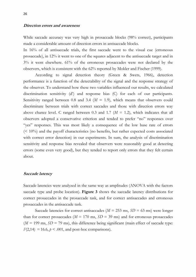
saccade type and probe location). Figure 3 shows the saccadic latency distributions for
correct prosaccades in the prosaccade task, and for correct antisaccades and erroneous
prosaccades in the antisaccade task.
Saccade latencies for correct antisaccades (M = 253 ms, SD = 63 ms) were longer
than for correct prosaccades (M = 170 ms, SD = 39 ms) and for erroneous prosaccades
(M = 199 ms, SD = 79 ms), this difference being significant (main effect of saccade type:
F(2,14) = 16.6, p < .001, and post-hoc comparisons).
27
Figure 3. Saccade latencies in Experiment 2. The histograms represent relative frequency distributions of saccade latencies (bin size: 5 ms) of correct prosaccades (N = 13003), correct antisaccades (N = 9665) and erroneous prosaccades (N = 2298). The vertical dotted lines correspond to the means.
Saccade amplitude
Amplitudes of primary saccades were subjected to a repeated measures ANOVA with the
factors saccade type (prosaccade, antisaccade, erroneous prosaccade) and probe location
(at cue, opposite cue). As in Experiment 1, erroneous prosaccades (in the antisaccade
task) had significantly shorter amplitudes (M = 5.7 deg, SD = 1.5 deg) than both correct
prosaccades (M = 6.7 deg, SD = 0.8 deg) and correct antisaccades (M = 6.7 deg, SD = 1.1
deg). The difference was statistically significant (main effect of saccade type, F(2,26) =
63.0, p < .001 and post-hoc comparisons). Within the group of erroneous prosaccades,
amplitudes were significantly shorter for unperceived errors (M = 5.2 deg, SD = 1.5 deg)
than for perceived errors (M = 6.1 deg, SD = 1.2 deg), t(15) = 6.7, p < .001.
Corrective saccades
Erroneous prosaccades having wrong direction and shorter amplitudes than the correct
saccades were often followed by corrective saccades. Indeed, our analysis revealed that
71% of all prosaccade errors were corrected in the direction of the intended antisaccade
goal (only saccades that crossed the midline were counted as corrective saccades). The
proportion of corrective saccades was considerably higher after unperceived errors (87%)
than after perceived errors (47%).
28
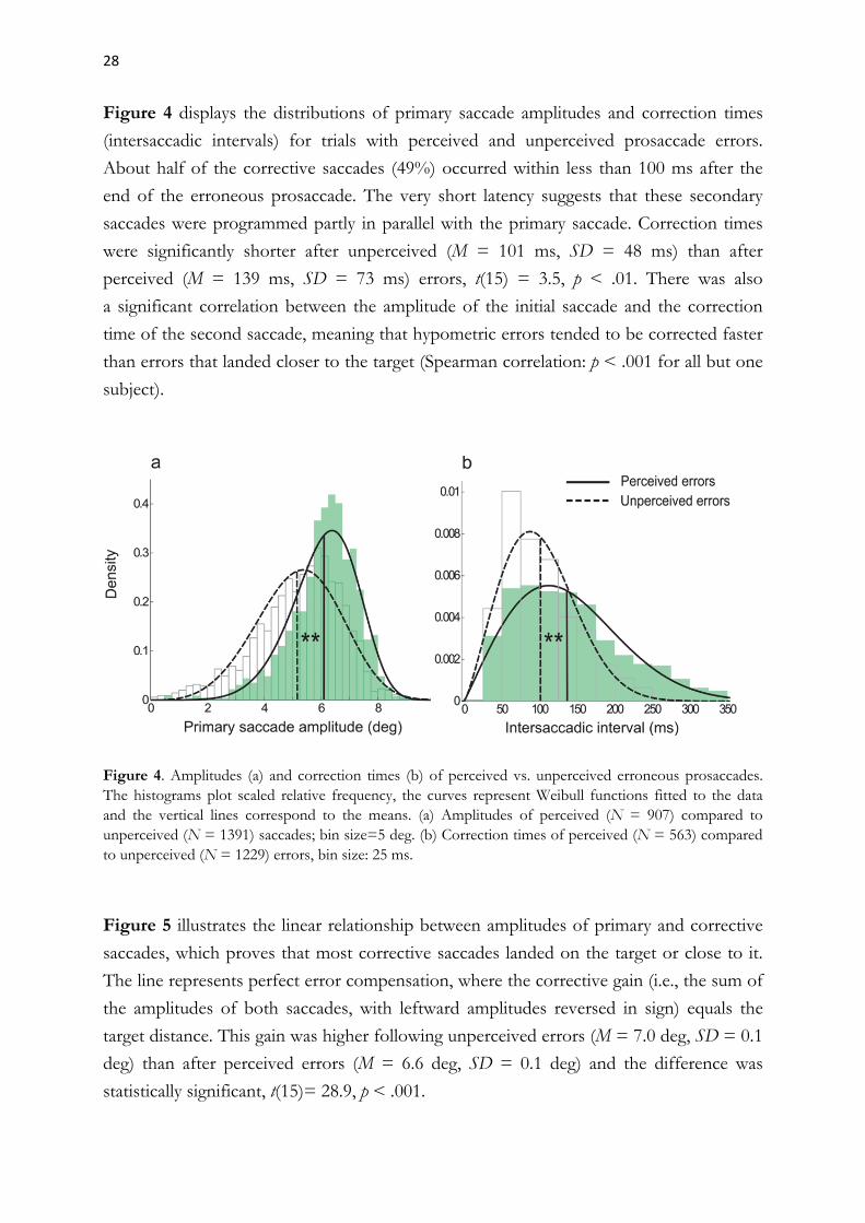
Figure 4 displays the distributions of primary saccade amplitudes and correction times
(intersaccadic intervals) for trials with perceived and unperceived prosaccade errors.
About half of the corrective saccades (49%) occurred within less than 100 ms after the
end of the erroneous prosaccade. The very short latency suggests that these secondary
saccades were programmed partly in parallel with the primary saccade. Correction times
were significantly shorter after unperceived (M = 101 ms, SD = 48 ms) than after
perceived (M = 139 ms, SD = 73 ms) errors, t(15) = 3.5, p < .01. There was also
a significant correlation between the amplitude of the initial saccade and the correction
time of the second saccade, meaning that hypometric errors tended to be corrected faster
than errors that landed closer to the target (Spearman correlation: p < .001 for all but one
subject).
Figure 4. Amplitudes (a) and correction times (b) of perceived vs. unperceived erroneous prosaccades. The histograms plot scaled relative frequency, the curves represent Weibull functions fitted to the data and the vertical lines correspond to the means. (a) Amplitudes of perceived (N = 907) compared to unperceived (N = 1391) saccades; bin size=5 deg. (b) Correction times of perceived (N = 563) compared to unperceived (N = 1229) errors, bin size: 25 ms.
Figure 5 illustrates the linear relationship between amplitudes of primary and corrective
saccades, which proves that most corrective saccades landed on the target or close to it.
The line represents perfect error compensation, where the corrective gain (i.e., the sum of
the amplitudes of both saccades, with leftward amplitudes reversed in sign) equals the
target distance. This gain was higher following unperceived errors (M = 7.0 deg, SD = 0.1
deg) than after perceived errors (M = 6.6 deg, SD = 0.1 deg) and the difference was
statistically significant, t(15)= 28.9, p < .001.

29
Interestingly, this effect remained present in the subgroup of very quickly corrected
saccades, which means that it cannot be explained by differences in correction time.
Figure 5. Scatterplot of the amplitudes of erroneous prosaccades and their corrections. The diagonal line represents full correction to the intended antisaccade target.
Discrimination performance
We performed a repeated measures ANOVA with saccade type (prosaccade, antisaccade,
erroneous prosaccade) and probe location (at cue, opposite cue, neutral) as the within-
subjects factors (see Figure 6 for a graphical summary of the results). The results showed
that probe discrimination mainly depended on the location of the probe (main effect of
probe location, F(2,26) = 28.7, p < .001, and revealed a significant interaction between
saccade type and probe location, F(4,52) = 10.7, p < .001). Before correct prosaccades,
discrimination performance was significantly better at the cued location, which was the
saccade goal, (M = 83.4%, SD = 8.3%) than both at the opposite location (M = 55.1%,
SD = 7.7%) and at the neutral location (M = 57.2%, SD = 7.4%), which were task-
irrelevant. Before correct antisaccades, discrimination at the cued location (M = 68.9%,
SD = 12.8%) and at the antisaccade goal (M = 69.4%, SD = 15.1%) were almost equal
and were both significantly better than at the neutral location (M = 55.3%, SD = 7.9%).
In contrast to this, probe discrimination before erroneous prosaccades was significantly
better at the cued location (M = 74.4%, SD = 13.9%) than at the opposite (M = 57.5%,
SD =14.8%) and neutral (M = 51.4%, SD = 11.7%) locations.

30
Figure 6. Discrimination performance in Experiment 2. The graph compares discrimination rates for probes presented between 100 and 200 ms after cue onset as a function of saccade type (correct pro-saccade, correct antisaccade, erroneous prosaccade) and probe location (at cue, opposite cue, neutral). Error bars represent standard errors of the mean. The dashed line denotes the chance performance level. The analysis was based on at least five trials per participant and condition (M = 98 for correct prosaccades, M = 102 for correct antisaccades, M = 19 for erroneous prosaccades).
In summary, the results on discrimination performance in Experiment 2 tell the same
story as in Experiment 1: Correct antisaccades were associated with pre-saccadic attention
at both locations. We further observed that errors were associated with more attention at
the cued location, where the saccade was made to, and less attention at the correct
antisaccade goal. Moreover, the significant difference between performance at the anti-
saccade goal and at the neutral location before correct antisaccades proves that attention
allocation to the antisaccade goal is mediated by oculomotor preparation rather than by
some strategy for maximizing discrimination performance.
As we were interested in whether the enhanced attention at the cued location or
rather the reduced amount of attention at the correct antisaccade goal was predictive of
errors, we performed post-hoc comparisons of discrimination performance at the cued
and opposite locations before correct antisaccades and before errors. The results revealed
that only the error-related decline in performance at the correct antisaccade goal, but not
the increase at the cued location, was statistically significant. This suggests that attention
at the antisaccade goal is crucial for correct antisaccade programming.

31
To investigate the question of whether error awareness is related to attention allocation,
as has been proposed in previous work (e.g., Deubel et al., 1999; Godijn & Theeuwes,
2003b; Mokler & Fischer, 1999), we compared discrimination performance in trials with
perceived and with unperceived errors. The results did not reveal any differences, except
for a non-significant trend towards better discrimination performance (at all locations) in
trials with unperceived errors. To see whether the allocation of attention in trials with
corrected errors depended on the latency of the corrective saccade, we compared
discrimination performance in trials with very fast (<= 90 ms) and longer (> 90 ms)
correction times. The results did not reveal any consistent differences.
DISCUSSION
The goal of this study was to investigate the allocation of spatial attention during the
programming of antisaccades. We employed a dual task, in which participants made
prosaccades or antisaccades and concurrently discriminated visual probes at the cued
location, the opposite location (i.e., the antisaccade goal), or at task-irrelevant locations.
First, we replicated the findings of previous antisaccade studies, such as the
substantially longer latency of antisaccades in comparison to prosaccades (Everling,
Dorris, & Munoz, 1998; Hallett, 1978) and the higher error rate in the antisaccade
condition (Hallett, 1978; Heath, Dunham, Binsted, & Godbolt, 2010). Second, we found
that most erroneous prosaccades were not perceived and were rapidly corrected,
suggesting that a large proportion of corrective antisaccades was programmed in parallel
with the erroneous prosaccades (Massen, 2004; Mokler et al., 2000; Mokler & Fischer,
1999). Our third and most important finding was that before antisaccades, attention was
allocated in parallel to the visual cue and the antisaccade goal, rather than being first
allocated to the cue and then to the antisaccade goal. Prosaccade errors were associated
with an attentional bias towards the prosaccade goal, which has important implications
concerning the relationship between attention and saccade programming.
In the following sections, we will discuss our results in the context of existing
theories and previous findings in this field and propose a model of how attention and
saccades could be influenced by a common competitive process.
32
Parallel programming of prosaccades and antisaccades
Parallel programming of two subsequent saccades can be inferred from very short
intersaccadic intervals (Becker & Jürgens, 1979) and has been reported not only in the
antisaccade task (Massen, 2004; Mokler & Fischer, 1999), but also in other tasks, such as
reading (Morrison, 1984), double-step paradigms (Becker & Jürgens, 1979; Walker &
McSorley, 2006), visual search (McPeek, Skavenski & Nakayama, 2000) and in the
oculomotor capture paradigm (Godijn & Theeuwes, 2002, Irwin, Colcombe, Kramer, &
Hahn, 2000; Theeuwes, Kramer, Hahn, & Irwin, 1998; Theeuwes, Kramer, Hahn, Irwin,
& Zelinsky, 1999).
Recent investigations into this topic mainly focused on situations where an
endogenous saccade is programmed along with an initial involuntary saccade to a visual
distractor. McPeek et al. (2000), for instance, asked their subjects to saccade to a red or
green color singleton presented along with two distractors of the opponent color (e.g.,
green or red). The fact that the same colors were used for target and distractors led to
many erroneous saccades towards one of the distractors, especially when the distractors
had the same color as the target in the previous trial. These erroneous saccades were often
hypometric and many were followed by short-latency corrective saccades to the target.
Based on these observations, McPeek et al. (2000) proposed a competition model of
saccade programming, in which both saccade goals were represented in a common motor
map, supposedly located in the superior colliculus. Mutual inhibitory connections between
neurons would make sure that any increase in neural activity at one location would result
in a decrease in activity at the other.
Investigations using the oculomotor capture paradigm (Godijn & Theeuwes, 2002;
Irwin et al., 2000; Theeuwes et al., 1998, 1999), where endogenous saccades to a color-
defined target compete with involuntary saccades to an onset distractor, yielded very
similar results: a substantial proportion of initial erroneous saccades to the distractor,
many of them followed by corrective saccades after less than 100 ms of fixation.
After the initial assumption that exogenous and endogenous saccades were
programmed in separate brain circuits and simply race towards a threshold (Theeuwes et
al., 1998, 1999), Godijn & Theeuwes (2002) formulated their competitive integration model
(also see Meeter, Van der Stigchel, & Theeuwes, 2010), which also postulates that the
rivalry takes place on a common collicular map with lateral inhibitory connections.
The idea of a parallel competition between erroneous prosaccades and subsequent
corrective saccades in the antisaccade task was first addressed by Mokler and Fischer
(1999) and further elaborated by Massen (2004).

33
Although Massen assumed mutual inhibition between the pro- and antisaccade programs,
her findings (see Introduction) could not rule out an independent race model (the only
evidence for mutual inhibition between prosaccades and antisaccades came from her
observation that slower and faster corrected erroneous prosaccades tended to have
shorter amplitudes, which could be due to interference from the second saccade
program). Kristjánsson and colleagues (Kristjánsson, Chen, & Nakayama, 2001;
Kristjánsson, Vandenbroucke, & Driver, 2004) showed that manipulations that slow
down the prosaccade component can lead to faster antisaccades, which is more
compatible with a model that assumes competitive interactions between both.
The results of the present study confirm many of the above mentioned findings,
such as the shorter amplitudes of erroneous saccades and the significant proportion of
very short correction times. In agreement with McPeek et al. (2000), we found
a significant correlation between the amplitudes of initial erroneous saccades and their
correction times: The faster a saccade was corrected, the smaller tended to be its
amplitude. This suggests that the first saccade was influenced or even disrupted by the
programming of the second saccade. Slower errors also tended to have shorter amplitudes
(although this relationship was less consistent). Taken together, our findings provide
further evidence that reflexive and endogenous saccades compete within the same or
overlapping neural networks.
Parallel attentional selection
The results of our experiments revealed that antisaccades are preceded by attention
allocation to both the visual cue and the antisaccade goal, thus suggesting that both
locations compete for attentional resources. Our findings are consistent with previous
evidence that visuospatial attention can be divided when this is beneficial for the task
(Awh & Pashler, 2000; Baldauf & Deubel, 2008a, 2008b, 2009; Baldauf, Wolf, & Deubel,
2006; Deubel, 2014; Godijn & Theeuwes, 2003a; Jefferies, Enns, & DiLollo, 2014;
Jonikaitis & Deubel, 2011). Moreover, our data rule out the serial hypothesis, according to
which attention first needs to be disengaged from the visual target, before it can shift to
the antisaccade goal (e.g., Crawford, Kean, Klein, & Hamm, 2006; Olk & Kingstone,
2003). If the serial hypothesis was true, we would have observed improved performance
at the antisaccade target and poor performance at the cued location shortly before saccade
onset. Instead, we found comparable performance at both locations.

34
The link between attention and (anti)saccades
Our findings on attention allocation before correct antisaccades and before prosaccade
errors have some important implications concerning the link between attention and
saccade programming. The fact that attention before correct antisaccades was equally
distributed among the cued location and the antisaccade goal is in conflict with the
premotor theory of attention (Rizzolatti, Riggio, Dascola, & Umiltà, 1987; Rizzolatti,
Riggio, & Sheliga, 1994), which regards attention as functionally equivalent to saccade
preparation. If this theory was correct, correct antisaccades would be associated with
more attention at the antisaccade goal than at the cued location, which was clearly not the
case. Nevertheless, our results suggest that attention and saccade programming are closely
linked, as attentional distribution was predictive of prosaccade errors.
Current views of visual attention are tied to the concept of priority maps (Fecteau
& Munoz, 2006; Serences & Yantis, 2006), which are thought to integrate information
about bottom-up saliency with top-down influences into a single real-time representation
of behavioral relevance. In agreement with this concept, we believe that during the
preparation of antisaccades, cue-related and antisaccade-related activity compete on such
a map (or several related maps) and that the resulting priority signal influences both
saccade programming (through modulatory influences on the oculomotor system) and
visual perception (through feedback to early visual areas).
Our findings are compatible with such a model, as discrimination performance was
clearly modulated by the saccade task and the ratio between cue-related and antisaccade-
related attention was predictive of erroneous prosaccades. Research indicates that such
a priority map could be represented in posterior parietal cortex. Single cell recording
studies in monkeys have shown that neurons in area LIP (lateral intraparietal area)
combine visual, cognitive and saccadic signals – and possibly others, such as information
about reward – into a topographic representation of behavioral priority, which can be
used to guide eye movements and attention (Bisley & Goldberg, 2010; Bisley, Ipata,
Krishna, Gee, & Goldberg, 2009; Ipata, Gee, Bisley, & Goldberg, 2009). Gottlieb and
Goldberg (1999) investigated LIP activity while monkeys performed antisaccades and
found that most neurons strongly responded to the visual stimulus, when it fell in their
receptive field, and some also fired in response to the antisaccade target. Recently, LIP
has been shown to implement center-surround suppression mechanisms that can account
for the type of competitive interactions between an endogenous saccade plan and
a visually salient distractor, as we assume are happening in the antisaccade task.

35
In humans, researchers have identified topographically organized areas within the
intraparietal sulcus (IPS) that most likely are the human homologues to monkey LIP
(Schluppeck, Glimcher, & Heeger, 2005; Sereno, Pitzalis, & Martinez, 2001; Silver, Ress,
& Heeger, 2005). Corbetta and Shulman (2002) identified IPS as a central part of the
brain’s network for endogenous attention. At the same time, IPS seems to play a role in
selection for perception, as it has been shown to modulate activity in primary visual
cortex via top-down attentional signals (Lauritzen, D’Esposito, Heeger, & Silver, 2009).
A recent study by Khan et al. (2009) established a link between these two roles by
showing that the well-documented facilitation of visual perception at the goal of
a planned saccade crucially depends on the parietal cortex. IPS lesions lead to prolonged
antisaccade latencies (Machado & Rafal, 2004) and a number of fMRI studies have found
enhanced IPS activation in antisaccades as compared to prosaccades (see a recent meta-
analysis by Jamadar, Fielding, & Egan, 2013). Of particular interest are the results of
a study by Anderson, Husain, and Sumner (2008), which suggest that human IPS
importantly contributes to the resolution of competition in the antisaccade task.
The idea that we try to convey here, namely that competitive integration can be
generalized beyond the eye movement system and could occur on a parietal priority map,
is not new, as it was already suggested by Hunt, von Mühlenen, and Kingstone (2007),
based on their results on parallels between attentional and oculomotor capture.
We would like to emphasize that this proposal does not contradict the idea that
saccade programs compete on a common collicular motor map (e.g., Findlay & Walker,
1999; Godijn & Theeuwes, 2002, McPeek et al., 2000; Trappenberg, Dorris, Munoz, &
Klein, 2001), since we do not assume that our putative priority signal can directly drive
eye movements. It rather seems that the SC motor map consists of a further competitive
stage, even more specialized on oculomotor selection. Interestingly, findings from
neurophysiological studies in monkeys revealed that stimulus-related activity bursts can be
larger than antisaccade-related bursts even in FEF and SC, which are known to directly
trigger eye movements (Everling, Dorris, Klein, & Munoz, 1999; Everling & Munoz,
2000). This indicates that the final threshold for saccade generation is not localized in SC
or FEF, but rather in the brainstem saccade generator, where outputs from the whole
oculomotor network are integrated (see Jantz, Watanabe, Everling, & Munoz, 2013, for
further evidence). The brain employs several strategies to down-weight target-related
activity and up-weight antisaccade-related activity, both at the level of the SC and
downstream from it. Examples are the increase in fixation-related and decrease in visual
SC activity in the preparation of an antisaccade (Everling, Dorris, & Munoz, 1998) or the
transient increase in omnipause neuron activity following the appearance of the visual
stimulus (Everling, Paré, Dorris, & Munoz, 1998).

36
It has also been suggested that input from other oculomotor areas, such as the
supplementary eye field (SEF), could boost the relatively weak antisaccade-related activity
downstream from the SC (Jantz et al., 2013; Munoz & Everling, 2004). The question
whether the antisaccade target is also favored by signals from a parietal priority map
remains to be clarified by further research.
Taken together, our results, as well as the research reviewed here, suggest that
attention and saccade programming are both inherently parallel processes in which
different spatial locations are selected along each other, rather than one after the other.
Although attentional and oculomotor selection seem to be linked, they are not identical,
as perceptual benefits are not limited to the goal of the upcoming saccade. In terms of
brain economy, such a distinction makes sense: While it may often be beneficial to attend
to several objects or locations at the same time, the eyes cannot go to more than one
target. This constraint, along with the relatively high costs associated with an erroneous
eye movement, entails a greater need to favor task demands over salience for saccade
programming. Nevertheless, a parallel accumulation of information until a very late stage
of oculomotor programming is still advantageous, as it allows the system to act fast and
flexibly and dramatically reduces planning costs, for example when several eye
movements are made in sequence.
Error awareness and attention
Some authors (Deubel, Irwin & Schneider, 1999; Mokler & Fischer, 1999) have proposed
that error recognition in the antisaccade task is mediated by visuospatial attention, in the
sense that our mind falsely attributes eye position to the current locus of attention. If,
according to this hypothesis, attention first moves to the visual cue, the participant would
recognize the saccade direction error. If, however, attention first shifts to the antisaccade
goal and only the eyes initially make a reflexive saccade in the wrong direction, the error
would not reach awareness and can be corrected faster, as attention does not need to
move to the correct location anymore. Mokler et al. (2000) observed that unperceived
erroneous prosaccades were associated with better visual discrimination performance at
the antisaccade goal than at the visual target, while the opposite was true for perceived
errors. From this they concluded that reflexive prosaccades can occur without a prior
attention shift to the target.
In our study, discrimination performance in error trials was always best at the cue
location and error perception was associated with slightly worse performance on the
discrimination task.
37
One attempt to explain these contradictory findings could be through the exact
comparison of the experimental designs used in their study and ours. Notably, Mokler
and her collaborators did not present probes at a neutral location, which makes it
impossible to judge the amount of task-related attention at the antisaccade goal. Second,
the 100% valid exogenous pre-cue shown 100-200 ms before the probe, which was
intended to increase the rate of erroneous prosaccades (see Fischer & Weber, 1996), may
have led to better discrimination performance at the cued location. Alternatively, the
process of error monitoring may have drawn attentional resources away from the
discrimination task.
Such an account could explain both the better discrimination performance in
association with unperceived errors and the shorter latencies of subsequent corrective
saccades observed in the present study. The results of a study by Taylor and Hutton
(2011) support this hypothesis by showing that error perception in the antisaccade task
may require top-down attentional control. The lower frequency and reduced gain of
corrective saccades that we observed following perceived errors would also be consistent
with such an explanation.
Based on their findings from the oculomotor capture paradigm, Godijn &
Theeuwes (2003b) proposed a weaker form of Mokler’s hypothesis, which states that
involuntary saccades to distractors may not be perceived when attention remains on the
distractor for too little time. Our results do not support any of the two accounts, since we
found neither a proof for attention being disengaged from the cued location nor for less
cue-related attention before unperceived errors. Rather, the main problem associated with
both hypotheses may be that they presume that attentional processes are strictly serial,
which reflects the persistent influence of the attentional spotlight metaphor. Only
recently, the notion of a single attentional focus that needs to be shifted in space has been
replaced by newer theories, in which activations corresponding to spatial locations can be
enhanced or suppressed through mutual interactions or through external modulatory
influences, leading to dynamic “attentional landscapes” that are adapted to the current
sensorimotor task (Baldauf & Deubel, 2010).
Acknowledgments
This work was funded by the Deutsche Forschungsgemeinschaft Research Training
Group GRK 1091 and a grant by the Deutsche Forschungsgemeinschaft (DE 336/5-1).

38
References
Anderson, E. J., Husain, M., & Sumner, P. (2008). Human intraparietal sulcus (IPS) and competition between endogenous and exogenous saccade plans. NeuroImage, 40, 838–851.
Awh, E., Armstrong, K. M., & Moore, T. (2006). Visual and oculomotor selection: links, causes and implications for spatial attention. Trends in Cognitive Science, 10(3), 124–130.
Awh, E., & Pashler, H. (2000). Evidence for split attentional foci. Journal of Experimental Psychology: Human Perception and Performance, 26, 834–846.
Baldauf, D., & Deubel, H. (2008a). Properties of attentional selection during the preparation of sequential saccades. Experimental Brain Research, 184(3), 411–425.
Baldauf, D., & Deubel, H. (2008b). Visual attention during the preparation of bimanual movements. Vision Research, 48, 549–563.
Baldauf, D., & Deubel, H. (2009). Attentional selection of multiple movement goal positions before rapid hand movement sequences: an event-related potential study. Journal of Cognitive Neuroscience, 21(1), 18–29.
Baldauf, D. & Deubel, H. (2010). Attentional landscapes in reaching and grasping. Minireview. Vision Research, 50, 999–1013.
Baldauf, D., Wolf, M., & Deubel, H. (2006). Deployment of visual attention before sequences of goal-directed hand movements. Vision Research, 46, 4355–4374.
Becker, W., & Jürgens, R. (1979). An analysis of the saccadic system by means of double-step stimuli. Vision Research, 19, 967–983.
Bell, A. H., Everling , S., & Munoz, D. P. (2000). Influence of stimulus eccentricity and direction on characteristics of pro- and antisaccades in non-human primates. Journal of Neurophysiology, 84(5), 2595–2604.
Bisley, J. W. & Goldberg, M. (2010). Attention, intention, and priority in the parietal lobe. Annual Reviews in Neuroscience, 33, 1–21.
Bisley, J. W., Ipata, A. E., Krishna, B. S., Gee, A. L., & Goldberg, M. E. (2009). The lateral intraparietal area: a priority map in posterior parietal cortex. In M. Jenkin & L. Harris (Eds.), Cortical Mechanisms of Vision (pp. 9–34), Cambridge: Cambridge University Press.
Brainard, D. H. (1997). The Psychophysics Toolbox. Spatial Vision, 10, 433–436.
39
Carrasco, M. (2006). Covert attention increases contrast sensitivity: psychophysical, neurophysiological, and neuroimaging studies. In S. Martinez-Conde, S. L. Macknik, L. M. Martinez, J. M. Alonso & P. U. Tse (Eds.), Progress in Brain Research, Vol. 154 (pp. 33–70), Amsterdam: Elsevier.
Carrasco, M. (2011). Visual Attention: The past 25 years. Vision Research, 51, 1484–1525.
Carrasco, M., Ling, S. & Read, S. (2004). Attention alters appearance. Nature Neuroscience, 7, 308–313.
Corbetta, M., & Shulman, G. L. (2002). Control of goal-directed and stimulus-driven attention in the brain. Nature Reviews Neuroscience, 3, 201–215.
Cornelissen, F. W., Peters., E., & Palmer, J. (2002). The Eyelink Toolbox. Behavior Research Methods, Instruments & Computers, 34, 613–617.
Crawford, T. J., Kean, M., Klein, R. M., & Hamm, J. P. (2006). The effects of illusory line motion on incongruent saccades: implications for saccadic eye movements and visual attention. Experimental Brain Research, 173, 498–506.
Deubel, H. (2008). The time course of presaccadic attention shifts. Psychological Research, 72, 630–640.
Deubel, H. (2014) Attention in action. In A. C. Nobre & S. Kastner (Eds.), The Oxford Handbook of Attention (pp. 865–889), Oxford: Oxford University Press.
Deubel, H., Irwin, D. E., & Schneider, W. X. (1999). The subjective direction of gaze shifts long before the saccade. In W. Becker, H. Deubel & T. Mergner (Eds.), Current Oculomotor Research: Physiological and Psychological Aspects (pp. 65–70). New York, London: Plenum.
Deubel, H., & Schneider, W. X. (1996). Saccade target selection and object recognition: Evidence for a common attentional mechanism. Vision Research, 36(12), 1827–1837.
Everling, S., Dorris, M. C., Klein, R. M., & Munoz, D. P. (1999). Role of primate superior colliculus in preparation and execution of anti-saccades and pro-saccades. The Journal of Neuroscience, 19, 2740–2754.
Everling, S., Dorris, M. C., & Munoz, D. P. (1998). Reflex suppression in the anti-saccade task is dependent on prestimulus neural processes. Journal of Neurophysiology, 80, 1584–1589.
Everling, S., & Munoz, D. P. (2000). Neuronal correlates for preparatory set associated with saccade generation in the frontal eye field. The Journal of Neuroscience, 20, 387–400.
40
Everling, S., Paré, M., Dorris, M. C., & Munoz, D. P. (1998). Comparison of the discharge characteristics of brain stem omnipause neurons and superior colliculus fixation neurons in monkey: implications for control of fixation and saccade behavior. Journal of Neurophysiology, 79, 511–528.
Fecteau, J. H., & Munoz, D. P. (2006). Salience, relevance, and firing: a priority map for target selection. Trends in Cognitive Science, 10(8), 382–390.
Findlay, J. M., & Walker, R. (1999). A model of saccade generation based on parallel processing and competitive inhibition. Behavioral and Brain Sciences, 22(4), 661–721.
Fischer, B., & Weber, H. (1996). Effects of procues on error rate and reaction time of antisaccades in human subjects. Experimental Brain Research, 109, 507–512.
Fischer, B., & Weber, H. (1997). Effects of stimulus conditions on the performance of antisaccades in man. Experimental Brain Research, 116(2), 191–200.
Forbes, K., & Klein, R. M. (1996). The magnitude of the fixation offset effect with endogenously and exogenously controlled saccades. Journal of Cognitive Neuroscience, 8(4), 344–352.
Godijn, R., & Theeuwes, J. (2002). Programming of exogenous and endogenous saccades: Evidence for a competitive integration model. Journal of Experimental Psychology: Human Perception and Performance, 28(5), 1039–1054.
Godijn, R., & Theeuwes, J. (2003a). Parallel allocation of attention prior to the execution of saccade sequences. Journal of Experimental Psychology: Human Perception and Performance, 29, 882–896.
Godijn, R., & Theeuwes, J. (2003b). The relationship between exogenous and endogenous saccades and attention. In J. Hyönä, R. Radach & H. Deubel (Eds). The Mind's Eyes: Cognitive and Applied Aspects of Eye Movements (pp. 3-26). Amsterdam: Elsevier/North-Holland.
Gottlieb J., & Goldberg, M. E. (1999). Activity of neurons in the lateral intraparietal area of the monkey during an antisaccade task. Nature Neuroscience, 2(10), 906–912.
Green, D. M., & Swets, J. A. (1966). Signal Detection Theory and Psychophysics. New York: Wiley.
Hallett, P. E. (1978). Primary and secondary saccades to goals defined by instructions. Vision Research, 18, 1279–1296.
Hallett, P. E., & Adams, B. D. (1980). The predictability of saccadic latency in a novel voluntary oculomotor task. Vision Research, 20, 329–339.

41
Heath, M., Dunham, K., Binsted, G., & Godbolt, B. (2010). Antisaccades exhibit diminished online control relative to prosaccades. Experimental Brain Research, 203(4), 743–752.
Hoffman, J. E., & Subramaniam, B. (1995). The role of visual attention in saccadic eye movements. Perception and Psychophysics, 57(6), 787–795.
Hunt, A.R., von Mühlenen, A., & Kingstone, A. (2007). The time course of attentional and oculomotor capture reveals a common cause. Journal of Experimental Psychology: Human Perception & Performance, 33, 271–284.
Ipata, A. E., Gee, A. L., Bisley, J. W., & Goldberg, M. E. (2009). Neurons in the lateral intraparietal area create a priority map by the combination of disparate signals. Experimental Brain Research, 192, 479–488.
Irwin, D. E., Colcombe, A. M., Kramer, A. F., & Hahn, S. (2000). Attentional and oculomotor capture by onset, luminance, and color singletons. Vision Research, 40, 1443–1458.
Jamadar, S. D., Fielding, J., & Egan, G. F. (2013). Quantitative meta-analysis of fMRI and PET studies reveals consistent activation in fronto-striatal-parietal regions and cerebellum during antisaccades and prosaccades. Frontiers in Psychology, 4:749.
Jantz, J. J., Watanabe, M., Everling, S., & Munoz, D. P. (2013). Threshold mechanism for saccade initiation in frontal eye field and superior colliculus. Journal of Neurophysiology, 109(11), 2767–2780.
Jefferies, L. N., Enns, J. T., & Di Lollo, V. (2014). The flexible focus: whether spatial attention is unitary or divided depends on observer goals. Journal of Experimental Psychology: Human Perception and Performance, 40, 465–470.
Jonikaitis, D., & Deubel, H. (2011). Independent allocation of attention to eye and hand targets in coordinated eye- hand movements. Psychological Science, 22(3), 339–347.
Jonikaitis, D., & Theeuwes, J. (2013). Dissociating oculomotor contributions to spatial and feature-based selection. Journal of Neurophysiology, 110(7), 1525–1534.
Khan, A. Z., Blangero, A., Rossetti, Y., Salemme, R., Luauté, J., Deubel, H., Schneider, W. X., Laverdure, N., Rode, G., Boisson, D., & Pisella, L. (2009). Parietal damage dissociates saccade planning from presaccadic perceptual facilitation. Cerebral Cortex, 19, 383–387.
King-Smith, P. E., Grigsby, S. S., Vingrys, A. J., Benes, S. C., & Supowit, A. (1994). Efficient and unbiased modifications of the QUEST threshold method: theory, simulations, experimental evaluation and practical implementation. Vision Research, 34 (7), 885–912.
42
Kleiner, M., Brainard, D., & Pelli, D. (2007). “What’s new in Psychtoolbox-3?”. Perception, 36, ECVP Abstract Supplement.
Kowler, E., Anderson, E., Dosher, B., & Blaser, E. (1995). The role of attention in the programming of saccades. Vision Research, 35(13), 1897–1916.
Kristjánsson, Á., Chen, Y., & Nakayama, K. (2001). Less attention is more in the preparation of antisaccades, but not prosaccades. Nature Neuroscience, 4(10), 1037–1042.
Kristjánsson, Á., Vandenbroucke, M. W. & Driver, J. (2004). When pros become cons for anti-versus prosaccades: Factors with opposite or common effects on different saccade types. Experimental Brain Research, 155, 231–244.
Lauritzen, T. Z., D’Esposito, M., Heeger, D. J., & Silver, M. A. (2009). Top-down flow of visual spatial attention signals from parietal to occipital cortex. Journal of Vision, 9(13):18.
Machado, L., & Rafal, R. D. (2004). Control of fixation and saccades during an anti-saccade task: an investigation in humans with chronic lesions of oculomotor cortex. Experimental Brain Research, 156, 55–63.
Massen, C. (2004). Parallel programming of exogenous and endogenous components in the antisaccade task. The Quarterly Journal of Experimental Psychology, Section A: Human Experimental Psychology, 57(3), 475–498.
McPeek, R. M., Skavenski, A. A., & Nakayama, K. (2000). Concurrent processing of saccades in visual search. Vision Research, 40, 2499–2516.
Meeter, M., Van der Stigchel, S., & Theeuwes, J. (2010). A competitive integration model of exogenous and endogenous eye movements. Biological Cybernetics, 102(4), 271–291.
Mokler, A., Deubel, H., & Fischer, B. (2000). Unintended saccades can be executed without presaccadic attention shifts. Perception, 29 (Suppl.), 54.
Mokler, A., & Fischer, B. (1999). The recognition and correction of involuntary prosaccades in an antisaccade task. Experimental Brain Research, 125, 511–516.
Montagnini, A., & Castet, E. (2007). Spatiotemporal dynamics of visual attention during saccade preparation: Independence and coupling between attention and movement planning. Journal of Vision, 7(14):8.
Morrison, R. E. (1984). Manipulation of stimulus onset delay in reading: Evidence for parallel programming of saccades. Journal of Experimental Psychology: Human Perception and Performance, 10, 667–682.

43
Müller, H. J., & Rabbit, P. M. (1989). Reflexive and voluntary orienting of visual attention: time course of activation and resistance to interruption. Journal of Experimental Psychology: Human Perception and Performance, 15(2), 315–330.
Munoz, D. P., & Everling, S. (2004). Look away: the anti-saccade task and the voluntary control of eye movement. Nature Reviews Neuroscience, 5(3), 218–228.
Nakayama, K., & Mackeben, M. (1989). Sustained and transient components of focal visual attention. Vision Research, 29, 1631–1647.
Noorani, I., & Carpenter, R. H. (2013). Antisaccades as decisions: LATER model predicts latency distributions and error responses. European Journal of Neuroscience, 37(2), 330–338.
Olk, B., & Kingstone, A. (2003). Why are antisaccades slower than prosaccades? A novel finding using a new paradigm. NeuroReport, 14, 151–155.
Pelli, D. G. (1997). The VideoToolbox software for visual psychophysics: Transforming numbers into movies, Spatial Vision, 10, 437–442.
Rizzolatti, G., Riggio, L. , & Sheliga, B. M. (1994). Space and selective attention. In Carlo Umiltà & Morris Moscovitch (Eds.), Attention and Performance XV (pp. 231-265). Cambridge, MA: MIT Press.
Rizzolatti, G., Riggio, L., Dascola, I., & Umiltà, C. (1987). Reorienting attention across the horizontal and vertical meridians: evidence in favor of a premotor theory of attention. Neuropsychologia, 25, 31–40.
Rolfs, M., Jonikaitis, D., Deubel, H., & Cavanagh, P. (2011). Predictive remapping of attention across eye movements. Nature Neuroscience, 14, 252–256.
Schluppeck, D., Glimcher, P., & Heeger, D. J. (2005). Topographic organization for delayed saccades in human posterior parietal cortex. Journal of Neurophysiology, 94(2), 1372–1384.
Serences, J. T., & Yantis, S. (2006). Selective visual attention and perceptual coherence. Trends in Cognitive Science, 10, 38–45.
Sereno, M. I., Pitzalis, S., & Martinez, A. (2001). Mapping of contralateral space in retinotopic coordinates by a parietal cortical area in humans. Science, 294, 1350–1354.
Sergent, C., Wyart, V., Babo-Rebelo, M., Cohen, L., Naccache, L., & Tallon-Baudry, C. (2013). Cueing attention after the stimulus is gone can retrospectively trigger conscious perception. Current Biology, 23(2), 150–155.

44
Silver, M. A., Ress, D., & Heeger, D. J. (2005). Topographic maps of visual spatial attention in human parietal cortex. Journal of Neurophysiology, 94(2), 1358–1371.
Taylor, A. J. G., & Hutton, S. B. (2011). Error awareness and antisaccade performance. Experimental Brain Research, 213, 27–34.
Theeuwes, J., Kramer, A. F., Hahn, S., & Irwin, D. E. (1998). Our eyes do not always go where we want them to go: capture of the eyes by new objects. Psychological Science, 9, 379–385.
Theeuwes, J., Kramer, A. F., Hahn, S., Irwin, D. E., & Zelinsky, G. J. (1999). Influence of attentional capture on oculomotor control. Journal of Experimental Psychology: Human Perception & Performance, 25, 1595–1608.
Thibault, L., Cavanagh, P., & Sergent, C. (2015). Retroactive Attention can trigger all-or-none conscious access to past sensory stimulus. Journal of Vision, 15(12): 547.
Trappenberg, T. P., Dorris, M. D., Munoz, D. P., & Klein, R. M. (2001). A model of saccade initiation based on the competitive integration of exogenous and endogenous signals in the superior colliculus. Journal of Cognitive Neuroscience, 13, 256–271.
Walker, R., & Mc Sorley, E. (2006). The parallel programming of voluntary and reflexive saccades. Vision Research, 46, 2082–2093.
Watson, A. B. & Pelli, D. G. (1983). QUEST: a Bayesian adaptive psychometric method. Perception & Psychophysics, 33(2), 113–120.
Wilder, J. D., Kowler, E., Schnitzer, B. S., Gersch, T. M., & Dosher, B. A. (2009). Attention during active visual tasks: counting, pointing, or simply looking. Vision Research, 49(9), 1017–1031.

45
2.2 Study 2: Attention reflects saccade decisions
Contributions:
The author of this dissertation participated in data collection and analysis, interpreted the
results and wrote the manuscript.
Donatas Jonikaitis designed and programmed the experiment, collected and analyzed
data, created plots, interpreted the results and participated in writing the manuscript.
Heiner Deubel supervised the project, interpreted results and commented on the
manuscript.

46
Manuscript for submission
Attention reflects saccade decisions
Donatas Jonikaitis1, Anna Klapetek2,3, Heiner Deubel2
1 Department of Neurobiology and Howard Hughes Medical Institute, Stanford University School of
Medicine, USA
2 Allgemeine und Experimentelle Psychologie, Ludwig-Maximilians-Universität München, Germany
3 Graduate School of Systemic Neurosciences, Ludwig-Maximilians-Universität München, Germany
Abstract
The present study investigated the allocation of visuospatial attention during free or rule-
based decisions between two memorized saccade targets. Participants were asked to
memorize two spatial locations, highlighted by different colors. After a delay period, the
color of the central fixation changed and either indicated one of the memorized locations
as the saccade target (rule-based choice), or indicated to freely choose between the two
locations. By probing discrimination performance at variable SOAs relative to this cue, we
were able to measure the time course of the attentional selection of both targets. In both
rule-based and free choice trials, we observed a parallel selection of the chosen and the
non-chosen saccade target, with a clear bias towards the chosen target. This bias was
evident before correct and before erroneous rule-based responses. Perceptual
performance during the memory delay was influenced by top-down and bottom-up
factors as well as by selection history and predicted the direction of saccades. During
saccade programming, performance increased at both potential saccade goals, consistent
with a race of both saccade programs towards a decision threshold. Our findings
demonstrate that saccade decisions have direct visual consequences and show that
decision making can be traced in the human oculomotor system well before choices are
made.

47
INTRODUCTION
Despite the fact that we, seemingly automatically, move our eyes several times per second,
each of our saccades has to be preceded by some kind of decision making process, in
which the saccade goal is selected amongst other possible goals. While traditional theories
of decision making postulated that decisions are first made by cognitive systems and only
then implemented in the form of motor actions, recent neurophysiological findings
challenge these serial models by supporting the view that motor decisions consist of
a biased competition between alternative motor plans that are represented in parallel in
sensorimotor brain areas (Andersen & Cui, 2009; Cisek, 2007; Cisek & Kalaska, 2010).
Computational models of the decision process assume that sensory evidence in
favor of each movement alternative is integrated over time and gradually accumulated
towards a threshold, at which the corresponding action is initiated (e.g., Brown &
Heathcote, 2007; Carpenter & Williams, 1995; Ratcliff & McKoon, 2008). The starting
point – or baseline – of the accumulation depends on the prior probability that the given
movement will be executed (Ludwig, 2011), while the rate of accumulation depends on
the strength of the sensory evidence and other variables, such as the value associated with
the given response (Gold & Shadlen, 2007).
Numerous single cell recordings from the monkey brain have supported these
assumptions by showing that the outcome of saccadic decisions directly depends on
sensory information (Newsome, Britten, & Movshon, 1989; Salzman, Britten, &
Newsome, 1990) and that this decision-related sensory evidence is accumulated in visuo-
motor brain areas, such as LIP (Gold & Shadlen, 2000; Ipata, Gee, Goldberg, & Bisley,
2009; Roitman & Shadlen, 2002; Shadlen & Newsome, 1996, 2001), FEF (Hanes &
Schall, 1996; Kim & Shadlen, 1999; Schall, 2003) and SC (Glimcher & Sparks, 1992;
Horwitz & Newsome, 2001; Munoz & Wurtz, 1995; Sparks, 1978; Wurtz & Goldberg,
1972). An fMRI study in humans, in which two eye movements had to be planned in
sequence, also provided evidence that the posterior parietal cortex (containing the human
homologue of LIP) represents goals of upcoming saccades (Medendorp, Goltz, & Vilis,
2006).
When monkeys have to decide between two saccade targets, neurons in visuo-
motor brain areas represent both response alternatives – with a stronger signal
corresponding to the selected goal (Kim & Basso, 2008; Platt & Glimcher, 1997) – and
the representations are modulated by the perceived probability or value of the responses
(Basso & Wurtz, 1998; Sugrue, Corrado, & Newsome, 2004).
48
Consistent with this, behavioral studies show that competitive visual environments not
seldom lead to the parallel programming of two, or possibly even more, saccades (Becker
& Jürgens, 1979; Godijn & Theeuwes, 2002; Irwin, Colcombe, Kramer & Hahn, 2000;
Klapetek, Jonikaitis, & Deubel, 2016; Massen, 2004; McPeek, Skavenski & Nakayama,
2000; Morrison, 1984; Theeuwes, Kramer, Hahn, Irwin, & Zelinsky, 1999; Walker &
McSorley, 2006).
Unfortunately, many of the studies that supposedly demonstrated decision-related
neural activity investigated saccades to visual targets, which makes it difficult to judge
whether the neural signals reflected perceptual decision making or saccade planning.
A possible way to distinguish between the two processes is to dissociate visual
information and saccade planning spatially (as for example in the antisaccade task, where
saccades have to be directed away from a visual stimulus) or temporally (by presenting the
saccade cue after the disappearance of the visual information). A few researchers recorded
from LIP neurons while monkeys performed antisaccades (Gottlieb & Goldberg, 1999;
Zhang & Barash, 2000, 2004), their results being contradictory. While Gottlieb and
Goldberg found that only few LIP neurons represented purely saccade-related activity
and most cells showed visual responses, Zhang and Barash (2000, 2004) observed that
most neurons could carry visual as well as motor activity, depending on the context.
To investigate saccade decisions in humans, one can take advantage of the tight
coupling between oculomotor and perceptual selection. During saccade preparation,
perception is automatically enhanced at the saccade target (Deubel & Schneider, 1996;
Hoffman & Subramaniam, 1995; Jonikaitis & Deubel, 2011; Klapetek et al., 2016; Kowler,
Anderson, Dosher, & Blaser, 1995; Rolfs, Jonikaitis, Deubel, & Cavanagh, 2011; Jonikaitis
& Theeuwes, 2013). On the other hand, the presentation of salient visual events, such as
irrelevant distractors, often leads to involuntary saccades in the direction of these stimuli,
which has been termed oculomotor capture (Theeuwes, Kramer, Hahn, & Irwin, 1998;
Theeuwes, Kramer, Hahn, Irwin, & Zelinsky, 1999). Such observations, along with
a wealth of other findings in monkeys and humans (for a review, see Ptak, 2012)
corroborate the hypothesis that oculomotor and visual selection are coupled through
a common attentional priority mechanism, which always selects the behaviorally most
relevant location (Fecteau & Munoz, 2006; Serences & Yantis, 2006). If this is true, we
should expect that any saccadic decision (reflected in accumulation of evidence in favor of
a particular location on the priority map) should also be reflected in gradually improving
perceptual performance at the corresponding spatial location.
In order to test this hypothesis, we designed an experiment, in which human
observers had to choose between two memorized saccade targets (either freely or on the
basis of a color rule) and simultaneously discriminate visual probes at the two competing

49
target locations or elsewhere in the display. By probing discrimination performance at
variable times during the trial, we were able to measure the time course of the perceptual
selection of both target locations. Consistent with the previously reviewed evidence on
the coupling between saccadic and perceptual selection, we expected to see a clear
perceptual benefit at the chosen target. We were also interested whether a perceptual
benefit would be evident at the competing (non-chosen) target location as well. It is
possible that both saccade targets (or the oculomotor programs towards them) are
simultaneously represented in visuomotor brain areas and facilitate perception at both
corresponding spatial locations. A second possibility, however, is that the non-chosen
target is treated as an irrelevant distractor and can be ignored or even inhibited from early
on. A third possibility is that the pattern of perceptual facilitation would differ in the rule-
based and in the free-choice conditions, for example due to smaller competition of the
non-chosen target in the rule-based condition than in the free-choice condition.
METHODS
Participants
Twelve observers (age 21-29, seven female) completed the experiment for payment. All
participants were naive as to the predictions of the study. The experiments were carried
out with the approval of the department’s ethic guidelines committee and in accordance
to the Declaration of Helsinki, and participants gave written informed consent.
Apparatus
The observers were seated in a quiet and dimly illuminated room in front of a gamma-
linearized 21-in CRT monitor (Sony GDM-F500R, 1024 x 768 pixels, 120 Hz), positioned
at a viewing distance of 60 cm. Right-eye gaze position was recorded with an EyeLink
1000 desktop mounted eye tracker (SR Research, Canada) at a sampling rate of 1000 Hz
while head movements were minimized through the use of a chin and forehead rest. The
eye tracker was calibrated before each new block and whenever it was necessary. Stimulus
presentation and response collection were controlled by an Apple Mac Mini, using
MATLAB software (MathWorks, Natick, USA) and the Psychophysics and Eyelink
Toolbox extensions (Brainard, 1997; Cornelissen, Peters, & Palmer, 2002; Kleiner,
Brainard, & Pelli, 2007; Pelli, 1997; see http://psychtoolbox.org). Manual responses were
collected via the arrow keys on the right hand side of the computer keyboard.
50
Procedure
Main task
Each trial started with a fixation target (black dot, diameter: 0.75° of visual angle)
presented on a gray background. Five square objects (diameter: 2.8°), were positioned on
an imaginary circle around the fixation (radius: 7°), the angular position of the first object
was 30° plus a random jitter between -10 to 10° and the distance between objects was 72°
(see Figure 1). Each square object consisted of an alternating stream of vertically oriented
Gabor patches (spatial frequency: 2.5 cycles per degree, 100% contrast, random phase
selected on each presentation) and grayscale noise masks (pixel luminance values
randomly drawn from a Gaussian distribution with minimum 0 (black) and maximum 255
(white), M = 128 and SD = 128). Gabor patches and noise masks alternated every 4
display refresh frames (33 ms).
During an initial memory cueing phase (Gaussian distribution with M = 2000 ms
and SD = 100 ms), two of the streams were highlighted by color frames (one blue and the
other green) and participants were asked to memorize their locations. The memory cueing
phase was followed by a delay of 500 to 1500 ms (selected randomly from a uniform
distribution), during which the colored frames were extinguished. After the delay,
a central saccade cue was presented for 700 ms: the fixation target changed color to an
equiluminant blue, green or orange color with equal probability. A green or blue fixation
instructed a saccade to the corresponding memorized green or blue target location (rule-
based choice), whereas an orange fixation indicated to saccade to either of the two
memorized locations (free choice). After 700 ms, the fixation and all square objects were
removed.
A probe display was presented -300 to 400 ms relative to saccade cue onset (time
selected randomly from a uniform distribution). The probe was a Gabor patch tilted
clockwise or counterclockwise (angle of tilt determined for each participant, see below)
and was presented for 33 ms in one of the five object streams (selected randomly with
equal probability).
After probe offset, no Gabor patches were further shown, so the object streams
consisted of alternating noise masks and blank sequences. Observers reported the
perceived probe orientation by pressing the left (counterclockwise tilt) or right (clockwise
tilt) arrow key. We instructed observers to focus on making fast and accurate saccades
and to guess whenever they were unsure about the probe orientation. Discrimination
responses were not speeded. A new trial started 200 ms after the discrimination response.

51
Pretest
We determined the probe tilt angle for each participant at the beginning of each
recording day. The visual stimuli in the pretest were identical as in the main task, except
that only one memory target was presented and it always predicted the location of the
probe (100% valid cue).
Observers were instructed to covertly attend to the memory target while
maintaining central fixation and to report the orientation of the probe. The tilt angle at
which observers reached 80% correct probe discrimination was determined using
a modified version of the QUEST staircase procedure (King-Smith, Grigsby, Vingrys,
Benes, & Supowit, 1994; Watson & Pelli, 1983) implemented in MATLAB Psychophysics
toolbox.
Observers participated in a minimum of 14 main task sessions, each consisting of
eight blocks of 50 trials. They usually performed one or two sessions on a single day.
Data analyses
Drift correction was performed offline using the average gaze direction (given that no
saccades of amplitude larger than 0.5º occurred) between 100 and 10 ms before memory
delay onset. Saccades were detected offline using an algorithm that evaluates eye velocity
changes (Engbert & Mergenthaler, 2006) with a velocity threshold of 6 SD to detect
saccade onset and a minimum saccade duration of 6 ms. We classified saccades as correct
if the starting point was less than 2º away from fixation, the endpoint was less than 3º
away from the target center and the onset latency was between 50 and 700 ms. We
removed trials due to breaking of fixation if a saccade larger than 2º occurred during the
last 200 ms before saccade cue onset or between cue onset and correct saccade initiation.
Trials were also removed if blinks occurred in the interval starting 100 ms before saccade
cue onset and ending with saccade onset. On trials where the two target locations were
adjacent (distance 72º of arc), the saccade was classified as either correct or incorrect,
depending on whether the first saccade landed closer to the cued or to the non-cued
location. Data from each participant was inspected manually for saccade and micro-
saccade detection accuracy and data recording noise. In total, participants selected the
correct target on 80155 and the erroneous target on 14912 out of the total 102451
recorded trials (7% of trials had to be removed for reasons mentioned above).
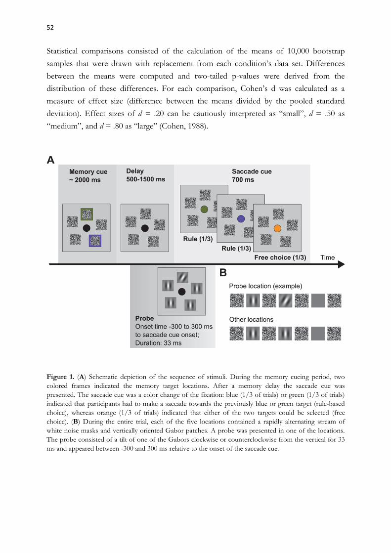
52
Statistical comparisons consisted of the calculation of the means of 10,000 bootstrap
samples that were drawn with replacement from each condition’s data set. Differences
between the means were computed and two-tailed p-values were derived from the
distribution of these differences. For each comparison, Cohen’s d was calculated as a
measure of effect size (difference between the means divided by the pooled standard
deviation). Effect sizes of d = .20 can be cautiously interpreted as “small”, d = .50 as
“medium”, and d = .80 as “large” (Cohen, 1988).
Figure 1. (A) Schematic depiction of the sequence of stimuli. During the memory cueing period, two colored frames indicated the memory target locations. After a memory delay the saccade cue was presented. The saccade cue was a color change of the fixation: blue (1/3 of trials) or green (1/3 of trials) indicated that participants had to make a saccade towards the previously blue or green target (rule-based choice), whereas orange (1/3 of trials) indicated that either of the two targets could be selected (free choice). (B) During the entire trial, each of the five locations contained a rapidly alternating stream of white noise masks and vertically oriented Gabor patches. A probe was presented in one of the locations. The probe consisted of a tilt of one of the Gabors clockwise or counterclockwise from the vertical for 33 ms and appeared between -300 and 300 ms relative to the onset of the saccade cue.

53
RESULTS
Properties of saccades
Response latency and accuracy are the two main measures of the outcome of any decision
making process. Further measures of motor performance, such as trajectory deviations,
have also been used as a basis for inferences about competitive processes underlying
choices between multiple motor plans (e.g., Chapman et al., 2010; McSorley & McCloy,
2009; Song & Nakayama, 2008; Welsh & Elliott, 2004).
Figure 2A shows the distributions of saccade latencies. Latencies (mean ± SE)
were shorter in the free-choice condition (234 ± 8 ms) than in the rule-based condition
(241 ± 7 ms, p = .004, Cohen’s d = .27). A comparison of saccade latencies on correct and
error trials in the rule-based condition (note that decision errors could not occur in the
free-choice condition) revealed shorter latencies on error trials (230 ± 8 ms) than on
correct trials (241 ± 7 ms, p < .001, d = .44).
Figure 2B shows the saccade trajectories. In human studies, saccade trajectories
tend to deviate away from competing stimuli if they are successfully suppressed on the
motor map of the superior colliculus, and towards distractors if this suppression fails (e.g.,
Sheliga, Riggio, Craighero & Rizzolatti, 1995; Van der Stigchel, Meeter, & Theeuwes,
2006).
We observed that saccade trajectories deviated away from the non-chosen target
both in the free-choice and in the rule-based condition. Surprisingly, this deviation away
was abolished on error trials. Saccade deviation on error trials (0.03 ± 0.03 deg) was
smaller than on both correct rule-based trials (0.30 ± 0.03 deg, p < .001, d = 2.56) and on
free-choice trials (0.26 ± 0.05 deg, p < .001, d = 1.78). This suggests that suppression of
the non-chosen target was reduced on error trials. However, two different reasons could
underlie this reduced suppression. First, it is possible that both targets were represented in
the oculomotor system and that the representation of the non-chosen target was not
suppressed. Second, only the erroneously chosen saccade target may have been
represented on error trials, while the correct target may simply not have been considered
from the beginning. Unfortunately, neither alternative can be ruled out, as motor
responses only reflect the final state of competition between possible movement targets,
shortly before or during movement execution.

54
Figure 2. (A) Saccade latency distributions in the free and rule-based choice tasks. Errors were defined as wrong saccade target selection during the rule-based condition. Shaded areas represent the standard error of the mean (SEM). (B) Saccade trajectories towards or away from the non-chosen target. Locations were rotated and flipped, so that all non-chosen targets appear to the left of the saccade goal in the figure. Saccade trajectories towards the non-chosen target fall on the shaded background area, whereas trajectories away from it fall on the white background area.
In summary, while the latency and trajectory differences observed in our study can be
cautiously interpreted, they represent the final state of motor decisions and hence allow us
to draw only limited conclusions on the decision making process itself. We will therefore
focus on attention allocation during decision making, which will change or disambiguate
some of the previously discussed interpretations.
Attentional selection
Based on the existing literature, we expected to find (a) that attention would be
maintained at the locations held in working memory during the delay interval, and (b) that
attention would shift to the saccade goal before saccade onset.
In order to measure attention allocation during the memory delay, we calculated
visual sensitivity (d-prime) in the probe discrimination task at all locations in the range
300-200 ms before the onset of the saccade cue. Visual sensitivity (M ± SE) was superior
at both memorized locations. When compared to the average of the three task-irrelevant
locations (0.1 ± 0.1), d-primes were higher at the location of the first (0.7 ± 0.1, p < .001,
d = 2.01), and the second memory target (0.8 ± 0.1, p < .001, d = 2.28).
This indicates that both locations were selected by visuospatial attention, possibly in order
to strengthen their representation in working memory by spatial rehearsal (Awh &
Jonides, 2001).

55
Next, we focused on discrimination performance after the appearance of the saccade cue.
In line with earlier studies on saccade preparation and spatial attention (Deubel &
Schneider, 1996; Hoffman & Subramaniam, 1995; Jonikaitis & Deubel, 2011; Klapetek et
al., 2016; Kowler et al., 1995; Rolfs et al., 2011; Jonikaitis & Theeuwes, 2013), we found
that saccade preparation was associated with improved target discrimination at the future
saccade goal. Visual sensitivity (100-0 ms before saccade onset) was higher at the chosen
target (2.3 ± 0.2) than at the non-chosen target (1.1 ± 0.2, p < .001, d = 1.64), as well as at
irrelevant locations (0.3 ± 0.1, p < .001, d = 3.89). Taken together, the pattern of
perceptual discrimination performance suggests that spatial attention shifted to the
saccade goal whenever a saccade was prepared.
Attentional selection is linked to decision making
Figure 3A-B shows the spatiotemporal distribution of the pre-saccadic visual sensitivity.
An increase in sensitivity occurred at both competing target locations, whereas there was
little to no sensitivity increase at the irrelevant locations. Furthermore, sensitivity at the
non-chosen location increased regardless of whether it was close to the chosen target
(radial distance 72 degrees) or further away from it (radial distance 144 degrees), with no
benefit at the location between the chosen and non-chosen target locations in the latter
case (Figure 3D). This indicates that target selection is spatially specific and occurs
before saccade onset.
To evaluate whether spatial attention reflects the process of saccadic decision
making, we measured how visual sensitivity after saccade cue onset changed relative to
visual sensitivity during the memory delay. For this purpose, we subtracted the average d-
prime during the memory delay (-300 to -100 ms before saccade cue onset) from the d-
prime in each time bin, separately for chosen, non-chosen, and task irrelevant locations as
well as for both choice conditions. Figure 3E-F shows the change in visual sensitivity in
the rule-based and free-choice conditions over time. Visual sensitivity increased at both
the chosen target and the non-chosen target after the appearance of the saccade cue.
Indeed, during rule-based decisions, the visual sensitivity increase relative to the memory
delay (change in d-primes 100 to 0 ms before saccade onset) was larger at the chosen
target (1.3 ± 0.1) than at irrelevant locations (0.2 ± 0.1, p < .001, d = 4.20) and this
benefit was also present at the non-chosen target (0.5 ± 0.1, p < .001, d = 1.20).
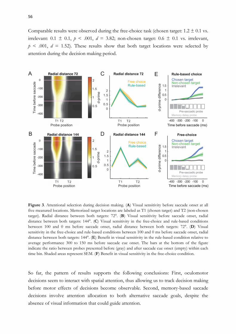
56
Comparable results were observed during the free-choice task (chosen target: 1.2 ± 0.1 vs.
irrelevant: 0.1 ± 0.1, p < .001, d = 3.82; non-chosen target: 0.6 ± 0.1 vs. irrelevant,
p < .001, d = 1.52). These results show that both target locations were selected by
attention during the decision making period.
Figure 3. Attentional selection during decision making. (A) Visual sensitivity before saccade onset at all five measured locations. Memorized target locations are labeled as T1 (chosen target) and T2 (non-chosen target). Radial distance between both targets: 72°. (B) Visual sensitivity before saccade onset, radial distance between both targets: 144°. (C) Visual sensitivity in the free-choice and rule-based conditions between 100 and 0 ms before saccade onset, radial distance between both targets: 72°. (D) Visual sensitivity in the free-choice and rule-based conditions between 100 and 0 ms before saccade onset, radial distance between both targets: 144°. (E) Benefit in visual sensitivity in the rule-based condition relative to average performance 300 to 150 ms before saccade cue onset. The bars at the bottom of the figure indicate the ratio between probes presented before (gray) and after saccade cue onset (empty) within each time bin. Shaded areas represent SEM. (F) Benefit in visual sensitivity in the free-choice condition.
So far, the pattern of results supports the following conclusions: First, oculomotor
decisions seem to interact with spatial attention, thus allowing us to track decision making
before motor effects of decisions become observable. Second, memory-based saccade
decisions involve attention allocation to both alternative saccade goals, despite the
absence of visual information that could guide attention.

57
Both in the free-choice and surprisingly also in the rule-based choice condition, where the
saccade target was defined by a simple stimulus-response mapping, we observed a
simultaneous representation of both potential saccade goals during the entire decision
making process.
Attentional selection is associated with saccade programming
We next investigated whether the observed attentional selection was directly associated
with saccade responses. For this purpose, we examined the relationship between saccade
latencies and the time course of visual sensitivity (Figure 4). If covert attention indeed
reflects decision making, then faster decisions, as indicated by saccade latencies, should be
preceded by earlier attentional selection. However, in this context it is important to
distinguish two factors that can influence the speed of decisions. One is the rate at which
sensory evidence is accumulated towards the decision boundary, and the other is the
starting point of the accumulation (or alternatively the threshold itself).
To compare trials with faster and slower saccades, we separated saccade latencies
at the median for each participant (faster latencies were on average 217 ms and slower
latencies were 269 ms). As can be seen in Figure 4A, we observed that after the onset of
the saccade cue, visual sensitivity increased earlier during fast saccadic responses and
improved later during slow saccadic responses. Visual sensitivity (averaged across 50-200
ms after saccade cue onset) was better during short latency trials (2.1 ± 0.2) than during
long latency trials (1.6 ± 0.2, p < .001, d = 1.04. Figure 4B shows discrimination
performance relative to saccade onset. We observed comparable rise rates for fast and
slower decisions. Visual sensitivity for the interval 150-0 ms before saccade onset was
comparable for short latency trials (2.0 ± 0.2) and long latency trials (1.9 ± 0.3), p = .52, d
= 0.09. These findings are compatible with the assumption that faster and slower
decisions differ in the starting point of the accumulation, which depends on the prior
probability of the associated movement vector (Ludwig, 2011), but not in the rate of the
sensory evidence accumulation.
Figure 4C shows that saccade latency differences were also associated with
discrimination performance differences at the non-chosen target. Visual sensitivity at the
non-chosen target 50-200 ms after saccade cue onset was higher before short latency
saccades (1.0 ± 0.2), than before long-latency saccades (0.7 ± 0.2, p = .006, d = .42),
suggesting that the attention shift to the non-selected target was also linked to saccade
onset. No such differences were present at task-irrelevant locations (Figure 4D).

58
Our results thus demonstrate that the timing of the attentional selection of the competing
saccade targets is directly related to the timing of the saccade decision. An apparent
interpretation is that participants were simply more alert on fast latency trials, which could
lead to both an earlier increase in discrimination performance and to faster saccade
programming. This explanation is unlikely, however, as discrimination performance at the
irrelevant locations was not modulated by saccade latency.
Visual sensitivity at the irrelevant locations (50-200 ms after saccade cue onset) did
not differ between short latency saccades (0.2 ± 0.1) and long latency saccades (0.1 ± 0.1,
p = .53, d = .24). The effect of saccade latency on discrimination performance was instead
restricted to the decision-relevant locations, suggesting that a spatially specific
competition between these two locations had taken place.
Figure 4. Attentional selection before saccades with short and with long latencies. (A) Visual sensitivity at the chosen target relative to saccade cue onset. (B) Visual sensitivity at the chosen target relative to saccade onset. (C) Visual sensitivity at the non-chosen target relative to saccade cue onset. (D) Visual sensitivity at irrelevant locations relative to saccade cue onset.

59
Choice errors are related to attentional biases
We observed that in the rule-based choice condition participants correctly selected the
instructed target on 78% of the trials, and made an erroneous selection on 22%. Given
the size of our data set, we recorded on average ~1200 error trials per participant, which
allowed us to perform an analysis of attentional selection during error trials. Our data on
saccade trajectory deviations (Figure 2B) permitted two opposing hypotheses – either the
non-chosen location was not suppressed, or it was not represented from the beginning.
Figure 5A compares visual sensitivity at the chosen target on correct and on error trials.
It is evident that the target was selected later on error trials than on correct trials. Due to
this, visual sensitivity (50-150 ms after saccade cue onset) was better at correctly chosen
targets (1.4 ± 0.2) than at erroneously chosen targets (0.7 ± 0.1, p < .001, d = 1.43).
However, discrimination performance at the non-chosen target also improved before
saccade onset, indicating that both targets still competed on error trials. Indeed,
discrimination performance at the non-chosen target (50-150 ms after saccade cue onset)
was comparable on correct trials (0.7 ± 0.1) and on error trials (0.8 ± 0.1, p = .62, d =
.16). This shows that due to later improvement at the chosen target locations on error
trials, the competition between chosen and non-chosen locations was resolved later on
error trials.
To determine whether saccade decisions were biased by selection history, we
measured whether target choice was influenced by choice on the previous trial (Figure
5B). For this purpose, we calculated the selection bias as the difference between the
percentages of same and different choices (target color or location) with respect to the
previous trial. We observed that free choices tended to be biased by the color of the
previously chosen target, as participants were less likely to choose the target of the same
color (bias: -3.8 ± 1.3 %, p = .003, d = 0.7). Color did not affect rule-based choices,
neither on correct (bias: 0.4 ± 0.4 %, p = .26, d = 0.26) nor on error trials (bias: 1.4 ±
1.8%, p = .42, d = .39). The location of the previously chosen target also affected saccade
choices, as participants were more likely to choose the same location as on the previous
trial. This was true for both free choice trials (bias: 4.4 ± 1.0 %, p < .001, d = 1.28) and
error trials of the rule-based condition (bias: 2.8 ± 1.5 %, p = .05, d = .84), but not for
correct rule-based trials (bias: 0.4 ± 0.4 %, p = .28, d = .18). Taken together, we observed
some small but detectable biases by choice history.

60
We also checked whether participants were more likely to choose a saccade target when it
coincided with the location of the discrimination probe (Figure 5C). Indeed, on trials
where the probe was presented at the location of one of the two targets 50 ms or earlier
before the saccade cue, saccades were more likely to be directed towards the probed
target than towards the non-probed target. This bias was present in the free-choice
condition (17 ± 5 %, p < .001, d = 1.58), as well as on correct trials of the rule-based
condition (8 ± 2 %, p < .001, d = 1.15), and was strongest on error trials of the rule-based
condition (32 ± 9 %, p < .001, d = 1.96). This suggests that the higher discrimination
performance at the upcoming saccade goal during the memory delay (Figure 5A) results
in large part from this bias.
The influence of the probe on decision making was greatly reduced when the
probe was presented 50 ms or later after the onset of the saccade cue. In this case, the
bias was not significant both on correct (0.3 ± 0.1 %, p = .78, d = .03) and on error trials
(5 ± 4 %, p = .74, d = 0.72) of the rule-based condition (5 ± 4 %, p = .74, d = 0.72) and it
was even reversed in the free choice condition (-6 ± 3 %, p = .04, d = 0.78). This can be
expected, as it is progressively more difficult to capture attention as more information in
favor of the saccade goal has been accumulated (Hunt, Von Mühlenen, & Kingstone,
2007).
Figure 5. (A) Visual sensitivity during rule-based decisions relative to saccade onset. (B) Saccade choice as a function of previous choice. Selection bias is defined as the percentage of trials on which participants were more likely (positive bias) or less likely (negative bias) to choose a target of the same color (upper panel ) or same location (lower panel) as in the preceding trial. Error bars represent the SEM. Bootstrap comparisons between two conditions: * indicates p<0.05, ns indicates p>0.05. (C) Target selection as a function of probe location. Selection bias is defined as the percentage of trials on which participants were more likely or less likely to choose the target at the same location as a probe presented during the memory delay period (upper panel) or as a probe presented after the saccade cue (lower panel).
61
DISCUSSION
The present study investigated the allocation of visuospatial attention during free or rule-
based choices between memorized saccade targets. Decision making has been extensively
studied in the presence of visual information, that is when one of two responses has to be
chosen while the strength of visual information in favor of both response alternatives is
being evaluated (for reviews see Glimcher, 2003; Gold & Shadlen, 2007; Schall, 2003), but
to our knowledge this is the first examination of motor decisions between two goals
stored in working memory. In our experiment, no external visuospatial information was
available to guide the decision, but we still observed a parallel attentional selection of the
competing saccade goals, both before and after the saccade cue.
To our surprise, we did not find any substantial differences between the rule-based
and free-choice conditions, except for a shorter saccadic latency under free choice
conditions.
Pre-selection during the memory delay
The parallel selection of both saccade targets during the memory delay is consistent with
evidence from other studies that spatial attention is allocated to locations maintained in
working memory (Awh & Jonides, 2001; Awh, Jonides, & Reuter-Lorenz, 1998; Herwig,
Beisert, & Schneider, 2010). The design of our experiment does not allow to disentangle
the effects of attention and working memory on discrimination performance, as this was
not one of our goals. Moreover, attention and visual working memory show so much
overlap at the neural level that many authors consider them to be a unitary mechanism
(Chun, 2011; Gazzaley & Nobre, 2012; Kiyonaga & Egner, 2013; Rensink, 2002;
Theeuwes, Belopolsky, & Olivers, 2009; Wheeler & Treisman, 2002).
Interestingly, we did not observe a balanced selection of both targets during the
memory delay, but one that was biased by future saccadic choice. This shows that on
some trials, one target was clearly preferred over the other, and that this target was more
likely to become the future saccade goal. Target selection was significantly biased by the
appearance of the visual probe at one of the two remembered locations and, to a lesser
degree, by the color of the saccade goal on the previous trial.

62
The finding that choice on the previous trial can bias motor decisions has been reported
by a number of authors (Gallivan, Barton, Chapman, Wolpert, & Flanagan, 2015; Klaes,
Westendorff, Chakrabarti, & Gail, 2011; de Lange, Rahnev, Donner, & Lau, 2013; Suriya-
Arunroj & Gail, 2015) and it has been explained by the tendency of subjects to form
probabilistic expectations that shift the starting point of the evidence accumulation closer
to the decision boundary (de Lange et al., 2013; Suriya-Arunroj & Gail, 2015).
Neurophysiological studies have shown that non-sensory variables, such as the prior
probability that a certain response will be required (Basso & Wurtz, 1998; Dorris &
Munoz, 1998; Platt & Glimcher, 1999), the reward associated with different response
alternatives (Platt & Glimcher, 1999), or learned space-reward associations (Chelazzi et al.,
2014), directly modulate the accumulation of saccade-related evidence in SC and LIP. It is
becoming increasingly clear that the traditional dichotomy between top-down and
bottom-up influences is not sufficient to explain how attention is allocated and that
selection and reward history have to be considered as equally important attentional biases
(Awh, Belopolsky, & Theeuwes, 2012). Our findings support this notion, as we observed
that saccade goal selection was biased by choice on the previous trial, although this bias
reduced performance.
We also observed that saccadic decisions in our study were biased by the
appearance of the discrimination probe during the memory delay period, but not during
saccade preparation. The latter finding is consistent with our earlier work, in which we did
not observe any effects of discrimination probe appearance on saccade target selection
(Rolfs et al, 2011, Jonikaitis, Szinte, Rolfs & Cavanagh, 2013, Jonikaitis & Theeuwes,
2013; Klapetek et al., 2016). In those studies, the probe did not capture attention, as
neither the location nor the time of probe onset affected the direction or latency of
saccades. Indeed, the reason we used continuous Gabor-mask streams was to prevent
disruption of saccade preparation due to probe onset on a static display during saccade
preparation. Therefore, the preferential selection of the probe location during the
memory delay in the present study indicates that the probe was not salient enough to
attract attention per se. Instead, it only affected decisions when participants were actively
sampling the two memorized locations in search of any evidence that could help to decide
for one of them. Such contingencies of attentional capture on endogenous attention have
been previously reported by other authors (Anderson & Folk, 2010; Eimer & Kiss, 2008;
Folk, Remington, & Johnston, 1992).
Taken together, our results on discrimination performance during the memory
delay show that, instead of comprising a mere representation of the possible saccade
goals, the memory interval involved some form of biased competition between the goals
(Desimone & Duncan, 1995).

63
Choice on the previous trial, probe appearance, and possibly other intrinsic factors, biased
participants to pre-select the saccade goal before the appearance of the saccade cue, even
when this was not appropriate (such as in the rule-based condition, where the correct
target was defined by the color of the cue).
Perceptual selection reflects the decision process
During the decision period, we observed an increase in visual sensitivity at the chosen
saccade goal (Castet, Jeanjean, Montagnini, Laugier, & Masson, 2006; Deubel, 2008;
Doré-Mazars, Pouget, & Beauvillain, 2004; Jonikaitis & Deubel, 2011; Montagnini &
Castet, 2007). The time course of this pre-saccadic visual sensitivity increase was linked to
saccade onset regardless of the saccade latency, which indicates that it reflected the
accumulation of sensory information in favor of the saccade goal and/or the saccade
program. Surprisingly, we also observed a saccade-related increase in visual sensitivity at
the non-chosen target, both in the free and rule-based choice tasks. The increase in visual
sensitivity at both the chosen and the non-chosen targets indicates that both locations
were evaluated in the oculomotor system as potential saccade targets, even though the
decision could have been simply made by retrieving a stimulus-response association
stored in memory (e.g., green fixation – look at the previously green location). This
suggests that, rather than representing the chosen saccade goal, the oculomotor system
represents the decision making process.
Our findings are compatible with previous neurophysiological results, which have
shown that visuomotor brain areas simultaneously represent competing or sequential
saccade goals (Basso & Wurtz, 1998; Kim & Basso, 2008; McPeek & Keller, 2002; Platt &
Glimcher, 1997; Sugrue et al., 2004), or reach goals (Baldauf, Cui & Andersen, 2008;
Cisek & Kalaska, 2005; Gallivan et al., 2015; Klaes et al., 2011; Scherberger & Andersen,
2007). Convergent evidence also comes from behavioral research, where parallel saccade
programming has been observed in a number of contexts (Becker & Jürgens, 1979;
Godijn & Theeuwes, 2002; Hodgson, Parris, Gregory, & Jarvis, 2009; Irwin et al., 2000;
Massen, 2004; McPeek, Skavenski & Nakayama, 2000; Morrison, 1984; Theeuwes,
Kramer, Hahn, & Irwin, 1998; Theeuwes, Kramer, Hahn, Irwin, & Zelinsky, 1999; Walker
& McSorley, 2006). We recently observed that parallel saccade programming in the
antisaccade task was accompanied by the simultaneous attentional selection of both
saccade goals and that the distribution of attention predicted erroneous prosaccades
(Klapetek et al., 2016).

64
An equivalent association between perception and saccade direction was evident in the
present study, as both correct and erroneous saccades were preceded by a discrimination
benefit at the saccade goal. The fact that vision and saccades tend to select the same
spatial locations suggests that they rely on shared decisional mechanisms.
Errors follow late decisions
The general pattern of attentional selection, by which attention was allocated in parallel to
both saccade targets, though with a clear benefit at the chosen target, did not differ
between trials with a correct and erroneous saccade. Nevertheless, visual sensitivity began
to rise significantly earlier on correct trials, where it also reached a plateau shortly before
the onset of the saccade. Sensitivity on error trials continued to rise until the moment of
saccade initiation, which suggests that the competition was resolved at a very late point.
Our results suggest that more than 30% of errors in the rule-based condition resulted
from a pre-selection during the delay period that was incongruent with the rule-defined
target (see Figure 5C). The increased competition should take additional time to be
resolved. However, the fact that saccadic latencies were shorter on error trials suggests
that errors may partly be a consequence of participants prioritizing speed over accuracy,
which could have modulated the accumulation rate of the evidence in favor of the
erroneous target (Cisek, Puskas, & El-Murr, 2009; Hanks, Kiani, & Shadlen, 2014; Heitz
& Schall, 2012). A faster accumulation of evidence for a target on the collicular motor
map could be associated with less suppression of other locations via local inhibitory
interactions (Sumner, 2011), which could explain why erroneous saccades tended to show
less deviation away from the non-selected target than correct saccades (Van der Stigchel
et al., 2006).
Attention, decision making, or saccade programming?
Although it is tempting to ask whether the modulatory effects that we observed on visual
discrimination performance reflect attention, the emerging saccadic decision, or saccade
programming, we cannot answer this question and we doubt that the three concepts can
be clearly distinguished.

65
The mechanism behind the spatially-selective perceptual benefits that we measured has
been traditionally called visuospatial attention. Current theories of attention are closely
linked to the concept of priority maps (Fecteau & Munoz, 2006; Serences & Yantis,
2006), assuming that certain topographically organized brain areas integrate bottom-up
and top-down signals into one or several online representations of the behavioral
relevance or priority of spatial locations. In contrast to this type of theories, many
decision-theoretic models of visual or saccadic choice posit that the same brain areas
transform sensory evidence into saccade programs that compete against each other on-
line. This assumption probably goes too far, as not every decision is automatically
transformed into a saccade program (Gold & Shadlen, 2003), but it is in principle
compatible with the priority map theory: Both frameworks describe the situation where
multiple stimuli or spatial locations compete for further processing and both assume that
the outcome of the competition is used to guide saccades. Their main difference lies in
the degree of their focus on motor actions and in the terminology used to describe the
competing neural representations: while the former speak of attentional or priority signals,
the latter call them decision variables or saccade programs (not claiming that these signals
directly drive eye movements). In agreement with this, attention has even been
conceptualized as an outcome or byproduct of decisional processes (Fernandez-Duque &
Johnson, 2002; Krauzlis, Bollimunta, Arcizet, & Wang, 2014).
The oculomotor system consists of a network of interconnected cortical and
subcortical structures, most of which are known to participate in both visual and
oculomotor selection. Neural correlates of decisions have been observed in all parts of
the network, including the frontal eye field (e.g., Kim & Shadlen, 1999; Schall, 2003), the
supplementary eye field (Coe, Tomihara, Matsuzawa, & Hikosaka, 2002), area LIP (e.g.,
Platt & Glimcher, 1999, Shadlen & Newsome, 2001), the superior colliculus (e.g., Horwitz
& Newsome, 2001, Kim & Basso, 2008), the prefrontal cortex (Hasegawa, Sawaguchi, &
Kubota, 1998; Watanabe & Funahashi, 2007) and the caudate nucleus (Ding & Gold,
2010; Isoda & Hikosaka, 2011). Most of these areas have also been proposed to
accommodate a priority map. The final decision where a saccade will be executed is
probably accomplished through a distributed consensus between the above mentioned
brain areas, possibly involving a progressive amplification of the difference between target
and non-target representations from the parietal to the frontal cortex and onto the
superior colliculus (Paré & Dorris, 2011). This process involves a top-down modulation
of neural activity in lower visual areas by higher visual areas as well as local neuronal
interactions, which have important perceptual consequences and are typically summarized
under the term attention (Carrasco, 2011; Maunsell, 2015).
66
In our view, this shows that it is not only impossible, but also unnecessary to make
a distinction between the terms attention, saccade programming and saccadic decision
making when referring to processes that are clearly related to the selection of the saccade
goal. We mainly consider important that theories move away from the traditional view
that attention or motor programming are guided by a winner-take-all mechanism, as this
view is incompatible with existing results on parallel movement planning and
simultaneous attention allocation to multiple locations. While a winner-take-all
mechanism must be necessarily applied during the final selection of the upcoming saccade
in motor neurons of the superior colliculus, covert processes, such as visual attention or
saccade programming, seem to be guided by the momentary priority of the competing
targets or response alternatives.
CONCLUSIONS
In the present study, we investigated the allocation of visuospatial attention during
decisions between two memorized saccade targets. Attention, as measured by visual
discrimination performance, was allocated in parallel to the two competing saccade
targets, both during the memory delay and in the pre-saccadic decision period, when the
saccade was being programmed.
The distribution of attentional resources was influenced by task requirements,
probe appearance and selection history and predicted the direction of future saccades.
During saccade programming, discrimination performance increased gradually at the two
potential saccade goals, consistent with a race of both saccade programs towards
a decision threshold. Our results therefore indicate that saccade decisions take the form
of a biased competition between potential saccade goals, which can begin by a pre-
selection before the saccade cue.
What remains unclear is whether both motor programs are really accumulated in
parallel until the last stage of oculomotor programming or whether the second motor
program becomes suppressed at some earlier stage. The former strategy would increase
the flexibility of saccade planning, for example when participants would want to change
their decision or if they decided to carry out both alternative eye movements in sequence,
while the latter could speed decisions and prevent errors. While our results seem to show
that both locations competed until the beginning of the saccade, we cannot definitely rule
out that one motor program was suppressed at some intermediate stage of oculomotor
programming, as the suppression may not have affected visual perception any more.

67
In the same way, we cannot rule out that attentional selection close to saccade onset did
not reflect the saccade decision, but rather some post-decisional processes related to
decision commitment or to the evaluation of outcomes (Ding & Gold, 2012; Resulaj,
Kiani, Wolpert, & Shadlen, 2009). These questions will hopefully be addressed by future
studies.
Acknowledgments
This research was supported by Deutsche Forschungsgemeinschaft (DFG) grants
JO 980/1–1 (to D.J. and H.D.) and DE 336/5–1 (to H.D.), and by the DFG Research
Training group “Orientation and Motion in Space” (GRK 1091).
References
Andersen, R. A., & Cui, H. (2009). Intention, action planning, and decision making in parietal frontal circuits. Neuron, 63, 568–583.
Anderson, B. A., & Folk, C. L. (2010). Variations in the magnitude of attentional capture: Testing a two-process model. Attention, Perception, & Psychophysics, 72, 342–352.
Awh, E., Belopolsky, A. V., & Theeuwes, J. (2012). Top-down versus bottom-up attentional control: a failed theoretical dichotomy. Trends in Cognitive Sciences, 16, 437−443.
Awh, E., & Jonides, J. (2001). Overlapping mechanisms of attention and spatial working memory. Trends in Cognitive Sciences, 5, 119−126.
Awh, E., Jonides, J., & Reuter-Lorenz, P. A. (1998). Rehearsal in spatial working memory. Journal of Experimental Psychology: Human Perception and Performance, 24, 780−790.
Baldauf, D., Cui, H., & Andersen, R.A. (2008). The posterior parietal cortex encodes in parallel both goals for double-reach sequences. The Journal of Neuroscience, 28, 10081–10089.
Basso, M.A., & Wurtz, R.H. (1998). Modulation of neuronal activity in superior colliculus by changes in target probability. The Journal of Neuroscience, 18, 7519–7534.
Becker, W., & Jürgens, R. (1979). An analysis of the saccadic system by means of double-step stimuli. Vision Research, 19, 967–983.
68
Brainard, D. H. (1997). The Psychophysics Toolbox. Spatial Vision, 10, 433–436.
Brown, S. D., & Heathcote, A. (2007). The simplest complete model of choice response time: linear ballistic accumulation. Cognitive Psychology, 57, 153–178.
Carpenter, R. H., & Williams, M. L. (1995). Neural computation of log likelihood in control of saccadic eye movements. Nature, 377(6544), 59–62.
Carrasco, M. (2011). Visual Attention: The past 25 years. Vision Research, 51, 1484–1525.
Castet, E., Jeanjean, S., Montagnini, A., Laugier, D., & Masson, G. S. (2006). Dynamics of attentional deployment during saccadic programming. Journal of Vision, 6(3):2, 196–212.
Chapman, C. S., Gallivan, J. P., Wood, D. W., Milne, J. L., Culham, J. C., & Goodale, M. A. (2010). Reaching for the unknown: Multiple target encoding and real-time decision-making in a rapid reach task. Cognition. 116(2), 168–176.
Chelazzi, L., Eštočinová, J., Calletti, R., Lo Gerfo, E., Sani, I., Della Libera, C., & Santandrea, E. (2014). Altering spatial priority maps via reward-based learning. The Journal of Neuroscience, 34, 8594–8604.
Chun, M. M. (2011). Visual working memory as visual attention sustained internally over time. Neuropsychologia, 49, 1407–1409.
Cisek, P. (2007). Cortical mechanisms of action selection: The affordance competition hypothesis. Philosophical Transactions of the Royal Society of London B: Biological Sciences, 362, 1585–1599.
Cisek, P., & Kalaska, J. F. (2005). Neural correlates of reaching decisions in dorsal premotor cortex: Specification of multiple direction choices and final selection of action. Neuron, 45(5), 801–814.
Cisek, P., & Kalaska, J. F. (2010). Interacting with a world full of action choices. Annual Review of Neuroscience, 33, 269–298.
Cisek, P., Puskas, G. A., & El-Murr, S. (2009). Decisions in changing conditions: The urgency-gating model. The Journal of Neuroscience, 29(37), 11560–11571.
Coe, B., Tomihara, K, Matsuzawa, M, & Hikosaka, O. (2002). Visual and anticipatory bias in three cortical eye fields of the monkey during an adaptive decision-making task. The Journal of Neuroscience, 22(12), 5081–5090.
Cohen, J. (1988). Statistical power analysis for the behavioral sciences (2nd ed.). Hillsdale, NJ: Lawrence Erlbaum Associates.
69
Cornelissen, F. W., Peters., E., & Palmer, J. (2002). The Eyelink Toolbox. Behavior Research Methods, Instruments & Computers, 34, 613–617.
Desimone, R., & Duncan, J. (1995). Neural mechanisms of selective visual attention. Annual Reviews of Neuroscience, 18, 193–222.
Deubel, H. (2008). The time course of presaccadic attention shifts. Psychological Research, 72, 630–640.
Deubel, H., & Schneider, W. X. (1996). Saccade target selection and object recognition: Evidence for a common attentional mechanism. Vision Research, 36(12), 1827–1837.
Ding, L., & Gold, J. I. (2010). Caudate encodes multiple computations for perceptual decisions. The Journal of Neuroscience, 30, 15747–15759.
Ding, L., & Gold, J. I. (2012). Neural correlates of perceptual decision making before, during, and after decision commitment in monkey frontal eye field. Cerebral Cortex, 22, 1052–1067.
Doré-Mazars, K., Pouget, P., & Beauvillain, C. (2004). Attentional selection during preparation of eye movements. Psychological Research, 69, 67–76.
Eimer, M., & Kiss, M. (2008). Involuntary attentional capture is determined by task set: Evidence from event-related brain potentials. Journal of Cognitive Neuroscience, 20, 1423–1433.
Engbert, R., & Mergenthaler, K. (2006). Microsaccades are triggered by low retinal image slip. PNAS, 103, 7192–7197.
Fecteau, J. H., & Munoz, D. P. (2006). Salience, relevance, and firing: a priority map for target selection. Trends in Cognitive Sciences, 10(8), 382–390.
Fernandez-Duque, D., & Johnson, M. L. (2002). Cause and effect theories of attention: the role of conceptual metaphors. Review of General Psychology, 6, 153–165.
Folk, C. L., Remington, R. W., & Johnston, J. C. (1992). Involuntary covert orienting is contingent on attentional control settings. Journal of Experimental Psychology: Human Perception and Performance, 18(4), 1030–1044.
Gallivan, J. P., Barton, K. S., Chapman, C. S., Wolpert, D. M., & Flanagan, J. R. (2015). Action plan co-optimization reveals the parallel encoding of competing reach movements. Nature Communications, 6, 7428.
Gazzaley, A., & Nobre, A. C. (2012). Top-down modulation: bridging selective attention and working memory. Trends in Cognitive Sciences, 16(2), 129–135.

70
Glimcher, P. W. (2003). The neurobiology of visual-saccadic decision making. Annual Reviews in Neuroscience, 26, 133–179.
Glimcher, P. W., & Sparks, D. L. (1992). Movement selection in advance of action in the superior colliculus. Nature, 355, 542–545.
Godijn, R., & Theeuwes, J. (2002). Oculomotor capture and inhibition of return: Evidence for an oculomotor suppression account of IOR. Psychological Research, 66(4), 234–246.
Gold, J. I., & Shadlen, M. N. (2000). Representation of a perceptual decision in developing oculomotor commands. Nature, 404, 390–394.
Gold, J. I., & Shadlen, M. N. (2003). The influence of behavioral context on the representation of a perceptual decision in developing oculomotor commands. The Journal of Neuroscience, 23, 632–651.
Gold, J. I., & Shadlen, M. N. (2007). The neural basis of decision making. Annual Review of Neuroscience, 30, 535–574.
Gottlieb, J., & Goldberg, M. E. (1999). Activity of neurons in the lateral intraparietal area of the monkey during an antisaccade task. Nature Neuroscience, 2, 906–912.
Hanes, D. P., & Schall, J. D. (1996). Neural control of voluntary movement initiation. Science, 274, 427–430.
Hanks, T., Kiani, R., & Shadlen, M. N. (2014). A neural mechanism of speed-accuracy tradeoff in macaque area LIP. eLife, 3:e02260.
Hasegawa, R., Sawaguchi, T., & Kubota, K. (1998). Monkey prefrontal neuronal activity coding the forthcoming saccade in an oculomotor delayed matching-to-sample task. Journal of Neurophysiology, 79, 322–333.
Heitz, R. P., & Schall, J. D. (2012). Neural mechanisms of speed-accuracy tradeoff. Neuron, 76(3), 616–628.
Herwig, A., Beisert, M., & Schneider, W. X. (2010). On the spatial interaction of visual working memory and attention: Evidence for a global effect from memory-guided saccades. Journal of Vision, 10(5):8, 1–10.
Hodgson, T. L., Parris, B. A., Gregory, N. J., & Jarvis, T. (2009). The saccadic Stroop effect: Evidence for involuntary programming of eye movements by linguistic cues. Vision Research, 49(5), 569–574.
Hoffman, J. E., & Subramaniam, B. (1995). The role of visual attention in saccadic eye movements. Perception and Psychophysics, 57(6), 787–795.
71
Hunt, A.R., von Mühlenen, A., & Kingstone, A. (2007). The time course of attentional and oculomotor capture reveals a common cause. Journal of Experimental Psychology: Human Perception & Performance, 33, 271–284.
Ipata, A. E., Gee, A. L., Goldberg, M. E., & Bisley, J. W. (2009). Activity in the lateral intraparietal area predicts the goal and latency of saccades in a free viewing visual search task. The Journal of Neuroscience, 26, 3656–3661.
Irwin, D. E., Colcombe, A. M., Kramer, A. F., & Hahn, S. (2000). Attentional and oculomotor capture by onset, luminance, and color singletons. Vision Research, 40, 1443–1458.
Isoda, M. & Hikosaka, O. (2008) A neural correlate of motivational conflict in the superior colliculus of the macaque. Journal of Neurophysiology, 100, 1332–1342.
Jonikaitis, D., & Deubel, H. (2011). Independent allocation of attention to eye and hand targets in coordinated eye- hand movements. Psychological Science, 22(3), 339–347.
Jonikaitis, D., Szinte, M., Rolfs, M., & Cavanagh, P. (2013). Allocation of attention across saccades. Journal of Neurophysiology, 109 (5), 1425–1434.
Jonikaitis, D., & Theeuwes, J. (2013). Dissociating oculomotor contributions to spatial and feature-based selection. Journal of Neurophysiology, 110(7), 1525–1534.
Kim, B., & Basso, M. A. (2008). Saccade target selection in the superior colliculus: a signal detection theory approach. The Journal of Neuroscience, 28(12), 2991–3007.
Kim, J. N., & Shadlen, M. N. (1999). Neural correlates of a decision in the dorsolateral prefrontal cortex of the macaque. Nature Neuroscience, 2(2), 176–185.
King-Smith, P. E., Grigsby, S. S., Vingrys, A. J., Benes, S. C., & Supowit, A. (1994). Efficient and unbiased modifications of the QUEST threshold method: theory, simulations, experimental evaluation and practical implementation. Vision Research, 34 (7), 885–912.
Kiyonaga, A., & Egner, T. (2013). Working memory as internal attention: toward an integrative account of internal and external selection processes. Psychonomic Bulletin & Review, 20(2), 228–242.
Klaes, C., Westendorff, S., Chakrabarti, S., & Gail, A. (2011). Choosing goals, not rules: deciding among rule-based action plans. Neuron, 70, 536–548.
Klapetek, A., Jonikaitis, D., & Deubel, H. (2016). Attention allocation before antisaccades. Journal of Vision, 13(9):1228.

72
Kleiner, M., Brainard, D., & Pelli, D. (2007). “What’s new in Psychtoolbox-3?”. Perception, 36, ECVP Abstract Supplement.
Kowler, E., Anderson, E., Dosher, B., & Blaser, E. (1995). The role of attention in the programming of saccades. Vision Research, 35(13), 1897–1916.
Krauzlis, R., Bollimunta, A., Arcizet, F., & Wang, L. (2014). Attention as an effect not a cause. Trends in Cognitive Sciences, 18(9), 457–464.
de Lange, F. P., Rahnev, D., Donner, T. H., & Lau, H. C. (2013). Prestimulus oscillatory activity over motor cortex reflects perceptual expectations. The Journal of Neuroscience, 33, 1400–1410.
Ludwig, C. J. H. (2011). Saccadic decision-making. In S. P. Liversedge, I. Gilchrist, & S. Everling (Eds.), The Oxford Handbook of Eye Movements (pp. 425-437). Oxford: Oxford University Press.
Massen, C. (2004). Parallel programming of exogenous and endogenous components in the antisaccade task. The Quarterly Journal of Experimental Psychology, Section A: Human Experimental Psychology, 57(3), 475–498.
Maunsell, J. H. R. (2015). Neuronal mechanisms of visual attention. Annual Review of Vision Science, 1, 373–391.
McPeek, R. M., & Keller, E. L. (2002). Superior colliculus activity related to concurrent processing of saccade goals in a visual search task. Journal of Neurophysiology, 87(4), 1805–1815.
McPeek, R. M., Skavenski, A. A., & Nakayama, K. (2000). Concurrent processing of saccades in visual search. Vision Research, 40, 2499–2516.
McSorley, E., & McCloy, R. (2009). Saccadic eye movements as an index of perceptual decision-making. Experimental Brain Research, 198(4), 513–520.
Medendorp, W. P., Goltz, H. C., & Vilis, T. (2006). Directional selectivity of BOLD activity in human posterior parietal cortex for memory-guided double step saccades. Journal of Neurophysiology, 95, 1645–1655.
Montagnini, A., & Castet, E. (2007). Spatiotemporal dynamics of visual attention during saccade preparation: Independence and coupling between attention and movement planning. Journal of Vision, 7(14):8.
Morrison, R. E. (1984). Manipulation of stimulus onset delay in reading: Evidence for parallel programming of saccades. Journal of Experimental Psychology: Human Perception and Performance, 10, 667–682.
73
Munoz, D.P., & Wurtz, R. H. (1995). Saccade-related activity in monkey superior colliculus. I. Characteristics of burst and buildup cells. Journal of Neurophysiology, 73(6), 2313–2333.
Newsome, W. T., Britten, K. H., & Movshon, J. A. (1989). Neuronal correlates of a perceptual decision. Nature, 341 (6237), 52–54.
Paré, M., & Dorris, M. C. (2011). Role of posterior parietal cortex in the regulation of saccadic eye movements. In S. P. Liversedge, I. D. Gilchrist, & S. Everling (Eds.), The Oxford Handbook of Eye Movements (pp. 257-278). Oxford: Oxford University Press.
Pelli, D. G. (1997). The VideoToolbox software for visual psychophysics: Transforming numbers into movies. Spatial Vision, 10, 437–442.
Platt, M. L., & Glimcher, P. W. (1997). Responses of intraparietal neurons to saccadic targets and visual distractors. Journal of Neurophysiology, 78(3), 1574–1589.
Platt, M. L., & Glimcher, P. W. (1999). Neural correlates of decision variables in parietal cortex. Nature, 400 (6741), 233–238.
Ptak, R. (2012). The frontoparietal attention network of the human brain: action, saliency, and a priority map of the environment. Neuroscientist, 18(5), 502–515.
Ratcliff, R., & McKoon, G. (2008). The diffusion decision model: Theory and data for two-choice decision tasks. Neural Computation, 20, 873–922.
Rensink, R. A. (2002). Change detection. Annual Review of Psychology, 53(1), 245–277.
Resulaj, A., Kiani, R., Wolpert, D. M, & Shadlen, M. N. (2009). Changes of mind in decision-making. Nature, 461, 263–268.
Roitman, J. D., & Shadlen, M. N. (2002). Response of neurons in the lateral intraparietal area during a combined visual discrimination reaction time task. The Journal of Neuroscience, 22(21), 9475–9489.
Rolfs, M., Jonikaitis, D., Deubel, H., & Cavanagh, P. (2011). Predictive remapping of attention across eye movements. Nature Neuroscience, 14, 252–256.
Salzman, C. D., Britten, K. H., & Newsome, W. T. (1990). Cortical microstimulation influences perceptual judgements of motion direction. Nature, 346(6280), 174–177.
Schall, J. D. (2003). Neural correlates of decision processes: Neural and mental chronometry. Current Opinion in Neurobiology, 13, 182–186.
74
Scherberger, H., & Andersen, R. A. (2007). Target selection signals for arm reaching in the posterior parietal cortex. The Journal of Neuroscience, 27(8), 2001–2012.
Serences, J. T., & Yantis, S. (2006). Selective visual attention and perceptual coherence. Trends in Cognitive Sciences, 10, 38–45.
Shadlen M. N., & Newsome, W. T. (1996). Motion perception: seeing and deciding. PNAS, 93, 628–633.
Shadlen M. N., & Newsome, W. T. (2001). Neural basis of a perceptual decision in the parietal cortex (area LIP) of the rhesus monkey. Journal of Neurophysiology, 86(4), 1916–1936.
Sheliga B. M., Riggio, L., Craighero, L. & Rizzolatti, G. (1995). Spatial attention-determined modifications in saccade trajectories. Neuroreport, 6, 585–588.
Song, J. H., & Nakayama, K. (2008). Target selection in visual search as revealed by movement trajectories. Vision Research, 48, 853–861.
Sparks, D. L. (1978). Functional properties of neurons in the monkey superior colliculus: Coupling of neuronal activity and saccade onset. Brain Research, 156, 1–16.
Sugrue, L. P., Corrado, G. S., & Newsome, W. T. (2004). Matching behavior and the representation of value in the parietal cortex. Science, 304, 1782–1787.
Sumner, P. (2011). Determinants of saccadic latency. In S. Liversedge, I. Gilchrist, & S. Everling (Eds.). The Oxford Handbook of Eye Movements (pp. 413-424). Oxford: Oxford University Press.
Suriya-Arunroj, L., & Gail, A. (2015). I plan, therefore I choose: Free-choice bias due to prior action-probability but not action-value. Frontiers in Behavioral Neuroscience, 9: 315, 1–17.
Theeuwes, J., Belopolsky, A., & Olivers, C. N. L. (2009). Interactions between working memory, attention and eye movements. Acta Psychologica, 132, 106–114.
Theeuwes, J., Kramer, A. F., Hahn, S., & Irwin, D. E. (1998). Our eyes do not always go where we want them to go: capture of the eyes by new objects. Psychological Science, 9, 379–385.
Theeuwes, J., Kramer, A. F., Hahn, S., Irwin, D. E., & Zelinsky, G. J. (1999). Influence of attentional capture on oculomotor control. Journal of Experimental Psychology: Human Perception & Performance, 25, 1595–1608.
Van der Stigchel, S., Meeter, M., & Theeuwes, J. (2006). Eye movement trajectories and what they tell us. Neuroscience & Biobehavioral Reviews, 30, 666–679.

75
Walker, R., & Mc Sorley, E. (2006). The parallel programming of voluntary and reflexive saccades. Vision Research, 46, 2082–2093.
Watanabe K., & Funahashi, S. (2007). Prefrontal delay-period activity reflects the decision process of a saccade direction during a free-choice ODR task. Cerebral Cortex, 17, 88–100.
Watson, A. B. & Pelli, D. G. (1983). QUEST: a Bayesian adaptive psychometric method. Perception & Psychophysics, 33(2), 113–120.
Welsh, T. N., & Elliott, D. (2004). Movement trajectories in the presence of a distracting stimulus: evidence for a response activation model of selective reaching. The Quarterly Journal of Experimental Psychology: A, 57, 1031–1057.
Wheeler, M. E., & Treisman, A. M. (2002). Binding in short-term visual memory. Journal of Experimental Psychology: General, 131, 48–64.
Wurtz, R. H., & Goldberg, M. E. (1972). Activity of superior colliculus in behaving monkey. III. Cells discharging before eye movements. Journal of Neurophysiology, 35, 575–586.
Zhang, M., & Barash, S. (2000). Neuronal switching of sensorimotor transformations for antisaccades. Nature, 408, 971–975.
Zhang, M., & Barash, S. (2004). Persistent LIP activity in memory antisaccades: working memory for a sensorimotor transformation. Journal of Neurophysiology, 91, 1424–1441.

76

77
2.3 Study 3: TMS of the left frontal eye field biases endogenous visual
attention independent of saccade programming
Contributions:
The author of this dissertation designed and programmed the experiment, collected and
analyzed data, created plots, interpreted the results and wrote the manuscript.
Donatas Jonikaitis helped designing and programming the experiment and interpreting
results.
Jasper Dezwaef participated in data collection, created a part of the plots and commented
on the manuscript.
Heiner Deubel supervised the project, participated in designing the experiment and in
interpreting results and commented on the manuscript.
Paul Taylor supervised the project, participated in designing the experiment and in
interpreting results and commented on the manuscript.
Bas Neggers supervised the project, helped with experimental setup and data collection,
participated in designing the experiment and in interpreting results and commented on
the manuscript.

78
Manuscript for submission
TMS of the left frontal eye field biases endogenous visual
attention independent of saccade programming
A. Klapetek1,2, D. Jonikaitis3, J. Dezwaef4, H. Deubel1,2, P.C.J. Taylor2,5,
and S.W.F. Neggers6
1 Allgemeine und Experimentelle Psychologie, Ludwig-Maximilians-Universität München, Germany
2 Graduate School of Systemic Neurosciences, Ludwig-Maximilians-Universität München, Germany
3 Department of Neurobiology and Howard Hughes Medical Institute, Stanford University School of
Medicine, USA
4 Department of Experimental Psychology, Ghent University, Belgium
5 German Center for Vertigo and Balance Disorders, Ludwig-Maximilians-Universität München, Germany
6 Brain Center Rudolf Magnus, Department of Psychiatry, University Medical Center Utrecht, The
Netherlands
Abstract
The present study investigated the effects of TMS of the frontal eye fields on the coupling
between visual attention and saccade programming. We employed a dual task paradigm,
in which participants made saccades towards a location indicated by a central arrow cue
(endogenous condition) or a peripheral onset cue (exogenous condition) and
simultaneously discriminated the orientation of a visual probe shown at the saccade goal
or at a neutral location 100 or 150 ms after the onset of the saccade cue. In each trial, we
applied a single TMS pulse to the left FEF, right FEF or to the vertex of the scalp 30 ms
prior to the onset of the discrimination probe. The results indicate that TMS of the left
FEF enhanced endogenous attention to the right visual field, independent of saccade
programming, which in turn reduced the well-known coupling of attention to saccade
programming in the left visual field. The fact that we observed a right-sided facilitation of
endogenous attention that was independent of saccade programming and that seemed to
compete with attention at the saccade goal suggests that endogenous attention and visual
selection as a part of saccade programming are separable within the frontal eye fields.

79
INTRODUCTION
The relation between covert visual attention and saccadic eye movements (overt
attention) has been a matter of longstanding scientific debate. Although there is abundant
evidence that both processes are closely linked, the precise nature of this coupling
remains controversial.
It is known that saccade programming and visual attention are controlled by
a common network of frontal and parietal cortical areas (e.g., Corbetta et al., 1998; Nobre,
Gitelman, Dias, & Mesulam, 2000; Perry & Zeki, 2000; de Haan, Morgan & Rorden,
2008, Wardak, Olivier, & Duhamel, 2011). One of the central nodes of this network are
the frontal eye fields (FEF), a structure in the left and right frontal lobes, where eye
movement-related activity can be recorded in single units and electrical stimulation often
elicits short latency saccades to the contralateral hemifield. In primates, the FEF is located
in the rostral bank of the arcuate sulcus and its area has been defined rather functionally
than anatomically, as the cortical area from which saccades can be elicited with currents of
less than 50 μA (Bruce & Goldberg, 1985). In humans, it is located in the rostral bank of
the precentral sulcus, laterally to its intersection with the superior frontal sulcus (Amiez &
Petrides, 2009; Neggers et al., 2012). The FEF is a suitable location for investigating the
link between saccades and visual attention, as it is known to play a crucial role in the
visual selection of goals for eye movements but also in the control of covert attentional
shifts in the absence of eye movements (for reviews, see Schall, 2015; Vernet, Quentin,
Chanes, Mitsumasu, & Valero-Cabré, 2014).
Behavioral studies in humans have demonstrated that during saccade planning
attentional resources are automatically coupled to the saccade goal, which is reflected in
enhanced visual perception at the saccade goal and poor visual perception at other spatial
locations (e.g., Deubel & Schneider, 1996; Hoffman & Subramaniam, 1995; Jonikaitis &
Deubel, 2011; Klapetek, Jonikaitis & Deubel, 2016; Kowler, Anderson, Dosher, & Blaser,
1995; Rolfs, Jonikaitis, Deubel, & Cavanagh, 2011; Jonikaitis & Theeuwes, 2013).
A recent EEG study has shown that this saccade-related benefit may be achieved through
a top-down modulation of visual activity in occipital areas by FEF activity (Gutteling, Van
Ettinger-Veenstra, Kenemans, & Neggers, 2010).
A number of studies investigated saccade programming and/or visual attention in
the human FEF with the help of transcranial magnetic stimulation (TMS), a method that
can non-invasively influence cortical activity and thus allows to draw causal inferences
about the function of specific brain areas. During TMS, very brief magnetic pulses are
sent out through a coil held directly over a person’s scalp, inducing an electric field in the
underlying brain tissue, which eventually alters the transmembrane potentials of axons.

80
TMS can be applied in a single-pulse mode or as a series of pulses at rates between 1 and
50 Hz (repetitive TMS, or rTMS). When compared to rTMS, single-pulse TMS produces
smaller outcomes, which is simply given by the fact that when several pulses are presented
in close temporal succession; their effects add up in a non-linear fashion (Moliadze,
Giannikopoulos, Eysel, & Funke, 2005). Due to the short duration of their consequences
(in the order of milliseconds), single pulses or short pulse trains are always applied in an
“on-line approach”, i.e., at previously defined time points during an experimental trial.
The opportunity to affect neural activity with such a high temporal resolution makes TMS
a very valuable tool for the study of the temporal dynamics of attentional and oculomotor
processes.
Several investigators took advantage of the fact that TMS can create “virtual
lesions” (Pascual-Leone, Bartres-Faz, & Keenan, 1999) and used it to transiently disrupt
functioning in the targeted brain areas. TMS over the FEF was found to reduce spatial
priming of pop-out (O‘Shea, Muggleton, Cowey, & Walsh, 2007) and to impair visual
search performance (Muggleton, Juan, Cowey, & Walsh, 2003; O’Shea, Muggleton,
Cowey, & Walsh, 2004).
The results of another line of research, which studied the effects of TMS on
oculomotor behavior, provided consistent evidence that FEF-TMS has inhibitory effects
on saccadic performance. When delivered before the execution of a saccade, single TMS
pulses over the FEF were shown to increase latencies of both prosaccades (Ro, Henik,
Machado, & Rafal, 1997; Thickbroom, Stell, & Mastaglia, 1996), and of antisaccades
(Müri, Hess, & Meienberg, 1991; Olk, Chang, Kingstone, & Ro, 2006; Terao et al., 1998).
Using exactly the same dual task paradigm as Deubel and Schneider (1996), but
with a train of three TMS pulses over the FEF shortly before the presentation of the
discrimination target, Neggers et al. (2007) demonstrated that the coupling between visual
selection and saccade preparation is weakened by TMS contralateral to the saccade and
discrimination goals. Van Ettinger-Veenstra et al. (2009), however, replicated their
experiment with a single TMS pulse delivered at three possible times during the
presentation of the discrimination target and found that early stimulation led to enhanced
selection of contralateral saccade targets. A number of other studies also provided
evidence that FEF-TMS can boost visual perception in the contralateral hemifield (Bosch,
Neggers, & Van der Stigchel, 2012; Chanes, Chica, Quentin & Valero-Cabré, 2012;
Grosbras & Paus, 2003) and that this enhancement might be achieved through top-down
modulations of different visual cortical areas (Ruff at al., 2006, Silvanto, Lavie, & Walsh,
2006; Taylor, Nobre, & Rushworth, 2007).

81
The results of previous research indicate that TMS of the FEF can either enhance or
disrupt visual processing, possibly depending on timing and intensity of the stimulation,
coil type and orientation, and the activation state of the targeted cortical area (Nyffeler et
al., 2004; Siebner, Hartwigsen, Kassuba, & Rothwell, 2009). Another critical factor could
be stimulation frequency: while the majority of studies that found performance-reducing
effects administered trains of several pulses, studies that found attention-boosting effects
mostly used only a single pulse per trial. This could also explain why Neggers et al. (2007)
and Van Ettinger-Veenstra et al. (2009) observed opposing effects in the same behavioral
task.
The goal of the present study was to replicate the results of Van Ettinger-Veenstra
et al.(2009), using a similar dual-task paradigm. Additionally, we aimed to compare the
effects of TMS on exogenously and endogenously cued pre-saccadic attention shifts and
to measure the time course of those shifts. The original version of Deubel and
Schneider’s (1996) dual-task, which was also used by Neggers et al. (2007) and Van
Ettinger-Veenstra et al. (2009), contained a 500-1000 ms interval between the cue that
defined the saccade goal and the go-signal for the saccade, which ensured that the saccade
could be fully prepared by the time of the go signal. In our paradigm, saccades were cued
by the onset of a central arrow (endogenous cue) or by a peripheral color change
(exogenous cue), without a separate go-signal. The preparation of the saccade thus
happened in the same interval where we measured attention allocation by probing
perceptual performance and where we applied the single TMS pulse. In order to be able
to sample discrimination performance at different times during saccade preparation, we
used a Gabor patch as a discrimination probe that could be shown for a much shorter
time than the perceptually more complex stimuli used in the previous studies. The reason
why we chose to compare the effects of endogenous and exogenous cues is that it has
been suggested that the FEF are less active during the programming of exogenously
triggered saccades compared to endogenous saccades (Mort et al., 2003) and may be less
needed for their generation (Dias & Segraves, 1999; Guitton, Buchtel, & Douglas, 1985;
Henik, Rafal, & Rhodes, 1994; Rivaud, Müri, Gaymard, Vermersch, & Pierrot-Deseilligny,
1994; Sommer & Tehovnik, 1997). We thus expected to find smaller effects of TMS in
trials with exogenous cues.
The goal of our purely behavioral Experiment 1 was to measure probe discri-
mination performance during the whole saccade programming process in order to obtain
a performance baseline before employing TMS. Moreover, we wanted to make sure that
all participants could perform the dual task satisfactorily, meaning that they would
perform fast and accurate saccades and discriminate probes at the cued location above
chance level.

82
Experiment 2 aimed to test the effects of single pulse TMS of the left and right FEF and
of the vertex (as a control) on visual attention and on saccade programming. We
measured discrimination performance and delivered TMS at two possible SOAs,
assuming that TMS at the first SOA could have stronger effects in trials with exogenous
saccade cues, while TMS at the second SOA could affect performance more in trials with
endogenous cues. The rationale behind this assumption was that exogenous cues typically
elicit faster attention shifts (Müller & Rabbit, 1989; Nakayama & Mackeben, 1989) and
saccades with shorter latencies (Forbes & Klein, 1996; Henik et al., 1994; Mort et al.,
2003; Rafal, Egly, & Rhodes, 1994; Walker, Walker, Husain, & Kennard, 2000) compared
to endogenous cues. We wanted to stimulate the FEF shortly before the onset of the
discrimination probe, as this would ensure that potentially modulated top-down signals
from the FEF would reach the visual cortex simultaneously with probe-related signals
from the retina. According to Neggers et al. (2007) the optimal moment to stimulate is
around 30 ms before the onset of the target, which is the difference between the neural
transmission time from FEF to visual cortex – estimated to be around 100 ms based on
animal models (Super et al., 2004; Thompson et al., 2005) – and the transmission time
from retina to cortex (66 ms, Leonard et al., 2011). Van Ettinger-Veenstra (2009) com-
pared three different TMS times and found the largest effects 20 ms before the onset of
the discrimination target. In agreement with these two studies, we decided to deliver the
TMS pulse 30 ms before the onset of the discrimination probe.
METHODS
Participants
Sixteen volunteers participated in the study after giving informed written consent. Two
participants were excluded due to poor quality of the eye-tracking data and bad task
performance (i.e. 40% of the saccades were not directed to the target). The data of the
other 14 participants (age: 20-33 years, M = 26 years, 5 females) were included in the
analysis. All participants had normal or corrected-to-normal vision, no history of mental
or neurological illness and were naïve to the purposes of the study (except for one of the
authors). They were screened for metal implants and general MRI compliance (UMC
Utrecht internal guidelines), as well as for TMS compliance (Keel, Smith, & Wassermann,
2001). The experiments were approved by the Medical Ethical Committee of the
University Medical Center Utrecht and the TMS protocol remained within the inter-
nationally accepted safety limits (Wassermann, 1998).

83
Apparatus
The participants were seated in a quiet and dimly illuminated room in front of a 24-in
LCD monitor (BenQ-XL2420T, spatial resolution: 1024 x 768 pixels, 100 Hz refresh
rate), positioned at a viewing distance of 75 cm. Right-eye gaze position was recorded
with an EyeLink 1000 desktop mounted eye tracker (SR Research, Canada) at a sampling
rate of 1000 Hz while the head was supported by a chin and forehead rest and
additionally restricted from movement by an elastic rubber band. The eye tracker was
calibrated before each new block and whenever it was necessary. Trials in which no
target-directed eye movement could be recorded were automatically repeated. Stimulus
presentation and response collection were controlled by a Pentium PC using MATLAB
software (MathWorks, Natick, MA) and the Psychophysics and Eyelink toolboxes
(Brainard, 1997; Cornelissen, Peters, & Palmer, 2002; Kleiner, Brainard, & Pelli, 2007;
Pelli, 1997; see http://psychtoolbox.org). Manual responses were collected via an external
response pad (containing four standard arrow keys) placed at the participant’s right hand.
TMS pulses were administered by a Magstim Rapid2 stimulator with a figure-of-8
TMS coil held by a mechanical arm. The coil was triggered by TTL pulses sent out
through the parallel port of the stimulus PC. The placement of the TMS coil was
stereotactically guided with the help of the Neural Navigator (by Brain Science Tools BV,
the Netherlands, also see Neggers, 2004 and www.neuralnavigator.com). For stereotactic
guidance, T1‐weighted MRI scans were acquired for all participants in a separate session
on a 3.0T Philips Achieva MRI scanner. Scanning parameters: whole-brain three-
dimensional fast-field echo T1-weighted scan; 200 slices; TR = 10 ms; TE = 4.6 ms; flip
angle = 8°; field of view, 240 x 240 x 160 mm; voxel size: 0.75 x 0.8 x 0.75 mm. The
Neural Navigator renders the skin of each participant individually and creates a cortical
surface through a grey matter map from that same T1-weighted scan. Once the skin and
the brain map were loaded in the graphical user interface, the positions of eight
anatomical landmarks on the head of the participant (tip and bridge of the nose, the inner
and outer meeting points of the upper and lower eye lids, and the upper adherence of the
left and right ear; see Figure 1A) were measured with a 3-D digitizer and mapped to
corresponding locations on the image of the skin rendering on the computer screen. In
the next step, the FEFs and the vertex were located on the cortical surface map and
labeled with target markets (see Figure 1B). Each FEF was labeled with two markers: one
at the medial and one at the later side of the superior frontal sulcus, where it connects to
the precentral sulcus (the likely location of human FEF according to Amiez & Petrides,
2009 and Neggers et al., 2012). The center of the coil was placed over the medial marker
and the coil handle was aligned with the lateral marker.

84
This ensures that TMS pulses generate currents perpendicularly to the sulcal walls of the
superior frontal sulcus, optimizing induced neuronal activation (Kammer, Vorwerg, &
Herrnberger, 2007). The spatial locations of the targets were then marked on a swim cap
the participants were wearing. The position of the coil was checked after every block and
was corrected, if necessary. In order to reduce the TMS-related noise disturbance, we
asked participants to wear earplugs.
Figure 1. (A) Facial landmarks used to calibrate the Neural Navigator. (B) Individually determined markers for the FEFs and the vertex site.
Procedure
Each trial started with the presentation of a central black fixation dot (diameter: 0.5
degrees of visual angle) and four green frames (edge length: 2°) on a gray background (see
Figure 2). The frames were positioned on the outline of an imaginary circle (radius: 7°)
around the fixation dot at angular positions of 45°, 135°, 225° and 315°. Each square
contained a flickering stream of vertically oriented Gabor patches (spatial frequency: 2.5
cpd, 100% contrast, random phase) and noise masks (pixel gray values randomly drawn
from a Gaussian distribution with M = 128 (RGB), SD = 128 and cut-offs at 0-black and
255-white), alternating every 3 frames (30 ms).
After a random fixation period of 700 to 1300 ms, the fixation dot was either
replaced by an arrow (central cue) or one of the object frames changed from green to
a more luminant blue (exogenous cue), indicating that a saccade was to be made to the
cued square as quickly as possible. The probe, consisting of a 30-ms leftward or rightward
tilt of the Gabor patch, was shown in one of the four objects (selected randomly with
equal probability) following a variable SOA relative to saccade cue onset.
A B

85
In Experiment 1, where we wanted to measure the time course of covert attention
deployment without applying TMS, the SOA was randomly drawn from 36 time points
between -100 and 250 ms. In Experiment 2, where the focus was mainly on TMS effects,
the SOA only varied between 100 and 150 ms. The orientation of the tilted Gabor pattern
(left or right) was also selected randomly and the tilt angle was chosen for each observer
individually in a threshold procedure at the beginning of each experimental session (see
Pretests). In Experiment 2, the probe was preceded (by 30 ms) by a TMS pulse to the left
or right frontal eye field or to the vertex. Immediately after probe presentation, the probe-
and distractor streams in all four squares were replaced by identical masking streams
(noise masks alternating with blank sequences) and the display turned black 700 ms after
the onset of the saccade cue. Participants responded to the perceived probe orientation by
pressing the left arrow key for a leftward tilt or the right arrow key for a rightward tilt.
They were instructed to focus on making fast and accurate saccades and to simply guess
the orientation of the probe whenever they could not discriminate it well. A new trial
started automatically 200 ms after the response.
Figure 2. (A) Schematic representation of the stimulus sequence and the probe and distractor streams (to the right). (B) Sequence of events with the two possible probe and TMS timings.

86
Participants first completed Experiment 1 in a single session (400 trials divided into eight
blocks of 50 trials) and then proceeded with Experiment 2 (comprising three sessions,
each consisting of 400 trials, divided as in Experiment 1). In each of these three sessions,
TMS was applied to a different scalp location (left FEF, right FEF, vertex), the order of
TMS conditions being carefully counterbalanced over participants. In both experiments,
the saccade cue type (endogenous or exogenous) was alternated blockwise in a rando-
mized order.
Before the experiment, the motor threshold (MT) of the right hemisphere was
determined for each participant as the minimal output intensity of the TMS device at
which 5 of 10 TMS pulses over the cortical motor area for the thumb evoked a visible
twitch in the contralateral thumb. The TMS output intensity during the experiment was
then set to 110% of the right hemisphere motor threshold.
Pretests
The pretests consisted of 60 trials, divided into two blocks of 30 trials with identical visual
stimuli as in the main experiment, except that the probe was always presented at the cued
location 150 ms after cue onset. Endogenous and exogenous cues were presented in
separate blocks. Participants were instructed to covertly attend to the cued square while
maintaining central fixation and to discriminate the orientation of the probe at the end of
the trial. A modified version of the QUEST procedure (King-Smith et al., 1994; Watson
& Pelli, 1983) was used to determine the tilt angles at which observers reached 82%
correct probe discrimination in the endogenous and exogenous cueing conditions. Tilt
angles ranged between 5 and 20 degrees in the endogenous (M = 10.6, SD = 5.4) and
between 3 and 18 degrees in the exogenous condition (M = 7.8, SD = 3.6). The pretests
were also used to train participants on the behavioral task in a separate practice session
(a threshold of 20° or less had to be reached before the experiment).
Data analyses
We analyzed all behavioral and eye movement data using Matlab software (MathWorks,
USA) and the Psychophysics and Eyelink toolboxes (Brainard, 1997; Cornelissen et al.,
2002; Kleiner et al., 2007; Pelli, 1997; see http://psychtoolbox.org). Eye movements were
evaluated offline using Eyelink’s in-built saccade detection algorithm. Trials with primary
saccade latencies (time from cue onset to saccade onset) below 80 ms or above 500 ms
and those where the saccade started more than 2° away from the fixation point or did not

87
land within a 3° window around the target center were removed from analysis. In total,
we excluded 4% of all trials of Experiment 1 and 11% of all trials of Experiment 2 due to
blinks, missing data or inadequate saccade responses.
In order to optimally test for our hypothesized lateralized effects, we chose to
exclude the uncued location ipsilateral to the saccade goal whenever we analyzed cueing
effects (i.e., whenever we compared attention at or saccades towards the cued location
and uncued locations). The “uncued” condition hence consisted of the average of the two
locations contralateral to the saccade goal.
Statistical analyses contained repeated-measures analyses of variance (ANOVA)
and post-hoc comparisons by t-tests with a Bonferroni correction. The Greenhouse-
Geisser correction was employed whenever the assumption of sphericity was not met.
RESULTS
Experiment 1
Saccade latency and amplitude
Individual latency and amplitude means were subjected to repeated measures ANOVAs
with the factors SACCADE DIRECTION (leftward, rightward), SOA (-100-0, 0-100,
100-200), CUE TYPE (endogenous, exogenous), and PROBE LOCATION (cued,
uncued).
The analysis of saccade latencies revealed a significant main effect of CUE TYPE
[F(1,11) = 16.83, p = .002], as saccade latencies (Mean ± SE) were longer in the
endogenous cueing condition (247 ± 9 ms) than in the exogenous cueing condition (235
± 10 ms), and a main effect of SOA [F(2,22) = 3.76, p = .039]. Saccade latencies tended
to be a little slower when the probe was shown between 100 and 200 ms after the saccade
cue (244 ± 9 ms), than when it was shown before the saccade cue (242 ± 10 ms), or
within 100 ms after the cue (241 ± 10 ms), but none of the differences reached
significance.
Finally, the analysis showed a significant interaction between CUE TYPE and
PROBE LOCATION [F(1,11) = 12.47, p = .005], resulting from the fact that in the
endogenous cueing condition saccades to the cued location (256 ± 9 ms) were slower
than saccades to contralateral uncued locations (235 ± 10 ms), while in the exogenous
condition saccades to the cued location (229 ± 12 ms) were faster compared to saccades
to uncued locations (250 ± 8 ms).
88
The analysis of the amplitude did not reveal any significant main effects nor interactions.
Saccades tended to slightly undershoot the distance to the target center, (mean gain =
0.97) but their amplitude was not modulated by any experimental variables.
Discrimination performance
As the probe was presented at various SOAs relative to the onset of the saccade cue, we
could determine the time courses of the covert attention shifts to the probed locations.
For this purpose, we sorted all SOAs into bins of 100 ms and calculated the proportion of
correct probe discriminations at the cued location (future saccade goal) and at
contralateral uncued locations.
An ANOVA of the data with the factors CUE TYPE (endogenous, exogenous),
PROBE LOCATION (cued, uncued) and SOA (-100-0, 0-100, 100-200) revealed
significant main effects of PROBE LOCATION [F(1,13) = 38.21, p < .001] and SOA
[F(2,26) = 4.99, p = .015], as well as a significant interaction between the two factors
[F(2,26) = 4.56, p < .002]. Probe discrimination (M ± SE) was better at cued locations (67
± 2) than at uncued locations (54 ± 1), but it only grew over time at the cued location,
where discrimination 100-200 ms after the cue was significantly better than in the earlier
two time bins. Additionally, there was an interaction between CUE TYPE and PROBE
LOCATION [F(1,13) = 6.61, p < .023], reflecting the larger cueing effect with exogenous
compared to endogenous cues.
While probe discrimination at uncued locations remained close to chance
throughout the whole trial, performance at the saccade goal began to increase from the
moment of cue appearance, peaking shortly before the onset of the saccade (see Figure
3A): In the endogenous cueing condition, cueing effects only emerged between 100 and
200 ms after cue onset, where there was a significant difference between cued and uncued
locations [t(13) = 2.84, p = .014]. This was later than with exogenous cues, where cueing
effects were present both between 0 and 100 ms [t(13) = 4.16, p = .001] and between 100
and 200 ms after cue onset [t(13) = 6.46, p < .001]. This is consistent with previous
reports of faster attention shifts following exogenous as compared to endogenous cues
(Müller & Rabbit, 1989; Nakayama & Mackeben, 1989).
The small cueing effects before the onset of the saccade cue most likely reflect
retro-active attention (see Sergent et al., 2013; Thibault, Cavanagh, & Sergent, 2015), but
they were not statistically significant. Figure 3B summarizes the influences of cueing
condition and SOA on the cueing effect.

89
Figure 3. (A) Probe discrimination performance in Experiment 1 at various time intervals relative to saccade cue onset. Correct discrimination (in %) is plotted as a function of probe location (cued location or contralateral visual field) and cue type (endogenous or exogenous). Error bars represent standard errors of the mean. The dashed line denotes the chance performance level. (B) Cueing benefit as a function of SOA interval, saccade latency (fast, slow) and cue type (endogenous, exogenous). Error bars represent standard errors of the mean.
Experiment 2
Saccade latency and amplitude
Individual latency and amplitude means were subjected to repeated measures ANOVAs
with the factors SACCADE DIRECTION (leftward, rightward), TMS (FEF-L, FEF-R,
vertex), SOA (100,150), CUE TYPE (endogenous, exogenous), and PROBE
LOCATION (cued, uncued).
Saccade latencies (M ± SE) in the endogenous cueing condition (249 ± 7 ms) were
substantially longer than in the exogenous cueing condition (217 ± 7 ms), [CUE TYPE:
F(1,9) = 28.64, p < .001]. This was an expected finding, consistent with the results of
Experiment 1 and with previous studies (e.g., Forbes & Klein, 1996; Henik et al., 1994;
Mort et al., 2003; Rafal et al., 1994; Walker et al., 2000).

90
Moreover, there was a significant effect of SOA [F(1,9) = 72.43, p < .001] and an
interaction of SOA and CUE TYPE [F(1,9) = 12.56, p = .006], resulting from the fact
that latencies in the exogenous condition were substantially shorter when the probe
appeared 100 ms after saccade cue onset (208 ± 7 ms) than when it appeared 150 ms after
cue onset (226 ± 8 ms), while there was no significant difference between the two SOAs
in the endogenous condition (247 ± 7 ms vs. 251 ± 7 ms). It thus seems that reflexive but
not voluntary saccade programming was to some degree contingent on probe appearance.
Importantly, saccade latency was not influenced by TMS [F(2,18) = 0.09, p = .91]
and there were no meaningful interactions between TMS site and any of the other factors.
Latencies for the different stimulation sites, cue types and SOAs are summarized in
Table 1.
The ANOVA of the amplitude data revealed no significant main effects nor
interactions. As in Experiment 1, the mean amplitude of saccades (6.72 ± 0.8 deg) was
slightly shorter than the target distance (mean gain = 0.96), but it was not influenced by
any experimental factors.
Table 1. Saccade latencies (means and standard deviations in milliseconds)
Left FEF Right FEF Vertex
Endogenous Exogenous Endogenous Exogenous Endogenous Exogenous
SOA
100 249 (64) 211 (59) 243 (53) 210 (56) 246 (49) 208 (54)
SOA
150 254 (66) 228 (60) 249 (54) 228 (61) 249 (55) 226 (58)
Discrimination performance
We calculated the mean correct discrimination performance for each subject and
experimental condition and subjected the data to a repeated measures ANOVA with the
factors SACCADE DIRECTION (leftward, rightward), TMS (FEF-L, FEF-R, vertex),
SOA (100, 150), CUE TYPE (endogenous, exogenous), and PROBE LOCATION (cued,
uncued). To ensure that the discrimination probe could not have been viewed foveally, we
excluded all trials where the eye moved away from fixation before the onset of the probe.

91
The analysis revealed that discrimination performance (M ± SE) was significantly better at
the saccade goal (77 ± 4 % ) than at uncued locations (55 ± 3 %), [PROBE LOCATION:
F(1,7) = 152.90, p < .001]. Moreover, performance at the saccade goal was better when
the saccade was cued exogenously (82 ± 5 %) than with endogenous cues (75 ± 8 %),
[CUE TYPE x PROBE LOCATION: F(1,7) = 12.84, p =.009]. TMS had no significant
effects [F(2,14) = 0.14, p =.87].
Since there also was a three-way interaction between SACCADE DIRECTION,
SOA and CUE TYPE [F(1,7) = 8.53, p =.022], we decided to run a separate ANOVA
with the factors TMS, CUE TYPE and PROBE LOCATION for each of the four
combinations of SACCADE DIRECTION and SOA. All four analyses revealed
significant effects of PROBE LOCATION and CUE TYPE (better performance at the
saccade goal than at uncued locations and with exogenous cues compared to endogenous
cues), but only the ANOVA that combined saccades to the left visual field and probes
shown at the first SOA (100 ms) disclosed an effect of TMS (see Figure 4).
More precisely, there was an interaction between PROBE LOCATION and TMS
[F(2,14) = 4.05, p = .031], because probe discrimination at the saccade goal was reduced
with TMS of the left (ipsilateral) FEF (71 ± 11 %) compared to TMS of the right FEF (78
± 9 %) or vertex (78 ± 10 %). As can be seen in Figure 4A (left panel), this impairment
was present both in the endogenous and exogenous cueing conditions, so there was no
significant interaction between all three factors. Discrimination performance at the
uncued contralateral locations did not vary depending on stimulation site (see Figure 4B
– left panel ).
When we compared discrimination performance at uncued locations before
saccades to the left and right visual hemifields (left vs. right panel of Figure 4A), it
became evident that performance was markedly better before leftward saccades,
particularly in the endogenous cueing condition. To confirm this statistically, we ran an
ANOVA (on discrimination performance at the SOA of 100 ms) with the factors
SACCADE DIRECTION (leftward, rightward), TMS (FEF-L, FEF-R, vertex), CUE
TYPE (endogenous, exogenous) and PROBE LOCATION (cued, uncued), which
showed a significant interaction between SACCADE DIRECTION and PROBE
LOCATION [F(1,10) = 6.32, p = .031], as there was a significant difference in the
discrimination of uncued probes before leftward and rightward saccades. We also
performed the same analysis for discrimination performance shortly before saccade onset
(at the SOA of 150 ms) and found a completely different pattern of results.
92
This time, there were no differences for uncued probes, but a striking asymmetry in the
opposite direction for cued probes: In the endogenous cueing condition, discrimination at
the saccade goal was significantly better before rightward saccades than before leftward
saccades [SACCADE DIRECTION x CUE TYPE x PROBE LOCATION: F(1,7) =
7.73, p = .027]. In our view, these finding must reflect hemispheric asymmetries in the
control of endogenous visuospatial attention and we will examine them in more detail in
the Discussion.
Figure 4C shows the results of two separate ANOVAs (for leftward saccades and
for rightward saccades, only at the first SOA) of the cueing effect (the absolute
discrimination benefit at the saccade goal compared to contralateral control locations) as a
function of TMS (FEF-L, FEF-R, vertex) and SACCADE DIRECTION (leftward,
rightward). The ANOVA for leftward saccades revealed a significant main effect of TMS
[F(2,24) = 4.05, p = .031]. The cueing effect was significantly smaller for left FEF
stimulation (12 ± 11 %) compared to vertex (22 ± 11 %), while right FEF stimulation (18
± 13 %) did not differ from left FEF or vertex stimulation. For rightward saccades, the
main effect of TMS was not significant [F(2,20) = 1.02, p = .378], as cueing effects for left
FEF (29 ± 12 %), right FEF (26 ± 13%) and vertex (31 ± 13 %) did not differ
substantially. Overall, the cueing effect (especially at the 100 ms SOA) was much higher
with exogenous cues (29 ± 12 % for leftward saccades at SOA 100) than with
endogenous cues (4 ± 10 % for leftward saccades at SOA 100), [CUE TYPE: F(1,12) =
49.17, p < .001]. The differences between the endogenous and exogenous conditions are
visible in Figure 4A.

93
Figure 4. Probe discrimination performance and cueing effect (100 ms after saccade cue onset) as a function of TMS site for saccades to the left and right visual field (left and right panels respectively). Error bars represent the standard error of the mean. (A) Discrimination performance as a function of cue type (endogenous, exogenous) and probe location (cued, uncued). (B) Discrimination performance (both cue types combined) as a function of probe location (cued, uncued). (C) Absolute cueing effect (difference between cued and uncued locations) for both cue types combined.
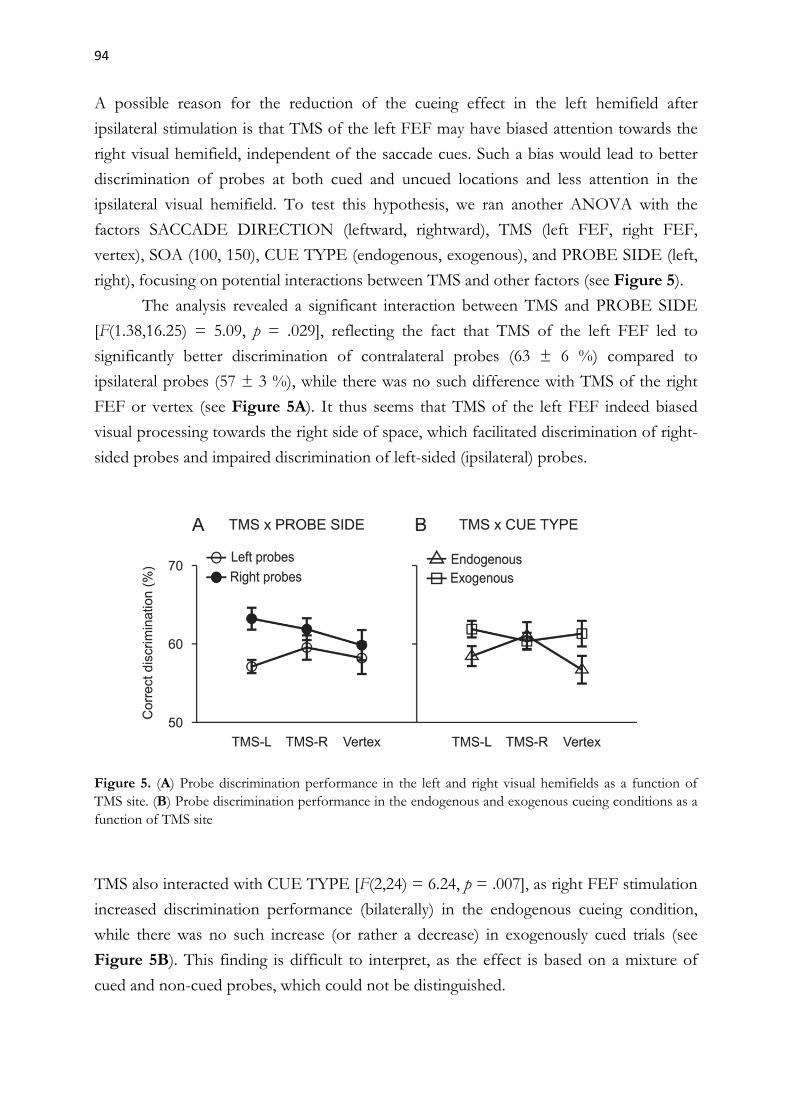
94
A possible reason for the reduction of the cueing effect in the left hemifield after
ipsilateral stimulation is that TMS of the left FEF may have biased attention towards the
right visual hemifield, independent of the saccade cues. Such a bias would lead to better
discrimination of probes at both cued and uncued locations and less attention in the
ipsilateral visual hemifield. To test this hypothesis, we ran another ANOVA with the
factors SACCADE DIRECTION (leftward, rightward), TMS (left FEF, right FEF,
vertex), SOA (100, 150), CUE TYPE (endogenous, exogenous), and PROBE SIDE (left,
right), focusing on potential interactions between TMS and other factors (see Figure 5).
The analysis revealed a significant interaction between TMS and PROBE SIDE
[F(1.38,16.25) = 5.09, p = .029], reflecting the fact that TMS of the left FEF led to
significantly better discrimination of contralateral probes (63 ± 6 %) compared to
ipsilateral probes (57 ± 3 %), while there was no such difference with TMS of the right
FEF or vertex (see Figure 5A). It thus seems that TMS of the left FEF indeed biased
visual processing towards the right side of space, which facilitated discrimination of right-
sided probes and impaired discrimination of left-sided (ipsilateral) probes.
Figure 5. (A) Probe discrimination performance in the left and right visual hemifields as a function of TMS site. (B) Probe discrimination performance in the endogenous and exogenous cueing conditions as a function of TMS site
TMS also interacted with CUE TYPE [F(2,24) = 6.24, p = .007], as right FEF stimulation
increased discrimination performance (bilaterally) in the endogenous cueing condition,
while there was no such increase (or rather a decrease) in exogenously cued trials (see
Figure 5B). This finding is difficult to interpret, as the effect is based on a mixture of
cued and non-cued probes, which could not be distinguished.

95
We therefore checked the results of our first ANOVA (with all experimental factors), and
found exactly the same pattern of results, both for cued and for uncued probes. However,
since the differences between TMS of the right FEF and the other two sites did not reach
significance in any of the analyses, we cannot rule out that the performance increase after
right FEF stimulation was only due to random variation.
DISCUSSION
In the present study, TMS delivered to the left FEF between an endogenous or
exogenous saccade cue and a visual target facilitated discrimination performance in the
right visual field and reduced discrimination performance at saccade goals in the left
visual field. TMS of the right FEF led to a tendency for better discrimination
performance in both visual hemifields in the endogenous cueing condition that was
independent of saccade programming, but the difference to the other two stimulation
sites did not reach statistical significance.
The main goal of this study was to investigate how TMS of the frontal eye fields
influences the coupling between visual attention and saccade programming. We used
a dual-task setting, in which participants prepared saccades to an endogenously or
exogenously cued location and simultaneously discriminated a visual probe that could be
presented at the saccade goal or at one of three other possible locations. We replicated the
well-established finding that attention during saccade preparation is coupled to the
saccade goal (Deubel & Schneider, 1996; Hoffman & Subramaniam, 1995; Jonikaitis &
Deubel, 2011; Klapetek et al., 2016; Kowler et al., 1995; Neggers et al., 2007; Rolfs et al.,
2011; Jonikaitis & Theeuwes, 2013; Van Ettinger-Veenstra et al., 2009), and found this
coupling to emerge earlier and to be stronger for exogenously cued saccades. Additionally,
we found that TMS of the left FEF biased attention towards the contralateral visual
hemifield, enhancing visual perception in the right hemifield and reducing the coupling of
attention to the saccade goal in the left hemifield.
Two previous studies (Neggers et al., 2007; Van Ettinger-Veenstra et al., 2009)
examined the same question using a behavioral task originally employed by Deubel and
Schneider (1996). Both reported contralateral effects of FEF-TMS on attentional
performance, but Neggers observed that FEF-TMS compromised saccade-related
attention, while Van Ettinger-Veenstra found that it enhanced attention at the saccade
goal.

96
The contradictory results are most likely a consequence of the different TMS protocols
(the first study used trains of three pulses while the second used single pulses), and if we
disregard them, the common conclusion of both studies is that both the left and the right
FEF play a crucial role in the control of attention in the contralateral visual hemifield.
The results of the present study are difficult to reconcile with these previous
findings. Although we also found that TMS of the left FEF biased visual attention
towards the right hemifield, this effect was not coupled to saccade programming, as in the
above mentioned studies. In contrary, there was a reduction of the saccade-related benefit
for leftwards saccades. For the interpretation of these differences it is crucial to realize
that the paradigm used in the preceding studies (Deubel & Schneider, 1996) and our dual
task differ in a very important aspect: In the former, discrimination probes always
appeared in the same visual hemifield as the saccade target and this hemifield was cued
with 100% validity, which means that participants could simply ignore the other side of
the display. In the present study, the cue was not predictive of the laterality of the
discrimination probe (25% validity), so the optimal strategy participants could adopt was
to split endogenous attention over both sides of the visual display, prioritizing the cued
location just enough to maintain good oculomotor performance. The finding that saccade
execution was contingent on probe appearance supports this view, as it shows that
participants tended to prioritize the discrimination task over the saccade task. As
a consequence, there was always some endogenous attention directed to uncued locations,
which could also be enhanced by TMS. The enhancement of contralateral probe
discrimination after left FEF stimulation could thus have been mediated by top-down
attentional control, even though it was not coupled to the saccade goal. Consequently, the
bias towards the contralateral visual field may have caused the ipsilateral impairment of
probe discrimination through some kind of interhemispheric inhibition. Our results may
therefore indirectly support the same conclusion as Neggers et al. (2007) and Van
Ettinger-Veenstra et al. (2009) that the left FEF exerts top-down control over contra-
lateral visual space.
In contrast to Van Ettinger-Veenstra et al. (2009), we did not find evidence for
a contralateral attentional benefit following TMS of the right FEF, but we found
a tendency towards a bilateral enhancement of attention that was also independent of
saccade programming. This enhancement was specific for the endogenous cueing
condition, most likely because attention in the exogenous cueing condition was so quickly
and automatically summoned by the sudden onset cues that there was no room left for
any modulation. (which also explains why the cueing effect was generally larger with
exogenous cues). Alternatively, the FEF may be less active during the programming of
exogenously cued saccades.

97
Although the tendency in our data cannot be taken as a reliable effect, it is consistent with
previous reports that single pulse TMS of the right FEF leads to a bilateral enhancement
of endogenous spatial attention (e.g., Chanes et al., 2012; Grosbras & Paus, 2002, 2003).
We therefore believe that further studies on a larger data set could come to this same
conclusion, reaching statistical significance.
Besides this asymmetrical pattern of TMS effects, the present study revealed
significant attentional asymmetries that were independent of TMS. At the earlier SOA,
participants were better at discriminating uncued probes when they were planning
a saccade to the left, while at the later SOA they were better at discriminating probes at
the saccade goal when planning a saccade to the right. We think that these effects may be
a consequence of the relative dominance of the right cerebral hemisphere in the control
of spatial attention.
It has been proposed that the right hemisphere mediates attention shifts to both
visual hemifields, while the left hemisphere only mediates attention shifts to the right
hemifield (Heilman & Abell, 1980; Heilman & Valenstein, 1979, Mesulam, 1981). Based
on a careful review of empirical evidence for and against this “hemispatial theory”,
Duecker and Sack (2015) proposed that the right hemisphere dominance applies to
frontal but not to the posterior parts of the brain’s attention network (also see
Szczepanski, Konen, & Kastner, 2010). A substantial part of the evidence comes from
TMS studies that found a right hemisphere dominance at the level of the FEF, with
bilateral effects on covert or overt attention after TMS of the right FEF and only
unilateral effects (affecting contralateral visual space) after TMS of the left FEF (Cazzoli
et al., 2015; Chanes et al., 2012; Duecker, Formisano, & Sack, 2013; Hung, Driver, &
Walsh, 2011; Grosbras & Paus, 2002, 2003; Ruff et al., 2009; Silvanto et al., 2006; Walker,
Techawachirakul, & Haggard, 2009).
The experimental design in the present study, where the saccade cue did not
predict the location of the discrimination probe, encouraged participants to distribute
attention over both visual hemifields. However, during saccade planning attentional
resources become increasingly engaged at the saccade goal (Castet, Jeanjean, Montagnini,
Laugier, & Masson, 2006; Deubel, 2008; Doré-Mazars, Pouget, & Beauvillain, 2004;
Jonikaitis & Deubel, 2011; Montagnini & Castet, 2007) and can only be allocated
elsewhere at the very beginning of the saccade programming process (Doré-Mazars et al.,
2004; Montagnini & Castet, 2007). This may explain why the pattern of asymmetries was
different at the early and later SOAs. It seems that 100 ms after the cue participants could
still split attention over both hemifields and they were better at it before leftward than
before rightward saccades.
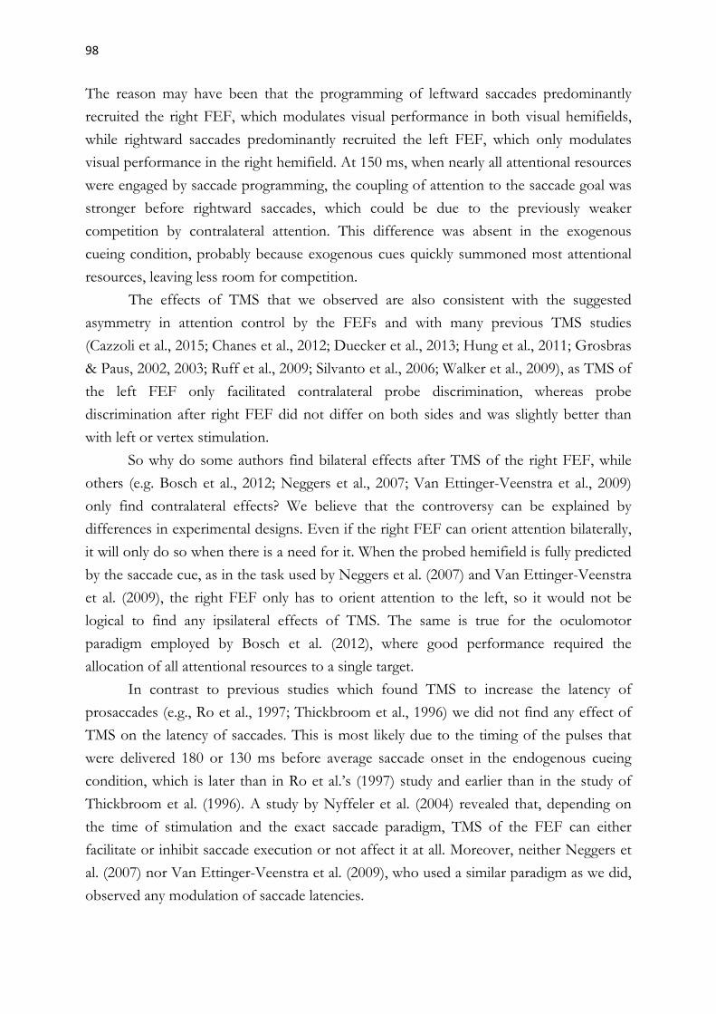
98
The reason may have been that the programming of leftward saccades predominantly
recruited the right FEF, which modulates visual performance in both visual hemifields,
while rightward saccades predominantly recruited the left FEF, which only modulates
visual performance in the right hemifield. At 150 ms, when nearly all attentional resources
were engaged by saccade programming, the coupling of attention to the saccade goal was
stronger before rightward saccades, which could be due to the previously weaker
competition by contralateral attention. This difference was absent in the exogenous
cueing condition, probably because exogenous cues quickly summoned most attentional
resources, leaving less room for competition.
The effects of TMS that we observed are also consistent with the suggested
asymmetry in attention control by the FEFs and with many previous TMS studies
(Cazzoli et al., 2015; Chanes et al., 2012; Duecker et al., 2013; Hung et al., 2011; Grosbras
& Paus, 2002, 2003; Ruff et al., 2009; Silvanto et al., 2006; Walker et al., 2009), as TMS of
the left FEF only facilitated contralateral probe discrimination, whereas probe
discrimination after right FEF did not differ on both sides and was slightly better than
with left or vertex stimulation.
So why do some authors find bilateral effects after TMS of the right FEF, while
others (e.g. Bosch et al., 2012; Neggers et al., 2007; Van Ettinger-Veenstra et al., 2009)
only find contralateral effects? We believe that the controversy can be explained by
differences in experimental designs. Even if the right FEF can orient attention bilaterally,
it will only do so when there is a need for it. When the probed hemifield is fully predicted
by the saccade cue, as in the task used by Neggers et al. (2007) and Van Ettinger-Veenstra
et al. (2009), the right FEF only has to orient attention to the left, so it would not be
logical to find any ipsilateral effects of TMS. The same is true for the oculomotor
paradigm employed by Bosch et al. (2012), where good performance required the
allocation of all attentional resources to a single target.
In contrast to previous studies which found TMS to increase the latency of
prosaccades (e.g., Ro et al., 1997; Thickbroom et al., 1996) we did not find any effect of
TMS on the latency of saccades. This is most likely due to the timing of the pulses that
were delivered 180 or 130 ms before average saccade onset in the endogenous cueing
condition, which is later than in Ro et al.’s (1997) study and earlier than in the study of
Thickbroom et al. (1996). A study by Nyffeler et al. (2004) revealed that, depending on
the time of stimulation and the exact saccade paradigm, TMS of the FEF can either
facilitate or inhibit saccade execution or not affect it at all. Moreover, neither Neggers et
al. (2007) nor Van Ettinger-Veenstra et al. (2009), who used a similar paradigm as we did,
observed any modulation of saccade latencies.

99
In conclusion, the present study revealed that TMS of the left FEF modulates
endogenous visual attention independently of saccade programming, even in a paradigm
where endogenous attention is strongly coupled to the saccade goal. Our results thus
support the notion that attention and saccade programming are separate functions that
are dissociable at the level of FEF neurons (Juan et al., 2008; Juan, Shorter-Jacobi, &
Schall, 2004; Sato & Schall, 2003; Wardak et al., 2011). Since the attentional modulation
was independent of cueing, we could not determine to what extent the FEF is involved in
the programming of exogenously cued saccades or in the shifts of attention that precede
them. Future studies will hopefully reveal more about the role of the FEF in exogenous
attention and uncover the exact mechanisms and pathways by which TMS affects the
activity of the FEF and of downstream visual areas.
Acknowledgments
This study was funded by Deutsche Forschungsgemeinschaft (DFG) grants JO 980/1–1
(to D.J. and H.D.) and DE 336/5–1 (to H.D.), and by the DFG Research Training group
“Orientation and Motion in Space” (GRK 1091).
References
Amiez, C., & Petrides, M. (2009). Anatomical organization of the eye fields in the human and non-human primate frontal cortex. Progress in Neurobiology, 89, 220–230.
Bosch, S. E., Neggers, S. F. W., & Van der Stigchel, S. (2012). The role of the frontal eye fields in oculomotor competition: image-guided TMS enhances contralateral target selection. Cerebral Cortex, 23(4), 824–832.
Brainard, D. H. (1997). The Psychophysics Toolbox. Spatial Vision, 10, 433–436.
Bruce, C. J., & Goldberg, M. E. (1985). Primate frontal eyefields: I. Single neurons discharging before saccades. Journal of Neurophysiology, 53, 603-635.
Cazzoli, D., Jung, S., Nyffeler, T., Nef, T., Wurtz, P., Mosimann, U. P., & Müri, R. M. (2015). The role of the right frontal eye field in overt visual attention deployment as assessed by free visual exploration. Neuropsychologia, 74, 37–41.
Chanes, L., Chica, A. B., Quentin, R., & Valero-Cabré, A. (2012). Manipulation of pre-target activity on the right frontal eye field enhances conscious visual perception in humans. PLOS ONE, 7(5), e36232.

100
Corbetta, M., Akbudak, E., Conturo, T. E., Snyder, A. Z., Ollinger, J. M., Drury, H. A., Linenweber, M. R., Petersen, S. E., Raichle, M. E., Van Essen, D. C. & Shulman, G. L. (1998). A common network of functional areas for attention and eye movements. Neuron, 21, 761–773.
Cornelissen, F. W., Peters., E., & Palmer, J. (2002). The Eyelink Toolbox. Behavior Research Methods, Instruments & Computers, 34, 613–617.
Deubel, H., & Schneider, W. X. (1996). Saccade target selection and object recognition: evidence for a common attentional mechanism. Vision Research, 36, 1827–1837.
Dias, E. C., & Segraves, M. A. (1999). Muscimol-induced inactivation of monkey frontal eye field: effects on visually and memory-guided saccades. Journal of Neurophysiology, 81, 2191-2214.
Doré-Mazars, K., Pouget, P., & Beauvillain, C. (2004). Attentional selection during preparation of eye movements. Psychological Research, 69, 67–76.
Duecker, F., Formisano, E., & Sack, A. T. (2013). Hemispheric differences in the voluntary control of spatial attention: direct evidence for a right-hemispheric dominance within frontal cortex. Journal of Cognitive Neuroscience, 25(8), 1332–1342.
Duecker, F., & Sack, A. T. (2015). The hybrid model of attentional control: New insights into hemispheric asymmetries inferred from TMS research. Neuropsychologia, 74, 21–29.
Forbes, K., & Klein, R. (1996). The magnitude of the fixation offset effect with endogenously and exogenously controlled saccades. Journal of Cognitive Neuroscience, 8, 344–352.
Grosbras, M. H., & Paus, T. (2002). Transcranial magnetic stimulation of the human frontal eye field: effects on visual perception and attention. Journal of Cognitive Neuroscience, 14(7), 1109–1120.
Grosbras, M. H., & Paus, T. (2003).Transcranial magnetic stimulation of the human frontal eye field facilitates visual awareness. European Journal of Neuroscience, 18, 3121–3126.
Guitton, D., Buchtel, H. A., & Douglas, R. M. (1985). Frontal lobe lesions in man cause difficulties in suppressing reflexive glances and in generating goal-directed saccades. Experimental Brain Research, 58, 455–472.
Gutteling, T. P., Van Ettinger-Veenstra, H. M., Kenemans, J. L.., & Neggers, S. F. W. (2010). Lateralized frontal eye field activity precedes occipital activity shortly before saccades: evidence for cortico-cortical feedback as a mechanism underlying covert attention shifts. Journal of Cognitive Neuroscience, 22(9), 1931–1943.

101
de Haan, B., Morgan, P. S. & Rorden, C. (2008). Covert orienting of attention and overt eye movements activate identical brain regions. Brain Research, 1204, 102–111.
Heilman, K. M., & Abell, T. V. D. (1980). Right hemisphere dominance for attention. Neurology, 30(3), 327.
Heilman, K. M., & Valenstein, E. (1979). Mechanism underlying hemispatial neglect. Annals of Neurology, 5(2), 166–170.
Henik, A., Rafal, R., & Rhodes, D. (1994). Endogenously generated and visually guided saccades after lesions of the human frontal eye fields. Journal of Cognitive Neuroscience, 6, 400–411.
Hoffman, J. E., & Subramaniam, B. (1995). The role of visual attention in saccadic eye movements. Perception and Psychophysics, 57(6), 787–795.
Hung, J., Driver, J., & Walsh, V. (2011). Visual selection and the human frontal eye fields: effects of frontal transcranial magnetic stimulation on partial report analyzed by Bundesen’s theory of visual attention. The Journal of Neuroscience, 31(44), 15904–15913.
Jonikaitis, D., & Deubel, H. (2011). Independent allocation of attention to eye and hand targets in coordinated eye- hand movements. Psychological Science, 22(3), 339–347.
Jonikaitis, D., & Theeuwes, J. (2013). Dissociating oculomotor contributions to spatial and feature-based selection. Journal of Neurophysiology, 110(7), 1525–1534.
Juan, C. H., Muggleton, N. G., Tzeng, O. J. L., Hung, D. L., Cowey, A., & Walsh, A. (2008). Segregation of visual selection and saccades in human frontal eye fields. Cerebral Cortex, 18, 2410–2415.
Juan, C. H., Shorter-Jacobi, S. M., & Schall, J. D. (2004). Dissociation of spatial attention and saccade preparation. PNAS, 101, 15541–15544.
Kammer, T., Vorwerg, M., & Herrnberger, B. (2007). Anisotropy in the visual cortex investigated by neuronavigated transcranial magnetic stimulation. Neuroimage, 36, 313–321.
Keel, J. C., Smith, M. J., & Wassermann, E. M. (2001). A safety screening questionnaire for transcranial magnetic stimulation. Clinical Neurophysiology, 112(4):720.
Klapetek, A., Jonikaitis, D., & Deubel, H. (2016). Attention allocation before antisaccades. Journal of Vision, 16(1):11.
Kleiner, M., Brainard, D., & Pelli, D. (2007). “What’s new in Psychtoolbox-3?”. Perception, 36, ECVP Abstract Supplement.

102
Kowler, E., Anderson, E., Dosher, B., & Blaser, E. (1995). The role of attention in the programming of saccades. Vision Research, 35(13), 1897–1916.
Mesulam, M. M. (1981). A cortical network for directed attention and unilateral neglect. Annals of Neurology, 10, 309–325.
Moliadze, V., Giannikopoulos, D., Eysel, U. T., & Funke, K. (2005) Paired-pulse transcranial magnetic stimulation protocol applied to visual cortex of anaesthetized cat: effects on visually evoked single-unit activity. Journal of Physiology, 566, 955–965.
Montagnini, A., & Castet, E. (2007). Spatiotemporal dynamics of visual attention during saccade preparation: Independence and coupling between attention and movement planning. Journal of Vision, 7(14):8.
Müller, H. J., & Rabbitt, P. M. A. (1989). Reflexive and voluntary orienting of visual attention: Time course of activation and resistance to interruption. Journal of Experimental Psychology: Human Perception and Performance, 15, 315–330.
Müri, R. M., Hess, C. W., & Meienberg, O. (1991). Transcranial magnetic stimulation of the human frontal eye field by magnetic pulses. Experimental Brain Research, 86, 219–223.
Muggleton, N. G., Juan, C. H., Cowey, A., & Walsh, V. (2003). Human frontal eye fields and visual search. Journal of Neurophysiology, 89, 3340–3343.
Mort, D. J., Perry, R. J., Mannan, S. K., Hodgson, T. L., Anderson, E., Quest, R., McRobbie, D., McBride, A., Husain, M., & Kennard, C. (2003). Differential cortical activation during voluntary and reflexive saccades. NeuroImage, 18, 231–246.
Nakayama, K., & Mackeben, M. (1989). Sustained and transient components of focal visual attention. Vision Research, 29, 1631–1647.
Neggers, S. W. F., Huijbers, W., Vrijlandt, C. M., Vlaskamp, B. N. S., Schutter, D. J. L. G., & Kenemans, J. L. (2007). TMS pulses on the frontal eye fields break coupling between visuospatial attention and eye movements. Journal of Neurophysiology, 98, 2765–2778.
Neggers, S. W. F., Langerak, T. R., Schutter, D. J., Mandl, R. C., Ramsey, N. F., Lemmens, P. J., & Postma, A. (2004). A stereotactic method for image-guided transcranial magnetic stimulation validated with fMRI and motor-evoked potentials. Neuroimage, 21(4), 1805–1817.
Neggers, S. W. F., van Diepen, R. M., Zandbelt, B. B., Vink, M., Mandl, R. C. W., & Gutteling, T. P. (2012). A functional and structural investigation of the human fronto-basal volitional saccade network. PLOS ONE 7(1): e29517.

103
Nobre, A. C., Gitelman, D. R., Dias, E. C. & Mesulam, M. M. (2000). Covert visual spatial orienting and saccades: overlapping neural systems. Neuroimage, 11, 210–216.
Nyffeler, T., Bucher, O., Pflugshaupt, T., Von Wartburg, R., Wurtz, P., Hess, C. W. & Müri, R. M. (2004). Single-pulse transcranial magnetic stimulation over the frontal eye field can facilitate and inhibit saccade triggering. European Journal of Neuroscience, 20(8), 2240–2244.
Olk, B., Chang, E., Kingstone, A., & Ro, T. (2006). Modulation of antisaccades by transcranial magnetic stimulation of the human frontal eye field. Cerebral Cortex, 16, 76–82.
O’Shea, J., Muggleton, N. G., Cowey, A., & Walsh, V. (2004). Timing of target discrimination in human frontal eye fields. Journal of Cognitive Neuroscience, 16, 1060–1067.
O’Shea, J., Muggleton, N. G., Cowey, A., & Walsh, V. (2007). Human frontal eye fields and spatial priming of pop-out. Journal of Cognitive Neuroscience, 19, 1140–1151.
Pascual-Leone, A., Bartres-Faz, D., & Keenan, J. P. (1999). Transcranial magnetic stimulation: Studying the brain-behaviour relationship by induction of 'virtual lesions'. Philosophical Transactions of the Royal Society of London: Biological Sciences, 354, 1229–1238.
Pelli, D. G. (1997). The VideoToolbox software for visual psychophysics: Transforming numbers into movies, Spatial Vision, 10, 437–442.
Perry, R. J. & Zeki, S. (2000). The neurology of saccades and covert shifts in spatial attention: an event-related fMRI study. Brain, 123, 2273–2278.
Rafal, R., Egly, R., & Rhodes, D. (1994). Effects of inhibition of return on voluntarily and visually guided saccades. Canadian Journal of Experimental Psychology, 48, 284–300.
Rivaud, S., Müri, R. M., Gaymard, B., Vermersch, A. I., & Pierrot-Deseilligny, C. (1994). Eye movement disorders after frontal eye field lesions in humans. Experimental Brain Research, 102, 110–120.
Ro, T., Henik, A., Machado, L., & Rafal, R. D. (1997). Transcranial magnetic stimulation of the prefrontal cortex delays contralateral endogenous saccades. Journal of Cognitive Neuroscience, 9, 433–440.
Rolfs, M., Jonikaitis, D., Deubel, H., & Cavanagh, P. (2011). Predictive remapping of attention across eye movements. Nature Neuroscience, 14, 252–256.
104
Ruff, C. C., Blankenburg, F., Bjoertomt, O., Bestmann, S., Freeman, E., Haynes, J. D., Rees, G., Josephs, O., Deichmann, R., & Driver, J. (2006). Concurrent TMS-fMRI and psychophysics reveal frontal influences on human retinotopic visual cortex. Current Biology, 16, 1479–1488.
Ruff, C. C., Blankenburg, F., Bjoertomt, O., Bestmann, S., Weiskopf, N., & Driver, J. (2009). Hemispheric differences in frontal and parietal influences on human occipital cortex: direct confirmation with concurrent TMS-fMRI. Journal of Cognitive Neuroscience, 21, 1146–1161.
Sato, T., & Schall, J. D. (2003). Effects of stimulus-response compatibility on neural selection in frontal eye field. Neuron, 38(4), 637–648.
Schall, J. D. (2015). Visuomotor functions in the frontal lobe. Annual Review of Vision Science, 1, 469–498.
Sergent, C., Wyart, V., Babo-Rebelo, M., Cohen, L., Naccache, L., & Tallon-Baudry, C. (2013). Cueing attention after the stimulus is gone can retrospectively trigger conscious perception. Current Biology, 23(2), 150–155.
Siebner, H. R., Hartwigsen, G., Kassuba, T., & Rothwell, J. C. (2009). How does transcranial magnetic stimulation modify neuronal activity in the brain? Implications for studies of cognition. Cortex, 45(9), 1035–1042.
Silvanto, J., Lavie, N., & Walsh, W. (2006). Stimulation of the human frontal eye fields modulates sensitivity of extrastriate visual cortex. Journal of Neurophysiology, 96, 941–945.
Sommer, M. A., & Tehovnik, E. J. (1997). Reversible inactivation of macaque frontal eye field. Experimental Brain Research, 116, 229–249.
Szczepanski, S. M., Konen, C. S., & Kastner, S. (2010). Mechanisms of spatial attention control in frontal and parietal cortex. The Journal of Neuroscience, 30, 148–160.
Taylor, P. C. J., Nobre, A. C., & Rushworth, M. F. S. (2007). FEF-TMS affects visual cortical activity. Cerebral Cortex, 17, 391–399.
Terao, Y., Fukuda, H., Ugawa, Y., Hikosaka, O., Hanajima, R., Furubayashi, T., Sakai, K., Miyauchi, S., Sasaki, Y. & Kanazawa, I. (1998). Visualization of the information flow through human oculomotor cortical regions by transcranial magnetic stimulation. Journal of Neurophysiology, 80, 936–946.
Thibault, L., Cavanagh, P., & Sergent, C. (2015). Retroactive Attention can trigger all-or-none conscious access to past sensory stimulus. Journal of Vision, 15(12), 547.
105
Thickbroom, G. W., Stell, R., & Mastaglia, F. L. (1996). Transcranial magnetic stimulation of the human frontal eye field. Journal of the Neurological Sciences, 144, 114–118.
Van Ettinger-Veenstra, H. M., Huijbers, W., Gutteling, T. P., Vink, M., Kenemans, J. L., & Neggers, S. F. W. (2009). fMRI-guided TMS on cortical eye fields: the frontal but not intraparietal eye fields regulate the coupling between visuospatial attention and eye movements. Journal of Neurophysiology, 102(6), 3469–3480.
Vernet, M., Quentin, R., Chanes, L., Mitsumasu, A., & Valero-Cabré, A. (2014). Frontal eye field, where art thou? Anatomy, function, and non-invasive manipulation of frontal regions involved in eye movements and associated cognitive operations. Frontiers in Integrative Neuroscience, 8: 66, doi: 10.3389/fnint.2014.00066.
Walker, R., Techwachirakul, P., & Haggard, P. (2009). Frontal eye field stimulation modulates the balance of salience between target and distractors. Brain Research, 1270, 54–63.
Walker, R., Walker, D., Husain, M., Kennard, C. (2000). Control of voluntary and reflexive saccades. Experimental Brain Research, 130, 540–544.
Wardak, C., Olivier, E. , & Duhamel, J. R. (2011). The relationship between spatial attention and saccades in the frontoparietal network of the monkey. European Journal of Neuroscience, 33(11), 1973–1981.
Wassermann, E. M. (1996). Risk and safety of repetitive transcranial magnetic stimulation: report and suggested guidelines from the International Workshop on the Safety of Repetitive Transcranial Magnetic Stimulation, June 5-7, 1996. Electroencephalography and Clinical Neurophysiology, 108(1), 1–16.

106

107
3 General Discussion
The present thesis investigated how visual attention is linked to saccade programming and
saccadic decision making. I will briefly summarize the results of the three studies and
discuss how they have helped to clarify the relationship between attention, saccade
decisions and saccade programming.
3.1 Summary of findings
The first study (Chapter 2.1) examined the coupling between oculomotor selection and
perceptual selection in an antisaccade paradigm. Previous studies provided evidence that
antisaccade programming involves a competition between exogenous and endogenous
influences, but none of them assessed the allocation of spatial attention by measuring
perceptual performance at the competing locations. Our results have filled this gap by
showing that attention is allocated in parallel to both the visual cue and the antisaccade
goal and that pre-saccadic attention at the antisaccade goal is predictive of initial saccade
direction. Besides this, we replicated previous findings on parallel saccade programming
of pro- and antisaccades and the relationship between the awareness of prosaccade errors
and their correction. Our study found no relationship between attention allocation and
awareness of saccade errors and thus disproves a previous hypothesis by Deubel &
Mokler (2000) that erroneous, unperceived prosaccades may occur without attentional
involvement.
The second study (Chapter 2.2) investigated oculomotor and attentional measures
in rule-based or free decisions between two memorized saccade goals. The main finding
was that attentional selection evolved in parallel at the final saccade goal and the
competing target, with a bias towards the saccade goal both before correct and before
erroneous rule-based choices. Moreover, we found that decisions emerged from the
competition of several types of biases and that they tended to form at a very early point in
time, before task-relevant information was made available. While several
neurophysiological studies provided evidence for such parallel competitive processes in
the monkey brain, our study was the first to show that the competition is also reflected in
visual perception and that saccade decisions can be predicted from the spatial distribution
of attention.
The third study (Chapter 2.3) tested whether interfering with activity of the
frontal eye fields (FEF), a brain region crucially involved in both visual and oculomotor
selection, can modulate the coupling between saccades and visual selection.
108
Previous studies have demonstrated that transcranial magnetic stimulation (TMS) of the
left or right FEF can either weaken (Neggers et al., 2007) or enhance (Van Ettinger-
Veenstra et al., 2009) the obligatory coupling of attention to a future saccade goal in the
contralateral visual hemifield, depending on the stimulation protocol. These studies used
the behavioral task introduced by Deubel and Schneider (1996), in which participants
have to saccade to one of three horizontally aligned locations in the left or right visual
hemifield and discriminate a target at one of these locations. Our study employed a novel
dual-task paradigm, in which the saccade cue was not predictive of the probe location and
saccade programming occurred in parallel to the attentional task. We therefore increased
the degree of attentional competition between both tasks and introduced an additional
component of interhemispheric rivalry. The results showed that TMS of the left FEF
enhanced endogenous attention to the contralateral visual field, independent of saccade
programming. Contrary to our expectations, the results could not help to discern the role
of the FEF in the control of exogenous attention, but they still contributed to the
understanding of the relationship between exogenous and endogenous attention by
providing evidence that they compete through interhemispheric connections.
3.2 Parallel saccade programming
In most situations, average fixation durations between two subsequent saccades lie
between 180 and 330 ms (Rayner & Castelhano, 2007). Sometimes, however, intervals
between subsequent saccades are so short that their programming must have overlapped
in time (e.g., Becker & Jürgens, 1979; Godijn & Theeuwes, 2002; Hodgson, Parris,
Gregory, & Jarvis, 2009; Irwin et al., 2000; Massen, 2004; McPeek, Skavenski &
Nakayama, 2000; Mokler & Fischer, 1999; Morrison, 1984; Theeuwes et al., 1998, 1999;
Walker & McSorley, 2006).
Amongst other examples, parallel saccade programming occurs when erroneous
prosaccades in the antisaccade task are followed by corrective antisaccades (Massen, 2004;
Mokler & Fischer, 1999). The first study of this dissertation (Chapter 2.1) replicated this
finding and extended it by showing that the parallel saccade programming is associated
with parallel attention allocation to both saccade goals. Moreover, we observed mutual
inhibitory interactions between the two parallel saccade programs and thus provided
behavioral evidence that reflexive and endogenous saccade programs in the antisaccade
task compete within the same neural structures.
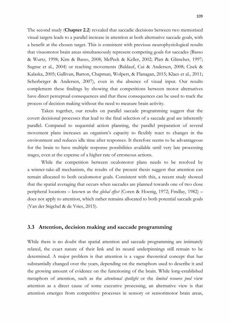
109
The second study (Chapter 2.2) revealed that saccadic decisions between two memorized
visual targets leads to a parallel increase in attention at both alternative saccade goals, with
a benefit at the chosen target. This is consistent with previous neurophysiological results
that visuomotor brain areas simultaneously represent competing goals for saccades (Basso
& Wurtz, 1998; Kim & Basso, 2008; McPeek & Keller, 2002; Platt & Glimcher, 1997;
Sugrue et al., 2004) or reaching movements (Baldauf, Cui & Andersen, 2008; Cisek &
Kalaska, 2005; Gallivan, Barton, Chapman, Wolpert, & Flanagan, 2015; Klaes et al., 2011;
Scherberger & Andersen, 2007), even in the absence of visual input. Our results
complement these findings by showing that competitions between motor alternatives
have direct perceptual consequences and that these consequences can be used to track the
process of decision making without the need to measure brain activity.
Taken together, our results on parallel saccade programming suggest that the
covert decisional processes that lead to the final selection of a saccade goal are inherently
parallel. Compared to sequential action planning, the parallel preparation of several
movement plans increases an organism’s capacity to flexibly react to changes in the
environment and reduces idle time after responses. It therefore seems to be advantageous
for the brain to have multiple response possibilities available until very late processing
stages, even at the expense of a higher rate of erroneous actions.
While the competition between oculomotor plans needs to be resolved by
a winner-take-all mechanism, the results of the present thesis suggest that attention can
remain allocated to both oculomotor goals. Consistent with this, a recent study showed
that the spatial averaging that occurs when saccades are planned towards one of two close
peripheral locations – known as the global effect (Coren & Hoenig, 1972; Findlay, 1982) –
does not apply to attention, which rather remains allocated to both potential saccade goals
(Van der Stigchel & de Vries, 2015).
3.3 Attention, decision making and saccade programming
While there is no doubt that spatial attention and saccade programming are intimately
related, the exact nature of their link and its neural underpinnings still remain to be
determined. A major problem is that attention is a vague theoretical concept that has
substantially changed over the years, depending on the metaphors used to describe it and
the growing amount of evidence on the functioning of the brain. While long-established
metaphors of attention, such as the attentional spotlight or the limited resource pool view
attention as a direct cause of some executive processing, an alternative view is that
attention emerges from competitive processes in sensory or sensorimotor brain areas,
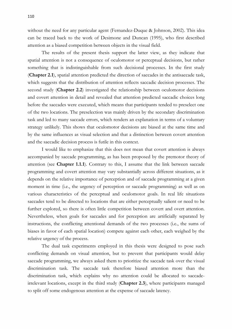
110
without the need for any particular agent (Fernandez-Duque & Johnson, 2002). This idea
can be traced back to the work of Desimone and Duncan (1995), who first described
attention as a biased competition between objects in the visual field.
The results of the present thesis support the latter view, as they indicate that
spatial attention is not a consequence of oculomotor or perceptual decisions, but rather
something that is indistinguishable from such decisional processes. In the first study
(Chapter 2.1), spatial attention predicted the direction of saccades in the antisaccade task,
which suggests that the distribution of attention reflects saccadic decision processes. The
second study (Chapter 2.2) investigated the relationship between oculomotor decisions
and covert attention in detail and revealed that attention predicted saccadic choices long
before the saccades were executed, which means that participants tended to preselect one
of the two locations. The preselection was mainly driven by the secondary discrimination
task and led to many saccade errors, which renders an explanation in terms of a voluntary
strategy unlikely. This shows that oculomotor decisions are biased at the same time and
by the same influences as visual selection and that a distinction between covert attention
and the saccadic decision process is futile in this context.
I would like to emphasize that this does not mean that covert attention is always
accompanied by saccade programming, as has been proposed by the premotor theory of
attention (see Chapter 1.1.1). Contrary to this, I assume that the link between saccade
programming and covert attention may vary substantially across different situations, as it
depends on the relative importance of perception and of saccade programming at a given
moment in time (i.e., the urgency of perception or saccade programming) as well as on
various characteristics of the perceptual and oculomotor goals. In real life situations
saccades tend to be directed to locations that are either perceptually salient or need to be
further explored, so there is often little competition between covert and overt attention.
Nevertheless, when goals for saccades and for perception are artificially separated by
instructions, the conflicting attentional demands of the two processes (i.e., the sums of
biases in favor of each spatial location) compete against each other, each weighed by the
relative urgency of the process.
The dual task experiments employed in this thesis were designed to pose such
conflicting demands on visual attention, but to prevent that participants would delay
saccade programming, we always asked them to prioritize the saccade task over the visual
discrimination task. The saccade task therefore biased attention more than the
discrimination task, which explains why no attention could be allocated to saccade-
irrelevant locations, except in the third study (Chapter 2.3), where participants managed
to split off some endogenous attention at the expense of saccade latency.

111
Belopolsky and Theeuwes (2009, 2012) proposed an updated version of the premotor
theory, suggesting that shifting covert attention to a location always involves a saccade
program to that location, whereas maintaining attention can lead to both activation or
suppression of saccades to the attended location. The first part of this claim is
corroborated by the results of our first study, which showed that shifting attention to the
antisaccade cue often led to involuntary prosaccades towards it. The second claim can be
reconciled with the results of our second study, where we found some evidence for the
suppression of saccade programming to the non-chosen target while it was selected by
attention. The finding that discrimination performance at the non-chosen target was
worse than at the chosen target, but still kept improving towards saccade onset could
mean that attentional representations of the non-chosen target were simultaneously
downweighed by top-down information about the task rule (Dhawan et al., 2013) and
upweighed due to their relevance for the decision task.
The third study (Chapter 2.3) demonstrated that the frontal eye fields (FEF),
which are crucially involved in the control of eye movements, also participate in
endogenous attention shifts that are independent of saccade programming. This indicates
that the role of the FEF in attentional control is separable from its role in saccade
planning, as has already been shown by other authors (Juan et al., 2008; Juan et al., 2004;
Sato & Schall, 2003; Wardak et al., 2011). The finding that attention at saccade-irrelevant
locations competed with attention at the saccade goal is compatible with the view that
saccade preparation constitutes an independent attentional bias that competes with other
types of biases (Desimone & Duncan, 1995). Consistent with this, a recent study in
monkeys provided evidence that saccade preparation in the FEF modulates activity in
area V4 at least as much as covert attention (Steinmetz & Moore, 2014).
Taken together, the results of this thesis support the view that covert attention,
saccadic decision making and saccade programming are consequences of a common
competitive process that aims to prioritize certain spatial locations over others. Several
theories of selective attention have suggested that visual selection and eye movements
may be guided by the output of a “priority map”, which represents the behavioral
relevance of locations in visual space based on a combination of their bottom-up visual
saliency as well as top-down biases reflecting task demands (e.g., Fecteau & Munoz, 2006;
Serences & Yantis, 2006). This concept is compatible with accumulator models of
decision making (see Chapter 1.1.3) and it has been proposed that the activity on the map
could also represent the evolution of parallel motor plans towards the represented
locations (Cisek, 2007), which is congruent with the premotor theory of attention.

112
In search for the neural substrate of the priority map, researchers have proposed several
candidate areas, including the FEF (Thompson & Bichot, 2005), SC (Bayguinov, Ghitani,
Jackson, & Basso, 2015; Krauzlis, Bollimunta, Arcizet, & Wang, 2014) and LIP (Bisley &
Goldberg, 2010; Bisley et al., 2009; Ipata et al., 2009; Goldberg et al., 2006). While these
areas fulfill the criteria (i.e., they are topographically organized, integrate bottom-up and
top-down signals and represent priority independent of task), it is likely that they
represent intermediate maps and that the final priority map emerges from the interaction
of several areas (Mirpour & Bisley, 2015).
The results of this doctoral thesis do not allow to conclude where the signals that
concurrently drive saccade preparation to selected locations and facilitate visual
perception at these locations originate and whether it is more appropriate to view them as
attentional or as premotor activity. They mainly show that the influence between attention
and saccade programming is mutual: On one hand, attentional selection of a location
increases the likelihood of saccades to that location, and on the other hand, saccade
programming automatically biases attention towards locations in space.
And what are the implications of the present results for the premotor theory of
attention? The premotor theory of attention was undoubtedly useful, as it challenged the
formerly prevalent view that attention constitutes an independent cognitive domain and
opened up a whole new field of research. Since then, many studies, including the ones
that comprise this thesis, have confirmed that visual and oculomotor selection tend to be
linked, but can also be dissociated. Consistent with this, single cell recordings have shown
that both processes tend to be carried out by the same neural structures, where they show
crosstalk but also partial independence (see Chapter 1.1.4). In the light of these findings,
the question whether attention and saccade programming are linked or independent has
become a philosophical one, as the answer mainly depends on how the two terms are
defined.
3.4 Endogenous and exogenous attention
One of the goals of this doctoral thesis was to examine the relationship between
endogenous and exogenous attention, particularly in relation to saccade programming, as
it has recently been suggested that endogenous attention is more independent from the
oculomotor system than exogenous attention (Smith et al., 2012, 2014; Smith & Schenk,
2012). Both the first and the third study (Chapters 2.1 and 2.3) employed experimental
designs that promoted the competition between endogenous and exogenous orienting,
and the third study contrasted the effects of endogenous and exogenous saccade cues.
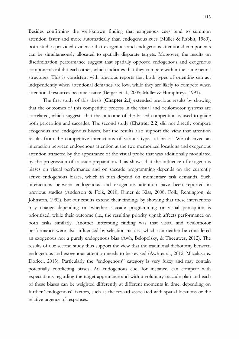
113
Besides confirming the well-known finding that exogenous cues tend to summon
attention faster and more automatically than endogenous cues (Müller & Rabbit, 1989),
both studies provided evidence that exogenous and endogenous attentional components
can be simultaneously allocated to spatially disparate targets. Moreover, the results on
discrimination performance suggest that spatially opposed endogenous and exogenous
components inhibit each other, which indicates that they compete within the same neural
structures. This is consistent with previous reports that both types of orienting can act
independently when attentional demands are low, while they are likely to compete when
attentional resources become scarce (Berger et al., 2005; Müller & Humphreys, 1991).
The first study of this thesis (Chapter 2.1) extended previous results by showing
that the outcomes of this competitive process in the visual and oculomotor systems are
correlated, which suggests that the outcome of the biased competition is used to guide
both perception and saccades. The second study (Chapter 2.2) did not directly compare
exogenous and endogenous biases, but the results also support the view that attention
results from the competitive interactions of various types of biases. We observed an
interaction between endogenous attention at the two memorized locations and exogenous
attention attracted by the appearance of the visual probe that was additionally modulated
by the progression of saccade preparation. This shows that the influence of exogenous
biases on visual performance and on saccade programming depends on the currently
active endogenous biases, which in turn depend on momentary task demands. Such
interactions between endogenous and exogenous attention have been reported in
previous studies (Anderson & Folk, 2010; Eimer & Kiss, 2008; Folk, Remington, &
Johnston, 1992), but our results extend their findings by showing that these interactions
may change depending on whether saccade programming or visual perception is
prioritized, while their outcome (i.e., the resulting priority signal) affects performance on
both tasks similarly. Another interesting finding was that visual and oculomotor
performance were also influenced by selection history, which can neither be considered
an exogenous nor a purely endogenous bias (Awh, Belopolsky, & Theeuwes, 2012). The
results of our second study thus support the view that the traditional dichotomy between
endogenous and exogenous attention needs to be revised (Awh et al., 2012; Macaluso &
Doricci, 2013). Particularly the “endogenous” category is very fuzzy and may contain
potentially conflicting biases. An endogenous cue, for instance, can compete with
expectations regarding the target appearance and with a voluntary saccade plan and each
of these biases can be weighted differently at different moments in time, depending on
further “endogenous” factors, such as the reward associated with spatial locations or the
relative urgency of responses.

114
A more elaborate taxonomy that would distinguish between implicit and voluntary biases
as well as between cognitive, perceptual and motor demands would already make it much
easier to evaluate the relationship between overt and covert attention.
The results of this dissertation neither clearly support nor refute the view that
exogenous but not endogenous attention depends on saccade programming (Smith et al.,
2012, 2014; Smith & Schenk, 2012), which is mainly based on evidence that exogenous
attention cannot be allocated to locations outside the oculomotor range. We observed
that exogenous cues elicited faster attentions shifts than endogenous cues (Chapter 2.3)
and tended to be followed by involuntary saccades (Chapter 2.1). This is consistent with
a bulk of neurophysiological evidence that salient visual information quickly activates
saccade-related motor neurons, for example in the superior colliculus (see Chapter 1.1.4).
However, there is also evidence that less salient visual information is represented within
the same spatial maps (at least up to the intermediate layers of the superior colliculus),
where it can either be outcompeted by the more salient information or override it and win
the competition, depending on the modulatory influences of endogenous biases. In
consequence, perception and behavior are never based exclusively on endogenous or
exogenous biases, but always on a weighted combination of both. I therefore disagree
with the view that only exogenous attention depends on the oculomotor system.
3.5 Conclusion and Future Perspectives
The present doctoral thesis investigated the relationship between covert attention, saccade
decisions and saccade programming. Our results are consistent with a model in which
saccade programming and visual perception are guided by a common attentional priority
map resulting from the weighted combination of multiple different biases. While saccade
execution is guided by the most prominent peak of the map, saccade programming and
visual processing occur at several locations in parallel, as they are biased by the map as a
whole. This explains why visual processing is always superior at an upcoming saccade
goal, but can still be allocated to other locations.
Further evidence is needed, however, to prove that this model is correct.
Hopefully, the experiments reported here will inspire further studies, which will address
some of the questions that we could not answer. Methodological approaches that would
combine a thorough assessment of the behavioral effects of attention with spatially and
temporally sensitive measures of brain activity could be particularly fruitful, as they could
reveal potential correlations between behavioral effects and brain activity in areas that
purportedly contain priority maps.

115
The second study (Chapter 2.2), for instance, demonstrated that temporal dynamics of
spatial attention reflect ongoing oculomotor decisions and that the perceptual
consequences of this attentional selection can serve as a window into the otherwise
hidden decision process. Future studies could strengthen our claims by showing that
saccadic decision processes are associated with ERP components that have been
repeatedly related to attention and saccade programmming (e.g., Eimer, Van Velzen,
Gherri, & Press, 2007; Van der Lubbe, Neggers, Verleger, & Kenemans, 2006; Van der
Stigchel, Heslenfeld, & Theeuwes, 2006).
More research is also needed to uncover the functional differences between spatial
maps in different parts of the brain. The current status of knowledge indicates that
attentional selection is achieved through some sort of distributed consensus between
multiple priority maps, but very little is known about the interactions that actually take
place. The same is true for the neural mechanisms that transform sensory information
into saccadic commands.
The experiments reported in this thesis also demonstrated that distinctions
between endogenous and exogenous attention are not always straightforward, for
example because the influence of exogenous biases may be contingent on endogenous
attention, or because different endogenous biases can compete within the same task. I am
therefore convinced that the traditional distinction between endogenous and exogenous
attention should be replaced by a new taxonomy that will include a distinction between
different kinds of voluntary biases and additional categories for implicit biases. Future
studies should continue to systematically test interactions between different types of
attentional biases, trying to isolate them as much as possible in order to avoid confounds
by related biases.
Finally, it would be of interest to see whether some of our findings also apply to
other types of movements, particularly to reaching, or if they are unique to the oculo-
motor system.

116

117
References
Andersen, R. A., Brotchie, P. R., & Mazzoni, P. (1992). Evidence for the lateral intraparietal area as the parietal eye field. Current Opinion in Neurobiology, 2, 840–846.
Anderson, B. A., & Folk, C. L. (2010). Variations in the magnitude of attentional capture: Testing a two-process model. Attention, Perception, & Psychophysics, 72, 342–352.
Arcizet, F., Mirpour, K., & Bisley, J. W. (2011). A pure salience response in posterior parietal cortex. Cerebral Cortex, 21, 2498-2506.
Armstrong, K. M., Fitzgerald, J. K., & Moore, T. (2006). Changes in visual receptive fields with microstimulation of frontal cortex. Neuron, 50, 791–798.
Awh, E., Belopolsky, A. V., & Theeuwes, J. (2012). Top-down versus bottom-up attentional control: a failed theoretical dichotomy. Trends in Cognitive Sciences, 16, 437−443.
Balan, P. F., & Gottlieb, J. (2006). Integration of exogenous input into a dynamic salience map revealed by perturbing attention. The Journal of Neuroscience, 26, 9239–9249.
Baldauf, D., Cui, H., & Andersen, R.A. (2008). The posterior parietal cortex encodes in parallel both goals for double-reach sequences. The Journal of Neuroscience, 28, 10081–10089.
Baldauf, D., & Deubel, H. (2008). Properties of attentional selection during the preparation of sequential saccades. Experimental Brain Research, 184(3), 411–425.
Basso, M.A., & Wurtz, R.H. (1998). Modulation of neuronal activity in superior colliculus by changes in target probability. The Journal of Neuroscience, 18, 7519–7534.
Bayer, H. M., Handel, A., & Glimcher, P. W. (2004). Eye position and memory saccade related responses in substantia nigra pars reticulata. Experimental Brain Research, 154, 428–441.
Bayguinov, P. O, Ghitani, N., Jackson, M. B., & Basso, M. A. (2015). A hard-wired priority map in the superior colliculus shaped by asymmetric inhibitory circuitry. Journal of Neurophysiology, 114(1), 662–676.
Becker, W., & Jürgens, R. (1979). An analysis of the saccadic system by means of double-step stimuli. Vision Research, 19, 967–983.
Belopolsky, A. V., & Theeuwes, J. (2009). When are attention and saccade preparation dissociated? Psychological Science, 20, 1340–1347.
118
Belopolsky, A. V., & Theeuwes, J. (2012). Updating the premotor theory: the allocation of attention is not always accompanied by saccade preparation. Journal of Experimental Psychology: Human Perception and Performance, 38, 902–914.
Bendiksby, M. S., & Platt, M. L. (2006). Neural correlates of reward and attention in macaque area LIP. Neuropsychologia, 44(12), 2411–2420.
Bennur, S., & Gold, J. I. (2011). Distinct representations of a perceptual decision and the associated oculomotor plan in monkey area LIP. The Journal of Neuroscience, 31, 913–921.
Berger, A., Henik, A., & Rafal, R. D. (2005). Competition between endogenous and exogenous orienting of visual attention. Journal of Experimental Psychology: General, 134(2), 207–221.
Bisley, J. W. & Goldberg, M. (2010). Attention, intention, and priority in the parietal lobe. Annual Reviews in Neuroscience, 33, 1–21.
Bisley, J. W., Ipata, A. E., Krishna, B. S., Gee, A. L., & Goldberg, M. E. (2009). The lateral intraparietal area: a priority map in posterior parietal cortex. In M. Jenkin & L. Harris (Eds.), Cortical Mechanisms of Vision (pp. 9–34). Cambridge: Cambridge University Press.
Brown, S. D., & Heathcote, A. (2007). The simplest complete model of choice response time: linear ballistic accumulation. Cognitive Psychology, 57, 153–178.
Bruce, C. J., Goldberg, M. E., Bushnell, M. C., & Stanton, G. B. (1985). Primate frontal eye fields. II. Physiological and anatomical correlates of electrically evoked eye movements. Journal of Neurophysiology, 54, 714–734.
Buneo, C. A., Jarvis, M. R., Batista, A. P., & Andersen, R. A. (2002). Direct visuomotor transformations for reaching. Nature, 416, 632–636.
Buschman, T. J. & Miller, E. K. (2007). Top-down versus bottom-up control of attention in the prefrontal and posterior parietal cortices. Science, 315, 1860–1862.
Carpenter, R. H., & Williams, M. L. (1995). Neural computation of log likelihood in control of saccadic eye movements. Nature, 377(6544), 59–62.
Castet, E., Jeanjean, S., Montagnini, A., Laugier, D., & Masson, G. S. (2006). Dynamics of attentional deployment during saccadic programming. Journal of Vision, 6(3):2, 196–212.
Cavanaugh, J., & Wurtz, R. H. (2004). Subcortical modulation of attention counters change blindness. The Journal of Neuroscience, 24(50), 11236–11243.

119
Cisek, P. (2005). Neural representations of motor plans, desired trajectories, and controlled objects. Cognitive Processing, 6, 15–24.
Cisek, P. (2007). Cortical mechanism of action selection: the affordance competition hypothesis. Philosophical Transactions of the Royal Society of London: B, 362(1485), 1585–1599.
Cisek, P., & Kalaska, J. F. (2005). Neural correlates of reaching decisions in dorsal premotor cortex: Specification of multiple direction choices and final selection of action. Neuron, 45(5), 801–814.
Coe, B., Tomihara, K, Matsuzawa, M, & Hikosaka, O. (2002). Visual and anticipatory bias in three cortical eye fields of the monkey during an adaptive decision-making task. The Journal of Neuroscience, 22(12), 5081–5090.
Colby, C. L., Duhamel, J. R. & Goldberg, M. E. (1996) Visual, presaccadic, and cognitive activation of single neurons in monkey lateral intraparietal area. Journal of Neurophysiology, 76, 2841–2852.
Constantinidis, C., & Steinmetz, M. A. (2005). Posterior parietal cortex automatically encodes the location of salient stimuli. The Journal of Neuroscience, 25(1), 233-238.
Corbetta, M., Akbudak, E., Conturo, T. E., Snyder, A. Z., Ollinger, J. M., Drury, H. A., Linenweber, M. R., Petersen, S. E., Raichle, M. E., Van Essen, D. C. & Shulman, G. L. (1998). A common network of functional areas for attention and eye movements. Neuron, 21, 761–773.
Coren, S., & Hoenig, P. (1972). Effect of non-target stimuli on the length of voluntary saccades. Perceptual and Motor Skills, 34, 499–508.
Craighero, L., Carta, A., & Fadiga, L. (2001). Peripheral oculomotor palsy affects orienting of visuospatial attention. NeuroReport, 12(15), 3283–3286.
Craighero, L., Nascimben, M., & Fadiga, L. (2004). Eye position affects orienting of visuospatial attention. Current Biology, 14(4), 331–333.
Dassonville, P., Schlag, J., & Schlag-Rey, M. (1992). The frontal eye field provides the goal of saccadic movement. Experimental Brain Research, 89, 300–310.
Desimone, R., & Duncan, J. (1995). Neural mechanisms of selective visual attention. Annual Reviews of Neuroscience, 18, 193–222.
Deubel, H. (2008). The time course of presaccadic attention shifts. Psychological Research, 72, 630–640.

120
Deubel, H., & Schneider, W. X. (1996). Saccade target selection and object recognition: evidence for a common attentional mechanism. Vision Research, 36, 1827–1837.
Dhawan, S., Deubel, H., & Jonikaitis, D. (2013). Inhibition of saccades elicits attentional suppression. Journal of Vision, 13(6):9, 1–12.
Doré-Mazars, K., Pouget, P., & Beauvillain, C. (2004). Attentional selection during preparation of eye movements. Psychological Research, 69, 67–76.
Dorris, M. C., & Glimcher, P. W. (2004). Activity in posterior parietal cortex is correlated with the relative subjective desirability of action. Neuron, 44, 365–378.
Eimer, M., & Kiss, M. (2008). Involuntary attentional capture is determined by task set: Evidence from event-related brain potentials. Journal of Cognitive Neuroscience, 20, 1423–1433.
Eimer, M., Van Velzen, J., Gherri, E., & Press, C. (2007). ERP correlates of shared control mechanisms involved in saccade preparation and in covert attention. Brain Research, 1135, 154–166.
Ekstrom, L. B., Roelfsema, P. R., Arsenault, J. T., Bonmassar, G., Vanduffel, W. (2008). Bottom-up dependent gating of frontal signals in early visual cortex. Science, 321, 414–417.
Ekstrom, L. B., Roelfsema, P. R., Arsenault, J. T., Kolster, H., Vanduffel, W. (2009). Modulation of the contrast response function by electrical microstimulation of the macaque frontal eye field. The Journal of Neuroscience, 29(34), 10683–10694.
Fecteau, J. H., & Munoz, D. P. (2006). Salience, relevance, and firing: a priority map for target selection. Trends in Cognitive Science, 10(8), 382–390.
Fernandez-Duque, D., & Johnson, M. L. (2002). Cause and effect theories of attention: the role of conceptual metaphors. Review of General Psychology, 6, 153–165.
Findlay, J. M. (1982). Global visual processing for saccadic eye movements. Vision Research, 22, 1033–1045.
Foley, N. C., Jangraw, D. C., Peck, C., & Gottlieb, J. (2014). Novelty enhances visual salience independently of reward in the parietal lobe. The Journal of Neuroscience , 34(23), 7947–7957.
Folk, C. L., Remington, R. W., & Johnston, J. C. (1992). Involuntary covert orienting is contingent on attentional control settings. Journal of Experimental Psychology: Human Perception and Performance, 18(4), 1030–1044.

121
Gabay, S., Henik, A., & Gradstein, L. (2010). Ocular motor ability and covert attention in patients with Duane Retraction Syndrome. Neuropsychologia, 48, 3102–3109.
Gallivan, J. P., Barton, K. S., Chapman, C. S., Wolpert, D. M., & Flanagan, J. R. (2015). Action plan co-optimization reveals the parallel encoding of competing reach movements. Nature Communications, 6, 7428.
Gaymard, B., Lynch, J., Ploner, C. J., Condy, C., Rivaud-Pechoux, S. (2003). The parieto-collicular pathway: anatomical location and contribution to saccade generation. European Journal of Neuroscience, 17, 1518–1526.
Godijn, R., & Theeuwes, J. (2002). Oculomotor capture and inhibition of return: Evidence for an oculomotor suppression account of IOR. Psychological Research, 66(4), 234–246.
Godijn, R., & Theeuwes, J. (2003). The relationship between exogenous and endogenous saccades and attention. In J. Hyönä, R. Radach & H. Deubel (Eds), The Mind's Eyes: Cognitive and Applied Aspects of Eye Movements (pp. 3-26). Amsterdam: Elsevier/North-Holland.
Goldberg, M. E., Bisley, J. W., Powell, K. D. & Gottlieb, J. (2006). Saccades, salience and attention: the role of the lateral intraparietal area in visual behavior. Progress in Brain Research, 155, 157–175.
Goldberg, M. E., & Wurtz, R. H. (1972). Activity of superior colliculus in behaving monkey. II. The effect of attention on neuronal responses. Journal of Neurophysiology, 35, 560–574.
Gottlieb, J. P., Kusunoki, M., & Goldberg, M. E. (1998). The representation of visual salience in monkey parietal cortex. Nature, 391, 481–484.
Goodale, M. A., & Milner, A. D. (1992). Separate visual pathways for perception and action. Trends in Neurosciences, 15(1), 20–25.
de Haan, B., Morgan, P. S. & Rorden, C. (2008). Covert orienting of attention and overt eye movements activate identical brain regions. Brain Research, 1204, 102–111.
Hallett, P. E. (1978). Primary and secondary saccades to goals defined by instructions. Vision Research, 18, 1279–1296.
Hanes, D. P., & Schall, J. D. (1996) Neural control of voluntary movement initiation. Science, 274, 427–430.
Hanks, T. D., Ditterich, J., & Shadlen, M. N. (2006). Microstimulation of macaque area LIP affects decision-making in a motion discrimination task. Nature Neuroscience, 9(5), 682–689.

122
Hikosaka, O., Nakamura, K., & Nakahara, H. (2006). Basal ganglia orient eyes to reward. Journal of Neurophysiology, 95, 567–584.
Hikosaka, O., Takikawa, Y., & Kawagoe, R (2000). Role of the basal ganglia in the control of purposive saccadic eye movements. Physiological Reviews, 80, 953–978.
Hikosaka, O., & Wurtz, R. H (1983). Visual and oculomotor functions of monkey substantia nigra pars reticulata. III. Memory-contingent visual and saccade responses. Journal of Neurophysiology, 49, 1268–1284.
Hodgson, T. L., Parris, B. A., Gregory, N. J., & Jarvis, T. (2009). The saccadic Stroop effect: Evidence for involuntary programming of eye movements by linguistic cues. Vision Research, 49(5), 569–574.
Hoffman, J. E., & Subramaniam, B. (1995). The role of visual attention in saccadic eye movements. Perception and Psychophysics, 57(6), 787–795.
Ibos, G., Duhamel, J. R., & Ben Hamed (2013). A functional hierarchy within the parietofrontal network in stimulus selection and attention control. The Journal of Neuroscience, 33(19), 8359–8369.
Ignashchenkova, A., Dicke, P. W., Haarmeier, T., & Thier, P. (2004). Neuron-specific contribution of the superior colliculus to overt and covert shifts of attention. Nature Neuroscience, 7, 56–64.
Ipata, A. E., Gee, A. L., Bisley, J. W., & Goldberg, M. E. (2009). Neurons in the lateral intraparietal area create a priority map by the combination of disparate signals. Experimental Brain Research, 192, 479–488.
Ipata, A. E., Gee, A. L., Gottlieb, J., Bisley, J. W., & Goldberg, M. E. (2006). LIP responses to a popout stimulus are reduced if it is overtly ignored. Nature Neuroscience, 9, 1071–1076.
Irwin, D. E., Colcombe, A. M., Kramer, A. F., & Hahn, S. (2000). Attentional and oculomotor capture by onset, luminance, and color singletons. Vision Research, 40, 1443–1458.
Jonides, J. (1981). Voluntary versus automatic control over the mind’s eye’s movement. In J. B. Long & A. D. Baddeley (Eds.), Attention and Performance, vol. IX (pp. 187–203). Hillsdale, NJ: Lawrence Erlbaum Associates.
Jonikaitis, D., & Deubel, H. (2011). Independent allocation of attention to eye and hand targets in coordinated eye- hand movements. Psychological Science, 22(3), 339–347.

123
Juan, C. H., Muggleton, N. G., Tzeng, O. J. L., Hung, D. L., Cowey, A., & Walsh, A. (2008). Segregation of visual selection and saccades in human frontal eye fields. Cerebral Cortex, 18, 2410–2415.
Juan, C. H., Shorter-Jacobi, S. M., & Schall, J. D. (2004). Dissociation of spatial attention and saccade preparation. PNAS, 101, 15541–15544.
Katsuki, F., Saito, M., & Constantinidis, C. (2014). Influence of monkey dorsolateral prefrontal and posterior parietal activity on behavioral choice during attention tasks. European Journal of Neuroscience, 40(6), 2910–2921.
Kim, B., & Basso, M. A. (2008). Saccade target selection in the superior colliculus: a signal detection theory approach. The Journal of Neuroscience, 28(12), 2991–3007.
Kim, Y. H., Gitelman, D. R., Nobre, A. C., Parrish, T. B., LaBar, K. S., & Mesulam, M. M. (1999). The largescale neural network for spatial attention displays multifunctional overlap but differential asymmetry. NeuroImage, 9(3), 269–277.
Kincade, J. M., Abrams, R. A., Astafiev, S. V., Shulman, G. L., & Corbetta, M. (2005). An event-related functional magnetic resonance imaging study of voluntary and stimulus-driven orienting of attention. The Journal of Neuroscience, 25, 4593–4604.
Klaes, C., Westendorff, S., Chakrabarti, S., & Gail, A. (2011). Choosing goals, not rules: deciding among rule-based action plans. Neuron, 70, 536–548.
Klein, R. (1980). Does oculomotor readiness mediate cognitive control of visual attention? In R. S. Nickerson (Ed.), Attention and performance VIII (pp. 259-276). Hillsdale, NJ: Erlbaum.
Klein, R. M., & Pontefract, A. (1994). Does oculomotor readiness mediate cognitive control of visual attention? Revisited! In C. Umiltà & M. Moskovitch (Eds.), Attention and performance XV (pp. 333-350). Cambridge, MA: MIT Press.
Kowler, E., Anderson, E., Dosher, B., & Blaser, E. (1995). The role of attention in the programming of saccades. Vision Research, 35(13), 1897–1916.
Krauzlis, R. J., Bollimunta, A., Arcizet, F., & Wang, L., (2014) Attention as an effect not a cause. Trends in Cognitive Sciences, 18(9), 457–464.
Kustov, A. A., & Robinson, D. L. (1996). Shared neural control of attention shifts and eye movements. Nature, 384(6604), 74–77.
Kusunoki, M., Gottlieb, J., & Goldberg, M. E. (2000). The lateral intraparietal area as a salience map: the representation of abrupt onset, stimulus motion, and task relevance. Vision Research, 40, 1459–1468.

124
Lovejoy, L. P., Krauzlis, R. J. (2010). Inactivation of primate superior colliculus impairs covert selection of signals for perceptual judgments. Nature Neuroscience, 13, 261–266.
Macaluso E., & Doricchi F. (2013). Attention and predictions: control of spatial attention beyond the endogenous-exogenous dichotomy. Frontiers in Human Neuroscience, 7:685.
Massen, C. (2004). Parallel programming of exogenous and endogenous components in the antisaccade task. The Quarterly Journal of Experimental Psychology, Section A: Human Experimental Psychology, 57(3), 475–498.
Mayer, A. R., Dorflinger, J. M., Rao, S. M., & Seidenberg, M. (2004). Neural networks underlying endogenous and exogenous visual-spatial orienting. NeuroImage, 23(2), 534–541.
McPeek, R. M., Han, J. H., & Keller, E. L. (2003). Competition between saccade goals in the superior colliculus produces saccade curvature. Journal of Neurophysiology, 89, 2577–2590.
McPeek, R. M., & Keller, E. L. (2002). Superior colliculus activity related to concurrent processing of saccade goals in a visual search task. Journal of Neurophysiology, 87(4), 1805–1815.
McPeek, R. M., & Keller, E. L. (2004). Deficits in saccade target selection after inactivation of superior colliculus. Nature Neuroscience, 7(7), 757–763.
McPeek, R. M., Skavenski, A. A., & Nakayama, K. (2000). Concurrent processing of saccades in visual search. Vision Research, 40, 2499–2516.
McSorley, E., & McCloy, R. (2009) Saccadic eye movements as an index of perceptual decision-making. Experimental Brain Research, 198 (4), 513–520.
Mirpour K., & Bisley J. W. (2015). Formation of the priority map by the reciprocal connections between LIP and FEF. Journal of Vision, 15 (12), 1257–1257.
Mishkin, M., Ungerleider, L. G., & Macko, K. A. (1983). Object vision and spatial vision: two cortical pathways. Trends in Neurosciences, 6, 414–417.
Mokler, A., Deubel, H., & Fischer, B. (2000). Unintended saccades can be executed without presaccadic attention shifts. Perception, 29 (Suppl.), 54.
Mokler, A., & Fischer, B. (1999). The recognition and correction of involuntary prosaccades in an antisaccade task. Experimental Brain Research, 125, 511–516.
125
Montagnini, A., & Castet, E. (2007). Spatiotemporal dynamics of visual attention during saccade preparation: Independence and coupling between attention and movement planning. Journal of Vision, 7(14):8.
Moore, T., & Armstrong, K. M. (2003). Selective gating of visual signals by microstimulation of frontal cortex. Nature, 421, 370–373.
Moore, T. & Fallah, M. (2001). Control of eye movements and spatial attention. PNAS, 98, 1273–1276.
Moore, T. & Fallah, M. (2004). Microstimulation of the frontal eye field and its effects on covert spatial attention. Journal of Neurophysiology, 91, 152–162.
Müller, J. R. Philiastides, M. G., & Newsome, W. T. (2005). Microstimulation of the superior colliculus focuses attention without moving the eyes. PNAS, 102(3), 524–529.
Müller, H. J., & Rabbitt, P. M. A. (1989). Reflexive and voluntary orienting of visual attention: Time course of activation and resistance to interruption. Journal of Experimental Psychology: Human Perception and Performance, 15, 315–330.
Munoz, D.P., & Wurtz, R. H. (1995). Saccade-related activity in monkey superior colliculus. I. Characteristics of burst and buildup cells. Journal of Neurophysiology, 73(6), 2313–2333.
Murthy, A., Thompson, K. G., Schall, J. D. (2001). Dynamic dissociation of visual selection from saccade programming in frontal eye field. Journal of Neurophysiology, 86, 2634–2637.
Neggers, S. W. F., Huijbers, W., Vrijlandt, C. M., Vlaskamp, B. N. S., Schutter, D. J. L. G., & Kenemans, J. L. (2007). TMS pulses on the frontal eye fields break coupling between visuospatial attention and eye movements. Journal of Neurophysiology, 98, 2765–2778.
Newsome, W. T., Britten, K. H., & Movshon, J. A. (1989). Neuronal correlates of a perceptual decision. Nature, 341 (6237), 52–54.
Nobre, A. C., Gitelman, D. R., Dias, E. C. & Mesulam, M. M. (2000). Covert visual spatial orienting and saccades: overlapping neural systems. NeuroImage, 11, 210–216.
Nummenmaa, L., & Hietanen, J. K. (2006). Gaze distractors influence saccadic curvature: Evidence for the role of the oculomotor system in gaze-cued orienting. Vision Research, 46(11), 3674–3680.

126
Paré, M., & Dorris, M. C. (2011). Role of posterior parietal cortex in the regulation of saccadic eye movements. In S. P. Liversedge, I. D. Gilchrist, & S. Everling (Eds.), Oxford Handbook of Eye Movements (pp. 257-278). Oxford: Oxford University Press.
Peck, C. J., Jangraw, D. C., Suzuki, M., Efem, R., Gottlieb, J. (2009). Reward modulates attention independently of action value in posterior parietal cortex. The Journal of Neuroscience, 29(36), 11182–11191.
Peelen, M., Heslenfeld, D. J., & Theeuwes, J. (2004). Endogenous and exogenous attention shifts are mediated by the same large-scale neural network. NeuroImage, 22, 822–830.
Perry, R. J. & Zeki, S. (2000). The neurology of saccades and covert shifts in spatial attention: an event-related fMRI study. Brain, 123, 2273–2278.
Pierrot-Deseilligny, C., Ploner, C. J., Müri, R, M., Gaymard, B. & Rivaud-Pechoux, S. (2002). Effect of cortical lesions on saccadic eye movements in humans. Annals of the New York Academy of Science, 956, 216–229.
Platt, M. L., & Glimcher, P. W. (1997). Responses of intraparietal neurons to saccadic targets and visual distractors. Journal of Neurophysiology, 78(3), 1574–1589.
Platt, M. L., & Glimcher, P. W. (1999). Neural correlates of decision variables in parietal cortex. Nature, 400 (6741), 233–238.
Posner, M. I. (1980). Orienting of attention. Quarterly Journal of Experimental Psychology, 32, 3–25.
Premereur, E., Vanduffel, W. & Janssen, P. (2014). The effect of FEF microstimulation on the responses of neurons in the lateral intraparietal area. Journal of Cognitive Neuroscience, 26(8), 1672–1684.
Rafal, R. D., Posner, M. I., Friedman, J. H., Inhoff, A. W., & Bernstein, E. (1988). Orienting of visual attention in progressive supranuclear palsy. Brain, 111(2), 267–280.
Ratcliff, R., & McKoon, G. (2008). The diffusion decision model: Theory and data for two-choice decision tasks. Neural Computation, 20, 873–922.
Rayner, K., & Castelhano, M. (2007). Eye movements. Scholarpedia, 2(10): 3649.
Remington, R. W. (1980). Attention and saccadic eye movements. Journal of Experimental Psychology: Human Perception & Performance, 6, 726–744.

127
Rizzolatti, G., Riggio, L., Dascola, I., & Umiltà, C. (1987). Reorienting attention across the horizontal and vertical meridians: evidence in favor of a premotor theory of attention. Neuropsychologia, 25, 31–40.
Rizzolatti, G., Riggio, L., & Sheliga, B. M. (1994). Space and selective attention. In C. Umiltà & M. Moscovitch (Eds.), Attention and Performance XV (pp. 231-265). Cambridge, MA: MIT Press.
Robinson, D. A., & Fuchs, A. F. (1969). Eye movements evoked by stimulation of frontal eye fields. Journal of Neurophysiology, 32, 637–648.
Roitman, J. D., & Shadlen, M. N. (2002). Response of neurons in the lateral intraparietal area during a combined visual discrimination reaction time task. The Journal of Neuroscience, 22(21), 9475–9489.
Rolfs, M., Jonikaitis, D., Deubel, H., & Cavanagh, P. (2011). Predictive remapping of attention across eye movements. Nature Neuroscience, 14, 252–256.
Rosen, A. C., Rao, S. M., Caffarra, P., Scaglioni, A., Bobholz, J. A., Woodley, S. J., Hammeke, T. A., Cunningham, J. M., Prieto, T. E., & Binder, J. R. (1999). Neural basis of endogenous and exogenous spatial orienting. A functional MRI study. Journal of Cognitive Neuroscience, 11(2), 135–152.
Sato, T., & Schall, J. D. (2003). Effects of stimulus-response compatibility on neural selection in frontal eye field. Neuron, 38(4), 637–648.
Schall, J. D. (2003). Neural correlates of decision processes: Neural and mental chronometry. Current Opinion in Neurobiology, 13, 182–186.
Schall, J. D., & Hanes, D. P (1993). Neural basis of saccade target selection in frontal eye field during visual search. Nature, 366, 467-469.
Schall, J. D., Hanes, D. P., Thompson, K. G., & King, D. J. (1995). Saccade target selection in frontal eye field of macaque. I. visual and premovement activation. The Journal of Neuroscience, 15, 6905-6918.
Scherberger, H., & Andersen, R. A. (2007). Target selection signals for arm reaching in the posterior parietal cortex. The Journal of Neuroscience, 27(8), 2001–2012.
Schiller, P. H. & Koerner, R. (1971). Discharge characteristics of single units in the superior colliculus of the alert rhesus monkey. Journal of Neurophysiology, 34, 920–936.
Schiller, P. H. & Stryker, M. (1972). Single-unit recording and stimulation in superior colliculus of the alert rhesus monkey. Journal of Neurophysiology, 35, 915–924.

128
Schneider, W. X. (1995). VAM: A neuro-cognitive model for visual attention control of segmentation, object recognition, and space-based motor action, Visual Cognition, 2, 331–375.
Schneider, W. X., & Deubel, H. (2002). Selection-for-perception and selection-for-spatial-motor-action are coupled by visual attention: A review of recent findings and new evidence from stimulus-driven saccade control. In W. Prinz & B. Hommel (Eds.), Attention and Performance XIX (pp. 609-627). Oxford: Oxford University Press.
Segraves, M. A. (1992). Activity of monkey frontal eye field neurons projecting to oculomotor regions of the pons. Journal of Neurophysiology, 68(6), 1967–1985.
Segraves, M. A., & Goldberg, M. E. (1987). Functional properties of corticotectal neurons in the monkey’s frontal eye field. Journal of Neurophysiology, 58(6), 1387–1419.
Serences, J. T., & Yantis, S. (2006). Selective visual attention and perceptual coherence. Trends in Cognitive Sciences, 10, 38–45.
Shadlen M. N., & Newsome, W. T. (1996). Motion perception: seeing and deciding. PNAS, 93, 628–633.
Shadlen M. N., & Newsome, W. T. (2001). Neural basis of a perceptual decision in the parietal cortex (area LIP) of the rhesus monkey. Journal of Neurophysiology, 86(4), 1916–1936.
Sheliga B. M., Riggio, L., Craighero, L. & Rizzolatti, G. (1995). Spatial attention-determined modifications in saccade trajectories. Neuroreport, 6, 585–588.
Sheliga, B. M., Riggio, L., & Rizzolatti, G. (1994). Orienting of attention and eye movements. Experimental Brain Research, 98, 507–522.
Shires, J., Joshi, S., & Basso, M. A. (2010). Shedding new light on the role of the basal ganglia-superior colliculus pathway in eye movements. Current Opinion in Neurobiology, 20, 717–725.
Smith, D. T., Ball, K., & Ellison, A. (2014). Covert visual search within and beyond the effective oculomotor range. Vision Research, 95, 11–17.
Smith, D. T., Ball, K., Ellison, A., & Schenk, T. (2010). Deficits of reflexive attention induced by abduction of the eye. Neuropsychologia, 48, 1269–1276.
Smith, D. T., Rorden, C., & Jackson, S. R. (2004). Exogenous orienting of attention depends upon the ability to execute eye movements. Current Biology, 14, 792–795.
Smith, D.T. & Schenk, T. (2007). Enhanced probe discrimination at the location of a colour singleton. Experimental Brain Research 181(2), 367–375.
129
Smith, D. T., & Schenk, T. (2012). The premotor theory of attention: Time to move on? Neuropsychologia, 50, 1104–1114.
Smith, D. T., Schenk, T., & Rorden, C. (2012). Saccade preparation is required for exogenous attention but not endogenous attention or IOR. Journal of Experimental Psychology: Human Perception and Performance, 38, 1438–1447.
Sommer, M. A., & Wurtz, R. H. (2000). Composition and topographic organization of signals sent from the frontal eye field to the superior colliculus. Journal of Neurophysiology, 83(4), 1979–2001.
Sommer, M. A., & Wurtz, R, H. (2001). Frontal eye field sends delay activity related to movement, memory, and vision to the superior colliculus. Journal of Neurophysiology, 85(4), 1673–1685.
Sparks, D. L. (1986). The neural translation of sensory signals into commands for the control of saccadic eye movements: The role of the primate superior colliculus. Physiological Reviews, 66, 118–171.
Steinmetz, N. A., & Moore, T. (2014). Eye movement preparation modulates neuronal responses in area V4 when dissociated from attentional demands. Neuron, 83, 496–506.
Sugrue, L. P., Corrado, G. S., & Newsome, W. T. (2004). Matching behavior and the representation of value in the parietal cortex. Science, 304(5678), 1782–1787.
Tehovnik, E. J., Sommer, M. A., Chou, I. H., Slocum, W. M., & Schiller, P. H. (2000). Eye fields in the frontal lobes of primates. Brain Research Reviews, 32, 413–448.
Theeuwes, J., Kramer, A. F., Hahn, S., & Irwin, D. E. (1998). Our eyes do not always go where we want them to go: capture of the eyes by new objects. Psychological Science, 9, 379–385.
Theeuwes, J., Kramer, A. F., Hahn, S., Irwin, D. E., & Zelinsky, G. J. (1999). Influence of attentional capture on oculomotor control. Journal of Experimental Psychology: Human Perception & Performance, 25, 1595–1608.
Thompson, K. G., & Bichot (2005). A visual salience map in the primate frontal eye field. Progress in Brain Research, 147, 251–262.
Thompson, K. G., Bichot, N. P., & Schall, J. D. (1997). Dissociation of visual discrimination from saccade programming in macaque frontal eye field. Journal of Neurophysiology, 77, 1046–1050.

130
Thompson, K. G., Hanes, D. P., Bichot, N. P., & Schall, J. D. (1997). Perceptual and motor processing stages identified in the activity of macaque frontal eye field neurons during visual search. Journal of Neurophysiology, 76, 4040–4055.
Tommasi, G., Fiorio, M., Yelnik, J., Krack, P., Sala, F., Schmitt, E., Fraix, V., Bertolasi, L., Le Bas J. F., Ricciardi, G. K., Fiaschi, A., Theeuwes, J., Pollak, P., & Chelazzi, L. (2015). Disentangling the role of cortico-basal ganglia loops in top-down and bottom-up visual attention: An investigation of attention deficits in Parkinson’s disease. Journal of Cognitive Neuroscience, 27, 1215–1237.
Toth, L. J. & Assad, J. A. (2002). Dynamic coding of behaviorally relevant stimuli in parietal cortex. Nature, 415 (6868), 165–168.
Van der Lubbe, R. H. J., Neggers, S. W. F., Verleger, R., & Kenemans, J. L. (2006). Spatiotemporal overlap between brain activation related to saccade preparation and attentional orienting. Brain Research, 1072(1), 133–152.
Van der Stigchel, S., Heslenfeld, D. J., & Theeues, J. (2006). An ERP study of preparatory and inhibitory mechanisms in a cued saccade task. Brain Research, 1105, 32–45.
Van der Stigchel, S., & Theeuwes, J. (2007). The relationship between covert and overt attention in endogenous cueing. Perception & Psychophysics, 69(5), 719–731.
Van der Stigchel, S., & de Vries, J. P. (2015). There is no attentional global effect: Attentional shifts are independent of the saccade endpoint. Journal of Vision, 15(17), 1-12.
Van Ettinger-Veenstra, H. M., Huijbers, W. , Gutteling, T. P., Vink, M., Kenemans, J. L., & Neggers, S. F. W. (2009). fMRI-guided TMS on cortical eye fields: the frontal but not intraparietal eye fields regulate the coupling between visuospatial attention and eye movements. Journal of Neurophysiology, 102(6), 3469–3480.
Van Schouwenburg, M. R., den Ouden, H. E. M., & Cools, R. (2010). The human basal ganglia modulate frontal-posterior connectivity during attention shifting. The Journal of Neuroscience, 30(29), 9910–9918.
Van Schouwenburg, M. R., den Ouden, H. E., Cools, R. (2015). Selective attentional enhancement and inhibition of fronto-posterior connectivity by the basal ganglia during attention switching. Cerebral Cortex , 25, 1527–1534.
Vokoun, C. R., Mahamed, S., & Basso, M. A. Saccadic eye movements and the basal ganglia. In S. P. Liversedge, I. D. Gilchrist, & S. Everling (Eds.), Oxford Handbook of Eye Movements (pp. 215-234). Oxford: Oxford University Press.
Walker, R., & Mc Sorley, E. (2006). The parallel programming of voluntary and reflexive saccades. Vision Research, 46, 2082–2093.

131
Wardak, C., Ibos, G., Duhamel, J. R., & Olivier, E. (2006). Contribution of the monkey frontal eye field to covert visual attention. The Journal of Neuroscience, 26(16), 4228–4235.
Wardak, C., Olivier, E. , & Duhamel, J. R. (2011). The relationship between spatial attention and saccades in the frontoparietal network of the monkey. European Journal of Neuroscience, 33(11), 1973–1981.
Wardak, C., Vanduffel, W. & Orban, G. A. (2010). Searching for a salient target involves frontal regions. Cerebral Cortex, 20(10), 2464–2477.
Wurtz, R. H., & Goldberg, M. E. (1972). Activity of superior colliculus in behaving monkey. III. Cells discharging before eye movements. Journal of Neurophysiology, 35, 575–586.
Wurtz, R. H., & Mohler, C. W. (1976). Organization of monkey superior colliculus: Enhanced visual response of superficial layer cells. Journal of Neurophysiology, 39, 745–765.
Zhao, M., Gersch, T. M., Schnitzer, B. S., Dosher, B. A. & Kowler, E. (2012). Eye movements and attention: The role of pre-saccadic shifts of attention in perception, memory and the control of saccades. Vision Research, 74, 40–60.

132

133
Curriculum Vitae
Anna Klapetek-Dünnweber (née Klapetek)
Born on 11.03.1981 in Köln, Germany
Education
2011 – 2016 Ph.D. in Systemic Neurosciences,
Graduate School of Systemic Neurosciences,
Ludwig-Maximilians-Universität München
2009 – 2011 MSc. in Neuro-Cognitive-Psychology,
Ludwig-Maximilians-Universität München
2004 – 2009 Mgr. in Psychology,
Charles University in Prague, Czech Republic
1999-2002 BcA. in Sound Design/Engineering
Film and TV Faculty (FAMU),
Academy of the Performing Arts in Prague, Czech Republic
1999 Abitur, Freiherr-vom-Stein Gymnasium Rösrath
Research Experience
2010 – present Assistant/ researcher in eye movement lab (Prof. Deubel),
Ludwig-Maximilians-Universität München
7/2010 – 9/2010 Visiting student in crossmodal research lab (Prof. Spence)
University of Oxford
10/2012 – 12/2012 Visiting researcher in STAR lab (Dr. Neggers)
University Medical Centre Utrecht

134

135
List of Publications
Journal articles
Klapetek, A., Jonikaitis, D., & Deubel, H. (2016). Attention allocation before
antisaccades. Journal of Vision, 16(1):11, doi: 10.1167/16.1.11.
Klapetek, A., Ngo, M. K., & Spence, C. (2012). Does crossmodal correspondence
modulate the facilitatory effect of auditory cues on visual search? Attention, Perception, &
Psychophysics, 74(6), 1154–1167.
Klapetek, A., & Viktorinová, M. (2009). Jak se pije a kouří v českém filmu (Alcohol and
tobacco (ab)use in Czech movies). Psychologie Dnes, 15(1), 53–55.
Conference abstracts
Klapetek, A., & Deubel, H. (2015). Distribution of attention and parallel saccade
programming in antisaccades (meeting abstract). Journal of Vision, 15(12):71. doi:
10.1167/15.12.71.
Klapetek, A., & Deubel, H. (2013). Parallel attentional allocation in antisaccades (meeting
abstract). Journal of Vision, 13(9):1228, doi: 10.1167/13.9.1228.
Chladová, H., Tesařová, T., Hodková, P., Weiss, P., Klapetek, A., & DeMartini, A., Fila,
L., Zemkova, D., Smolikova, L., & Lekes, M. (2008). Sexuality in CF patients in Czech
Republic. Journal of Cystic Fibrosis, 7(2), 109.

136

137
Eidesstattliche Versicherung/Affidavit
Hiermit versichere ich an Eides statt, dass ich die vorliegende Dissertation „The link
between visual and oculomotor selection: Evidence from competitive tasks“ selbstständig
angefertigt habe, mich außer der angegebenen keiner weiteren Hilfsmittel bedient und alle
Erkenntnisse, die aus dem Schrifttum ganz oder annähernd übernommen sind, als solche
kenntlich gemacht und nach ihrer Herkunft unter Bezeichnung der Fundstelle einzeln
nachgewiesen habe.
I hereby confirm that the dissertation “The link between visual and oculomotor selection:
Evidence from competitive tasks” is the result of my own work and that I have only used
sources or materials listed and specified in the dissertation.
München, den
Munich, date Unterschrift/Signature

138
139
Author Contributions
Chapter 2.1
A version of this chapter has been published as Klapetek, A., Jonikaitis, D., & Deubel, H. (2016). Attention allocation before antisaccades. Journal of Vision, 16(1):11.
The author of this dissertation participated in designing the experiments, programmed the experiments, collected and analyzed the data, created plots, interpreted the results and wrote the journal article.
Donatas Jonikaitis participated in designing the experiments, in analyzing and interpreting the results, and he commented on and helped revising the manuscript.
Heiner Deubel conceived and supervised the project, participated in designing the experiments and interpreting the results, and commented on the manuscript.
Chapter 2.2
The author of this dissertation participated in data collection and analysis, interpreted the results and wrote the manuscript.
Donatas Jonikaitis designed and programmed the experiment, collected and analyzed data, created plots, interpreted the results and participated in writing the manuscript.
Heiner Deubel supervised the project, interpreted results and commented on the manuscript.
Chapter 2.3
The author of this dissertation designed and programmed the experiment, collected and analyzed data, created plots, interpreted the results and wrote the manuscript.
Donatas Jonikaitis helped designing and programming the experiment and interpreting results.
Jasper Dezwaef participated in data collection, created a part of the plots and commented on the manuscript.
Heiner Deubel supervised the project, participated in designing the experiment and in interpreting results and commented on the manuscript.
Paul Taylor supervised the project, participated in designing the experiment and in interpreting results and commented on the manuscript.
Bas Neggers supervised the project, helped with experimental setup and data collection, participated in designing the experiment and in interpreting results and commented on the manuscript.
The above contributions to the doctoral thesis of Anna Klapetek-Dünnweber are all correct as stated above.
Hjhjhjhjhjhhjhjhjjjjjjjjjjjjjjj j Hjhjhjhjhjhhjhjhjjjjjjjjjjjjjjj j
Anna Klapetek-Dünnweber Prof. Dr. Heiner Deubel


