

Accepted Manuscript
The Neuroethology of electrocommunication: how signal background influen‐
ces sensory encoding and behaviour in Apteronotus leptorhynchus
Henriette Walz, Ginette Hupe, Jan Benda, John Lewis
PII: S0928-4257(12)00040-X
DOI: http://dx.doi.org/10.1016/j.jphysparis.2012.07.001
Reference: PHYSIO 524
To appear in: Journal of Physiology - Paris
Please cite this article as: Walz, H., Hupe, G., Benda, J., Lewis, J., The Neuroethology of electrocommunication:
how signal background influences sensory encoding and behaviour in Apteronotus leptorhynchus, Journal of
Physiology - Paris (2012), doi: http://dx.doi.org/10.1016/j.jphysparis.2012.07.001
This is a PDF file of an unedited manuscript that has been accepted for publication. As a service to our customers
we are providing this early version of the manuscript. The manuscript will undergo copyediting, typesetting, and
review of the resulting proof before it is published in its final form. Please note that during the production process
errors may be discovered which could affect the content, and all legal disclaimers that apply to the journal pertain.

The Neuroethology of electrocommunication: how signal background influences sensory
encoding and behaviour in Apteronotus leptorhynchus
Henriette Walz1, Ginette Hupe2, Jan Benda3*, John Lewis2
1Bernstein Center for Computational Neuroscience Munich, 82152 Martinsried, Germany
2Department of Biology and Centre for Neural Dynamics, University of Ottawa, Ottawa, ON, K1N 6N5, Canada
3Institute of Neurobiology, University of Tübingen, 72076 Tübingen, Germany
The Neuroethology of electrocommunication: how signal background influences sensory encoding and behaviour in Apteronotus leptorhynchus
Abstract
Weakly-electric fish are a well-established model system for neuroethological studies on
communication and aggression. Sensory encoding of their electric communication signals, as well
as behavioural responses to these signals, have been investigated in great detail under laboratory
conditions. In the wave-type brown ghost knifefish, Apteronotus leptorhynchus, transient increases
in the frequency of the generated electric field, called chirps, are particularly well-studied, since
they can be readily evoked by stimulating a fish with artificial signals mimicking conspecifics.
When two fish interact, both their quasi-sinusoidal electric fields (called electric organ discharge,
EOD) superimpose, resulting in a beat, an amplitude modulation at the frequency difference
between the two EODs. Although chirps themselves are highly stereotyped signals, the shape of the
amplitude modulation resulting from a chirp superimposed on a beat background depends on a
number of parameters, such as the beat frequency, modulation depth, and beat phase at which the
chirp is emitted. Here we review the influence of these beat parameters on chirp encoding in the
three primary stages of the electrosensory pathway: electroreceptor afferents, the hindbrain
electrosensory lateral line lobe, and midbrain torus semicircularis. We then examine the role of
these parameters, which represent specific features of various social contexts, for the behavioural
responses of A. leptorhynchus. Some aspects of the behaviour may be explained by the coding
properties of early sensory neurons to chirp stimuli. However, the complexity and diversity of
behavioural responses to chirps in the context of different background parameters cannot be
explained solely on the basis of the sensory responses and thus suggest that critical roles are played
by higher processing stages.
1. Introduction
During social encounters, many animals use communication signals to transmit a variety of
information, such as individual identity and motivational state, that is used to dynamically modulate
behavioural strategies. Across taxa, signals involving mechanical (including acoustic and
vibrational stimuli; Hill, 2009; Kelley and Bass, 2010), visual (Osorio and Vorobyev, 2008),
chemical (Stacey et al., 2003; Johansson and Jones, 2007) and electric modalities as well as a
mixture of them (Bro-Jorgensen, 2010) have been characterized. Responding to these signals
appropriately can be crucial for reproductive success, as well as the survival of an individual
(Kelley and Bass, 2010). Accordingly, understanding why and how signals are produced has been a
central goal in animal ethology.
The accurate detection of communication signals depends crucially on signal encoding by
the nervous system which can be limited by internal and external noise (Waser and Brown, 1986;
Schmidt et al., 2011). In the auditory and electrosensory systems, communication signals can be
produced in the presence of an ongoing background signal that is a consequence of the interaction
itself (Zupanc and Maler, 1993; Kelley and Bass, 2010). Different aspects of this background signal,
including its frequency and contrast also provide behaviourally relevant information about social
context, i.e. the identity and proximity of interacting individuals (Engler and Zupanc, 2001; Bastian
et al., 2001; Yu et al, in press).
To explore both the meaning of communication signals, and the mechanisms by which they
are encoded, it is necessary to consider an integrated description of how sensory stimuli, neural
responses, and behaviour change during the social interactions. The study of communication also
offers a framework for studying the encoding of sensory stimuli, in that encoding principles and
stimulus sensitivities can be inferred directly from behavioural experiments. Behavioural
adjustments produced in response to conspecific or simulated communication signals provide
evidence that the receiving individual has detected the sensory stimuli. A combined analysis of
neuronal encoding and behaviour is therefore profitable for both neurophysiology and ethology.
In this review, our goal is to exemplify this neuroethological approach in the context of
electrocommunication among the Gymnotiform weakly electric fish Apteronotus leptorhynchus.
Environmental conditions involving low-light and low electrosensory signal-to-noise ratio set a
premium on efficient detection and processing of electrocommunication signals. For decades,
studies examining the neurophysiological systems of weakly electric fish have provided insights
into how natural behaviours are generated using relatively simple sensorimotor circuits (for recent
reviews see: Chacron et al., 2011; Fortune and Chacron 2011; Marsat et al., 2012). Further,
electrocommunication signals are relatively easy to describe, classify and simulate, facilitating
quantification and experimental manipulation. Weakly electric fish are therefore an ideal system for
examining how communication signals influence sensory scenes, drive sensory system responses,
and consequently exert effects on conspecific behaviour.
Electric communication signals can be analyzed by measuring properties of the complex
electric field that results from the interaction of nearby fish. In A. leptorhynchus, the dipole-like
electric field (electric organ discharge, EOD) oscillates in a quasi-sinusoidal fashion at frequencies
from 700-1100Hz (Zakon et al., 2002) with males emitting at higher frequencies than females
(Meyer et al., 1987). When two fish with different EOD frequencies interact, the combination of
their signals results in an amplitude modulation called a “beat”; the beat signal oscillates at the
frequency difference between the fish. Beat signals are a direct consequence of social interactions
and thus set the background of the electrosensory scene. In addition, through the individual EOD
frequencies, information about sex, relative size and individual identities are represented in the beat
signal. Physical movements result in slow amplitude modulations of the beat that can encode,
among other things, aggressive approach and retreat behaviours (Yu et al., in press).
Electrocommunication signals are produced in these social contexts and thus must be detected
amidst the resulting complex background.
One type of electrocommunication signal, the chirp, involves brief amplitude and frequency
modulations of the EOD and thus induces transient perturbations of the ongoing beat signal (Zupanc
and Maler, 1993). Chirp production in this species is sexually dimorphic: males emit chirps at high
rates during agonistic encounters, while females do not. Chirp production is strongly influenced by
steroid hormones (e.g. testosterone; Dulka & Maler, 1994; Dunlap 2002) and neuromodulators (e.g.
serotonin; Maler and Ellis, 1987; Smith and Combs, 2008). Recent physiological results suggest
that encoding is influenced by serotonin as well (Deemyad et al., 2011).
Behavioural studies have focused on chirping behaviours under diverse conditions: from
stimulating a restrained fish with signals mimicking a conspecific (Zupanc & Maler 1993; Bastian
et al., 2001; Engler and Zupanc, 2001) to observing freely-moving fish during social interactions
(Dunlap and Larkins-Ford, 2003; Hupé and Lewis, 2008; Triefenbach and Zakon, 2008). The neural
encoding of chirps has also been studied at successive stages from electroreceptor afferents (Benda
et al., 2005, 2006), through the hindbrain (Marsat et al., 2009; Marsat and Maler, 2010, 2011), and
up to the midbrain (Vonderschen and Chacron, 2011), albeit in limited and simplified background
contexts. Furthermore, the neural circuitry that controls the production of these signals is well
known (Zupanc, 2002).
We here focus on how context-dependent properties of the beat signal influence the neural
encoding of chirps and correlate with chirp production and aggression responses to chirp stimuli.
We begin with a description of the different beat perturbations that are generated by the interplay of
chirps with the different background beat parameters encountered during interactions. Following
this, we review how chirps are encoded at successive stages of the electrosensory pathway in
different background conditions. We then integrate findings from behavioural studies to reveal how
chirp production varies under different social contexts. In the final section, we incorporate

principles from both neurophysiological and behavioural studies, to explore relationships between
communication signal encoding and behaviour.
2. Signals and backgrounds in electrocommunication
Weakly electric fish use active electroreception to navigate and communicate under low
light conditions (Zupanc et al., 2001). In active electroreception, animals produce an electric field
using an electric organ (and this electric field is therefore called the electric organ discharge, EOD)
and infer, from changes of the EOD, information about the location and identification of objects and
conspecifics in their vicinity (e.g. MacIver et al., 2001; Kelly et al., 2008). However, perturbations
result not only from objects and other fish, but also from self-motion and other factors. All of these
together make up the electrosensory scene. The perturbed version of the fish’s own field on its skin
is called the electric image (Caputi and Budelli, 2006) which is sensed via specialized receptors
distributed over the body surface (Carr et al., 1982). In the following, we will describe the
modulations caused by the superposition of the electric fields of two interacting fish and by the
production of specific communication signals.
2.1 Chirps involve transient increases in EOD frequency
Some weakly electric fish, the pulse-type fish, emit EODs in discrete pulses, while wave-
type electric fish produce an EOD continuously, with a potential that oscillates with a specific
frequency (the EOD frequency, EODf) that remains stable in time (exhibiting a coefficient of
variation as low as 2*10-4; Moortgat et al., 1998). During social encounters, wave-type fish often
modulate the frequency as well as the amplitude of their field to communicate (Hagedorn and
Heiligenberg, 1985). Several different types of electrocommunication signals have been identified,
varying in the type and pattern of frequency and amplitude modulations of the EOD (Zakon et al.,

2002; Zupanc, 2002). Communication signals in A. leptorhynchus have been classified into two
classes: chirps are transient and stereotyped EODf excursions over tens of milliseconds (Zupanc et
al., 2006), while rises are longer duration and more variable modulations of EODf, typically lasting
for hundreds of milliseconds to seconds (Hagedorn and Heiligenberg, 1985; Tallarovic and Zakon,
2002). Here, we focus on chirps because chirp encoding in the nervous system, in contrast to that of
rises, has been the subject of a number of physiological studies and the behaviour is more
stereotyped and is easier to quantify.
Several types of chirps have been distinguished (Zupanc et al., 2006, Types 1-6). Under
most experimental conditions, the most commonly produced type is the �small chirp� (Type 2
chirp), with males producing these signals at high rates during agonistic interactions (e.g. Larimer
and MacDonald, 1968; Hagedorn and Heiligenberg, 1985; Hupé et al., 2008; Triefenbach and
Zakon, 2008). A small chirp is traditionally defined as a short duration (10-20ms) increase in EODf
of about 60-150Hz (Fig. 1A; Zupanc and Maler, 1993; Engler and Zupanc, 2001). The only other
chirp type observed across a number of experimental contexts and also studied
electrophysiologically, is the big chirp (Type 1 chirp), so called because of the much larger increase
in EODf (>350Hz, Zupanc and Maler, 1993; Engler et al., 2000; Cuddy et al., 2012). The big chirp
is accompanied by a marked decrease in EOD amplitude that is not seen in small chirps. Although
the behavioural relevance of chirps remains unclear, researchers are beginning to gain insights
about the relationship between chirping behaviours and aggression using a diversity of experimental
approaches (see below).
2.2 The beat background and its social context
During the interaction of two wave-type fish, their electric fields superimpose and summate
at every point in space. Measured across the skin of each fish, the combined signal consists of a

carrier determined by its own EOD with a periodic amplitude modulation (AM) at a frequency
equal to the difference of the two individual EODfs, the beat frequency (Fig. 1C, D). The beat
frequency has been suggested to reflect different aspects of the social encounter (Bastian et al.,
2001; Kolodziejski et al., 2007). Crucial to this idea is that EODf correlates with identifying
characteristics of the emitting fish including sex and dominance status. Given that EODfs are
sexually dimorphic in A. leptorhynchus, slower beat frequencies are more common in same-sex
interactions. In addition, EODf has been found to be correlated with size and dominance (Hagedorn
and Heiligenberg, 1985; Dunlap and Oliveri, 2002; Triefenbach and Zakon, 2008; Fugère et al.,
2011), suggesting that the beat frequency also provides information about relative size and
dominance status.
The depth of an AM signal (its peak to trough distance) is referred to as its contrast. The
contrast of the beat, as well as its phase, are determined by the position and orientation of the two
fish with respect to each other (Kelly et al., 2008), with contrast decreasing as the distance
separating two fish increases (see Fig. 1B, D). During social interactions, fish experience increases
and decreases in beat contrast due to their own movements and those of interacting conspecifics.
More aggressive interactions involve more frequent and longer-lasting approach behaviours that are
associated with similar changes in contrast. The contrast also depends on the amplitude of the EODs
of both fish. At a given distance, fish with larger EOD amplitudes produce larger contrasts than do
fish with lower amplitude EODs. The beat phase varies spatially along the fish's body in a manner
that depends on their orientation (i.e. whether fish are positioned parallel or perpendicular to one
another; Kelly et al., 2008; Heiligenberg, 1986).
2.3 Chirps modulate the beat background
Chirps involve brief changes in EOD frequency and thus directly influence the amplitude,

frequency and the phase of the underlying beat (Benda et al., 2005; Zupanc and Maler, 1993). Even
chirps of the same duration having identical frequency and amplitude modulations can induce very
different effects on the composite signal received by the other fish depending on the specific beat
parameters (Fig. 1). Classically, a small chirp has been described in the context of a slow beat and
generated by the higher frequency fish (Fig. 1B, for a beat frequency of 20Hz), and in the example
shown it causes a fast amplitude upstroke. However, the amplitude modulation looks different if the
underlying beat is fast. The chirp still accelerates the beat, but now does so over multiple beat
cycles (Fig 1C, frequency difference of 100Hz). Because the distance between the two fish
influences the contrast, the AM caused by the chirp is smaller when fish are farther apart (compare
Fig. 1B, D). However, the position of the chirping fish relative to the other fish also plays a critical
role: the beat phase is 180° out of phase between the right and left sides of the receiving fish, so the
same chirp will occur at two different phases on each side of the body (Fig. 1B, E). In all these
cases, the chirp is produced by the fish with the higher EODf. A different picture emerges if the
chirping fish emits the lower EODf because under these conditions, a chirp transiently decreases the
beat frequency and decelerates the beat (Fig. 1F). In summary, the beat signal is not simply a static
background noise source over which a chirp must be detected, but rather, it dynamically interacts
with the chirp signal in a way that depends on social context. Thus, reliably detecting and encoding
chirps presents a significant challenge for the electrosensory system.
3. Electrosensory pathways and principles of chirp encoding
Central to the detection and discrimination of a chirp is its representation in the nervous
system. Chirp encoding has been studied in electroreceptor afferents called P-units (Benda et al.,
2005, 2006; Hupé et al., 2008), and in primary electrosensory nuclei called the electrosensory
lateral line lobe, ELL (Marsat et al., 2009; Marsat and Maler, 2010; Marsat and Maler, 2012) and
torus semicircularis, TS (Vonderschen and Chacron, 2011). In this section, we summarize how beat

frequency, contrast and phase influence the processing of chirps at these different stages.
3.1 Electrosensory pathways
As all Gymnotiform fish, A. leptorhynchus possesses two kinds of electroreceptors on its
skin that are activated by electric signals with different properties. Ampullary receptors are tuned to
the low frequencies and DC signals associated with the passive electric sense, while tuberous
receptors are tuned to the EOD frequency and comprise the active electric sense. In contrast to those
in other species (Eigenmannia, see Hopkins, 1974; Metzner and Heiligenberg, 1991; Naruse and
Kawasaki, 1998), A. leptorhynchus chirps do not contain DC components and are thus thought to be
encoded by tuberous receptors.
Each electroreceptor organ is made up of several electroreceptor cells and innervated by
afferents that make up the octavolateralis nerve (Zakon, 1986) projecting to the brain. Among the
tuberous receptor afferents, two subpopulations can be discriminated (Scheich et al., 1973): P-type
electroreceptor afferents called P-units respond by phase-locking to the EOD, firing an action
potential with a probability that depends on the amplitude of the EOD received at the skin surface
(Bullock, 1969; Nelson et al., 1997), while T-type electroreceptor afferents fire in response to every
EOD cycle at a particular phase in the cycle. Electroreceptor afferents project to the ELL of the
hindbrain, the first stage in which electrosensory information is processed in the central nervous
system (see Fig. 2A). Here, the axons of P-unit afferents trifurcate to connect to pyramidal neurons
in three different maps of the electroreceptive body surface (Heiligenberg and Dye, 1982; Carr et
al., 1982), represented in regions called the centromedial segment (CMS), centrolateral segment
(CLS) and lateral segment (LS), respectively. A fourth segment, the medial segment (MS) processes
information carried by ampullary receptors and will not be described in detail here. ELL pyramidal
neurons can be further categorized as superficial, intermediate and deep cells based on their

morphology and physiology (Bastian and Courtright, 1991; Harvey-Girard et al., 2007).
Pyramidal ELL neurons then project to higher processing areas including the nucleus
praeeminentialis (nP) and torus semicircularis (TS, an inferior colliculus homologue, Fig. 2A;
Metzner and Heiligenberg, 1991; Maler et al., 1991; Rose, 2004). nP provides direct and indirect
(via the eminentia granularis pars posterior, EGp) feedback that is involved in reafference
suppression and enhanced feature detection (Berman and Maler, 1998; Bastian et al., 2004; Lewis et
al., 2007; Bol et al., 2011). In the TS, the pyramidal cells of the lateral segment converge together
with cells of other types and all four ELL maps (Maler et al., 1982).
The TS projects to the tectum, to the diencephalic nucleus electrosensorius (nE), as well as
back to nP (Maler et al., 1991; Rose, 2004). The sensorimotor nE integrates convergent
electrosensory information and sends projections to two prepacemaker nuclei: the sublemniscal
prepacemaker nucleus (sPPn) and the diencephalic prepacemaker nucleus (PPn) that are responsible
for controlling the frequency of the EOD set by the medullary pacemaker nucleus (Pn). Spatially
specific stimulation of the nE by glutamate iontophoresis results in EODf modulations (rises and
chirps) via distinct inputs to the PPn (Rose, 2004). The sPPn and PPn project to the medullary
pacemaker nucleus (Pn). The Pn contains electrotonically-coupled pacemaker neurons, whose
endogenously oscillating membrane potential sets the EODf, and relay cells which propagate these
signals to the electric organ (Smith and Zakon, 2000; Smith, 2006).
The most direct route that information can flow from sensory input to motor output is from
electroreceptors to ELL, TS, nE, prepacemaker nuclei and then to the pacemaker nucleus. This
direct route is indeed thought to form the basis of the jamming avoidance response (Heiligenberg,
1986; Rose, 2004), a behaviour that involves the fish changing their EOD frequency when
stimulated with an EOD of similar frequency.
3.2 Chirps are encoded by electroreceptor afferents
To date, the afferent encoding of chirps has exclusively been studied in the tuberous P-unit
receptors. In response to a step increase in EOD amplitude, P-units exhibit pronounced spike
frequency adaptation (Xu et al., 1996; Nelson et al., 1997; Benda et al., 2005). Spike-frequency
adaptation involves a strong peak in firing response to the onset of a constant stimulus, followed by
a decrease to a lower steady state response. Thus, adaptation acts as a high-pass filter, reducing the
response to low stimulus frequencies, such as beat frequencies lower than about 25Hz (Xu et al.,
1996; Nelson et al., 1997; Benda et al., 2005). When produced by the higher frequency fish, chirps
transiently increase the frequency content of the beat signal such that adaptation is transiently
overcome. The result is a strong response similar to those evoked by the onset of a constant
stimulus – provided the chirp is emitted during a sufficiently slow beat background (see Fig. 2C).
The increase in firing rate is accompanied by an increase in P-unit population synchrony (Benda et
al., 2006). The degree of synchrony between P-units is maximal for an intermediate range of beat
frequencies (30Hz to 80Hz) and decays for higher beat frequencies. Small chirps at beats faster than
30Hz accelerate the beat frequency into a regime in which the synchrony between P-units decreases
relative to their response to the beat. Hence, while P-units are synchronized by chirps occurring at
beats slower than approx. 30Hz, they are desynchronized by the same chirps presented in
conjunction with faster beats (Hupé et al., 2008; Walz et al., 2010).
The increase of the EOD frequency associated with big chirps is so large that they decrease
the rate as well as the synchrony of P-units regardless of the underlying beat frequency (although
there seems to be an increase in single unit reliability at beats < 10Hz, Benda et al., 2006); this
effect is enhanced by the concomitant decrease in EOD amplitude typical of big chirps. The
enhanced response to small chirps at slow beats, as well as the decrease in response to small and big

chirps at fast beats, are seen in measures of the firing rate as well as in measures of synchronization
(Benda et al., 2006).
3.3 Chirp encoding in the electrosensory lateral line lobe
The next processing stage is the electrosensory lateral line lobe (ELL). There are two main
classes of pyramidal neurons in each segment of the ELL. E-cells receive direct input from P-units
and are excited when P-units increase their rate (i.e. during EOD amplitude increases), while I-cells
receive the P-unit input via disynaptic connections from interneurons and are inhibited by an
increase in afferent rate (Maler, 1979; Shumway and Maler, 1989).
As a consequence of differential ion channel distributions (Ellis et al., 2007; Mehaffey et al.,
2008) as well as different connectivity to the afferent neurons (Maler, 2009), E-cells of all three
segments exhibit very different response properties to P-unit inputs. From the CMS to the LS,
neurons are increasingly responsive to higher frequency AMs (Krahe et al., 2008) and have larger
receptive fields. Both characteristics, high-pass frequency tuning and large receptive fields, make
neurons of the LS most responsive to communication signals (Marsat et al., 2009); compared to
signals encountered during navigation and hunting, communication signals are much higher in
frequency and more spatially broad. Not surprisingly, the LS has been shown to be crucial for
communication behaviour (Metzner and Juranek, 1997).
Feedback to ELL from nP and EGp plays an important role in chirp encoding. Superficial E-
cells of the LS respond with a highly reliable and synchronous burst of spikes to small chirps
emitted at slow beats (Fig. 2D; Marsat et al., 2009). The second spike of the burst is not phase-
locked to the EOD, indicating that it is not caused by input from P-units. The bursting mechanism
relies on a depolarizing after potential (DAP) that stems from backpropagating action potentials

from the dendrites (Turner et al., 2002; Marsat and Maler, 2012). In these cells, the indirect
feedback from EGp provides a negative image of a low frequency beat (Bastian et al., 2004).
During an ongoing beat, feedback and input are antiphase, but the chirp shifts the phase of the beat
stimulus. When this occurs, the feedback coincides with the DAP and a spike in response to a chirp
is more likely to be followed by a second one (Marsat and Maler, 2012). Such bursts may facilitate
chirp detection, similar to many systems where bursts enhance signal detection by increasing the
signal to noise ratio (for a review, see Krahe and Gabbiani, 2004). The feedback, however, is only
present in response to beats of frequencies up to 20Hz (Bol et al., 2011, Bastian et al., 2004). The
enhancement of the ELL response by feedback to small chirps is therefore likely to be even more
confined to low beat frequencies than the P-unit response.
Big chirps are encoded by a strong increase in firing rate in I-cells of all maps and types
(superficial, intermediate and deep, Marsat et al., 2009). This is expected since they cause a
decrease in the response of P-units and because, in contrast to E-cells, I-cells of different maps and
morphology do not show strong differences in frequency tuning (Krahe et al., 2008).
3.4 Higher level processing of chirps
The main target area of the ELL for further information processing is the TS. TS cells can be
grouped into two categories according to their baseline firing rate and selectivity to different chirp
stimuli (Vonderschen and Chacron, 2011; Chacron et al., 2011). One category, the densely coding
neurons, produce responses that resemble those of ELL pyramidal cells (Fig. 2E, left), while cells in
the other category respond much more sparsely, i.e. with a higher selectivity (Fig. 2E, right).
Compared to the dense coding TS cells and ELL pyramidal cells, sparsely coding TS cells do not
respond during the beat and respond similarly to chirps with certain attributes, but not at all to those
with others (see also Fig. 2 in Vonderschen and Chacron, 2011). This population of TS cells can

thus, in principle, detect the presence of certain categories of chirps and differentiates between
them. How this selectivity arises is currently unknown. The synapses between ELL pyramidal cells
and TS neurons show pronounced short-term synaptic plasticity that can act as a temporal filter
passing low or high frequencies (Fortune and Rose, 2000, 2001, shown for Eigenmannia). This
synaptic plasticity has been shown to create direction selectivity to moving electrosensory images in
TS neurons (Chacron et al., 2009; Chacron and Fortune, 2010). Whether synaptic plasticity
sharpens responses to chirps is unknown. Cells that respond selectively to chirps are not direction
selective and vice versa (Vonderschen and Chacron, 2011).
3.5 Large contrasts enhance the encoding of beats and chirps
So far, we have primarily considered the effect of beat frequency on chirp encoding. We will
now turn to the influence of beat phase and contrast. The encoding of beats and chirps at different
contrasts has been studied only in P-units. P-units respond linearly to increasing contrasts within a
certain range (Gussin et al., 2007). The responses of P-units to chirps and beats are greater for larger
contrasts, however, the relative response to chirps is qualitatively independent of the contrast
(Benda et al., 2006). At higher contrasts, the responses of P-units become nonlinear (due to
rectification) suggesting that responses to chirps will change (Savard et al., 2011). For example, at
beat frequencies in which the chirp elicits a stronger response than the underlying beat, the response
is cut off and both chirp and beat elicit responses that are equally strong.
Heterogeneity in the characteristics of P-units (Gussin et al., 2007) as well as in spatial
properties of the signal (Kelly et al., 2008), however, result in saturation of their responses at
different levels of beat contrast. Using whole-nerve recordings, Benda et al. (2006) showed that the
population response begins saturating at contrasts around 20%. Whether or not saturation effects
make the detection of chirps more difficult for interacting A. leptorhynchus remains to be tested

with behavioural experiments.
3.6 The phase of the beat influences chirp encoding at low frequencies
The timing within the beat cycle at which a chirp is produced strongly affects the shape of
the AM at beat frequencies lower than 30Hz (Fig. 1), causing either transient upstrokes or
downstrokes. In response to a chirp that causes an upstroke, a strong increase in firing rate is seen in
P-units, whereas in response to a chirp that results in a downstroke, P-units cease firing. However,
when chirps occur at times between a trough and peak, the resulting AM contains parts of an
upstroke as well as a downstroke and the response is similar to the case of a pure upstroke (Benda et
al., 2005).
One way to characterize whether a certain attribute of a chirp significantly influences its
encoding, is to analyze whether a response is sufficient to differentiate between chirps of different
values of this attribute. This is traditionally done in a discrimination analysis (Green and Swets,
1974) and such an analysis has been conducted using responses in the ELL and TS (Marsat and
Maler, 2010; Vonderschen and Chacron, 2011). At both processing stages, responses to the same
chirp presented at different phases of the beat are significantly different. The beat phase selectivity
of ELL and TS neurons to chirp stimuli suggests that there may be a behavioural relevance of chirps
produced at different times in the beat phase cycle and that fish could therefore control chirp
production to influence perception by the other fish. If this were the case, one would expect a
nonuniform distribution of chirps over the beat cycle (see behavioural section for further
discussion).
As discussed earlier, the amplitude modulation of a chirp depends on whether the emitting
fish carries the higher or lower EODf (the sign of the frequency difference, see Fig. 1E). The results
from electrophysiological studies about the encoding mechanisms suggest that the responses of
Punits, ELL and TS to chirps will also depend on whether the chirping fish has the lower or higher
EODf. However, this remains to be shown as all electrophysiological studies thus far have been
conducted using positive difference frequencies.
4. Behavioural Responses to Chirp Stimuli
In the previous section we reviewed how electrophysiological responses to chirps in
electrosensory afferents and primary integration centers are influenced by parameters of the
background signal, including beat frequency, contrast, and beat phase. In this section, we will
review evidence from the literature that describes how behavioural responses to chirps are
influenced by these same parameters. We will begin with a brief discussion of A. leptorhynchus
responses to different chirp stimuli characterized using a simplified experimental design, addressing
how chirp delivery influences chirp production rates depending on stimulus parameters (the beat
frequency and contrast, and the rates and pattern of chirps delivered). following this, we will
examine how chirping and aggressive responses to conspecific chirps are influenced by beat
parameters under more natural contexts. We focus primarily on small chirps, with some discussion
of big chirps at the end of the section. Throughout, we consider whether what is known about
sensory encoding is sufficient for explaining the relationships between behavioural responses to
chirps and the background beat parameters.
4.1 Chirping in chirp chambers
Stimulus specific behavioural responses have been characterized using “chirp chamber”
experiments, wherein the EOD modulations produced by individual fish restrained in tubes are
recorded in response to electrical stimuli (sinusoidal or EOD mimics) of varying frequency and

amplitude (Dulka et al., 1995; Zupanc and Maler 1993; Engler and Zupanc, 2001). In these
conditions, chirp production rates of males decrease with increasing beat frequency (Engler and
Zupanc, 2001; Bastian et al., 2001) regardless of the sign of the frequency difference. This selective
behavioural response corresponds well with the range of beat frequencies over which chirps lead to
the greatest increases in P-unit synchrony and enhancement of chirp encoding in the ELL by
feedback mechanisms. Beat contrast also influences the chirp production rates of fish in chirp
chambers. These experiments have suggested stimulus intensities greater than 50μV/cm are
required to elicit chirp responses in A. leptorhynchus (Dunlap et al., 1998; Engler and Zupanc,
2001; Zupanc et al., 2006). Further, chirp production rates of males increase with increasing
stimulus intensity i.e. increasing contrast (Zupanc and Maler, 1993; Engler and Zupanc, 2001).
Chirp chambers have also been used to characterize the occurrence of chirps relative to the
phase of the beat. In chirp chamber experiments using slow beat stimuli, chirps were produced at all
beat phases (Zupanc and Maler, 1993). To investigate whether there might be effects of beat phase
on chirp production at other beat frequencies, we performed chirp chamber experiments using
various stimulation frequencies (Fig. 3). The data shows that chirps were produced at equal rates
across all beat phases (Fig. 3C). Thus, fish will likely be exposed to chirps at all different beat
phases. Marsat and Maler (2010) suggested that the high variability in the response of pyramidal
cells, resulting from chirps at different beat phases, hinders the fish’s ability to distinguish between
chirps of different parameters. However, fish often emit chirps in bursts and the electric image
evoked by the chirp is heterogeneous along the body (Kelly et al., 2008). This means that receiver
fish likely have access to information about a chirp at multiple beat phases. The absence of a
pronounced relationship between chirp production times and beat phase does not necessarily imply
that beat phase is not behaviourally relevant. Even if the fish might not control the production at
certain beat phases, chirps emitted at certain beat phases could still be represented better in the
nervous system and evoke stronger behavioural responses. In free-swimming conditions, the exact
phase of the beat at the skin of a fish is hard to infer and no such analysis has been done so far.
However, such experiments could give important information in this direction.
Chirp chamber experiments have also been performed using playback stimuli containing
chirps. In these experiments, chirping also decreases with increasing beat frequency similar to the
response to stimulus EODs that do not contain chirps (Engler and Zupanc, 2001; Triefenbach,
2005). Interestingly though, overall chirping rates are lower when playbacks contain chirps,
suggesting that in these conditions, chirp reception inhibits the chirp production rates of receiving
fish (Dunlap and Larkins-Ford, 2003; Triefenbach, 2005). Chirp production rates in response to
playbacks with chirps approximately 3cm and 10 cm from the receiving fish (resulting in field
intensities of 0.5mV/cm and 0.075mV/cm, respectively, near the receiving fish) also increase with
higher intensity stimuli (Dunlap and Larkins-Ford, 2003). However, as will be discussed in the
following section, these relationships are more complicated during natural interactions.
4.2 Behavioural responses to chirps under more natural experimental conditions
The stimuli presented and experimental conditions used in chirp chamber experiments are
similar to those used in electrophysiological experiments, but both lack many features common to
natural interactions. While many of the observations found in chirp chambers carry over to more
natural experimental conditions involving staged social interactions (Dunlap and Larkins-Ford,
2003; Zupanc et al., 2006; Triefenbach and Zakon, 2008; Hupé et al., 2008), there are also
important differences, suggesting that the complexity of chirping behaviours produced under
increasingly naturalistic conditions involves the integration of multiple features of a social
interaction. As described earlier, the beat background during conspecific interactions can be quite
complex because fish constantly change position relative to one another and produce rapid
frequency and amplitude modulations in varying temporal patterns.

4.2.1 Chirp response rates and patterns are influenced by the experimental setting and behaviour
of interacting conspecifics
When two fish confined to separate tubes interact electrically (but not physically), the chirp
production pattern of one fish is correlated with that of the other fish (Zupanc et al., 2006).
Correlation analyses of the instantaneous chirp rates of fish responding to chirps suggest that
following chirp reception there is a short-term inhibition of chirping (~100-200ms) which precedes
a subsequent period of chirp rate enhancement (Zupanc et al., 2006; Hupé and Lewis, 2008; Gama-
Salgado and Zupanc, 2011).
From a sensory coding perspective, this so-called “echo response” implies that conspecific
(or artificial) chirps are discriminated by the sensory system of a receiving individual amongst
various background beat modulations. It is thus a convenient measure of sensory detection at the
behavioural level. Using EOD playbacks, Gama-Salgado and Zupanc (2011) found that 20ms-long
chirp mimics with a frequency increase of just 1.2%, delivered with an interchirp interval of 0.6s,
were sufficient to induce a robust echo response. This indicates that the typical frequency excursion
associated with small chirps (~50-100Hz) is at least five times greater than the behavioural
threshold for chirp detection. These results were characterized with beat background conditions
optimal for chirp encoding: in response to a signal delivered at a high stimulus intensity (mimicking
an inter-individual distance of approximately 1-2cm) with an EODf similar to that of the stimulated
fish (±10Hz) (Gama-Salgado and Zupanc, 2011).
The pattern of chirp stimuli also influences both the chirping and aggressive responses of
free-swimming fish. With EOD playbacks containing chirps delivered in a random sequence, the
chirp and aggressive responses of male A. leptorhynchus decrease with the number of chirps
delivered (Hupé, 2012). In addition, fish echo more often in response to higher randomly patterned
stimulus chirp rates, produce fewer chirps and are less aggressive towards stimulus mimics (Hupé,
2012). This inhibition of chirping was not observed in fish responding to playbacks during which
chirps are delivered interactively (stimulus chirps echo those produced by the real fish with a
latency of 200ms). These observations suggest that both the rate and pattern of chirps delivered
differentially influence behaviour and provide evidence that chirps received are temporally
integrated in electrosensory systems.
4.2.2 The influence of beat frequency
The relationship between chirp rate and beat frequency characterized in chirp chamber
studies persists across a number of behavioural scenarios (Dunlap and Larkins-Ford, 2003; Zupanc
et al., 2006; Hupé et al., 2008). These results imply that chirps are produced at high rates during
stimulus conditions that represent more aggressive same-sex contexts. Given that EODf is related to
indicators of dominance among males, increased chirping and physical escalation are expected
between more closely matched individuals (see section 2, Fugère et al., 2011).
Along these lines, it follows that chirp rates should be asymmetrical with respect to the sign
of the frequency difference. Contrary to this prediction, results from chirp chamber studies reveal
no significant dependence of chirp rates on the sign of the frequency difference (Engler and Zupanc,
2001; Bastian et al 2001). However, contrary to the results from chirp chambers, there is growing
evidence that under more natural experimental conditions, fish respond in a way that depends on the
sign of the frequency difference. During experiments in which one fish is restrained in the center of
a tank in an electrically transparent hammock, and another fish swims freely around it, the chirp
rates of the free-swimming fish correlate significantly with the magnitude and the sign of the
frequency difference (Hupé, 2012), Additionally, playbacks of lower frequency EODs without
chirps elicit more approach behaviours from fish than do playbacks of an EOD of the same
frequency with chirps, or playbacks of higher frequency EODs regardless of whether they contain
chirps or not (Triefenbach, 2005).
Although small chirps are produced infrequently in response to large beat frequencies,
analysis of the chirp echo response has demonstrated that free-swimming fish reciprocate chirps at
rates significantly greater than chance even during social pairings that result in high beat
frequencies (Hupé et al., 2008), evidence that small chirps can be encoded across the range of all
beat frequencies encountered.
4.2.3 The effect of beat contrast
Chirp rates of pairs of males, each confined to a separate tube, change significantly only
when the tubes are positioned within 10cm of one another, suggesting that the dependence of chirp
production on contrast characterized in chirp chambers extends to dyadic interactions (Zupanc et
al., 2006). When one or both of two interacting fish are unrestrained, contrasts change dynamically
throughout the interaction and can provide fish with information about conspecific motion and
proximity (Yu et al., in press). During such free swimming interactions, there are significant
correlations between measures of aggression (associated with large increases in contrast) and
average chirping rates (Triefenbach and Zakon, 2008; Hupé and Lewis, 2008).
To further characterize the temporal relationship between chirping and contrast, we
quantified, over time, the relationship between chirp production and the distance separating a
chirping fish and the playback mimic to which it is responding (Methods described in Hupé, 2012).
Figure 4 shows the mean distances centered at the time of chirp production (Fig. 4A), and centered
at the time of delivered chirps (Fig. 4B), in one free-swimming fish responding to a low beat

frequency EOD playback with chirps delivered to echo those of the real fish, calculated for every
minute of a ten minute trial (Hupé, 2012). At the onset of the trial (within the first couple of
minutes), the fish remains at a distance from the mimic, and during this time chirps are produced
when the distance separating the fish and mimic is largest (when contrasts are small). Further into
the trial (from 3-4 minutes onward), the fish spends more time in close proximity to the mimic and
produces its chirps during, or slightly following, times when mean distances are the smallest
(corresponding to large contrasts that occur during approach behaviours). This suggests that the
timing of chirps produced does not depend only on the absolute contrast or on specific types of
contrast changes. Further, it is expected that the distance relationships in Fig. 4A and 4B should be
similar, because chirps delivered echo those produced by the fish. However, the relationship
between chirp time and distance is more pronounced for chirps produced than for chirps delivered,
suggesting that chirp production may be influenced more strongly by contrast than by conspecific
chirps. The strength of these relationships may also depend on the experimental and social
conditions under which the behaviours are examined. Future studies should address how aggressive
behaviours are differentially patterned with chirping under conditions that more closely represent
natural interactions.
4.3 Big and small chirps: Differential chirp production and associated behaviours
Up to now we have exclusively considered the behavioural responses to stimulation with
EODs containing small chirps. However, behavioural results from playback experiments suggest
that the chirp types are differentially encoded. Playbacks with big chirps increase the production of
big chirps relative to small chirps, and evoke approach behaviours in both male and female fish
(Triefenbach, 2005). This is consistent with the idea that big chirps are an attractive signal, as
predicted from observations of interacting fish during reproductive contexts (Hagedorn and
Heiligenberg, 1985), chirp chamber studies (Bastian et al., 2001; Engler and Zupanc, 2001), and
freely interacting A. leptorhynchus (Hupé, 2012).
The relative production rates of small chirps and big chirps are also influenced by the
frequency and contrast of the beat background, chirp stimulus parameters, as well as experimental
setting (Triefenbach, 2005; Hupé and Lewis, 2008). Under a variety of conditions, males
preferentially produce big chirps in response to stimulation with high beat frequencies (Engler and
Zupanc, 2001; Bastian et al., 2001). High beat frequencies occur during opposite sex-interactions
and during interactions between same sex individuals with large differences in EODf, providing
additional evidence that big chirps function in attraction, reproduction (Engler and Zupanc, 2001;
Zakon et al., 2002) and/or to signal subordinance (Cuddy et al., 2012). This proposed function is
further supported by results from dyadic experiments in which less aggressive males produce
significantly more big chirps than do more aggressive males (Hupé, 2012).
If two communication signals convey different meaning, they must be discriminated in
sensory systems. Signals that convey opposite behavioural states (i.e. aggression and submission)
often comprise opposite extremes of some variable in signal space (i.e. frequency, duration)
presumably to reduce receiver error (Morton, 1977; Hurd et al., 1995; Triefenbach and Zakon,
2003). As described in the previous sections, responses to big and small chirps can be discriminated
at all stages in the early electrosensory pathway when they are emitted on a slow beat background.
During fast beats, both small and big chirps desynchronize P-unit electroreceptors, so how these
two signals are distinguished in subsequent processing stages is not clear.
4.4 Behavioural responses to chirps depend on the interplay of individual propensities and stimulus
condition
Pronounced individual differences in chirp responses have been observed in multiple

contexts (Dunlap and Larkins-Ford, 2003; Gama-Salgado and Zupanc, 2011); and interestingly, the
chirp rates of fish to EOD playbacks with and without chirps are correlated (Dunlap and Larkins-
Ford, 2003). Some variation is explained by body size, as larger males chirp more overall, maintain
higher chirp rates, and are less likely to decrease chirp rate in response to chirp containing EOD
playbacks compared to smaller males (Triefenbach, 2005). This is consistent with the idea that the
chirping by more dominant males is less affected by threatening stimuli than that of less dominant
(smaller, low EODf) males (Triefenbach and Zakon, 2008; Hupé, 2012). These selective responses
to different chirp stimuli suggest that responses to chirps can be influenced by the threat potential
and the condition of the receiver (Triefenbach, 2005). Individual differences could at least in part be
a consequence of differential chirp encoding and processing by electrosensory pathways.
A complex behavioural repertoire is revealed through a comparison of behavioural
responses to chirp stimuli presented under different experimental conditions. The information
contained in the beat frequency and beat contrast influence both chirp encoding and chirp
production behaviours. While differences in chirp encoding under specific beat background
conditions may account for some of this variation in behavioural response, it appears that higher
processing of conspecific chirps may be categorical (small versus big chirp) and subject to
modification by a number of influences.
5. Integration of Encoding and Behaviour
Characterizations of chirping behaviours in male and female A. leptorhynchus have revealed
that chirp production patterns can be very complex, influenced by a variety of internal and external
factors. As demonstrated, some of the complexity of chirping behaviour may be explained by
features of chirp encoding in early sensory pathways. Many aspects of chirping behaviour, however,
might only be reflected in higher processing stages, downstream from the primary integration

centers that have been studied so far.
5.1 What encoding can tell us about behaviour
Describing the physical properties of the sensory environment of an animal provides
information about the nature of the stimuli that activate the receptor cells of a certain modality.
However, the internal representation of these stimuli is ultimately responsible for the information an
animal has access to about the outside world. Here we have examined how conspecific signals are
encoded when presented in conjunction with different background parameters and discuss which
aspects of encoding may influence behavioural responses. Small chirps are particularly well-
encoded when they are emitted at slow beats (Benda et al., 2005; Marsat et al., 2009; Marsat and
Maler, 2010). Behavioural investigations show, however, that they are detected by the fish even
when occurring on faster beats, during which chirps have an opposite effect on the response of
receptor cells (Hupé et al., 2008). Although the ELL responses have only been studied in a limited
context, i.e. a chirp on a 5Hz beat (Marsat and Maler, 2010), and the question of how chirps are
encoded in conjunction with different beat frequencies has not been analyzed in detail in the TS, the
distinct responses to a chirp at a slow and a fast beat suggest that chirp encoding at low and high
beat frequencies is routed through different streams. In the ELL, for example, we would expect E
cells to respond to small chirps occurring at low beat frequencies (as shown by Marsat and Maler,
2010) and I cells to be responsive to small chirps occurring at high beat frequencies. Furthermore,
the way small chirps are encoded by P-units at high beat frequencies seems to be similar to the
encoding of big chirps at these frequencies.
Chirp encoding in the early electrosensory pathway suggests two aspects that future
behavioural investigations should consider. First, since the effect or relevance of a chirp might
depend on whether they are emitted at low or high beat frequencies, a more careful analysis of

behavioural responses at different beat frequencies is warranted. Second, the categorical distinction
between small and big chirps might depend on beat frequency and should be examined further. If
behavioural studies confirm a clear distinction between big and small chirps at high beat
frequencies, the encoding of big and small chirps at these frequencies might rely on mechanisms
and effects that have not been examined in physiological studies so far.
The encoding principles investigated so far in the early electrosensory pathway can only
provide hints to the overall representation of the stimulus. Processing at the neural population level
could lead to enhanced detection or discrimination in successive stages. Even at the initial stage
from P units to the ELL, there is a high degree of convergence (by a factor of 1:1000 in LS; Maler
2009); this is also most likely occurring between ELL and TS. Additionally, in the TS, the
information encoded by P-units converges with information about low frequencies and phase
differences in the EOD signal that is encoded by ampullary receptors and in T-units, respectively
(Metzner and Heiligenberg, 1991; Kawasaki, 1986). Beats as well as the amplitude modulations
caused by chirps generate no low-frequency signals that might be detected by the ampullary system.
However, Dunlap et al. (2010) demonstrated that A. leptorhynchus also chirps in response to the low
frequency signals preferred by ampullary receptors. This behaviour suggests that information from
the ampullary system could be used to trigger chirp production. Chirp encoding in T-units has not
been studied to date, but could provide a complementary stream of information.
5.2 What behaviour can tell us about encoding
Studies of the encoding of sensory stimuli shed light onto the mechanisms by which sensory
information may be represented in a nervous system. However, only behavioural studies can
ultimately show whether a signal is detected and differentiated by the animal.

The complex temporal patterning observed between chirp production and physical
aggression, occurs over subsecond timescales (Triefenbach and Zakon, 2008; Hupé et al., 2008;
Gama-Salgado and Zupanc, 2011). This provides evidence that the electrosensory system is able to
encode and respond to chirps occurring at rates as high as 3 to 5 chirps per second (Hupé and
Lewis, 2008; Gama-Salgado and Zupanc, 2011). In many systems, antiphonal exchanges such as the
chirping echo response, mediate mutual assessment of individual status. Coordinated signalling
behaviours are often exchanged during confrontations as a means to prevent the costs associated
with escalation (Triefenbach and Zakon, 2008) and necessitate that signal timing and quality are
rapidly and faithfully represented in sensory pathways.
An even faster control of chirp production time than observed in behavioural experiments
seems, however, not possible or necessary. Although at early electrosensory stages the phase in the
beat at which a chirp occurs strongly influences chirp encoding, chirps do not appear to be produced
with any phase preference. This does not necessarily imply that beat phase is irrelevant. Chirps
emitted at certain beat phases could still be represented better in the nervous system and therefore
potentially evoke stronger behavioural responses. In free-swimming conditions, the exact phase of
the beat at the skin of a fish is hard to infer and no such analysis has been done so far. However,
experiments investigating the influence of beat phase on the echo response, for example, could give
important information in this direction.
Under various experimental conditions, fish tend to produce chirps in bursts (Zupanc et al.,
2006; Hupé and Lewis, 2008). Bursts of chirps might allow for neural responses to integrate over
successive chirps in higher processing stages, leading to a larger signal to noise ratio. Up to the
level of the TS, this is clearly not the case. All time scales of the responses are still fast and chirps
separated by 400 ms will be processed as separate signals. Alternatively, emitting chirps in bursts
might simply increase the chance of some chirps occurring at beat phases at which they are

perceived best.
The difference between chirping responses to playback chirps and to those produced by two
physically interacting fish suggests that spatiotemporal electric field complexities resulting from
relative motion significantly influence chirping and aggressive responses to chirps (Dunlap and
Larkins-Ford, 2003). Furthermore, during dyadic interactions, chirps are produced preferentially
when fish are positioned in a head-to-tail orientation compared to when oriented head-to-head
(Triefenbach and Zakon, 2008). Future studies should characterize the electric image modulations
produced during chirping in each of these orientations, and electrosensory responses to these
different stimuli.
In other systems, signal attributes such as maximal frequency excursion and duration
provide information about the identity and attractiveness of conspecifics: individual identity in
damselfish (Myrberg and Riggio, 1985) and attractiveness in crickets (Hennig 2003). This could
also be the case for chirping in A. leptorhynchus (Dulka et al., 1995). However, the great variability
involved in encoding one chirp at different beat phases at the level of the ELL has led to the
suggestion that the differentiation between chirps of different attributes is impossible for the fish
(Marsat and Maler, 2010). To ultimately evaluate this possibility, the whole parameter space of
chirp patterning and beat backgrounds must be taken into account. Also, other parameters such as
EODf, beat frequency and chirp production rates already convey redundant information about
identity and dominance status, suggesting that specific chirp attributes may be less important.
Evidence from choice experiments in which females prefer males with higher EODf (Bargeletti,
Gogarten and Krahe, personal communication) show that this information seems to be relevant in
reproductive contexts as does the observation that fish increase their EODf in breeding conditions
(Cuddy et al., 2012).
However, negative results from behavioural experiments do not necessarily mean that a
chirp has not been detected by a receiving fish. Chirping is not a reflexive behaviour and whether or
not a fish chirps in response to a stimulus chirp or EOD does not only depend on signal detectability
but also on the receiver’s motivation and behavioural strategy, as well as the experimental context
and various other factors. Carefully designed experiments are required to tease out the relative
effects of these different factors on behavioural thresholds.
5.3 The complexity of chirp encoding and behaviour: future directions
The diversity and context specificity of behavioural responses to chirps under more realistic
experimental conditions demonstrate that many factors are integrated to influence these responses.
Despite the extensive description of chirp encoding in the first three stages of electrosensory
processing and the growing body of behavioural characterizations, many open questions about chirp
encoding remain. There is a need for a description of electrosensory responses to chirping in higher
brain areas as well as behavioural and physiological experiments performed under increasingly
natural conditions.
Male and female A. leptorhynchus behave very differently to chirp stimuli, with only males
producing chirps (Dulka and Maler 1994; Dulka et al., 1995). These behavioural differences are
likely a consequence of hormonal modulation of chirp production pathways (Telgkamp et al., 2007;
Smith and Combs 2008). Recent evidence suggests that encoding pathways are sensitive to
neuromodulation by circulating hormone levels (Deemyad et al., 2011). This in vitro study showed
that serotonin increases the excitability and the burst firing of the ELL E-cells that are responsible
for encoding chirps. The effects of neuromodulation on chirp encoding is an exciting finding that
should be investigated in vivo and in more detail in future studies.

So far, physiological experiments have characterized responses to stimuli containing chirps
on a beat with a constant contrast, presented in conditions similar to those used in chirp chamber
behavioural experiments. Certain aspects of movement that are reflected in contrast changes of the
beat are correlated with chirping (Hupé, 2012). Whether or not contrast changes will influence chirp
encoding is another important question for future physiological or modeling studies (see Yu et al, in
press), in particular at higher processing stages. Future studies should also examine
electrophysiological responses to stimulus chirps that incorporate elements of the spatiotemporal
electric field complexities generated during conspecific interactions, and compare these to the
responses to self-generated chirps. Clearly, behavioural responses to chirps are influenced by the
context under which they are characterized, and stimulus paradigms that represent more natural
electric scenes should be a priority.
Figure Captions
Figure 1. Beat modulations induced by chirps during representative encounters between
different pairs of fish. (A) shows one example of a small chirp as measured in a chirp chamber (for
method descriptions see Fig. 3). When the instantaneous EOD frequency is plotted over time (upper
panel), an increase from around 710Hz to 810Hz is seen. The amplitude is almost unchanged during
the chirp, as seen when the EOD waveform is plotted over time (lower panel). (B-F) In each
scenario, one fish emits the chirp shown in Fig. 1A, but under different simulated background
conditions. The sketches of the fish demonstrate the encounter, with the chirping fish shown in red
and the size of each fish reflecting its EODf (a higher EODf is indicated by a bigger size). (B)
shows the encounter with a beat frequency of 20Hz and a contrast of about 40%; (C) with a beat
frequency of 100Hz and 40% contrast; (D) shows the same encounter as in B but with a contrast of
20%; (E) shows an encounter similar to B but at a beat phase shifted 180 ; (F) as in C, but the fish
with the smaller EODf emits a chirp. (The fish sketches are modified from Hagedorn and
Heiligenberg, 1985)
Figure 2. The electrosensory processing stages and their response to a chirp. (A) Connectivity
between the different brain nuclei involved is indicated by arrows, with black arrows depicting
ascending projections and red arrows feedback. ELL, electrosensory lateral line lobe; EGp,
eminentia granularis pars posterior; nP, nucleus praeeminentialis; TS, torus semicircularis; nE,
nucleus electrosensorius; SPPn, sublemniscal prepacemaker nucleus; Ppn, prepacemaker nucleus.
(B). The same chirp stimulus was used to stimulate cells of the different processing stages. It
consisted of a chirp with a frequency excursion of 60Hz and a beat frequency of 20Hz. The
responses of P-unit electroreceptor afferents (C), pyramidal cells of the hindbrain electrosensory
lateral line lobe (ELL; D) and of two types of neurons in the midbrain torus semicircularis (TS; the
dense and sparse coding cells in the left and right column, respectively) to this chirp stimulus are
shown as raster plots. The data from p-units was recorded by H. Walz following the methods
described in Benda et al. 2005; data from ELL and TS were kindly provided by M. Chacron (for
methods see Vonderschen & Chacron, 2011).
Figure 3. Beat phase and chirp production. (A) shows a histogram of all chirps over beat phase
recorded in 66 chirp chamber experiments with a stimulation of 4Hz above the fish's own EODf.
Fish were placed in a tube and stimulated with mimics of conspecifics using two carbon electrodes,
one on either side of its body. The fish's field was measured with silver chloride electrodes placed
near the head and the tail of the fish and chirps were detected as frequency increases of more than
10Hz of the EODf using custom made software. To exclude effects of an overall higher chirp rate of
individual fish, we normalized the histograms with the overall chirp rate for each fish. Shown are
the number of chirps in each phase bin (of 36°) divided by the number of all emitted chirps of this
fish, then summed over all experimental conditions. For a more detailed description of chirp
chamber experiments see Bastian et al., 2001. (B) shows the results from the same experiments
under a stimulation with 48Hz above the fish EODf. (C) For each stimulation frequency we
calculated the vector strength of the histogram. The vector strength is a measure for phase locking
and ranges from 0 to 1. As we find values of 0.1 for all stimulation frequencies, this shows that
chirp production rates do not depend on beat phase.
Figure 4. Chirp patterning over time. Chirps are patterned with contrast changes that result from
physical movements in a manner that changes over time. The mean distance separating a free-
swimming fish and a playback mimic calculated over twenty seconds centered at the time of (A)
chirp production and (B) chirp delivery. Distances are depicted in the colour of each 100ms bin
centered at the time of chirp production or delivery, averaged over one minute bins for every minute
of a ten minute interactive chirp playback trial. The colour bar denotes the linearly distributed

representation of distances. Playback stimuli EODs were delivered through a mimic at a frequency
slightly higher (+10Hz) than that of the real fish, with an amplitude matching that of the real fish,
and chirps were delivered to echo those produced by the real fish with a latency of 200ms (Methods
described in Hupé, 2012).

References Bastian J, Chacron MJ, and Maler L (2004) Plastic and nonplastic pyramidal cells perform unique roles in a network capable of adaptive redundancy reduction. Neuron 41: 767-779. Bastian J, Courtright J (1991) Morphological correlates of pyramidal cell adaptation rate in the electrosensory lateral line lobe of weakly electric fish. J Comp Physiol A 168: 393– 407. Bastian J, Schniederjan S, Nguyenkim J (2001) Arginine vasotocin modulates a sexually dimorphic communication behavior in the weakly electric fish Apteronotus leptorhynchus. J Exp Biol 204: 1909–1923. Benda J, Longtin A, Maler L (2005) Spike-frequency adaptation separates transient communication signals from background oscillations. J Neurosci 25: 2312–2321. Benda J, Longtin A, Maler L (2006) A synchronization-desynchronization code for natural communication signals. Neuron 52: 347–358. Berman NJ, Maler L (1998) Inhibition evoked from primary afferents in the electrosensory lateral line lobe of the weakly electric fish (Apteronotus leptorhynchus). J Neurophysiol 80: 3173–3196. Bol K, Marsat G, Harvey-Girard E, Longtin A, Maler L (2011) Frequency-tuned cerebellar channels and burst-induced LTD lead to the cancellation of redundant sensory inputs. J Neurosci 31: 11028–11038. Bro-Joergensen J (2010) Dynamics of multiple signalling systems: animal communication in a world in flux. Trends Ecol Evol 25: 292–300. Bullock TH (1969) Species differences in effect of electroreceptor input on electric organ pacemakers and other aspects of behavior in electric fish. Brain Behav. Evol. 2: 85–118. Caputi AA, Budelli R (2006) Peripheral electrosensory imaging by weakly electric fish. J Comp Physiol A Neuroethol Sens Neural Behav Physiol 192: 587–600. Carr CE, Maler L, Sas E (1982) Peripheral organization and central projections of the electrosensory nerves in gymnotiform fish. J Comp Neurol 211: 139–153. Chacron MJ, Fortune ES (2010) Subthreshold membrane conductances enhance directional selectivity in vertebrate sensory neurons. J Neurophysiol 104: 449–462. Chacron MJ, Longtin A, Maler L (2001) Negative interspike interval correlations increase the neuronal capacity for encoding time-dependent stimuli. J Neurosci 21: 5328–5343. Chacron MJ, Longtin A, Maler L (2011) Efficient computation via sparse coding in electrosensory neural networks. Curr Opin Neurobiol 21: 752–760. Chacron MJ, Toporikova N, Fortune ES (2009) Differences in the time course of short-term depression across receptive fields are correlated with directional selectivity in electrosensory neurons. J Neurophysiol 102: 3270–3279. Cuddy M, Aubin-Horth N, Krahe R (2012) Electrocommunication behaviour and non-invasively Apteronotus leptorhynchus. Horm Behav 61: 4–11.

Deemyad T, Maler L, Chacron MJ (2011) Inhibition of SK and M channel-mediated currents by 5-HT enables parallel processing by burtsts and isolated spikes. J Neurophysiol 105:1276-1294. Dunlap KD (2002) Hormonal and body size correlates of electrocommunication behavior during dyadic interactions in a weakly electric fish, Apteronotus leptorhynchus. Horm Behav 41: 187– 194. Dunlap KD, Larkins-Ford J (2003) Production of aggressive electrocommunication signals to progressively realistic social stimuli in male Apteronotus leptorhynchus. Ethology 109: 243– 258. Dulka JG, Maler L (1994) Testosterone modulates female chirping behavior in the weakly electric fish, Apteronotus leptorhynchus. J Comp Physiol A 174(3): 331-343. Dulka JG, Maler L, Ellis W (1995) Androgen-induced changes in electrocommunicatory behavior are correlated with changes in substance P-like immunoreactivity in the brain of the electric fish Apteronotus leptorhynchus. J Neurosci 15(3): 1879-1890. Ellis LD, Mehaffey WH, Harvey-Girard E, Turner RW, Maler L, Dunn RJ (2007) Sk channels provide a novel mechanism for the control of frequency tuning in electrosensory neurons. J Neurosci 27: 9491–9502. Engler G, Fogarty CM, Banks JR, Zupanc GK (2000) Spontaneous modulations of the electric organ discharge in the weakly electric fish, Apteronotus leptorhynchus: a biophysical and behavioral analysis. J Comp Physiol A 186: 645–660. Engler G, Zupanc GK (2001) Differential production of chirping behavior evoked by electrical stimulation of the weakly electric fish, Apteronotus leptorhynchus. J Comp Physiol A 187: 747–756. Fortune ES, Chacron MJ (2011). Physiology of Tuberous Electrosensory Systems. In: Farrell A.P.,(ed.), Encyclopedia of Fish Physiology: From Genome to Environment. 1: 366–374. Fortune ES, Rose GJ (2000) Short-term synaptic plasticity contributes to the temporal filtering of electrosensory information. J Neurosci 20: 7122–7130. Fortune ES, Rose GJ (2001) Short-term synaptic plasticity as a temporal filter. Trends Neurosci 24: 381–385. Fugère V, Ortega H, Krahe R (2011) Electrical signalling of dominance in a wild population of electric fish. Biol Lett 7: 197-200. Gama Salgado JA, Zupanc GKH (2011) Echo response to chirping in the weakly electric brown ghost knifefish (Apteronotus leptorhynchus): role of frequency and amplitude modulations. Can J Zool 89(6): 498-508. Green DM, Swets JA (1974) Signal Detection Theory and Psychophysics. Robert Krieger Publ. Comp. Gussin D, Benda J, Maler L (2007) Limits of linear rate coding of dynamic stimuli by electroreceptor afferents. J Neurophysiol 97: 2917–2929.

Hagedorn M, Heiligenberg W (1985) Court and spark: electric signals in the courtship and mating of gymnotoid fish. Animal Behavior 33: 254–265. Harvey-Girard E, Dunn RJ, Maler L (2007) Regulated expression of N-methyl-D-aspartate receptors and associated proteins in teleost electrosensory system and telencephalon. J Comp Neurol 505: 644 –668. Heiligenberg W (1986) Electroreception. John Wiley & Sons. Heiligenberg W, Dye J (1982) Labelling of electroreceptive afferents in a gymnotoid fish by intracellular injection of HRP: The mystery of multiple maps. J Comp Physiol A 148: 287-296. Hennig RM (2003) Acoustic feature extraction by cross-correlation in crickets? J Comp Physiol A 189: 589-598. Hill PSM (2009) How do animals use substrate-borne vibrations as an information source? Naturwissenschaften 96: 1355–1371. Hopkins CD (1974) Electric communication: Functions in the social behavior of eigenmannia virescens. Behaviour 50: 270–305. Hupé GJ (2012) Electrocommunication in a species of weakly electric fish, Apteronotus leptorhynchus: Signal patterning and behaviour. PhD Thesis, University of Ottawa, Ottawa ON Hupé GJ, Lewis JE (2008) Electrocommunication signals in free swimming brown ghost knifefish, Apteronotus leptorhynchus. J Exp Biol 211: 1657–1667. Hupé GJ, Lewis JE, Benda J (2008) The effect of difference frequency on electrocommunication: chirp production and encoding in a species of weakly electric fish, Apteronotus leptorhynchus. J Physiol Paris 102: 164–172. Hurd PL, Wachtmeister CA, Enquist M (1995) Darwin�s Principle of Antithesis revisited: A role for perceptual biases in the evolution of intraspecific signals. Proc R Soc L 259: 1355, 201-205. Johansson BG, Jones TM (2007) The role of chemical communication in mate choice. Biol Rev Camb Philos Soc 82: 265–289. Kawasaki M, Rose G, Heiligenberg W (1988) Temporal hyperacuity in single neurons of electric fish. Nature 336: 173–176. Kelley DB, Bass AH (2010) Neurobiology of vocal communication: mechanisms for sensorimotor integration and vocal patterning. Curr Opin Neurobiol 20: 748–753. Kelly M, Babineau D, Longtin A, Lewis JE (2008) Electric field interactions in pairs of electric fish: modeling and mimicking naturalistic input. Biol Cybern 98: 479–490. Kolodziejski JA, Sanford SE, Smith GT (2007) Stimulus frequency differentially affects chirping in two species of weakly electric fish: implications for the evolution of signal structure and function. J Exp Biol 210: 2501–2509. Krahe R, Bastian J, Chacron MJ (2008) Temporal processing across multiple topographic maps in the electrosensory system. J Neurophysiol 100: 852–867.

Krahe R, Gabbiani F (2004) Burst firing in sensory systems. Nat Rev Neurosci 5: 13–23. Larimer JL, MacDonald JA (1968) Sensory feedback from electroreceptors to electromotor pacemaker centers in gymnotids. Am J Physiol 214: 1253–1261. Lewis JE, Maler L (2004) Synaptic dynamics on different time scales in a parallel fiber feedback pathway of the weakly electric fish. J Neurophysiol 91: 1064-1070. Lewis JE, Lindner B, Laliberté B, Groothuis S (2007) Control of neuronal firing by dynamic parallel fiber feedback: implications for electrosensory reafference suppression. J Exp Biol 210: 4437–4447. MacIver MA, Sharabash NM, Nelson ME (2001) Prey-capture behavior in gymnotid electric fish: motion analysis and effects of water conductivity. J Exp Biol 204: 543–557. Maler L (1979) The posterior lateral line lobe of certain gymnotoid fish: quantitative light microscopy. J Comp Neurol 183: 323–363. Maler L (2009) Receptive field organization across multiple electrosensory maps. i. columnar organization and estimation of receptive field size. J Comp Neurol 516: 376–393. Maler L, Ellis WG (1987) Inter-male aggressive signals in weakly electric fish are modulated by monoamines. Behav Brain Res 25: 75–81. Maler L, Sas E, Carr CE, Matsubara J (1982) Efferent projections of the posterior lateral line lobe in gymnotiform fish. J Comp Neurol 211: 154–164. Maler L, Sas E, Johnston S, Ellis W (1991) An atlas of the brain of the electric fish Apteronotus leptorhynchus. J Chem Neuroanat 4: 1–38. Marsat G, Longtin A, Maler L (2012) Cellular and circuit properties supporting different sensory coding strategies in electric fish and other systems. Curr Opin Neurobiol 22:1-7 Marsat G, Maler L (2010) Neural heterogeneity and efficient population codes for communication signals. J Neurophysiol 104: 2543–2555. Marsat G, Maler L (2012) Preparing for the unpredictable: Adaptive feedback enhances the response to unexpected communication signals. J Neurophysiol 107: 1241-1246. Marsat G, Proville R, Maler L (2009) Transient signals trigger synchronous bursts in an identified population of neurons. J Neurophysiol 102: 714–723. Mehaffey WH, Maler L, Turner RW (2008) Intrinsic frequency tuning in ell pyramidal cells varies across electrosensory maps. J Neurophysiol 99: 2641–2655. Metzner W, Heiligenberg W (1991) The coding of signals in the electric communication of the gymnotiform fish eigenmannia: from electroreceptors to neurons in the torus semicircularis of the midbrain. J Comp Physiol A 169: 135–150. Metzner W, Juranek J (1997) A sensory brain map for each behavior? Proc Natl Acad Sci U S A 94: 14798–14803.

Meyer JH, Leong M and Keller CH (1987) Hormone-induced and ontogenetic changes in electric organ discharge and electroreceptor tuning in the weakly electric fish Apteronotus. J. Comp. Physiol. A 160, 385–394. Moortgat KT, Keller CH, Bullock TH, Sejnowski TJ (1998) Submicrosecond pacemaker precision is behaviorally modulated: the gymnotiform electromotor pathway. Proc Natl Acad Sci U S A 95: 4684–4689. Morton ES (1977) On the occurrence and significance of motivation-structural rules in some bird and mammal sounds. Amer. Nat. 111(981): 855-869. Myrberg AA, Riggio JR (1985) Acoustically mediated individual recognition by a coral reef fish (Pomacentrus partitus). Anim. Behav. 33: 411-416.. Naruse M, Kawasaki M (1998) Possible involvement of the ampullary electroreceptor system in detection of frequency-modulated electrocommunication signals in eigenmannia. J Comp Physiol A 183: 543–552. Nelson ME, Xu Z, Payne JR (1997) Characterization and modeling of P-type electrosensory afferent responses to amplitude modulations in a wave-type electric fish. J Comp Physiol A 181: 532–544. Osorio D, Vorobyev M (2008) A review of the evolution of animal colour vision and visual communication signals. Vision Res 48: 2042–2051. Ratnam R, Nelson ME (2000) Nonrenewal statistics of electrosensory afferent spike trains: implications for the detection of weak sensory signals. J Neurosci 20: 6672–6683. Requarth T, Sawtell NB (2011) Neural mechanisms for filtering self-generated sensory signals in cerebellum-like circuits. Curr Opin Neurobiol 21: 602–608. Rose GJ (2004) Insights into neural mechanisms and evolution of behaviour from electric fish. Nat Rev Neurosci 5: 943–951. Savard M, Krahe R, Chacron MJ (2011) Neural heterogeneities influence envelope and temporal coding at the sensory periphery. Neuroscience 172: 270–284. Scheich H, Bullock TH, Hamstra RH (1973) Coding properties of two classes of afferent nerve fibers: high-frequency electroreceptors in the electric fish, eigenmannia. J Neurophysiol 36: 39–60. Schmidt AKD, Riede K, Römer H (2011) High background noise shapes selective auditory filters in a tropical cricket. J Exp Biol 214: 1754–1762. Shumway CA, Maler L (1989) GABAergic inhibition shapes temporal and spatial response properties of pyramidal cells in the elctrosensory lateral line lobe of gymnotiform fish. J Comp Physiol A 164: 391-407. Smith GT (2006) Pharmacological characterization of ionic currents that regulate high-frequency spontaneous activity of electromotor neurons in the weakly electric fish, Apteronotus leptorhynchus. J Neurobiol 66: 1–18.

Smith, G.T. and Combs, N. (2008) Serotonergic activation of 5HT1A and 5HT2 receptors modulates sexually dimorphic communication signals in the weakly electric fish Apteronotus leptorhynchus. Horm and Behavior 54:69-82. Smith GT, Zakon HH (2000) Pharmacological characterization of ionic currents that regulate the pacemaker rhythm in a weakly electric fish. J Neurobiol 42: 270–286. Stacey N, Chojnacki A, Narayanan A, Cole T, Murphy C (2003) Hormonally derived sex pheromones in fish: exogenous cues and signals from gonad to brain. Can J Physiol Pharmacol 81: 329–341. Tallarovic SK, Zakon HH (2002) Electrocommunication signals in female brown ghost electric knifefish, Apteronotus leptorhynchus. J Comp Physiol A Neuroethol Sens Neural Behav Physiol 188: 649–657. Tallarovic SK, Zakon HH (2005) Electric organ discharge frequency jamming during social interactions in brown ghost knife Apteronotus leptorhynchus. Animal Behavior 70: 1355–1365. Telgkamp, P., Combs, N., and Smith, G.T. (2007). Serotonin in a diencephalic nucleus controlling communication in an electric fish: Sexual dimorphism and relationship to indicators of dominance. Dev Neurobiol 67:339-354. Triefenbach FA (2005) Communication in the weakly electric brown ghost knifefish, Apteronotus leptorhynchus. PhD Thesis, University of Texas at Austin, Austin TX Triefenbach F, Zakon H (2003) Effects of sex, sensitivity and status on cue recognition in the weakly electric fish Apteronotus leptorhynchus. Animal Behavior 65: 19–28. Triefenbach F, Zakon H (2008) Changes in signalling during agonistic interactions between male weakly electric knifefish, Apteronotus leptorhynchus. Animal Behavior 75: 1263–1272. Turner RW, Lemon N, Doiron B, Rashid AJ, Morales E, Longtin A, Maler L, Dunn RJ (2002) Oscillatory burst discharge generated through conditional backpropagation of dendritic spikes. J Physiol Paris 96: 517–530. Vonderschen K, Chacron MJ (2011) Sparse and dense coding of natural stimuli by distinct midbrain neuron subpopulations in weakly electric fish. J Neurophysiol 106: 3102–3118. Walz H, Grewe J, Benda J (2010) Communication signals in a wave-type electric fish are encoded by synchronization and desynchronization depending on the social context. In Front. Comput. Neurosci. Conference Abstract: Bernstein Conference on Computational Neuroscience. Waser P, Brown C (1986) Habitat acoustics and primate communication. American Journal of Primatology 10: 135–154. Xu Z, Payne JR, Nelson ME (1996) Logarithmic time course of sensory adaptation in electrosensory afferent nerve fibers in a weakly electric fish. J Neurophysiol 76:2020–2032. Yu N, Hupé GJ, Lewis JE, Longtin A (in press) Coding conspecific identity and motion in the electric sense. PLoS Comput Biol: PCOMPBIOL-D-11-01595 Zakon H, Oestreich J, Tallarovic S, Triefenbach F (2002) Eod modulations of brown ghost electric
fish: JARs, chirps, rises, and dips. J Physiol Paris 96: 451–458. Zakon HH, Dunlap KD (1999) Sex steroids and communication signals in electric fish: a tale of two species. Brain Behav Evol 54: 61–69. Zakon HH (1986) The emergence of tuning in newly generated tuberous electroreceptors. J Neurosci. 6: 3297-308. Zupanc G, Maler L (1993) Evoked chirping in the weakly electric fish, Apteronotus leptorhynchus: a biophysical and behavioral analysis. Can. J. Zool. 71: 2301–2310. Zupanc MM, Engler G, Midson A, Oxberry H, Hurst LA, Symon MR, Zupanc GKH (2001) Lightdark changes in spontaneous modulations of the electric organ discharge in the weakly electric fish, Apteronotus leptorhynchus. Animal Behaviour 62, 1119-1128 Zupanc GKH (2002) From oscillators to modulators: behavioral and neural control of modulations of the electric organ discharge in the gymnotiform fish, apteronotus leptorhynchus. J Physiol Paris 96: 459–472. Zupanc GKH, Sîrbulescu RF, Nichols A, Ilies I (2006) Electric interactions through chirping behavior in the weakly electric fish, Apteronotus leptorhynchus. J Comp Physiol A Neuroethol Sens Neural Behav Physiol 192: 159–173.

A B
C D E F
680 700 720 740 760 780 800
EO
D fr
eque
ncy
[Hz]
-0.2
-0.1
0
0.1
0.2
-30 -25 -20 -15 -10 -5 0 5 10 15 20 25 30Time [ms]
EO
D [m
V/c
m]
10ms
0.1m
V/c
m
chirp
EODAM

P-unit
TS dense cell
B
B
A
ELL E cell
EO
D a
mpl
[mV
/cm
]
electroreceptor a�erents
ELL TS
TectumEGp nP
nE
PPn
SPPn
Pacemakerelectrocytes
0
10
20
30
40
-100 -50 0 50 100
0
10
20
30
40
-100 -50 0 50 100
Tria
ls [#
]
Time [ms]
0
10
20
30
40
-100 -50 0 50 100Time [ms]
TS sparse cell
0
10
-100 -50 0 50 100
Tria
ls [#
]
C D
E

0 0.2 0.4 0.6 0.8
0 90 180 270 360
Chi
rp fr
actio
n
A
B
C
0
0.1
0.2
0.3
0 90 180 270 360
Chi
rp fr
actio
n
Phase [°]
A
B
C
0 0.2 0.4 0.6 0.8
1
-200-100 0 100 200
Vec
tor
stre
ngth
Beat frequency [Hz]
A
B
C
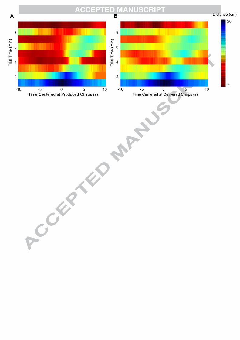
Time Centered at Produced Chirps (s) Time Centered at Delivered Chirps (s)-10 -5 0 5 10 -10 -5 0 5 10
Tria
l Tim
e (m
in)
Tria
l Tim
e (m
in)
2
4
6
8
2
4
6
8
7
26
Distance (cm)A B

Wave-type electric fish produce communication signals called chirps. We review chirping
behaviour as well as how sensory neurons encode these signals.
We consider the electrosensory background resulting from the interaction of two fish and
discuss how various social interactions shape both behaviour and sensory encoding.
We show how the encoding properties can explain some aspects of the behavior, but, to fully
understand the complexities of the behavior will require more detailed knowledge of higher
processing stages.


![Journal of Comparative Physiology A- Neuroethology, Sensory, Neural, and Behavioral Physiology Volume 121 issue 3 1977 [doi 10.1007%2Fbf00613014] DeForest Mellon -- The anatomy and](https://static.documents.pub/doc/80x56/55cf8c845503462b138d2f31/journal-of-comparative-physiology-a-neuroethology-sensory-neural-and-behavioral.jpg)


