

Page ‹#›
The Protists
A diverse assemblage of eukaryotesthat ARENʼT
fungi, plants, or animals
In what ways are protists important?
Base of many “food chains” - especiallyin aquatic settings
Clarify water by filtering out smallparticles
Some are parasites that causediseases in other organisms
Some have economic uses for humansSome are involved in important
symbiotic relationships…
Why can termites eat wood?
Because ofsymbiotichypermastigotes(a group ofparabasilids) livingin the termite gutworking togetherwith Archaeanmethanogens Fig 28.26 (SEM)
And… they are a spectacular groupof organisms
Where Did Eukaryotic Cells come from?
First found in fossil record about 2.1billion years ago
(Prokaryote fossils to 3.5 BYA)Two major features to explain:
- membrane-bounded organelles(mitochondria and plastids)
- internal membrane systems
Origin of Organelles
Idea is that the ancestors ofeukaryotic cells were symbioticconsortiums of prokaryotic cells
Has come to be called the“endosymbiont theory”

Page ‹#›
Lynn Margulis
Person who led the development of the endosymbiont theory
The Ideas of the EndosymbiontTheory (Fig 25.9)
Mitochondria are the descendents ofaerobic heterotrophic bacteria
Chloroplasts are the descendants ofphotosynthetic bacteria - very likelycyanobacteria
Origin ofEukaryotes
Fig.25.9
Evidence that Supports theEndosymbiont Theory
Endosymbiotic relationships exist inthe modern world, e.g., somespecies of dinoflagellates areendosymbiotic in corals
Plastids and mitochondria about thesame size as typical prokaryoticcells
Evidence (cont.)
Similar membrane proteins (innermembrane)
Reproduce by a process similar tobinary fission
Contain circular DNA moleculesRibosomal RNA sequences in
organelles more similar toprokaryotes
What organisms have eukaryotic cells?
Animals (mitochondria) Plants (mitochondria and plastids) Fungi (mitochondria) Protists (mitochondria, some have
plastids)

Page ‹#›
The ProtistsIncredible diversity of organisms -
your text recognizes 21 clades atprobably the Phylum or Kingdomlevel
Typically found in aquatic or dampenvironments, or in body fluids,tissues, or cells of host organisms
Most have flagella or cilia at somestage in their life cycle
Flagella and Cilia (Fig 6.23)
Structurallydistinct fromthe flagella ofprokaryotes
Eukaryoticflagella andcilia have asimilarstructureinvolvingmicrotubules
Human sperm
Ciliate
Cilia and Flagella in Action
Cilia and Flagella
Protist Size
Most are single-celled, but their cellstructure can be very complex
Ciliates (e.g., Paramecium,Vorticella) are among the mostcomplex of all cells
Some are multicellular andindividuals can be as large as 60meters in length - the “kelps”(brown algae)
Kelp (Brown Algae)
Definitely donʼt need a microscope to see this protist!
Protist Nutrition
Nutritionally diverse- photoautotrophs- chemoheterotrophs
Also are“mixotrophs”e.g., Euglena

Page ‹#›
Nutrition
Three major means of obtainingnutrition amongst protists:
- Ingestive (“animal-like”), sometimescalled “protozoa”
- Absorptive (“fungus-like”) - Photosynthetic (“plant-like”),
sometimes called “algae”Distinct nutritional mechanisms may be
found within one Clade
Protistan Phylogeny“Kingdom Protista” was a diverse group of
organisms that were, in many cases, notclosely related
Phylogeny is currently in a “state of flux”DNA sequence data have been, and will
continue to be, very helpfulSplitting of “Kingdom Protista” into 21
clades (Phyla? Kingdoms?) has beenproposed
These clades have been placed into 5“supergroups” in your text
Fig 28.3
Protistan Diversity
A quick look at 9 of the 21 protistclades described in Campbell et al.
Why not look at ALL 21 clades?
Getting a Ph.D. - Thatʼs where you learnmore and more about less and less untilyou know everything about nothing
Intro Bio Course - Thatʼs where you learnless and less about more and more untilyou know nothing about everything
I want you to know something aboutsomething...
Supergroup Excavata
Evidence:- Excavated feeding groove- DNA sequence similarities
Evidence supporting this “supergroup” israther weak and investigation is on-going
Page ‹#›
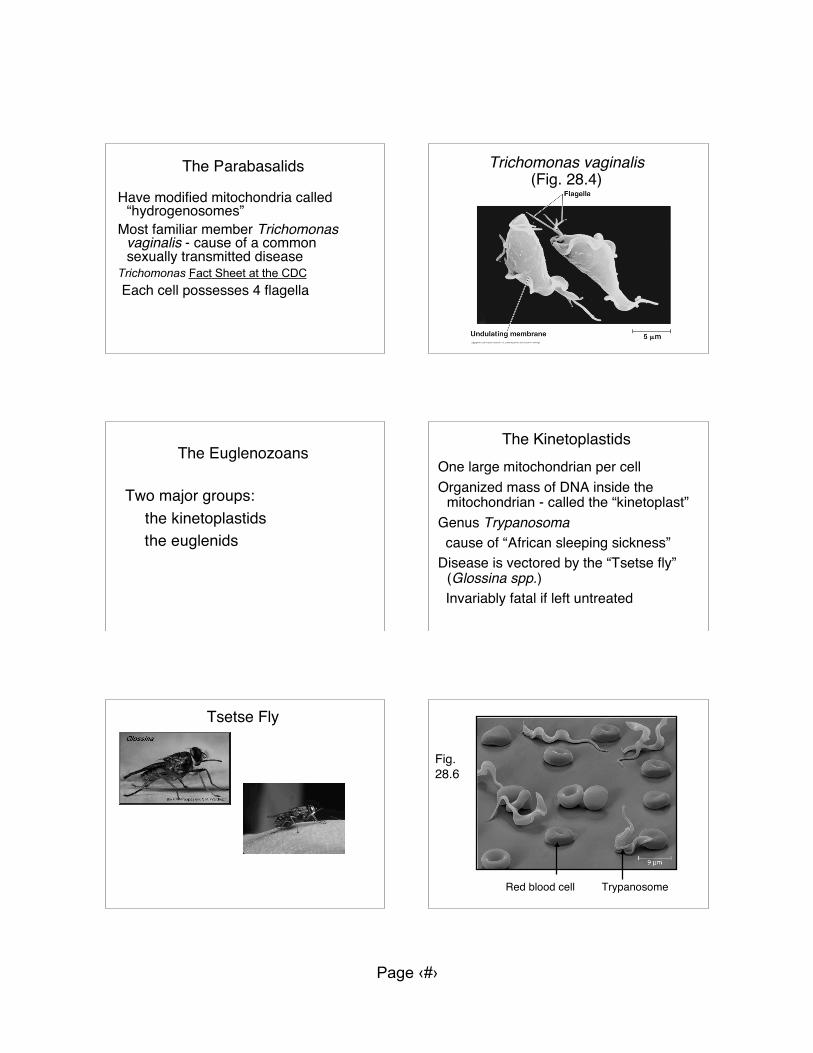
The Parabasalids
Have modified mitochondria called“hydrogenosomes”
Most familiar member Trichomonasvaginalis - cause of a commonsexually transmitted disease
Trichomonas Fact Sheet at the CDC Each cell possesses 4 flagella
Trichomonas vaginalis(Fig. 28.4)
The Euglenozoans
Two major groups: the kinetoplastids
the euglenids
The KinetoplastidsOne large mitochondrian per cellOrganized mass of DNA inside the
mitochondrian - called the “kinetoplast”Genus Trypanosoma cause of “African sleeping sickness”Disease is vectored by the “Tsetse fly”
(Glossina spp.) Invariably fatal if left untreated
Tsetse Fly
Red blood cell Trypanosome
Fig.28.6

Page ‹#›
Supergroup Chromalveolata
Evidence:- DNA sequence similarities- Chloroplast structure similarities
Highly controversial “supergroup”
The Alveolates
Characterized by the presence ofsmall membrane-bounded cavitiesunder their cell membrane
Three major groups: Dinoflagellates Apiocomplexans Ciliates
The DinoflagellatesBoth marine and freshwaterMost species unicellularImportant component of “plankton”About 50% of known species are
photosyntheticMost species have elaborate cell
walls
Dinoflagellates
Ceratium (lightmicroscope)
Peridinium(SEM)
“Red Tide”
Dead FishBoat
Red Tide
Dinoflagellate population explosionsWater stained brownish-red
(xanthophylls)Toxins produced by the
dinoflagellates can kill fish,invertebrates, seabirds
Some types of toxins canaccumulate in shellfish - causingpoisoning in humans

Page ‹#›
Karenia brevis
One species of dinoflagellate thatcauses red tides
Produces a toxin that kills fish andinvertebrates
Human exposure to the toxin maycause a variety of symptoms,including death - Called “neurotoxicshellfish poisoning”
Karenia brevis (SEM)
Location of Karenia blooms(data from December 2004)
Unit 1 Exam
Available Monday 15 Septemberthrough Tuesday 23 September
READ: “COLL Testing Facility Policiesand Procedures” in the CourseIntroduction Learning Module
Go to Center for On-Line Learning,room 60 Carver Hall to take the exam
The Cilates
Many beautiful freshwater speciesUse cilia to move and feedHave very complex cells, e.g., each
cell has one micronucleus and onemacronucleus
Micronuclei participate in sexualreproduction; macronuclei incontrolling cell functions
Ciliates
Stentor spp. Paramecium spp.

Page ‹#›
Paramecium feeding
The Stramenopiles
Some species are photoautotrophic,some are heterotrophic
Characterized by the presence hair-like projections on one of their(typically) two flagella
Stramenopile Flagella (Fig 28.12)Four major groups: Diatoms Brown algae (includes “kelp”) Golden algae Oomycetes (water molds)
The Diatomsglass-like cell walls - made of
hydrated silicaimportant photosynthetic organisms
in “plankton”fresh water and marinelarge number of species (estimated
to be ~ 100,000)
Diatoms
Diatom Diversity(Fig 28.3)
Diatom Art

Page ‹#›
Diatomaceous Earth
Huge amounts of ancient diatom cell wallsVarious uses:
filtering medium metal polishes reflective paint pesticide nanotechnology
SEM of Diatom
Supergroup Archaeplastida
Evidence:- DNA sequence similarity- Chloroplast structure similarities
This “supergroup” is well supportedby the available evidence
The Red Algae
No flagella present at any stage of thelife cycle
Most abundant in tropical oceansMost are multicellular~ 6,000 described speciesSome species are heterotrophic
Red Algae (Fig 28.19)
Red Algae
Accessorypigments allowphotosynthesisat great depths -as deep as 260meters
Effective atabsorbing bluelight
Human Uses
Cell wall extracts: carageenan - commonly
eaten by people… agar - microbiological
culturing media

Page ‹#›
How do you feel about sushi?
Fig. 28.19
The Green AlgaeMost species (~7,000) found in fresh
waterCell walls with a relatively high
percent of celluloseCan be unicellular,
colonial/filamentous, or multicellularCan be motile (flagella) or non-motile
Single-celled Green Alga -Eremosphaera viridis
Nucleus
Chloroplasts
Chlamydomonas
Unicellular and motile green alga Important model genetic system - much
research is done with this organism
Colonial Green Alga - Volvox (Fig. 28.3)
Volvox

Page ‹#›
Filamentous Green Alga - Ulothrix spp.
MulticellularGreen Alga -
Ulva spp.(Fig. 28.21)
Green Algal Life CyclesCan be quite complex with both sexual
and asexual reproductionMost gametes have two flagellaGametes may be isogamous or
anisogamousSome multicellular species exhibit
alternation of generations (as do allplants)
- may be heteromorphic or isomorphic
An example of a green algallife cycle
Oedogonium is a genus offilamentous green algae
Oedogonium Life Cycle Oedogonium life cycle
AnisogamousMeiosis leads to production of
“zoospores” (not gametes)Gametes are produced by mitosisAsexual “macrozoospores” are also
produced by mitosis

Page ‹#›
Supergroup Unikonta
Evidence:- DNA sequence similarities
This “supergroup” is well supported bythe available evidence
The Amoebozoans
Four major groups: Plasmodial slime molds Cellular slime molds Gymnamoebas (free-living) Entamoebas (parasitic)
Plasmodial Slime Molds
Feeding stage is an called a“plasmodium” (Fig. 28.24)
The plasmodium is a“coenocytic mass”
Multinucleatecytoplasm undividedby walls ormembranes
Live in moist habitats,e.g., rotting logs
The plasmodiumengulfs food by“phagocytosis” as doameobas
Cool Slime Mold Slime mold in “action”
Planet Earth - Jungles 23:20

Page ‹#›
Response to the Environment
Growth away from detergent
Start End ~ 48 hours
Slime Mold Reproduction
If available water or food insufficient,produces resistant spores throughmeiosis
Eachsporangiumproducesmany spores(Fig. 28.24)
Gymnamoebas
UnicellularFound in soil, freshwater, and marine
habitatsHeterotrophs that often consume
prokaryotes and other protists astheir food
Move by producing pseudopodia
Amoeba spp. (Fig. 28.3)
Attack of the Killer Amoeba
Studying organisms too small to seewithout a microscope is…
1. Boring beyondhuman tolerance
2. Very boring3. More interesting
than I expected - butstill boring
4. Remarkablyinteresting
5. More interestingthan any previousexperience in my life

Page ‹#›
The most interesting (or least boring)group weʼve studied so far is
1. Archaea2. Bacteria3. Excavata4. Chromalveolata5. Archaeplastida6. Unikonta


