

The Role of Cardiac Myocyte Dimensions in the Transition
from Hypertensive Hypertrophy to Cardiac Dilatation
Raúl José Correia
Dissertation submitted to the Faculty of Health Sciences, University of the
Witwatersrand, Johannesburg, in fulfilment of the requirements for the degree of
Master of Science in Medicine
Johannesburg, 2010

ii
DECLARATION
I, Raúl José Correia declare that this dissertation is my own, unaided work. It is being submitted
for the degree of Master of Science in Medicine, in the Faculty of Medicine, University of the
Witwatersrand, Johannesburg. The work contained in this thesis has not been submitted before for
any degree or examination in this university, or any other university.
………………………………………………………………………………………………….
RAÚL JOSÉ CORREIA
……………………………day of …………………………., 2010
I certify that the studies contained in this thesis have the approval of the Animal Ethics Committee
of the University of the Witwatersrand, Johannesburg. The ethics approval numbers are: 97:44:5,
99:01:2b, 2002:37:5, 2002:39:5 and 2006:41:05.
………………………………………………………………………………………………….
RAÚL JOSÉ CORREIA
……………………………day of …………………………., 2010
………………………………….. ………………………..
ANGELA J. WOODIWISS (Supervisor) GAVIN R. NORTON (Supervisor)

iii
Dedication:
For my parents,
Augusto and Jeanette
de Paiva Correia

iv
PUBLICATIONS AND PRESENTATIONS
Data presented in this dissertation have been published in a manuscript of which I am third author,
namely,
Veliotes DGA, Norton GR, Correia RJ, Strijdom H, Badenohorst D, Brooksbank R &
Woodiwiss AJ (2010). Impact of aldosterone receptor blockade on the deleterious effects of
adrenergic activation in hypertensive rats. J Cardiovasc Pharmacol 56, 203-211.
In addition data presented in this dissertation have been presented in the form of an oral
presentation, as well as in a poster at the 33rd
Meeting of the Physiology Society of Southern
Africa Conference in Cape Town, September 2005. The titles of these presentations were,
Correia RJ, Norton GR & Woodiwiss AJ. Cardiomyocyte lengthening does not contribute to the
development of cardiac dilatation (oral presentation)
Woodiwiss A, Correia R, Norton G, Muller C & Strijdom H. Determination of cardiomyocyte
length using flow cytometry (poster presentation)

v
ABSTRACT
The progression from compensated cardiac hypertrophy to decompensation and cardiac failure is
accompanied by cardiac dilatation. As cardiac failure has a poor prognosis, it is imperative to
prevent the progression to cardiac dilatation and heart failure. In this regard, an understanding of
the mechanisms of cardiac dilatation is vital to guide optimal therapy to prevent heart failure.
Although a number of factors have been shown to contribute to the development of cardiac
dilatation, to date the role of alterations in cardiac myocyte dimensions remains unclear. Hence,
the aim of the current study was to determine whether changes in cardiac myocyte dimensions
contribute to the process of cardiac dilatation.
Methods: Two models of cardiac dilatation in pressure-overload induced cardiac hypertrophy were
assessed. One model was a natural progression model, in which 18 spontaneously hypertensive rats
(SHR), were assessed at 23 months of age (an age when left ventricular hypertrophy is noted to
have progressed to left ventricular decompensation, dilatation and heart failure in approximately
50% of rats). The second model, a pharmacological model, was induced in 14 month old SHR
(n=9) by chronic beta-adrenoreceptor activation [0.02mg/kg isoproterenol (ISO) twice daily for 4.5
months]. Chronic beta-adrenoreceptor activation in SHR, enhances the progression from
compensated left ventricular hypertrophy to left ventricular dilatation. Nine normotensive Wistar
Kyoto (WKY) rats were the controls for both models. Left ventricular dilatation was defined as an
increase in left ventricular radius determined at controlled filling pressures using piezo-electric
transducers. The classification of rats as being in heart failure was based upon the presence of
pleuropericardial effusions and / or atrial thrombi. Cardiac myocytes were isolated and dimensions
determined using both light microscopy and flow cytometry.
Results: Left ventricular radius was increased in SHR-Failure compared to SHR-Non-Failure
(p<0.01), and in SHR-ISO compared to SHR-Control (saline administration) (p<0.01), hence
confirming the presence of cardiac dilatation in both models. Although, cardiac myocyte length

vi
was increased in all SHR groups compared to WKY (p<0.001), no differences were observed
between SHR-Failure and SHR-Non-Failure, or between SHR-ISO and SHR-Control. No
differences in cell length:width ratios or in cell widths were evident between the groups. The flow
cytometry data confirmed the results obtained for cardiac myocyte lengths using microscopy.
Moreover, a linear correlation (r=0.46, p=0.002) between flow cytometry and microscopy cardiac
myocyte lengths was observed. Importantly, no relationships were evident between left ventricular
radius and cardiac myocyte length (r=0.12, p=0.42 and r=0.14, p=0.35 for microscopic and flow
cytometry lengths respectively).
Conclusion: The results from the present study show that although pressure-overload hypertrophy
is associated with lengthening of cardiac myocytes, no further changes occur with cardiac
dilatation. Hence, alterations in cardiac myocyte dimensions do not contribute to the development
of cardiac dilatation in pressure-overload models.

vii
ACKNOWLEDGEMENTS
I am grateful for the assistance of the Central Animal Services of the University of the
Witwatersrand. I would like to thank Mr Ernest Somya and Dr Oleg Osadchii for their invaluable
assistance. I would also like to thank Prof. A Woodiwiss and Prof. G Norton for their guidance.
Funding for these studies was obtained from grants awarded to Prof. A Woodiwiss from the South
African National Research Foundation, and to Prof. G Norton from the Medical Research Council
of South Africa. The Cardiovascular Pathophysiology and Genomics Research Unit, the School of
Physiology and Faculty of Health Sciences of the University of the Witwatersrand also supported
these studies.

viii
TABLE OF CONTENTS
Declaration ii
Dedication iii
Publications and Presentations iv
Abstract v
Acknowledgements vii
Table of Contents viii
List of Figures x
List of Tables xi
Abbreviations xii
Preface xiv
CHAPTER 1 1
1.0 INTRODUCTION 2
1.1 CARDIAC DILATATION AND HEART FAILURE 4 1.1.1 Definition of Cardiac Dilatation 6 1.1.2 Appropriate Measurements of Cardiac Dilatation 6 1.1.3 Role of Cardiac Dilatation in the Development of Heart Failure 9
1.1.4 How Does Cardiac Dilatation Produce Pump Dysfunction? 11
1.2 MEDIATORS OF CARDIAC HYPERTROPHY AND ADVERSE CARDIAC
REMODELLING 13 1.2.1 Role of Neurohormones in Compensatory Cardiac Hypertrophy and the Progression to Cardiac
Decompensation and Heart Failure 14 1.2.1.1 Role of the Sympathetic Nervous System 15
1.2.1.2 Role of the Renin-Angiotensin-Aldosterone System 18
1.2.2 Role of Growth Factors and Inflammatory Cytokines 19
1.2.3 Role of Stretch Receptors (Cardiac Myocyte Stretch) 20
1.3 PROPOSED MECHANISMS OF COMPENSATORY CARDIAC
HYPERTROPHY 20 1.3.1 Role of Collagen and Interstitial Changes 20
1.3.2 Role of Cardiac Myocyte Hypertrophy Due to Increases in Cell Width 21
1.4 PROPOSED MECHANISMS OF ADVERSE CARDIAC REMODELLING 22 1.4.1 Role of Collagen and Interstitial Changes 23
1.4.2 Role of Cardiac Myocyte Apoptosis and Necrosis 24
1.4.3 Role of Cardiac Myocyte Hypertrophy Due to Increases in Cell Length 25
1.4.3.1 Are Changes in Cardiac Dimensions Associated with Changes in Cardiac Myocyte Length? 25
1.4.3.1.1 Data from Human Studies 25
1.4.3.1.2 Data from Animal Experimental Models 29
1.5 PROBLEM STATEMENT AND STUDY OBJECTIVES 35
CHAPTER 2 39
2.0 METHODS 40
2.1 RAT STRAINS AND GROUPS 40
2.1.1 Natural Progression Model 43
2.1.2 Pharmacological Model 43
2.2 SYSTOLIC BLOOD PRESSURE 44
2.3 LEFT VENTRICULAR GEOMETRY 44 2.3.1 Identification of Failure and Non-Failure Rats 48
2.4 TISSUE SAMPLING 49
2.5 MYOCYTE ISOLATION 50
2.6 LIGHT MICROSCOPY 52
2.7 FLOW CYTOMETRY 56

ix
2.8 STATISTICAL ANALYSES 57
CHAPTER 3 62
3.0 RESULTS 63
3.1 LEFT VENTRICULAR GEOMETRY 63 3.1.1 Natural Progression Model 63
3.1.1.1 Body and Tissue Weights and Blood Pressures 63
3.1.1.2 LV Dimensions 63
3.1.2 Pharmacological Model 66 3.1.2.1 Body and Tissue Weights 66 3.1.2.2 LV Dimensions 68
3.2 MYOCYTE DIMENSIONS 68 3.2.1 Natural Progression Model 68
3.2.1.1 Light Microscopy 68 3.2.1.2 Flow Cytometry 71
3.2.2 Pharmacological Model 74 3.2.2.1 Light Microscopy 75 3.2.2.2 Flow Cytometry 78
3.3 CORRELATIONS 78 3.3.1 Left Ventricular End Diastolic Radius and Cardiac Myocyte Length 78
3.3.2 Cardiac Myocyte Lengths Obtained Using Light Microscopy versus Flow Cytometry 78 3.3.3 Left Ventricular Weight (mg/100g Body Weight) versus Cardiac Myocyte Length 82
CHAPTER 4 84
4.0 DISCUSSION 85
CHAPTER 5 94
5.0 REFERENCES 95
APPENDICES (Animal Ethics Screening Committee Clearance Certificates) 116

x
LIST OF FIGURES
Figure Page Chapter 1
1.1 Example of a right-shift in the diastolic pressure-volume relationship 7
1.2 Example of changes in the Frank-Starling relationship 12
1.3 Schematic representation showing possible mechanisms of cardiac dilatation 16
1.4 Schematic representation of the factors known to contribute to cardiac dilatation 37
Chapter 2
2.1 Flow chart detailing the groups of rats 42
2.2 Intraventricular pressure monitoring and piezo-electric ultrasonic transducers 46
2.3 Example of recordings of left ventricular external diameter measurements 47
2.4 Examples of isolated cardiac myocytes 53
2.5 Photograph of digital camera and microscope 54
2.6 An example of striations of an isolated cardiac myocyte 55
2.7 Flow cytometer 58
2.8 Example of flow cytometer dot plot 59
2.9 Diagrammatic representation of a cardiac myocyte in the path of the flow cytometer
laser beam 60
2.10 Plot of cardiac myocyte counts versus time of flight 61
Chapter 3
3.1 Left ventricular end diastolic radius – pressure relations in the natural progression
model 65
3.2 Left ventricular end diastolic wall thickness to radius ratio – pressure relations in the
natural progression model 67
3.3 Left ventricular end diastolic radius – pressure relations in the pharmacological
model 69
3.4 Left ventricular end diastolic wall thickness to radius ratio – pressure relations in the
pharmacological model 70
3.5 Cardiac myocyte lengths and frequency distribution of cardiac myocyte lengths in
the natural progression model 72
3.6 Cardiac myocyte widths and length to width ratios in the natural progression model 73
3.7 Cardiac myocyte lengths as assessed by flow cytometry in the natural progression
model 74
3.8 Cardiac myocyte lengths and frequency distribution of cardiac myocyte lengths in
The pharmacological model 76
3.9 Cardiac myocyte widths and length to width ratios in the pharmacological model 77
3.10 Cardiac myocyte lengths as assessed by flow cytometry in the pharmacological model 79
3.11 Linear correlation analyses between left ventricular end diastolic radius and cardiac
myocyte length 80
3.12 Linear correlation analysis between cardiac myocyte length measured using light
microscopy and flow cytometry 81
3.13 Linear correlation analyses between left ventricular weight normalised to 100g body
weight and cardiac myocyte length as assessed by light microscopy and flow cytometry 83

xi
LIST OF TABLES
Table Page
Chapter 1
1.1 Summary of human data addressing possible association of cardiac remodelling with
alterations in myocyte morphology 26
1.2 Summary of data from animal experimental models addressing possible association of
cardiac remodelling with alterations in myocyte morphology 30
Chapter 3
3.1 Body and tissue weights in the natural progression model and in the pharmacological
model 64

xii
ABBREVIATIONS
ACE – Angiotensin-converting enzyme
AESC – Animal ethics screening committee
ANOVA – Analysis of variance
ATP – Adenosine triphosphate
CaCl2 – Calcium chloride
cAMP – Cyclic adenosine monophosphate
CAS – Central animal service
CHF – Congestive heart failure
CO2 – Carbon dioxide
EDP – End-diastolic ventricular pressure
EDV – End-diastolic ventricular volume
ESV - End-systolic ventricular volume
HEPES- (4-(2-hydroxyethyl)-piperazine-1-ethanesulfonic acid hemisodium salt
HOCM –Hypertrophic obstructive cardiomyopathy
h/r – relative wall thickness; i.e. LVED wall thickness to LVED radius ratio
IDCM - Idiopathic dilated cardiomyopathy
ISO - Isoproterenol
KCl – Potassium chloride
KH2PO4 – Potassium dihydrogen phosphate
LV – Left ventricle
LVAD – Left ventricular assist device
LVDP – Left ventricular diastolic pressure

xiii
LVED – Left ventricular end-diastolic
LVEDh – Left ventricular end-diastolic wall thickness
LVEDP – Left ventricular end-diastolic pressure
LVEDr – Left ventricular end-diastolic radius
LVEF – Left ventricular ejection fraction
LVextD – Left ventricular external diameter
MI – Myocardial infarction
MgCl2 – Magnesium chloride
NaCl – Sodium chloride
NaOH – Sodium hydroxide
O2 - Oxygen
PNS – Parasympathetic nervous system
PSS - Physiological saline solution
RAAS – Renin-angiotensin-aldosterone system
SBP – Systolic blood pressure
SEM – Standard error of the mean
SHHF – Spontaneously hypertensive heart failure
SHR – Spontaneously hypertensive rat
SNS – Sympathetic nervous system
TNF-α - Tumour Necrosis Factor-α
WKY - Wistar-Kyoto

xiv
PREFACE
Cardiovascular disease is one of the leading causes of morbidity and mortality in all parts of the
world today. Almost all forms of cardiovascular disease progress to heart failure, which is the
terminal endpoint of cardiovascular diseases. Hence, the progression to heart failure needs to be
prevented. Chronic cardiovascular disease is initially accompanied by cardiac hypertrophy which
is considered compensatory in that cardiac wall stress is maintained within normal levels; however
compensatory cardiac hypertrophy progresses to cardiac decompensation and heart failure, a state
in which cardiac wall stress is elevated. The increase in cardiac wall stress is primarily due to
enlargement of the cardiac chamber volume and thinning of the cardiac chamber wall, a process
termed detrimental cardiac remodelling or dilatation. As cardiac failure has a poor prognosis, it is
imperative to prevent the progression to cardiac dilatation and heart failure. In this regard, an
understanding of the mechanisms of cardiac dilatation is vital to guide optimal therapy to prevent
heart failure.
The mechanisms by which the cardiac tissue remodels to cause the dilatation are the topic of much
debate. Both changes in the cardiac interstitium as well as changes in the cardiac myocytes are
believed to play a role. In previous studies, adverse cardiac remodelling has been associated with
increased cardiac interstitial fibrosis, but of the non-cross-linked form; cardiac myocyte apoptosis
and necrosis; as well as cardiac myocyte hypertrophy due to changes in cell length. Although,
some consensus has been reached regarding the role of the cardiac interstitium and cardiac
myocyte death (apoptosis or necrosis); to date the role of alterations in the dimensions of cardiac
myocytes remains unclear.
Although, cardiac dilatation is thought to be mediated by increases in the length of cardiac
myocytes, not all studies support this hypothesis. Indeed, although some studies show that
increases in cardiac myocyte length are associated with increases in cardiac chamber dimensions
or heart failure; a number of other studies show no relationship. Furthermore, data from

xv
intervention studies failed to show changes in cardiac dimensions in parallel with changes in
cardiac myocyte length. A number of reasons may explain the controversial results regarding the
role of alterations in cardiac myocyte length in cardiac dilatation.
Firstly, in some studies the increased cardiac myocyte length in patients with heart failure
compared to healthy controls could be as a consequence of the differences in body weight and
hence left ventricular (LV) weight between these two groups. The differences in body weight were
due to a greater proportion of males in the heart failure group compared to the control group.
Secondly, as the phase of the cardiac cycle determines cardiac chamber dimensions and cardiac
myocyte length, possible differences between the heart failure and control groups in the phase of
the cardiac cycle at the time of cardiac arrest may account for the differences in cardiac chamber
diameter and cardiac myocyte length observed between these two groups in some human studies.
Thirdly, in a number of studies in spontaneously hypertensive heart failure (SHHF) rats showing
an association between cardiac myocyte length and cardiac chamber dimension, the animals in
heart failure were significantly older than the control animals. As LV weight increases with age in
hypertensive rats, and cardiac myocyte length is strongly associated with age and LV weight, the
increases in cardiac myocyte length observed in the rats in heart failure may reflect age induced
changes, rather than an association with adverse chamber remodelling (cardiac dilatation) and the
development of heart failure.
Fourthly, other than the studies in SHHF rats, no other studies have made direct comparisons of
animals with cardiac pathology which have heart failure to animals with the same cardiac
pathology but without heart failure. However, as discussed the increased cardiac myocyte length in
SHHF rats may reflect increased age and hence increased LV weight compared to controls, rather
than increased cardiac dimensions. Indeed, it has been shown that cardiac myocyte lengthening
increased with age and occurred well before the development of heart failure in SHHF rats.
Although the latter study suggests that changes in cardiac myocyte length are not responsible for

xvi
the development of heart failure, no measurements of cardiac dimensions were made in this study.
To my knowledge no study to date has compared age-matched animals with the same cardiac
pathology which have heart failure to those without heart failure.
Lastly, the use of load dependent measures of cardiac chamber dimensions (echocardiography in
vivo) in the presence of increased preloads may also contribute to the controversy. In this regard
cardiac myocyte lengthening occurs as a consequence of the stretching of cardiac myocytes during
increased preloads (filling volume or pressure). Moreover, when pulmonary capillary wedge
pressure was normalised as a consequence of LV assist device support, a normalisation of both LV
end diastolic diameter and cardiac myocyte length has been reported.
Moreover, data from various intervention studies failed to show changes in cardiac dimensions in
parallel with changes in cardiac myocyte length and hence the results of these studies do not
support a role of changes in cardiac myocyte length in cardiac dilatation. Indeed, if increases in
cardiac myocyte length were causally related to adverse cardiac remodelling, a reduction in cardiac
myocyte length should be accompanied by a decrease in cardiac chamber dimensions. However,
these intervention data need to be interpreted with caution in view of the use of load dependent
measures of cardiac dimensions in these studies.
The data to date provides no clear conclusion as to the possible role of cardiac myocyte length in
adverse chamber remodelling. Hence, the aim of the current study was to determine whether
changes in cardiac myocyte length contribute to the progression from compensatory cardiac
hypertrophy to adverse chamber remodelling (cardiac dilatation) as measured using load
independent methods in rats with heart failure compared to those not in heart failure.

1
CHAPTER 1

2
1.0 INTRODUCTION
One of the leading causes of morbidity and mortality in all parts of the world today is
cardiovascular disease (American Heart Association, 2010). Over 80 million adults (more
than one in three), in America alone have some form of cardiovascular disease (American
Heart Association, 2010). Moreover, cardiovascular disease accounts for 16,7 million of
deaths (29.2%) worldwide per annum (Frey et al., 2004). In South Africa, cardiovascular
disease was reported to account for ~90 to 100 deaths per 100 000 population in 2006
(Mayosi et al., 2009). In addition, of the total cases reported on in a cardiology unit, at a
hospital that services an urban developing community in South Africa, 43% were de novo
presentations of heart failure (Stewart et al., 2008). Importantly, almost all forms of
cardiovascular disease progress to heart failure, which is the terminal endpoint of
cardiovascular diseases. Hence, the progression to heart failure needs to be prevented.
In order to adequately prevent morbidity and mortality from heart failure, an understanding of
the mechanisms underlying the progression to heart failure are paramount. In this regard a
number of different models of heart failure have been investigated. A major predictor of
progressive heart disease and an adverse prognosis is cardiac hypertrophy (Levy et al., 1990).
Although, cardiac hypertrophy is initially a compensatory response to alterations in loading
conditions (Grossman et al., 1975), prolonged hypertrophy ultimately leads to cardiac
dilatation (increase in cardiac chamber dimensions), heart failure and subsequent death (Bing
et al., 1995; Inoko et al., 1994; Lorell 1997; Spann et al., 1967). Factors which have been
associated with the development of cardiac dilatation and heart failure (decompensation) in
various models of cardiac hypertrophy, include unfavourable changes in the cardiac
interstitium; enhanced cardiac myocyte apoptosis and necrosis; realignment of cardiac

3
myocytes within the ventricular walls (myocardial slippage) expression of foetal genes; and
alterations in cardiac myocyte dimensions (Cohn et al., 2000; Ferrari et al., 2009; Frigerio &
Roubina, 2005; Remme 2003). These changes occur in response to sustained pathological
stress signals such as neurohumoral activation, the release of growth factors and
inflammatory cytokines, and mechanical stretch (Cohn et al., 2000; Ferrari et al., 2009;
Frigerio & Roubina, 2005; Remme 2003). These pathological signals are termed the
mediators of adverse cardiac remodelling.
Despite a plethora of studies investigating the various factors associated with the
development of heart failure, the role of alterations in cardiac myocyte dimensions in the
progression from compensatory cardiac hypertrophy to cardiac dilatation is unclear. In this
regard, compensatory cardiac hypertrophy is generally associated with increases in cardiac
myocyte width (Onodera et al., 1998; Zierhut et al., 1991); whereas alterations in cardiac
myocyte length are thought to contribute to cardiac dilatation (Gerdes 2002). However, most
studies showing increases in cardiac myocyte length in association with increases in left
ventricular (LV) dimensions have used load dependent measures of cardiac chamber
dimensions (Chen et al., 2010; Gerdes et al., 2010; Kajstura et al., 1995; Tamura et al., 1998;
Wang et al., 1999; Yarbrough et al., 2010). Thus whether cardiac dilatation has indeed
occurred in these studies is debatable. Moreover, although in some studies increases in load
dependent measures of cardiac chamber diameter are associated with increases in cardiac
myocyte dimensions (Gerdes et al., 2010; Janczewski et al., 2003; Kajstura et al., 1995;
Schultz et al., 2007; Tamura et al., 1998; Toischer et al., 2010; Wang et al., 1999); other
studies fail to show such relationships (Li et al., 2010; Schultz et al., 2007; Tamura et al.,
2000; Yarbrough et al., 2010). Nevertheless, an understanding of the role of cardiac myocyte
hypertrophy in cardiac dilatation is essential to guide choices of optimal therapy to prevent

4
the progression from compensatory hypertrophy to decompensation.
Hence, the aim of my studies was to determine the role of alterations in cardiac myocyte
dimensions in the progression from concentric cardiac hypertrophy to cardiac dilatation. As
the most prevalent form of heart failure is that associated with hypertensive heart disease
(Remme et al., 2003), I chose to assess two models of cardiac dilatation in spontaneously
hypertensive rats (SHR) with cardiac hypertrophy. Therefore, in the present chapter of my
dissertation, I will discuss the role of cardiac dilatation in the development of heart failure
and the importance of defining cardiac dilatation using load independent measurements. I
will then review the literature on the factors (mediators and mechanisms) associated with the
development of cardiac hypertrophy and the progression to decompensation and heart failure,
with specific reference to the controversial role of alterations in cardiac myocyte dimensions.
1.1 CARDIAC DILATATION AND HEART FAILURE
In response to chronic elevations in cardiac wall stress (increases in loading conditions, such
as chronic hypertension or post myocardial infarction), the heart undergoes hypertrophy
(thickening of the ventricular wall) in an attempt to normalise wall stress. According to the
law of La Place, wall tension or stress, is proportional to the product of pressure (P) and
radius (r) and inversely proportional to wall thickness (h). Hence, an increased wall thickness
in cardiac hypertrophy maintains a normal wall stress in the presence of increments in either
pressure or volume (radius) within the cavity (Grossman et al., 1975). This process, termed
compensatory cardiac hypertrophy, is generally associated with adequate cardiac systolic
function, and a normal or increased ventricular wall thickness to radius ratio (Janicki et al.,
2004). However, diastolic function may be decreased due to the restriction of ventricular

5
filling by a thickened ventricular wall (Kai et al., 2005; Norton et al., 1993). The
development of compensatory hypertrophy is due to the load-induced activation of various
mediators which initiate cellular, molecular and genetic processes.
As will be discussed in more detail later, persistent activation of these mediators over time,
and consequently alterations in cellular, molecular and genetic processes eventually leads to
cardiac decompensation (Remme, 2003). Cardiac decompensation is associated with a
reduced ventricular wall thickness to radius ratio, ventricular enlargement and depressed
cardiac systolic function (Janicki et al., 2004). This process of adverse cardiac remodelling
(reduced ventricular wall thickness to radius ratio and ventricular enlargement), also termed
cardiac dilatation, progresses with time to heart failure. Hence, cardiac dilatation is an
important negative prognostic factor in patients with heart failure (Cohn et al., 2000; Udelson
et al., 2002). Moreover, even in asymptomatic subjects without a prior history of heart
failure, LV dilatation is associated with an increased risk of the development of heart failure
(Vasan et al., 1997). It is therefore important to understand how cardiac remodelling
contributes to the development of heart failure.
One of the limitations of many studies assessing the role of cardiac remodelling in heart
failure, and the mechanisms thereof, is the inability to define cardiac dimensions in a load-
independent model. In this regard, increases in cardiac filling volumes (increased preloads)
would manifest as increased cardiac dimensions. Moreover, increases in cardiac afterload
(increased cardiac wall stress as discussed above) would reduce stroke volume thereby
increasing ventricular volumes at the end of systole, which would also manifest as increased
cardiac dimensions. Hence, the correct definition of cardiac remodelling is an increase in
cardiac diameter, or a decrease in the wall thickness to radius ratio, at a given filling volume.

6
Therefore, before discussing the impact of cardiac dilatation on cardiac function and the
development of heart failure, I will first discuss in more detail the definition of cardiac
dilatation and appropriate measurements thereof.
1.1.1 Definition of Cardiac Dilatation
Cardiac chamber dilatation is defined as an increase in chamber cavity volume or dimension
as a consequence of a right shift in the diastolic pressure-volume relationship (Figure 1.1).
Importantly, cardiac dilatation is not an increase in only the chamber cavity volume, as this
may result from an enhanced blood volume or venous return (increased preload) without
necessarily being accompanied by a right shift in the diastolic pressure-volume relationship.
In addition, cardiac dilatation is not a right shift in the diastolic pressure-volume relationship
produced by alterations in the slope of this relationship. Changes in the slope of the cardiac
diastolic pressure-volume relationship occur as a consequence of alterations in chamber
stiffness, which are usually mediated by modifications in the material properties of the
myocardium (Gilbert and Glantz, 1989). However, cardiac chamber dilatation is the
consequence of a right shift in the diastolic pressure-volume relationship due to an increase in
the volume intercept of this relationship (Gibbs et al., 2004) (Figure 1.1).
1.1.2 Appropriate Measurements of Cardiac Dilatation
As increases in blood volume, venous return, and blood pressure (may decrease stroke
volume); and decreases in cardiac contractility can result in increases in ventricular volumes,
it is important that filling volumes are controlled when determining the relationship between
diastolic pressure and volume. Hence, the in vivo assessment of the diastolic pressure-volume
relationship using echocardiographic measures of cardiac dimensions is not appropriate.
Indeed, blood volumes are related to body size, which may differ between for example

7
Figure 1.1 Example of a right-shift in the diastolic pressure-volume relationship
The normal diastolic pressure-volume relationship is indicated by the solid line, and a right
shift in the pressure-volume relationship, indicative of cardiac dilatation, is indicated by the
dashed line. The volume intercept [volume at which left ventricular (LV) end diastolic
pressure equals zero (V0)] is increased in the right shifted relationship.

8
normotensive and hypertensive rats (Badenhorst et al., 2003a; Tsotetsi et al., 2001; Veliotes
et al., 2005). In addition, increases in cardiac afterload, such as increases in blood pressure or
peripheral resistance in hypertension, would reduce stroke volume and hence increase filling
volumes (more blood is left behind at the end of systole). Moreover, the measurement of
cardiac dimensions alone, without the measurement of the accompanying diastolic pressures
is not appropriate. Indeed, as diastolic pressure increases, so does diastolic volume (Figure
1.1). Hence, increases in chamber dimensions may be a manifestation of increases in filling
pressures (increased preload) rather than measures of true cardiac dilatation.
Nevertheless, many clinical and experimental studies have defined the presence of dilatation
using non-invasive measurements such as echocardiography and ventriculography. Hence,
whether cardiac dilatation was indeed present is questionable. Indeed increases in filling
pressures were noted in many of these studies (Chen et al., 2010; Gerdes et al., 2010;
Kajstura et al., 1995; Tamura et al., 1998; Wang et al., 1999; Yarbrough et al., 2010;
Zefeiridis et al. 1998). Although in clinical studies there is no alternative to non-invasive
measurements; of greater concern is the use of such measurements in terminal experimental
studies. In this regard, in many animal based studies, the groups differ in age (Gerdes et al.,
1996; Tamura et al., 1998). As age is associated with increased LV weight (Gerdes et al.
1996), and increased left ventricular weight is correlated with cardiac myocyte length
(Campbell et al. 1991; Capasso et al. 1992), and age (Tamura et al., 1998), any differences in
cardiac dimensions could be attributed to differences in age and hence heart weight rather
than true differences in the dimensions of the heart. In other words the increased left
ventricular weight and hence cardiac myocyte length is likely to reflect growth effects during
aging.

9
Therefore, in order to appropriately define the presence of cardiac dilatation, ventricular
pressures need to be determined and ventricular filling volumes need to be controlled. In this
manner the diastolic pressure-volume relationship can be constructed and hence the volume
at which pressure is 0 mm Hg (ie. the volume intercept) can be determined. Importantly, very
few studies have accurately defined dilatation using pressure-volume relationships. In this
regard, no study assessing the role of cardiac myocyte dimensions in heart failure, has
accurately defined the presence of dilatation.
1.1.3 Role of Cardiac Dilatation in the Development of Heart Failure
Initially it was thought that cardiac dilatation was a remodeling process that began in order to
prevent the progressive increases in filling pressures associated with heart failure (Ertl et al.,
1991). In essence, it was believed that the decreased contractility in heart failure and hence
increased filling volumes (Patterson and Adams, 1996), were necessary in order to maintain
stroke volume via the Frank-Starling effect (Grossman et al., 1975). However, the increased
filling volumes would be accompanied by increases in filling pressures and hence pulmonary
capillary hydrostatic pressures may be elevated resulting in pulmonary congestion. It was
therefore believed that in order to accommodate enhanced filling volumes at normal filling
pressures a right shift in the diastolic pressure-volume occurred.
However, more recently, cardiac dilatation has been shown to be a precursor of pump
dysfunction and clinical heart failure (Gaudron et al., 1993; Pfeffer et al., 1992; Vasan et al.,
1997). In a 3 year prospective study in patients post myocardial infarction, those patients who
had progressive dilatation also had a progressive decline in ejection fraction and an increase
in pulmonary capillary wedge pressure (Gaudron et al., 1993); whereas in those patients with
no dilatation, LV ejection fraction did not decline and pulmonary wedge pressure remained

10
within normal values. Hence cardiac dilatation post myocardial infarction results in the
development of heart failure. In addition, in an 11-year follow-up study of people who had
not sustained a myocardial infarction and who did not have congestive heart failure at
enrolment, increments in LV internal dimension increased the risk of development of
congestive heart failure (adjusted hazard ratio of 1.47 for a one standard deviation increase in
LV end diastolic diameter indexed for height) (Vasan et al., 1997). Moreover, intervention
studies have shown that the alleviation of LV enlargement post myocardial infarction
prolongs survival and reduces mortality and morbidity due to major cardiovascular events
(Pfeffer et al., 1992; St John Sutton et al., 1997). Hence, the process of cardiac dilatation has
to be seen as a cause of heart failure as opposed to its consequence.
There are a number of additional observations which support the role of cardiac dilatation in
the development of heart failure. Firstly, in the presence of compensatory cardiac
hypertrophy (increases in wall thickness) in response to pressure overload, there is no
evidence of systolic heart failure (Wang et al., 1999; Onodera et al., 1998; Woodiwiss et al.,
1995; Yousef et al., 2000). Secondly, the neurohumoral factors that maintain systolic
function in the hypertrophied heart, in the long-term are detrimental to the myocardium.
Indeed, these neurohumoral factors promote cardiac dilatation and ultimately lead to systolic
dysfunction (Woodiwiss et al., 1995; Yousef et al., 2000). Lastly, there is evidence to show
that maladaptive changes in myocardial tissue occur long before symptoms of heart failure
(Onodera et al., 1998). Hence adverse cardiac remodelling, which consists of both
macroscopic and microscopic changes in the myocardium, precedes heart failure and
therefore contributes to, instead of results from, heart failure (Onodera et al., 1998). The
question of how cardiac dilatation produces pump dysfunction and heart failure therefore
arises.

11
1.1.4 How Does Cardiac Dilatation Produce Pump Dysfunction?
Changes in pump function are best explained by the Frank-Starling relationship. Figure 1.2
illustrates the normal Frank-Starling relationship and the changes that occur in association
with either an enhanced pump function or a decreased pump function. A left and upward shift
of the curve compared to normal (an enhanced pump function) occurs when intrinsic
myocardial contractility increases (such as in the presence of increased circulating
catecholamines as may occur with exercise); or afterload decreases (such as following
vasodilatation); or the relationship between wall thickness and internal radius increases (such
as with compensatory cardiac hypertrophy). In contrast, displacement to the right and
downward from the normal occurs when ventricular contractility is depressed; or afterload is
increased; or the heart dilates as is the case in most forms of heart failure due to systolic
functional abnormalities. Although the impact of changes in intrinsic myocardial contractility
and the resistance to blood flow on systolic function, are relatively easy concepts to grasp,
cardiac dilatation is sometimes a conceptually difficult issue. Hence, how does cardiac
dilatation produce deleterious effects on pump function?
As chamber dilatation is associated with an increased cavity volume (and hence radius), and a
reduced wall thickness, according to La Place’s law, wall tension or stress (afterload) will be
increased. As wall stress determines myocardial oxygen consumption, the myocardial oxygen
demand-to-supply ratio may be increased in a dilated ventricle, and a demand-to-supply
mismatch may subsequently decrease cardiac contraction. However, when systolic function
was measured using a stress (or load)-independent measure of pump function (end systolic
elastance) in an animal model of congestive cardiac failure and pump dysfunction associated
with massive cardiac dilatation (Norton et al., 2002), pump function was reduced without
parallel changes in myocardial contractility. These data would suggest that a mechanism

12
Figure 1.2 Example of changes in the Frank-Starling relationship
13
unrelated to stress or load-induced effects contributes to pump dysfunction in cardiac
dilatation. It is possible that inappropriate force transduction occurs in dilated ventricles
during myocyte contraction, which in-turn leads to pump dysfunction (Sallin 1969).
Alternatively, in a dilated ventricle, larger chamber volumes may be required to produce
cardiomyocyte stretch and hence to recruit the Frank-Starling effect. Indeed, when the
structure of the ventricle changes, the mechanics of systolic output are affected, thus resulting
in a low-output state (Laskey et al., 1984; Cohn et al., 2000).
Having established that cardiac dilatation is a cause rather than a consequence of pump
dysfunction and heart failure; in order to reduce morbidity or mortality from progressive
heart failure treatments which prevent or reverse adverse cardiac remodelling are required.
The choice of effective therapy is based on the knowledge of the mediators and the
mechanisms responsible for cardiac remodelling. I will therefore discuss what is known to
date regarding the mediators of cardiac dilatation and the role of various potential
mechanisms.
1.2 MEDIATORS OF CARDIAC HYPERTROPHY AND ADVERSE
CARDIAC REMODELLING
The generally accepted theory is that the mediators responsible for compensatory cardiac
hypertrophy, when sustained are ultimately responsible for the progression to adverse cardiac
remodelling and heart failure. I will therefore discuss what is known regarding the mediators
of compensatory hypertrophy as well as the how these factors are thought to mediate cardiac
dilatation and the development of heart failure.

14
Compensatory cardiac hypertrophy and cardiac dilatation occur due to the independent and
interactive effects of three extrinsic mediators on the heart, namely: (1) neurohormones; (2)
growth factors and inflammatory cytokines; and (3) mechanical stretch receptors in the cell
membranes, which activate signalling pathways intracellularly (Cohn et al., 2000; Ferrari et
al., 2009; Frigerio & Roubina, 2005; Remme 2003).
These extrinsic mediators act via various intracellular pathways [mitogen activated protein
(MAP) kinase; nuclear factor ĸB; protein kinase B] to activate nuclear transcription, which
leads to cellular hypertrophy, necrosis, apoptosis and fibrosis (Katz 2002; Opie et al., 2006;
Yousef et al., 2000). The nature of the signalling stimulus is believed to determine which
intracellular pathways are activated and hence whether compensatory cardiac
hypertrophy or cardiac dilatation occurs (Ferrari et al., 2009; Hill & Olson 2008; Opie et
al., 2006).
1.2.1 Role of Neurohormones in Compensatory Cardiac
Hypertrophy and the Progression to Cardiac Decompensation
and Heart Failure
Neurohormonal activation [activation of the sympathetic nervous system (SNS) and the
renin-angiotensin-aldosterone system (RAAS)] is known to occur in response to increases in
cardiac wall stress. Indeed, circulating concentrations of noradrenaline are increased in
persons with hypertension and LV hypertrophy (Agabiti-Rosei et al., 1987; Kelm et al.,
1996). In addition, the RAAS is activated in the hypertrophied and failing heart (Danser et
al., 1997; Iwai et al., 1995). Initially, the increased activity of the SNS and RAAS occur in
order to normalise wall stress and to preserve contractile performance; however continual
activation of the SNS (Badenhorst et al., 2003b; Gibbs et al., 2004; Veliotes et al., 2005) and

15
the RAAS (Mizuno et al., 2001; Schunkert et al., 1993) have been shown to induce cardiac
dilatation and heart failure. Indeed, neurohumoral activation in heart failure (Hasking et al.,
1986), is a major factor responsible for the progression of heart failure (Bristow 1997; Cohn
et al., 1984).
1.2.1.1 Role of the Sympathetic Nervous System
Initially in compensatory hypertrophy, in response to increased catecholamines, the inotropy
of the cardiac myocytes is increased through post receptor activation of adenylate cyclase and
consequent increases in the intracellular concentration of the second messenger cyclic
adenosine monophosphate (cAMP). This response will improve the cardiac output through an
increased myocardial contraction. However, sustained elevations in catechoamines in the
presence of increased pressure loads will increase myocardial oxygen demand (a
consequence of increased inotrope as well as increased afterload, due to alpha adrenergic
mediated vasoconstriction), which may outstrip myocardial oxygen supply, as increased
vascularisation does not occur in parallel with myocardial hypertrophy (Weisman et al.,
1988). One of the consequences of oxygen demand-to-supply imbalance is tissue necrosis. As
cardiac myocytes within the syncitium die, the viable cardiac myocytes within the syncitium
are stretched hence possibly resulting in side-to-side slippage and ultimately cardiac
dilatation (Figure 1.3). Indeed, myocyte slippage in end stage dilated cardiomyopathy has
been well documented (Beltrami et al., 1995; Linzbach 1960).
Sustained (or chronic) activation of the SNS not only induces cardiac myocyte death through
hemodynamic mechanisms as described above, but also via direct mechanisms. Indeed,
excessive concentrations of adrenergic agonists promote necrosis (Esler et al., 1997) and
apoptosis (Communal et al., 1998; Singh et al., 2001). Adrenergic-induced cardiac myocyte

16
Figure 1.3 Schematic representation showing possible mechanisms of cardiac
dilatation.

17
apoptosis is mediated via activation of β1-adrenergic receptor cAMP-dependent protein
kinase A and mitogen-activated protein kinase (MAPK) pathways. Adrenergic activation may
also promote cardiomyocyte apoptosis indirectly via stimulation of the RAAS (see 1.2.1.2
below) or through increases in myocardial expression of inflammatory cytokines (see 1.2.2
below). Similar to necrosis the consequences of apoptosis are stretching of the viable
cardiac myocytes within the syncitium are stretched hence possibly resulting in side-to-side
slippage and ultimately cardiac dilatation (Figure 1.3). In addition to cardiac myocyte death,
chronic β-adrenergic activation results in unfavourable alterations in the cardiac interstitium,
which result in decreased tethering of the cardiac myocytes (Figure 1.3). Indeed, as will be
discussed below (see 1.4.1) increases in the non-cross-linked collagen content of the
myocardium have been demonstrated in association with cardiac dilatation after chronic
administration of the β-adrenergic agonist isoproterenol (Woodiwiss et al., 2001). Although,
isoproterenol, has been shown to stimulate the activity of matrix metalloproteinases (MMPs)
in isolated cardiac myocytes (Coker et al., 2001), which would increase collagen turnover
thereby reducing time available for cross-linking to occur (Woodiwiss et al., 2001); it is more
likely that the changes in the characteristics of myocardial collagen are mediated by
activation of the RAAS, as the decreased collagen cross-linking could be prevented by both
angiotensin-converting enzyme inhibitor administration (Woodiwiss et al., 2001) as well as
aldosterone receptor blockade (Veliotes et al., 2005).
Although, adrenergic activation, together with activation of inflammatory cytokines, has been
associated with cardiac myocyte hypertrophy (increased cell width) (Tarone & Lembo,
2003); to date there is no evidence to implicate adrenergic activation in promoting
lengthening of cardiac myocytes.

18
Evidence of the potential role of activation of the SNS in mediating adverse cardiac
remodelling is provided by the detection of substantially increased plasma concentrations of
noradrenaline and adrenaline in patients with heart failure (Anand et al., 2003; Cohn et al.,
1984; Francis et al., 1993; Swedberg et al., 1990), and the relationship of these
concentrations to the severity of pump dysfunction (Kluger et al., 1982) and heart failure
(Sigurdsson et al., 1994). Furthermore, a number of studies have demonstrated that β-
adrenergic receptor blocking agents reduce cardiac cavity dimensions (Gerson et al., 2002;
Metra et al., 2003; Packer et al., 1996; Toyama et al., 2003; Waagstein et al., 1993a;
Waagstein et al., 1993b).
1.2.1.2 Role of the Renin-Angiotensin-Aldosterone System
Initially activation of the RAAS is compensatory in nature in that RAAS mediated fluid
retention improves venous return and thus cardiac output (via the Frank-Starling effect).
However sustained elevations in the RAAS are detrimental. Indeed, both angiotensin II and
aldosterone have been shown to promote cardiac myocyte apoptosis (De Angelis et al.,
2002), and hence the potential for side-to-side slippage of cardiac myocytes (Figure 1.3).
Activation of the RAAS is also likely to play a role in the unfavourable changes in the
characteristics of myocardial collagen, as decreased collagen cross-linking associated with
cardiac dilatation could be prevented by both angiotensin-converting enzyme inhibitor
administration (Woodiwiss et al., 2001) as well as aldosterone receptor blockade (Veliotes et
al., 2005). In addition, angiotensin II and aldosterone have been shown to stimulate collagen
synthesis (Brilla et al., 1994).
Although, angiotensin II acting as a growth factor has been shown to mediate cardiac

19
myocyte hypertrophy by increasing cell width (Sadoshima et al., 1997; Serneri et al., 1999),
the role of the RAAS in cardiac myocyte lengthening is as yet unclear.
Nevertheless, the role of the RAAS in adverse cardiac remodelling is substantiated by
intervention data showing reductions in cardiac dimensions following the administration of
ACEI to patients in heart failure (Greenberg et al., 1995; Konstam et al., 1992).
1.2.2 Role of Growth Factors and Inflammatory Cytokines
Growth factors such as insulin-like growth factor are thought to mediate cellular hypertrophy
via the protein kinase B pathway (Opie et al., 2006). Activation of this pathway promotes
cardiac myocyte growth (increased cell width) and inhibits apoptosis (Matsui & Rosenzweig,
2005). Hence, activation of insulin-like growth factor is important in mediating compensatory
hypertrophy, but possibly plays no role in the transition to cardiac decompensation. In
comparison transforming growth factor β, similar to angiotensin II, activates fibroblasts
hence promoting collagen formation and fibrosis (Hein et al., 2003; Kuwahara et al., 2004).
Increased fibrosis (as discussed below) is important in both compensatory hypertrophy and
dilatation. Inflammatory cytokines, such as tumour necrosis factor α, seem to have a dual
role. At low concentrations these cytokines mediate compensatory hypertrophy; whereas at
high concentrations they may play a role in mediating cardiac dilatation and heart failure
(Tarone & Lembo, 2003). Lastly, cardiac myocyte apoptosis may be promoted by increased
myocardial expression of inflammatory cytokines as a consequence of adrenergic activation
(Baumgarten et al., 2000). However, intervention studies targeting inflammatory cytokines
proved disappointing in patients with heart failure, in that no differences were observed in
rates of death or hospitalisation due to chronic heart failure compared to placebo (Anker &
Coats 2002).

20
1.2.3 Role of Stretch Receptors (Cardiac Myocyte Stretch)
The hypertrophy of cardiac myocytes is also regulated by stretch mediated by hemodynamic
loading conditions (Russell et al., 2010); in association with increased activation of the
RAAS (Kudoh et al., 1997; Sadoshima et al, 1993. In this regard increases in ventricular
volume (increased diastolic strain) are thought to be responsible for increases in cardiac
myocyte length (Russell et al., 2010); whereas increases in pressure (increased systolic stress)
produce increases in cardiac myocyte width (Russell et al., 2010). Consequently,
compensated cardiac hypertrophy is associated with increases in cardiac myocyte width and
cross-sectional area in proportion to increases in length (Onodera et al., 1998; Zierhut et al.,
1991). In comparison, in the decompensated state, changes in cardiac myocyte length may
exceed increments in cardiac myocyte width (Fedak et al., 2005; Gerdes & Capasso 1995).
1.3 PROPOSED MECHANISMS OF COMPENSATORY
CARDIAC HYPERTROPHY
Mechanisms of compensatory cardiac hypertrophy include changes in the cardiac interstitium
as well as changes in the cardiac myocytes. In essence compensatory cardiac hypertrophy is
associated with increased cardiac interstitial fibrosis, increased collagen cross-linking, as well
as cardiac myocyte hypertrophy (increased cell width). These mechanisms will be discussed
separately below.
1.3.1 Role of Collagen and Interstitial Changes
In compensatory cardiac hypertrophy, in response to increased angiotensin II, aldosterone,
transforming growth factor β and noradrenaline, cardiac fibroblasts are activated (Hein et al.,

21
2003; Kuwahara et al., 2004; Weber et al., 1990; Weber et al., 1993). Consequently, collagen
synthesis is enhanced. In addition, matrix metalloproteinases (MMPs), which are responsible
for collagen degradation (Gunasinghe et al., 2001) are inhibited (Janicki et al., 2004), hence
collagen deposition exceeds collagen degradation. In addition, compensatory hypertrophy is
associated with increased collagen cross-linking, mediated by increased activity of the cross-
linking enzyme lysyl oxidase (Hermida et al., 2009). As a consequence of increased fibrosis
and collagen cross-linking, there is adequate tethering of cardiac myocytes to the interstitial
matrix. Hence in the presence of increased loading conditions the structural morphology of
the cardiac chamber in maintained.
1.3.2 Role of Cardiac Myocyte Hypertrophy Due to Increases in Cell
Width
The initial response to high pressures in the cardiovascular system is cellular hypertrophy by
means of increases in cardiac myocyte width, which result in increases in chamber wall
thickness (Grossman et al., 1975). In response to increased afterload (increased cardiac wall
stress due to high pressures in the cardiovascular system), cardiac hypertrophy occurs in
order to increase cardiac wall thickness and hence to decrease cardiac wall stress (via La
Place’s law) (Grossman et al., 1975). In addition, cellular hypertrophy occurs in an attempt to
relieve the heart of the raised filling pressures (increased preload) by increasing the output of
the heart through an increased stroke volume or systolic function. The greater stroke volume
is achieved by the increased muscle mass which generates a stronger muscle force. Katz et al.
(2002), in a review article, refer to this process of hypertrophy by the heart and its myocytes
as an attempt to ‘grow their way out of trouble’. In other words, in the presence of increased
loading conditions, the ventricles need to grow (through hypertrophy) in order to reduce the
loading conditions.

22
The initial response of cardiac muscle tissue to increased pressure load conditions is to
increase cardiac myocyte width through parallel additions of cardiac myofilaments (Onodera
et al., 1998; Zierhut et al., 1991), thus resulting in thickening of the myocardial wall. In
models of pressure overload (hypertension), both Onodera et al. 1998 and Zierhut et al. 1991,
showed that initially cardiac hypertrophy was accompanied by increases in cardiac myocyte
width but not by changes in cardiac myocyte length. Hence, the initial response of the cardiac
myocyte to augmented afterload is to increase in width but not in length (Onodera et al.,
1998; Zierhut et al., 1991). As a consequence of increases in cardiac wall thickness
subsequent to cellular hypertrophy, the luminal radius is reduced (Janicki et al., 2004). An
increase in the width of the ventricular wall with a concomitant reduction in the luminal
radius is commonly referred to as concentric or compensatory hypertrophy (Janicki et al.,
2004). This initial form of hypertrophy in response to increased cardiovascular loading
conditions is considered compensatory in nature (Janicki et al., 2004).
1.4 PROPOSED MECHANISMS OF ADVERSE CARDIAC
REMODELING
The mechanisms by which the cardiac tissue remodels to cause the dilatation are the topic of
much debate. However, as with compensatory cardiac hypertrophy both changes in the
cardiac interstitium as well as changes in the cardiac myocytes are believed to play a role. In
essence adverse cardiac remodelling is associated with increased cardiac interstitial fibrosis,
but of the non-cross-linked form; cardiac myocyte apoptosis and necrosis; as well as cardiac
myocyte hypertrophy due to changes in cell length. These mechanisms will be discussed
separately below.
23
1.4.1 Role of Collagen and Interstitial Changes
Sustained elevations in noradrenaline, angiotensin II, aldosterone and transforming growth
factor β increase collagen synthesis (Boluyt et al., 1995; Hein et al., 2003; Weber & Brilla
1991; Weber et al., 1993). However, sustained elevations in these neurohormones are also
accompanied by increases in MMP activity (Banfi et al., 2005; Mujundar & Tyagi, 1999;
Spinale et al., 1998). Therefore, both collagen synthesis and degradation are enhanced.
Indeed, reductions in cardiac chamber dimensions following the use of LV assist devices are
generally accompanied by increases rather than decreases in myocardial collagen
concentrations (Li et al., 2001; Scheinin et al., 1992); whereas pacing-induced cardiac
dilatation (Spinale et al., 1991) and adrenergic-induced cardiac dilatation (Woodiwiss et al.,
2001) are accompanied by decreases in myocardial collagen concentrations. Hence,
alterations in the characteristics of myocardial collagen, rather than in myocardial collagen
concentrations, are more likely to contribute toward chamber dilatation.
As a consequence of enhanced collagen turnover (increased synthesis and degradation), the
time available for collagen cross-linking to occur is decreased (Woodiwiss et al., 2001). A
reduction in collagen cross-linking reduces the capacity to tether cardiac myocytes (Li et al.,
2001; Mann & Spinale 1998), resulting in side-to-side slippage of cardiac myocytes and
hence the development of cardiac dilatation (Olivetti et al., 1990; Woodiwiss et al., 2001)
(Figure 1.3). Indeed, non cross-linked collagen is associated with cardiac dilatation and
systolic dysfunction (Capasso et al., 1989; Gunja-Smith et al., 1996; Spinale et al., 1996;
Woodiwiss et al., 2001).
In addition, non cross-linked collagen may be more susceptible to degradation by MMPs thus
resulting in decreased tethering of cardiac myocytes and cardiac dilatation (Badenhorst et al.,

24
2003b; Woodiwiss et al., 2001). Indeed, genetic decreases in the susceptibility of collagen to
degradation, reduce the degree of dilatation which accompanies pressure-overload states
(Papadimitriou et al., 1974). In addition, increased myocardial expression and activation of
MMPs has been demonstrated in patients with heart failure or in patients with a reduced
systolic function and cardiac dilatation (Li et al., 2001; Polyakova et al., 2004; Reddy et al.,
2004; Spinale et al., 2000; Spinale, 2002), and in animal models of pump dysfunction and
cardiac dilatation (King et al., 2003; Mukherjee et al., 2003; Peterson et al., 2001; Rohde et
al., 1999; Sakata et al., 2004; Spinale et al., 1998). Moreover, MMP inhibition attenuates left
ventricular dilatation in animal models of pacing-induced heart failure (Spinale et al., 1999),
myocardial infarction (Mukherjee et al., 2003; Rohde et al., 1999) and heart failure in the
spontaneously hypertensive rat (Peterson et al., 2001); and a loss of MMP inhibitory control
of MMPs, through a gene deletion of the tissue inhibitor of the matrix metalloproteinase-type
1 (TIMP-1), has been demonstrated to lead to ventricular dilatation in mice (Roten et al.,
2000).
1.4.2 Role of Cardiac Myocyte Apoptosis and Necrosis
Cardiac myocyte cell death may occur via an active, regulated, energy demanding process
controlled by an inherited genetic program (Sabbah & Sharov, 1998) resulting in apoptosis.
Alternatively cardiac myocyte death may occur via the unregulated process of necrosis (Kang
& Izumo, 2000). Due to sustained increases in noradrenaline and angiotensin II, and the
consequence of myocardial oxygen demand exceeding supply, necrosis and apoptosis of
cardiac myocytes occurs. Cardiac myocyte death could reduce the capacity to tether cardiac
myocytes hence promoting side-to-side slippage of cardiac myocytes (Figure 1.3). Hence,
cardiac myocyte death mediated either by tissue apoptosis or necrosis may promote the
development of cardiac dilatation (Yussman et al., 2002). Indeed, cardiac dilatation occurs

25
following myocardial infarction and cellular necrosis (Anversa et al., 1985; Zimmer et al.,
1990; Olivetti et al., 1990).
1.4.3 Role of Cardiac Myocyte Hypertrophy Due to Increases in Cell
Length
Although, cardiac dilatation is thought to be mediated by increases in the length of cardiac
myocytes, not all studies support this hypothesis. Indeed, although some studies show that
increases in cardiac myocyte length are associated with increases in cardiac chamber
dimensions or heart failure (Beltrami et al., 1994; Gerdes et al., 1996; Gerdes et al., 2010;
Janczewski et al., 2003; Kajstura et al., 1995; Pangonyte et al., 2008; Schultz et al., 2007;
Tamura et al., 1998; Toischer et al., 2010; Wang et al., 1999; Zefeiridis et al., 1998); a
number of other studies show no relationship (Li et al., 2010; Schultz et al., 2007; Tamura et
al., 2000; Yarbrough et al., 2010). Furthermore, data from intervention studies failed to show
changes in cardiac dimensions in parallel with changes in cardiac myocyte length (Kuzman et
al., 2007; Li et al., 2010; Schultz et al., 2007; Tamura et al., 2000). Therefore, it is important
to discuss possible reasons for the controversial results regarding the possible role of
alterations in cardiac myocyte length in cardiac dilatation.
1.4.3.1 Are Changes in Cardiac Dimensions Associated with Changes
in Cardiac Myocyte Length?
1.4.3.1.1 Data From Human Studies:
Previous data obtained in human studies on the possible association between cardiac myocyte
length and cardiac dimensions are summarised in Table 1. The data from human studies
indicate that increases in cardiac myocyte length accompany increases in cardiac chamber
26
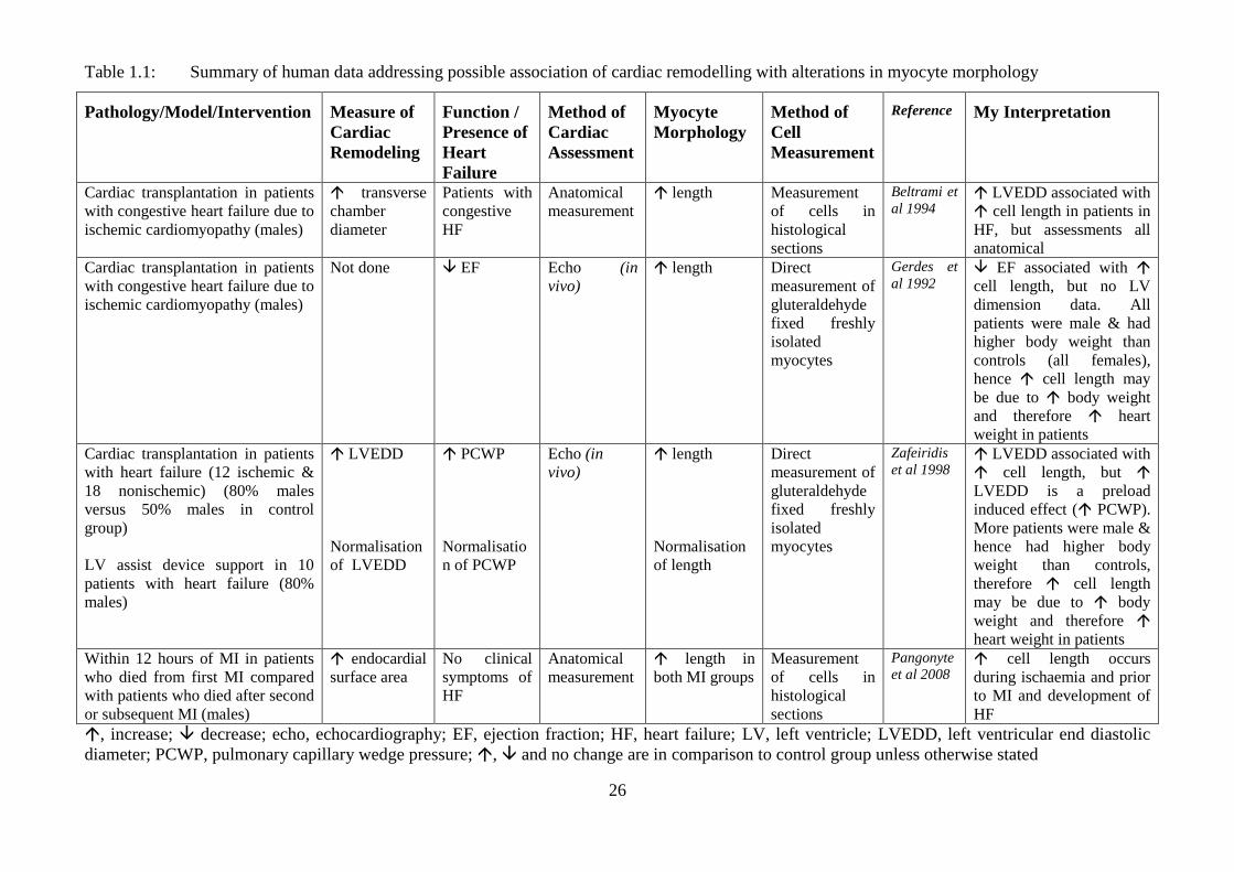
Table 1.1: Summary of human data addressing possible association of cardiac remodelling with alterations in myocyte morphology
Pathology/Model/Intervention Measure of
Cardiac
Remodeling
Function /
Presence of
Heart
Failure
Method of
Cardiac
Assessment
Myocyte
Morphology
Method of
Cell
Measurement
Reference My Interpretation
Cardiac transplantation in patients
with congestive heart failure due to
ischemic cardiomyopathy (males)
transverse
chamber
diameter
Patients with
congestive
HF
Anatomical
measurement
length
Measurement
of cells in
histological
sections
Beltrami et
al 1994 LVEDD associated with
cell length in patients in
HF, but assessments all
anatomical
Cardiac transplantation in patients
with congestive heart failure due to
ischemic cardiomyopathy (males)
Not done EF Echo (in
vivo)
length
Direct
measurement of
gluteraldehyde
fixed freshly
isolated
myocytes
Gerdes et
al 1992 EF associated with
cell length, but no LV
dimension data. All
patients were male & had
higher body weight than
controls (all females),
hence cell length may
be due to body weight
and therefore heart
weight in patients
Cardiac transplantation in patients
with heart failure (12 ischemic &
18 nonischemic) (80% males
versus 50% males in control
group)
LV assist device support in 10
patients with heart failure (80%
males)
LVEDD
Normalisation
of LVEDD
PCWP
Normalisatio
n of PCWP
Echo (in
vivo) length
Normalisation
of length
Direct
measurement of
gluteraldehyde
fixed freshly
isolated
myocytes
Zafeiridis
et al 1998 LVEDD associated with
cell length, but
LVEDD is a preload
induced effect ( PCWP).
More patients were male &
hence had higher body
weight than controls,
therefore cell length
may be due to body
weight and therefore
heart weight in patients
Within 12 hours of MI in patients
who died from first MI compared
with patients who died after second
or subsequent MI (males)
endocardial
surface area
No clinical
symptoms of
HF
Anatomical
measurement length in
both MI groups
Measurement
of cells in
histological
sections
Pangonyte
et al 2008 cell length occurs
during ischaemia and prior
to MI and development of
HF
, increase; decrease; echo, echocardiography; EF, ejection fraction; HF, heart failure; LV, left ventricle; LVEDD, left ventricular end diastolic
diameter; PCWP, pulmonary capillary wedge pressure; , and no change are in comparison to control group unless otherwise stated

27
diameter (Beltrami et al., 1994; Pangonyte et al., 2008; Zefeiridis et al., 1998). However, it
would be incorrect to draw a conclusion that adverse chamber remodelling (cardiac
dilatation) is associated with increases in cardiac myocyte length, from this data for a number
of reasons.
Firstly, in one these studies (Zefeiridis et al., 1998), echocardiography in vivo was used to
determine left ventricular end diastolic diameters and hence the cardiac chamber dimension
measurements were load dependent. As increases in pulmonary capillary wedge pressure
were noted in the patients in this study (Zefeiridis et al., 1998), the increased chamber
dimensions are likely to be due to increases in LV preload. In this regard, as discussed
previously, cardiac myocyte lengthening occurs as a consequence of the stretching of cardiac
myocytes during increased preloads (Ferrari et al., 2009). Indeed, Zefeiridis et al. (1998)
showed a normalisation of both LV end diastolic diameter and cardiac myocyte length, when
pulmonary capillary wedge pressure was normalised as a consequence of LV assist device
support. Left ventricular assist devices divert blood from the left atrium to the aorta, thereby
removing the preload and afterload to the left ventricle. Hence these devices are used to
provide haemodynamic support for patients with end-stage heart failure awaiting
transplantation.
Secondly, in the study of Beltrami et al. (1994), the hearts were collected at autopsy for the
control group and during cardiac transplantation for the heart failure group. Measurements of
cardiac chamber dimensions and cardiac myocyte length were made anatomically; however
no mention was made as to whether the hearts were arrested in diastole or systole. It is
possible that during transplantation the hearts were arrested in diastole, as this was part of the
surgical procedure at the time; whereas in the control group the phase of the cardiac cycle at
the time of arrest was unknown as 70% of the control hearts were obtained from individuals
in whom death was sudden due to traumatic injury. As the phase of the cardiac cycle

28
determines cardiac chamber dimensions and cardiac myocyte length, possible differences
between the heart failure and control groups in the phase of the cardiac cycle at the time of
cardiac arrest may have accounted for the differences in cardiac chamber diameter and
cardiac myocyte length between these two groups.
Thirdly, in some studies the increased cardiac myocyte length in patients compared to
controls could be as a consequence of the differences in body weight and hence LV weight
between these two groups (Gerdes et al., 1992; Zafeiridis et al., 1998). Indeed, in one study
all patients in heart failure were males; whereas all healthy controls were females (Gerdes et
al., 1992). As the males had greater body weights than the controls (Gerdes et al., 1992), the
increased cardiac myocyte length may be attributed to the greater body size in the males
rather than to the presence of heart failure. Similarly, in another study the increased cardiac
myocyte length in the patients compared to the controls may in part be attributed to the
greater proportion of males in the heart failure group compared to the control group
(Zafeiridis et al., 1998).
Lastly, in a study assessing cardiac myocyte length and cardiac dimensions within 12 hours
of death due to a first myocardial infarction compared to within 12 hours of death from a
second or subsequent myocardial infarction, increases in cardiac myocyte length were noted
in both groups with myocardial infarction compared to a control group who died from non-
cardiovascular causes (Pangonyte et al., 2008). Importantly neither of the myocardial
infarction groups had clinical symptoms of heart failure. Therefore, from this study it can be
concluded that increases in cardiac myocyte length occur during ischaemia before myocardial
infarction and the development of heart failure.
Hence, from human studies published to date, the potential relationship between adverse
chamber remodelling (cardiac dilatation) and cardiac myocyte length is unclear.

29
1.4.3.1.2 Data from Animal Experimental Models:
Previous data on the possible association between cardiac myocyte length and cardiac
dimensions obtained from animal experimental models are summarised in Table 2. Although
a number of studies have attempted to address the question of the relationship between
cardiac myocyte length and cardiac dimensions in various animal experimental models; the
data is controversial. Some studies show that increases in cardiac myocyte length are
associated with increases in cardiac chamber dimensions or heart failure (Gerdes et al., 1996;
Gerdes et al., 2010; Janczewski et al., 2003; Kajstura et al., 1995; Schultz et al., 2007;
Tamura et al., 1998; Toischer et al., 2010; Wang et al., 1999); whereas a number of other
studies show no relationship (Li et al., 2010; Schultz et al., 2007; Tamura et al., 2000;
Yarbrough et al., 2010). As with the human studies, a number of possible reasons may
explain the controversial data.
One of the reasons for the contrasting findings is the use of load dependent measures of
cardiac chamber dimensions (echocardiography in vivo) in the presence of increased preloads
(Chen et al., 2010; Gerdes et al., 2010; Kajstura et al., 1995; Tamura et al., 1998; Wang et
al., 1999; Yarbrough et al., 2010). As discussed above with respect to human studies, the
increased cardiac chamber diameters and cardiac myocyte length noted in these studies may
be indicative of the increased preloads rather than the presence of adverse chamber
remodelling (cardiac dilatation).
Secondly, in a number of studies showing an association between cardiac myocyte length and
cardiac chamber dimension, the experimental animals (spontaneously hypertensive heart
failure, SHHF rats) were from 12 to 16 months older than the control animals (Gerdes et al.,
1996; Tamura et al., 1998). As LV weight increases with age in hypertensive rats (Gerdes et

30
Table 1.2: Summary of data from animal experimental models addressing possible association of cardiac remodelling with alterations in myocyte
morphology
Pathology/Model/Intervention Measure of
Cardiac
Remodeling
Function /
Presence of
Heart Failure
Method of
Cardiac
Assessment
Myocyte
Morphology
Method of Cell
Measurement
Reference My Interpretation
Dogs with pacing-induced heart
failure (gender not stated)
chamber
diameter
LVEDP,
LVSP
LV
dimensions of
cardiac rings
following
perfusion
fixation at
LVEDP
measured in
vivo
length Direct
measurement of
freshly isolated
myocytes
Kajstura et
al. 1995 As LVEDP in dogs with
pacing-induced HF, LV
dimensions likely to be due
to preload
Rats genetically predisposed to
developing heart failure with
increasing age (spontaneously
hypertensive heart failure, SHHF)
(females)
Not done Clinical signs
of HF
(dyspnoea,
cyanosis),
liver weight,
lung wet
weight, LV
systolic
pressure
Catheter plus
pressure
transducer (in
vivo)
length
Direct
measurement of
gluteraldehyde
fixed freshly
isolated
myocytes
Gerdes et al.
1996 cell length in rats in HF,
but these rats were 12
months older than rats not
in HF. As cardiac myocyte
length is strongly correlated
with age & LV weight, and
LV weight with age in
hypertensive rats; cell
length possibly due to
age & LV weight rather
than LV dimensions
Rats genetically predisposed to
developing heart failure with
increasing age (spontaneously
hypertensive heart failure, SHHF)
(females)
Not done No clinical
signs of HF
Echo (in vivo) length
Direct
measurement of
gluteraldehyde
fixed freshly
isolated
myocytes
Onodera et
al. 1998
(Gerdes
laboratory)
cell length with age
associated with heart
weight/body weight with
age in hypertensive rats
without HF; therefore
cell length possibly due to
age & heart weight rather
than LV dimensions

31
Pathology/Model/Intervention Measure of
Cardiac
Remodeling
Function /
Presence of
Heart Failure
Method of
Cardiac
Assessment
Myocyte
Morphology
Method of Cell
Measurement
Reference My Interpretation
Rats genetically predisposed to
developing heart failure with
increasing age (spontaneously
hypertensive heart failure, SHHF)
(females)
chamber
circumference
Clinical signs
of HF (eg.
dyspnoea,
ascites, pleural
effusion,
pericardial
effusion,
cyanosis)
Anatomical
measurement
of chamber
circumference
in formalin
fixed heart
slices
length
Direct
measurement of
gluteraldehyde
fixed freshly
isolated
myocytes &
myocytes
isolated from
formalin fixed
tissue using
potassium
hydroxide
Tamura et
al. 1998
(Gerdes
laboratory)
cell length in rats in HF,
but these rats were 16
months older than rats not
in HF. As cardiac myocyte
length is strongly correlated
with age & LV weight, and
LV weight with age in
hypertensive rats; cell
length possibly due to
age & LV weight rather
than LV dimensions
Constriction of thoracic aorta in
guinea pigs (males)
LVEDD
FS, EF,
LVEDP
Echo (in vivo) length Direct
measurement of
gluteraldehyde
fixed freshly
isolated
myocytes
Wang et al
1999
(Gerdes
laboratory)
As LVEDP in guinea
pigs with FS & EF,
LVEDD likely to be due to
preload
Rats genetically predisposed to
developing heart failure with
increasing age (spontaneously
hypertensive heart failure, SHHF)
(females)
LVEDD at
22 months
No change in
FS
Echo (in vivo) No change in
length
Direct
measurement of
gluteraldehyde
fixed freshly
isolated
myocytes
Tamura et
al. 2000
(Gerdes
laboratory)
Despite LVEDD at 22
months, no change in cell
length observed, hence
LV dimensions not
associated with cell
length
Angiotensin II type 1 receptor
blocker administration to SHHF
(females)
No change in
LVEDD
No change in
FS
Echo (in vivo) length cell length associated
with heart weight /body
weight but no change in
LVEDD, suggests that
cell length in SHHF is due
to heart weight rather
than LV dimensions

32
Pathology/Model/Intervention Measure of
Cardiac
Remodeling
Function /
Presence of
Heart Failure
Method of
Cardiac
Assessment
Myocyte
Morphology
Method of Cell
Measurement
Reference My Interpretation
Transgenic mice with cardiac
specific overexpression of tumour
necrosis alpha (results in the
development of HF)
(males compared to females)
LVEDD
only in males
FS Echo (in vivo) length in
males &
females
Direct
measurement of
freshly isolated
myocytes
Janczewski
et al 2003
In females, cell length
but no change in LVEDD,
hence cell length not
associated with LVEDD
Cardiomyopathic hamsters (male)
L-thyroxine administration
Diiodothyropropionic acid (thyroid
hormone analogue) administration
LVEDD
Normalisation
of LVEDD
EF
EF
Echo (in vivo) No change in
length
No change in
length
Direct
measurement of
gluteraldehyde
fixed freshly
isolated
myocytes
Kuzman et
al 2007
(Gerdes
laboratory)
Cell length not associated
with LVEDD
Spontaneously hypertensive heart
failure (SHHF) adult rats (females)
Long-term exercise (16 months) in
SHHF adult rats
LVEDD
LVEDD,
and > in non-
exercise SHHF
EF & FS
EF & FS,
and < in non-
exercise SHHF;
ascites in 42%
of rats
Echo (in
vivo); catheter
plus pressure
transducer (in
vivo)
length
length,
but not
different
from non-
exercise
SHHF
Direct
measurement of
gluteraldehyde
fixed freshly
isolated
myocytes
Schultz et al
2007
(Gerdes
laboratory)
LVEDD, EF & FS
associated with cell
length
Despite greater LVEDD,
EF, FS & presence of
ascites, no difference in cell
length compared to non-
exercise SHHF
Myocardial infarction (MI) via left
coronary artery ligation in adult rats
(males compared to females)
LVEDD FS & LV
systolic
pressure;
LVEDP
Echo (in
vivo); catheter
plus pressure
transducer (in
vivo)
length
Direct
measurement of
gluteraldehyde
fixed freshly
isolated
myocytes
Chen et al
2010
(Gerdes
laboratory)
LVEDD, FS, LVSP
& LVEDP associated
with cell length; but
LVEDD is a likely to be a
preload induced effect
Myocardial infarction (MI) via left
coronary artery ligation in adult mice
(males)
LVEDD
FS
Echo (in vivo) No change in
length
Direct
measurement of
freshly isolated
myocytes
Li et al 2010
LVEDD not associated
with cell length

33
Pathology/Model/Intervention Measure of
Cardiac
Remodeling
Function /
Presence of
Heart Failure
Method of
Cardiac
Assessment
Myocyte
Morphology
Method of Cell
Measurement
Reference My Interpretation
Intramyocardial delivery of
mesenchymal stem cells to mice
with MI
Prevention of
LVEDD
Prevention of
FS
Echo (in vivo) length Direct
measurement of
freshly isolated
myocytes
Li et al 2010
Normalisation of LVEDD
associated with cell
length
preload via aortocaval shunt in
adult mice (females)
LVEDD
No change in
FS
Echo (in vivo) length
Direct
measurement of
freshly isolated
myocytes
Toischer et
al 2010 cell length prior to FS
MI via ligation of circumflex
coronary artery in adult pigs (gender
not stated)
Caspase inhibition to pigs with MI
LVEDD
LVEDD, but
< in absence of
caspase
inhibition
LVEDP
LVEDP
Echo (in
vivo); catheter
plus pressure
transducer (in
vivo)
No change in
length
length
Direct
measurement of
myocytes from
formalin-fixed
tissue
Yarbrough
et al 2010 LVEDD & in HF (
LVEDP) but no change in
cell length
LVEDD & in HF (
LVEDP) but cell length
, increase; decrease; echo, echocardiography; EF, ejection fraction; FS, fractional shortening; HF, heart failure; LV, left ventricle; LVEDD, left
ventricular end diastolic diameter; LVEDP, left ventricular end diastolic pressure; LVSP, left ventricular systolic pressure; PCWP, pulmonary capillary
wedge pressure; PWTd, posterior wall thickness in diastole; SHHF, spontaneously hypertensive heart failure; , and no change are in comparison to
control group unless otherwise stated

34
al. 1996), and cardiac myocyte length is strongly associated with age in SHHF rats (Tamura
et al., 1998) and with LV weight (Campbell et al. 1991; Capasso et al. 1992), the increases in
cardiac myocyte length observed in the experimental groups may reflect age induced
changes, rather than an association with adverse chamber remodelling (cardiac dilatation) and
the development of heart failure.
Thirdly, data from various intervention studies failed to show changes in cardiac dimensions
in parallel with changes in cardiac myocyte length. In intervention studies where increases in
cardiac chamber diameter were prevented by thyroid hormone administration (Kuzman et al.,
2007) or intramyocardial delivery of mesenchymal stem cells (Li et al., 2010), either no
change (Kuzman et al., 2007) or even increases in cardiac myocyte length (Li et al., 2010)
were noted. Alternatively, in one study decreases in cardiac myocyte length were noted
subsequent to angiotensin II receptor blockade, but no changes in cardiac chamber
dimensions were noted (Tamura et al., 2000). Furthermore, in a study of the effects of long-
term exercise in rats with heart failure, further increases in cardiac chamber dimensions due
to exercise training were not accompanied by further increases in cardiac myocyte length
(Schultz et al., 2007). The results from the intervention studies discussed above, although
different from each other, do not show parallel changes in cardiac chamber dimensions and
cardiac myocyte length. Therefore the results of these studies do not support a role of changes
in cardiac myocyte length in cardiac dilatation. Indeed, if increases in cardiac myocyte length
were causally related to adverse cardiac remodelling, a reduction in cardiac myocyte length
should be accompanied by a decrease in cardiac chamber dimensions. Hence the data
provides no clear conclusion as to the possible role of cardiac myocyte length in adverse
chamber remodelling. In fact, it could be concluded that there is no causal relationship.

35
However, the data need to be interpreted with caution in view of the use of load dependent
measures of cardiac dimensions in these studies.
Lastly, few studies have made direct comparisons of animals with cardiac pathology which
have heart failure to animals with the same cardiac pathology but without heart failure. The
only studies which have used this study design have been done in SHHF rats (Gerdes et al.,
1996; Tamura et al., 1998 and 2000). However, in these studies the rats in heart failure were
from 12 to 22 months older than the rats not in heart failure. As discussed above, as cardiac
myocyte length is associated with age (Tamura et al., 1998), the increased cardiac myocyte
length may reflect increased age rather than increased cardiac dimensions. Indeed, a study by
Onodera et al. (1998) showed that cardiac myocyte lengthening increased with age and
occurred well before the development of heart failure in SHHF rats. Although the study by
Onodera et al. (1998) suggests that changes in cardiac myocyte length are not responsible for
the development of heart failure, no measurements of cardiac dimensions were made in this
study. To my knowledge no study to date has compared age-matched animals with the same
cardiac pathology which have heart failure to those without heart failure.
Hence the data obtained from animal experimental models to date are equally confusing and
hence the question of the relationship between cardiac myocyte length and adverse cardiac
chamber remodelling remains unresolved.
1.5 PROBLEM STATEMENT AND STUDY OBJECTIVES
In summary, despite a plethora of studies aimed to address the relationship between cardiac
dimensions and cardiac myocyte length, to date the data are controversial (Tables 1.1 and 1.2,

36
and Figure 1.4). As discussed the data are limited by the use of load dependent measures of
cardiac dimensions; the comparison of hearts from humans of different gender and body size;
the comparison of hearts from rat groups of different age and body size; and a failure to make
direct comparisons between age-matched animals with cardiac pathology who are in heart
failure and those who are not in heart failure. Hence, the role of cardiac myocyte length in
adverse cardiac remodelling needs to defined using load independent measures of chamber
dimensions and in age-matched animals where direct comparisons are made between animals
with cardiac pathology who are in heart failure and those who are not in heart failure.
To address the limitations discussed above, I chose to study two models of cardiac dilatation
in pressure-overload induced cardiac hypertrophy in rats. The first model of cardiac dilatation
was a natural progression model in that it involved the use of 23 month old SHR. In SHR
older than 21 months of age, left ventricular hypertrophy is noted to progress to left
ventricular decompensation, dilatation and heart failure in approximately half of the rats
(Bing et al. 1995; Norton et al. 1997; Tsotetsi et al. 2001). The second model of cardiac
dilatation, a pharmacological model, was induced in 14 month old SHR by chronic beta-
adrenoreceptor activation [daily administration of the beta-adrenergic agonist isoproterenol
(ISO) for a period of 4.5 months]. Chronic beta-adrenoreceptor activation in SHR, has been
shown enhance the progression from compensated left ventricular hypertrophy to left
ventricular dilatation (Badenhorst et al. 2003b; Gibbs et al. 2004). In both of these models the
rats in heart failure were age-matched to the rats not in heart failure and comparisons were
made with an age-matched healthy control group. Importantly, in my studies I used load
independent measures (determination of filling pressures at controlled volumes) to define
cardiac dilatation.

37
Figure 1.4 Schematic representation of the factors known to contribute to cardiac
dilatation; whereas the role of cardiac myocyte lengthening is still
controversial.

38
The studies in this dissertation aimed to determine the role that cardiac myocyte lengthening
in the development of adverse chamber remodelling (cardiac dilatation) that occurs in
pressure-overload states in rats.
The specific objectives of my studies were:
to compare the length of cardiac myocytes isolated from SHR rats in heart failure to
those isolated from SHR rats without heart failure (natural progression model)
to compare the length of cardiac myocytes isolated from SHR rats receiving
isoproterenol (rats in heart failure) to those isolated from SHR rats not receiving
isoproterenol (rats without heart failure) (pharmacological model)
to compare the cardiac dimensions (measured using load independent measures), of
SHR rats in heart failure to those isolated from SHR rats without heart failure
(natural progression model)
to compare the cardiac dimensions (measured using load independent measures), of
SHR rats receiving isoproterenol (rats in heart failure) to those isolated from SHR
rats not receiving isoproterenol (rats without heart failure) (pharmacological model)
to determine the relationship between cardiac myocyte length and cardiac dimensions
measured using load independent measures (both natural progression model and
pharmacological model)

39
CHAPTER 2
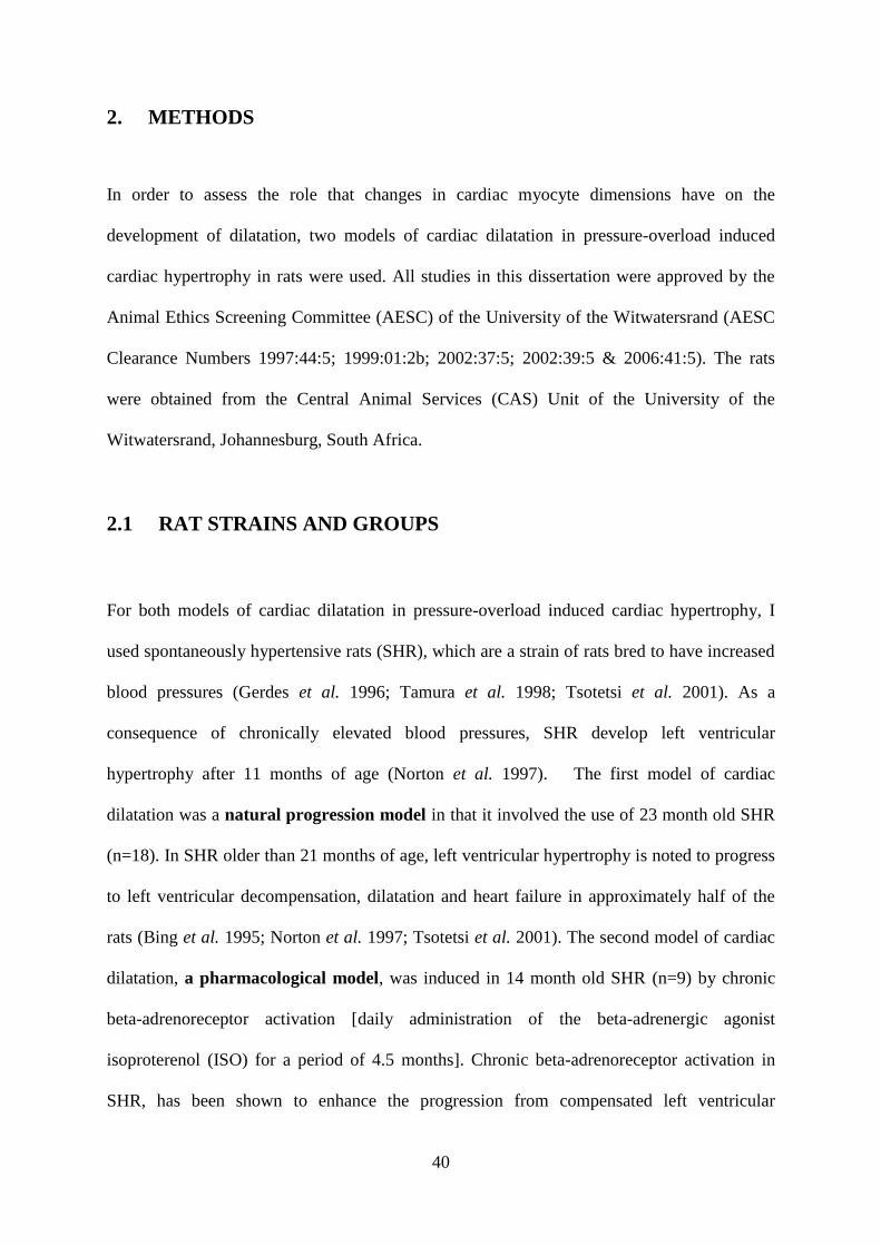
40
2. METHODS
In order to assess the role that changes in cardiac myocyte dimensions have on the
development of dilatation, two models of cardiac dilatation in pressure-overload induced
cardiac hypertrophy in rats were used. All studies in this dissertation were approved by the
Animal Ethics Screening Committee (AESC) of the University of the Witwatersrand (AESC
Clearance Numbers 1997:44:5; 1999:01:2b; 2002:37:5; 2002:39:5 & 2006:41:5). The rats
were obtained from the Central Animal Services (CAS) Unit of the University of the
Witwatersrand, Johannesburg, South Africa.
2.1 RAT STRAINS AND GROUPS
For both models of cardiac dilatation in pressure-overload induced cardiac hypertrophy, I
used spontaneously hypertensive rats (SHR), which are a strain of rats bred to have increased
blood pressures (Gerdes et al. 1996; Tamura et al. 1998; Tsotetsi et al. 2001). As a
consequence of chronically elevated blood pressures, SHR develop left ventricular
hypertrophy after 11 months of age (Norton et al. 1997). The first model of cardiac
dilatation was a natural progression model in that it involved the use of 23 month old SHR
(n=18). In SHR older than 21 months of age, left ventricular hypertrophy is noted to progress
to left ventricular decompensation, dilatation and heart failure in approximately half of the
rats (Bing et al. 1995; Norton et al. 1997; Tsotetsi et al. 2001). The second model of cardiac
dilatation, a pharmacological model, was induced in 14 month old SHR (n=9) by chronic
beta-adrenoreceptor activation [daily administration of the beta-adrenergic agonist
isoproterenol (ISO) for a period of 4.5 months]. Chronic beta-adrenoreceptor activation in
SHR, has been shown to enhance the progression from compensated left ventricular

41
hypertrophy to left ventricular dilatation (Badenhorst et al. 2003b; Gibbs et al. 2004). In this
arm of the study, an SHR group receiving twice daily administration of the vehicle of
isoproterenol (saline) for a period of 4.5 months (SHR-Control, n=9), served as the ‘non-
failure’ group (see Figure 2.1 below).
The control group for this study comprised of 23 month old Wistar Kyoto (WKY) rats (n = 9;
Kleinterfarm Madorin Ltd, Germany). These rats are the genetic controls for SHR (Tsotetsi et
al. 2001). The WKY control group acted as such for both the natural progression model as
well as the pharmacological model of the study (see Figure 2.1 below), as these rats are
normotensive, and are not known to develop heart failure. Although the WKY control group
of rats were older (23 months old) than the SHR-Control and SHR-ISO rats (both 18.5
months old), in normotensive rats in which body weight and heart weight are stable, age has
been shown to have no impact on myocyte dimensions (Bai et al. 1990; Onodera et al. 1998;
Tamura et al. 1998). We have previously reported no differences in body weight or left
ventricular weight in 21-22 month old WKY compared to 12 month old WKY (Badenhorst et
al. 2003b). Moreover, as it has previously been shown that chronic administration of a beta-
adrenergic agonist to the WKY strain of rats does not induce cardiac dilatation (Badenhorst et
al. 2003b; Gibbs et al. 2004) I did not include a WKY group receiving chronic beta-
adrenergic agonist administration.
The rats were housed in pairs in cages in the CAS Unit of the University of the
Witwatersrand and given food and water ad libitum until termination at 18.5 or 23 months of
age.

42
Figure 2.1 Flow chart detailing the groups of rats
.
The control group acted as such for both models. In the natural progression model, rats found
to have pathological features of cardiac failure (see 2.2.1 below) were assigned to the failure
group, and rats without any features of cardiac failure were assigned to the non-failure group.
In the pharmacological model, the beta-adrenergic agonist, isoproterenol (or its vehicle,
saline) was administered from 14 months of age until termination at 18.5 months of age.
ISO, isoproterenol; SHR, spontaneously hypertensive rats; WKY, Wistar-Kyoto rats.
Non - (n = 9)
Rats
18 months old at termination
Pathological features
of heart failure
Vehicle administered daily
for 4 ½ months
Pharmacological
Model
Natural Progression
Model Control
SHR
(n = 18)
WKY
(n = 9)
SHR (13 months)
(n = 18)
Vehicle + Isoproterenol
4 ½ months
Failure (n = 9)
WKY (n = 9)
SHR - Control (n = 9)
SHR - (n = 9)
Non-Failure (n = 9)
Rats
23 months old at termination 18.5 months old at termination
Assessment for pathological
features of heart failure
Vehicle
administered daily
for 4.5 months
Pharmacological
Model
Natural Progression
Model Control
SHR (14 months)
(n = 18)
WKY (14 months)
(n = 9)
SHR (14 months)
(n = 18)
Failure (n = 9)
WKY (n = 9)
SHR - Control (n = 9)
SHR-ISO (n = 9)
Isoproterenol
administered daily
for 4.5 months

43
2.1.1 Natural Progression Model
Eighteen 14 month old SHR (OLAC, UK) were housed in the CAS Unit of the University of
the Witwatersrand. At 23 months of age the rats were terminated. At the time of termination,
the rats were assessed for signs and symptoms of cardiac failure (see 2.2.1 below). Rats noted
to have pathological features of cardiac failure were assigned to the failure group (SHR
Failure, n=9), and rats without any features of cardiac failure were assigned to the non-failure
group (SHR Non-Failure, n=9).
2.1.2 Pharmacological Model
Eighteen 14 month old SHR (OLAC, UK) were randomly assigned to two groups of nine rats
each. One group (SHR-ISO) received a twice daily intraperitoneal injection of the beta-
adrenergic agonist isoproterenol (ISO) (Imuprel, Adcock Ingram), at a concentration of 0.02
mg/kg/injection, in a volume of approximately 0.2 ml for 4.5 months. The other group (SHR-
Control) received a twice daily injection of the vehicle of ISO (0.2 ml of 0.9% saline) for 4.5
months. The rats were terminated at 18.5 months of age.
2.2 SYSTOLIC BLOOD PRESSURE
Non-invasive systolic blood pressures (SBP) were assessed in all rats prior to the end of the
study using a tail-cuff technique. In the pharmacological model, these measurements were
obtained at least 24-hours after the last dose of ISO. To familiarise the rats to the procedure,
rats were placed in restrainers, the tail pre-warmed and the tail cuff inflated every 15 minutes
for an hour a day on five separate days prior to the measurements. To determine SBP, rat tails
were pre-warmed until the tail artery pulse could be detected with a photoelectric diode
coupled to a Model 29 pulse amplifier (IITC Inc.). A tail-cuff coupled to a pressure
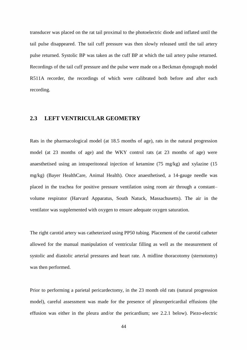
44
transducer was placed on the rat tail proximal to the photoelectric diode and inflated until the
tail pulse disappeared. The tail cuff pressure was then slowly released until the tail artery
pulse returned. Systolic BP was taken as the cuff BP at which the tail artery pulse returned.
Recordings of the tail cuff pressure and the pulse were made on a Beckman dynograph model
R511A recorder, the recordings of which were calibrated both before and after each
recording.
2.3 LEFT VENTRICULAR GEOMETRY
Rats in the pharmacological model (at 18.5 months of age), rats in the natural progression
model (at 23 months of age) and the WKY control rats (at 23 months of age) were
anaesthetised using an intraperitoneal injection of ketamine (75 mg/kg) and xylazine (15
mg/kg) (Bayer HealthCare, Animal Health). Once anaesthetised, a 14-gauge needle was
placed in the trachea for positive pressure ventilation using room air through a constant–
volume respirator (Harvard Apparatus, South Natuck, Massachusetts). The air in the
ventilator was supplemented with oxygen to ensure adequate oxygen saturation.
The right carotid artery was catheterized using PP50 tubing. Placement of the carotid catheter
allowed for the manual manipulation of ventricular filling as well as the measurement of
systolic and diastolic arterial pressures and heart rate. A midline thoracotomy (sternotomy)
was then performed.
Prior to performing a parietal pericardectomy, in the 23 month old rats (natural progression
model), careful assessment was made for the presence of pleuropericardial effusions (the
effusion was either in the pleura and/or the pericardium; see 2.2.1 below). Piezo-electric

45
ultra-sonic transducers, attached to a previously validated apparatus (Woodiwiss & Norton
1995; Trifunovic et al. 1995), were then placed across the short axis of the beating left
ventricle (LV). These transducers emit ultra-sonic waves through one transducer (anteriorly,
B in Figure 2.2) and receive the ultra-sonic waves through the other (posteriorly, C in Figure
2.2). The distance between the transducers was measured using a calibrated sonomicrometer
(Triton Technology). The transducers were used to measure the external diameter of the left
ventricle through the phases of the cardiac cycle over a range of left ventricular filling
pressures (upper panel Figure 2.3).
The filling pressures were measured by means of the insertion of a 21-gauge needle through
the apex of the heart (A in Figures 2.2 and 2.3). The 21-gauge needle was attached to a
saline-filled PP25 polyethylene catheter which was coupled to a Gould P50 pressure
transducer. The fluid filled catheter had amplitude-frequency responses which were uniform
to 10Hz (Norton et al. 1997). Filling volumes were modified by the infusion of an iso-oncotic
solution via the carotid catheter (Badenhorst et al. 2003b; Norton et al. 1997; Woodiwiss &
Norton 1995; Tsotetsi et al. 2001). The frequent occurrence of extra systolic beats at filling
pressures below 2 mm Hg prevented the collection of data at pressures below 2 mm Hg. The
left ventricular end diastolic (LVED) diameters were therefore measured over a range of
filling pressures between 2 mm Hg and 9 mm Hg.

46
Figure 2.2 Intraventricular pressure monitoring and piezo-electric ultrasonic
transducers
Diagrammatic representation of the apical catheter (A) and the apparatus used for the
placement of the piezo-electric ultrasonic transducers (B and C) across the short axis of the
left ventricle. A, apical catheter attached to 21-gauge needle; B, anterior (transmitting)
transducer; C, posterior (receiving) transducer.
A
B
C

47
Figure 2.3 Example of recordings of left ventricular external diameter
measurements
Example of recordings of LV external diameter (LVExtD) obtained from the piezo-electric
ultrasonic transducers (B and C in figure 2.2) and of LV diastolic pressure obtained from the
apical catheter (A in figure 2.2) inserted into the left ventricle.
LVEDP, LV end diastolic pressure

48
All recordings were made on a Beckman Dynograph Recorder (type R511A). Left ventricular
end diastolic radius (LVEDr) was calculated from the LVED external diameter
measurements, using the following equation (Weber et al. 1988),
LVED radius =
where,
LV wall volume = 0.943 x LV wet weight
and where 0.943 represents the density of cardiac tissue (Tsotetsi et al. 2001; Woodiwiss and
Norton 1995).
Left ventricular end diastolic wall thickness (LVEDh), was then determined using the
equation,
LVEDh = [(LVED external diameter-2 x LVEDr]/2
(Tsotetsi et al. 2001; Woodiwiss & Norton 1995); and from these measurements, LVED wall
thickness to radius ratio (h/r) was calculated.
Using these values LVEDP-LVEDr and LVEDP-LVEDh/r relations were constructed in
order to assess left ventricular geometry.
2.3.1 Identification of Failure and Non-failure Rats
In the natural progression model, rats were classified as in heart failure based upon the
presence of pleuropericardial effusions and / or atrial thrombi. As stated above (see 2.2), at
the time of thoracotomy and prior to pericardectomy careful assessment was made to identify
3 ] Volume [¾ wall LV -
3 ] diameter / 2 external [LVED π

49
the presence or absence of pleuropericardial effusions. In addition, the presence or absence of
atrial thrombi was identified at post-mortem (see 2.3 below).
Those animals that were found to have one or more of these pathological features of heart
failure were assigned to the ‘SHR-Failure’ group (see Figure 2.1). Those rats that did not
exhibit any pathological features of heart failure were assigned to the ‘SHR-Non-Failure’
group (see Figure 2.1). In order to confirm the presence of heart failure wet liver weights
were determined in the SHR rats in the natural progression model and in the WKY control
rats.
2.4 TISSUE SAMPLING
Once the collection of data to assess left ventricular geometry had been completed, a high
dose (16mM) of potassium chloride (Hearse et al. 1975) was infused via the catheter into the
carotid artery, hence arresting the heart in diastole. The heart was then removed, blotted dry
and weighed. The right ventricular free wall was dissected away from the left ventricle and
the left and right ventricles were weighed. During the dissection of the heart, the atria were
removed and carefully inspected for the presence of thrombi (one of the features of heart
failure).
Samples from the left ventricle were stored at –70 ˚C for later analysis. The samples were
carefully labelled so that cardiac myocytes could be isolated from the same region of each
heart for accurate comparisons.

50
2.5 MYOCYTE ISOLATION
Cardiac myocytes were isolated from the frozen tissue samples using a modification of a
technique previously described (Diffee & Nagle 2003; Gerdes et al. 1998; Tamura et al.
1998). A sharp blade was used to carefully section off a 20 mg (wet weight, for light
microscopy) or 100 mg (wet weight, for flow cytometry) piece of the LV posterior wall from
the same region of each rat heart. Each piece of LV tissue was of full thickness, in that it
extended from epicardium to endocardium.
Calcium-free physiological saline solution (PSS) was made for the cardiac myocyte isolation
process. PSS consisted of NaCl 120 mmol, KCl 10 mmol, KH2PO4 1.2 mmol, MgCl2 2.6
mmol, glucose 10 mmol, taurine 20 mmol, pyruvate 6.2 mmol and HEPES (4-(2-
hydroxyethyl)-piperazine-1-ethanesulfonic acid hemisodium salt) 4.8 mmol dissolved in 100
ml of distilled water (all chemicals obtained from Sigma Chemical Co., St. Louis, MO).
100ml of PSS was prepared for every 20mg of LV tissue. The PSS solution was titrated with
10 mM NaOH droplets until the pH of the solution was equal to a pH of 7.40 (physiological
pH). The pH was measured using a pH meter (Beckman Φ32 pH meter, Beckman
Instruments, Inc. USA). For every 20 mg of LV tissue, 50 ml of incubation solution and 50
ml of wash solution were prepared as described below.
The incubation solution consisted of 15 mg Collagenase Type 2 (Worthington Biochemical
Corporation, Lakewood NJ, USA) at a concentration of 317 U/mg, 14mg of Hyaluronidase
(Worthington Biochemical Corporation, Lakewood NJ, USA) at a concentration of 581
U/mg, 125 mg of bovine serum albumin (Fraction V, Sigma Chemical Co., St. Louis, MO)
and 0.0227 ml of 110 mM CaCl2 dissolved in 50 ml of PSS. The CaCl2 (Sigma Chemical

51
Co., St. Louis, MO) was prepared by dissolving 1.617g of CaCl2 into 100 ml of distilled
water.
The wash solution consisted of 0.045 ml of 110 mM CaCl2 and 250 mg of bovine serum
albumin.
The sample of LV tissue was minced and then placed in a tissue culture flask containing 10
ml of the incubation solution. The tissue culture flask was then placed in a preheated (37 ˚C)
oscillating water bath. The sample was oscillated in the water bath at a rate of 60 cycles per
minute (slow shake), for a period of ten minutes. The incubation solution was then aspirated
and a further 10 ml of fresh incubation solution was added to the flask with the tissue,
followed by a second ten minute period of oscillations at the same rate. The incubation was
then aspirated, and a 10 ml of fresh incubation solution was added. For the third period of
oscillations the cycle rate was increased to 120 cycles per minute for a period of 15 minutes.
Throughout the oscillation procedure the incubation solution containing the tissue was gassed
with 95% O2 – 5% CO2.
The flask was then removed from the water bath, two thirds of the incubation solution
aspirated and the remainder of the contents of the flask (incubation solution containing
digested tissue and tissue precipitants) were flushed through a 250-μm nylon mesh into a 15-
ml polypropylene test tube. The remaining tissue was then gently flushed through the nylon
mesh using ~10 ml of the wash solution. The sample was then left to settle for 15 minutes
after which the top two thirds of the supernatant was aspirated and ~10 ml of fresh wash
solution was added to the test tube. Following a second settling step of 15 minutes the
supernatant was aspirated without disturbing the pellet of cells. The cells were then diluted in

52
PSS containing 0.01% bromophenyl blue (The Coleman and Bell Co., USA) which stains
proteins, hence allowing for the easy identification of striations (sarcomere bands) within the
cardiac myocytes (Figure 2.4). The diluted cell sample was then aliquoted into tissue culture
dishes for analysis under light microscopy. Approximately 55-75% of the cells in the
suspension were rod-shaped cardiac myocytes (Figure 2.4).
2.6 LIGHT MICROSCOPY
LV tissue from all rats in both models (natural progession and pharmacological) plus LV
tissue from the WKY control group of the study, was assessed under light microscopy. Using
an inverted light microscope (A, Figure 2.5), images seen under the microscope were relayed
to a computer program using a digital camera (B, Figure 2.5) (Nikon Digital Sight DS-U1 &
DS-5M, Nikon Corporation, Japan). The cells were viewed at 400x magnification and cardiac
myocytes were selected based on the ability to see and count most of the sarcomere bands
from one end of the cardiac myocyte to the other (Figure 2.6). Between 25 and 30 cardiac
myocytes were selected from each LV tissue sample. Using the program, Act2U (Ver. 1.70
Nikon Corporation, Japan), the dimensions of each cardiac myocyte were measured. The
software was calibrated using a 0.01 x 100 = 1mm graticule (Graticules Ltd, Tonbridge, Kent,
England). All images were saved on hard drive before being assessed. Saving of the images
ensured that the cardiac myocyte images were not moving during the measurements and that
the measurements could be reassessed at anytime. The dimensions measured were the length
(A, Figure 2.6), width (B, Figure 2.6) and sarcomere length (distance between Z-bands) (C,
Figure 2.6) of each cardiac myocyte. The length to width ratio of each cardiac myocyte was
calculated. The 30-35 cardiac myocyte dimensions obtained were then averaged for each
sample of LV tissue. From these group means were then calculated.

53
Figure 2.4 Examples of isolated cardiac myocytes
An example of ~70% rod-shaped isolated cardiac myocytes (A) surrounded by cell debris,
non-viable cardiac myocytes or cells which are not cardiac myocytes (B) (upper panel) and a
rod-shaped isolated cardiac myocyte (lower panel)
A
A
B
B

54
B
AA
C
Figure 2.5 Photograph of digital camera and microscope
Photograph showing inverted microscope (A) to which digital camera (B) is attached. The
digital camera is coupled to a computer running Act2U software and an image of a cardiac
myocyte is displayed on the computer screen (C).

55
Figure 2.6 An example of striations of an isolated cardiac myocyte
Photograph of a cardiac myocyte showing striations and the measurement of length (A),
width (B) and sarcomere length (C).
A
B
C

56
2.7 FLOW CYTOMETRY
As using light microscopy, relatively few cardiac myocytes can be measured in comparison
to the total number of cardiac myocytes, I wished to also determine cardiac myocyte
dimensions using a procedure which allows for the measurement of vast numbers of cells
(~30 000 cells) in a short period of time. In order to do this I devised a method to measure
cardiac myocyte dimensions using flow cytometry based upon previously described methods
(Diez & Simm 1998; Nash et al. 1979). For the assessment of cardiac myocyte dimensions
using flow cytometry, cardiac myocytes were isolated from 100mg of LV tissue from each of
the rats (those in the natural progression model; those in the pharmacological model and the
WKY control rats) using the same procedure as described above (see 2.4). However, after
cardiac myocyte isolation, the cells were not stained with bromophenol blue; they were
suspended in 1ml of PSS and placed in 5ml Polystyrene round bottom tubes (Falcon, Becton-
Dickinson, USA) rather than in cell culture dishes.
Cardiac myocyte morphometry was assessed using a Becton-Dickinson flow cytometer
(FACSCalibur®, Franklin Lakes, NJ) (see Figure 2.7). Fluorescence in the FL1 channel was
quantified at a single cell level and data were analysed using Cellquest® version 3.3 (Becton
Dickinson software). The flow cytometer was preset to measure a maximum of 30 000 cells
per sample. The cardiac myocytes were not stained as they autofluoresce, hence allowing for
their detection and the assessment of their forward and side scatter. Side scatter gives an
indication of cell granularity (number of intracellular organelles and/or proteins) and forward
scatter a measure of relative cell size (based upon the shape of the cell and the degree of
folding of the cell membrane). A dot plot of side versus forward scatter (Figure 2.8) was then
drawn. The side versus forward scatter dot plot was then gated (A, Figure 2.8) to exclude
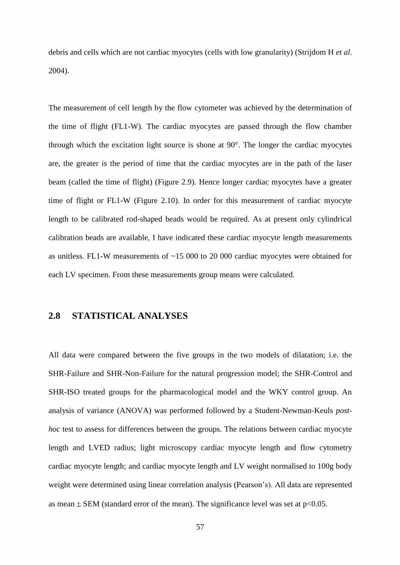
57
debris and cells which are not cardiac myocytes (cells with low granularity) (Strijdom H et al.
2004).
The measurement of cell length by the flow cytometer was achieved by the determination of
the time of flight (FL1-W). The cardiac myocytes are passed through the flow chamber
through which the excitation light source is shone at 90°. The longer the cardiac myocytes
are, the greater is the period of time that the cardiac myocytes are in the path of the laser
beam (called the time of flight) (Figure 2.9). Hence longer cardiac myocytes have a greater
time of flight or FL1-W (Figure 2.10). In order for this measurement of cardiac myocyte
length to be calibrated rod-shaped beads would be required. As at present only cylindrical
calibration beads are available, I have indicated these cardiac myocyte length measurements
as unitless. FL1-W measurements of ~15 000 to 20 000 cardiac myocytes were obtained for
each LV specimen. From these measurements group means were calculated.
2.8 STATISTICAL ANALYSES
All data were compared between the five groups in the two models of dilatation; i.e. the
SHR-Failure and SHR-Non-Failure for the natural progression model; the SHR-Control and
SHR-ISO treated groups for the pharmacological model and the WKY control group. An
analysis of variance (ANOVA) was performed followed by a Student-Newman-Keuls post-
hoc test to assess for differences between the groups. The relations between cardiac myocyte
length and LVED radius; light microscopy cardiac myocyte length and flow cytometry
cardiac myocyte length; and cardiac myocyte length and LV weight normalised to 100g body
weight were determined using linear correlation analysis (Pearson’s). All data are represented
as mean SEM (standard error of the mean). The significance level was set at p<0.05.

58
Figure 2.7 Flow cytometer
A photograph of the Becton-Dickinson flow cytometer (FACSCalibur®).

59
Figure 2.8 Example of flow cytometer dot plot
Dot plot of side scatter height (SSC-H) and forward scatter height (FSC-H) of cardiac
myocytes. The dot plot was gated (A) from a SSC-H of above 200 in order to exclude debris
and cells which are not cardiac myocytes [cells with low granularity (SSC-H)].
A

60
1 2 3 4
5
1 2 3 4 5
Time of flight
(indicative of cell length as longer cells
spend more time in the path of the laser beam)
A
BA
Figure 2.9 Diagrammatic representation of a cardiac myocyte in the path of the flow
cytometer laser beam
Diagrammatic representation of a cardiac myocyte (A) in the path of the laser beam (B). As
indicated in the graph, the time of flight counts (y-axis) increase as the cardiac myocyte
passes from ~⅓ (1) to the whole (3) of the cell being in the path of the laser beam.
Counts

61
Figure 2.10 Plot of cardiac myocyte counts versus time of flight
Plot of counts versus time of flight (FL1-W) for cardiac myocytes from a normotensive WKY
rat (purple), a SHR-NF rat (light pink) and a SHR-F rat (dark pink).

62
CHAPTER 3

63
3.0 RESULTS
3.1 LEFT VENTRICULAR GEOMETRY
3.1.1 Natural Progression Model
3.1.1.1 Body and Tissue Weights and Blood Pressures
Both heart weight and LV weight were increased in the SHR-Failure and SHR-Non-Failure
groups when compared to the WKY control group (Table 3.1). However, the body weights of
the WKY control group were greater than those of both the SHR-Failure and the SHR-Non-
Failure groups. Nevertheless, LV weight standardised to 100g body weight remained
increased in both the SHR-Failure and the SHR-Non-Failure groups compared to the WKY
control group (Table 3.1). Consistent with the increased heart and LV weights, both SHR
groups had increased SBP compared to the WKY group (Table 3.1).
Despite similar body weights; heart weight, LV weight and LV weight normalised to 100g
body weight ratio were greater in the SHR-Failure group compared to the SHR-Non-Failure
group. Consistent with the presence of heart failure, both RV weight and wet liver weight
were increased in the SHR-Failure group compared to the SHR-Non-Failure group and the
WKY control group (Table 3.1).
3.1.1.2 LV Dimensions
The LV end diastolic radius of the SHR-Failure group was significantly greater than that of
the SHR-Non-Failure group throughout the measured LV end diastolic pressure range of 2 –
9 mm Hg (Figure 3.1, upper panel), but was only greater than that of the WKY control group
at an LV end diastolic pressure of 9 mm Hg. Bearing in mind the increased body weight in
the WKY control group, the LV end diastolic radius was normalised to 100g body weight.
Throughout the measured LV end diastolic pressure range of 2-9 mm Hg, the LV end
diastolic radius normalised to 100g body weight was greater in the SHR-Failure group

64
Table 3.1 Body and tissue weights in the natural progression model and in the pharmacological model
Normotensive control Natural progression model Pharmacological model
WKY SHR-Non-Failure SHR-Failure SHR-Control SHR-ISO
Number 9 9 9 9 9
Body weight (g) 418±6 344±9*** 336±8*** 360±6*** 353±10***
Heart weight (g) 1.35±0.03 1.75±0.03*** 1.96±0.03***†‡ 1.67±0.07** 2.01±0.13***##§
LV weight (g) 0.99±0.02 1.32±0.02*** 1.50±0.02***†‡ 1.29±0.06*** 1.53±0.11***#§
LV weight / 100g body weight 0.238±0.006 0.383±0.009*** 0.446±0.011***†‡ 0.359±0.013*** 0.437±0.034***##§
RV weight (g) 0.36±0.01 0.41±0.01 0.48±0.01***††‡‡ 0.38±0.02 0.48±0.02***###§§
Liver weight (g) 13.10±0.38 13.48±0.48 15.41±0.75*† - -
Systolic blood pressure (mmHg) 128±8 188±7*** 190±8*** 186±6*** 188±9***
Values are mean ± SEM. The WKY normotensive group was the control group for both models.
*p<0.05, **p<0.01, ***p<0.001 compared to the WKY control group; †p<0.05, ††p<0.001 compared to the SHR-Non-Failure group; #p<0.05,
##p<0.01, ###p<0.001 compared to the SHR-Control group; ‡p<0.05, ‡‡p<0.01 versus SHR-Control group; §p<0.05, §§p<0.01 versus SHR-
Non-Failure group.

65
Figure 3.1 Left ventricular end diastolic radius – pressure relations in the natural
progression model
Left ventricular (LV) end diastolic radius – pressure relations (upper panel) and these
relations after normalising LV radius to 100g body weight (BW) (lower panel) for the three
groups in the natural progression model.
*p<0.05, **p<0.01, ***p<0.001 compared to the WKY control group; †p<0.05, ††p<0.01,
†††p<0.001 compared to the SHR-Non-Failure group.

66
compared to those of the SHR-Non-Failure group and the WKY control group (Figure 3.1,
lower panel). The increased LV end diastolic radius normalised to 100g body weight in the
SHR-Failure group is indicative of the presence of LV dilatation.
Throughout the measured LV end diastolic pressure range of 2-9 mm Hg, the LV end
diastolic wall thickness to radius ratio was higher in the SHR-Non-Failure group compared to
that of the WKY control group, consistent with compensatory concentric geometry (Figure
3.2). Moreover the LV end diastolic wall thickness to radius ratio in the SHR-Failure group
was decreased compared to that of the SHR-Non-Failure group and was no different to that of
the WKY controls (Figure 3.2). The decreased LV end diastolic wall thickness to radius ratio
in the SHR-Failure group is consistent with the presence of LV decompensation and
dilatation.
3.1.2 Pharmacological Model
3.1.2.1 Body and Tissue Weights
Body weight was lower but heart weight and LV weight were greater in the SHR-Control and
SHR-ISO groups when compared to the WKY control group (Table 3.1). In addition, the
SHR-ISO group had significantly greater heart weight and LV weight than the SHR-Control
group (Table 3.1). Furthermore, the LV weight normalised to 100g body weight of the SHR-
Control and SHR-ISO groups was greater than that of the WKY control group. Moreover, the
LV weight normalised to 100g body weight of the SHR-ISO group was greater than that of
the SHR-Control group (Table 3.1). In the SHR-ISO group, RV weight was increased
compared to both the SHR-Control and the WKY groups, consistent with the presence of LV
failure in the SHR-ISO group.

67
Figure 3.2 Left ventricular end diastolic wall thickness to radius ratio – pressure
relations in the natural progression model
Left ventricular (LV) end diastolic wall thickness to radius ratio – pressure relations for the
three groups in the natural progression model.
*p<0.05, **p<0.01 compared to the WKY control group; †p<0.05, ††p<0.01 compared to the
SHR-Non-Failure group.

68
3.1.2.2 LV Dimensions
The LV end diastolic radius of the SHR-ISO group were increased compared to those of both
the WKY control and the SHR-Control groups throughout the measured LV end diastolic
pressure range of 2 – 9 mm Hg (Figure 3.3, upper panel), indicative of the presence of LV
dilatation in the SHR-ISO group. Similarly, the LV end diastolic radius normalised to 100g
body weight was increased in the SHR-ISO group compared to both the SHR-Control and the
WKY control groups throughout the measured LV end diastolic pressure range of 2-9 mm Hg
(Figure 3.3, lower panel). No differences were noted between the SHR-Control and WKY
control groups.
Indicative of the presence of compensatory concentric geometry, the LV end diastolic wall
thickness to radius ratio at LV end diastolic pressures of 2-6 mm Hg was increased in the
SHR-Control group (Figure 3.4). In comparison, at LV end diastolic pressures of 2-6 mm Hg,
the LV end diastolic wall thickness to radius ratio in the SHR-ISO group was lower than that
of the SHR-Control group and was no different from that of the WKY control group (Figure
3.4). The decreased LV end diastolic wall thickness to radius ratio in the SHR-ISO group is
indicative of LV decompensation and dilatation.
3.2 MYOCYTE DIMENSIONS
3.2.1 Natural Progression Model
3.2.1.1 Light microscopy
There was a significant increase in the length of the cardiac myocytes of the hypertensive
groups (SHR-Failure and SHR-Non-Failure groups) when compared to the normotensive
(WKY) group (Figure 3.5, upper panel). Importantly, there was no difference in the cardiac
myocyte lengths between the two hypertensive groups. Hence, despite the presence of LV

69
Figure 3.3 Left ventricular end diastolic radius – pressure relations in the
pharmacological model
Left ventricular (LV) end diastolic radius – pressure relations (upper panel) and these
relations after normalising LV radius to 100g body weight (BW) (lower panel) for the three
groups in the pharmacological model.
*p<0.05, **p<0.01, ***p<0.001 compared to the WKY control group; ##p<0.01, ###p<0.001
compared to the SHR-Control group.

70
Figure 3.4 Left ventricular end diastolic wall thickness to radius ratio – pressure
relations in the pharmacological model
Left ventricular (LV) end diastolic wall thickness to radius ratio – pressure relations for the
three groups in the pharmacological model.
*p<0.05, **p<0.01 compared to the WKY control group; #p<0.05, ##p<0.01 compared to the
SHR-Control group.

71
dilatation, the SHR-Failure rats had cardiac myocytes of similar length to those of the SHR-
Non-Failure rats (absence of LV dilatation) (Figure 3.5, upper panel). Consistent with an
increased cardiac myocyte length in the hypertensive groups, a right shift in the frequency
distribution of cardiac myocyte lengths was noted in the SHR-Non-Failure and SHR-Failure
groups in comparison to the WKY control group (Figure 3.5, lower panel).
No differences were noted for either cardiac myocyte width (Figure 3.6, upper panel) or the
cardiac myocyte length to width ratio (Figure 3.6, lower panel) between the SHR-Failure,
SHR-Non-Failure and WKY groups. In addition no differences in sarcomere length (μm)
(WKY: 1.87±0.04; SHR-Non-Failure: 1.99±0.04; SHR-Failure: 2.00±0.04) or sarcomere
number (WKY: 45±2; SHR-Non-Failure: 46±2; SHR-Failure: 46±2) were noted between the
three groups.
3.2.1.2 Flow Cytometry
The data collected from the same rat hearts using flow cytometry indicated a similar pattern
as that shown with light microscopy. The length (FL1W) of the WKY cardiac myocytes was
significantly decreased as compared to that of the two hypertensive groups, SHR-Failure and
SHR-Non-Failure (Figure 3.7). Moreover, no differences in cardiac myocyte length (FL1W)
were noted between the SHR-Failure and the SHR-Non-Failure groups (Figure 3.7),
indicating that despite differences in LV dimensions (Figures 3.1 and 3.2), no differences in
cardiac myocyte length (FL1W) were noted.

72
Figure 3.5 Cardiac myocyte lengths and frequency distribution of cardiac myocyte
lengths in the natural progression model
Cardiac myocyte lengths (upper panel) and frequency distribution of cardiac myocyte lengths
(lower panel) as assessed by light microscopy in the three groups of the natural progression
model.
***p<0.001 compared to the WKY control group.

73
Figure 3.6 Cardiac myocyte widths and length to width ratios in the natural
progression model
Cardiac myocyte widths (upper panel) and length to width ratios (lower panel) as assessed by
light microscopy in the three groups of the natural progression model.
No significant differences were noted between the three groups.

74
Figure 3.7 Cardiac myocyte lengths as assessed by flow cytometry in the natural
progression model
Cardiac myocyte lengths as assessed by flow cytometry (FL1W) in the three groups of the
natural progression model.
*p<0.05, **p<0.01 compared to the WKY control group.

75
3.2.2 Pharmacological model
3.2.2.1 Light microscopy
There was a significant increase in the length of the cardiac myocytes of the SHR-Control
and SHR-ISO groups when compared to those of the normotensive WKY control group
(Figure 3.8, upper panel). Importantly, there was no difference in the length of the cardiac
myocytes of the SHR-ISO group compared to those of the SHR-Control groups (Figure 3.8,
upper panel). Hence, despite the presence of cardiac dilatation in the SHR-ISO group, but not
in the SHR-Control group, the cardiac myocyte length was no different between these two
groups. Consistent with an increased cardiac myocyte length in the hypertensive groups, a
right shift in the frequency distribution of cardiac myocyte lengths was noted in the SHR-
Control and SHR-ISO groups in comparison to the WKY control group (Figure 3.8, lower
panel).
The cardiac myocyte widths of the SHR-ISO and SHR-Control groups were no different from
those of the WKY control group (Figure 3.9, upper panel). In addition, no differences in the
cell length to width ratios were noted between the three groups (Figure 3.9, lower panel). A
modest increase (p<0.05) in sarcomere length (μm) was noted in SHR-Control (2.10±0.04)
and SHR-ISO (2.13±0.04) in comparison to WKY (1.87±0.04). However, no differences in
sarcomere number (WKY: 45±2; SHR-Control: 40±2; SHR-ISO: 40±2) were noted between
the three groups.

76
Figure 3.8 Cardiac myocyte lengths and frequency distribution of cardiac myocyte
lengths in the pharmacological model
Cardiac myocyte lengths (upper panel) and frequency distribution of cardiac myocyte lengths
(lower panel) as assessed by light microscopy in the three groups of the pharmacological
model.
*p<0.05 compared to the WKY control group.
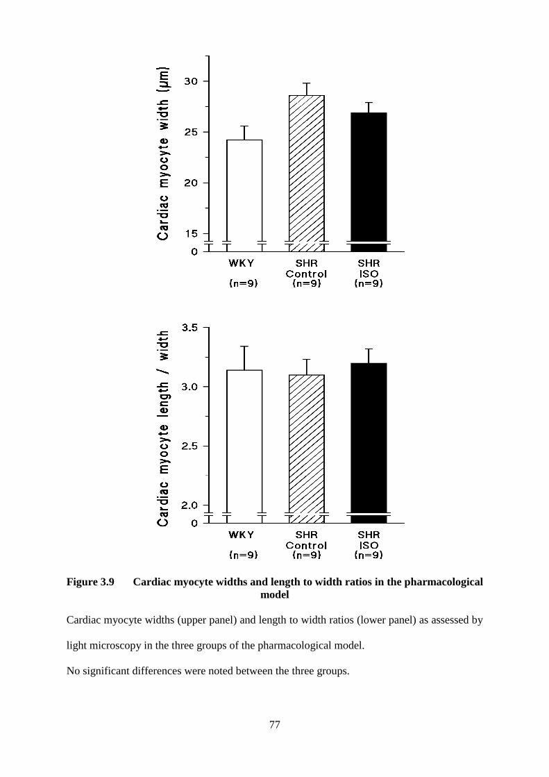
77
Figure 3.9 Cardiac myocyte widths and length to width ratios in the pharmacological
model
Cardiac myocyte widths (upper panel) and length to width ratios (lower panel) as assessed by
light microscopy in the three groups of the pharmacological model.
No significant differences were noted between the three groups.

78
3.2.2.2 Flow Cytometry
The data collected from the same rat hearts using flow cytometry indicated a similar pattern
as that shown with light microscopy. The length (FL1W) of the WKY cardiac myocytes was
significantly shorter as compared to that of the SHR-Control and SHR-ISO groups (Figure
3.10). Importantly, despite differences in LV dimensions (Figures 3.3 and 3.4) no difference
in cardiac myocyte length (FL1W) was evident between the SHR-ISO and SHR-Control
groups (Figure 3.10).
3.3 CORRELATIONS
3.3.1 Left ventricular end diastolic radius and cardiac myocyte length
Linear correlation analyses between LV end diastolic radius and cardiac myocyte length
(Figure 3.11, light microscopy, upper panel and flow cytometry, lower panel) were performed
using data from all groups (SHR-Non-Failure, SHR-Failure, WKY, SHR-Control, SHR-ISO).
Despite a range of light microscopy mean myocyte lengths (~70-110 μm) and mean LV end
diastolic radii (~0.1-0.6 cm), no correlation was observed (Figure 3.11, upper panel).
Furthermore, no correlation was observed between flow cytometry myocyte lengths and LV
end diastolic radii (Figure 3.11, lower panel).
3.3.2 Cardiac myocyte lengths obtained using light microscopy versus flow
cytometry
The two techniques used to measure cardiac myocyte length were linearly correlated (Figure
3.12). The cardiac myocytes from the WKY control rats were the smallest (in the left of
Figure 3.12) and no differences between the four SHR groups were evident (there is
considerable overlap of the data obtained in these four groups, Figure 3.12). Using these two
techniques the same differences were shown between the groups (compare figures 3.5 and
3.7; compare Figures 3.8 and 3.10). Hence, although absolute measurements of cell length

79
Figure 3.10 Cardiac myocyte lengths as assessed by flow cytometry in the
pharmacological model
Cardiac myocyte lengths as assessed by flow cytometry (FL1W) in the three groups of the
pharmacological model.
*p<0.05 compared to the WKY control group.

80
Figure 3.11 Linear correlation analyses between left ventricular end diastolic radius
and cardiac myocyte length
Linear correlation analyses between left ventricular (LV) end diastolic radius and cardiac
myocyte length as assessed by light microscopy (upper panel) and flow cytometry (lower
panel) using data obtained in all groups.

81
Figure 3.12 Linear correlation analysis between cardiac myocyte length measured
using light microscopy and flow cytometry
Linear correlation analysis between cardiac myocyte length measured using light microscopy
(y-axis) and cardiac myocyte length assessed using flow cytometry (FL1W, x-axis) using
data obtained in all groups.

82
cannot be obtained using flow cytometry, due to the inability to calibrate at present,
differences in cardiac myocyte length between groups can be assessed using this technique.
3.3.3 Left ventricular weight (mg/100g body weight) versus cardiac
myocyte length
A strong association was noted between LV weight normalised to 100g body weight and
cardiac myocyte length measured using either light microscopy (Figure 3.13, upper panel) or
flow cytometry (Figure 3.13, lower panel).

83
Figure 3.13 Linear correlation analyses between left ventricuar weight normalised to
100g body weight and cardiac myocyte length as assessed by light microscopy and flow
cytometry
Linear correlation analyses between left ventricular (LV) weight normalised to 100g body
weight and cardiac myocyte length as assessed by light microscopy (upper panel) and flow
cytometry (lower panel) using data obtained in all groups.

84
CHAPTER 4

85
4.0 DISCUSSION
The main findings of the present study are as follows: The length of cardiac myocytes
isolated from rats with LV dilatation (SHR-Failure and SHR-ISO) was no different from the
length of cardiac myocytes isolated from rats without LV dilatation (SHR-Non-Failure and
SHR-Control). The two models of LV dilatation used [natural progression model (SHR-
Failure) and pharmacological model (SHR-ISO)] revealed the same outcome that LV
dilatation (increased LV end diastolic radius and decreased LV wall thickness to radius ratio)
is not associated with an increase in cardiac myocyte length. Furthermore, the data were
confirmed using two methods (light microscopy and flow cytometry) to determine cardiac
myocyte length. However, the hypertensive rats (SHR-Non-Failure; SHR-Failure; SHR-
Control; SHR-ISO) did have an increased cardiac myocyte length compared to that of
normotensive rats (WKY control).
The present study clarifies that although alterations in cardiac myocyte dimensions
accompany hypertension-induced cardiac hypertrophy (Bishop et al. 1979; McCrossan et al.
2004; Olivetti et al. 1994; Zierhut et al. 1991), further increases in cardiac myocyte length are
not a mechanism of the development of cardiac dilatation and heart failure. The present study
is the first study to assess cardiac myocyte dimensions in age-matched animals with cardiac
dilatation and to accurately determine LV dilatation using load independent measurements.
Our data showing that LV dilatation is not associated with increases in cardiac myocyte
length are in contrast to a number of studies mostly from the same group (Chen et al., 2010;
Gerdes et al. 1996; Onodera et al. 1998; Schultz et al., 2007; Tamura et al. 1998, 1999 and
2000) that report increases in cardiac myocyte length in rats in heart failure.

86
As a model of heart failure, Gerdes and his colleagues (Gerdes et al. 1996; Onodera et al.
1998; Tamura et al. 1998, 1999 and 2000) have used rats that are genetically predisposed to
the development of heart failure with increasing age (spontaneously hypertensive heart
failure, SHHF). Consequently, in all of their studies the rats with heart failure were 3 to 16
months older than those rats not in heart failure (Gerdes et al. 1996: 24 versus 12 months;
Onodera et al. 1998: 12 versus 9 months; Tamura et al. 1998, 1999 and 2000: 18 versus 2, 16
versus 6 and 22 versus 18 months respectively). As cardiac myocyte length is strongly
correlated with age in SHHF rats (Tamura et al. 1998) and with LV weight (Campbell et al.
1991; Capasso et al. 1992; current study), and LV weight increases with age in hypertensive
rats (Gerdes et al. 1996), the increased cardiac myocyte length observed in the SHHF rats
with heart failure may be attributed to the increased age and hence LV weight of these rats
rather than the presence of increased LV dimensions and heart failure. Moreover, in a study
to assess reverse remodelling, an angiotensin II type 1 receptor blocker resulted in a decrease
in myocyte length associated with a decrease in heart weight but no change in LV internal
dimensions in age-matched SHHF rats (Tamura et al. 2000). In addition, although differences
in cardiac myocyte length and heart weight between the angiotensin II type 1 receptor blocker
treatment group and the hydralazine treatment group were noted, no differences in LV
internal dimensions between these two groups were evident (Tamura et al. 2000).
Few previous studies have simultaneously measured cardiac myocyte length and LV
dimensions. Most studies reporting increases in cardiac myocyte length in rats with heart
failure compared to rats without heart failure have failed to measure LV dimensions (Gerdes
et al. 1996; Onodera et al. 1998; Tamura et al. 1998 and 1999). Hence, whether increases in
cardiac myocyte length accompany LV dilatation is not evident from these studies. Moreover,
the few studies which have assessed LV dimensions have used in vivo echocardiography,

87
which is dependent on loading conditions and heart rate. Hence the increases in LV
dimensions reported previously (Tamura et al. 2000) may be as a consequence of increases in
loading conditions rather than the presence of LV remodelling and dilatation. Indeed in
SHHF rats increases in LV internal dimensions are accompanied by increases in LV systolic
wall stress (afterload) (Tamura et al. 2000) and decreases in LV internal dimensions occurred
when LV systolic wall stress was reduced (Tamura et al. 2000). Similarly, in both aortic
banded guinea pigs (Wang et al. 1999) and supraventricular tachycardia-induced
cardiomyopathy in pigs (Spinale et al. 1996), increases in LV internal dimensions were
measured in the presence of increases in afterload (systolic wall stress) and preload (LV end
diastolic pressure). Furthermore, the reductions in LV end diastolic diameter reported
following LV assist device support are likely to be due to the decreases in pulmonary
capillary wedge pressure (preload) as a consequence of LV assist device support (Zafeiridis
et al. 1998). Although, one previous study did not use echocardiography, they measured LV
dimensions of cardiac rings following perfusion fixation (Kajstura et al. 1995), in which the
perfusion pressure was maintained equal to the LV end diastolic pressure measured in vivo.
As the LV filling pressure was increased in the dogs with pacing-induced heart failure, the
increased LV dimensions reported are likely to be attributed to the increased preload
(Kajstura et al. 1995). Moreover, reductions in cardiac myocyte length have been reported
when cardiac preload and/or afterload are decreased (Campbell et al. 1991; Cooper et al.
1982 and 1984; Thompson et al.1984). In contrast in the present study, LV dilatation was
defined as the presence of an increased LV end diastolic radius and a decreased LV end
diastolic wall thickness to radius ratio determined over a range of controlled LV filling
volumes in vivo. Hence, the increases in LV dimensions in the current study are an accurate
indication of the presence of LV dilatation independent of loading conditions.

88
A further explanation for the discrepancy between our data and that of others may be the
difference in the strain of rats used. In the studies by Gerdes and colleagues (Gerdes et al.
1996; Onodera et al. 1998; Tamura et al. 1998, 1999 and 2000) the SHHF model has been
used predominantly. However, the genetic background of the SHHF rat strain differs from
that of the SHR and WKY rat strains (McCune et al. 1990; 1995a and 1995b). Moreover, the
SHHF rat strain is reported to have longer cardiac myocytes than normal adult rats (Gerdes et
al. 1996), a difference that was noted long before the development of the clinical signs and
decreased hemodynamic function associated with heart failure (Onodera et al. 1998).
Typically SHHF rats not in heart failure have cardiac myocyte lengths of ~140 to 150 μm
(Capasso et al. 1992; Tamura et al. 1999), whereas mean cardiac myocyte lengths of 109 to
145 μm and 94 to 110 μm have been reported in the Sprague-Dawley (Bai et al. 1990;
Korecky & Rakusan 1978; Nash et al. 1979) and Fischer (Bishop & Drummond 1979) strains
of rats respectively. Similar to the results of the present study, previous studies reported
longer cardiac myocytes in SHR compared to WKY rats (Aiello et al. 2004; Bishop et al.
1979; Brooksby et al. 1992; McCrossan et al. 2004). In these previous studies cardiac
myocyte lengths ranged from 93 to 140 μm in WKY and from 105 to 150 μm in SHR (Aiello
et al. 2004; Bishop et al. 1979; Brooksby et al. 1992; McCrossan et al. 2004) and in the
present study we report mean cardiac myocyte lengths of 76 μm in WKY rats and 86 to 96
μm in SHR. Hence, even within rat strain there is a wide range of cardiac myocyte lengths.
Differences within strain may be related to differences in the origin of the rats, as those SHR
and WKY obtained from the same source have similar cardiac myocyte lengths (Bishop et al.
1979: WKY=93 μm and SHR=105 μm; Brooksby et al. 1992: WKY=95 μm and SHR=115
μm; both from Charles River Breeding Laboratory); whereas WKY and SHR from a different
source (bred in the researchers own facility as opposed to from Charles River Breeding
Laboratory) have different cardiac myocyte lengths (WKY=136 μm and SHR=147 μm;

89
Aiello et al. 2004). Similarly, Sprague-Dawley rats obtained from different sources (Bio-
Breeding Laboratories: Korecky & Rakusan 1978 versus Holtman Company: Bai et al. 1990)
have different cardiac myocyte lengths (120 μm: Korecky & Rakusan 1978; 145 μm: Bai et
al. 1990). In this regard the SHR used in the present study were bred in the Central Animal
Services at the University of the Witwatersrand. The original breeding pairs were obtained
over 20 years ago from OLAC, UK. It has also been reported that differences in cardiac
myocyte size can be present in different shipments of a given strain of rats (Campbell et al.
1991). In addition, cardiac myocyte lengths vary across species. Mean cardiac myocyte
lengths of 122 to 137 μm have been reported in normal adult hamsters (Sorenson et al. 1985)
and in normal adult guinea pigs mean cardiac myocyte length is 168 μm (Wang et al. 1999).
It may also be argued that the method of cell isolation may impact upon cardiac myocyte
dimensions. Fixation after isolation does not appear to impact upon cardiac myocyte length
(freshly isolated: 109 μm; fixed: 109 μm; Nash et al. 1979); however, plated cardiac
myocytes are longer (147 μm, Fraticelli et al. 1989) than those freshly isolated (110 μm,
Sorenson et al. 1985) from the same strain of rat of the same age (Wistar). Nevertheless,
Diffee and Nagle (2003), who used a similar method of isolation as in the present study (ie.
from frozen tissue) report cardiac myocyte lengths of 123 μm which are no different from
those of cardiac myocytes freshly isolated (120 μm, Korecky and Rakusan 1978) from the
same strain (Sprague Dawley) of rats of the same age. Hence it is unlikely that the method of
cell isolation used in the present study had an impact on the cardiac myocyte lengths
recorded. Furthermore, the slack (relaxed) sarcomere lengths of 1.87 μm (WKY) and 1.99 to
2.13 μm (SHR) recorded in the present study are similar to those recorded in previous studies
1.60 μm (Fischer: Bishop and Drummond, 1979), 1.62 μm (Wistar, WKY and SHR: Bishop
et al. 1979), 1.80 μm (Sprague-Dawley: Korecky & Rakusan 1978) 1.81 μm (WKY) and 1.85

90
μm (SHR) (McCrossan et al. 2004), 1.83 μm (Wistar: Fraticelli et al. 1989), 1.88 μm (SHHF:
Gerdes et al. 1996), 1.90 μm (Sprague-Dawley: Diffee & Nagle 2003; SHHF: Tamura et al.
1998) and 1.95 μm (WKY and SHR: Brooksby et al. 1992).
Lastly, the discrepancy between the results of our study and that of previous studies may be
related to differences in the models of LV dilatation and heart failure. Besides the SHHF rat
model (Gerdes et al. 1996; Onodera et al. 1998; Tamura et al. 1998, 1999 and 2000), cardiac
myocyte lengths have been assessed in two other models of heart failure, namely
supraventricular tachycardia-induced cardiomyopathy in pigs (Spinale et al. 1996) and dogs
(Kajstura et al. 1995), and constriction of the descending thoracic aorta in guinea pigs (Wang
et al. 1999). Consistent with the SHHF model, in these two models increases in cardiac
myocyte length have been reported in the group with increased LV end diastolic dimensions
(Kajstura et al. 1995; Spinale et al. 1996; Wang et al. 1999). However, as cardiac dilatation
has been attributed to alterations in both cardiac myocyte dimensions and myocardial fibrosis
in dilated cardiomyopathy in humans (Beltrami et al. 1995), and changes in myocyte volume
are reported to correlate with alterations in heart mass unless there is extensive fibrosis
(Gerdes, 2002), it is important to assess the relative contribution of each of these factors to
LV remodelling. In this regard, Campbell et al. (1991) attribute increases in heart weight
despite no change in cardiac myocyte size to increases in myocardial fibrosis. Importantly, in
the SHHF model only modest increases in myocardial collagen content (an increase from 1.9
to 2.5%; Onodera et al. 1998; Tamura et al. 1998) have been noted. Similarly, there is a lack
of myocardial fibrosis in the aortic banding model (Wang et al. 1999), and in tachycardia-
induced cardiomyopathy myocardial collagen concentrations are either decreased (Spinale et
al. 1996) or modestly increased (from 0.5 to 6%, Kajstura et al. 1995). In contrast, in both the
natural progression (SHR-Failure) and the pharmacological (SHR-ISO) model used in the

91
present study, we have previously shown that alterations in the characteristics of myocardial
collagen play a major role in determining LV remodelling and dilatation (Anamourlis et al.
2006; Badenhorst et al. 2003a and 2003b; Tsotetsi et al. 2001; Veliotes et al. 2005;
Woodiwiss et al. 2001), data which is supported by studies showing increased fibrosis in
SHR in heart failure (Boluyt et al. 1995; Conrad et al. 1995; Engelmann et al. 1987). Hence
it is not surprising that we found no contribution of cardiac myocyte length to LV dilatation;
whereas in models in which fibrosis plays little role (Kajstura et al. 1995; Onodera et al.
1998; Spinale et al. 1996; Tamura et al. 1998; Wang et al. 1999), alterations in cardiac
myocyte length were observed.
In agreement with previous studies conducted in SHR (Boluyt et al. 1995; Conrad et al.
1995; Engelmann et al. 1987), no changes in cardiac myocyte dimensions were noted in SHR
with LV dilatation in the present study. In this regard, Engelmann et al. (1987) report no
change in cardiac myocyte cross-sectional area but substantial increases in myocardial
fibrosis in SHR with decreased LV wall thickness. Similarly, Boluyt et al. (1995) and Conrad
et al. (1995) report decreases in cardiac myocyte fractional area and increased myocardial
fibrosis in SHR in heart failure.
Furthermore, in support of the lack of association between cardiac myocyte length and LV
dimensions observed in the present study; Schultz et al. (2007) showed that 16 months of
exercise in SHHF rats resulted in a 21% increase in LV diastolic diameter, but no change in
myocyte length. The deleterious effects of exercise on cardiac remodelling (enhanced LV
dilatation) were however associated with a profound increase in cardiac fibrosis (~140%).
Similarly, the prevention of increases in cardiac chamber diameter, by thyroid administration
in cardiomyopathic hamsters, was associated with no change in cardiac myocyte length

92
(Kuzman et al., 2007). In a study assessing the impact of TNF-α overexpression on cardiac
remodelling; although cardiac myocyte length was increased in both male and female rats,
increases in LV diastolic diameter were only observed in the male rats (Janczewski et al.
2003). Moreover, Tamura et al., (2000) reported decreases in cardiac myocyte length
subsequent to angiotensin II receptor blockade; however no changes in cardiac dimensions
were noted. Lastly, increases rather than decreases in cardiac myocyte length were reported
in a recent study in which intramyocardial delivery of mesenchymal stem cells resulted in the
prevention of cardiac dilatation following myocardial infarction in mice (Li et al., 2010).
Although a limitation of the present study may be the use of cardiac myocytes isolated from
frozen tissue, this is unlikely. As previously discussed the lengths of cardiac myocytes
isolated from frozen tissue (123 μm, Diffee & Nagle, 2003) are no different from those of
freshly isolated cardiac myocytes (120 μm, Korecky & Rakusan 1978). In addition, the
method used to isolate cardiac myocytes is more likely to impact on absolute length
measurements than on comparisons between groups. Hence, the lack of difference in cardiac
myocyte lengths between rats with LV dilatation (SHR-Failure and SHR-ISO) and those
without LV dilatation (SHR-Failure and SHR-Control) is not likely to be as a consequence of
the method of cardiac myocyte isolation. In contrast, the strengths of the present study
include the use of two methods of measurement of cardiac myocyte length; the investigation
of two models of cardiac dilatation; and the assessment of LV dimensions using load and
heart rate independent measures.
From a clinical perspective the present study suggests that in hypertension-induced and beta-
adrenergic induced heart failure, models in which the cardiac interstitium plays a major role,
alterations in cardiac myocyte dimensions do not contribute to LV dilatation. Hence, in these

93
models therapy should be aimed at targeting the extracellular matrix rather than growth or
hypertrophy of cardiac myocytes.
In conclusion, the present study indicates that although cardiac hypertrophy (in both
hypertension-induced and beta-adrenergic-induced models) is associated with increases in
cardiac myocyte length, cardiac dilatation in these models is not attributed to alterations in
cardiac myocyte dimensions.

94
CHAPTER 5

95
5.0 REFERENCES
1. Agabiti-Rosei E, Muiesan ML, Romanelli G, Castellano M, Beschi M, Alicandri C &
Muiesan G (1987). Cardiac function as related to adrenergic activity in hypertensive
left ventricular hypertrophy. J Clin Hypertens 3, 203-210.
2. Aiello EA, Villa-Abrille MC, Escudero EM, Portiansky EL, Pérez NG, de Hurtado MC &
Cingolani HE (2004). Myocardial hypertrophy of normotensive Wistar-Kyoto rats.
Am J Physiol Heart Circ Physiol 286, H1229-H1235.
3. American Heart Association (2010). Heart Disease and Stroke Statistics – 2010 Update.
Dallas, Texas: American Heart Association.
4. Anamourlis C, Badenhorst D, Gibbs M, Correia R, Veliotes D, Osadchii O, Norton GR &
Woodiwiss AJ (2006). Phosphodiesterase inhibition promotes the transition from
compensated hypertrophy to cardiac dilatation in rats. Eur J Physiol 451, 526–533.
5. Anand IS, Fisher LD, Chiang YT, Latini R, Masson S, Maggioni AP, Glazer RD,
Tognoni G & Cohn JN (2003). Changes in brain natriuretic peptide and
norepinephrine over time and mortality and morbidity in the Valsartan Heart Failure
Trial (Val-HeFT). Circulation 107, 1278-1283.
6. Anker SD & Coats AJ (2002). How to RECOVER from RENAISSANCE? The
significance of the results of RECOVES, RENAISSANCE, RENEWAL and
ATTACH. Int J Cardiol 85, 195-197.
7. Anversa P, Loud AV, Levicky V & Guideri G (1985). Left ventricular failure induced by
myocardial infarction. Myocyte hypertrophy. Am J Physiol Heart Circ Physiol 248,
H876-H882.
8. Badenhorst D, Maseko M, Tsotetsi OJ, Naidoo A, Brooksbank R, Norton GR &
Woodiwiss AJ (2003a). Cross-linking influences the impact of quantitative changes

96
in myocardial collagen on cardiac stiffness and remodelling in hypertension in rats.
Cardiovasc Res 57, 632-641.
9. Badenhorst D, Veliotes D, Maseko M, Tsotetsi OJ, Brooksbank R, Naidoo A, Woodiwiss
AJ & Norton GR (2003b). β-adrenergic activation initiates chamber dilatation in
concentric hypertrophy. Hypertension 41, 499–504.
10. Bai SL, Campbell SE, Moore JA, Morales MC & Gerdes AM (1990). Influence of age,
growth, and sex on cardiac myocyte size and number in rats. Anat Rec 226, 207-
212.
11. Banfi C, Cavalca V, Veglia F, Brioschi M, Barcella S, Mussoni L, Boccotti L, Temoli E,
Biglioloi P & Agostoni PG (2005). Neurohumoral activation is associated with
increased levels of plasma matrix metalloproteinase-2 in human heart failure. Eur
Heart J 26, 481-488.
12. Baumgarten G, Knuefermann P & Mann DL (2000). Cytokines as emerging targets in the
treatment of heart failure. Trends Cardiovasc Med 10, 216-223.
13. Beltrami CA, Finato N, Rocco M, Feruglio GA, Puricelli C, Cigola E, Quaini EH,
Sonnenblick EH, Olivetti G & Anversa P (1994). Structural basis of end-stage
failure in ischemic cardiomyopathy in humans. Circulation 89, 151-163.
14. Beltrami CA, Finato N, Rocco M, Feruglio GA, Puricelli C, Cigola E, Sonnenblick EH,
Olivetti G & Anversa P (1995). The cellular basis of dilated cardiomyopathy in
humans. J Mol Cell Cardiol 27, 291-305.
15. Bing OH, Brooks WW, Robinson KG, Slawsky MT, Hayes JA, Litwin SE, Sen S &
Conrad CH (1995). The spontaneously hypertensive rat as a model of the transition
from compensated left ventricular hypertrophy to failure. J Mol Cell Cardiol 27,
383-396.

97
16. Bishop SP, Oparil S, Reynolds RH & Drummond JL (1979). Regional myocyte size in
normotensive and spontaneously hypertensive rats. Hypertension 1, 378-383.
17. Bishop SP & Drummond JL (1979). Surface morphology and cell size measurement of
isolated rat cardiac myocytes. J Mol Cell Cardiol 11, 423-433.
18. Boluyt MO, Bing OH & Lakatta EG (1995). The ageing spontaneously hypertensive rat
as a model of the transition from stable compensated hypertrophy to heart failure.
Eur Heart J 16, 19-30.
19. Brilla CG, Zhou G, Matsubara L & Weber KT (1994). Collagen metabolism in cultured
adult rat cardiac fibroblasts: Response to angiotensin II and aldosterone. J Mol Cell
Immunol 26, 809-820.
20. Bristow MR (1997). Mechanism of action of beta-blocking agents in heart failure. Am J
Cardiol. 80, 26L-40L.
21. Brooksby P, Levi AJ & Jones JV (1992). Contractile properties of ventricular myocytes
isolated from spontaneously hypertensive rat. J Hypertens 10, 521-527.
22. Campbell SE, Korecky B & Rakusan K (1991). Remodeling of myocyte dimensions in
hypertrophic and atrophic rat hearts. Circ Res 68, 984-996.
23. Capasso JM, Robinson TF & Anversa P (1989). Alterations in collagen cross-linking
impair myocardial contractility in the mouse heart. Circ Res 65, 1657-1664.
24. Capasso JM, Fitzpatrick D & Anversa P (1992). Cellular mechanisms of ventricular
failure: myocyte kinetics and geometry with age. Am J Physiol Heart Circ Physiol
262, H1770-1781.
25. Chen YF, Redetzke RA, Sivertson RM, Coburn TS, Cypher LR & Gerdes AM (2010).
Post-myocardial infarction left ventricular myocyte remodeling: are there gender
differences in rats? Cardiovasc Pathol Nov 15 (Epub ahead of print).

98
26. Cohn JN, Ferrari R & Sharpe N (2000). Cardiac remodeling - concepts and clinical
implications: A consensus paper from an International Forum on Cardiac
Remodeling. J Am Coll Cardiol 35, 569–582.
27. Cohn JN, Levine TB, Olivari MT, Garberg V, Lura D, Francis GS, Simon AB & Rector T
(1984). Plasma norepinephrine as a guide to prognosis in patients with chronic
congestive heart failure. N Engl J Med 311, 819-823.
28. Coker ML, Jolly JR, Joffs C, Etoh T, Holder JR, Bond BR & Spinale FG (2001). Matrix
metalloproteinase expression and activity in isolated myocytes after neurohormonal
stimulation. Am J Physiol Heart Circ Physiol 281, H543-H551.
29. Communal C, Singh K, Pimentel DR & Colucci WS (1998). Norepinephrine stimulates
apoptosis in adult rat ventricular myocytes by activation of the beta-adrenergic
pathway. Circulation 98, 1329-1334.
30. Conrad CH, Brooks WW, Hayes JA, Sen S, Robinson KG & Bing OH (1995).
Myocardial fibrosis and stiffness with hypertrophy and heart failure in the
spontaneously hypertensive rat. Circulation 91, 161-170.
31. Cooper G 4th & Marino TA (1984). Complete reversibility of cat right ventricular chronic
progressive pressure overload. Circ Res 54, 323-331.
32. Cooper G 4th & Tomanek RJ (1982). Load regulation of the structure, composition, and
function of mammalian myocardium. Circ Res 50, 788-798.
33. Danser AHJ, van Kesteren CAM, Bax WA, Tavenier M, Derkx FH, Saxena PR &
Schalekamp MA (1997). Prorenin, renin, angiotensinogen, and angiotensin-
converting enzyme in normal and failing human hearts. Evidence for renin binding.
Circulation 96, 220-236.
34. De Angelis N, Fiordaliso F, Latini R, Calvillo L, Funicello M, Gobbi M, Mennini T &
Masson S (2002). Appraisal of the role of angiotensin II and aldosterone in

99
ventricular myocyte apoptosis in adult normotensive rat. J Mol Cell Cardiol 34,
1655-1665.
35. Diez C & Simm A (1998). Gene expression in rod shaped cardiac myocytes, sorted by
flow cytometry. Cardiovasc Res 40, 530-537.
36. Diffee GM & Nagle GF (2003). Exercise training alters length dependence of contractile
properties in rat myocardium. J Appl Physiol 94, 1137-1144.
37. Engelmann GL, Vitullo JC & Gerrity RG (1987). Morphometric analysis of cardiac
hypertrophy during development, maturation, and senescence in spontaneously
hypertensive rats. Circ Res 60, 487-494.
38. Ertl G, Gaudron P, Eilles C, Schorb W & Kochsiek K (1991). Compensatory mechanisms
for cardiac dysfunction in myocardial infarction. Basic Res Cardiol 86, 159-165.
39. Esler M, Kaye D, Lambert G, Esler D & Jennings G (1997). Adrenergic nervous system
in heart failure. Am J Cardiol 80, 7L-14L.
40. Fedak PWM, Verma S, Weisel RD, Skrtic M & Li RK (2005). Cardiac remodelling and
failure. From molecules to man (Part III). Cardiovasc Path 14, 109-119.
41. Ferrari R, Ceconi C, Campo G, Cangiano E, Cavazza C, Secchiero P & Tavazzi L (2009).
Mechanisms of remodelling. A question of life (stem cell production) and death
(myocyte apoptosis). Circ J 73, 1973-1982.
42. Francis GS, Cohn JN, Johnson G, Rector TS, Goldman S & Simon A (1993). Plasma
norepinephrine, plasma renin activity, and congestive heart failure. Relations to
survival and the effects of therapy in V-HeFT II. The V-HeFT VA Cooperative
Studies Group. Circulation 87, VI40-48.
43. Fraticelli A, Josephson R, Danziger R, Lakatta E & Spurgeon H (1989). Morphological
and contractile characteristics of rat cardiac myocytes from maturation to
senescence. Am J Physiol Heart Circ Physiol 257, H259- H265.

100
44. Frey N, Katus HA, Olson EN & Hill JA (2004). Hypertrophy of the heart: a new
therapeutic target? Circulation 109, 1580-1589.
45. Frigerio M & Roubina E (2005). Drugs for left ventricular remodelling in heart failure.
Am J Cardiol 96(suppl), 10L-18L.
46. Gaudron P, Eilles C, Kugler I & Ertl G (1993). Progressive left ventricular dysfunction
and remodelling after myocardial infarction. Potential mechanisms and early
predictors. Circulation 87, 755-763.
47. Gerdes AM (2002). Cardiac myocyte remodeling in hypertrophy and progression to
failure. J Card Fail 8, S264-S268.
48. Gerdes AM & Capasso JM (1995). Structural remodeling and mechanical dysfunction of
cardiac myocytes in heart failure. J Mol Cell Cardiol. 27, 849-856.
49. Gerdes AM & Iervasi G (2010). Thyroid replacement therapy and heart failure.
Circulation 122, 385-393.
50. Gerdes AM, Kellerman SE, Moore JA, Muffly KE, Clark LC, Reaves PY, Malec KB,
McKeown PP & Schoken DD (1992). Structural remodelling of cardiac myocytes in
patients with ischemic cardiomyopathy. Circulation 86, 426-430.
51. Gerdes AM, Onodera T, Tamura T, Said S, Bohlmeyer TJ, Abraham WT & Bristow MR
(1998). New method to evaluate myocyte remodelling from formalin-fixed biopsy
and autopsy material. J Card Fail 4, 343-348.
52. Gerdes AM, Onodera T, Wang X & McCune SA (1996). Myocyte remodelling during the
progression to failure in rats with hypertension. Hypertension 28, 609-614.
53. Gerson MC, Craft LL, McGuire N, Suresh DP, Abraham WT & Wagoner LE (2002).
Carvedilol improves left ventricular function in heart failure patients with idiopathic
dilated cardiomyopathy and a wide range of sympathetic nervous system function as
measured by iodine 123 metaiodobenzylguanidine. J Nucl Cardiol 9, 608-615.

101
54. Gibbs M, Veliotes DG, Anamourlis C, Badenhorst D, Osadchii O, Norton GR &
Woodiwiss AJ (2004). Chronic beta-adrenoreceptor activation increases cardiac
cavity size through chamber remodeling and not via modifications in myocardial
material properties. Am J Physiol Heart Circ Physiol 287, H2762-H2767.
55. Gilbert JC & Glantz SA (1989). Determinants of left ventricular filling and of the
diastolic pressure-volume relation. Circ Res 64, 827-852.
56. Greenberg B, Quinones MA, Koilpillai C, Limacher M, Shindler D, Benedict C &
Shelton B (1995). Effects of long-term enalapril therapy on cardiac structure and
function in patients with left ventricular dysfunction. Results of the SOLVD
echocardiography substudy. Circulation 91, 2573-2581.
57. Grossman W, Jones D & McLaurin LP (1975). Wall Stress and patterns of hypertrophy in
the human left ventricle. J Clin Invest 56, 56-64.
58. Gunasinghe SK, Ikonomidis J & Spinale FG (2001). Contributory role of matrix
metalloproteinases in cardiovascular remodeling. Curr Drug Targets Cardiovasc
Haematol Disord 1, 75-91.
59. Gunja-Smith Z, Morales AR, Romanelli R & Woessner JF Jr (1996). Remodeling of
human myocardial collagen in idiopathic dilated cardiomyopathy. Role of
metalloproteinases and pyridinoline cross-links. Am J Pathol 148, 1639-1648.
60. Hasking GJ, Esler MD, Jennings GL, Burton D, Johns JA & Korner PI (1986).
Norepinephrine spillover to plasma in patients with congestive heart failure:
evidence of increased overall and cardiorenal sympathethic nervous activity.
Circulation 73, 615-621.
61. Hearse DJ, Stewart DA & Braimbridge MV (1975). Hypothermic arrest and potassium
arrest: metabolic and myocardial protection during elective cardiac arrest. Circ Res
36, 481-489.
102
62. Hein S, Amon E, Kostin S, Schönburg M, Elsässer A, Polyakova V, Bauer EP,
Klövekorn WP & Schaper J (2003). Progression from compensated hypertrophy to
failure in the pressure-overloaded human heart. Structural deterioration and
compensatory mechanisms. Circulation 107, 984-991.
63. Hermida N, López B, González A, Dotor J, Lasarte JJ, Sarobe P, Borrás-Cuesta F &
Díez J (2009). A synthetic peptide from transforming growth factor-beta1 type III
receptor prevents myocardial fibrosis in spontaneously hypertensive rats.
Cardiovasc Res 81, 601-609.
64. Hill JA & Olson EN (2008). Cardiac plasticity. N Engl J Med 358, 1370-1380.
65. Inoko M, Kihara Y, Morii I, Fujiwara H, Sasayama S (1994). Transition from
compensatory hypertrophy to dilated, failing left ventricles in Dahl salt-sensitive
rats. Am J Physiol Heart Circ Physiol 267, H2471-H2482.
66. Iwai N, Shimoike H & Kinoshita M (1995). Cardaic rennin-angiotensin system in the
hypertrophied heart. Circulation 92, 2690-2696.
67. Janczewski AM, Kadokami T, Lemster B, Frye CS, McTiernan CF & Feldman AM
(2003). Morphological and functional changes in cardiac myocytes isolated from
mice overexpressing TNF-α. Am J Physiol Heart Circ Physiol 284, H960-H969.
68. Janicki JS, Brower GL, Gardner JD, Chancey AL & Stewart JA Jr (2004). The dynamic
interaction between Matrix Metalloproteinase Activity and adverse myocardial
remodeling. Heart Failure Reviews 9, 33-42.
69. Kai H, Kuwahara F, Tokuda K & Imaizumi T (2005). Diastolic dysfunction in
hypertensive hearts: Roles of perivascular inflammation and reactive myocardial
fibrosis. Hypertens Res 28, 483-490.

103
70. Kajstura J, Zhang X, Liu Y, Szoke E, Cheng W, Olivetti G, Hintze TH & Anversa P
(1995). The cellular basis of pacing-induced dilated cardiomyopathy. Myocyte cell
loss and myocyte cellular reactive hypertrophy. Circulation 92, 2306-2317.
71. Kang PM & Izumo S (2000). Apoptosis and heart failure: A critical review of the
literature. Circ Res 86, 1107-1113.
72. Katz AM (2002). Proliferative signalling and disease progression in heart failure. Circ J
66, 225-231.
73. Kelm M, Schafer S, Mingers S, Heydthausen M, Vogt M, Motz W & Strauer BE (1996).
Left ventricular mass is linked to cardiac noradrenaline in normotensive and
hypertensive patients. J Hypertens 14, 1365-1367.
74. King MK, Coker ML, Goldberg A, McElmurray JH, 3rd
, Gunasinghe HR, Mukherjee R,
Zile MR, O'Neill TP & Spinale FG (2003). Selective matrix metalloproteinase
inhibition with developing heart failure: effects on left ventricular function and
structure. Circ Res 92, 177-185.
75. Kluger, J., Cody, R. J. & Laragh, J. H. (1982). The contributions of sympathetic tone and
the renin-angiotensin system to severe chronic congestive heart failure: response to
specific inhibitors (prazosin and captopril). Am J Cardiol 49, 1667-1674.
76. Konstam MA, Rousseau MF, Kronenberg MW, Udelson JE, Melin J, Stewart D, Dolan
N, Edens TR, Ahn S, Kinan D, Howe DM, Kilcoyne L, Metherall J, Benedict C,
Yusuf S & Pouleur H (1992). Effects of the angiotensin converting enzyme inhibitor
enalapril on the long-term progression of left ventricular dysfunction in patients
with heart failure. SOLVD Investigators. Circulation 86, 431-438.
77. Kudoh S, Komuro I, Mizuno T, Yamazaki T, Zou Y, Shiojima I, Takekoshi N & Yazaki
Y (1997). Angiotensin II stimulates c-Jun NH2-terminal kinase in cultured cardiac
myocytes of neonatal rats. Circ Res 80, 139-146.

104
78. Korecky B & Rakusan K (1978). Normal and hypertrophic growth of the rat heart:
changes in cell dimensions and number. Am J Physiol Heart Circ Physiol 234,
H123-H128.
79. Kuwahara F, Kai H, Tokuda K, Takeya M, Takeshita, Egashira K & Imaizumi T (2004).
Hypertensive myocardial fibrosis and diastolic dysfunction: another model of
inflammation? Hypertension 43, 739-745.
80. Kuzman JA, Tang Y, Vogelsang KA, Said S, Anderson BE, Morkin E & Gerdes AM
(2007). Thyroid hormone analog, diiodothyropropionic acid (DITPA), exerts
beneficial effects on chamber and cellular remodelling in cardiomyopathic
hamsters. Can J Physiol Pharmacol 85, 311-318.
81. Laskey WK, St. John Sutton M, Zeevi G, Hirshfeld JW & Reichek N (1984). Left
ventricular mechanics in dilated cardiomyopathy. Am J Cardiol 54, 620-625.
82. Levy D, Garrison RJ, Savage DD, Kannel WB & Castelli WP (1990). Prognostic
implications of echocardiographically determined left ventricular mass in the
Framingham Heart Study. N Engl J Med 322, 1561-1566.
83. Li YY, Feng Y, McTiernan CF, Pei W, Moravec CS, Wang P, Rosenblum W, Kormos
RL & Feldman AM (2001). Downregulation of matrix metalloproteinases and
reduction in collagen damage in the failing human heart after support with left
ventricular assist devices. Circulation 104, 1147-1152.
84. Li Q, Turdi S, Thomas DP & Ren J (2010). Intra-myocardial delivery of mesenchymal
stem cells ameliorates left ventricular and cardiomyocyte contractile dysfunction
following myocardial infarction. Toxicology Letters 195, 119-126.
85. Linzbach AJ (1960). Heart failure from the point of view of quantitative anatomy. Am J
Cardiol 5, 370-382.
86. Lorell BH (1997). Transition from hypertrophy to failure. Circulation 96, 3824-3827.
105
87. Mann DL & Spinale FG (1998). Activation of matrix metalloproteinases in the failing
human heart: breaking the tie that binds. Circulation 98, 1699-1702.
88. Matsui T & Rosenzweig A (2005). Convergent signal transduction pathways controlling
cardiomyocyte survival and function: the role of PI-3-kinase and Akt. J Mol Cell
Cardiol 38, 63-71.
89. Mayosi BM, Flisher AJ, Lalloo UG, Sitas F, Tollman SM & Bradshaw D (2009). The
burden of non-communicable diseases in South Africa. Lancet 374, 934-947.
90. McCrossan ZA, Billeter R & White E (2004). Transmural changes in size, contractile and
electrical properties of SHR left ventricular myocytes during compensated
hypertrophy. Cardiovasc Res 63, 283-292.
91. McCune SA, Baker PB & Stills HF Jr (1990). SHHF/Mcc-cp rat: model of obesity, non-
insulin-dependent diabetes, and congestive heart failure. ILAR News 32, 23–27.
92. McCune SA, Radin MJ, Jenkins JE, Chu YY & Park S (1995a). SHHF/Mcc-fa cp rat
model: effects of gender and genotype on age of expression of metabolic
complications and congestive heart failure and on response to drug therapy. In:
Shafir E, ed. Lessons From Animal Diabetes V. Smith-Gordon, 255–270.
93. McCune SA, Park S, Radin MJ & Jurin RR (1995b). The SHHF/Mcc-facp
rat model: a
genetic model of congestive heart failure. In: Singal PK, Dixon IMC, Beamish RE,
Dhalla NS, eds. Mechanisms of Heart Failure. Kluwer Academic Publishers, 91–
106.
94. Metra M, Nodari S, Parrinello G, Giubbini R, Manca C & Dei Cas L (2003). Marked
improvement in left ventricular ejection fraction during long-term beta-blockade in
patients with chronic heart failure: clinical correlates and prognostic significance.
Am Heart J 145, 292-299.

106
95. Mizuno Y, Yoshimura M, Yasue H, Sakamoto T, Ogawa H, Kugiyama K, Harada E,
Nakayama M, Nakamura S, Ito T, Shimasaki Y, Saito Y & Nakao K (2001).
Aldosterone production is activated in failing ventricle in human. Circulation 103,
72-77.
96. Mujundar VS & Tyagi SC (1999). Temporal regulation of extracellular matrix
components in transition from compensatory hypertrophy to decompensatory heart
failure. J Hypertens 17, 261-270.
97. Mukherjee R, Brinsa TA, Dowdy KB, Scott AA, Baskin JM, Deschamps AM, Lowry AS,
Escobar GP, Lucas DG, Yarbrough WM, Zile MR & Spinale FG (2003).
Myocardial infarct expansion and matrix metalloproteinase inhibition. Circulation
107, 618-625.
98. Nash GB, Tatham PE, Powell T, Twist VW, Speller RD & Loverock LT (1979). Size
measurements on isolated rat heart cells using Coulter analysis and light scatter flow
cytometry. Biochim Biophys Acta 587, 99-111.
99. Norton GR, Tsotetsi OJ & Woodiwiss AJ (1993). Hydrochlorotiazide improves
ventricular compliance and thus performance without reducing hypertrophy in renal
artery stenosis in rats. Hypertension 21, 638-645.
100. Norton GR, Tsotetsi J, Trifunovic B, Hartford C, Candy GP & Woodiwiss AJ (1997).
Myocardial stiffness is attributed to alterations in cross-linked collagen rather than
total collagen or phenotypes in spontaneously hypertensive rats. Circulation 96,
1991-1998.
101. Norton GR, Woodiwiss AJ, Gaasch WH, Mela T, Chung ES, Aurigemma GP & Meyer
TE (2002). Heart failure in pressure overload hypertrophy: The relative roles of
ventricular remodeling and myocardial dysfunction. J Am Coll Cardiol 39, 664-671.

107
102. Olivetti G, Capasso JM, Sonnenblick EH & Anversa P (1990). Side-to-side slippage of
myocytes participates in ventricular wall remodelling acutely after myocardial
infarction in rats. Circ Res 67, 23-34.
103. Olivetti G, Melissari M, Balbi T, Quaini F, Cigola E, Sonnenblick EH & Anversa P
(1994). Myocyte cellular hypertrophy is responsible for ventricular remodelling in
the hypertrophied heart of middle aged individuals in the absence of cardiac failure.
Cardiovasc Res 28, 1199-1208.
104. Onodera T, Tamura T, Said S, McCune SA & Gerdes AM (1998). Maladaptive
remodelling of cardiac myocyte shape begins long before failure in hypertension.
Hypertension 32, 753-757.
105. Opie LH, Commerford PJ & Pfeffer MA (2006). Controversies in cardiology 4.
Controversies in ventricular remodelling. Lancet 367. 356-367.
106. Packer M, Bristow MR, Cohn JN, Colucci WS, Fowler MB, Gilbert EM & Shusterman
NH (1996). The effect of carvedilol on morbidity and mortality in patients with
chronic heart failure. U.S. Carvedilol Heart Failure Study Group. N Engl J Med 334,
1349-1355.
107. Papadimitriou JM, Hopkins BE & Taylor RR (1974). Regression of left ventricular
dilation and hypertrophy after removal of volume overload: morphological and
ultrastructural study. Circ Res 35, 127-135.
108. Pangonyte D, Stalioraityte E, Ziuraitiene R, Kazlauskaite D, Palubinskiene J & Balnyte
I (2008). Cardiomyocyte remodelling in ischemic heart disease. Medicina (Kaunas)
44, 848-854.
109. Patterson JH & Adams KF, Jr (1996). Pathophysiology of heart failure: changing
perceptions. Pharmacotherapy 16, 27S-36S.

108
110. Peterson JT, Hallak H, Johnson L, Li H, O'Brien PM, Sliskovic DR, Bocan TMA,
Coker ML, Etoh T & Spinale FG (2001). Matrix metalloproteinase inhibition
attenuates left ventricular remodeling and dysfunction in a rat model of progressive
heart failure. Circulation 103, 2303-2309.
111. Pfeffer MA, Braunwald E, Moye LA, Basta L, Brown EJ Jr, Cuddy TE, Davis BR,
Geltman EM, Goldman S, Flaker GC, Klein M, Lamas GA, Packer M, Rouleau J,
Rouleau JL, Rutherford J, Wertheimer JH & Hawkins CM on behalf of SAVE
investigators (1992). Effect of captopril on mortality and morbidity in patients with
left ventricular dysfunction after myocardial infarction. Results of the Survival and
Ventricular Enlargement Trial. N Engl J Med 327, 669-677.
112. Polyakova V, Hein S, Kostin S, Ziegelhoeffer T & Schaper J (2004). Matrix
metalloproteinases and their tissue inhibitors in pressure-overloaded human
myocardium during heart failure progression. J Am Coll Cardiol 44, 1609-1618.
113. Reddy HK, Tjahja IE, Campbell SE, Janicki JS, Hayden MR & Tyagi SC (2004).
Expression of matrix metalloproteinase activity in idiopathic dilated
cardiomyopathy: a marker of cardiac dilatation. Mol Cell Biochem 264, 183-191.
114. Remme WJ (2003). Pharmacological modulation of cardiovascular remodelling. A
guide to heart failure therapy. Cardiovasc Drugs Ther 17, 349-360.
115. Rohde LE, Ducharme A, Arroyo LH, Aikawa M, Sukhova GH, Lopez-Anaya A,
McClure KF, Mitchell PG, Libby P & Lee RT (1999). Matrix metalloproteinase
inhibition attenuates early left ventricular enlargement after experimental
myocardial infarction in mice. Circulation 99, 3063-3070.
116. Roten L, Nemoto S, Simsic J, Coker ML, Rao V, Baicu S, Defreyte G, Soloway PJ, Zile
MR & Spinale FG (2000). Effects of gene deletion of the tissue inhibitor of the

109
matrix metalloproteinase-type 1 (TIMP-1) on left ventricular geometry and function
in mice. J Mol Cell Cardiol 32, 109-120.
117. Russell B, Curtis MW, Koshman YE & Samarel AM (2010). Mechanical stress-induced
sarcomere assembly for cardiac muscle growth in length and width. J Mol Cell
Cardiol 48, 817-823.
118. Sabbah HN & Sharov VG (1998). Apoptosis in heart failure. Prog Cardiovasc Dis 40,
549-562.
119. Sadoshima J & Izumo S (1997). The cellular and molecular response of cardiac
myocytes to mechanical stress. Annu Rev Physiol 59, 551-571.
120. Sakata Y, Yamamoto K, Mano T, Nishikawa N, Yoshida J, Hori M, Miwa T &
Masuyama T (2004) Activation of matrix metalloproteinases precedes left
ventricular remodeling in hypertensive heart failure rats: its inhibition as a primary
effect of angiotensin-converting enzyme inhibitor. Circulation 109, 2143-2149.
121. Sallin EA (1969). Fiber orientation and ejection fraction in the human left ventricle.
Biophys J 9, 954-964.
122. Scheinin SA, Capek P, Radovancevic B, Duncan JM, McAllister HA & Frazier OH
(1992). The effect of prolonged left ventricular support on myocardial
histopathology in patients with end-stage cardiomyopathy. ASAIO Journal 38,
M271-M274.
123. Schultz RL, Swallow JG, Waters RP, Kuzman JA, Redetzke RA, Said S, Morreale de
Escobar G & Gerdes AM (2007). Effects of excessive long-term exercise on cardiac
function and myocyte remodelling in hypertensive heart failure rats. Hypertension
50, 410-416.

110
124. Schunkert H, Jackson B, Tang SS, Schoen FJ, Smits JF, Apstein CS & Lorell BH
(1993). Distribution and functional significance of cardiac angiotensin converting
enzyme in hypertrophied rat hearts. Circulation 87, 1328-1339.
125. Serneri GG, Modesti PA, Boddi M, Cecioni I, Paniccia R, Coppo M, Galanti G,
Simonetti I, Vanni S, Papa L, Bandinelli B, Migliorini A, Modesti A, Maccherini
M, Sani G & Toscano M (1999). Cardiac growth factors in human hypertrophy:
Relations with myocardial contractility and wall stress. Circ Res 85, 57-67.
126. Sigurdsson A, Amtorp O, Gundersen T, Nilsson B, Remes J & Swedberg K (1994).
Neurohormonal activation in patients with mild or moderately severe congestive
heart failure and effects of ramipril. The Ramipril Trial Study Group. Br Heart J 72,
422-427.
127. Singh K, Xiao L, Remondino A, Sawyer DB & Colucci WS (2001). Adrenergic
regulation of cardiac myocyte apoptosis. J Cell Physiol 189, 257-265.
128. Sorenson AL, Tepper D, Sonnenblick EH, Robinson TF & Capasso JM (1985). Size
and shape of enzymatically isolated ventricular myocytes from rats and
cardiomyopathic hamsters. Cardiovasc Res 19, 793-799.
129. Spann JF Jr, Buccino RA, Sonnenblick EH, Braunwald E (1967). Contractile state of
cardiac muscle obtained from cats with experimentally produced ventricular
hypertrophy and heart failure. Circ Res 21, 341-343.
130. Spinale FG (2002). Matrix metalloproteinases: regulation and dysregulation in the
failing heart. Circ Res 90, 520-530.
131. Spinale FG, Coker ML, Heung LJ, Bond BR, Gunasinghe HR, Etoh T, Goldberg AT,
Zellner JL & Crumbley AJ (2000). A matrix metalloproteinase induction/activation
system exists in the human left ventricular myocardium and is upregulated in heart
failure. Circulation 102, 1944-1949.

111
132. Spinale FG, Coker ML, Krombach SR, Mukherjee R, Hallak H, Houck WV, Clair MJ,
Kribbs SB, Johnson LL, Peterson JT & Zile MR (1999). Matrix metalloproteinase
inhibition during the development of congestive heart failure: effects on left
ventricular dimensions and function. Circ Res 85, 364-376.
133. Spinale FG, Coker ML, Thomas CVT, Walker JD, Mukherjee R & Hebbar (1998).
Time dependent changes in matrix metalloproteinase activity and expression during
the progression of congestive heart failure: relation to ventricular and myocyte
function. Circ Res 82, 482-495.
134. Spinale FG, Crawford, Jr. FA, Hewett KW & Carabello BA (1991). Ventricular failure
and cellular remodeling with chronic supraventricular tachycardia. J Thorac
Cardiovasc Surg 102, 874-882.
135. Spinale FG, Zellner JL, Johnson WS, Eble DM & Munyer PD (1996). Cellular and
extracellular remodeling with the development and recovery from tachycardia-
induced cardiomyopathy: changes in fibrillar collagen, myocyte adhesion capacity
and proteoglycans. J Mol Cell Cardiol 28, 1591-1608.
136. Stewart S, Wilkinson D, Hansen C, Vaghela V, Mvungi R, McMurray J & Sliwa K
(2008). Predominance of heart failure in the heart of Soweto study cohort.
Circulation 118, 2360-2367.
137. St John Sutton M, Pfeffer MA, Moye L, Plappert T, Rouleau JL, Lamas G, Rouleau J,
Parker JO, Arnold MO, Sussex B & Braunwald E (1997). Cardiovascular death and
left ventricular remodeling two years after myocardial infarction: baseline predictors
and impact of long-term use of captopril: information from the Survival and
Ventricular Enlargement (SAVE) trial. Circulation 96, 3294-3299.

112
138. Strijdom H, Muller C & Lochner A (2004). Direct intracellular nitric oxide detection in
isolated adult cardiomyocytes: flow cytometric analysis using the fluorescent probe,
diaminofluorescein. J Mol Cell Cardiol 37, 897-902.
139. Swedberg K, Eneroth P, Kjekshus J & Wilhelmsen L (1990). Hormones regulating
cardiovascular function in patients with severe congestive heart failure and their
relation to mortality. CONSENSUS Trial Study Group. Circulation 82, 1730-1736.
140. Tamura T, Onodera T, Said S & Gerdes AM (1998). Correlation of myocyte
lengthening to chamber dilation in spontaneously hypertensive heart failure (SHHF)
rat. J Mol Cell Cardiol 30, 2175-2181.
141. Tamura T, Said S & Gerdes AM (1999). Gender-related differences in myocyte
remodeling in progression to heart failure. Hypertension 33, 676-680.
142. Tamura T, Said S, Harris J, Lu W & Gerdes AM (2000). Reverse remodelling of
cardiac myocyte hypertrophy in hypertension and failure by targeting of the Renin-
Angiotensin System. Circulation 102, 253-259.
143. Tarone G & Lembo G (2003). Molecular interplay between mechanical and humoral
signalling in cardiac hypertrophy. Trends Mol Med 9, 376-382.
144. Thompson EW, Marino TA, Uboh CE, Kent RL & Cooper G 4th (1984). Atrophy
reversal and cardiocyte redifferentiation in reloaded cat myocardium. Circ Res 54,
367-377.
145. Toischer K, Rokita AG, Unsold B, Zhu W, Kararigas G, Sossalla S, Reuter SP, Becker
A, Teucher N, Seidler T, Grebe C, Preub L, Gupta SN, Schmidt K, Lehnart SE,
Kruger M, Linke WA, Backs J, Regitz-Zagrosek V, Schafer K, Filed LJ, Maier LS
& Hasenfuss (2010). Differential cardiac remodelling in preload versus afterload.
Circulation 122, 993-1003.

113
146. Toyama T, Hoshizaki H, Seki R, Isobe N, Adachi H, Naito S, Oshima S & Taniguchi K
(2003). Efficacy of carvedilol treatment on cardiac function and cardiac sympathetic
nerve activity in patients with dilated cardiomyopathy: comparison with metoprolol
therapy. J Nucl Med 44, 1604-1611.
147. Trifunovic B, Norton GR, Duffield MJ, Avraam P & Woodiwiss AJ (1995). An
androgenic steroid decreases left ventricular compliance in rats. Am J Physiol Heart
Circ Physiol 268, H1096-H1105.
148. Tsotetsi OJ, Woodiwiss AJ, Netjhardt M, Qubu D, Brooksbank R & Norton GR (2001).
Attenuation of cardiac failure, dilatation, damage, and detrimental interstitial
remodeling without regression of hypertrophy in hypertensive rats. Hypertension
38, 846-851.
149. Udelson JM & Konstam MA (2002). Relation between left ventricular remodelling and
clinical outcomes in heart failure patients with left ventricular systolic dysfunction.
J Card Fail 8, 465-471.
150. Vasan RS, Larson MG, Benjamin EJ, Evans JC & Levy D (1997). Left ventricular
dilatation and the risk of congestive heart failure in people without myocardial
infarction. N Engl J Med 336, 1350-1355.
151. Veliotes DGA, Woodiwiss AJ, Deftereos DAJ, Gray D, Osadchii O & Norton GR
(2005). Aldosterone receptor blockade prevents the transition to cardiac pump
dysfunction induced by β-Adrenoreceptor activation. Hypertension 45, 914-920.
152. Waagstein F (1993a). Beta blockers in heart failure. Cardiology 82 Suppl 3, 13-18.
153. Waagstein F, Bristow MR, Swedberg K, Camerini F, Fowler MB, Silver MA, Gilbert,
EM, Johnson MR, Goss FG & Hjalmarson A (1993b). Beneficial effects of
metoprolol in idiopathic dilated cardiomyopathy. Metoprolol in Dilated
Cardiomyopathy (MDC) Trial Study Group. Lancet 342, 1441-1446.

114
154. Wang X, Li F & Gerdes AM (1999). Chronic pressure overload cardiac hypertrophy
and failure in guinea pigs: I. Regional hemodynamics and myocyte remodeling. J
Mol Cell Cardiol 31, 307-317.
155. Weber KT & Brilla CG (1991). Pathological hypertrophy and cardiac interstitium.
Fibrosis and renin-angiotensin-aldosterone system. Circulation 83, 1849-1865.
156. Weber KT, Brilla CG, Campbell SE, Guarda E, Zhou G & Sririam K (1993).
Myocardial fibrosis: role of angiotensin II and aldosterone. Basic Res Cardiol 88
(suppl I), 129-137.
157. Weber KT, Brilla CG & Janicki JS (1990). Structural remodeling of myocardial
collagen in systemic hypertension: Functional consequences and potential therapy.
Heart Failure 6, 129-137.
158. Weber KT, Janicki JS, Shroff SG, Pick R, Chen RM & Bashey RI (1988). Collagen
remodeling of the pressure-overloaded, hypertrophied nonhuman primate
myocardium. Circ Res 62, 757-765.
159. Weisman HF, Bush DE, Mannisi JA, Weisfeldt ML & Healy B (1988). Cellular
mechanisms of myocardial infarct expansion. Circulation 78, 186-201.
160. Woodiwiss AJ & Norton GR (1995). Exercise-induced cardiac hypertrophy is
associated with an increased myocardial compliance. J Appl Physiol 78, 1303-1311.
161. Woodiwiss AJ, Tsotetsi OJ, Sprott S, Lancaster EJ, Mela T, Chung ES, Meyer TE &
Norton GR (2001). Reduction in myocardial collagen cross-linking parallels left
ventricular dilatation in rat models of systolic chamber dysfunction. Circulation
103, 155-160.
162. Yarbrough WM, Mukherjee R, Stroud RE, Meyer EC, Escobar GP, Sample JA,
Hendrick JW, Mingoia JT & Spinale FG (2010). Caspase inhibition modulates left

115
ventricular remodelling following myocardaial infarction through cellular and
extracellular mechanisms. J Cardiovasc Pharmacol 55, 408-416.
163. Yousef ZR, Redwood SR & Marber MS (2000). Postinfarction left ventricular
remodelling: where are the theories and trials leading us? Heart 83, 76-80.
164. Yussman MG, Toyokawa T, Odley A, Lynch RA, Wu G, Colbert MC, Aronow BJ,
Lorenz JN & Dorn GW, 2nd (2002). Mitochondrial death protein Nix is induced in
cardiac hypertrophy and triggers apoptotic cardiomyopathy. Nat Med 8, 725-730.
165. Zafeiridis A, Jeevanandam V, Houser SR & Margulies KB (1998). Regression of
cellular hypertrophy after left ventricular assist device support. Circulation 98, 656-
662.
166. Zierhut W, Zimmer H-G & Gerdes AM (1991). Effect of Angiotensin Converting
Enzyme inhibition on pressure-induced left ventricular hypertrophy in rats. Circ Res
69, 609-617.
167. Zimmer H-G, Gerdes AM, Lortet S & Mall G (1990). Changes in heart function and
cardiac cell size with chronic myocardial infarction. J Mol Cell Cardiol 22, 1231-
1243.

116
APPENDICES
(Animal Ethics Screening Committee Clearance Certificates)

117

118

119

120

121


