

The Structure and Function of Macromolecules
AP Biology – Chapter 5

What are Macromolecules?
They are ENORMOUS…as far as molecules go. Many are composed of thousands of atoms
Extremely complex Shape is often vital to function
Most biological molecules are macromolecules This does NOT mean that smaller and/or inorganic
molecules are unimportant to life.

Polymers
Monomer Many smaller subunits that are either similar
OR identical to each other
Polymer is a long molecule Covalent bonds link monomers (subunits)
Making a Polymer
Dehydration/Condensation Reactions Joining of monomers with a covalent bond Water is lost as a result


Breaking Down a Polymer
Hydrolysis Reactions Monomers separated by adding water Covalent bond between monomers is broken For more on dehydration and hydrolysis reactio
ns, click here.

Variety of Organic Macromolecules
Relatively few building blocks still lead to incredible variety in the molecules made
This is due to the ARRANGEMENT of the molecules – HOW they are put together.
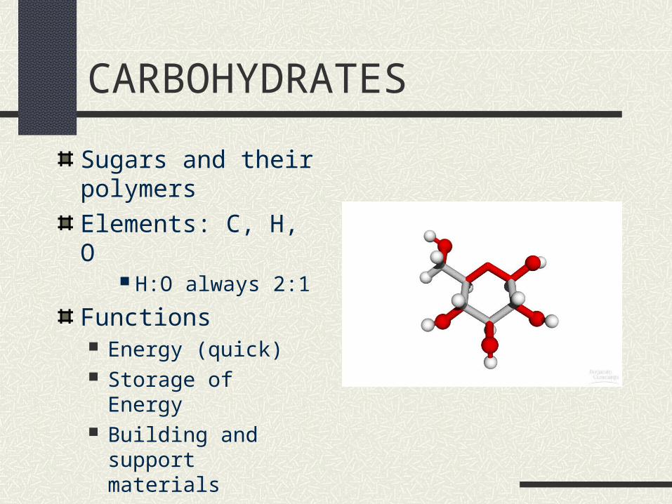
CARBOHYDRATES
Sugars and their polymers
Elements: C, H, O H:O always 2:1
Functions Energy (quick) Storage of Energy Building and support
materials

MonosaccharidesMONOMERS of the carbohydrate Monosaccharide = simple sugar CH2O
Glucose (C6H
12O
6) most common
(and arguably most important)
• FUNCTION: quick energy and as monomers for all other carbohydrate molecules
Side note: can function as raw materials for making other compounds like amino acids and fatty acids.

Monosaccharides
Can be shown as a linear molecule More realistic representation is as a ring Sugars form rings in aqueous solution Note that each bend in the ring is a carbon atom Note that Hydrogens and Hydroxyl (OH) groups extend from each
carbon (except one).
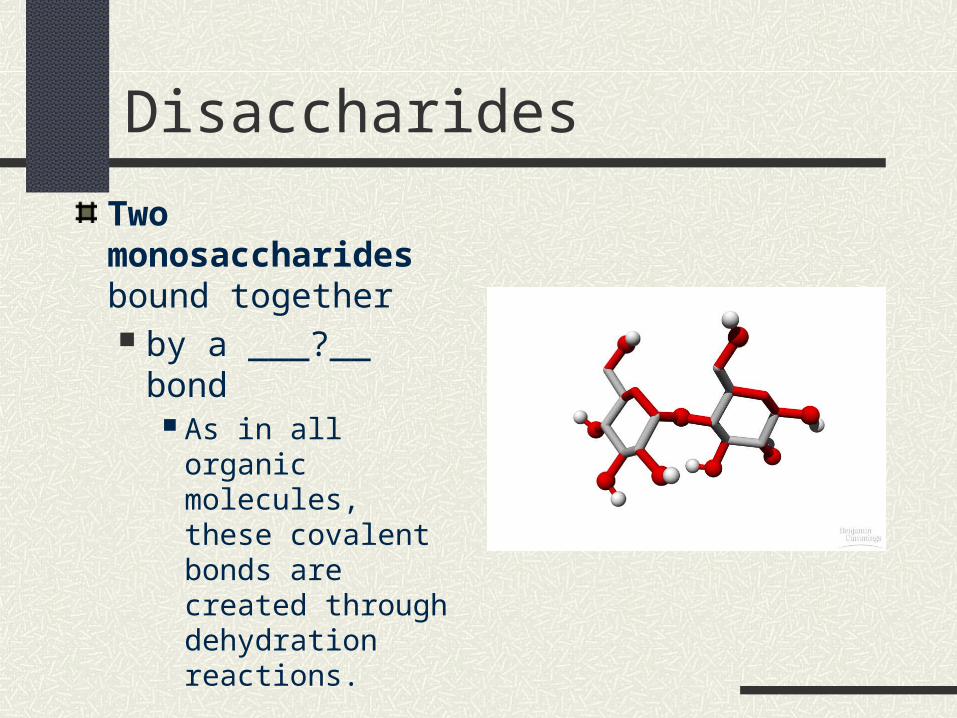
Disaccharides
Two monosaccharides bound together by a ___?__ bond
As in all organic molecules, these covalent bonds are created through dehydration reactions.

Examples of Disaccharides
Glucose + Glucose = Maltose
Glucose + Galactose = Lactose
Glucose + Fructose = Sucrose

Examples of Disaccharides

Functions of Disaccharides
Function 1 Transport in plants Sugar being transported from leaves to roots is
more safe (resists being consumed by the plant) when transported as sucrose.
Side note: Few adult mammals have the necessary
enzymes to break down lactose Preserves milk supply for young who need it

Polysaccharides
macromolecule - few hundred to a few thousand monosaccharides linked covalently (glycosidic linkages)
FUNCTION 1 Energy Storage
FUNCTION 2 Building and support material

Storage Polysaccharides
Starch Made only by plants
(Animals can break down starch , but they cannot make it)

Storage Polysaccharides
Glycogen Storage polysaccharide created and used by
ANIMALS Found in the liver and muscles Highly branched chains of glucose
Only about a day’s supply of glycogen is stored in the body
Note: Polysaccharides are NOT the major energy storage compound in animals that they are in plants

Glycogen – diagram and photo

Structural Polysaccharides
Structural polysaccharides are those that are used in building physical structures in an organism
Most often we think of cell walls in plants, but there are others.
Cellulose
Structural polysaccharide that makes up plant cell walls
The bulk of the woody part of a plant
Cellulose structure Long chains of glucose – similar to starch Glucose molecules are linked differently from starch Difference makes cellulose indigestible to almost all
organisms EXCEPT bacteria and some other microbes

Starch/Cellulose Comparison

Starch/Cellulose Comparison
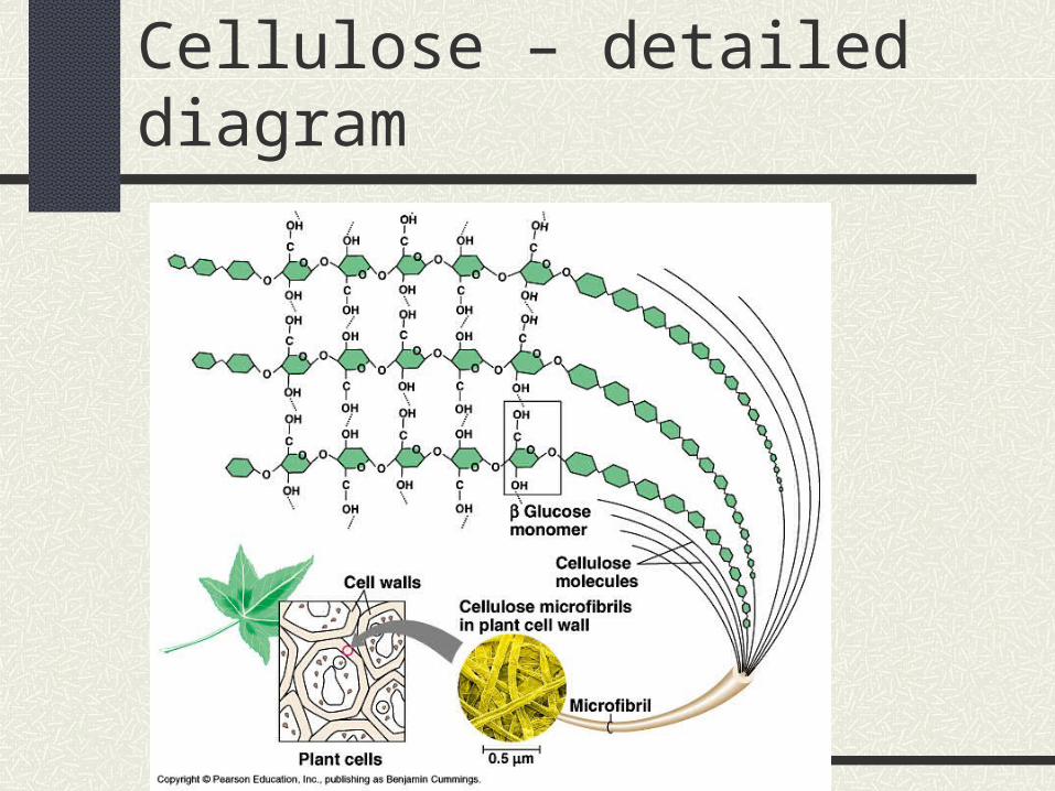
Cellulose – detailed diagram
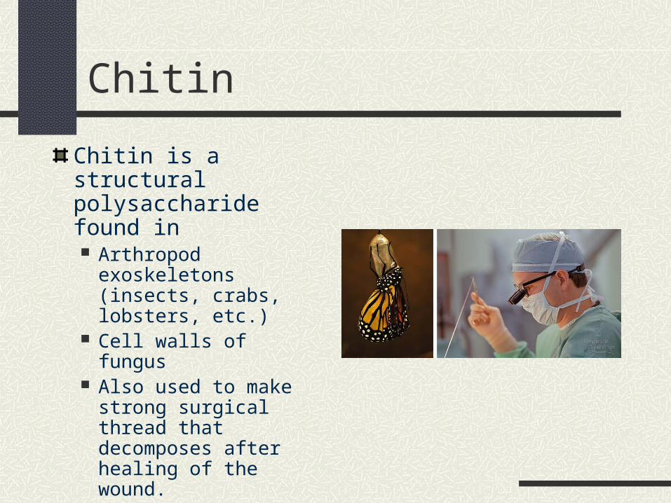
Chitin
Chitin is a structural polysaccharide found in Arthropod
exoskeletons (insects, crabs, lobsters, etc.)
Cell walls of fungus Also used to make
strong surgical thread that decomposes after healing of the wound.

LIPIDS
Elements : C, H, OMajor types Fats and oils Waxes Phospholipids steroids
Functions Energy storage Insulation Cushioning Cell communication Make up membranes

Lipid Structure
Composed of two kinds of smaller molecules Glycerol
An alcohol 3 carbons each with an –OH group
Fatty acids LONG carbon / hydrogen chains Carboxyl group at one end Hydrocarbon tail makes up bulk of the fatty acid
Glycerol linked to 3 fatty acids with ester bonds/linkages
Ester bond = type of covalent bond To view formation of lipids, click HERE

Lipid structure
Lipid structure relates to function
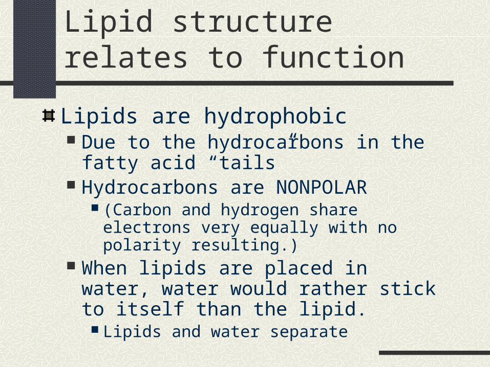
Lipids are hydrophobic Due to the hydrocarbons in the fatty acid
“tails” Hydrocarbons are NONPOLAR
(Carbon and hydrogen share electrons very equally with no polarity resulting.)
When lipids are placed in water, water would rather stick to itself than the lipid.
Lipids and water separate

Saturated vs Unsaturated fats(fatty acid tail comparison)
Saturated fats Each carbon is “holding hands” with the max
number of hydrogen atoms NO double bonds between carbon atoms of the
fatty acid tails Tails are STRAIGHT as a result
Straight tails allow for tight packing Solid at room temperature

Saturated fat – diagram
For more on lipids and saturated/unsaturated fats, click here

Saturated fat – diagram and photo

Saturated vs unsaturated fats
Unsaturated fats At least two carbons in the fatty acid chain are
NOT “holding hands” with the maximum number of hydrogens they can
Instead two of the carbons (or more) are DOUBLY covalently bound to each other.
This results in a bending of the fatty acid tail Crooked tails prevent tight packing Liquid at room temperature

Unsaturated fat – diagram and photo

Functions of Fat
Primary function = Energy Storage One gram of fat stores twice the energy of a
gram of polysaccharide Advantageous to animals that have to move
around – unlike plants that can have unlimited bulk without concern for mobility.
Cells that store fat – adipose cells

Functions of Fat
Other functions specifically related to FAT Cushioning Insulation

Phospholipids
Structure Glycerol TWO fatty acid tails ONE phosphate group – “polar head”
Results in a molecule that is BOTH hydrophobic AND hydrophilic Fatty acids are nonpolar and hydrophobic Phosphate group is polar and hydrophilic

Phospholipid diagram

Phospholipid
Hydrophilic/phobic nature causes phospholipids to naturally form membranes when placed in water (aqueous solution)
To view membrane formation click here.

Steroids
Structure 4 fused carbon rings Various functional groups extend from carbon rings
Functions Roles in cell membrane structure
CHOLESTEROL Maintains cell membrane structure in animals Also is a precursor to other hormones
Cell communication

Steroids - cholesterol

PROTEINS
MANY Important Functions Structural proteins – support
Silk in cocoons/webs; collagen in connective tissue
Storage proteins – storage of amino acids Albumin in egg white; casein in milk
Transport proteins – transport many substances across cell membranes or through the body
hemoglobin Hormonal proteins – coordination of activities
Insulin – controls concentration of sugar in the blood Receptor proteins – receive chemical stimuli and respond Contractile proteins – movement Defensive proteins – protection against disease Enzymatic proteins – speed up chemical reactions!!

Variety of Proteins
Variety within the different types of proteins is staggering! There are many thousands of different types of
enzymes alone – each specifically designed for a particular chemical reaction.

Importance of Shape
Conformation – term for the unique 3-D shape of a protein
Shape is absolutely critical to protein function!!

Protein StructureElements: CHONMonomers = AMINO ACIDS
POLYPEPTIDE is a polymer of amino acids Polypeptide may or may NOT be a fully functional
protein
One or more polypeptides configured in it’s particular shape = a protein
Protein Structure – AMINO ACIDS
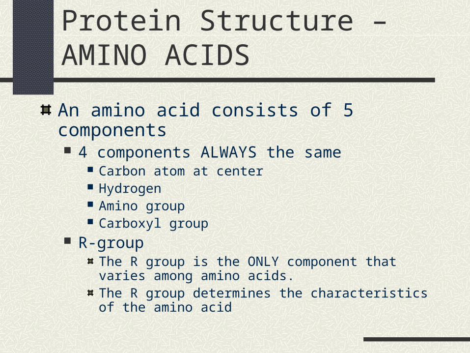
An amino acid consists of 5 components 4 components ALWAYS the same
Carbon atom at center Hydrogen Amino group Carboxyl group
R-groupThe R group is the ONLY component that varies among amino acids.The R group determines the characteristics of the amino acid

Nonpolar Amino Acids

Polar Amino Acids

Forming a Polypeptide
20 different amino acids exist
Can be assembled in any order
Options for HUGE variety of polypeptides

Forming a Polypeptide
To join two amino acids: Carboxyl group of one must meet the amino
group of another An enzyme will join them via a dehydration
reaction The resulting bond is called a peptide bond Repeating the process over and over creates a
polypeptide

Forming a Polypeptide

Formation of a Polypeptide
The repeated sequence of atoms that remains constant from one amino acid to the next is the polypeptide backbone.
The different appendages attached to the backbone are the R groups The reactivity of the R groups with each other
determines many unique properties of each polypeptide chain
Four Levels of Protein Structure
A functional protein is NOT just a polypeptide chain It is one or MORE polypeptide chains precisely
twisted, folded and coiled into a uniquely shaped molecule
ORDER OF AMINO ACIDS determine the 3-D SHAPESHAPE determines how the protein WORKS.
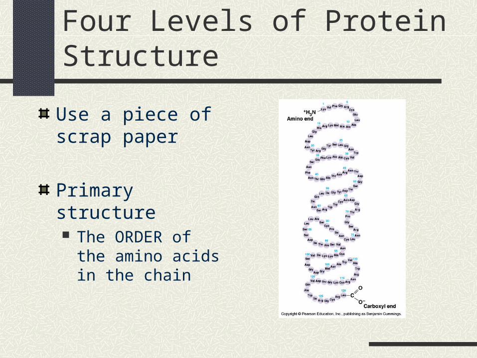
Four Levels of Protein Structure
Use a piece of scrap paper
Primary structure The ORDER of the
amino acids in the chain

Four Levels of Protein Structure
Secondary Structure Result from the
regularly repeating structure of the backbone
Hydrogen bonds between the constant parts of the amino acids
Results in Alpha helix (spiral) OR Beta pleated sheets
(fan)

Four Levels of Protein Structure
Tertiary Structure Results from interactions
between R-groups Hydrophobic interactions
Also involves van der Waals attractions
Disulfide bridges Hydrogen bonds Results in COMPLEX
folding and twisting of the polypeptide

Four Levels of Protein Structure
Quaternary Structure Results when two or more
polypeptide chains combine to make a functional protein
Example – Hemoglobin is composed of 4 chains.
For a protein structure animation, click HERE
Or HERE

Overview of Protein Structure - diagram

Different representations of a protein’s conformation - Lysozyme

Environment and Protein Conformation (SHAPE)
Environment plays an important role in shape of a protein
Environment unsuitable, protein can DENATURE – loss of a protein’s SHAPE (conformation)

What can cause a protein to denature?pH pH changes in the environment can interfere with the ability of a
polypeptide chain to hold its shape by interfering with the hydrogen bonds or other types of bonds within the molecule
Temperature Extremes Temperature extremes, especially HOT temperatures, cause an
increase in molecular movement which can cause the protein to lose its shape
Other causes of denaturing Changes in salinity Moving a protein from an aqueous to some organic solution
Hydrophilic regions of the protein that were once on the outside would move inside and vice versa
• For more on denaturing of proteins, click HERE.• Or HERE

Denatutration of a Protein - diagram

NUCLEIC ACIDS
Genes give the information for constructing proteins
Genes are made of nucleic acids

64

Two types of Nucleic Acid
DNA Deoxyribonucleic Acid
RNA Ribonucleic Acid

DNA
The genetic material inherited from parents

Nucleic Acid Structure
Nucleic Acids (both RNA and DNA) are polymers
The monomers making up these polymers are nucleotides

Nucleotide structure
3 parts Nitrogenous base Sugar (ribose; which is a 5-carbon [or
penotose]sugar) Phosphate group
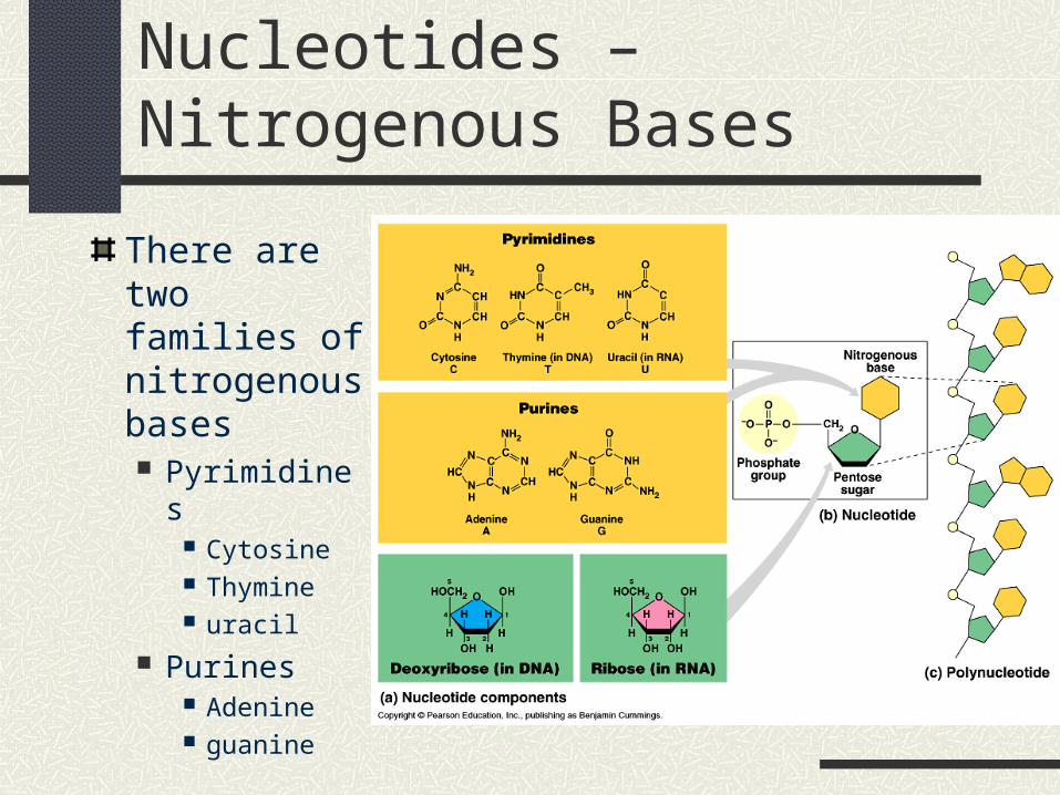
Nucleotides – Nitrogenous Bases
There are two families of nitrogenous bases Pyrimidines
Cytosine Thymine uracil
Purines Adenine guanine

Shape of the DNA Molecule
Double helix
For an animation showing the structure of DNA, click HERE.

http://www.tvcc.edu/depts/biology/HotPot/Biol%201406/biomolecule_structure.htm


