

1
Touch and the body
Andrea Serino 1 & Patrick Haggard 2
1 – Dipartimento di Psicologia and Centro studi e ricerche in Neuroscienze Cognitive ,
Università degli Studi di Bologna.
3 Department of Psychology and Institute of Cognitive Neuroscience, University
College London
Acknowledgements: PH was supported by BBSRC project grant D009529, and by a
research grant from Bial Foundation.
2
Abstract
The dual nature of touch has long been understood. The sense of touch seems to carry
information at the same time about the external object touching our skin, and also about
our body itself. However, the nature of this interaction has remained obscure. We
present an analytic model of how tactile information interacts with mental body
representations in the brain. Four such interactions are described: the link between the
body surface and the maps in primary somatosensory cortex, the contribution of
somatosensory cortical information to mental body representations, the feedback
pathway from such higher representations back to primary tactile processing in
somatosensory cortex, and the modulation of tactile object perception by mental body
representations.
3
Introduction and model
Touch is often considered by neuroscientists under the general heading of
somatosensation. This already reveals a strong link between tactile sensation and
perception on the one hand, and the body on the other. Indeed, the receptor organ for
touch, the skin, also forms the surface of the physical body. Although the
interdependence between the sense of touch and the body is well recognised, this
interaction can take place at a range of different levels within the nervous system, with
quite different consequences and mechanisms. In this review, we first present an
analytical model of the relation between touch and the body, and then use this to
distinguish four different ways in which tactile afferent information can either be
influenced by, or have influence upon, the mental representation of the body.
INSERT FIGURE 1 ABOUT HERE
The model is shown in figure 1. External stimuli (“objects”) impinge upon the
physical body, through contact with the skin. Tactile afferents convey information
about this contact via the medial lemniscal-thalamocortical pathway to the primary
somatosensory cortex (SI) of the contralateral hemisphere. This area therefore contains
an essentially spatial representation of the physical body surface, and is responsible for
primary tactile sensations. That is, the physical body structures tactile sensation,
because the physical body is the receptor surface (Pathway 1 in figure 1).
Mounting neurophysiological and psychophysical evidence shows that other brain
areas house additional mental body representations (MBRs). We will show that MBRs
depend on afferent inputs, relayed through primary representations (Pathway 2 in figure

4
1). However, MBRs are typically multimodal rather than unimodal, and persist even in
the absence of current stimulation. This abstraction from primary sensation allows
MBRs to contribute to cognitive functions including memory, mental imagery etc.
Here, we make the further strong claim that these MBRs reciprocally influence primary
tactile processing in SI (Pathway 3 in figure 1). A second strong claim from this
review will be that MBRs contribute not only to perception of one’s own body, but also
to perception of other objects in the external world. Specifically, tactile and visual
perception of external objects may be body-referenced (Pathway 4 in figure 1).
The structure of our review is based on these four critical pathways linking the
physical body, the sense of touch and the mental representation of one’s own body.
Therefore, we review, in turn, key psychophysical and neurophysiological evidence
that (1) the physical body structures tactile sensation, (2) that tactile sensations
contribute to a Mental Body Representation (MBR), (3) that MBRs reciprocally
influence primary tactile processing, and (4) that MBRs mediate the formation of a
object representations from primary tactile sensations.

5
Pathway 1 - THE PHYSICAL BODY STRUCTURES TACTILE SENSATION
The sense of touch is the phenomenal counterpart of afferent input from
mechanoceptors on the body surface. Peripheral signals from the skin are transmitted
trough the dorsal column of the medulla, and project, via the thalamus, to primary (SI;
area 3 in the monkey) and secondary (SII) somatosensory cortices, respectively in the
postcentral gyrus and lateral sulcus. For details on the mechanoreceptors and the
afferent pathways see the reviews by XXX in this volume. Here we concentrate on the
cortical mechanisms of touch only.
SI neurons encode physical proprieties of tactile stimuli within a spatial map.
Tactile sensations are localized on a given part of the body thanks to the organization of
primary somasensory cortex. SI of each hemisphere contains a complete topographical
organized representation of the controlateral side of the body. This “somatosensory
homunculus” is inverted relative to the physical body, with the legs represented
medially and the face and hands more laterally (Penfield & Boldrey, 1937; see Figure,
1). The relationship between space on the body and in SI homunculus has been clearly
demonstrated by classic neurophysiological experiments. Tactile stimuli administered
on a given body part elicits a neural response in a specific portion of SI homunuculus,
matching the same body part. Conversely, the electrical stimulation of the same region
of SI induces a tactile perception localized on the corresponding part of the body (see
also Kaas et al., 1979 and Tommerdhal et al., 1993 for data on animals and Yang et al.,
1993; Shoham & Grinvald 2001; Sato et al., 2005 for data on humans).
.

6
Neurophysiological studies on the structure of post central sulcus showed that SI is
organized in cortical columns, up to 500 m wide, which receive projections from a
restricted population of mechanoceptors (Kaas et al., 1979; Mountcastle, 1997). Thus
each column has a well defined receptive field. Adjacent neurons on SI surface tend to
have adjacent receptive fields on the body (Blakenburg et al., 2003; Penfield et al.,
1950).
Within this somatotopic map, there is a threeway relation between receptive field
size, extent of representation in SI, and tactile acuity., The size of individual receptive
fields on the skin size varies among body parts and therefore among regions of SI also.
At the same time the size of SI representations varies among body parts. For instance,
skin regions such as lips and fingers have large SI representations, while the back and
torso have small representations. Skin regions differ in the degree of tactile information
they supply: tactile spatial acuity on the finger is for instance about twenty times
greater than on the back. Tactile acuity is an inverse function of the receptive field size
of SI neurons (Brown et al., 2004): body parts with high tactile acuity have densely
packed mechanoreceptors on the skin, thus they have smaller cortical receptive fields,
and are largely represented in SI. Other skin regions have fewer mechanoreceptors,
small SI representations, with large receptive fields, and low tactile acuity. This
produces the classic correlation between size of SI representation and tactile spatial
resolution (Weinstein et al., 1968): body parts extensively represented in SI have better
tactile acuity.
Thus, the phenomenal experience of touch on a given part of the body depends on a
close linkage between peripheral signals from the skin and firing properties of a given
population of neurons in SI. Sometimes, this correspondence may be disturbed, because
7
of lesions or experimental manipulations. In these cases, very interesting phenomena
arise both at level of neural networks and of phenomenal sensation. These effects might
contribute to elucidate the link between primary sensory activity and conscious
experience.
Amputation
The “phantom limb” experience is a striking example of the link between primary
cortical activity and conscious experirence. This term, initially introduced by Mitchel
(1871), refers to patients’ experience of an amputated limb as still present. –Patients
feel tactile and also sensations to arise from their missing limb. Importantly, patients
are aware of their amputation and that these sensations are not veridical. They
experience an illusion, not a delusion, as Ramachandran (1998) well underlined.
Phantoms limb sensations are initially reported in the vast majority of amputated
patients, and then progressively disappear in few days or months, although illusions
persisting for decades have also been described (Sunderland, 1978). Normally
phantoms are reported following amputation of an arm or leg, but also cases involving
other body parts have been described (Scholtz, 1993; Aglioti et al., 1994; Hoffman,
1955; Sacks 1992). Phantoms are experienced as occupying the “habitual” posture of
the amputated limb. In many cases, especially in the first period after amputation,
patients claim they can generate voluntary movements in their phantom. This suggests
that phantoms closely mirror proprieties of real limbs.
One interesting feature of phantom limb is referred sensation, i.e. sensation
localized to a phantom body part after tactile stimulation of a remote part of the
subject’s body (Ramachandran et al., 1992; Halligan et al., 1994; Aglioti et al., 1997).

8
In patients with amputation of the upper limb, referred sensations are often reported
after stimulation of the lower face, ipsilaterally to amputation: tactile stimuli
administered on adjacent points of the face elicit both a tactile sensation on the face and
a referred sensation on the phantom hand, in a one to one spatial correspondence.
Moreover, referred sensation closely reflects perceptual characteristics, such as
intensity and frequency, of delivered stimulation.
Referred sensations have been interpreted as a sign of cortical reorganization of
primary sensory cortices following amputation. Absence of normal afferent inputs
from amputated body regions to the matching portion of SI leads to a reorganization of
neighbouring portions of the SI map. , as the latter regions “invade” the cortical
territory previously representing the amputated body part. In the case of phantom arm,
SI area mapping the face expands into the territory of the hand. Thus, the hand area
now responds also to stimuli delivered to face area, this resulting in a “duplicated”
tactile sensation on the face and on the phantom hand. Neuroimaging studies in
amputees confirm that tactile inputs from the face elicit neural responses also in the SI
hand territory (Fuhr et al., 1992; Kew et al., 1994; Yang et al.,1994; see Ramachandran
1993 for a review).
Findings with patients suggest considerable plasticity in human somatosensory
cortex. Experiments in animals have allowed more controlled investigation of these
phenomena, and have shed light on the neural mechanisms of plasticity. . Merzenich
and colleagues extensively studied the effects of amputation on topographic
representations of the body in areas 3b and 1 (corresponding to SI) in the monkey. Two
months after amputation of one digit most of the cortex that originally responded only
to the skin surface on the amputated digit, now responded to inputs from adjacent digits

9
or the subjacent palm. The cortical area undergoing topographic changes was confined
to the immediate boundaries of the amputated digit representation. Moreover, most
changes involved new representation of immediately adjacent digits, while there was
no significant increase in the representation of more distant digits (Merzenich et al.,
1984). Therefore, cortical reorganization after amputation is quite rapid, systematic and
follows the organisation of the SI map, even when large portions of somatosensory
cortex are involved.
A candidate mechanism for this form of plasticity is the unmasking of existing
synaptic connections between adjacent areas of SI (Ramachandran & Histein, 1998;
Buonamano & Merzenich, 1998; see Kew et al., 1997 for a discussion). Adjacent
columns of SI may be laterally interconnected. These lateral inputs may normally be
silent or below threshold, because they are masked from stronger inputs from
periphery. The existence of silent connections in normal conditions between hand and
face representations, for instance, has been recently demonstrated by Tanosaki et al.
(2003). They showed that in healthy subjects the somatosensory magnetic field evoked
by tactile sensation on the face was modulated by concurrent electrical stimulation of
the thumb. However, when a portion of SI is no longer “fed” by its proper signals,
lateral connections from neighbouring representations are unmasked and therefore
produce an over-threshold activity, in relationship with stimuli presented on a different
body location.
Interestingly, enlargement in cortical representation after amputation may not
improve functional touch. Indeed, studies which assessed tactile acuity in body parts
proximal to the amputation, and therefore likely to benefit from enalarged cortical
representation, both in monkeys (Vega-Bermudez & Johnoson, 2002) and in humans

10
(Teuber et al. 1949; Haber, 1958; Braune and Schady, 1993; Flor et al., 1998; Grusser
et al., 2001) found no significant improvements in tactile sensitivity. Thus the
relationship between plastic changes in the extension of cortical representation and
functional tactile processing remains unclear.
Transplantation
In principle, restoring peripheral inputs after trauma right re-establish, the
relationship between neural activity and tactile sensation. Indeed, effects of limb
amputation can be reversible in cases of transplantation.
This surgical procedure is based on regenerative proprieties of peripheral nerves.
Since pioneering studies by Head and Rivers (1908), it has been known that a divided
and reunited nerve regenerates. Head and Rivers recorded the recovery of sensation
after the division of nerves in Head's own arm. They described two definite stages in
the return of sensibility. In the first stage, the sensations are vague and crude in
character, without clear perceptual ability of either discrimination or localisation. The
second stage of the process of regeneration is characterised by the return of those
features of normal cutaneous sensibility, such as exact discrimination and localisation,
which underly normal touch and haptic interaction with objects. They concluded that
progressive nerve regeneration is related to recovery of sensation and functional touch.
This basic mechanism is effective also when all sensory afferents from an
amputated limb have been absent. An extraordinary demonstration comes from the case
of a patient (C.D.) who underwent a bilateral hand transplantation (Giraux et et al.,
2001). C.D.’s ability to localize tactile stimuli was tested 5 months and 11 months after
11
surgery (Farnè et al., 2005). Initially C.D. was able to perceive touch on his hands,
however sensation on one hand was hampered when concurrent stimuli were
administered on the ipsilateral cheek: right face stimulation extinguished right hand
perception. This was due to competition between hand and face representations
following long term absence of afferent inputs from the hands (see Amputation section
above). However, 11 months after surgery, face-hand extinction was no longer present.
This suggests that incoming sensory stimuli from re-planted hands drove a
reorganization of somatosensory representations.
Congenital absent limbs.
Afferent input from the physical body therefore plays a key role in structuring the
spatial maps in SI. This raises the question whether the neural representation of the
body is constrained by an intrinsic organising principle, or whether its organisation is
derived from experience of afferent inputs. Studies of phantom sensations in people
with congenitally absent limbs suggest that some innate structure of tactile
representation exists even in the absence of any relevant peripheral inputs. (Weinstein
& Sersen, 1961; Weinstein et al., 1964; Poeck, 1964; Vetter & Weinstein, 1967; see
Brugger, 2006 for a recent review). Thus, some individuals with congenital phantoms
claim to be able to move their phantoms. fMRI data showed that phantom limbs
movements activated cortical areas similar to those described in traumatic amputees.
TMS stimulation of controlateral motor cortex evoked sensations of movement in the
phantom limbs (Brugger et al., 2000). However, a recent study suggests that the neural
substrates of these sensations are primarily in motor rather than somatosensory brain
areas (Bestmann et al. 2006 ).

12
These findings have been taken as an evidence for an innate representation of the
structure of human body (Melzack, 1997). In this view, peripheral stimulation or
movement of a body part are not strictly necessary to drive representation in primary
cortices. The alternative possibility is that visual experience of other people moving
their extremities have activated networks mediating visuo-motor limb representation
(Brugger et al., 2000). The mirror system (see Rizzolatti & Cragheiro, 2004 for a
review) would be the mechanism underlying this effect.
While most studies have focussed on movement sensations in congenital phantoms,
one study has reported tactile experiences in the phantom limb evoked by touching the
stump (Melzack et al., 1997).
Elongation
Gradual changes in the physical body occur throughout the life span, in addition to
sudden changes such as amputations. These changes are normally too slow to produce
measurable effects in tactile maps within SI within the time-frame of most studies. In
addition, the most obvious gradual change, i.e., growth and maturation, involves
expansion of the physical body. In contrast, traumatic changes such as amputation
involve contraction. Therefore, post-amputation plasticity may be a poor model of
plasticity during natural growth. However, in the case of achondroplastic dwarfs, body
parts may be surgically elongated within a time interval of some months. This provides
a good experimental model for studying developmental neuroplastic change, because
there is an appropriate time-scale, a well-defined and controlled intervention, and a
healthy brain. Di Russo at al., (2006) recently studied cortical and perceptual
reorganization following progressive elongation of lower limbs in two patients. In this
13
procedure, an external device is fixed on the bones and progessively separates two bone
segments by about 1mm per day, up to an elongation of 10-15 cm of the lower limb.
Patients were tested before and after this surgical procedure, and at six month follow-
up. Somatosensory evoked potential and FMRI scans following tactile stimulation of
the knee and the foot changed after surgery. In particular, the foot representation
enlarged and shifted medially after the lengthening phase. This shift would allow
additional cortical territory for representing touch on the now-enlarged leg.
Interestingly, these SI changes also had implications for mental body representation,
which are discussed in the next section.
Deafferentation
The relationship between peripheral inputs and SI representation has been also
extensively investigated through deafferentation. When afferent inputs from the body
surface cannot reach matching portions of primary somatosensory cortices due to nerve
resection or anaesthesia, these regions plastically re-organize so that they respond to
stimuli presented on adjoining body parts.
For example, transecting the median nerve of a monkey, removes inputs to
somatosensory areas from ventral portion of digits D1-D3. Just immediately after this
manipulation, a limited sector of cortex previously mapping these body parts,
responded to inputs from the dorsal skin and the bordering zone on digits D3 and D4
(Jekins et al., 1990; Merzenich et al., 1993). A similar demonstration in on a larger
scale has been reported by Pons et al., (1991) who mapped the cortex of monkeys that
had undergone deafferentation of the dorsal roots (C2-T4) several years before. This
14
manipulation resulted in deprivation of a cortical area of over 1cm2 of its normal inputs
from arm and hand. Cortical maps reorganized so that deprived areas developed novel
responses to neighbouring skin areas, including face and chin.
Forms of short term cortical reorganization due to deafferentation have also been
shown in humans after anesthesia. Rossini et al. (1994) recorded Somatosensory
Evoked Fields (SEFs) during electrical stimulation of the 1st, 3rd, or 5th finger after a
complete ischemic anesthesia of the 4 non-stimulated fingers. They observed that
cortical responses from the unanesthetized fingers were increased following a relatively
brief period of anaesthesia of the adjacent finger.
These changes also influence tactile perception. Cutaneous anaesthesia of the right
hand improves spatial tactile acuity in the left hand. This effect was associated with a
change in evoked cortical potentials recorded from the right somatosensory cortex
(Werhanhn et al., 2002; see also Bjorkman et al., 2004). Importantly, these effects
follow a somatotopic principle, since anaesthesia of the foot did not affect touch on the
hand. Bjorkman et al, demonstrated that this manipulation is also effective in patients
suffering partial somatosensory loss due to injuries in median or ulnar nerves,
suggesting possible clinical applications.
These effects have been interpreted as a consequence of unmasking of existing
horizontal connections between homologues regions of somatosensory cortices of the
two hemispheres via corpus callosum, similar to those evoked by amputation (see
above). These projections makes excitatory contacts onto pyramidal cells and
interneurons: they provide both monosynaptic excitation in SI, as well as disynaptic
inhibition through excitatory synapses with inhibitory interneurons (Carr and Sesack
1998; Somogyi et al. 1983). Depressing the activity of the contralateral SI cortex by
15
means of peripheral anaesthesia might reduce controlateral inhibition and thus
"unmask" normally suppressed responses (Pluto et al., 2005). These processes should
act on the gain of neural populations, changing the spatial tuning of neurons’ receptive
fields, thus affecting tactile sensitivity.
Besides inter-hemispheric interaction, similar effects have been also demonstrated
intra-hemispherically. Indeed an improvement of tactile sensation has been shown
within the same hemisoma: anaesthesia of a limited portion of the right forearm by
means of a local anaesthetic cream, resulted in tactile improvement on the right hand
(Bjorkman et al., 2004).
These effects occur after some minutes of anaesthesia and are totally reversible
after the end of anaesthesia. When tactile afferents recovered the system reorganized,
to its original balance. This evidence further supports the idea SI organization is
determined by competition between neighbouring population of neurons. The activity
of each population depends on peripheral inputs from the skin.
Experience
Finally, the relationship between the physical body and SI structure is also shown
by the effects of tactile experience. The structure of afferent inputs affects both SI
representation and tactile sensitivity.
It is well known that subjects who exercise their tactile abilities to an extraordinary
degree in everyday life have an expanded representation of the trained body part in
primary somatosensory cortices. Pascual-Leone & Torres (1993) demonstrated that a
sample of Braille readers, who show superior tactile abilities as a group (Van Boven et
al., 2000), showed enlarged somatosensory representation of the right index finger,

16
which they used in Braille reading, compared to the left index finger, which was not
used in reading.. In the same vein, Ebert et al. (1995), showed that in professional
string players, tactile stimulation of left hand digits elicited a larger and enhanced
activation in SI corresponding regions in comparison to non-expert controls. The
effects were specific for the left hand, used to finger the strings.
An analogous plasticity has been shown in the monkey: prolonged training of
tactile stimulation to a restricted part of the distal pad of one of two phalanges,
produced an expanded cortical representation in area 3b. The effect was specific for the
portion of the cortex mapping the part of the digit which had undergone the training
(Recanzone et al., 1992).
Similar results have been shown in humans. In Braun et al.’s study, (2000) subjects
performed for 4 weeks, 1 hour per day, a tactile orientation task, administered on the 1st
and the 5th finger. Neuroelectric source imaging showed that SI representations of the
two fingers were further apart from each other than before the training, this suggesting
an expanded representation.
Plastic reorganization of somatosensory maps has been shown also in conditions of
entirely passive tactile stimulation. In anesthetized rats, a few hours of simultaneous
passive stimulation of two adjacent parts of hindpaw resulted in an increase of
corresponding SI territory (Godde et al., 1996). The effect was reversible after the end
of stimulation. In a further study on humans, similar “co-activation” stimuli were
applied for two hours on the right index finger. This passive stimulation produced an
improvement of tactile acuity, specific for the stimulated finger, which lasted for some
hours. If stimulation was repeated for 3 consecutive days, the duration of the effect
increased. Stimulation lasting less than 30 minutes was not effective (Godde et al.,

17
2000). Somatosensory evoked potentials showed that simultaneous stimulation induced
a shift in the source of electrical activation related to the stimulated finger, compatible
with an enlargement on SI representation. The degree of the shift was correlated with
the improvement of tactile performance after simultaneous stimulation (Pleger et al.,
2001; see also Pleger et al., 2003; Godde et al., 2003 and Hodzic et al. 2004). These
changes appeared not to depend on attention or motivation. Rather, they have been
interpreted in terms of automatic plastic reorganization of somatosensory maps based
on a Hebbian mechanism (Hebb, 1949): temporally-correlated inputs to adjacent skin
regions body induce reorganization of corresponding regions of SI.
A strong demonstration of this proposal comes from the evidence that in the
monkey prolonged simultaneous stimulation of different two fingers resulted in a
fusion of SI representation of the two fingers, whereas asynchronous stimulation
resulted in segregation (Wang, 1995). Analogously, Clark and colleagues demonstrated
that surgical fusion of two digits in the monkey resulted in a fusion of SI digits’
representations (Clark et al., 1998; Allard et al., 1991).
An elegant demonstration of the co-activation principle in humans came from Sterr
et al. (1998a; 1998b), who compared the ability of localizing tactile stimuli
administered on different fingers in Braille readers who normally used three fingers and
in Braille readers who use only one finger to read. Three finger Braille readers more
frequently confused which finger was touched than one finger readers. At the same
time, finger representations in SI differed between the two groups: in three-finger
readers the representations of the 3 reading digits were disordered compared to those in
one-finger readers.
18
At the same time, a number of experimental results indicate important constraints
on general principles of experience-dependent plasticity. First, touch improvement in
normal subjects has been obtained also by asynchronous stimulation (Blake et al.,
2005), suggesting that the temporal window which constitutes correlated tactile
experience is ill-defined. Second, simultaneous stimulation leads to improvement in
spatial discrimination within a body part, but also to confusion between body parts in
tactile localisation tasks (Braun et al., 2000). The length of lateral inhibitory
connections may constrain the effects of hebbian plasticity in SI. Closely adjacent skin
regions, for instance on the same finger, are likely to be connected by such
interneurons. Lateral inhibition then tends amplify even small differences between the
responses of simultaneously-activated afferents. Conversely, afferents projecting from
skin regions that are far apart are presumably less interconnected by such interneurons,
so are less sensitive to this effect.
A single general mechanism of competitive inhibition might underlie the
interactions between physical body structure and SI body maps revealed by amputation,
elongation, transplantation, deafferentation and experience. Specifically, competition
between afferent signals from different body parts or skin regions is resolved by lateral
inhibition provided notably by intracortical interneurons (Brecht et al., 2003). As a
result, a given cortical neuron will respond preferentially to touch on a given skin
region (i.e., its receptive field), but could potentially respond to touch on other,
adjacent regions if the dominant input were suppressed or removed. Thus, tactile
sensation in SI reflects a dynamic balance between excitatory afferents and inhibitory
interneurons. Alterations of the physical body induce reorganization. Therefore, inputs

19
from neighbouring regions rapidly invade the territory previously mapping a body part
that is removed, producing referral of phenomenal experience. This shows that the
relationship between touch and body representation is bidirectional. The peripheral
origins of tactile afferents define the neural maps that give rise to tactile experience on
the body. At the same time, the organisation of the neural map in SI determines how
tactile stimulation of a particular body location will be experienced.

20
Pathway 2 - TACTILE SENSATIONS CONTRIBUTE TO MENTAL BODY
REPRESENTATIONS
Processing of tactile information does not end at the primary somatosensory cortex.
Broadly speaking, brain areas showing tactile responses beyond SI can be classified in
two ways, following Katz’s (1925) distinction between exteroceptive and interoceptive
touch. On the one hand, areas such as SII are concerned with further processing of
tactile object features. Neurons in these areas have larger receptive fields but more
precise tuning properties than SI, suggesting a role in feature extraction (Fitzgerald et
al., 2006a,b). Other areas appear to contain representations related to the body itself.
While neurons in these areas may have tactile responses, they appear to code the state
of the body, rather than the properties of the external object. We use the term Mental
Body Representation (MBR) to refer to such representations. The evidence for MBRs,
and the various forms of MBR that may exist has been discussed in detail elsewhere
and is beyond the scope of this article (Haggard & Wolpert., 2005; Berlucchi & Aglioti,
1997; Gallagher, 2005; Dijkerman & de Haan 2007). For present purposes, we use the
term MBR to refer to an abstract representation of one’s own body, derived from
sensory input but capable of being dissociated from it, and playing a functional role in
perception and/or action.
A broad distinction has been drawn between two types of MBR, often called body
schema and body image respetively (Gallagher, 2005). The body schema represents the
positions of body parts in space, relative to each other. It is of primarily proprioceptive
origin, short-lived, and updated as our bodies move. It serves to guide our actions and
our interactions with the external world. The body image represents the canonical
21
appearance of the body as an object in third person perspective, is primarily of visual
origin, and remains relatively fixed over time and as the body moves and changes. It
may contribute to distinguishing between the body and external objects, and contribute
more widely to sense of self and personal identity. Often however, the neutral term
MBR is preferable to body image or body schema, since representations of the body
may include features of both types.
Rubber hand illusions and mental body representations
Pathway 2 of figure 1 shows that tactile information provides an important input to
MBRs. A simple demonstration of this point comes from the Rubber Hand Illusion
(RHI; Botvinick & Cohen, 1998). View a rubber had being stroked in synchrony with
the unseen stroking of one’s own hand produces the strong phenomenal experience that
the rubber hand is in fact one’s own hand. An external object (the rubber hand)
becomes phenomenally incorporated into one’s own body. A convenient quantitative
proxy for the illusion is a shift in the proprioceptively-perceived position of one’s own
hand towards the rubber hand. Correlated primary tactile and visual input therefore
clearly influence the MBR (pathway 2 in figure 1). Here we focus only on two studies
which considered the specific contribution of touch to the RHI:.the neural basis of the
RHI and the conditions that induce it have been discussed elsewhere (Ehrsson, 2004;
Tsakiris et al.,, 2006).
Tsakiris, Prabhu and Haggard (2005) compared the RHIs induced by tactile and by
somatosensory stimulation with those induced by voluntary action. In the tactile
condition, subjects were stroked on the index or little finger while viewing a
synchronous or delayed video image of the stroking. The RHI was measured as a

22
proprioceptive drift of the stimulated finger, or the unstimulated finger towards the
video image. Results showed that only the stimulated finger exhibited an RHI, and that
this did not transfer across the hand. For example, stroking the index finger produced
an RHI for the index but not for the little finger. Similar results were found for passive
extension and flexion of the same fingers. In contrast, RHIs generated by volulntary
extension and flexion actions of the same fingers generalised more successfully across
the hand, such that moving the index finger generated RHIs for both index and little
fingers. Action then, produced a coherent, generalised change in the MBR, whereby
the entire video hand was incorporated. Primary tactile stimulation, in contrast,
produced a purely local MBR, confined to the touched finger. This result suggests that
touch does contribute to MBRs in a feedforward fashion. However, the normal unity
and coherence with which we represent our own body, and which may underlie the
unity of self-consciousness, comes from efference, rather than from afferent sensation.
Costantini and Haggard (2007) investigated the frame of reference used to construct
MBRs from tactile input, using a sensitivity analysis. They gradually rotated the
subject’s hand through 0, 10 or 20 degrees while inducing the RHI using tactile
stroking. They found a partial level of RHI in the 10 degree rotation condition. They
also independently manipulated the direction of tactile stimulation of the subject’s
hand. Either stroking direction could rotate with the subject’s hand, or it could remain
aligned with the viewed stimulation of the rubber hand. In the former case, tactile and
visual stimulation would be congruent in hand-space though incongruent in external
space. In the latter case, tactile and visual stimulation would be congruent in external
space but incongruent in hand-space. The results showed that visual and tactile
stroking produced larger RHIs when congruent in hand space (stroking direction

23
changed as hand orientation changed) than when congruent in external space. Touch
again induced a change in MBR. However, the key point for present purposes is that
tactile information was first transformed into the spatial frame of reference of the
subject’s own hand, and only then matched with visual stimulation to update the MBR
by incorporation of the rubber hand. Tactile stimulation is first referenced to an MBR,
and the MBR is then updated according to principles of multisensory integration. Just
as the receptive fields of bimodal neurons move with the hand (Graziano et al., 1994),
so do the processes that construct the MBR. Put another way, pathway 2 does not
simply involve a feedforward adjustment of MBRs on the basis of tactile input. Rather,
MBRs assimilar current tactile input when this is coherent with the general description
of the body that they maintain. Since the MBRs in this study were based on
proprioceptively-perceived orientation of the hand, these results constitute a body
schema effect rather than a body image effect.
Volume of afferent transmission influences MBRs
Several lines of evidence suggest that the amount of tactile information transmitted
from the body to the cortex directly affects MBRs. Gandevia and Phegan (1999)
reported a change in body image induced by digital anaesthesia following nerve block,
cutaneous topical application or cooling. Subjects were asked to draw the size of their
thumb before and after each of these anaesthetic interventions. Reduced afferent
transmission due to anaesthesia lead to an increase in the perceived size of the thumb.
Interestingly, the perceived size of the lips, which overlap the thumb in SI
representations, showed a similar increase, while the perceived size of both index
fingers was not affected. The nature of this effect suggests that an MBR used to

24
represent body part size depends directly on tactile inputs The topography of the effect
moreover suggests that this input comes from SI. Our argument that MBRs are
abstracted from sensory input, rather than direct reflections of it, is bolstered by the fact
that none of the intrinsic somatoreceptors can provide a direct representation of the size
of body parts. The MBR specifying size is constructed from primary representations,
but is not merely a trivial recoding of such representations.
We have reviewed above the changes in primary tactile representation caused by
surgical elongation of the limbs (Di Russo et al., 1996). The same study also provides
evidence that the changing information about the body, including changing tactile
afferent input, produced a change in the patients’ mental representation of their own
body. The authors used a “Test of body schema” (Daurqat-Hmeljiak et al., 1978) to
investigate this point. Note, though, that by the definitions above the test may
investigate both body image and body schema. In this task subjects are requested to
put one tile depicting a body part in the appropriate position on an empty board where
just face contours were drawn. Nine different tiles, corresponding to the major body
parts such as limbs, trunk etc., are placed in succession and them removed, so that only
one tile is visible at any one time The task therefore involves knowledge of the spatial
arrangement of body parts relative to a putative cephalic “egocentre”. The subjective
body representation corresponded fairly closely to the patient’s actual body form before
surgery, but was deeply disturbed immediately after surgery. At six-month follow up
the representation again corresponded fairly closely to the patients now-elongated
actual body. This suggests that surgical elongation rapidly altered patients’ subjective
body perception. However, within 6 months, patients’ MBRs progressively adapted to
their altered physical body.
25
Pathway 3: MENTAL BODY REPRESENTATIONS RECIPROCALLY INFLUENCE PRIMARY
TACTILE PROCESSING
So far we have reviewed evidence showing that the tactile representation of the
body contributes to more abstract, multimodal representations of the body in the brain.
In this section we will show how MBRs in turn influence primary levels of tactile
processing. In particular, we focus on how visual information related to the body
affects tactile sensation.
Visual enhancement of touch
It is a common experience in everyday life to gather concurrent visual and tactile
information during objects manipulation, grasping, and tool-use. Visual information
often concerns objects we interact with, and effects of our interaction on the external
environment. However, we often see our own body parts, particularly the hands,
during such interactions. Therefore, many functional interactions with objects involve
touch and simultaneous vision of both body and touched object. Visual and tactile
information related to the body are integrated in specialized brain structures (see
Pathway 2). Several pieces of evidence show that these integrated inputs have an effect
on primary tactile processing.
First, viewing the body accelerates tactile processing. Tipper et al., (1998) showed
that reaction times to tactile stimuli on the hand were faster when subjects could see
their stimulated hand in a video monitor, even when the tactile stimuli themselves were
invisible.

26
Further studies showed that visual information related to the body also improves
tactile acuity. Kennett et al., (2001) assessed tactile acuity on normal participants’
forearms by means of two point discrimination threshold (2pdt), while subjects viewed
either their forearm, or a neutral object, presented in the same spatial location as their
arm, or were blindfolded. Tactile acuity improved when subjects viewed their
stimulated arm, compared to conditions of viewing the object or no visual stimulation.
These results clearly show that viewing the body boosts tactile processing; this effect
has been termed visual enchacement of touch (VET). The basic effect has been
replicated a number times (see Taylor-Clarke et al., 2002; Press et al., 2003; Serino et
al., 2006).
Importantly, VET does not involve visual information about the stimulus: vision is
always non-informative. This feature makes VET different from other forms of cross-
modal interaction between vision and touch, (see Spence and Driver, 2004) which
focus on optimal integration between information from different sensory modalities
about the same stimulus. In VET, visual information is not related to any particular
external stimulus, but rather defines a context to which tactile stimulation is referenced
(see below).
Moreover, VET cannot be a simple effect of spatial attention, in the sense of
convergence of visual and tactile attention towards the same location. The VET effect
exists even when effect of attention is experimentally controlled. Typically, tactile
performance when viewing the stimulated body part is compared with that when
viewing a neutral, non-body object presented in the same spatial position as the
stimulated body part, by means of mirrors or cameras. Thus, since visuo-spatial
27
attention is oriented towards the same location in external space in both conditions,
general mechanisms of spatial attention cannot explain the effect.
Thus, we suggest that viewing the body facilitates tactile perception, independently
from visuo-spatial orienting to the location of the body. We now consider some
possible mechanisms underlying VET. First, viewing a part of the body subject to
tactile stimulation seems to improve representation of tactile space for that body part.
(Haggard et al., 2003). This interpretation is strongly supported by a further result from
Kennett et al,’s (2001). They found that: tactile acuity further improved in a condition
in which a magnified view of the forearm was shown by means of a lens, compared to
just viewing the arm directly. This suggests that increasing spatial details of visual
information further enhanced spatial resolution of tactile system.
The interpretation of VET we propose has a strong implication: the relationship
between visual information related to the body and tactile sensation should strictly
concern spatial proprieties of perceived stimuli. Therefore, tactile spatial acuity can be
modulated by viewing the body, whereas other physical proprieties of tactile
perception, such as simple detection or perception of intensity or temporal frequency of
tactile stimuli, should be less affected. Press et al. (2003) confirmed this prediction,
since they demonstrated that viewing the body affects tactile perception only for a
spatial discrimination task and not for a simple detection task. Indeed subjects were
faster in discriminating between the spatial position of two tactile stimuli given on the
forearm when they looked at their forearm rather than at a neutral object, presented in
the same spatial location. On the contrary, reaction time to simple detection of tactile
stimuli, without any spatial discrimination, did not vary among experimental
conditions.

28
In addition, Press et al.’s work described another interesting propriety of VET, that
is the benefit of vision on touch occur only when spatial discrimination tasks are close
to perceptual limits. Viewing the arm improved reaction time only if spatial separation
between tactile stimuli to be discriminated was close to the participant’s discrimination
threshold, and not when stimuli were widely separated. This finding suggests that
visual information related to the body increases spatial sensitivity of touch. This
conclusion has been confirmed by a recent study by Serino et al. (2006), who showed
that the VET effect varies among individuals in inverse proportion to their tactile
acuity. Subjects showing poor tactile ability while viewing a neutral stimulus, taken as
a baseline condition, showed greater improvements when viewing the stimulated body
part. In close agreement with this result in normal subjects, Serino et al. (2006) further
showed that viewing the body was effective in patients whose tactile sensation was
reduced following brain lesions. This last result suggests a potential cross-modal
therapy based on the VET effect.
VET thus follows an inverse effectiveness law. This very general feature of
multisensory integration has been shown in both single cell recordings and
psychophysical studies (Stein & Meredith, 1993; Stein, Jiang & Stanford, 2004;
Stanford, Quessy & Stein, 2005; Frassinetti et al., 2002). According to the inverse
effectiveness principle, signals from different modalities are more strongly integrated
close to unimodal thresholds, when modality-specific signals are individually less
effective in producing a unisensory response. In the case of visuo-tactile interaction
related to the body, visual information is used to boost tactile sensation only when
touch alone cannot solve spatial discriminations close to perceptual limits. When more

29
tactile information is available, and tactile tasks are easy, the benefit is no longer
apparent.
INSERT FIGURE 2 ABOUT HERE
We now present possible neural and functional mechanisms underlying VET effects
(figure 2). Performing a tactile spatial discrimination task while viewing the body
implies perceiving and integrating tactile and visual information. These unimodal
inputs are first processed in segregated unisensory brain areas: visual information
should be processed in high order visual areas representing the body and body parts
(Downing et al., 2001; 2007), whereas tactile stimuli are processed in somatosensory
cortices. Then these unimodal signals are believed to converge probably in
multisensory brain areas in parietal and pre-frontal cortex underlying mental body
representations (see Pathway 2).
In addition, recurrent feedback processes from multisensory to unisensory areas
may exist (see Macaluso & Driver, 2005 for a recent review). Several findings show
that the activity of primary sensory cortex is modulated in condition of multimodal
stimulation as compared with unimodal stimulation. These feedback mechanisms
may underlie VET. Specifically, primary somatosensory cortex may be modulated by
recurrent projections from multisensory regions representing the body. This would be a
neural correlates of the modulation exerted by MBRs on primary tactile processing
defined in our model as pathway 3.
VET involves modulation of primary somatosensory processing

30
Several results indicate that touch enhancement due to vision of the body acts at
level of primary somatosensory cortex. Taylor-Clarke et al. (2002) used event-related
cortical potentials (ERP) to compare cortical activity in somatosensory regions during
2pdt task, while subjects viewed either their stimulated arm or a neutral object. The
first wave of afferent input to the cortex from the skin, occurring 50 ms post stimulus,
did not vary across viewing conditions. However, a later component of the brain
response, 80 ms post-stimulus, was significantly enhanced when vision of the arm was
available. This component has been identified with a second wave of cortical
processing within SI. This effect was strictly related to tactile processing, since it
occurred only when subjects had to make explicit judgements about the tactile inputs,
and not when subjects ignored them.
Consistent with these results, Schafer et al. have recently demonstrated, using
MEG, that viewing the index finger being touched while receiving corresponding
tactile stimulation results in a different activation of SI region mapping the index
finger, as compared to conditions of no visual stimulation (2005b) or of asynchronous
visuo-tactile stimulation (2005c).
A causal role of SI modulation in VET was suggested by Fiorio & Haggard (2005).
They demonstrated that VET is abolished if SI activity is disturbed by transcranial
magnetic stimulation (TMS). In this study, single pulses of TMS were administered on
the scalp above the post central gyrus, in order to interfere with SI activity, when
subjects performed a 2pdt task. TMS was delivered in a brief dark period after viewing
either their stimulated forearm or a neutral object, and 250 ms before tactile
stimulation. Single TMS pulse over SI reduced subjects’ performance when viewing
the hand, but not when viewing the object. Identical TMS over SII, which receives

31
strong input from SI and may be the second cortical relay for tactile processing had no
effect. This suggests that the enhancement effect occurred in SI itself, rather than in
areas upstream from SI but affected remotely by propagated effects of SI TMS.
Therefore, SI seems to be the critical structure underlying enhancement of touch from
viewing the body.
An indirect confirmation that VET occurs within primary somatosensory cortex
comes from a recent psychophysical study by Serino et al., (submitted). This work
addresses the issue whether VET acts accordingly to a somatotopic principle, that is
whether VET is specific for the viewed body part or extends to other body parts not
directly viewed. In this study, subjects viewed either their hand or a neutral object,
while performing 2pdt tasks on the hand, the face, and foot. These body parts were
chosen on the basis of their location in SI body representation. The hand and face
representations lie adjacent to each other on the lateral aspect of the postcentral gyrus,
whereas the foot representation is distant and more medial. When viewing the hand,
2pdt improved on the hand and the face, but not on the foot. This suggests that the
neural signal underlying VET spreads across body parts that are co-represented in SI.
Therefore, visual information related to the body modulates neural activity in a
local portion of SI. We suggest that such modulation reflects the state of a local
network of lateral interneuronal connections within that region. Tactile acuity depends
on receptive field size of SI neurons. The effect of vision on tactile acuity suggests that
viewing the body might influences somatosensory neurons by reducing their receptive
field size. This prediction has been directly tested in a recent study by Haggard et al.
(under revision). The authors used the effectiveness of vibrotactile maskers positioned
at different distances from a tactile target stimulus as a behavioural proxy for SI
32
receptive field size. The assumption was that maskers interfered with tactile task only
if they fell within the receptive field of the putative population of somatosensory
neurons responsible for spatial representation of the target. The hypothesis that
viewing the body modulates receptive field size predicts a steeper spatial gradient of
masking when viewing the body than when viewing a neutral object at the same
location. This prediction was confirmed, suggesting that receptive field size was
reduced when visual information related to the body was available.
In summary, taken together these findings suggest that viewing the body affects SI
activity, probably inducing a re-tuning of somatosensory neural activity via a local
interneuronal network responsible for tactile acuity judgement.. We speculate that this
effect is due to visual information concerning the body in the sense that vision
contributes to better define the bodily space to which tactile information is referenced.
This visual modulation may reach somatosensory areas via feedback projections from
multisensory regions involved in representing the body.
Multisensory body representations mediate modulation of SI processing.
Acomplex network of areas in the frontal and parietal lobe is activated in visuo-
tactile interactions (see Pathway 2). Converging evidence now demonstrates that these
multimodal areas can modulate neural activity in brain regions traditionally considered
purely unisensory (see Macaluso & Driver, 2005 and Schroeder & Foxe, 2005 for
reviews). Ventral parietal cortex integrates visuo-tactile information related to the body
both in monkeys (Graziano et al., 2000) and in humans (Lloyd et al., 2003; Ehrsson et
al., 2004; see Pathway 2). We therefore speculate that this area might be a key region
exerting backward projections to SI. This proposal is supported by results form a recent

33
TMS study in which a quite different visuo-tactile interaction regarding the body was
investigated (Ro et al., 2004). In this experiment subjects gazed towards their
unstimulated left hand, but viewed their right hand being brushed via a mirror. This
condition induces a conflict, since vision suggests that the left hand is being touched,
while no touch is felt on the left hand. Some subjects reported to feel stimulation on the
left hand, although they knew that only their right hand was brushed. Before and after
stimulation, tactile sensitivity on the left hand was assessed. An improvement was
found after the training on the unstimulated left hand, and lasted for over 3 minutes.
The authors speculate that this effect depends on a change in the gain of SI circuits in
order to solve the conflict between tactile and visual information during training. An
alternative explanation might be that viewing the apparent stimulation of the left hand
activated multisensory representations of the left hand, which in turn activated local
interneuronal networks in the right SI, producing enhanced tactile processing. Ro et al.
also demonstrated that TMS pulses interfering with posterior parietal cortex 50ms prior
to tactile stimulation, abolished such improvements in tactile perception. This result
indicates that PPC represents a critical site in modulating changes in tactile sensation
under conditions of visuo-tactile stimulation related to the body.
It is worth noting that from neurophysiological and neuroimaging studies, as
previously reviewed, another cortical site potentially able to modulate somatosensory
cortex when viewing the body, might be the ventral premotor cortex. However to our
knowledge, no study so far directly assessed this hypothesis.
In summary, visual enhancement of touch is a robust phenomenon, shown for
different body parts and with different measures of tactile sensation. It arises for tactile
tasks requiring spatial judgements when the tactile system is close to perceptual limits.

34
The neural correlates of this effect may involve activation of multimodal brain areas
representing the body, which results in a modulation of neural activity in primary
somatosensory cortex. The effect appears to differ from other forms of multisensory
integration, since it involves a visual context for touch, rather than feedforward
combination of information from different sensory modalities.

35
Pathway 4: MENTAL BODY REPRESENTATIONS MEDIATE THE FORMATION OF OBJECT
REPRESENTATIONS FROM PRIMARY TACTILE SENSATIONS.
The tactile interpretation of an object touching the skin is often mediated by a
description of one’s own body. That is, exteroceptive tactile perception depends on,
and implicitly includes, information from MBRs. In this sense, tactile perceptions are
always referenced to the body, even if the content of the perception is an external
object.
This body-referencing can take at least four forms, which we argue are conceptually
quite distinct. First, touch is inevitably body referenced in the sense that the receptor
surface, the skin, itself forms part of the body. Therefore, for example, a physical
change in body posture or body condition will inevitably influence transduction by
mechanoreceptors. Second, tactile information may be combined with other
somatosensory signals to produce a multimodal percept of an object. In particular,
tactile sensations can be combined with proprioceptive information about body
configuration to produce a spatial, volumetric description of a tactile object. For
example, Martin (1992) has suggested that tactile perception of spatial properties of
objects, such as the circularity of a wine glass held in the hand, depends crucially on
spatial information about body posture provided by proprioception. This may simply be
an instance of the general bottom-up process of combining sources of information
through multisensory perception, and may not involve any special interaction between
touch and body representation. Third, and most importantly, active movement of the
body allows more efficient acquisition of tactile information about the geometric
36
properties of an object. However, haptics might not change tactile processing, but just
provide more and better information to process. The haptic component of touch has
been reviewed extensively elsewhere (Lederman and Klatzky, 1993; 2004). Here we
focus on a fourth possibility with a different cognitive and physiological interest. This
involves an influence of cognitive representations of the body on the way that a
primary tactile sensation is interpreted. In such situations, the physical input to the
mechanoreceptors, and primary tactile sensation may both be constant, but the
perception of a tactile object evoked by the stimulation may vary depending on the
mental representation of the stimulated body part. That is, perceptual judgements about
an object touching the skin may depend on the perceiver’s representation of the body
part that the object touches. The MBRs mediate tactile perception, and the tactile
percept is body-referenced. This situation corresponds to pathway 4 in figure 1.
Importantly, the sensory information that contributes to the MBR does not provide any
direct information about the tactile object itself. Therefore, body-referencing more
closely resembles a top-down contextual modulation of perception than a bottom-up
multisensory fusion.
We review here 3 experiments on body-referencing of tactile perception. All
suggest that tactile exteroceptive judgements are made by relating tactile inputs to an
implicit internal, multisensory model of one’s own body, or MBR. In a first
experiment of this kind, Taylor-Clarke and Haggard (2004), investigated judgements of
the distance between two tactile stimuli presented simultaneously to the finger or the
arm. This task effectively involves estimating the size or length of a tactile object.
They replicated a result originally attributed to Weber (1877): an object of a given size
feels larger when presented to an area with dense tactile innervation, such as the
37
fingers, than to an area with sparse innervation such as the arm. The perception of the
tactile object varied according to the different primary tactile sensations provided by
each body part. However, tactile object perception also depended on an internal model
of the true physical size of the body part touched. When blindfolded subjects compared
tactile distances between finger and forearm after a period of visual experience of these
body parts, the perceived visual size of the body part influenced the perceived tactile
size of the object touching that body part. Thus, when subjects viewed their arm
through a device which selectively enlarged the visual size of the arm and not the hand,
the tendency to underestimate tactile distances on the arm relative to the hand was
significantly reduced. Importantly, this distorted visual experience never involved
seeing touch or seeing the tactile object, and indeed had no effect on primary measures
of tactile acuity. The authors therefore proposed that tactile object perception involves
a transformation between primary tactile sensations on the skin, which clearly vary
according to the skin region touched, and an allocentric description of the tactile object.
This transformation requires independent information about the physical size of the
body part from which the tactile sensation derives. No physiological receptors provide
direct evidence about the size of our own body parts. Instead, this information is
synthesised from multiple sensory sources, in this case vision in particular, and then
stored internally as an MBR. Information about one’s own body is then retrieved from
the MBR to interpret current tactile inputs, and generate a perceptual representation of
the object touching the skin.
A further study by de Vignemont, Ehrsson and Haggard (2005) extended this work in
two ways. First, they showed that proprioception, as well as vision, contributes to the

38
MBR mediating tactile distance judgement. Second, they showed that the MBR used
for body-referencing of touch is not simply a stored body-image or template stored in
long term memory, but is updated on-line to integrate current sensory information.
They asked volunteer subjects to hold the tip of the left index finger with their right
hand, while estimating tactile distances between two points delivered to the left index
finger. They then applied tendon vibration to the biceps or triceps tendon of the right
arm, thus generating the somatic illusion of the left index finger lengthening or
shortening respectively. Although the somatic illusion persisted for only a few minutes,
it significantly biased tactile distance judgement. Somatic illusions of finger
lengthening produced overestimation of tactile distance judgement. Interestingly, this
effect was asymmetric: although triceps vibration induced subjective finger shortening,
this was not associated with underestimation of tactile distance. In conclusion,
perception of tactile object dimensions such as distance makes reference to an implicit
model of body part size, to which proprioception makes a powerful and immediate
contribution.
On this last point, de Vignemont’s et al.’s study is consistent with the finding of
Gandevia and Phegan (1999, see section 2 of this paper), that changes in afferent
information from the periphery, including cutaneous afferents, produce rapid
adjustments in the cognitive representation of one’s own body geometry. Thus, the
MBR used for body referencing of touch appears to involve rapid updating by at least
visual, and proprioceptive inputs.
39
The previous studies both show changes in tactile interpretation induced by unimodal
information about body part size. A recent study by Haggard and Jundi (in preparation)
extends these findings in two important ways. First the MBR used for body referencing
involves multisensory integration, not merely interactions between one modality and
another. Second, the body-referencing of touch is not restricted to size judgements
alone. Haggard and Jundi used a multisensory Rubber Hand Illusion to induce changes
in the perceived size of the subject’s hand. Subjects watched a large or a small glove
being stroked in synchrony with stroking of their own unseen hand. They then grasped
and lifted one of several unseen objects of fixed size but varying weight. Subjects
estimated the object weight. The rubber hand illusion was intended to induce a size-
weight illusion. For example, viewing a large glove being stroked should induce the
perception that the subject’s own hand is large, and therefore that the grasped object is
small in comparison. A small object should feel heavier than a large object of the same
size, due to the size-weight illusion. This was indeed found. That is, the size of the
viewed hand during the RHI induction caused a directly proportional change in object
weight estimates. Importantly, subjects in this experiment were never asked about the
size of their own hand. That is, the relevant representation of the body was completely
implicit, and independent of the task, yet but produced systematic changes in
tactile/haptic exteroception. The finding of transfer from MBR to object properties, and
from size to weight provides strong evidence that body-referencing is a pervasive and
important feature of tactile perception
To conclude, body-referencing of touch provides an interesting example of a more
general phenomenon in perception. Philosophers of perception have maintained

40
(Bermudez, 1998), that a non-conceptual form of self-consciousness is implicit in
primary sensory experiences involving perceptions of external stimuli. A staple
example is the referencing of visual percepts to an egocentre, or point of view. The
perceptual content of my view of the garden depends on me as well as the garden:
where I am, and how I orient my head and eyes provide the clearest example of this
dependence. We suggest that representations of one’s own body, that is self-
representations, mediate exteroceptive touch in an even more implicit way. I can partly
de-reference my visual representation of the garden from my own current state, for
example by moving my body to add a new perspective. No analogous de-referencing is
possible in touch: I cannot feel an object other than through my skin. Touch implies
“the same old body always there” (James, 1890). Katz (1925) importantly commented
on the dual interoceptive and exteroceptive aspects of touch. In our view, interoception
does not involve a separate perceptual content of touch, but instead involes referencing
for a single, exteroceptive perceptual content. Being touched by an object does not
provide any information about my own body. Rather, my own body provides a
reference or context against which the tactile object is perceived.
Overall Conclusion
To conclude, we have shown that the sense of touch has a close and interactive
relation with higher cognitive representations of our own body. Indeed, studies of
tactile perception offer one of the few ways to study mental body representations in a
well-controlled and quantitative way. Modulations of tactile perception often reflect the
contribution of a mental body representation. We have presented an analytic and
41
neurally plausible model, suggesting four key pathways whereby touch and the body
interact. Not only does this model clarify the traditional distinction between tactile
exteroception and self-perception, it also accounts for several neuroplasticity and
multisensory phenomena. Touch is our most immediate and extensive interaction with
the world in which we live, but also a crucial agent in the construction of our self-
conscicousness.

42
References
Aglioti, S., Cortese, F., & Franchini, C. (1994). Rapid sensory remapping in the adult human brain as inferred from phantom breast perception. Neuroreport, 5(4), 473-476. Aglioti, S., Smania, N., Atzei, A., & Berlucchi, G. (1997). Spatio-temporal properties of the pattern of evoked phantom sensations in a left index amputee patient. Behav Neurosci, 111(5), 867-872. Allard, T., Clark, S. A., Jenkins, W. M., & Merzenich, M. M. (1991). Reorganization of somatosensory area 3b representations in adult owl monkeys after digital syndactyly. J Neurophysiol, 66(3), 1048-1058. Berlucchi, G., & Aglioti, S. (1997). The body in the brain: neural bases of corporeal awareness. Trends Neurosci, 20(12), 560-564. Bermúdez, J.L., 1998. The Paradox of Self-Consciousness, MIT Press, Cambridge, MA. Bestmann, S., Oliviero, A., Voss, M., Dechent, P., Lopez-Dolado, E., Driver, J., et al. (2006). Cortical correlates of TMS-induced phantom hand movements revealed with concurrent TMS-fMRI. Neuropsychologia, 44(14), 2959-2971. Bjorkman, A., Rosen, B., & Lundborg, G. (2004). Acute improvement of hand sensibility after selective ipsilateral cutaneous forearm anaesthesia. European Journal of Neuroscience, 20(10), 2733-2736. Blake, D. T., Strata, F., Kempter, R., & Merzenich, M. M. (2005). Experience-Dependent Plasticity in S1 Caused by Noncoincident Inputs. J Neurophysiol, 94(3), 2239-2250. Blankenburg, F., Ruben, J., Meyer, R., Schwiemann, J., & Villringer, A. (2003). Evidence for a Rostral-to-Caudal Somatotopic Organization in Human Primary Somatosensory Cortex with Mirror-reversal in Areas 3b and 1. Cereb. Cortex, 13(9), 987-993. Botvinick, M., & Cohen, J. (1998). Rubber hands 'feel' touch that eyes see. Nature, 391(6669), 756. Boven, R. W. V., Hamilton, R. H., Kauffman, T., Keenan, J. P., & Pascual-Leone, A. (2000). Tactile spatial resolution in blind Braille readers. Neurology, 54(12), 2230-2236. Braun, C., Schweizer, R., Elbert, T., Birbaumer, N., & Taub, E. (2000). Differential Activation in Somatosensory Cortex for Different Discrimination Tasks. J. Neurosci., 20(1), 446-450.

43
Braune, S., & Schady, W. (1993). Changes in sensation after nerve injury or amputation: the role of central factors. J Neurol Neurosurg Psychiatry, 56(4), 393-399. Brecht, M., Roth, A., & Sakmann, B. (2003). Dynamic receptive fields of reconstructed pyramidal cells in layers 3 and 2 of rat somatosensory barrel cortex. J Physiol (Lond), 553(1), 243-265. Brown, P. B., Koerber, H. R., & Millecchia, R. (2004). From innervation density to tactile acuity: 1. Spatial representation. Brain Research, 1011(1), 14-32. Brugger, P., Kollias, S. S., Muri, R. M., Crelier, G., Hepp-Reymond, M.-C., & Regard, M. (2000). Beyond re-membering: Phantom sensations of congenitally absent limbs. PNAS, 97(11), 6167-6172. Brugger, P., 2005, From Phantom Limb to Phantom Body: Varieties of Extracorporeal Awareness. In: Knoblich, G., Thornton, I., Grosjean, M., Shiffrar, M. (Eds.), Human Body Perception from the Inside Out. Oxford Univerisity Press, New York. Buonomano, D. V., & Merzenich, M. M. (1998). Cortical plasticity: from synapses to maps. Annu Rev Neurosci, 21, 149-186. Carr, D. B., & Sesack, S. R. (1998). Callosal terminals in the rat prefrontal cortex: synaptic targets and association with GABA-immunoreactive structures. Synapse, 29(3), 193-205. Clark, S. A., Allard, T., Jenkins, W. M., & Merzenich, M. M. (1988). Receptive fields in the body-surface map in adult cortex defined by temporally correlated inputs. 332(6163), 444-445. Costantini, M., & Haggard, P. (2007). The rubber hand illusion: Sensitivity and reference frame for body ownership. Conscious Cogn. de Vignemont, F., Ehrsson, H. H., & Haggard, P. (2005). Bodily illusions modulate tactile perception. Curr Biol, 15(14), 1286-1290. Di Russo, F., Committeri, G., Pitzalis, S., Spitoni, G., Piccardi, L., Galati, G., et al. (2006). Cortical plasticity following surgical extension of lower limbs. NeuroImage, 30(1), 172-183. Dijkerman, H. C., de Haan, E.H.F. (2007). Somatosensory processes subserving perception and action. Behav Brain Sci, 30(2). Downing, P. E., Jiang, Y., Shuman, M., & Kanwisher, N. (2001). A cortical area selective for visual processing of the human body. Science, 293(5539), 2470-2473.

44
Downing, P. E., Wiggett, A. J., & Peelen, M. V. (2007). Functional Magnetic Resonance Imaging Investigation of Overlapping Lateral Occipitotemporal Activations Using Multi-Voxel Pattern Analysis. J. Neurosci., 27(1), 226-233. Driver, J., Grossenbacher, P.G., 1996. Multimodal spatial constraints on tactile selective attention. In: Inui T, McClelland JL (Eds). Information integration in perception and communication. (Attention and performance XVI). MIT Press, Cambridge, MA: pp:209-235. Daurat-Hmeljiak C., Stamnback, M., Berges, J., 1978, Il test dello schema corporeo. Una prova di conoscenza e costruzione dell'immagine del corpo, Organizzazioni Speciali, Firenze. Ehrsson, H. H., Holmes, N. P., & Passingham, R. E. (2005). Touching a Rubber Hand: Feeling of Body Ownership Is Associated with Activity in Multisensory Brain Areas. J. Neurosci., 25(45), 10564-10573. Ehrsson, H. H., Spence, C., & Passingham, R. E. (2004). That's My Hand! Activity in Premotor Cortex Reflects Feeling of Ownership of a Limb. Science, 305(5685), 875-877. Elbert, T., Pantev, C., Wienbruch, C., Rockstroh, B., & Taub, E. (1995). Increased cortical representation of the fingers of the left hand in string players. Science, 270(5234), 305-307. Farne, A., & Ladavas, E. (2000). Dynamic size-change of hand peripersonal space following tool use. Neuroreport, 11(8), 1645-1649. Farne, A., Roy, A. C., Giraux, P., Dubernard, J. M., & Sirigu, A. (2002). Face or hand, not both: perceptual correlates of reafferentation in a former amputee. Curr Biol, 12(15), 1342-1346. Fiorio, M., & Haggard, P. (2005). Viewing the body prepares the brain for touch: effects of TMS over somatosensory cortex. Eur J Neurosci, 22(3), 773-777. Fitzgerald, P. J., Lane, J. W., Thakur, P. H., & Hsiao, S. S. (2006). Receptive field (RF) properties of the macaque second somatosensory cortex: RF size, shape, and somatotopic organization. J Neurosci, 26(24), 6485-6495. Fitzgerald, P. J., Lane, J. W., Thakur, P. H., & Hsiao, S. S. (2006). Receptive field properties of the macaque second somatosensory cortex: representation of orientation on different finger pads. J Neurosci, 26(24), 6473-6484. Flor, H., Elbert, T., Knecht, S., Wienbruch, C., Pantev, C., Birbaumer, N., et al. (1995). Phantom-Limb Pain as a Perceptual Correlate of Cortical Reorganization Following Arm Amputation. Nature, 375(6531), 482-484.

45
Frassinetti, F., Bolognini, N., & Ladavas, E. (2002). Enhancement of visual perception by crossmodal visuo-auditory interaction. Exp Brain Res, 147(3), 332-343. Fuhr, P., Cohen, L. G., Dang, N., Findley, T. W., Haghighi, S., Oro, J., et al. (1992). Physiological analysis of motor reorganization following lower limb amputation. Electroencephalogr Clin Neurophysiol, 85(1), 53-60. Gallagher, S., 2005, How the Body Shapes the Mind. Oxford University Press, New York. Gandevia, S. C., & Phegan, C. M. L. (1999). Perceptual distortions of the human body image produced by local anaesthesia, pain and cutaneous stimulation. J Physiol (Lond), 514(2), 609-616. Giraux, P., Sirigu, A., Schneider, F., & Dubernard, J.-M. (2001). Cortical reorganization in motor cortex after graft of both hands. 4(7), 691-692. Godde, B., Ehrhardt, J., & Braun, C. (2003). Behavioral significance of input-dependent plasticity of human somatosensory cortex. Neuroreport, 14(4), 543-546. Godde, B., Spengler, F., & Dinse, H. R. (1996). Associative pairing of tactile stimulation induces somatosensory cortical reorganization in rats and humans. Neuroreport, 8(1), 281-285. Godde, B., Stauffenberg, B., Spengler, F., & Dinse, H. R. (2000). Tactile coactivation-induced changes in spatial discrimination performance. J Neurosci, 20(4), 1597-1604. Graziano, M. S., Cooke, D. F., & Taylor, C. S. (2000). Coding the location of the arm by sight. Science, 290(5497), 1782-1786. Graziano, M. S., Yap, G. S., & Gross, C. G. (1994). Coding of visual space by premotor neurons. Science, 266(5187), 1054-1057. Graziano, M. S. A. (1999). Where is my arm? The relative role of vision and proprioception in the neuronal representation of limb position. PNAS, 96(18), 10418-10421. Grusser, S. M., Winter, C., Muhlnickel, W., Denke, C., Karl, A., Villringer, K., et al. (2001). The relationship of perceptual phenomena and cortical reorganization in upper extremity amputees. Neuroscience, 102(2), 263-272. Haber, W. B. (1958). Reactions to loss of limb: physiological and psychological aspects. Ann N Y Acad Sci, 74(1), 14-24. Haggard P, Christakou A & Serino A (in press). Viewing the body modulates tactile receptive fields. Experimental Brain Research, in press. NOTE: THIS IS IN RESUBMISSION REVIEW, BUT I THINK WE CAN COUNT ON IT BEING IN PRESS BY THE TIME THE PRESENT PAPER IS PUBLISHED.
46
Haggard P & Jundi S (in preparation). The body and the world. Article in preparation. NOTE: THIS IS IN PREPARATION, SUBMISSION PLANNED FOR APRIL 07. I THINK WE CAN COUNT ON IT BEING IN PRESS BY THE TIME THE PRESENT PAPER IS PUBLISHED. Haggard, P., Taylor-Clarke, M., & Kennett, S. (2003). Tactile perception, cortical representation and the bodily self. Curr Biol, 13(5), R170-173. Haggard, P., Wolpert, D.M., 2005. Disorders of Body Scheme. In Freund, H.J., Jeannerod, M., Hallett, M., Leiguarda R., (Eds.), Higher-Order Motor Disorders, Oxford University Press, New York. Halligan, P. W., Marshall, J. C., & Wade, D. T. (1994). Sensory disorganization and perceptual plasticity after limb amputation: a follow-up study. Neuroreport, 5(11), 1341-1345. Hodzic, A., Veit, R., Karim, A. A., Erb, M., & Godde, B. (2004). Improvement and decline in tactile discrimination behavior after cortical plasticity induced by passive tactile coactivation. J Neurosci, 24(2), 442-446. Hoffman, J. (1955). Facial phantom phenomenon. J Nerv Ment Dis, 122(2), 143-151. Honore, J., Bourdeaud'hui, M., & Sparrow, L. (1989). Reduction of cutaneous reaction time by directing eyes towards the source of stimulation. Neuropsychologia, 27(3), 367-371. Jenkins, W. M., Merzenich, M. M., & Recanzone, G. (1990). Neocortical representational dynamics in adult primates: Implications for neuropsychology. Neuropsychologia, 28(6), 573-584. James, W., 1890. The Principles of Psychology (2 vols.). Henry Holt, New York. Kaas, J., Nelson, R., Sur, M., Lin, C., & Merzenich, M. (1979). Multiple representations of the body within the primary somatosensory cortex of primates 10.1126/science.107591. Science, 204(4392), 521-523. Katz, D., 1925, The World of Touch, Lawrence Erlbaum, Hillsdale, NJ. Kennett, S., Taylor-Clarke, M., & Haggard, P. (2001). Noninformative vision improves the spatial resolution of touch in humans. Curr Biol, 11(15), 1188-1191. Kew, J. J., Halligan, P. W., Marshall, J. C., Passingham, R. E., Rothwell, J. C., Ridding, M. C., et al. (1997). Abnormal access of axial vibrotactile input to deafferented somatosensory cortex in human upper limb amputees. J Neurophysiol, 77(5), 2753-2764. Lederman, S. J., & Klatzky, R. L. (1993). Extracting object properties through haptic exploration. Acta Psychol (Amst), 84(1), 29-40.
47
Lederman, S. J., & Klatzky, R. L. (2004). Haptic identification of common objects: effects of constraining the manual exploration process. Percept Psychophys, 66(4), 618-628. Lloyd, D., Morrison, I., & Roberts, N. (2006). Role for Human Posterior Parietal Cortex in Visual Processing of Aversive Objects in Peripersonal Space. J Neurophysiol, 95(1), 205-214. Macaluso, E., & Driver, J. (2005). Multisensory spatial interactions: a window onto functional integration in the human brain. Trends Neurosci, 28(5), 264-271. Martin, M., 1992, Sight and Touch. In Crane, T., (Ed). The Contents of Experience, Cambridge University Press, Cambridge, MA. Melzack, R., Israel, R., Lacroix, R., & Schultz, G. (1997). Phantom limbs in people with congenital limb deficiency or amputation in early childhood. Brain, 120(9), 1603-1620. Merzenich, M. M., & Jenkins, W. M. (1993). Reorganization of cortical representations of the hand following alterations of skin inputs induced by nerve injury, skin island transfers, and experience. J Hand Ther, 6(2), 89-104. Merzenich, M. M., Nelson, R. J., Stryker, M. P., Cynader, M. S., Schoppmann, A., & Zook, J. M. (1984). Somatosensory cortical map changes following digit amputation in adult monkeys. J Comp Neurol, 224(4), 591-605. Mitchell, S.W., 1871, Phantom limbs, Lippincott's Mag Popular Literature & Science 8: 563-569 Mountcastle, V. (1997). The columnar organization of the neocortex. Brain, 120(4), 701-722. Pascual-Leone, A., & Torres, F. (1993). Plasticity of the sensorimotor cortex representation of the reading finger in Braille readers. Brain, 116 ( Pt 1), 39-52. Penfield, W., & Boldrey, E. (1937). Somatic motor and sensory representation in the cerebral cortex of man as studied by electrical stimulation. Brain, 60(4), 389-443. Penfield, W., Rasmussen, T., 1950. The cerebral cortex of man. A clinical study of localization of function. MacMillan, New York. Pleger, B., Dinse, H. R., Ragert, P., Schwenkreis, P., Malin, J. P., & Tegenthoff, M. (2001). Shifts in cortical representations predict human discrimination improvement. Proc Natl Acad Sci U S A, 98(21), 12255-12260.

48
Pleger, B., Foerster, A. F., Ragert, P., Dinse, H. R., Schwenkreis, P., Malin, J. P., et al. (2003). Functional imaging of perceptual learning in human primary and secondary somatosensory cortex. Neuron, 40(3), 643-653. Pluto, C. P., Chiaia, N. L., Rhoades, R. W., & Lane, R. D. (2005). Reducing Contralateral SI Activity Reveals Hindlimb Receptive Fields in the SI Forelimb-Stump Representation of Neonatally Amputated Rats. J Neurophysiol, 94(3), 1727-1732. Poeck, K. (1964). Phantoms following amputation in early childhood and un congenital absence of limbs. Cortex, 1, 269-275. Pons, T., Garraghty, P., Ommaya, A., Kaas, J., Taub, E., & Mishkin, M. (1991). Massive cortical reorganization after sensory deafferentation in adult macaques. Science, 252(5014), 1857-1860. Press, C., Taylor-Clarke, M., Kennett, S., & Haggard, P. (2004). Visual enhancement of touch in spatial body representation. Exp Brain Res, 154(2), 238-245. Ramachandran, V. (1993). Behavioral and Magnetoencephalographic Correlates of Plasticity in the Adult Human Brain. PNAS, 90(22), 10413-10420. Ramachandran, V., & Hirstein, W. (1998). The perception of phantom limbs. The D. O. Hebb lecture. Brain, 121(9), 1603-1630. Ramachandran, V. S., Rogers-Ramachandran, D., & Stewart, M. (1992). Perceptual correlates of massive cortical reorganization. Science, 258(5085), 1159-1160. Recanzone, G. H., Merzenich, M. M., Jenkins, W. M., Grajski, K. A., & Dinse, H. R. (1992). Topographic reorganization of the hand representation in cortical area 3b owl monkeys trained in a frequency-discrimination task. J Neurophysiol, 67(5), 1031-1056. Rivers, W. H. R., & Head, H. (1908). A human experiment in nerve division. Brain, 31(3), 323-450. Rizzolatti, G., & Craighero, L. (2004). The mirror-neuron system. Annu Rev Neurosci, 27, 169-192. Ro, T., Wallace, R., Hagedorn, J., Farne, A., & Pienkos, E. (2004). Visual enhancing of tactile perception in the posterior parietal cortex. J Cogn Neurosci, 16(1), 24-30. Rossini, P. M., Martino, G., Narici, L., Pasquarelli, A., Peresson, M., Pizzella, V., et al. (1994). Short-term brain 'plasticity' in humans: transient finger representation changes in sensory cortex somatotopy following ischemic anesthesia. Brain Res, 642(1-2), 169-177. Sacks, O. (1992). Phantom faces. BMJ, 304(364).

49
Sato, K., Nariai, T., Tanaka, Y., Maehara, T., Miyakawa, N., Sasaki, S., et al. (2005). Functional representation of the finger and face in the human somatosensory cortex: intraoperative intrinsic optical imaging. NeuroImage, 25(4), 1292-1301. Scholtz, M.J, 1993, Phantom brest pain following mastectomy (news) RN, 56:78. Schaefer, M., Heinze, H. J., & Rotte, M. (2005). Seeing the hand being touched modulates the primary somatosensory cortex. Neuroreport, 16(10), 1101-1105. Schaefer, M., Heinze, H. J., & Rotte, M. (2005). Task-relevant modulation of primary somatosensory cortex suggests a prefrontal-cortical sensory gating system. Neuroimage, 27(1), 130-135. Schaefer, M., Heinze, H. J., & Rotte, M. (2005). Viewing touch improves tactile sensory threshold. Neuroreport, 16(4), 367-370. Schroeder, C. E., & Foxe, J. (2005). Multisensory contributions to low-level, 'unisensory' processing. Curr Opin Neurobiol, 15(4), 454-458. Serino, A., Farne, A., Rinaldesi, M. L., Haggard, P., & Ladavas, E. (2007). Can vision of the body ameliorate impaired somatosensory function? Neuropsychologia, 45(5), 1101-1107. Serino, A., Padiglioni, S., Haggard, P. & Làdavas, E. submitted. Seeing the hand boosts feeling on the cheek. J. Neurophysiol. Shoham, D., & Grinvald, A. (2001). The Cortical Representation of the Hand in Macaque and Human Area S-I: High Resolution Optical Imaging. J. Neurosci., 21(17), 6820-6835. Somogyi, P., Kisvarday, Z. F., Martin, K. A., & Whitteridge, D. (1983). Synaptic connections of morphologically identified and physiologically characterized large basket cells in the striate cortex of cat. Neuroscience, 10(2), 261-294. Spence C. Driver J. (2004) Crossmodal space and crossmodal attention. Oxford University Press, New York. Stanford, T. R., Quessy, S., & Stein, B. E. (2005). Evaluating the operations underlying multisensory integration in the cat superior colliculus. J Neurosci, 25(28), 6499-6508. Stein, B.E., Meredith, M.A., 1993. The Merging of the Senses. MIT Press, Cambridge, MA. Sterr, A., Muller, M. M., Elbert, T., Rockstroh, B., Pantev, C., & Taub, E. (1998). Changed perceptions in Braille readers. 391(6663), 134-135.

50
Sterr, A., Muller, M. M., Elbert, T., Rockstroh, B., Pantev, C., & Taub, E. (1998). Perceptual Correlates of Changes in Cortical Representation of Fingers in Blind Multifinger Braille Readers. J. Neurosci., 18(11), 4417-4423. Sunderland, S., 1978. In: Nerves and nerve injuries, second ed. Churchill Livingstone, Edinburgh. Tanosaki, M., Iguchi, Y., Hoshi, Y., & Hashimoto, I. (2003). Tactile interference to the face affects magnetic responses elicited by electric thumb stimulation. Clin Neurophysiol, 114(11), 2118-2123. Taylor-Clarke, M., Jacobsen, P., & Haggard, P. (2004). Keeping the world a constant size: object constancy in human touch. 7(3), 219-220. Taylor-Clarke, M., Kennett, S., & Haggard, P. (2002). Vision modulates somatosensory cortical processing. Curr Biol, 12(3), 233-236. Taylor-Clarke, M., Kennett, S., & Haggard, P. (2004). Persistence of visual-tactile enhancement in humans. Neuroscience Letters, 354(1), 22-25. Teuber, H. L., Kreiger, H.P., Bender, M.B. (1949). Reorganization of sensory function in amputation stumps: two-point discrimination. Fed Proc, 8(156). Tipper, S. P., Lloyd, D., Shorland, B., Dancer, C., Howard, L. A., & McGlone, F. (1998). Vision influences tactile perception without proprioceptive orienting. Neuroreport, 9(8), 1741-1744. Tipper, S. P., Phillips, N., Dancer, C., Lloyd, D., Howard, L. A., & McGlone, F. (2001). Vision influences tactile perception at body sites that cannot be viewed directly. Exp Brain Res, 139(2), 160-167. Tommerdahl, M., Favorov, O., Whitsel, B. L., Nakhle, B., & Gonchar, Y. A. (1993). Minicolumnar Activation Patterns in Cat and Monkey SI Cortex 10.1093/cercor/3.5.399. Cereb. Cortex, 3(5), 399-411. Tsakiris, M., & Haggard, P. (2005). The rubber hand illusion revisited: visuotactile integration and self-attribution. J Exp Psychol Hum Percept Perform, 31(1), 80-91. Tsakiris, M., Hesse, M. D., Boy, C., Haggard, P., & Fink, G. R. (2006). Neural Signatures of Body Ownership: A Sensory Network for Bodily Self-Consciousness. Cereb Cortex. Vega-Bermudez, F., & Johnson, K. O. (2002). Spatial acuity after digit amputation. Brain, 125(Pt 6), 1256-1264. Vetter, R. J., Weinstein, S. (1967). The history of phantom in congenitally absent limbs. Neuropsychologia, 5, 335-338.

51
Wang, X., Merzenich, M. M., Sameshima, K., & Jenkins, W. M. (1995). Remodelling of hand representation in adult cortex determined by timing of tactile stimulation. Nature, 378(6552), 71-75. Weber, E.H., 1978, The Sense of Touch (H.E. Ross & D.J. Murray, trans.) Academic, London, UK. Weinstein, S., & Sersen, E. A. (1961). Phantoms in cases of congenital absence of limbs. Neurology, 11, 905-911. Werhahn, K. J., Mortensen, J., Van Boven, R. W., Zeuner, K. E., & Cohen, L. G. (2002). Enhanced tactile spatial acuity and cortical processing during acute hand deafferentation. Nature Neuroscience, 5(10), 936-938. Yang, T., Gallen, C., Schwartz, B., & Bloom, F. (1993). Noninvasive Somatosensory Homunculus Mapping in Humans by Using a Large- Array Biomagnetometer. PNAS, 90(7), 3098-3102. Yang, T. T., Gallen, C., Schwartz, B., Bloom, F. E., Ramachandran, V. S., & Cobb, S. (1994). Sensory maps in the human brain. 368(6472), 592-593.
52
Figure captions
Figure 1.
An analytical model of the relation between touch and the body. Four different
pathways by which tactile afferent information can interact with mental representations
of the body. See text for explanation.
Figure 2. A putative mechanism for Visual Enhancement of Touch. See text for
explanation.
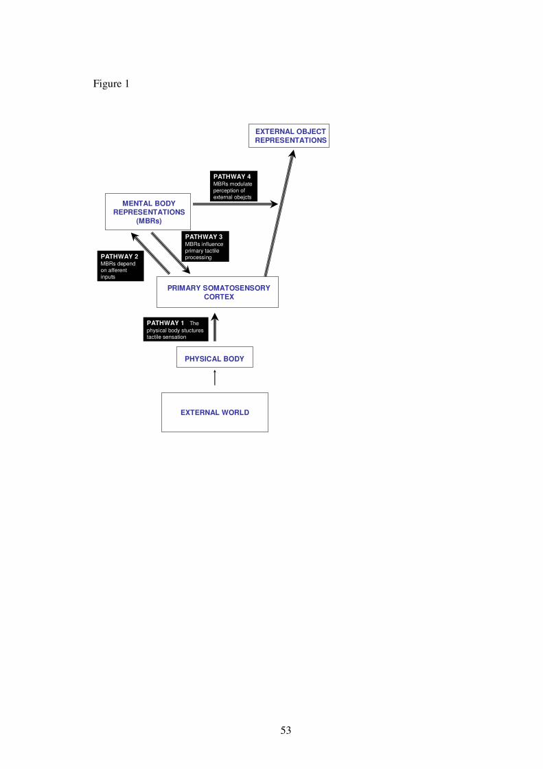
53
Figure 1
EXTERNAL WORLD
PHYSICAL BODY
PATHWAY 1 The physical body stucturestactile sensation
MENTAL BODY REPRESENTATIONS
(MBRs)
PRIMARY SOMATOSENSORYCORTEX
PATHWAY 3MBRs influenceprimary tactileprocessing
EXTERNAL OBJECT REPRESENTATIONS
PATHWAY 4MBRs modulate perception of external obejcts
PATHWAY 2MBRs dependon afferentinputs
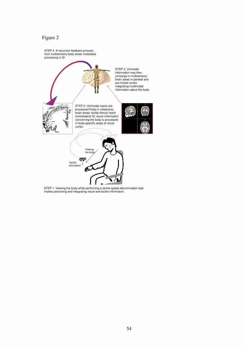
54
Figure 2
STEP 1. Viewing the body while performing a tactile spatial discrimination task implies perceiving and integrating visual and tactile information.
Tactilestimulation
Viewingthe body
STEP 2. Unimodal inputs are processed firstly in unisensorybrain areas: tactile stimuli reach contralateral SI; visual information concerning the body is processed in body-specific areas of visual cortex
STEP 3. Unimodalinformation may then converge in multisensorybrain areas in parietal and pre-frontal cortex integrating multimodal information about the body
STEP 4. A recurrent feedback process from multisensory body areas modulates processing in SI


