
Trauma and Pregnancy
William Schecter, MD
Trauma and Pregnancy
• ATLS Protocol the same
• Physiologic and Anatomic changes of
pregnancy change the pattern of injury and
the physiologic response to injury
• Two patients requiring treatment!!!

Anatomic Changes
16 weeks 24 weeks 32weeks http://www.bellaonline.com/articles/art7113.asp

Changes in Blood Volume and
Composition
• 40% increase in blood volume
• 25% increase in red cell mass
• Relative anemia (Hct 31-35)
• The mother may lose up to 1500 cc of blood
without hemodynamic instability BUT the
fetus may be in SHOCK!!!!
Changes in Blood Volume and
Composition
• White Blood Count elevated in pregnancy
(15,000)
• Fibrinogen and clotting factors increased
• Albumin level 2.2-2.8
Hemodynamic Changes in
Pregnancy
• Cardiac Output is increased by 1.0-1.5
liters/minute after the 10th week of
pregnancy
• Hypotension may be due to vena caval
compression by the uterus—Place patient
left side down!!
Hemodynamic Changes in
Pregnancy
• Heart rate increases 10-15 beats/minute—
consider ―tachycardia of pregnancy‖ when
evaluating Heart Rate during Stage ―C‖ of
the Primary Survey.
Blood Pressure
• Should be relatively normal.
• If patient is hypotensive, turn patient to the left thereby releasing uterine pressure from the vena cava decreasing venous return to the heart.
• Treat hypotension with aggressive fluid resuscitation if blood pressure does not improve rapidly.

Venous Pressure
• CVP variable
• Venous hypertension in lower extremities

Respiratory Changes
• Increased 02 Consumption
• Elevated diaphragm
• 30-40% increase in tidal volume and minute
ventilation
• PaC02 = 30-35 mm Hg
• Intubation may be challenging b/o airway edema
• Relaxed LES + Delayed Gastric Emptying =
Increased Risk of Aspiration
Renal Function
• Glomerular Filtration Rate increased in
pregnancy
• BUN and Creatinine decrease in pregnancy
• Glycosuria common
• Mild hydronephrosis a physiologic response
to uterine compression of the ureters
Musculoskeletal
• Symphysis pubis widens by the 7th month.
Sacroilicac joint spaces increase – may
create confusion in interpretation of Pelvic
X-rays
Eclampsia
• Seizures
• Hypertension, hyperreflexia, proteinuria,
peripheral edema
• May mimic Head Injury in the Trauma
Patient!!

Thrombotic Disease and
Pregnancy • Pregnancy may induce a hypercoagulable state
– Increased activity of Clotting Factors
– Decreased Fibrinolysis
• Venous Hypertension due to Uterine Pressure on the Inferior Vena Cava
• Incidence of DVT of 0.1-0.2%
• Lower Extremity Sequential Compression Devices recommended
• Heparin and Low Molecular Heparin ok in pregnancy
• Coumadin CONTRAINDICATED because of severe fetal malformations

Anesthetic Considerations
• Teratogenicity of Anesthetic Agents
• Anesthetic Drugs and Maternal Physiology
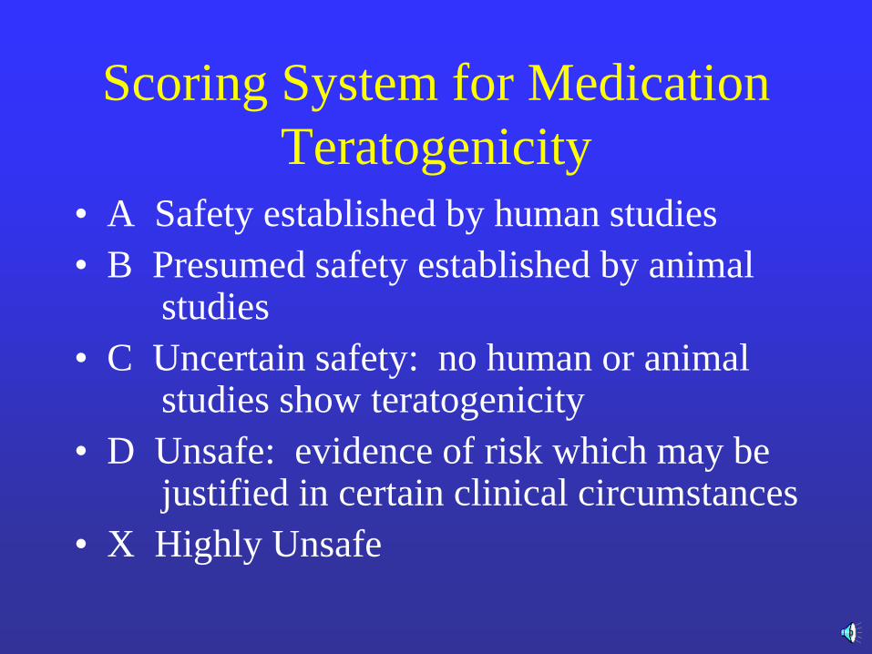
Scoring System for Medication
Teratogenicity
• A Safety established by human studies
• B Presumed safety established by animal studies
• C Uncertain safety: no human or animal studies show teratogenicity
• D Unsafe: evidence of risk which may be justified in certain clinical circumstances
• X Highly Unsafe

Teratogenicity and Anesthetics
• Almost all anesthetic drugs are Category C
drugs. No anesthetic drugs have been listed
as definitely teratogenic

Anesthetic Drugs and Maternal
Physiology
• Paralytic drugs do NOT cross the placenta
• Drugs used in Anesthesia are (with reasonable certainty) safe in pregnancy
– Inhalation anesthetics
– Local anesthetics
– Muscle relaxants
– Narcotics
– Benzodiazepines
Melnick DM, Wahl WL, Dalton VK. Management of general surgical problems in the pregnant
Patient. Am J Surg 2004;187:170-180.

Radiology, Trauma and
Pregnancy
Benefits to the Mother outweigh small
risks to the fetus

Radiation Risk to Fetus
• Teratogenicity
• Birth Defects (not proven)
• Increased Lifetime risk of malignancy
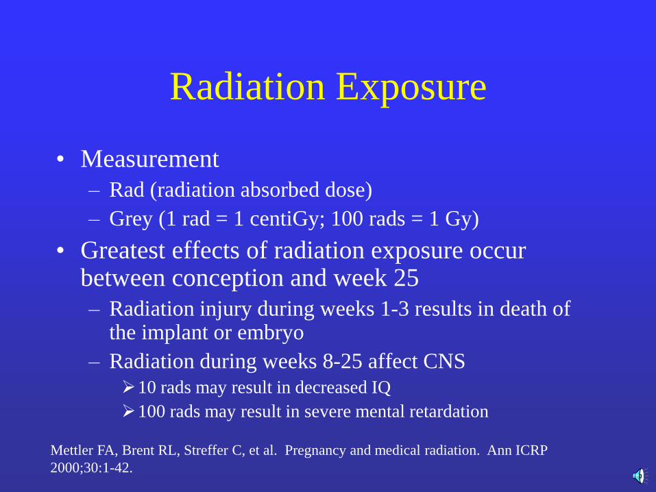
Radiation Exposure
• Measurement
– Rad (radiation absorbed dose)
– Grey (1 rad = 1 centiGy; 100 rads = 1 Gy)
• Greatest effects of radiation exposure occur between conception and week 25
– Radiation injury during weeks 1-3 results in death of the implant or embryo
– Radiation during weeks 8-25 affect CNS
10 rads may result in decreased IQ
100 rads may result in severe mental retardation
Mettler FA, Brent RL, Streffer C, et al. Pregnancy and medical radiation. Ann ICRP
2000;30:1-42.

Radiation Exposure
• After 25 weeks, greatest risk is childhood hematologic malignancy
– Background incidence is 0.2-0.3%
– Risk increases to 0.3-0.4% if exposure > 1 Gy
– Risk increases by 0.06% per 1 Gy of fetal exposure
• Risk negligible < 5 rads exposure
• Risk increases > 15 rads exposure
• Most diagnostic procedures have no measurable risk
• Therapeutic Procedures have greatest risk
Mettler FA, Brent RL, Streffer C, et al. Pregnancy and medical radiation. Ann ICRP
2000;30:1-42.
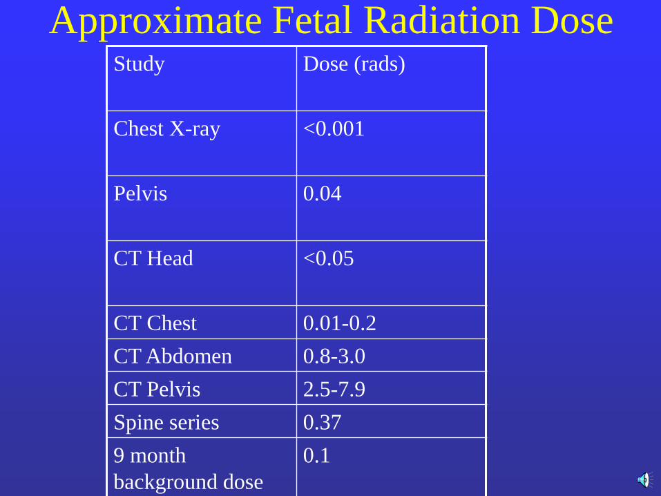
Approximate Fetal Radiation Dose Study
Dose (rads)
Chest X-ray
<0.001
Pelvis
0.04
CT Head <0.05
CT Chest 0.01-0.2
CT Abdomen 0.8-3.0
CT Pelvis 2.5-7.9
Spine series 0.37
9 month
background dose
0.1
Primary Survey
Airway: as per all patients
Breathing: High diaphragms in late stages of pregnancy
Circulation: If low risk of spinal injury, nurse left side down
REMEMBER: THE PREGANT PATIENT CAN LOSE A LOT OF BLOOD BEFORE ABNORMAL BP AND PULSE!!!

Additional Monitors
• Fetal Heart Monitoring
• Fetal Ultrasound
• Maximum fetal radiation dose = 5 rads

Fetomaternal Hemorrhage???
• Kleihauer-Betke Test: used to detect fetal
cells in the mother’s serum
• If mother is Rh negative and possible
fetomaternal hemorrhage: give Rh
immunoglobulin even if Kleihauer-Betke
Test negative.
Primary Concerns with Blunt
Abdominal Trauma
• Abruptio Placenta
– Leading cause of fetal death in injured mother
– DIC may occur
• Ruptured Uterus
– 0.6% of blunt abdominal trauma in pregnancy
Goals of Treatment of the Severely
Injured Pregnant Patient
• Goal 1
– SAVE THE MOTHER
• Goal 2
– Save the Fetus if possible
Emergency Cesarean Section
• Limited Role
• Primarily in unstable mother who is not responding to Fluid Management given in the Primary Survey
• Little role for perimortem cesarean section if mother has been in shock—the fetus has already been severely hypoperfused for a long period of time!!!!
Summary
• Primary Survey
• Stage of Resuscitation
• Secondary Survey
• SAVE THE MOTHER FIRST!!!
• Limit fetal radiation to 5 rads
• Limited role for emergency cesarean section


