

doi.org/10.26434/chemrxiv.9991937.v1
Tropoelastin-Inspired, Non-Ionic, Self-Coacervating Polyesters as StrongUnderwater AdhesivesAmal Narayanan, Joshua Menefee, Qianhui Liu, Ali Dhinojwala, Abraham Joy
Submitted date: 16/10/2019 • Posted date: 21/10/2019Licence: CC BY-NC-ND 4.0Citation information: Narayanan, Amal; Menefee, Joshua; Liu, Qianhui; Dhinojwala, Ali; Joy, Abraham (2019):Tropoelastin-Inspired, Non-Ionic, Self-Coacervating Polyesters as Strong Underwater Adhesives. ChemRxiv.Preprint.
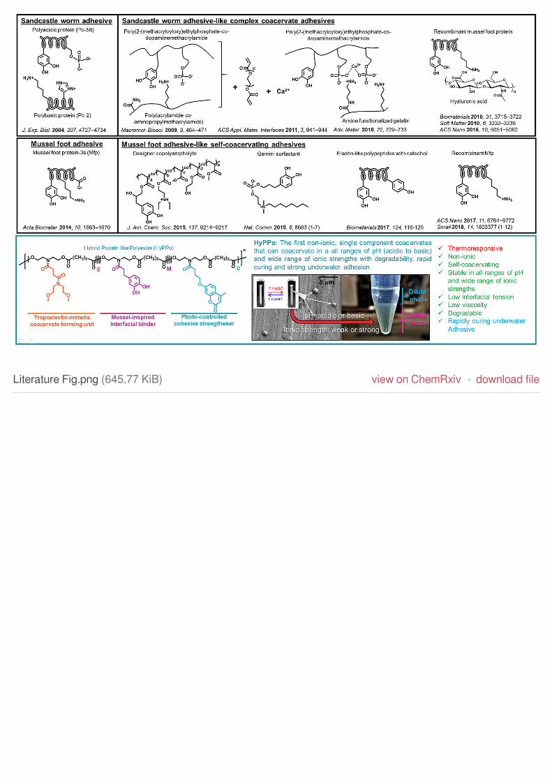
Inspired from the one-component self-coacervation of tropoelastin and mussel foot protein-3s, we created thefirst non-ionic, single component coacervates that can coacervate in a all ranges of pH (acidic to basic) andwide range of ionic strengths with degradability, rapid curing and strong underwater adhesion. In contrast tothe complex coacervates, these ‘charge-free’ coacervates are potential candidates as tissue adhesives andsealants, adhesives for sensor attachment to wet skin, and as sprayable adhesives. Their potential use in theclinic arises from their enhanced stability to changes in external conditions, cytocompatibility, biodegradabilityand modular nature in incorporating various functional groups and crosslinkers.
File list (5)
download fileview on ChemRxivManuscript.pdf (524.05 KiB)
download fileview on ChemRxivLiterature Fig.png (645.77 KiB)
download fileview on ChemRxivSupporting Information.pdf (1.12 MiB)
download fileview on ChemRxivMovie S1.m2ts (28.11 MiB)
download fileview on ChemRxivMovie S2.m2ts (98.65 MiB)

Tropoelastin-Inspired, Non-Ionic, Self-Coacervating Polyesters as
Strong Underwater Adhesives
Amal Narayanan, Joshua R. Menefee, Qianhui Liu, Ali Dhinojwala,* Abraham Joy*[a]
Abstract: Mussels and sandcastle worms utilize the advantages of
coacervation to deliver concentrated protein-rich adhesive cocktails in
aqueous environment that enables their attachment to underwater
substrates. By taking advantage of the principle of polymer
coacervation, we have created mussel foot protein-inspired,
tropoelastin-like, bioabsorbable, non-ionic, self-coacervating
polyesters that overcome the challenges of adhesion in wet or
underwater environments. Herein, we describe the rationale for their
design, and the underwater adhesive properties of these non-ionic
adhesives. Compared to previously reported coacervate adhesives,
these ‘charge-free’ polyesters coacervate in all ranges of pH and wide
ranges of ionic strengths, and rapidly (< 300 s) adhere to substrates
submerged underwater.
Coacervation is the macroscopic phase separation of a
solution to form two distinct fluid-fluid phases, namely, dilute and
dense.[1] The dense phase of the coacervate has unique
characteristics such as high density,[2] low viscosity,[3] and low
interfacial tension.[4] Nature has employed protein coacervates as
a means to overcome the challenge of adhesion between
interfaces in a wet environment.[5] For example, the low interfacial
tension and low viscosity of the dense phase allow it to even
spontaneously prime rough surfaces residing underwater.[6]
Aquatic life forms such as sandcastle worms[7] and mussels[8]
employ coacervation for the efficient delivery of protein-rich
adhesives in water resulting in robust underwater adhesion. For
example, sandcastle worms secrete a mixture of oppositely
charged proteins, which form a thermodynamically stable dense
phase in the presence of metal salts at neutral pH known as
complex coacervate. This complex coacervate is delivered to
surfaces for initiating interfacial adhesion and the corresponding
cohesive strength of the adhesive is obtained through enzymatic
and mineral-mediated crosslinking reactions.[7] Inspired by the
sandcastle worm adhesive, multi-component complex
coacervates formed from recombinant proteins[3,6,9] and synthetic
polymers[10–13] have been used to create adhesive joints. The
formation of complex coacervates with consistent physical
properties requires careful tuning of the molar ratios of oppositely
charged polymers, maintaining the correct pH and ionic strength,
and temperature.[14] This reduces the stability of complex
coacervates to variations in pH and ionic strength. The instability
to external factors and potential cytotoxic activity of charged
polymers[15] restricts the eventual application of multicomponent
complex coacervates in dynamic wet environments and on
biological surfaces.
Recently, Wei et al.[8] demonstrated that one of the adhesive
primer proteins (Mfp-3s) secreted by mussels undergoes
coacervation. The coacervation behavior displayed by the Mfp-3s
is distinct from the sandcastle worm adhesives since Mfp-3s
undergoes one component self-coacervation from pH 3 - 6 and
monovalent ionic strength ~ 100 - 600 mM. Seo et al.[16] translated
the self-coacervation of Mfp-3s to synthetic polymers using
designer copolyampholytes with strong cohesive interactions in
wet conditions. However, these Mfp-3s-inspired
copolyampholytes show coacervation only in narrow ranges of pH
(4 - 5) and ionic strength (< 20 mM) due to the strong columbic
nature of self-association.[16] What has been conspicuously
absent is the demonstration of ‘charge-free’ coacervate
adhesives that can self-coacervate in all ranges of pH and ionic
strength.[17] Such non-ionic, single component coacervates have
significant advantages over the charged coacervates since their
formation does not require optimization of the molar ratio of two
or more components. Also, their increased stability to changes in
external conditions allows the use of ‘charge-free’ coacervates in
applications where the interfacial pH and ionic strength are
susceptible to fluctuations, such as ruptured vascularized organs.
This unmet need was the motivating factor for development of the
non-ionic coacervates described below.
We base this study on our previously reported serendipitous
discovery that a library of thermoresponsive polyesters designed
in our lab show ‘tropoelastin-like’ coacervation behavior.[18–20]
Tropoelastin, the soluble precursor to elastin, shows lower critical
solution temperature (LCST) in aqueous medium, and above
LCST, the protein solution segregates to both dilute and protein-
rich dense phases.[21] Similarly, our polyester library also shows
hydrophobically-driven, single component coacervation. Their
non-ionic, bioabsorbable,[19] cell-compatible,[20] and modular
nature, allows incorporation of various functional groups[18] and
hence this platform provides significant advantages over any
other reported coacervates to date. In this study, we report an
extension of the above polyesters, wherein incorporation of
appropriate functional groups provides a new class of non-ionic,
self-coacervating polyesters that demonstrate rapid, water-
tolerant crosslinking, resulting in strong underwater adhesion.
These non-ionic polyesters coacervate in all ranges of pH and a
wide range of ionic strengths. To the best of our knowledge, there
are no reported non-ionic coacervates with strong underwater
adhesion.
Inspired by single component coacervation of Mfp-3s and
non-ionic coacervation of tropoelastin, we synthesized ‘Hybrid
Protein-like Polyester’ (HyPPo, Figure 1A) coacervates and
explored their interfacial, viscoelastic, and wet adhesive
properties. HyPPo is a class of statistical copolyesters made from
two or more N-functionalized diethanolamides (diols) and succinic
acid (diacid) using N,N-diisopropylcarbodiimide mediated
polyesterification reactions (Scheme S1).[22] The first diol contains
a tropoelastin-mimetic domain (E). The polyesters made from E
show self-coacervation in aqueous medium at temperatures
above the LCST.[20] From our library of diols that imbue
‘tropoelastin-like’ coacervation in polyesters, we chose bis(2-
[a] A. Narayanan, J. R. Menefee, Q. Liu, Prof. Dr. A. Dhinojwala, Prof.
Dr. A. Joy
Department of Polymer Science
The University of Akron
Akron, Ohio, United States
E-mail: [email protected]
Supporting information for this article is given via a link at the end of
the document.
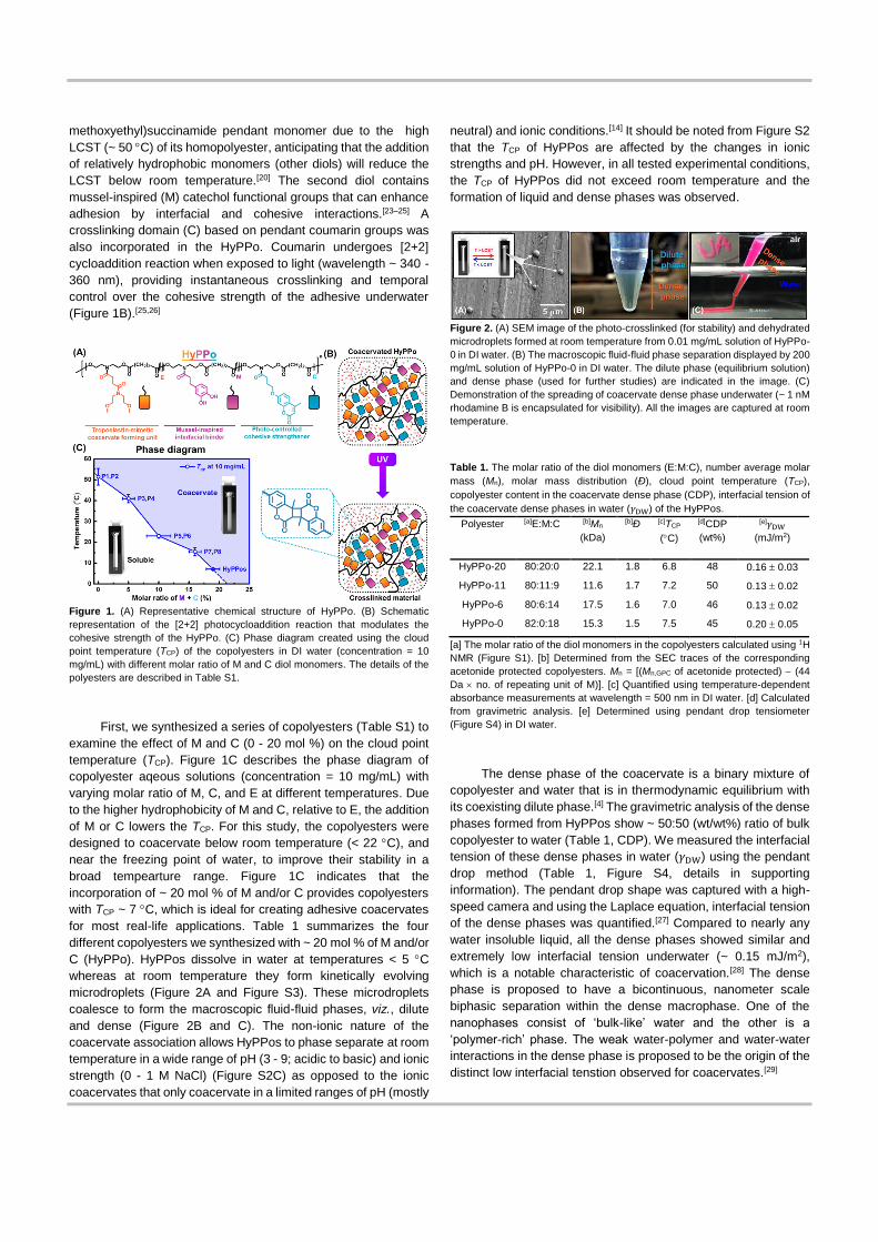
methoxyethyl)succinamide pendant monomer due to the high
LCST (~ 50 C) of its homopolyester, anticipating that the addition
of relatively hydrophobic monomers (other diols) will reduce the
LCST below room temperature.[20] The second diol contains
mussel-inspired (M) catechol functional groups that can enhance
adhesion by interfacial and cohesive interactions.[23–25] A
crosslinking domain (C) based on pendant coumarin groups was
also incorporated in the HyPPo. Coumarin undergoes [2+2]
cycloaddition reaction when exposed to light (wavelength ~ 340 -
360 nm), providing instantaneous crosslinking and temporal
control over the cohesive strength of the adhesive underwater
(Figure 1B).[25,26]
Figure 1. (A) Representative chemical structure of HyPPo. (B) Schematic
representation of the [2+2] photocycloaddition reaction that modulates the
cohesive strength of the HyPPo. (C) Phase diagram created using the cloud
point temperature (TCP) of the copolyesters in DI water (concentration = 10
mg/mL) with different molar ratio of M and C diol monomers. The details of the
polyesters are described in Table S1.
First, we synthesized a series of copolyesters (Table S1) to
examine the effect of M and C (0 - 20 mol %) on the cloud point
temperature (TCP). Figure 1C describes the phase diagram of
copolyester aqeous solutions (concentration = 10 mg/mL) with
varying molar ratio of M, C, and E at different temperatures. Due
to the higher hydrophobicity of M and C, relative to E, the addition
of M or C lowers the TCP. For this study, the copolyesters were
designed to coacervate below room temperature (< 22 C), and
near the freezing point of water, to improve their stability in a
broad tempearture range. Figure 1C indicates that the
incorporation of ~ 20 mol % of M and/or C provides copolyesters
with TCP ~ 7 C, which is ideal for creating adhesive coacervates
for most real-life applications. Table 1 summarizes the four
different copolyesters we synthesized with ~ 20 mol % of M and/or
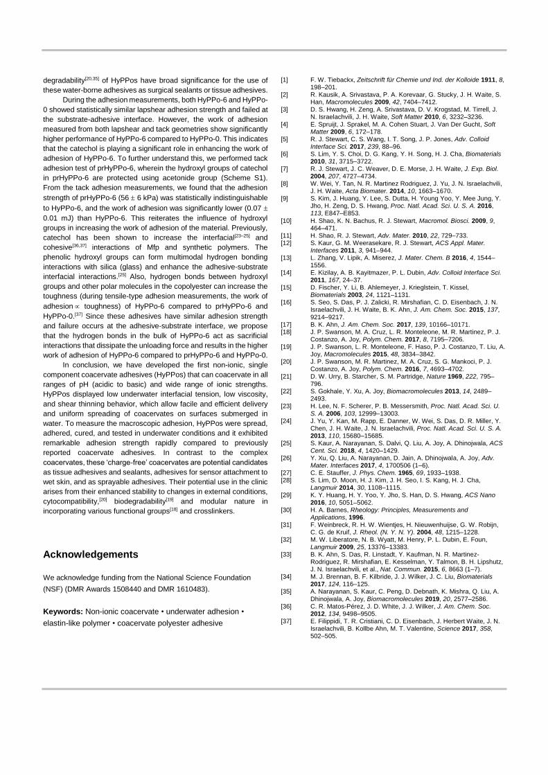
C (HyPPo). HyPPos dissolve in water at temperatures < 5 C
whereas at room temperature they form kinetically evolving
microdroplets (Figure 2A and Figure S3). These microdroplets
coalesce to form the macroscopic fluid-fluid phases, viz., dilute
and dense (Figure 2B and C). The non-ionic nature of the
coacervate association allows HyPPos to phase separate at room
temperature in a wide range of pH (3 - 9; acidic to basic) and ionic
strength (0 - 1 M NaCl) (Figure S2C) as opposed to the ionic
coacervates that only coacervate in a limited ranges of pH (mostly
neutral) and ionic conditions.[14] It should be noted from Figure S2
that the TCP of HyPPos are affected by the changes in ionic
strengths and pH. However, in all tested experimental conditions,
the TCP of HyPPos did not exceed room temperature and the
formation of liquid and dense phases was observed.
Figure 2. (A) SEM image of the photo-crosslinked (for stability) and dehydrated
microdroplets formed at room temperature from 0.01 mg/mL solution of HyPPo-
0 in DI water. (B) The macroscopic fluid-fluid phase separation displayed by 200
mg/mL solution of HyPPo-0 in DI water. The dilute phase (equilibrium solution)
and dense phase (used for further studies) are indicated in the image. (C)
Demonstration of the spreading of coacervate dense phase underwater (~ 1 nM
rhodamine B is encapsulated for visibility). All the images are captured at room
temperature.
Table 1. The molar ratio of the diol monomers (E:M:C), number average molar
mass (Mn), molar mass distribution (Ð), cloud point temperature (TCP),
copolyester content in the coacervate dense phase (CDP), interfacial tension of
the coacervate dense phases in water (𝛾DW) of the HyPPos.
Polyester [a]E:M:C [b]Mn
(kDa)
[b]Ð [c]TCP
(C)
[d]CDP
(wt%)
[e]𝛾DW
(mJ/m2)
HyPPo-20 80:20:0 22.1 1.8 6.8 48 0.16 0.03
HyPPo-11 80:11:9 11.6 1.7 7.2 50 0.13 0.02
HyPPo-6 80:6:14 17.5 1.6 7.0 46 0.13 0.02
HyPPo-0 82:0:18 15.3 1.5 7.5 45 0.20 0.05
[a] The molar ratio of the diol monomers in the copolyesters calculated using 1H
NMR (Figure S1). [b] Determined from the SEC traces of the corresponding
acetonide protected copolyesters. Mn = [(Mn,GPC of acetonide protected) − (44
Da no. of repeating unit of M)]. [c] Quantified using temperature-dependent
absorbance measurements at wavelength = 500 nm in DI water. [d] Calculated
from gravimetric analysis. [e] Determined using pendant drop tensiometer
(Figure S4) in DI water.
The dense phase of the coacervate is a binary mixture of
copolyester and water that is in thermodynamic equilibrium with
its coexisting dilute phase.[4] The gravimetric analysis of the dense
phases formed from HyPPos show ~ 50:50 (wt/wt%) ratio of bulk
copolyester to water (Table 1, CDP). We measured the interfacial
tension of these dense phases in water (𝛾DW) using the pendant
drop method (Table 1, Figure S4, details in supporting
information). The pendant drop shape was captured with a high-
speed camera and using the Laplace equation, interfacial tension
of the dense phases was quantified.[27] Compared to nearly any
water insoluble liquid, all the dense phases showed similar and
extremely low interfacial tension underwater (~ 0.15 mJ/m2),
which is a notable characteristic of coacervation.[28] The dense
phase is proposed to have a bicontinuous, nanometer scale
biphasic separation within the dense macrophase. One of the
nanophases consist of ‘bulk-like’ water and the other is a
‘polymer-rich’ phase. The weak water-polymer and water-water
interactions in the dense phase is proposed to be the origin of the
distinct low interfacial tenstion observed for coacervates.[29]

Figure 3. The steady-state flow measurements of (A) bulk HyPPo melts and
their corresponding (B) coacervate dense phases formed in DI water at 25 C.
The viscoelastic properties of the bulk copolyesters (HyPPos
in their melt state) and their corresponding coacervate dense
phases were analyzed using steady-state flow and small
amplitude oscillatory shear (SAOS) measurements at 25 C.
Figures 3A and B show the viscosity (𝜂) response to shear rate
(�̇�) of the bulk copolyesters and the coacervate dense phases
formed from 200 mg/mL aqueous solution of the corresponding
bulk copolyesters, respectively. The zero-shear viscosity (𝜂0, 𝜂 at
low �̇�) of the bulk HyPPo-20, HyPPo-11, HyPPo-6, and HyPPo-0
were found to be 1.0 105, 9.5 104, 8.8 103, and 8.1 103 Pa-
s, respectively. The 𝜂 of the bulk polyesters remained relatively
constant at �̇� between 10-2 to 101 s-1. The coacervate dense
phases formed from HyPPo-20, HyPPo-11, HyPPo-6, and
HyPPo-0 showed 𝜂0 ~ 2.5, 0.3, 1.4, and 0.9 Pa-s, respectively
(Figure 3B). An abrupt shear thinning behavior was observed at
�̇� (�̇�C) of 4, 25, 7, and 10 s-1 for HyPPo-20, HyPPo-11, HyPPo-6,
and HyPPo-0, respectively. The shear thinning is an indication of
the shear induced structural changes in the material.[30,31] When �̇�
> �̇�C , we hypothesize that the dense macrophase might be
restructuring along the plane of shear and causing the observed
decrease in viscosity (Figure 3B).[32] This non-Newtonian behavior
attains a limiting and constant viscosity (𝜂L)[30], which is observed
from �̇� > 25, 40, 150, and 100 s-1 till 200 s-1 for HyPPo-20, HyPPo-
11, HyPPo-6, and HyPPo-0, respectively.
SAOS measurements of the bulk copolyester melts show that
the loss modulus (G”) > storage modulus (G’) at angular
frequency (𝜔) between 0.1 to 100 rad/s, indicating the Newtonian
liquid-like behavior of the bulk copolyesters (Figure S6A).[30]
Figure S6B describes the response of the coacervate dense
phases to 𝜔 . A viscous behavior (G” > G’) was observed
throughout the experimental regime and G” and G’ scaled to ~
𝜔1.1 and ~ 𝜔0.9, respectively between 0.1 to 100 rad/s. From the
rheological measurements, we found that 𝜂0 and G” of the bulk
copolyesters reduced by more than 103 times when coacervated.
The low viscosity and shear thinning behavior of these
coacervates allow delivery of coacervates as continuous
filaments without any mass loss. Movie S1 shows the ease in
delivering the HyPPo-0 dense phase from a polypropylene
(hydrophobic) micropipette tip without any resistance underwater.
The density being higher than water and the low interfacial tension
allows immediate wetting of coacervates to hydrophilic surfaces
submerged underwater.
Figure 4. (A) Representative force curves of HyPPo-6 and HyPPo-0 during the
unloading of lapshear adhesion measurements and the (B) lapshear adhesion
strength and work of adhesion quantified from the force curves. (C)
Representative force curves of HyPPo-11, HyPPo-6, and HyPPo-0 during tack
adhesion measurements and the (B) tack adhesion strength and work of
adhesion quantified from the force curves. All the measurements were
performed in DI water and repeated at least five times. The data represented in
the bar diagrams are in terms of mean standard deviation, and ‘*’ represents
the statistical significance among the samples using a Tukey mean comparison
test (p < 0.05).
The adhesive performance of the HyPPos on glass were
quantified using lapshear and tack adhesion geometries underwater.
From the force-distance curve (Figure 4A and 4C), the adhesion
strength and the work of adhesion were calculated. The substrates
adhered using HyPPo-20 and HyPPo-11 showed negligible adhesion
strength and failed cohesively before testing. HyPPo-6 and HyPP-0
showed lapshear adhesion strength of 96 16 and 101 22 kPa,
respectively. The lapshear work of adhesion was found to be 7.8
1.9, and 3.8 1.1 mJ for HyPPo-6 and HyPPo-0, respectively (Figure
4B). In contrast to the lapshear strength measurements, the tack
adhesion test was able to capture the adhesion behavior of HyPPo-
11 (Figure 4C). The tack adhesion strengths of HyPPo-11, HyPPo-6,
and HyPPo-0 were quantified to be 7 3, 66 4, and 54 3 kPa,
respectively (Figure 4D). The tack work of adhesion measured from
the unloading curve in the force-distance profile were found to be 2
10-3, 0.21 0.06, and 0.09 0.02 mJ for HyPPo-11, HyPPo-6, and
HyPPo-0, respectively (Figure 4D).
In the literature, there are very few reports on coacervate
adhesives that carry out all the steps required to create adhesive
joints such as coating, bonding, curing, and testing in underwater
conditions.[6,10–12,33] Kaur et al.[12] have developed complex coacervate
formulations encapsulated with polyethylene glycol-diacrylate that are
applied outside water, but cured and tested underwater, which show
~ 1 MPa lapshear adhesion strength for bonded aluminum substrates.
Lim et al.[6] performed all steps of adhesive joint fabrication
underwater using complex coacervate made from association of
recombinant Mfp and hyaluronic acid and obtained lapshear adhesion
strength of ~ 200 kPa underwater.
Since most of the reported coacervate adhesives studies rely
on slow oxidative and free radical polymerization reactions for
cohesive strength, they require extended curing time (~ 24 h) for
making robust lap-joints.[34] Movie S2 demonstrates the rapid curing
of HyPPos (~ 3 min) to devise strong adhesive joints underwater. The
rapid and temporal nature of the adhesive curing and
degradability[20,35] of HyPPos have broad significance for the use of
these water-borne adhesives as surgical sealants or tissue adhesives.
During the adhesion measurements, both HyPPo-6 and HyPPo-
0 showed statistically similar lapshear adhesion strength and failed at
the substrate-adhesive interface. However, the work of adhesion
measured from both lapshear and tack geometries show significantly
higher performance of HyPPo-6 compared to HyPPo-0. This indicates
that the catechol is playing a significant role in enhancing the work of
adhesion of HyPPo-6. To further understand this, we performed tack
adhesion test of prHyPPo-6, wherein the hydroxyl groups of catechol
in prHyPPo-6 are protected using acetonide group (Scheme S1).
From the tack adhesion measurements, we found that the adhesion
strength of prHyPPo-6 (56 6 kPa) was statistically indistinguishable
to HyPPo-6, and the work of adhesion was significantly lower (0.07
0.01 mJ) than HyPPo-6. This reiterates the influence of hydroxyl
groups in increasing the work of adhesion of the material. Previously,
catechol has been shown to increase the interfacial[23–25] and
cohesive[36,37] interactions of Mfp and synthetic polymers. The
phenolic hydroxyl groups can form multimodal hydrogen bonding
interactions with silica (glass) and enhance the adhesive-substrate
interfacial interactions.[25] Also, hydrogen bonds between hydroxyl
groups and other polar molecules in the copolyester can increase the
toughness (during tensile-type adhesion measurements, the work of
adhesion toughness) of HyPPo-6 compared to prHyPPo-6 and
HyPPo-0.[37] Since these adhesives have similar adhesion strength
and failure occurs at the adhesive-substrate interface, we propose
that the hydrogen bonds in the bulk of HyPPo-6 act as sacrificial
interactions that dissipate the unloading force and results in the higher
work of adhesion of HyPPo-6 compared to prHyPPo-6 and HyPPo-0.
In conclusion, we have developed the first non-ionic, single
component coacervate adhesives (HyPPos) that can coacervate in all
ranges of pH (acidic to basic) and wide range of ionic strengths.
HyPPos displayed low underwater interfacial tension, low viscosity,
and shear thinning behavior, which allow facile and efficient delivery
and uniform spreading of coacervates on surfaces submerged in
water. To measure the macroscopic adhesion, HyPPos were spread,
adhered, cured, and tested in underwater conditions and it exhibited
remarkable adhesion strength rapidly compared to previously
reported coacervate adhesives. In contrast to the complex
coacervates, these ‘charge-free’ coacervates are potential candidates
as tissue adhesives and sealants, adhesives for sensor attachment to
wet skin, and as sprayable adhesives. Their potential use in the clinic
arises from their enhanced stability to changes in external conditions,
cytocompatibility,[20] biodegradability[19] and modular nature in
incorporating various functional groups[18] and crosslinkers.
Acknowledgements
We acknowledge funding from the National Science Foundation
(NSF) (DMR Awards 1508440 and DMR 1610483).
Keywords: Non-ionic coacervate • underwater adhesion •
elastin-like polymer • coacervate polyester adhesive
[1] F. W. Tiebackx, Zeitschrift für Chemie und Ind. der Kolloide 1911, 8, 198–201.
[2] R. Kausik, A. Srivastava, P. A. Korevaar, G. Stucky, J. H. Waite, S. Han, Macromolecules 2009, 42, 7404–7412.
[3] D. S. Hwang, H. Zeng, A. Srivastava, D. V. Krogstad, M. Tirrell, J. N. Israelachvili, J. H. Waite, Soft Matter 2010, 6, 3232–3236.
[4] E. Spruijt, J. Sprakel, M. A. Cohen Stuart, J. Van Der Gucht, Soft Matter 2009, 6, 172–178.
[5] R. J. Stewart, C. S. Wang, I. T. Song, J. P. Jones, Adv. Colloid Interface Sci. 2017, 239, 88–96.
[6] S. Lim, Y. S. Choi, D. G. Kang, Y. H. Song, H. J. Cha, Biomaterials 2010, 31, 3715–3722.
[7] R. J. Stewart, J. C. Weaver, D. E. Morse, J. H. Waite, J. Exp. Biol. 2004, 207, 4727–4734.
[8] W. Wei, Y. Tan, N. R. Martinez Rodriguez, J. Yu, J. N. Israelachvili, J. H. Waite, Acta Biomater. 2014, 10, 1663–1670.
[9] S. Kim, J. Huang, Y. Lee, S. Dutta, H. Young Yoo, Y. Mee Jung, Y. Jho, H. Zeng, D. S. Hwang, Proc. Natl. Acad. Sci. U. S. A. 2016, 113, E847–E853.
[10] H. Shao, K. N. Bachus, R. J. Stewart, Macromol. Biosci. 2009, 9, 464–471.
[11] H. Shao, R. J. Stewart, Adv. Mater. 2010, 22, 729–733. [12] S. Kaur, G. M. Weerasekare, R. J. Stewart, ACS Appl. Mater.
Interfaces 2011, 3, 941–944. [13] L. Zhang, V. Lipik, A. Miserez, J. Mater. Chem. B 2016, 4, 1544–
1556. [14] E. Kizilay, A. B. Kayitmazer, P. L. Dubin, Adv. Colloid Interface Sci.
2011, 167, 24–37. [15] D. Fischer, Y. Li, B. Ahlemeyer, J. Krieglstein, T. Kissel,
Biomaterials 2003, 24, 1121–1131. [16] S. Seo, S. Das, P. J. Zalicki, R. Mirshafian, C. D. Eisenbach, J. N.
Israelachvili, J. H. Waite, B. K. Ahn, J. Am. Chem. Soc. 2015, 137, 9214–9217.
[17] B. K. Ahn, J. Am. Chem. Soc. 2017, 139, 10166–10171. [18] J. P. Swanson, M. A. Cruz, L. R. Monteleone, M. R. Martinez, P. J.
Costanzo, A. Joy, Polym. Chem. 2017, 8, 7195–7206. [19] J. P. Swanson, L. R. Monteleone, F. Haso, P. J. Costanzo, T. Liu, A.
Joy, Macromolecules 2015, 48, 3834–3842. [20] J. P. Swanson, M. R. Martinez, M. A. Cruz, S. G. Mankoci, P. J.
Costanzo, A. Joy, Polym. Chem. 2016, 7, 4693–4702. [21] D. W. Urry, B. Starcher, S. M. Partridge, Nature 1969, 222, 795–
796. [22] S. Gokhale, Y. Xu, A. Joy, Biomacromolecules 2013, 14, 2489–
2493. [23] H. Lee, N. F. Scherer, P. B. Messersmith, Proc. Natl. Acad. Sci. U.
S. A. 2006, 103, 12999–13003. [24] J. Yu, Y. Kan, M. Rapp, E. Danner, W. Wei, S. Das, D. R. Miller, Y.
Chen, J. H. Waite, J. N. Israelachvili, Proc. Natl. Acad. Sci. U. S. A. 2013, 110, 15680–15685.
[25] S. Kaur, A. Narayanan, S. Dalvi, Q. Liu, A. Joy, A. Dhinojwala, ACS Cent. Sci. 2018, 4, 1420–1429.
[26] Y. Xu, Q. Liu, A. Narayanan, D. Jain, A. Dhinojwala, A. Joy, Adv. Mater. Interfaces 2017, 4, 1700506 (1–6).
[27] C. E. Stauffer, J. Phys. Chem. 1965, 69, 1933–1938. [28] S. Lim, D. Moon, H. J. Kim, J. H. Seo, I. S. Kang, H. J. Cha,
Langmuir 2014, 30, 1108–1115. [29] K. Y. Huang, H. Y. Yoo, Y. Jho, S. Han, D. S. Hwang, ACS Nano
2016, 10, 5051–5062. [30] H. A. Barnes, Rheology: Principles, Measurements and
Applications, 1996. [31] F. Weinbreck, R. H. W. Wientjes, H. Nieuwenhuijse, G. W. Robijn,
C. G. de Kruif, J. Rheol. (N. Y. N. Y). 2004, 48, 1215–1228. [32] M. W. Liberatore, N. B. Wyatt, M. Henry, P. L. Dubin, E. Foun,
Langmuir 2009, 25, 13376–13383. [33] B. K. Ahn, S. Das, R. Linstadt, Y. Kaufman, N. R. Martinez-
Rodriguez, R. Mirshafian, E. Kesselman, Y. Talmon, B. H. Lipshutz, J. N. Israelachvili, et al., Nat. Commun. 2015, 6, 8663 (1–7).
[34] M. J. Brennan, B. F. Kilbride, J. J. Wilker, J. C. Liu, Biomaterials 2017, 124, 116–125.
[35] A. Narayanan, S. Kaur, C. Peng, D. Debnath, K. Mishra, Q. Liu, A. Dhinojwala, A. Joy, Biomacromolecules 2019, 20, 2577–2586.
[36] C. R. Matos-Pérez, J. D. White, J. J. Wilker, J. Am. Chem. Soc. 2012, 134, 9498–9505.
[37] E. Filippidi, T. R. Cristiani, C. D. Eisenbach, J. Herbert Waite, J. N. Israelachvili, B. Kollbe Ahn, M. T. Valentine, Science 2017, 358, 502–505.

Table of Content
Non-ionic coacervates: The first non-ionic, single component coacervates that can coacervate in a all ranges of pH (acidic to basic) and wide range of ionic strengths with degradability, rapid curing and strong underwater adhesion.
Tropoelastin-Inspired, Non-Ionic, Self-Coacervating Polyesters as Strong Underwater Adhesives
A. Narayanan, J. R. Menefee, Q.
Liu, Prof. Dr. A. Dhinojwala,*
Prof. Dr. A. Joy*

download fileview on ChemRxivManuscript.pdf (524.05 KiB)
download fileview on ChemRxivLiterature Fig.png (645.77 KiB)

Supporting Information
Tropoelastin-Inspired, Non-Ionic, Self-Coacervating Polyesters as Strong
Underwater Adhesives
Amal Narayanan, Joshua R. Menefee, Qianhui Liu, Ali Dhinojwala,* Abraham Joy*
Department of Polymer Science, The University of Akron, Akron, OH 44325, USA
Materials
N,N′-Diisopropylcarbodiimide (DIC, 99+ %) was purchased from Chem-Impex Int’l Inc.
Succinic acid (99+%) was purchased from Fisher Scientific. Trifluoroacetic acid (TFA, 99%) and
triisopropylsilane (TIPS, 98 %) were purchased from Oakwood Chemical. Dichloromethane
(Sigma-Aldrich, 99.5 %) was dried by distilling over anhydrous CaH2. CDCl3 and Acetone-d6 (D,
99.85 %) were purchased from Cambridge Isotope Lab. Millipore water deoxygenated by nitrogen
purging was used for preparing coacervates. The lapshear samples and tack adhesion samples were
prepared in DI water. NaCl, HCl, and NaOH used for making solutions with different ionic strength
and pH were obtained from VWR analytical. 4-(dimethylamino) pyridinium-4-toluene sulfonate
(DPTS),[1] N1,N1-bis(2-hydroxyethyl)-N4,N4-bis(2-methoxyethyl)succinamide (E),[2] 3-(2,2-
dimethylbenzo[d][1,3]dioxol-5-yl)-N,N-bis(2-hydroxyethyl)propenamide (Mpr),[3] and N,N-
bis(2-hydroxyethyl)-4-((4-methyl-2-oxo-2H-chromen-7-yl)oxy)butanamide (C)[4] were prepared
as per reported procedures.

Instrumentation
1H NMR spectra of the monomers and polyesters were recorded on a Varian Mercury 500
MHz spectrometer. The molar mass (Mn,GPC) and dispersity (Ð) of the polyesters were calculated
from a TOSOH EcoSec HLC-8320 GPC using refractive index detector (RI) detector. Separation
occurred over two PSS Gram Analytical GPC Columns in series using 25 mM LiBr in DMF as
eluent at a flow rate of 0.8 mL/min. The columns and detectors temperatures were maintained at
50 °C. Molar masses were obtained relative to narrow disperse polystyrene standards. The cloud
point temperature (TCP) of the polyesters were analyzed using a Shimadzu UV-1800 UV-Vis
spectrophotometer equipped with a Shimadzu S-1700 thermoelectric single cell holder in a 1 cm
quartz cell with nitrogen chamber. The nanoparticles were imaged with a scanning electron
microscope (SEM JSM7401). The interfacial tension of the coacervate dense phases in water were
measured by pendant drop shape analysis using a Rame-Hart drop shape analyzer. Rheological
experiments were performed on a TA ARES-G2 rheometer. The lapshear and tack adhesion
strength measurements were performed on a TA.XT texture analyzer from StableMicroSystems
with 10 Kg load cell.
Experimental Section

Scheme S1. Schematic representation of the polyesterification reaction and the deprotection of
3,4-acetonide groups.
Table S1. The molar ratio of the monomers, weight average molar mass (Mn,GPC), cloud point
temperature (TCP) of the polyesters used for creating the phase diagram (Figure 1B).
Polymer aE:M:C Mn,GPC (kDa) dTCP (C)
P1 100:0:0 95.7b 55
P2 100:0:0 33.7b 48
P3 95:0:4 56.8b 41
P4 95:6:0 19.6c 39
P5 89:5:6 17.3c 24
P6 90:10:0 48.1c 25
P7 85:17:0 8.1c 17
P8 85:15:0 28.7c 14
HyPPo-20 80:20:0 22.1c 7
HyPPo-10 80:11:9 17.6c 7
HyPPo-15 80:6:14 13.5c 7
HyPPo-20 82:0:18 15.3b 8
aCalculated using 1H NMR. bDetermined from the SEC traces or cfrom the corresponding acetonide
protected copolyesters. Mn,GPC = Mn,GPC of acetonide protected − 44 Da no. of repeating unit of M. dQuantified using temperature-dependent absorbance measurements at wavelength = 500 nm.

(A)
(B)

(C)
(D)

Figure S1. 1H NMR of the polyesters (A) HyPP0-20, (B) HyPPo-11, (C) HyPPo-6, and (D)
HyPPo-0.
Synthesis
Polymerization
The polymerization of the N-functionalized diethanolamides were carried out in similar
method as reported by Gokhale et al.[6] The preparation of prHyPPo-11 is described as an example
(Scheme S1). To a 100 mL round bottom flask (r.b.) equipped with magnetic stir bar, added E
(3.015 g, 9.82 mmol, 0.8 eq.), Mpr (380 mg, 1.23 mmol, 0.1 eq.), C (429 mg, 1.23 mmol, 0.1 eq.),
and DPTS (1.445 g, 4.91 mmol, 0.4 eq.). The r.b. was then sealed with a rubber septum and
connected to a Schlenk line. The reaction vessel was kept under vacuum for 10 min and back filled
with dry N2. This cycle was repeated for three times. Under N2, anhydrous DCM (35 mL) was
added to the r.b. and allowed to homogenize for 30 min. Then the r.b. was cooled down using an
ice bath for 10 min prior to the dropwise addition of DIC (7.69 mL, 49.09 mmol, 4 eq.). The
reaction mixture was stirred at room temperature for 48 h. After the completion of reaction, urea
formed during the esterification reaction was filtered off and the crude was concentrated under
reduced pressure. The concentrated reaction mixture was then dissolved in methanol (~ 20 mL)
and transferred to a regenerated cellulose membrane dialysis bag (MWCO = 3.5 kDa) and dialyzed
against methanol for 72 h. The solvent was changed regularly during this time. The polymer
solution was collected from the dialysis membrane and concentrated under reduced pressure and
precipitated against diethyl ether (2). The obtained polymer was then dried under high vacuum
for overnight to obtain colorless polyesters. The polyesters were characterized using 1H NMR and
GPC.

Deprotection
The acetonide protected polyesters were deprotected using similar methods as reported
previously.[7][3] Typically, in a sealed two neck r.b. with one neck connected to an addition funnel,
concentrated acetonide-protected polymer solution in anhydrous DCM (~ 2 g polymer in 1 mL
DCM) was transferred under N2. To this set up, vacuum was applied to remove the solvents and
air. After 15 min of vacuum, the reaction container was back filled with N2. The vacuum-nitrogen
cycle was repeated for three times. Under N2 conditions, 20 mL of dry DCM was then transferred
to the reaction vessel and allowed to homogenize for 10 min. After the homogenization, the r.b.
was kept at – 20 C for 10 min prior to the slow dropwise addition of TFA (5.0 mL). After the
complete addition of TFA, 0.2 mL of TIPS was added to the reaction mixture and the reaction was
stirred at room temperature for 2 h under N2. After the predetermined time, the volatile compounds
were removed under reduced pressure. The concentrated solution was dissolved in acetone and
precipitated against diethyl ether (3). The precipitate was then dried under high vacuum overnight
to obtain colorless polyesters. The obtained polyesters were characterized using 1H NMR
spectroscopy (Figure S1 A-D).
Figure S2. The temperature-dependent normalized absorbance of light at wavelength = 500 nm
by (A) HyPP0-20, HyPPo-11, HyPPo-6, and HyPPo-0, (B) HyPPo-0 at pH = 3.0, 6.8 and 9.0, and
(C) HyPPo-0 in 0 mM NaCl, 0.1 mM NaCl, 1.0 M NaCl.

Cloud point temperature analysis
The cloud point temperature (TCP) of the polyesters in aqueous medium was determined
using a Shimadzu UV-1800 UV-VIS spectrophotometer equipped with a Shimadzu S-1700
thermoelectric single cell holder in a 1 cm quartz cell with nitrogen chamber. The polyester
solutions (10 mg/mL) were prepared in degassed Millipore water (unless noted) and kept at 3 C
overnight. This solution was transferred to a precooled quartz cuvette. The cuvette was kept at 3
C in the cell holder with nitrogen flow till the absorbance was equilibriated. During the
experiment, the temperature was raised from 3 to 30 C at a rate of 1.0 C/min and the absorbance
was recoded at wavelength = 500 nm with reference to Millipore water. The temperature at
which the normalized absorbance reach 50 % was recorded as the TCP.
Figure S3. The confocal microscope image of the kinetically stable (before settling to from the
macroscopic dilute and dense phases) coacervate droplets.
Imaging the nanodroplets
Scanning electron microscopy (SEM) was used to image the kinetically stable nanodroplets
formed during the separation. A 0.01 mg/mL aqueous solution of HyPPo-0 was cooled below its
TCP and quickly casted on stainless steel SEM stubs pre-warmed using UV lamp. The solution was
irradiated with light of wavelength ~ 320 – 420 nm and intensity at the substrate 300 mW/cm2 for

5 min. The sample was dried in ambient condition overnight and further dried under vacuum for
24 h. Prior to imaging the samples were sputter coated with gold for contrast.
Liquid-liquid phase separation
The liquid-liquid phase separation (Figure 2B) is a characteristic feature of coacervation.
The dense phase for the experiments were separated in the following way. To a centrifuge tube
with 2.0 g HyPPo added 10 mL of degassed Millipore water and kept at - 4 C for 30 min. The
HyPPo-water mixture was taken out and thoroughly mixed at room temperature. With the melting
of ice formed inside the centrifuge, HyPPo start dissolving. The freeze-melt cycle was repeated
multiple times to achieve homogenous solution at temperature below TCP. The solution (200
mg/mL) was then kept at room temperature overnight for obtaining the dilute and dense phases.
Figure S4. The representative images of the (A) pendant drop shape (used for calculating
interfacial tension) and the (B) contact angle made on PTFE sheet by dense phase formed from
HyPPo-0 in DI water.
Interfacial tension measurement using pendant drop method
We used pendant drop shape analysis to quantify the interfacial tension of the coacervate
dense phases (Figure S4A). The dense phases of the coacervate was transferred to a syringe and a
drop was created underwater from 30-gauge steel needle. The drop shape analysis was performed

using a Rame-hart drop shape analyzer. Young-Laplace equation was used to covert the drop shape
to interfacial tension (𝛾DW).[5] Ten independent droplets of each polyester dense phases immersed
in water were analyzed for 1 min after equilibration.
Figure S5. The stress response of the (A) bulk HyPPos and their corresponding (A) coacervate
dense phases during the steady-state flow measurements.
Figure S6. The small amplitude oscillatory sweep response of the (A) bulk HyPPos and their
corresponding (B) coacervate dense phases at 25 C.

Rheology
The viscoelastic properties of the bulk HyPPos and their corresponding coacervate dense
phases were studied using a TA ARES-G2 rheometer. For the bulk polyesters, 8.0 mm (diameter),
0.0872 rad cone plate was used at top. The viscous HyPPos were loaded to a preheated (~ 50 C)
8.0 mm bottom parallel plate. The HyPPos were equilibriated at 50 C for 15 min and then cooled
to 25 C for the experiments.
For the coacervate dense phases, 25.0 mm, 0.04 rad cone plate was used at top to minimize
the error due to low viscosity values. The samples were loaded to a custom made 30.0 mm
(diameter) 5 mm (height) closed cup to minimize the evaporation of water.

Adhesion measurements
Figure S7. Macroscopic underwater adhesion measurements of the coacervate dense phases. (A)
Schematic representation of the lapshear sample and the (B) representative force-distance curves
of HyPPo-6 and HyPPo-0 obtained during lapshear measurement. (C) Schematic representation of
the custom-built setup used for the underwater tack adhesion measurement and the (D)
representative force-distance curves of HyPPo-11, HyPPo6, and HyPPo-0 obtained during the tack
adhesion strength measurement. The maximum load, pull-off force and the work adhesion are
described in the force-distance curves.

Lapshear adhesion test
The lapshear strength measurements were carried out by following ASTM D1002 with
minor modifications. For the lapshear measurement, microscope glass slides from VWR were
used. The substrates were cleaned by sonicating it in hexane, acetone, and ethanol. The dried
substrates were then further oxidized by Piranha treatment. Afterwards, the substrates were
thoroughly cleaned with DI water and dried at 110 C. The dry substrates were then exposed to 5
min air plasma treatment prior to use. The substrates were immersed in water and the coacervate
dense phase (40 L on each surface) was deposited to the marked area (3.2 cm2). The coacervate
was allowed to spread on surfaces underwater for 5 min. Afterwards, two substrates were joined
and left underwater till the joined area was then exposed to UV-A irradiation ( = 320-420 nm,
intensity at substrate = 150 mW/cm2) for initiating the cross-linking reaction for 10 min
underwater. To demonstrate the robustness of underwater adhesion by HyPPos, prior to the force
measurements, the substrates were remained immersed underwater for 12 h. The wet substrates
were quickly attached to the clamps using a sand paper and the sheared at a rate of 1.3 mm/min.
The force values were recorded (Figure S7B) and the lapshear strength was calculated from the
following equation.
Lapshear strength = Maximum force load
Lap joint area
The lapshear work of adhesion was calculated from the are under the force-distance curve (Figure
S7B). For each HyPPo dense phases, the experiment was repeated for at least five times.

Tack adhesion test
The tack adhesion test was performed using a custom-built adapter for the texture analyzer.
First, the coacervate dense phases (40 L) were loaded on to microscope slides (cleaned similarly
as lapshear test substrates) sandwiched between plexiglass underwater. The top segment of the
plexiglass has holes (12 mm diameter) through which underwater contact can be made with the
dense phase. After allowing the coacervate to spread for 10 min, the force sensor arm of the texture
analyzer equipped with UV-A LED ( = 340-355 nm, intensity at substrate = 50 mW/cm2) and
quartz light guide was brought in contact to the dense phase (Figure S7C). The deposited layer of
coacervates were loaded to - 0.5 N. Once the determined preload (- 0.5 N) was reached, the UV-
A LED was started and photoirradiated the dense phases under the preload for 10 min. The force
arm was then retracted at a rate of 5 mm/min and the maximum load value was recorded as the
pull-off force (Figure S7D) and the tack adhesion strength was calculated from the following
equation.
Tack adhesion strength =Pull − off force
Area of contact
The tack work of adhesion was calculated from the area under the force-distance curve (Figure
S7D). For each HyPPo dense phases, the experiment was repeated for at least seven times.

download fileview on ChemRxivSupporting Information.pdf (1.12 MiB)

Other files
download fileview on ChemRxivMovie S1.m2ts (28.11 MiB)
download fileview on ChemRxivMovie S2.m2ts (98.65 MiB)


