
UNIVERSITY OF THESSALY
DEPARTMENT OF BIOCHEMISTRY AND BIOTECHNOLOGY
LAZARIDOU DIMITRA
Title of project: “Obtainment and characterization of null Hexim
mutants in Drosophila Melanogaster”
Project supervisor: Patricia Uguen
Responsible members of this project: Catherine Dreux Balatsos Nikolaos Mathiopoulos Konstantinos
Institutional Repository - Library & Information Centre - University of Thessaly09/12/2017 03:04:10 EET - 137.108.70.7
brought to you by COREView metadata, citation and similar papers at core.ac.uk
provided by University of Thessaly Institutional Repository

Responsible members of this project:
1) Dr Catherine Dreux : Erasmus Coordinator of Biology, University of Paris
Sud 11, Department of Biology
2) Dr Balatsos Nikolaos: Professor of Biochemistry, Department of
Biochemistry and Biotechnology, University of Thessaly
3) Dr Mathiopoulos Konstantinos: Professor of Molecular Biology,
Department of Biochemistry and Biotechnology, University of Thessaly
Supervisor of Project:
Dr Patricia Uguen: Professor and researcher in genetic expression and
development , Department of Biology, University of Paris Sud 11
This project has been done in University of Paris South 11.
Address: Bat.445, Université de Paris-Sud XI, 91405, Orsay Cedex
Institutional Repository - Library & Information Centre - University of Thessaly09/12/2017 03:04:10 EET - 137.108.70.7
CONTENTS
Remerciements…………………………………….…………………………………………1
Abbreviations…………………………………………………………….…………………..2 Abstract………………………………………………………………………………………...3
Introduction
1. Eukaryotic Transcription………………………………………….……..4 2. 7SK snRNA in vertebrates ……………………………………………....4
2.1 Characteristics of 7SK snRNA………………………………….…...4 2.2 Putative structure of 7SK snRNA.....................................4
2.3 Function of 7SK snRNA………..……………………………………...5
3. P-TEFb ...................................................................................6 4. The 7SK-HEXIM- P•TEFb complex ............................................7
5. Two Hexim isoforms ................................................................8
6. P element ……………………………………………………………………...9
7. Objectives of the project………..……………………………………………..10
Materials and Methods 1. MATERIALS 1.1 Model animal: Drosophila Melanogaster ......................................11 1.2 Balancer Chromosomes .............................................................13
2. METHODS
2.1 Crossings- Mutagenesis…………………………………………………………….14 2.2 Characterization of mutants by PCR…………………………………………..17 2.3 Deficiency (Deletion) of Hexim...................................................18
2.4 Characterization of lethality step by using GFP ....................18
Results and discussion 1.Mutagenesis ………………………………………………..……………………………20 2. Characterization of mutants by PCR ……………………………………………21
3. Deficiency (Deletion) of Hexim ....................................................22
4. Characterization of lethality step by using GFP ......................23
Conclusion and Perspectives ..............................................25
References ..........................................................................26
Annexes 1. Protocol of Mutagenesis ..................................................28 2. Protocol of PCR ..............................................................30
Institutional Repository - Library & Information Centre - University of Thessaly09/12/2017 03:04:10 EET - 137.108.70.7

- 1 -
REMERCIEMENTS
Je tiens à exprimer mes remerciements et ma respectueuse gratitude au docteur
Patricia Uguen qui m’ a fait le grand bonheur d’ accepter de diriger ce stage.
J’exprime également ma reconnaisance, du fond de mon coeur à Pierrette
Lercore, Marriane Malartre et Duy Nguyen qui m’ont fait profiter de meilleurs
conditions de travail et d'aides judicieux, qu’ils veuillent bien accepter ma
profonde gratitude et mes remerciements les plus vifs.
En fin, je remercie aussi Docteur Catherine Dreux qui m'a aidé à trouver ce
stage.
I would also like to thank my greek professors, who support me and accept my
project.
Institutional Repository - Library & Information Centre - University of Thessaly09/12/2017 03:04:10 EET - 137.108.70.7
- 2 -
ABBREVIATIONS
7SK snRNA: 7SK small nuclear RNA
CTD: Carboxyl Terminal Domain
Cdk9: Cyclin Dependent Kinase 9
P-TEFb : Positive Transcription Elongation Factor b
HEXIM: Hexamethylene Bisacetamide- Inducible protein
hm7SK snRNA: Human 7SK small nuclear RNA
Bin3: Bicoid Interacting protein 3
GFP: Green Fluorescent protein
LARP7: La-related Protein 7
Institutional Repository - Library & Information Centre - University of Thessaly09/12/2017 03:04:10 EET - 137.108.70.7

- 3 -
Abstract
Last years it is increasingly shown the importance of non-coding RNAs in
essential activities of eukaryotic cells. The 7SK snRNA is a non-coding
RNA, which even if it was known for more than 30 years, just recently it
proved its importance in the regulation of transcriptional elongation by
RNA polymerase II in mammals. 7SK RNA is responsible for inhibition
of P-TEFb. However, 7SK snRNA is not sufficient to inhibit P-TEFb
alone . Thay means another protein is necessary for the above regulation,
which is Hexim protein. The aim of this project was to investigate the
importance of Hexim protein in development by using Drosophila
Melanogaster as a model. This aim achieved by obtaining of null mutants
with deletion in Hexim, which is a factor that takes part in the above
regulation. Experimental method was mutagenesis by P element. The
obtained results indicated that Hexim is really important not only for the
development of the organism, but first of all for its viability.
Institutional Repository - Library & Information Centre - University of Thessaly09/12/2017 03:04:10 EET - 137.108.70.7

- 4 -
Introduction
1. Eukaryotic Transcription
In Eukaryotes the genetic material ( DNA) is primarily localized to the nucleus,
where is separated from the cytoplasm by the nuclear membrane. DNA is
transcribed into different RNAs ( m-RNA, r-RNA, t-RNA, sn-RNA) from
different RNA Polymerases. Specifically:
- RNA Polymerase I : synthesizes r-RNAs
- RNA Polymerase II: synthesizes m-RNAs
- RNA Polymerase III: synthesizes t-RNAs, small RNAs (r-RNA 5S), snRNAs
(p.ex. 7SK)
Eukaryotic transcription takes place in three steps: initiation, elongation and
termination. Therefore, when termination is completed successfully, maturation
of RNAs starts, before they will be released into the cytoplasm.
2. 7SK snRNA in vertebrates
2.1 Characteristics of 7SK snRNA
Human 7SK RNA is an abundant non-coding 331nt snRNA[1]
(approximately 2*105 per cell)
, which is transcribed from a single human gene
on chromosome 6 by RNA Polymerase III [3]
. 7SK RNA was considered as a
highly conserved vertebrate innovation. It has been found that there are
numerous repeated truncated 7SK pseudogenes, which are dispersed in
vertebrate genomes[4]
.
The hm7SK snRNA ribonucleoprotein particle (RNP) is found in cell
extracts as a 12S RNP complex composed of RNA with other proteins [5]
. The
hm7SK snRNA specifically interacts with up to eight different proteins
including CDK9 and Cyclin T that make up the P-TEFb complex[6]
together
with some others that have been recently discovered (Bin3, hnRNPs and
LARP7)
Recent studies have revealed the presence of 7SK snRNA in other
mammals, birds, reptiles, amphibians [4]
, echinoderms, as well as in Drosophila
Melanogaster.
2.2 Putative structure of 7SK snRNA
Institutional Repository - Library & Information Centre - University of Thessaly09/12/2017 03:04:10 EET - 137.108.70.7

- 5 -
Similar to spliceosomal U6 gene, the hm7SK gene belongs to the class III genes
that are transcribed by RNA Polymerase III. Class III genes posses a promoter
located exclusively upstream of the transcription initiation site [7]
. The 7SK
promoter contains three common elements including a proximal sequence
element, a TATA box-like element and a distal enhancer element. The putative
secondary structure of the 7SK snRNA displays several hairpin loops involved
in protein interactions.
Figure1: The schematic structure of the 7SK gene
Figure 2: The putative secondary structure of the hm7SK snRNA with hairpin loops involved
in protein interaction. The structure is quite well-conserved across animal phyla despite
the extreme divergence at sequence level.
2.3 Function of 7SK snRNA
7SK snRNA was discovered in 1976 by Zieve and Penman, but its role
was unknown for a lot of years. Thanks to Bensaude and Zhou groups (2001)
now it is well characterized and it is known that its human form acts in the
inhibition of transcription elongation [3].
It is transcribed by RNA polymerase III (RNA PIII) and is located in the
nucleus. Together with associated cellular proteins, 7SK snRNA regulates the
activity of the positive transcription elongation factor b (P-TEFb). In humans,
this regulation is accomplished by the recruitment of P-TEFb by the 7SK
snRNA-binding proteins, hexamethylene bisacetamide (HMBA)-induced
Institutional Repository - Library & Information Centre - University of Thessaly09/12/2017 03:04:10 EET - 137.108.70.7

- 6 -
mRNA 1 or 2 (HEXIM1 or HEXIM2), which inhibit the kinase activity of P-TEFb
[8] 7SK snRNA should be viewed as the RNA scaffold on which an
elaborate P-TEFb regulatory machine is assembled and the reversible
association of P-TEFb with 7SK snRNP is an important regulatory mechanism
for eukaryotic gene expression. [8]
3. P-TEFb
The positive transcription elongation factor P-TEFb is a pivotal regulator
of gene expression in higher cells. [9]
Specifically, it plays a key role in RNA
Polymerase II elongation control. [10]
There are two distinct P-TEFb complexes in vivo which differ in size,
composition and activity. The small P-TEFb complex has a strong kinase
activity (active form) and is composed of CDK9 and one of four C-type cyclin
regulatory subunits termed Cyclin T1, Cyclin T2a, Cyclin T2b and Cyclin K. [11,12]
The active form is free of Hexim and 7SK, and interacts with a variety of
cellular factors, including NF-κB , c-Myc, MyoD and Brd4, in order to regulate
gene transcription.[10]
In contrast, the large P-TEFb complex (inactive form) has
a very weak kinase activity of CDK9 (at least 15-fold weaker than the active
form). Last complex is that one contains 7SK snRNA and Hexim proteins. [6,9,10,13,14]
P-TEFb is required for the transition from abortive elongation into
productive elongation of most class II genes. [15]
One of the major targets of the
kinase activity of P-TEFb is the carboxyl-terminal domain (CTD) of the largest
subunit of RNA Polymerase II, and this phosphorylation of the CTD by P-TEFb
occurs during transcription elongation. [10]
4. The 7SK-HEXIM- P•TEFb complex
In the current model of elongation regulation by the 7SK-Hexim complex, P-
TEFb is maintained in a functional equilibrium by dynamic associations with its
positive and negative regulators. In the nucleus, a major fraction of P-TEFb is
sequestered by the 7SK-Hexim complex where the kinase activity of P-TEFb is
inhibited. P-TEFb, therefore, is unable to phosphorylate the RNA Polymerase II
and is channeled into an abortive elongation mode. Under certain stress or
signals, the rapid disruption of the 7SK-HEXIM- P•TEFb complex results in the
release of P-TEFb and the formation of Brd4- P•TEFb complex (active form).
This conversion results in the increased recruitment of P•TEFb by Brd4 to
transcriptional templates, and the stimulation of productive elongation.
However, also under a certain signal, P•TEFb can be shifted to the inactive
7SK-Hexim complex. Thus, the dynamic associations of P•TEFb with its
positive and negative regulators are kept under tight cellular control in response
to the transcriptional demand of the cell. (Figure 3)
Institutional Repository - Library & Information Centre - University of Thessaly09/12/2017 03:04:10 EET - 137.108.70.7

- 7 -
Figure 3: Elongation regulation by the 7SK – HEXIM complex in vivo. In the left side P-TEFb acts as negative regulator and in the right side as positive regulator. Regulations depend on
triggering signals and the response of the cell under a certain condition.
5.Two Hexim isoforms
Hexim 1
Hexim 1 is a highly conserved protein containing an RNA binding
domain, a nuclear localization signal and many other highly conserved regions
with unknown function [12]
. Hexim 1, consisting of 359 amino acids (80 kDa)
and its role is to regulate the elongation of transcription by RNA Polymerase II,
by associating with P-TEFb. Specifically, Hexim 1 has been identified as the
third protein component of the 7SK- P-TEFb snRNP formed in vivo, potently
and specifically inhibits the kinase and transcriptional activities of P-TEFb in a
7SK-dependent manner[16]
, by binding in one of the four Cyclins in P-TEFb.
Hexim 1 by itself assembles into dimers in vivo[11]
, and remains as dimers after
binding to 7SKsnRNA. Each dimer can bind to only one through their RNA
binding motifs and to two P-TEFb complexes [17].
(Figure 4 )
Hexim 1 can be found in both cytoplasm and nucleus. Hexim 1 m-RNA
and protein levels markedly increase in murine leukemia cells undergoing
terminal differentiation and during differentiation of neuroblastoma cells.
Futhermore, the ectopic expression of Hexim 1 causes growth inhibition and
Institutional Repository - Library & Information Centre - University of Thessaly09/12/2017 03:04:10 EET - 137.108.70.7

- 8 -
promotes neuronal differentiation [18]
. In addition, it has been suggested that
most of Hexim 1 could be associated with a variety of RNAs, and even form a
distinct complex with glucocorticoid receptor (GR) without involving the 7SK
snRNA, CDK9, nor Cyclin T1[19]
. However, the 7SK snRNA is probably the
major Hexim1 ligand [20]
Recently, Hexim 1 has been reported as a protein
accumumlating in heart tissue during early embryogenesis in mouse and also as
a growth inhibitor that is down-regulated in breast cancer.[12]
Hexim 2
Hexim 2 is similar with Hexim 1 (paralogue genes), but its difference is
that it is less abundant and shorter than Hexim 1 by 73 amino acids at the N-
terminal domain [21]
. The human Hexim 2 gene is localized less than 10kb
downstream of the Hexim 1 gene, on chromosome 17 .[12]
Hexim 2 regualtes P-TEFb activity, like Hexim1, throught its association
with 7SK snRNA. Thus, when Hexim 1 is knocked down, Hexim 2 is able to
fuctionally compensate for the loss of Hexim 1 [22]
to inactive the P-TEFb
complex resulting in the abortive elongation. Sequence analysis reveals that
Hexim proteins are highly conserved throughout vertebrates [12]
.
Moreover, Hexim1 and Hexim2 were found to form stable homo and
hetero-oligomers [23]
possessing different physiological functions. Until now,
there is no clear answer for the remaining question about the existence of two
Hexim genes in the cells. It is supposed that these genes might allow more
diverse control of P-TEFb and the utilization between Hexim 1 and Hexim 2 can
be differently regulated in vivo [12]
. This point of view suggests that the
expression levels of these two Hexims should be different in various tissues and
cells [11]
.
Institutional Repository - Library & Information Centre - University of Thessaly09/12/2017 03:04:10 EET - 137.108.70.7

- 9 -
Figure 4: Interacting partners of 7SK snRNA. Regardless of the inactive or active form of
P-TEFb, MePCE and LARP7 stably bind to 7SK snRNA while CDK9 and Cyclin T dissociate from this complex and Hexim to switch on active form.
6. P element
A P element is a transposon that is present specifically in the fruit fly
Drosophila melanogaster and is used widely for mutagenesis and the creation of
genetically modified flies used for genetic research.
The P element is a class II transposon, which means that its movement within
the genome is made possible by a transposase. The complete element is 2907 bp
and is autonomous because it encodes a functional transposase; non-autonomous
P elements which lack a functional transposase gene due to mutation also exist.
Non-autonomous P elements can still move within the genome if there are
autonomous elements to produce transposase. The P element can be identified
by its terminal 31-bp inverted repeats, and the 8 bp direct repeat produced by its
movement into and out of the DNA sequence. A typical P-strain fly has 30-50
copies of the P element in its genome. However many of these copies contain
internal deletions meaning that they do not encode the transposase. They
therefore rely on other P elements to produce transposases in order for them to
move.
The P element has found wide use in Drosophila research as a mutagen. The
mutagenesis system typically uses an autonomous but immobile element, and a
mobile nonautonomous element.Naturally-occurring P elements contain:
Coding sequence for the enzyme transposase
Recognition sequences for transposase action
Transposase is an enzyme that regulates and catalyzes the excision of a P
element from the host DNA, cutting at two recognition sites, and then reinserts
Institutional Repository - Library & Information Centre - University of Thessaly09/12/2017 03:04:10 EET - 137.108.70.7
- 10 -
randomly. It is the random insertion that may interfere with existing genes, or
carry an additional gene, that can be used for genetic research.
To use this as a useful and controllable genetic tool, the two parts of the P
element must be separated to prevent uncontrolled transposition.
7. Objectives of the Project
Until now, almost all of the studies about 7SK snRNA/HEXIM complex have
been carried out in cells cultures or in vitro. It is very difficult to assess globally
and properly about the role of 7SK snRNA/Hexim complex in living organisms.
With this project we propose an alternative model in Drosophila Melanogaster
that not only overcomes the impediments of study on human model but also
makes a chance to understand and assess to developmental role and function of
7SK snRNA/Hexim complex during development of a whole living organism in
vivo.
The first aim of this project is to investigate the importance of Hexim protein for
development by obtaining null mutants with deletion in Hexim gene.
Specifically, that is coming possible by mutagenesis for imprecisely excision of
P element.
And the second aim is to ensure our hypothesis that when deletion of Hexim is
homozygous, is responsible for lethality. In that case, GFP analysis allow us to
follow Drosophila in its life cycle and to understand in which step it deceases.
Institutional Repository - Library & Information Centre - University of Thessaly09/12/2017 03:04:10 EET - 137.108.70.7
- 11 -
MATERIALS AND METHODS
1.MATERIALS
1.1 Model animal: Drosophila Melanogaster
Drosophila Melanogaster is a two-winged insect that belongs to the Diptera and is
commonly known as the fruit fly. It is one of the most widely used and genetically best-
known of all eukaryotic organisms in Biology. Thanks to many advantages it has,
Drosophila is a useful tool in genetics and developmental biology:
-It is small and easy to grow in the laboratory
-It has a short generation time (10 days at room temperature- 25 C)
-It has a high fecundity (one egg every 30 minutes with sufficient food)
-It has only four pairs of chromosomes: three autosomes and one sex chromosome
-Males do not show meiotic recombination, facilitating genetic studies
-The mature larvae show giant chromosomes in the salivary glands called polytene
chromosomes
-Its complete genome is sequenced
-You can anesthetize them easily and manipulated individuals with very unsophisticated
equipment
-Drosophila are sexually dimorphic (males and females are different), making it is quite
easy to differentiate the sexes
-Virgin females are easily isolated because they are physically distinctive from mature
adults
Life cycle of Drosophila melanogaster
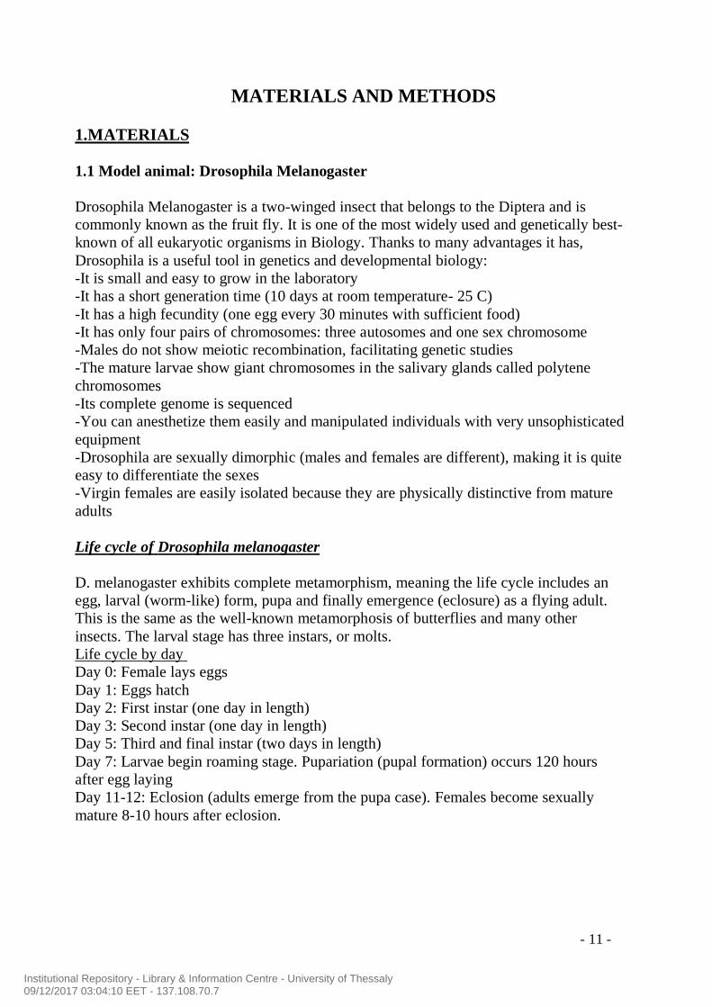
D. melanogaster exhibits complete metamorphism, meaning the life cycle includes an
egg, larval (worm-like) form, pupa and finally emergence (eclosure) as a flying adult.
This is the same as the well-known metamorphosis of butterflies and many other
insects. The larval stage has three instars, or molts.
Life cycle by day
Day 0: Female lays eggs
Day 1: Eggs hatch
Day 2: First instar (one day in length)
Day 3: Second instar (one day in length)
Day 5: Third and final instar (two days in length)
Day 7: Larvae begin roaming stage. Pupariation (pupal formation) occurs 120 hours
after egg laying
Day 11-12: Eclosion (adults emerge from the pupa case). Females become sexually
mature 8-10 hours after eclosion.
Institutional Repository - Library & Information Centre - University of Thessaly09/12/2017 03:04:10 EET - 137.108.70.7

- 12 -
In our experiments Drosophila is sustained at 25C on a growth medium of the following
composition:
-1kg maize
-1kg yeast
-155g agar
-6L distilled water
-600ml of Neplagine -1L pure alcohol 100% with 100gr of methylhydroxy-4-benzoate).
Neplagine prevents the growth of bacteria.
Institutional Repository - Library & Information Centre - University of Thessaly09/12/2017 03:04:10 EET - 137.108.70.7

- 13 -
1.2 Balancer Chromosomes
Drosophila has one more advantage that makes her a really great organism model and that is
the use of Balancer Chromosomes. Recessive lethal “balancer chromosomes” carrying
visible genetic markers can be used to keep stocks of lethal alleles in a heterozygous state
without recombination due to multiple inversions in the balancer. These markers allow to
following the deletion of Hexim (in our case), as they are located in the same Chromosome
and they are easily identifiable either with the naked eye or under a microscope.
Many balancers exist for the X, 2 and 3 chromosomes, but they are not necessary for
chromosome 4 because there is no exchange on that chromosome.
For this experiment, the balancers which are used are : Tm3 and Tm6.
Specifically, both of them are in the third chromosome and they carry on a marker:
-Tm3: Serrate (Ser), that expressed as wings notched
-Tm6: SbTb → Sb: stubble (hairs are shorter and thicker than wild type)
Tb: tubby (small body- good marker for larval and pupal stages)
Institutional Repository - Library & Information Centre - University of Thessaly09/12/2017 03:04:10 EET - 137.108.70.7

- 14 -
2. METHODS
2.1 Crossings- Mutagenesis
Mutagenesis took place in four steps of crossings: (protocol 1-Annexes)
1st cross: In this step P element (transposon) and transposase (Dr Δ2-3) are not in the
same fly. After those crossings, it is expected for having offspring flies that carry on
both of them. Specifically:
Female: w/w; +/+ ; PW+ / PW
+ X Male: w/Y ; CyO/ Sp; DrΔ2-3/ Tm6 Ubx
( red eyes ) ( white eyes )
( red eyes) (red eyes)
In new generation there are four different phenotypical males and females, but we are
interested only in males with above genotypes.
Chr I Chr II Chr III
CyO
+
w DrΔ2-3
P Hexim w
Chr I Chr II Chr III
+
+
w
w
P Hexim
P Hexim
Chr I Chr II Chr III
CyO +
+ Sp
w DrΔ2-3
Ubx
+
Chr I Chr II Chr III
Sp
+
w DrΔ2-3
P Hexim
Institutional Repository - Library & Information Centre - University of Thessaly09/12/2017 03:04:10 EET - 137.108.70.7

- 15 -
2nd
Cross: Crossing of virgin females that carry on a balancer with the males of first
cross(offspring), which have in the third chromosome P element and transposase.
Female: w/w; +/+ ; Tm3 Ser/Tm6 SbTb X Male: w/Y ; CyO or Sp/ +; DrΔ2-3/PW
+
(white eyes ) (red eyes )
(white eyes)
We are interested only in males with above genotypes.
3rd
cross: Crossing virgin females with same genotype like in the second cross with
males from the second cross (offspring)
Female: w/w; +/+; Tm3 Ser/Tm6 SbTb X Male: w/Y; CyO or Sp/ +; PΔW
+/Tm3 or
Tm6
(white eyes ) (white eyes )
Chr I Chr II Chr III
Sp or CyO
+
w DrΔ2-3
+
Tm3 or Tm6
Chr I Chr II Chr III
Sp
+
w Tm3 or Tm6
+ w
Chr I Chr II Chr III
Sp or CyO
+
w DrΔ2-3
P Hexim Hexim
Hexim
Institutional Repository - Library & Information Centre - University of Thessaly09/12/2017 03:04:10 EET - 137.108.70.7

- 16 -
(white eyes) (whites eyes)
We are interested only in males and females with above characteristics, even if we can
recognise and other phenotypes under the microscope. Throw away different
phenotypes.
4th
cross: Crossing of the descendants from cross 3. This crossing will allow the creation
of stock where included only flies with desirable deletion if the experiment is successful.
Female: w/w; CyO or Sp/ +; PΔW+/Tm3 or Tm6 SbTb
(white eyes )
X
Male: w/Y; CyO or Sp/ +; PΔW
+/Tm3 or Tm6
( white eyes )
Chr I Chr II Chr III
Sp or CyO
+
w
DrΔ2-3
+
Tm6 w
Chr I Chr II Chr III
Sp or CyO
+
w
DrΔ2-3
+
Tm6
Chr I Chr II Chr III
Sp
+
w Tm3 or Tm6
+ w
Chr I Chr II Chr III
Sp or Cyo
+
w
DrΔ2-3 +
Tm3 or Tm6
Chr I Chr II Chr III
Sp or CyO
+
w
DrΔ2-3
+
Tm6 w
Chr I Chr II Chr III
+
w DrΔ2-3
+ Sp or CyO
Tm6
Institutional Repository - Library & Information Centre - University of Thessaly09/12/2017 03:04:10 EET - 137.108.70.7

- 17 -
DrΔ2-3 Transposase
P element Transposon located in Chr III
Tm3 Balancer in chromosome III
Tm6 Balancer in chromosome III
Cyo Wings curled upward instead of flat
Ubx Haltere larger and rounder than normal
*In every step we keep the crossings in 25 ᵒC.
2.2 Characterization of mutants by PCR
There are four samples that are coming from mutagenesis, where mutants are homozygous
viable for a deletion. It is supposed that this deletion is not in Hexim, because there is the
hypothesis when the mutant is homozygous for Hexim deletion is lethal. To verify this
hypothesis, the samples are checked by PCR for presence or absence of Hexim gene.
The PCR will compare the presence or absence, but also the level of expression among the
samples: wild type flies(Canton S), flies 20799(mutants with overexpression of Hexim)
and homozygous viable samples. Hexim gene is detected at 300bp.
PCR allows the amplification of specific DNA sequences and dramatically increase the
amount of them. It is necessary to be known the sequence of regions which delimit the
DNA in order to determine the sequence of DNA primers. The number of PCR cycles
realized by DNA polymerase is generally between 25 and 40. Every cycle of PCR consists
of three different phases in three different temperatures. (Protocol 2-Annexes)
In the picture are depicted the limits of primers on Hexim gene. Specifically:
primers PU63 and PU53 recognising a part in the beginning of the gene
primers PU65 and PU66 recognising a part late in the gene
primers PU67 and PU68 are the positive control of PCR
Institutional Repository - Library & Information Centre - University of Thessaly09/12/2017 03:04:10 EET - 137.108.70.7

- 18 -
Electrophoresis in agarose gel 1,2%
For verifying and visualizing the size and the quantity of PCR’s products, it has been
used analysis by electrophoresis in agarose gel and they are compared with a score of size
(Smart Ladder SF). The detection of DNA on this kind of gel is possible when it is
exposed in UV radiation, after reaction with BET (ethidium promide), which is
intercalating in the DNA strains.
2.3 Deficiency (Deletion) of Hexim
Crossing flies that have deficiences of Hexim with the mutants of mutagenesis in order
to see if the mutants of mutagenesis have a deletion in Hexim. Hypothesis is that the loss
of Hexim is homozygous lethal. However, deficiencies do not have a deletion only to
Hexim, but also in other genes around Hexim. That is why, it is necessary to cross also
deficiencies together, in order to ensure that they have the same deletion.
In the picture is depicted the location of Hexim and the size of deciciences.
2.4 Characterization of lethality step by using GFP
The female mutants of mutagenesis that carry on a deletion in Hexim gene are
crossed with males that carry on a balancer in X Chromosome (FM7) and two
balancers in Chromosome III (Tm6 and Tm3).
FM7 balancer carries the dominant marker Bar, which is recognized as oval
shape of Drosophila’s eye (eye narrower than usual)
B D 88A 87 89
Def1 -7978 (88C10-88D6)
90 91 92 93
Def3- 1422 (87D1-2;88E5-6)
Hexim gene 88C10
Def2- 1534 (87E8F1;93C)
Institutional Repository - Library & Information Centre - University of Thessaly09/12/2017 03:04:10 EET - 137.108.70.7

- 19 -
Left: Bar eye Right: normal eye
Tm6 balancer carries on the marker Btb
, which is recognised by two long
bristles.
Tm3 balancer carries on the GFP.
Crossings:
1st step:
Female: w/w; +/+ ; ΔHexim/ Tm6 SbTb
X
Male: FM7,GFP ; +/+;Tm6 Btb
/Tm3 GFP
After doing the above crosses appeared many phenotypes, but we are interested only in:
females with genotype FM7,GFP/+; ΔHexim/Tm3-GFP
and males with genotype: +/+; ΔHexim/Tm3-GFP
2nd
step:
Female: FM7,GFP/+; ΔHexim/Tm3-GFP X Male: +/+; ΔHexim/Tm3-GFP
Crossing of descendants from first cross obtaining mutants that are:
homozygous for deletion of Hexim (it is supposed to be lethal)
heterozygous for deletion of Hexim and carry on Tm3 balancer with GFP marker
homozygous for balancer Tm3-GFP (lethal)
3rd
step:
Under specific microscope observing every developmental stage of Drosophila in order to
find out until which stage homozygous Drosophila for deletion of Hexim will stay alive.
This fly do not fluorescent under the suitable microscope.
Institutional Repository - Library & Information Centre - University of Thessaly09/12/2017 03:04:10 EET - 137.108.70.7

- 20 -
RESULTS AND DISCUSSION
1. Mutagenesis
In order to study what is the impact of the Hexim’s gene deletion in Drosophila, it is
necessary to obtain mutants that are homozygous for this deletion. The hypothesis is
when Hexim deletion is homozygous , the phenotype is lethal, because Hexim takes
part in a very important complex for transcriptional elongation. Specifically, the
obtainment of the null mutants in Hexim deletion needs four steps of crossings:
1 step: female fly has the transposon ( P element) and male the transposase (DrΔ2-
3) . Transposon needs to be in the same chromosome with transposase in the same
fly in order to be activated and jump. This goal has been successed in this cross.
2step: Collection of males from first cross that carry on transposon and transposase
and cross them with virgin females, which have a balancer chromosome ( Tm3 or
Tm6) in Chromosome III. We expect that transposase will activate transposon and it
will jump with Hexim gene or at least with a part of it. P element is located before
the beginning of Hexim. We verified that P element jumped with Hexim, when
white eyes appeared to descendants. However, P element is possible to jump
precisely that means without Hexim gene. In that case, there will not be any change
in eye colour.
3step: Collection of males that carry on the transposon, the transposase and one of
the chromosome balancers (either Tm3 or Tm6). Cross of these males with virgin
females as in the second step.
4 step: In this step, by crossing the descendants of the third cross that carry on a
deletion in Hexim, we create stock of them. Tm3 balancer is less credible than Tm6,
that is why in this step we prefer mutants carrying Tm6 and we reject mutants with
Tm3 balancer.
We suppose that our mutagenesis is successful because we have mutants with white
eyes (deletion), that means P element jumped with a part of a gene. But still we
cannot be sure if the deletion is in our candidate gene or in another close to P
element.
Institutional Repository - Library & Information Centre - University of Thessaly09/12/2017 03:04:10 EET - 137.108.70.7

- 21 -
Candidates Number of samples
Initial candidates 131
Deletion of the gene 50
Homozygous lethal 2 Table1: The number of candidates in every step of mutagenesis
Figure 5: Above Drosophila with deletion, down wild type Drosophila.
2. Characterization of mutants by PCR
We would like to verify our hypothesis that the homozygous deletion of Hexim is
lethal. But, we observed in our mutant that some of them have a deletion (white
eyes), nevertheless, this deletion is “homozygous viable”. That is coming in
contrast with our hypothesis. By using PCR we checked if these mutants have
deletion of Hexim or not.
The results we obtained are:
Institutional Repository - Library & Information Centre - University of Thessaly09/12/2017 03:04:10 EET - 137.108.70.7

- 22 -
Picture 6: There is detection of Hexim gene in every mutant apart from control sample that does
not contain DNA.
1,2,3,4: Homozygous viable mutants
5: Canton S. – wild type fly
6: 20799 fly- fly that has over-expression of Hexim gene
7: Control- no sample
So, after obtaining the above result we made sure that there is no deletion in Hexim
and mutants are “homozygous viable” for other gene deletion. That allows us to
continue our hypothesis about “homozygous lethal” phenotype.
3. Deficiency (Deletion) of Hexim
In order to check if the deletion in homozygous lethal phenotypes of mutagenesis
is in Hexim, we cross these mutants with deficiencies, which include deletion of
Hexim. Deficiencies is a kind of measure for the area of deletion. A cross between
the mutant of mutagenesis and the deficiency is supposed to give lethal phenotype.
However, it is necessary to cross deficiencies together in order to be sure that they
have deletion in same area and in our case to have deletion in Hexim (lethal
phenotype).
Deficiencies Observed phenotype
Candidate 1 Candidate 2
1 Not lethal Not lethal
2 Lethal Lethal
3 Lethal Lethal Table 2: Observed results of deficiencies
Institutional Repository - Library & Information Centre - University of Thessaly09/12/2017 03:04:10 EET - 137.108.70.7

- 23 -
We conclude that Deficiency 1 does not have the same deletion with our candidates,
but Deficiencies 2 and 3 appeared lethal phenotypes probably means there is Hexim
deletion. However, to ensure this hypothesis firstly we cross these two deficiencies
together and expecting for observing lethal phenotype. The result of this cross was
lethal phenotype, so we purpose both of them have Hexim deletion.
About Deficiency 1, we purpose that it has deletions in other regions but not in
region of our candidate gene. So, the problem in this case is that Deficiency
includes Hexim gene. After using PCR method by Patricia Uguen(supervisor of the
stage), she realized that our hypothesis is right.
However, still we cannot be absolutely sure that the deletion in common among our
candidates, deficiency 2 and deficiency 3 is the right deletion. It is possible that P
element has jumped with a region of another gene that is in common with both
deficiencies and it is observed lethal babies. If it can be true this hypothesis, we
have a wrong deletion.
4. Characterization of lethality step by using GFP
With this method we obtain mutants that can fluorescent under specific
microscope. But, these are not the mutants that are homozygous for the deletion,
because GFP is not in the same allele with the Hexim deletion. Observing the
mutants in every developmental stage we could see some mutants fluorescent and
some others not. But this observation was until first instar larva (L1). In candidates
we were interested in were these do not fluorescent, because that means were
homozygous for the deletion and they did not carry on the allele with GFP marker.
Specifically:
Stage Mutants-GFP Mutants-homozygous
lethal
Embryogenesis present present
First instar larva ( L1) present present
Second instar larva (L2) present absence
Third instar larva (L3) present absence Table 3: Presence and Absence of mutants in every developmental stage
According to above results we conclude that the mutant, which is homozygous for
the Hexim deletion can survive until L1 stage but not more. We purposed that
Institutional Repository - Library & Information Centre - University of Thessaly09/12/2017 03:04:10 EET - 137.108.70.7
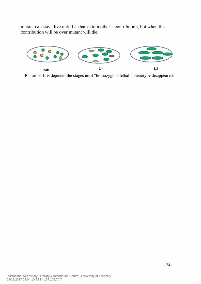
- 24 -
mutant can stay alive until L1 thanks to mother’s contribution, but when this
contribution will be over mutant will die.
Picture 7: It is depicted the stages until “homozygous lethal” phenotype disappeared.
Institutional Repository - Library & Information Centre - University of Thessaly09/12/2017 03:04:10 EET - 137.108.70.7

- 25 -
CONCLUSION AND PERSPECTIVES
This project had two aims firstly to investigate the importance of Hexim
gene in viability and secondly to examine the stage of life that Drosophila
can reach without production of Hexim protein. Both of these aims have
been successed, as we managed obtaining mutants with deletion of Hexim
in both alleles. It is difficult to obtain these mutants, because you cannot be
sure for the imprecise jump of P element. Also, needs a lot of time (3
months) and many crossings.
Futhermore, we observed that Hexim is really important gene for
Drosophila’s viability and development, as its absence causes death. That
probably means that deletion of Hexim gene provokes modifications in the
regulation of transcription. Consequently, this impact on transcription
affects in some other genes that are important for life and death is coming.
However, Drosophila manages to be alive at embryogenesis and first larva
stage (L1) thanks to mother’s contribution, but after that stage it dies.
In the future, this project could continue by investigating more details
about Hexim’s function and specifically, which part of Hexim is deleted or
in other words deletion of which part is responsible for death. For example,
using the method “sequencing” we could have some information about the
exact region of deletion in Hexim, but also we can discover if there is
deletion and in other genes.
Another thing, that we could do in order to ensure the importance of
Hexim gene for viability is to cross the “homozygous lethal” mutants with
wild type flies. In this case, every descendant will have an allele with
Hexim gene and it supposed to be viable. (rescue of mutants)
Institutional Repository - Library & Information Centre - University of Thessaly09/12/2017 03:04:10 EET - 137.108.70.7

- 26 -
REFERENCES
1. Diribarne G., Bensaude O. (2009) 7SK, a non-coding RNA regulating P-TEFb, a
general transcription factor. RNA Biol. 2: 122-128
2. Manja Marz, Alexander Donath, Nina Verstraete, Van Trung Nguyen, Peter
F. Stadler and Olivier Bensaud (2009) Evolution of 7SK RNA and Its Protein
Partners in Metazoa. Mol Biol Evol 26: 2821-2830
3. Driscoll C.T. , G J Darlington and R J Maraia (1994) The conserved 7SK
snRNA gene localizes to human chromosome 6 by homolog exclusion probing of
somatic cell hybrid RNA. Nucleic Acids Reseacrh 22: 722-725
4. Humphries P. , S. E. Russel, P. Mewilliam, S. Mcquaid, C. Pearson, and M. M.
Humpries (1987) Observations on the Structure of two Human 7SK Pseudogenes
and on Homologous Transcripts in Vertebrate Species. Bioch. J. 245: 281-284
5. Herreweghe, E. V., S.Egloff, I. Goiffon, B.E. Jady, C.Froment, B. Monsarrat,
and T. Kiss (2007) Dynamic remodelling of human 7SK snRNA controls the
nuclear level of active P-TEFb. The EMBO Journal 26: 3570-3580
6. Nguyen, V. T., T. Kiss, A. A. Michels and O. Bensaude (2001) 7SK small
nuclear RNA binds to and inhibits the activity of CDK9/cyclin T complexes.
Nature 414: 322-325
7. Wassarman, D. A. and J. A. Steitz (1991) Structural Analyses of the 7SK
Ribonucleoprotein (RNP), the Most Abundant Human Small RNP of Unknown
Function. Molecular an Cellular Biology 11: 3432-3445
8. B. MATIJA PETERLIN, JOHN E. BROGIE, DAVID H. PRICE (2011) 7SK
snRNA: a noncoding RNA that plays a major role in regulating eukaryotic
transcription. WIREs RNA 3: 92-103
9. Young TM, Tsai M. , Tian B, Mathews MB, Pe’ery T. (2007) Cellular m-RNA
activates transcription elongation by displacing 7SK RNA. PLoS One 10: e1010
10. Qintong Li, Jeffrey J. Cooper, Gary H. Altwerger, Michael D. Feldkamp,
Madeline A. Shea and David H. Price (2007) Hexim1 is a promiscuous double-
stranded RNA-binding protein and interacts with RNAs in addition to 7SK in
cultured cells. Nucleic Acids Research 35: 2503-2512
11. Blazek, D., M. Barboric, J. Kohoutek, I. Oven and B. M. Peterlin (2005)
Oligomerization of Hexim 1 via 7SK snRNA and coiled-coil region directs the
inhibition of P-TEFb. Nucleic Acids Research 33: 7000-7010
12. Byers, S. A., J.P. Price, J. J. Cooper, Q. Li and D. H. Price (2005) Hexim2, a
Hexim 1 Related Protein, Regulates P-TEFb Through Association with 7SK. The
journal of Biological Chemistry Manuscript M500424200
13. Barboric, M., J. Kohoutek, J. P. Price, D. Blazek, D. H. Price and B. M.
Peterlin (2005) Interplay between 7SK snRNA and oppositely charged regions in
Hexim1 direct the inhibition of P-TEFb. The EMBO Journal 24: 4291-4303
14. Peterlin, B.M. AND D.H. Price (2006) Controlling the elongation phase of
transcription with P-TEFb. Molecular Cell 23: 297-305
15. Michels, A. A., V.T. Nguyen, A. Fradli, V. Labas, M. Edwards, F. Bonet, L.
Lania and O. Bensaude. (2003) MAQ1 and 7SK RNA interact with CDK9/Cyclin
T Complexes in a transcription-Dependent Manner. Molecular and Cellular
Biology 23: 4859-4869
16. Jasper H., N. Y., R. C., Andrea C. Pezda, Craig S. Samfordand Qiang Zhou
(2004) A human Immunodeficieny Virus Type1 Tat-Like Arginine-Rich RNA –
Institutional Repository - Library & Information Centre - University of Thessaly09/12/2017 03:04:10 EET - 137.108.70.7

- 27 -
Binding is essential for Hexim 1 to inhibit RNA Polymerase II transcription
through 7SK snRNA-Mediated inactivation of P-TEFb. Molecular and Cellular
Biology p.5094-5105
17. Li, Q., J. P. Price, S. A. Byers, D. Cheng, J. Peng and D. H. Price (2005)
Analysis of the Large Inactive P-TEFb complex indicates that it contains one 7SK
molecule, a dimer of Hexim 1 or Hexim2 and two P-TEFb molecules containing
CDK9 Phosphrylated threonine 186 . The journal of Biological Chemistry 280:
28219-28826
18. Turano, M., G. Napolitano, C. DUlac, B. Majello, O. Bensaude and L.
Lania(2006) Increased Hexim1 expression during erythroleukemia and
Neuroblastoma cell differentiation. Journal of Cellular Physiology 206: 603-610
19. Shimizu, N., R. Ouchida, N. Yoshikawa, T. H., H. W. , K. Okamoto, M. K., H.
Handa, C. Morimoto and H.Tanaka (2005) Hexim1 forms a transcriptionally
abortive complex with glucocorticoid receptor without involving 7SK RNA and
positive transcription elongation factor b. PNAs 102: 8555-8560
20. Li, Q., J. Cooper, G. H. Altwerger, M. D. F., M. A. Shea and D. H. Price (2007)
Hexim1 is a promiscuous double-stranded RNA-binding protein and interacts with
RNAs in addition to 7SK in cultured cells. Nucleic Acids Research 1-11
21. Zhou, Q. ,and J. H. N. Yik (2006) The Yin and Yang of P-TEFb regulation:
Implications for human immunodeficiency Virus gene expression and global
control of cell growth and differentiation. Microbiology and Molecular Biology
Reviews 70: 646-659
22. Fraldi, A., F.V., G. Napolitano, A. A. M., B. Majello, O. Bensaude and L.
Lania (2005) Inhibition of the Tat activity by the Hexim1 protein Retrovirology 2:
42.22
23. Dulac, C., A.A. Michels, A. Fraldi, F. Bonnet, V. T. Nguyen, G. Napolitano, L.
Lania and O. Bensaude (2005) Transcription-dependent Association of multiple
Positive Transcription Elongation factor units to a Hexim multimer. The Journal of
Biological Chemistry 280: 30619-30629
Institutional Repository - Library & Information Centre - University of Thessaly09/12/2017 03:04:10 EET - 137.108.70.7

- 28 -
ANNEXES
Protocol 1: Mutagenesis
Cross 1: female vierge W/W; +/+; PW+/PW+ (red eyes)
X male W/Y; CyO/Sp; Dr2-3/Tm6 Ubx (white eyes)
Prepare 20 tubes, where every one will contain one female with 4-5 males. Remove parents in new tube.
male w; CyO; Dr2-3 w; CyO; Tm6 Ubx w; Sp; Dr2-3 w; Sp; Tm6 Ubx
ou (1/4) ou (1/4) ou (1/4) ou (1/4)
female Y; CyO; Dr2-3 Y; CyO; Tm6 Ubx Y; Sp; Dr2-3 Y; Sp; Tm6 Ubx
w; CyO; Dr2-3 w; CyO; Tm6 Ubx w; Sp ; Dr2-3 w; Sp ; Tm6 Ubx
w; + ; PW+ w; + ; PW+ w; + ; PW+ w; + ; PW+
w; +; PW+ (Fem, Curly, Dr, YO) (Fem, Curly, Ubx, YO) (Fem, Sp, Dr, YO) (Fem, Sp, Ubx, YO)
1 ou ou ou ou
w; CyO; Dr2-3 w; CyO; Tm6 Ubx w; Sp ; Dr2-3 w; Sp ; Tm6 Ubx
Y; + ; PW+ Y; + ; PW+ Y; + ; PW+ Y; + ; PW+
(male, Curly, Dr,
YO) (male, Curly, Ubx, YO) (male, Sp, Dr, YO) (male, Sp, Ubx, YO)
We are interested in: male, Curly (ou Sp), Dr, YO
Keep the males that you collect at 18°C , don’t use them immediately for second cross.
Cross 2: 1 male w/Y; CyO ou Sp/+; Dr2-3/PW+ (red eyes)
X female w/w; +/+; Tm3 Ser/Tm6 SbTb (white eyes)
Put one only male in a tube with 4-5 females. Prepare more than 50 tubes.
male w; CyOouSp; Dr2-3 w; CyOouSp; PW+ w; CyOouSp; PW+
ou (1/2) ou (1/2) ou (?)
female Y; CyOouSp; Dr2-3 Y; CyOouSp; PW+ Y; CyOouSp; PW+
w; CyOouSp; Dr2-3 w; CyOouSp; PW+ w; CyOouSp; PW+
w; + ; Tm3 Ser w; + ; Tm3 Ser w; + ; Tm3 Ser
w; +; Tm3 Ser (Fem, Curly ou Sp, Dr, Ser, RE) (Fem, Curly ou Sp, Ser, RE) (Fem, Curly ou Sp, Ser, WE)
(1/2) ou ou ou
w; CyOouSp; Dr2-3 w; CyOouSp; PW+ w; CyOouSp; PW+
Y; + ; Tm3 Ser Y; + ; Tm3 Ser Y; + ; Tm3 Ser
(male, Curly ou Sp, Dr, Ser, RE) (male, Curly ou Sp, Ser, RE) (male, Curly ou Sp, Ser, WE)
w; CyOouSp; Dr2-3 w; CyOouSp; PW+ w; CyOouSp; PW+
w; + ; Tm6 SbTb w; + ; Tm6 SbTb w; + ; Tm6 SbTb
(Fem, Curly ou Sp, Dr, SbTb, RE) (Fem, Curly ou Sp, SbTb, RE) (Fem, Curly ou Sp, SbTb, WE)
w; +; Tm6 SbTb ou ou ou
(1/2) w; CyOouSp; Dr2-3 w; CyOouSp; PW+ w; CyOouSp; PW+
Y; + ; Tm6 SbTb Y; + ; Tm6 SbTb Y; + ; Tm6 SbTb
(male, Curly ou Sp, Dr, SbTb,RE) (male, Curly ou Sp, SbTb, RE) (male, Curly ou Sp, SbTb, WE)
We are interested in : males Curly ou Sp, Ser ou SbTb, white eyes
Institutional Repository - Library & Information Centre - University of Thessaly09/12/2017 03:04:10 EET - 137.108.70.7

- 29 -
Cross 3: 1 male w/Y; CyO ou Sp/+; PW+/Tm3 ouTm6 (white eyes)
X femelle w/w; +/+; Tm3 Ser/Tm6 SbTb (white eyes)
Put one only male in a tube with 4-5 females. Prepare more than 50 tubes.
male w; CyOouSp; PW+ w; CyOouSp;Tm3 ouTm6
ou (1/2) ou (1/2)
femelle Y; CyOouSp; PW+ Y; CyOouSp; Tm3 ouTm6
w; CyOouSp; PW+ w; CyOouSp; Tm3 ouTm6
w; + ; Tm3 Ser w; + ; Tm3 Ser
w; +; Tm3 Ser (Fem, Curly ou Sp, Ser, WE) (Fem, Curly ou Sp, Ser, SbTb ou lethal, WE)
(1/2) Ou ou
w; CyOouSp; PW+ w; CyOouSp; Tm3 ouTm6
Y; + ; Tm3 Ser Y; + ; Tm3 Ser
(male, Curly ou Sp, Ser, WE) (male, Curly ou Sp, Ser, SbTb ou lethal, WE)
w; CyOouSp; PW+ w; CyOouSp; Tm3 ouTm6
w; + ; Tm6 SbTb w; + ; Tm6 SbTb
(Fem, Curly ou Sp, SbTb, WE) (Fem, Curly ou Sp, Ser, SbTb ou lethal, WE)
w; +; Tm6 SbTb Ou ou
(1/2) w; CyOouSp; PW+ w; CyOouSp; Tm3 ouTm6
Y; + ; Tm6 SbTb Y; + ; Tm6 SbTb
(male, Curly ou Sp, SbTb, WE) (male, Curly ou Sp, Ser, SbTb ou lethal, WE)
We are interested in : male et female Curly ou Sp, SbTb, white eyes. We could collect also the mutants that
have deletion of Hexim and as balancer Tm3, but Tm3 balancer is less good than Tm6.
Cross 4: Cross together the brothers and sisters from the third cross. In this way, you will have a stock with desirable mutants.
Institutional Repository - Library & Information Centre - University of Thessaly09/12/2017 03:04:10 EET - 137.108.70.7

- 30 -
Protocol 2: PCR
Test 50ng of genomic DNA from each sample: four “homozygous viable”, one
wild type, one mutant with overexpression of Hexim, control.
Synthesis of DNA template:
For final volume of 25μL:
- Distilled water H2O : 17,7 μL
- Tp 10X TaqAM : 2,5 μL
- MgCl2 (25mM): 1.5 μL
- dNTPs (2,5 Mm/each) : 1 μL
- Taq AM: 0,3 μL
- Genomic DNA (100ng/ μL) : 0,5 μL
- Primer 1 (10 μM) : 0,75 μL
- Primer 2 (10 μM) : 0,75 μL
For each pair of oligos you mix all the elements together except for genomic
DNA.
Set up the PCR program:
-94 C 5min
-94 C 30sec
-58 C 30 sec repeated 30 cycles
-72C 30 sec
-72 C 7 min
Migrate 10 μL of each PCR sample + 2 μL of loading buffer on gel agarose
1.2% with wider wells. Also, use 6 μL of ladder of size (Smart Ladder SF) for
electrophoresis.
Set up electrophoresis program: 100V for 25min.
Institutional Repository - Library & Information Centre - University of Thessaly09/12/2017 03:04:10 EET - 137.108.70.7


