
CHAPTER 17: THE DIGESTIVE SYSTEM
VERY Basic
But first… A video
http://www.youtube.com/watch?v=bfOyLuknD_4
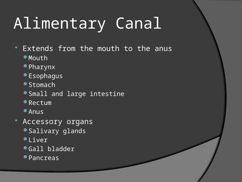
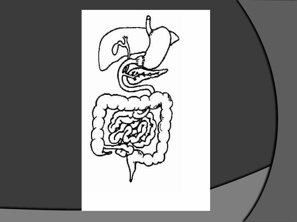
Alimentary Canal Extends from the mouth to the anus
MouthPharynxEsophagusStomachSmall and large intestineRectumAnus
Accessory organs Salivary glandsLiverGall bladderPancreas
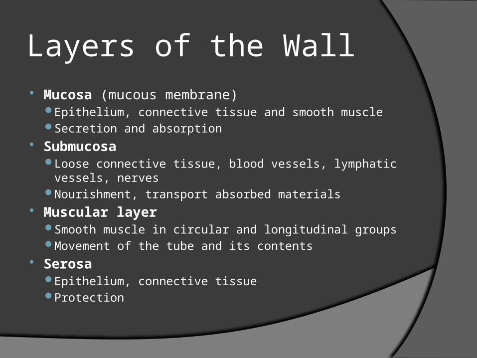
Layers of the Wall Mucosa (mucous membrane)
Epithelium, connective tissue and smooth muscle Secretion and absorption
Submucosa Loose connective tissue, blood vessels, lymphatic vessels,
nerves Nourishment, transport absorbed materials
Muscular layer Smooth muscle in circular and longitudinal groups Movement of the tube and its contents
Serosa Epithelium, connective tissue Protection
Movements of the Alimentary Canal Mixing Segmentation Peristalsis

Mouth Site of both mechanical
and chemical digestionTeeth and chewing
(mastication)
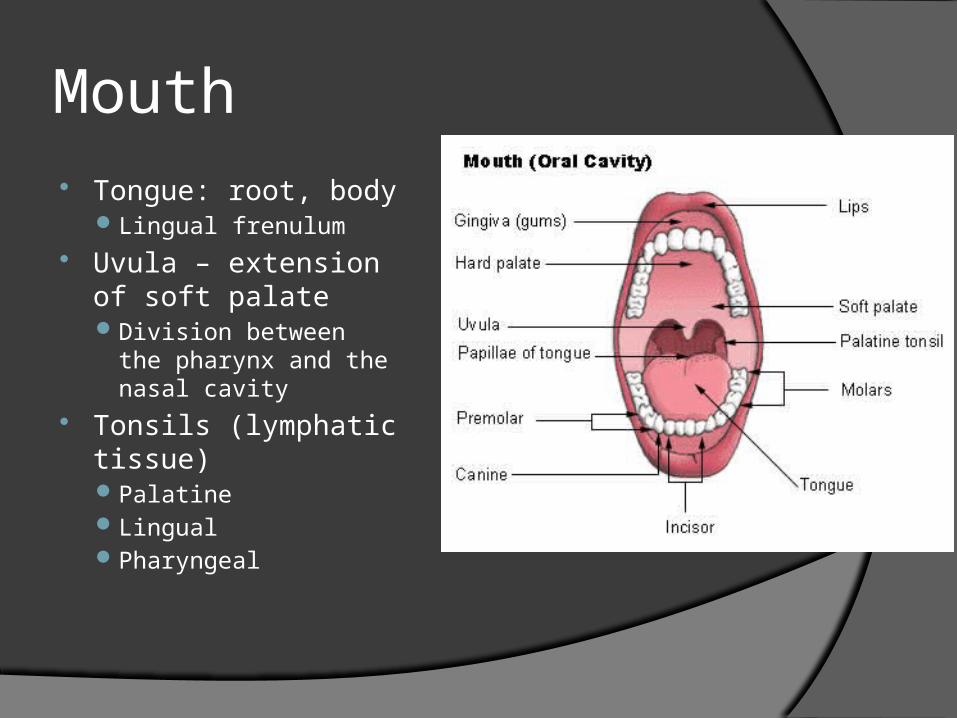
Mouth Tongue: root, body
Lingual frenulum Uvula – extension
of soft palateDivision between the
pharynx and the nasal cavity
Tonsils (lymphatic tissue)PalatineLingual Pharyngeal

Teeth
Incisor Canine Premolar Molar
Adult has 32 teeth (permanent) Children have 20 teeth (deciduous)
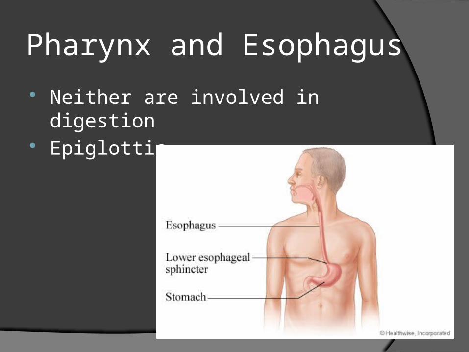
Pharynx and Esophagus
Neither are involved in digestion Epiglottis
Pharynx
Connects the nasal and oral cavities with the larynx and esophagus
Skeletal muscles control swallowingUnder voluntary control
Swallowing
3 stages:1. Food is chewed and mixed with saliva
○ Formation of a bolus
2. Food is pushed to the pharynx to enter the esophagus
○ Triggers a swallowing reflex
3. Peristalsis transports food from esophagus to stomach

Esophagus
Connects mouth to stomach
Lined with mucous glands
Cardiac sphincter (Lower esophageal sphincter)
Recap
Food enters the mouth…then what?
What types of digestion takes place?
What organs/structures are involved?
Is it voluntary or involuntary?
Stomach
Receives food from the esophagus Digestion
Muscle fibers run in all directionsCircularLongitudinalOblique
Very minimal absorption

Stomach
Inner lining: rugae – thick folds Greater curvature Lesser curvature Regions of the stomach:
Cardia FundusBodyPylorus
Pyloric sphincter
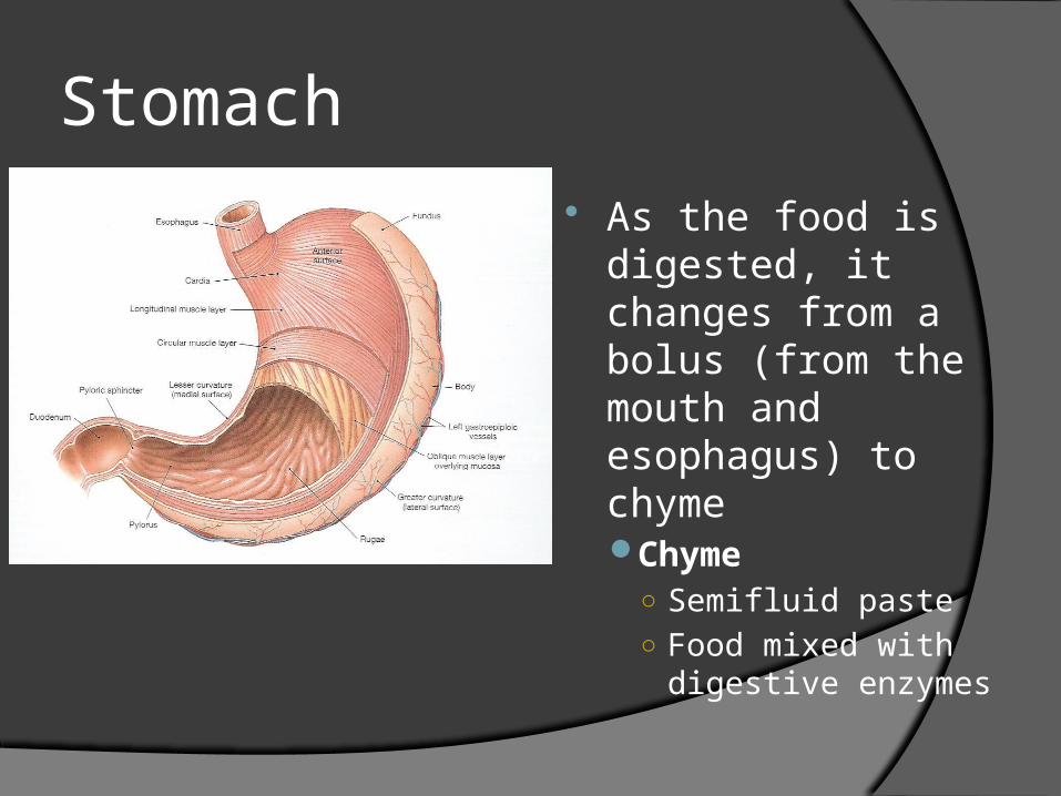
Stomach
As the food is digested, it changes from a bolus (from the mouth and esophagus) to chymeChyme
○ Semifluid paste ○ Food mixed with
digestive enzymes

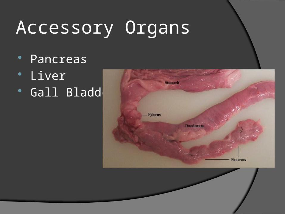
Accessory Organs
Pancreas Liver Gall Bladder

Pancreas
Secretion of pancreas juice Aids in digestion of
CarbohydratesFatsProteinNucleic acid
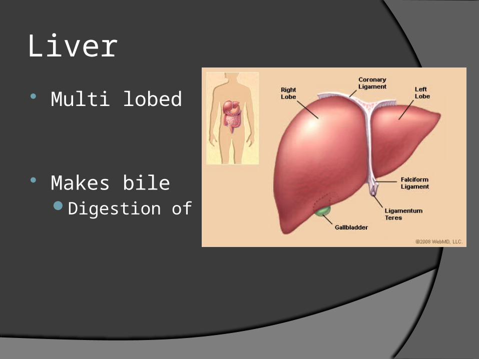
Liver
Multi lobed
Makes bileDigestion of fats
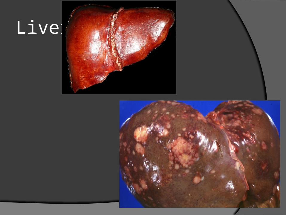
Liver
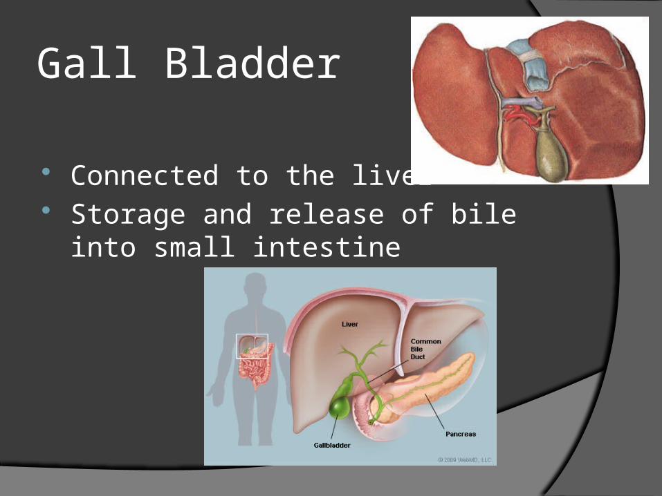
Gall Bladder
Connected to the liver Storage and release of bile into small
intestine

Small Intestine
Fills most of the abdominal cavity 5.5 – 6 meters long Receives secretions from pancreas, liver Absorption
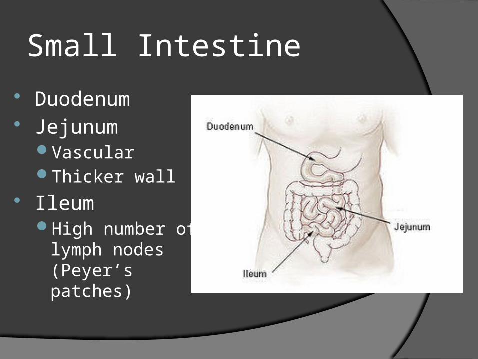
Small Intestine
Duodenum Jejunum
VascularThicker wall
Ileum High number of
lymph nodes (Peyer’s patches)

Small Intestine
Mesentary Connective tissue around the small intestine
Greater omentumMembrane drapes over lower digestive tractIf infection occurs, omentum will seal off
portion of digestive systemPrevents spread of infection to abdominal
cavity

Greater Omentum
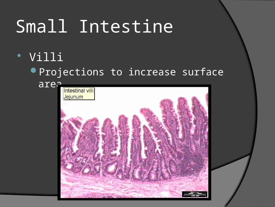
Small Intestine
VilliProjections to increase surface area

Large Intestine
Joins small intestine at the ileum
Water and electrolyte absorption Formation of feces
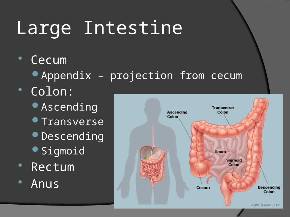
Large Intestine
CecumAppendix – projection from cecum
Colon:AscendingTransverseDescendingSigmoid
Rectum Anus
Digestive Enzyme Table
Digestive System Dissection


