
CRANIOTOMIES Op300 (1)
Craniotomies Last updated: September 5, 2017
SURGICAL ANATOMY OF CRANIUM........................................................................................................ 2 PATIENT POSITIONS ................................................................................................................................ 2
SIZE OF CRANIOTOMY ............................................................................................................................ 2
INCISION FOR CRANIOTOMY ................................................................................................................... 2 PERICRANIAL FLAP ................................................................................................................................. 3
CLOSURE OF CRANIOTOMY .................................................................................................................... 3
APPROACHES, CHOICE OF ...................................................................................................................... 3 CONVEXITY............................................................................................................................................ 3 SKULL BASE .......................................................................................................................................... 3
Anterior Skull Base .......................................................................................................................... 3
Middle Skull Base ............................................................................................................................ 4 Mesial Temporal Region ....................................................................................................... 4
POSTERIOR FOSSA .................................................................................................................................. 4 Internal Auditory Canal ± CP angle ...................................................................................... 4
Jugular Foramen .................................................................................................................... 4
SPECIAL SITUATIONS ............................................................................................................................... 4 Venous Sinus Injury ......................................................................................................................... 4
Entry into Frontal Sinus ................................................................................................................... 4 Entry into Mastoid Air Cells ............................................................................................................ 4
AWAKE CRANIOTOMIES ........................................................................................................................... 4 Indications ........................................................................................................................................ 5 Contraindications ............................................................................................................................. 5
Alternative ........................................................................................................................................ 5 Equipment ........................................................................................................................................ 5
Anesthesia ........................................................................................................................................ 5
Scalp regional anesthesia ....................................................................................................... 5 Positioning ........................................................................................................................................ 6
Electrode Placement ......................................................................................................................... 6 Craniotomy ....................................................................................................................................... 6
Mapping ........................................................................................................................................... 6
After Discharge (AD) Threshold Determination ................................................................... 6
Somatosensory Evoked Potentials Mapping ......................................................................... 7
Motor Mapping ...................................................................................................................... 7 Sensory Mapping ................................................................................................................... 7
Language Mapping ................................................................................................................ 7 Complication Avoidance .................................................................................................................. 8
TRANSNASAL ENDOSCOPIC ACCESS TO ANTERIOR CRANIAL FLOOR ................................................... 9 Indications ............................................................................................................................. 9 Preop ...................................................................................................................................... 9
Details .................................................................................................................................. 10 Postop .................................................................................................................................. 10
FRONTAL (UNILATERAL) CRANIOTOMY ............................................................................................... 10 Positioning, Pinning, Incision ............................................................................................. 10
BIFRONTAL CRANIOTOMY .................................................................................................................... 10 Indications ........................................................................................................................... 10 Positioning, Pinning, Incision ............................................................................................. 10
FRONTO-TEMPORO-ZYGOMATIC CRANIOTOMY ................................................................................... 11 SUBFRONTAL CRANIOTOMY .................................................................................................................. 11
PARIETAL CRANIOTOMY ....................................................................................................................... 12 (SUB)TEMPORAL CRANIOTOMY ............................................................................................................ 12 PTERIONAL CRANIOTOMY .................................................................................................................... 13 PTERIONAL CRANIOTOMY WITH ORBITOZYGOMATIC EXTENSION (“OZ CRANIOTOMY”) ................. 14 ANTERIOR CLINOIDECTOMY ................................................................................................................. 15
TRANSORAL CLIVAL APPROACH ........................................................................................................... 15 TRANSPETROSAL APPROACHES TO POSTERIOR FOSSA ....................................................................... 16
Anterior (medial) transpetrosal approaches (Kawase, s. anterior petrosectomy) ................ 16 Posterior transpetrosal (presigmoid) approaches ................................................................ 16
RETROLABYRINTHINE (PRESIGMOID) APPROACH ............................................................................... 16 TEMPOROPOLAR (HALF-AND-HALF) APPROACH TO THE BASILAR ARTERY AND THE RETROSELLAR
SPACE ..................................................................................................................................................... 18 INDICATIONS ........................................................................................................................................ 18 CONTRAINDICATIONS ........................................................................................................................... 19 PROCEDURE ......................................................................................................................................... 19
POSTERIOR FOSSA CRANIECTOMY / MIDLINE SUBOCCIPITAL CRANIOTOMY .................................... 20 Indications ........................................................................................................................... 20
Preop .................................................................................................................................... 20 Position ................................................................................................................................ 20
Technique ....................................................................................................................................... 20 Dissection ............................................................................................................................ 20 Craniectomy ........................................................................................................................ 21
C1 laminectomy .................................................................................................................. 21
Fibrous band ........................................................................................................................ 21
Dural Opening, Duraplasty .................................................................................................. 21 Cerebellum intervention ...................................................................................................... 23
Closure ................................................................................................................................. 23 Postoperatively ............................................................................................................................... 23
LATERAL SUBOCCIPITAL CRANIOTOMY ............................................................................................... 23 Indications ...................................................................................................................................... 23 Anesthesia ...................................................................................................................................... 23
Technique ....................................................................................................................................... 23 Postoperatively ............................................................................................................................... 25
RETROSIGMOID (RETROMASTOID) CRANIOTOMY ............................................................................... 25 INDICATIONS ........................................................................................................................................ 25
PROCEDURE ......................................................................................................................................... 25 Location of asterion ............................................................................................................. 25
Position ................................................................................................................................ 25
Electrophysiological monitoring ......................................................................................... 25 Pin placement ...................................................................................................................... 25
Incision ................................................................................................................................ 26 Craniotomy ..................................................................................................................................... 27
EXTENDED RETROSIGMOID CRANIOTOMY ........................................................................................... 27
Preop .................................................................................................................................... 27 Technique ............................................................................................................................ 27
SUPRACEREBELLAR INFRATENTORIAL APPROACH .............................................................................. 28 INDICATIONS ........................................................................................................................................ 28
CONTRAINDICATIONS ........................................................................................................................... 28
PREOPERATIVE ..................................................................................................................................... 28 PROCEDURE ......................................................................................................................................... 29
Anesthesia ...................................................................................................................................... 29 Positioning ...................................................................................................................................... 29 Dissection ....................................................................................................................................... 29
COMPLICATIONS .................................................................................................................................. 30 Air embolism ....................................................................................................................... 30
FAR-LATERAL SUBOCCIPITAL APPROACH ........................................................................................... 30 INDICATIONS ........................................................................................................................................ 31

CRANIOTOMIES Op300 (2)
PROCEDURE ......................................................................................................................................... 31
TRANSCONDYLAR APPROACH ............................................................................................................... 34 TRAUMA PROCEDURES (burr hole washout, decompressive craniectomies, etc) – see p. Op320 >>
SUBDURAL TAP THROUGH FONTANEL – see p. TrH13 >>
PATIENT POSITIONING, PINNING – see p. Op100 >>
MEDICATIONS for craniotomies – see p. Op100 >>
SURGICAL ANATOMY OF CRANIUM
ASTERION – sigmoid-transverse sinus junction most commonly (but not always) hugs it anteriorly-
superiorly (so it is safe to drill here if targeting venous sinus angle) – on the line from root of zygoma
to inion where it is intersected with vertical line just behind mastoid process.
Occipito-sigmoid suture (extends down from asterion) – posterior edge of sigmoid sinus is 1 cm
anterior from it.
Torcular is 1 cm above inion tip.
Jugular foramen – nerves occupy centra portion of it; jugular vein is posterior.
PATIENT POSITIONS
¾ = park bench
SIZE OF CRANIOTOMY
goal of “keyhole” surgery - not to perform small incision and craniotomy for sake of small opening
but to permit adequate access to skull base while limiting trauma to surrounding structures.
McCarty keyhole
size of craniotomy:
surface lesions typically require craniotomies as large as lesion.
deep-seated lesions can be accessed through much smaller craniotomy since intracranial field
widens with increasing distance from skull.
Minimally Invasive Transcranial Operative Corridors: Techniques:
http://www.neurosurgicalatlas.com/grand-rounds/minimally-invasive-transcranial-operative-corridors-
techniques
INCISION FOR CRANIOTOMY
instead of straight (slash) incisions, use “S” incisions – will get much more length and thus can
retract scalp more for larger craniotomy.
incise epithelium with knife, then use Bovie for hemostatic cut.
Alternative (Dr. Broaddus) – single cut down to bone* then Raney clips.
*except temporalis muscle – better to open with Bovie to prevent bleeding
avoid Raney clips – may cause permanent incisional alopecia; if used – remove at stepwise fashion
along of scalp closure (minimize blood loss).
use Metz over temporalis muscle – insert scissor tips between scalp and temporalis fascia and cut
over it.

CRANIOTOMIES Op300 (3)
for lifting up temporalis fascia and leaving cuff – how to find optimal cuff: incise fascia along
fibers – this way will see where fascia inserts and thus will know how big cuff needs to be.
PERICRANIAL FLAP
Dr. Graham likes to dissect pericranium from scalp flap at the end of case using Bovie; he
sometimes leaves only narrow pedicle for pericranial flap – still enough for vascular supply but lets
to advance flap much more posteriorly.
CLOSURE OF CRANIOTOMY
staples for skin (esp. Dr. Broaddus – does not like Monocryl!)
for kids < 3 yo, important to close dura water tight – to prevent leptomeningeal cyst (Dr. Collins
likes to rotate pericranial flap on pedicle to cover dura).
APPROACHES, CHOICE OF
CONVEXITY
ENTIRE CONVEXITY:
Twist drill craniostomy >>
Bur hole washout >>
Decompressive hemicraniectomy (“Trauma Flap”) / Frontotemporoparietal craniotomy >>
FRONTAL CONVEXITY:
Decompressive Bifrontal craniectomy (Kjellberg) >>
Frontal (Unilateral) craniotomy >>
FRONTOTEMPORAL CONVEXITY:
Fronto-temporo-zygomatic craniotomy >>
SKULL BASE
ANTERIOR SKULL BASE
Transnasal endoscopic access to anterior cranial floor >>
Midline Anterior Skull Base approach
NREF video >>
Bifrontal craniotomy
Subfrontal craniotomy
Supraorbital keyhole craniotomy - through eyebrow incision
Interhemispheric approach

CRANIOTOMIES Op300 (4)
transbasal approach – extending down through cribriform plate into nasal cavity (up to hard palate);
now obsolete due to ENT endoscopic capabilities.
MIDDLE SKULL BASE
Fronto-temporo-zygomatic craniotomy >>
Frontotemporal craniotomy (e.g. Dandy’s frontotemporal “macrosurgical approach”)
Pterional craniotomy (e.g. Yasargil’s microsurgical pterional approach) >>
Orbitozygomatic craniotomy >>
Infratemporal craniotomy
NREF video >>
Designing a Lateral Skull Base Approach – NREF video >>
MESIAL TEMPORAL REGION
- divided into 3 areas:
1. Anterior - transsylvian-transinsular approach
2. Middle - transtemporal approach
3. Posterior - supracerebellar-transtentorial approach
POSTERIOR FOSSA
A. Suboccipital craniotomy >>
Supracerebellar approach >>
B. Retrosigmoid (retromastoid) >>
Far Lateral approach >>
C. Presigmoid (in order of increasing temporal bone drilling):
a) retrolabyrinthine
b) translabyrinthine
c) transcochlear
D. Transoral Clival approach >>
Designing a Posterolateral Skull Base Approach: Presigmoid vs. Retrosigmoid Approaches – NREF
video >>
Designing a Posterior Fosse Approach – NREF video >>
INTERNAL AUDITORY CANAL ± CP ANGLE
JUGULAR FORAMEN
Approach to jugular foramen – NREF video >>
SPECIAL SITUATIONS
VENOUS SINUS INJURY
Small tears can be closed using bipolar cautery.
Medium tears can be managed by placing Gelfoam (Pfizer, Inc., NY, NY), Surgicel (Ethicon, Inc.,
Cornelia, GA), or Avitene Flour MCH (Davol, Inc., a subsidiary of C.R. Bard, Inc., Warwick, RI) over
opening followed by wet cottonoid until bleeding stops.
Larger tears can be sutured* over a muscle plug.
*esp. if contralateral flow is not patent on preoperative imaging.
before elevating fracture over sinus notify anesthesia (risk of bleeding + air embolism) and have
large piece of Gelfoam and rapid infuser ready.
if large circular sinus is encountered while crossing foramen magnum, this should be controlled
with Weck clips, divided, and oversewn with dural sutures. see below
if bleeding is too brisk and finger pressure blocks view for repair → proximal sinotomy to allow
temporary placement of inflatable Fogarty balloon catheter.
if interposed graft is necessary for permanent repair of large sinus laceration that cannot be
repaired any other way, autogenous saphenous vein graft may be utilized after temporarily
shunting blood through interposed shunt to allow time to sew graft into place.
bleeding from CAVERNOUS SINUS – inject fibrin glue into it!
ENTRY INTO FRONTAL SINUS
carefully remove sinus mucosa from bone flap pockets (may use diamond drill bit for thermal kill)
rest of sinus in orbitofrontal bar is cover with wet long patty for the duration of case; at the end
check to make sure no bone dust or clots are in frontal sinus; leave mucosa intact; do not place any
Gelfoam in it; M. Couldwell places muscle plug into sinus; cover with vascularized pericranial flap
→ Tisseel / DuraSeal.
add additional antibiotic coverage.
if sinus mucosa is inflamed (sinus is no longer sterile) – need sinus cranialization:
remove entire mucosa down to ostia
remove posterior wall of frontal sinus
cover ostia with pericranial flap
Japanese neurosurgeons suture-repair sinus mucosal entry to close sinus intracranial access.
ENTRY INTO MASTOID AIR CELLS
wax well or fill with HydroSet.
AWAKE CRANIOTOMIES
Used sources:
Connolly “Fundamentals of Operative Techniques in Neurosurgery” 2nd ed. (2010), ch. 60 (275-278
pages)
Badie 'Neurosurgical operative atlas - Neuro-oncology' 2nd ed. (2007), ch. 14 (pages 115, 117-119)
R. Jandial “Core Techniques in Operative Neurosurgery” (2011), procedure 39
CRANIOTOMIES Op300 (5)
General anesthesia is preferred for most patients with minimal to moderate motor and/or sensory
deficits and with lesions outside language regions.
Language function can only be assessed in awake patients.
Awake craniotomy should be considered for patients with more severe motor or sensory deficits,
provided that the patient is cooperative
INDICATIONS
Lesions / cortical resections in or near motor, somatosensory, or language cortex.
Wada test to determine hemisphere of language dominance.
object naming, at 4 sec per image, must be better than 75%.
motor mapping requires (near) normal power (at least 4-/5 for mapping under general anesthesia)
somatosensory mapping requires (near) normal sensation.
CONTRAINDICATIONS
1. Obesity, sleep apnea, airway problems.
2. Psychiatric issues.
3. Children < 10 years:
1) awake craniotomies are not as well tolerated
2) cortical stimulation mapping may not elicit motor responses (H: SSEPs are more useful).
• Patients whose preoperative language baseline is less than 80% of objects named correctly at 4-second intervals. Because stimulation language mapping relies on the ability to block object naming, language cannot be localized when baseline errors are too high. Although some object slides can be discarded from the specific patient’s slide set, the final set should have at least 50 slides. When the patient has normal naming ability (i.e., 100% of slides named correctly), slides are presented at 3-second intervals. This allows quicker mapping with a higher current because of less temporal current summation.
ALTERNATIVE
- subdural grid electrodes and extraoperative functional mapping.
EQUIPMENT
iced irrigation fluid and IV midazolam (to abort seizures during cortical stimulation)
brain diagram for drawing electrode montage on the brain
15-30 small (3 to 5 mm) paper numbered tickets
Ojemann Cortical Stimulator
for language mapping:
1) Grass CE-1 electrode holder (Grass Technologies, West Warwick, RI) and cortical
electrodes
2) EEG machine
3) slide projector (or computer slide show) with 50-100 object drawings, presented at a rate
of one object every 3-4 seconds (depending on patient’s verbal ability)
for somatosensory evoked potentials: 8-contact strip electrode (with cable and connector), SSEP
machine.
ANESTHESIA
A) patients awake for entire duration of surgical intervention (awake-awake-awake craniotomy,
AAA)
B) patients initially sedated (asleep-awake-asleep craniotomy, SAS) - may compromise
electrophysiological brain mapping and thus endanger patient's neurological outcome Ott C “The impact of sedation on brain mapping: a prospective, interdisciplinary, clinical
trial.” Neurosurgery. 2014 Aug;75(2):117-23
if patient was not on anticonvulsants preoperatively, therapeutic load of AED should be
administered
20% mannitol IV - maximum 0.5 g/kg; higher doses will cause nausea and vomiting.
paralytics cannot be used (other than for induction).
general anesthetic mapping cases - inhalation anesthetics must be minimized.
language mapping – only propofol and/or dexmedetomidine - facilitate mild sedation during
opening and closing, and maximum cooperation during mapping and resection phases; no
narcotics* or additional anesthetic medications!
*thus, good field block with 0.25-0.5% bupivacaine + 0.5-1% lidocaine + 1:200,000
epinephrine before draping – along incision, base of scalp flap, ± nerve blocks (e.g.
supraorbital, occipital).
laryngeal masked airway (LMA) may be employed depending on patient and preferences of
anesthesiologist.
SCALP REGIONAL ANESTHESIA
After induction of propofol or midazolam sedation, without intubation, the local anesthetic field block is placed, beginning in the regions of the preauricular, postauricular, and supraorbital nerves. By starting at these points, placement of the remainder of the block is less painful. The entry point of the one-inch, 25 gauge needle (just superior to the lateral aspect of the right eyebrow) is used for anterior scalp fi eld block.
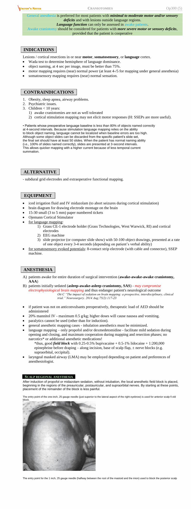
The entry point for the 1 inch, 25 gauge needle (halfway between the root of the mastoid and the inion) used to block the posterior scalp

CRANIOTOMIES Op300 (6)
POSITIONING
patient supine with head on horseshoe headholder; legs and head of bed elevated.
Mayfield pin head holder is applied using local anesthesia; alternative – use AxiEM or Neuro
FrameLock systems without pinning. see p. Op30 >>
The patient’s head must always be lateral or angled slightly above the horizon so that the airway is well protected and the patient can see the computer screen. Attention is directed toward positioning the head to optimize the patient’s airway during sedation.
ELECTRODE PLACEMENT
3 electrodes are placed on the neck as reference for EEG
median nerve or tibial nerve stimulating electrodes are placed contralateral to hemisphere where
SSEP testing is to be performed
CRANIOTOMY
standard temporal or frontal craniotomy, but opening must provide access to all areas to be
mapped.
warn pathologist not to announce frozen path results over loudspeaker.
Because some patients awaken confused or slightly combative, the dura is not opened until the patient is completely awake and calm. To hold the electrode, two options are available: an epidural post, which clamps to the skull, or a post that screws into the bone. We prefer the post that screws into the skull (shown) because it is more stable and avoids potential epidural bleeding.
After the brain is exposed, cortical electrodes are placed on the brain surface, followed by small numbers that identify which area has been stimulated:
There are also many electrode options available, including carbon-tip electrodes (as shown), cotton-tip electrodes, and strip/grid electrodes. We prefer carbon-tip electrodes because they maintain good contact with the cortex while permitting the surgeon excellent access for cortical stimulation
MAPPING
See also p. E13 >>
AFTER DISCHARGE (AD) THRESHOLD DETERMINATION
CRANIOTOMIES Op300 (7)
using bipolar stimulator, beginning at 2 mA current, stimulate 3 to 5 areas of brain region to be
mapped, calling out nearest cortical electrode to EEG team to record.
watch EEG for ADs: if none, increase current by 2 mA increments until ADs are elicited (this is
AD threshold); if persistent ADs occur, irrigate brain with cold irrigation fluid.
current 1-2 mA below AD threshold is used for mapping.
Determining the AD threshold helps to prevent evoking clinical seizure activity and false localization. A, The cortex is stimulated while electrocorticography is performed. Beginning with 2-mA current, several spots on the cortex are stimulated for the same duration as the planned object image presentation epoch (3 or 4 seconds). Current is increased by 2 mA after several areas are tested without evoking ADs. This sequence is repeated until ADs are seen, then mapping is performed with current 1 to 2 mA below the AD threshold. The surgeon stimulates the brain as the patient names the object slides. After each stimulation, the surgeon calls out the number on the nearest ticket. B, The neurologist monitors electroencephalogram for ADs and seizures. If either ADs or a frank clinical seizure occurs, the brain is irrigated with iced irrigation fluid. If a seizure persists, midazolam (Versed) is administered in 1- to 2-mg increments until clinical seizure activity ceases. It is very rare to evoke seizures that persist or become problematic for continued mapping If a seizure persists, despite reasonable doses of midazolam and irrigation of the brain with cold fluid, an airway should be placed and the seizures stopped with other drugs as needed
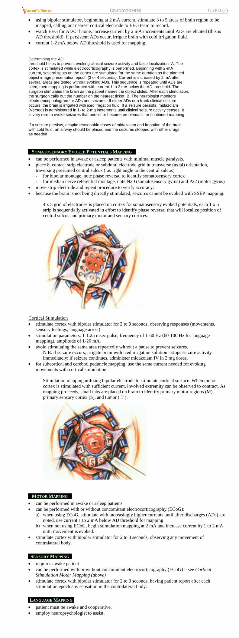
SOMATOSENSORY EVOKED POTENTIALS MAPPING
can be performed in awake or asleep patients with minimal muscle paralysis.
place 8–contact strip electrode or subdural electrode grid in transverse (axial) orientation,
traversing presumed central sulcus (i.e. right angle to the central sulcus)
- for bipolar montage, note phase reversal to identify somatosensory cortex
- for median nerve referential montage, note N20 (somatosensory gyrus) and P22 (motor gyrus)
move strip electrode and repeat procedure to verify accuracy.
because the brain is not being directly stimulated, seizures cannot be evoked with SSEP mapping.
4 x 5 grid of electrodes is placed on cortex for somatosensory evoked potentials, each 1 x 5
strip is sequentially activated in effort to identify phase reversal that will localize position of
central sulcus and primary motor and sensory cortices:
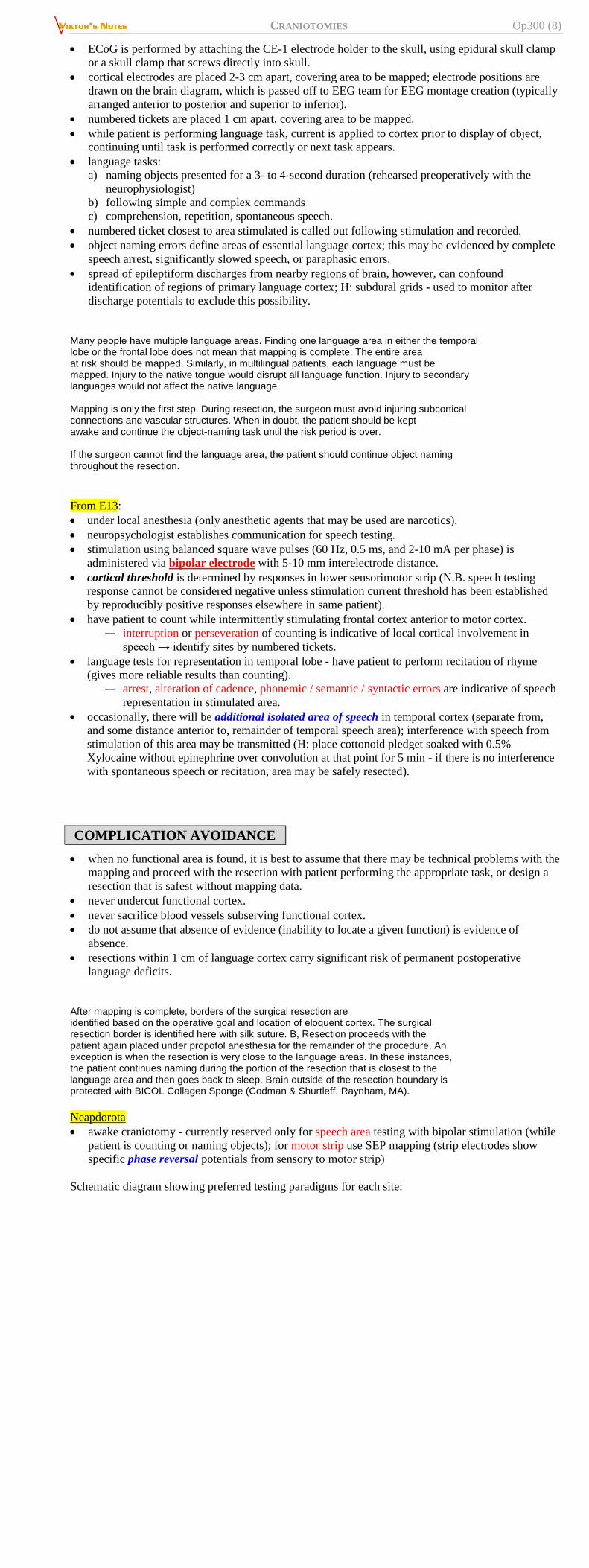
Cortical Stimulation
stimulate cortex with bipolar stimulator for 2 to 3 seconds, observing responses (movements,
sensory feelings, language arrest)
stimulation parameters: 1-1.25 msec pulse, frequency of 1-60 Hz (60-100 Hz for language
mapping), amplitude of 1-20 mA.
avoid stimulating the same area repeatedly without a pause to prevent seizures.
N.B. if seizure occurs, irrigate brain with iced irrigation solution - stops seizure activity
immediately; if seizure continues, administer midazolam IV in 2 mg doses.
for subcortical and cerebral peduncle mapping, use the same current needed for evoking
movements with cortical stimulation.
Stimulation mapping utilizing bipolar electrode to stimulate cortical surface. When motor
cortex is stimulated with sufficient current, involved extremity can be observed to contract. As
mapping proceeds, small tabs are placed on brain to identify primary motor regions (M),
primary sensory cortex (S), and tumor ( T ):
MOTOR MAPPING
can be performed in awake or asleep patients
can be performed with or without concomitant electrocorticography (ECoG):
a) when using ECoG, stimulate with increasingly higher currents until after discharges (ADs) are
noted, use current 1 to 2 mA below AD threshold for mapping
b) when not using ECoG, begin stimulation mapping at 2 mA and increase current by 1 to 2 mA
until movement is evoked.
stimulate cortex with bipolar stimulator for 2 to 3 seconds, observing any movement of
contralateral body.
SENSORY MAPPING
requires awake patient
can be performed with or without concomitant electrocorticography (ECoG) – see Cortical
Stimulation Motor Mapping (above)
stimulate cortex with bipolar stimulator for 2 to 3 seconds, having patient report after each
stimulation epoch any sensation in the contralateral body.
LANGUAGE MAPPING
patient must be awake and cooperative.
employ neuropsychologist to assist.
CRANIOTOMIES Op300 (8)
ECoG is performed by attaching the CE-1 electrode holder to the skull, using epidural skull clamp
or a skull clamp that screws directly into skull.
cortical electrodes are placed 2-3 cm apart, covering area to be mapped; electrode positions are
drawn on the brain diagram, which is passed off to EEG team for EEG montage creation (typically
arranged anterior to posterior and superior to inferior).
numbered tickets are placed 1 cm apart, covering area to be mapped.
while patient is performing language task, current is applied to cortex prior to display of object,
continuing until task is performed correctly or next task appears.
language tasks:
a) naming objects presented for a 3- to 4-second duration (rehearsed preoperatively with the
neurophysiologist)
b) following simple and complex commands
c) comprehension, repetition, spontaneous speech.
numbered ticket closest to area stimulated is called out following stimulation and recorded.
object naming errors define areas of essential language cortex; this may be evidenced by complete
speech arrest, significantly slowed speech, or paraphasic errors.
spread of epileptiform discharges from nearby regions of brain, however, can confound
identification of regions of primary language cortex; H: subdural grids - used to monitor after
discharge potentials to exclude this possibility.
Many people have multiple language areas. Finding one language area in either the temporal lobe or the frontal lobe does not mean that mapping is complete. The entire area at risk should be mapped. Similarly, in multilingual patients, each language must be mapped. Injury to the native tongue would disrupt all language function. Injury to secondary languages would not affect the native language. Mapping is only the first step. During resection, the surgeon must avoid injuring subcortical connections and vascular structures. When in doubt, the patient should be kept awake and continue the object-naming task until the risk period is over. If the surgeon cannot find the language area, the patient should continue object naming throughout the resection.
From E13:
under local anesthesia (only anesthetic agents that may be used are narcotics).
neuropsychologist establishes communication for speech testing.
stimulation using balanced square wave pulses (60 Hz, 0.5 ms, and 2-10 mA per phase) is
administered via bipolar electrode with 5-10 mm interelectrode distance.
cortical threshold is determined by responses in lower sensorimotor strip (N.B. speech testing
response cannot be considered negative unless stimulation current threshold has been established
by reproducibly positive responses elsewhere in same patient).
have patient to count while intermittently stimulating frontal cortex anterior to motor cortex.
— interruption or perseveration of counting is indicative of local cortical involvement in
speech → identify sites by numbered tickets.
language tests for representation in temporal lobe - have patient to perform recitation of rhyme
(gives more reliable results than counting).
— arrest, alteration of cadence, phonemic / semantic / syntactic errors are indicative of speech
representation in stimulated area.
occasionally, there will be additional isolated area of speech in temporal cortex (separate from,
and some distance anterior to, remainder of temporal speech area); interference with speech from
stimulation of this area may be transmitted (H: place cottonoid pledget soaked with 0.5%
Xylocaine without epinephrine over convolution at that point for 5 min - if there is no interference
with spontaneous speech or recitation, area may be safely resected).
COMPLICATION AVOIDANCE
when no functional area is found, it is best to assume that there may be technical problems with the
mapping and proceed with the resection with patient performing the appropriate task, or design a
resection that is safest without mapping data.
never undercut functional cortex.
never sacrifice blood vessels subserving functional cortex.
do not assume that absence of evidence (inability to locate a given function) is evidence of
absence.
resections within 1 cm of language cortex carry significant risk of permanent postoperative
language deficits.
After mapping is complete, borders of the surgical resection are identified based on the operative goal and location of eloquent cortex. The surgical resection border is identified here with silk suture. B, Resection proceeds with the patient again placed under propofol anesthesia for the remainder of the procedure. An exception is when the resection is very close to the language areas. In these instances, the patient continues naming during the portion of the resection that is closest to the language area and then goes back to sleep. Brain outside of the resection boundary is protected with BICOL Collagen Sponge (Codman & Shurtleff, Raynham, MA).
Neapdorota
awake craniotomy - currently reserved only for speech area testing with bipolar stimulation (while
patient is counting or naming objects); for motor strip use SEP mapping (strip electrodes show
specific phase reversal potentials from sensory to motor strip)
Schematic diagram showing preferred testing paradigms for each site:

CRANIOTOMIES Op300 (9)
Proposal of intraoperative tasks based on relationships between tumor location and white matter
connectivity: projection pathways (pyramidal tract, thalamocortical radiations, optic radiations) and
subcallosal fasciculus:
Proposal of intraoperative tasks based on relationships between tumor location and association
pathways: inferior frontooccipital fascicle (IFOF), superior longitudinal fascicle (SLF), inferior
longitudinal fascicle (ILF), and uncinate fascicle:
Excellent article:
Coello et al “Selection of intraoperative tasks for awake mapping based on relationships
between tumor location and functional networks” published online September 20, 2013; DOI:
10.3171/2013.6.JNS122470.
TRANSNASAL ENDOSCOPIC ACCESS TO
ANTERIOR CRANIAL FLOOR
NREF video >>
INDICATIONS
Olfactory groove meningioma.
PREOP
lumbar drain, leave clamped; Dr. Broaddus prefers no.
N.B. lumbar drain helps but does not save poor closure

CRANIOTOMIES Op300 (10)
DETAILS
register for neuronavigation (e.g. Stryker mask)
bilateral nasal cavities decongested with NeoSynephrine moistened pledgets.
ENT part – operative 0-degree endoscope:
bilateral superior turbinate removal, partial superior posterior nasal septum removal, bilateral
maxillary antrostomies and total ethmoidectomies
nasoseptal mucosal flap is developed on intact vascular pedicle.
ethmoidal cribriform plate drilled off with high speed diamond bur to create anterior skull base
defect right under the tumor.
Neurosurgery part:
coagulated dura → durotomy over the tumor with # 11 blade.
tumor removed in piecemeal fashion using ring curettes and pituitary rongeurs.
30 degree endoscope to inspect the tumor cavity for additional tumor tissue.
ENT part - reconstruction:
pack base of the tumor bed with AlloDerm patch, followed by septal cartilage patch, DuraSeal
glue, and overlay of well vascularized nasoseptal mucosal flap again reinforced with DuraSeal
glue.
nasal passages packed with Gelfoam, followed by Merocel packings into both nostrils; packings
were lubricated with bacitracin ointment and inflated with some saline.
orogastric tube to decompress the stomach and suction out the oropharynx.
deep extubation, no cough.
1980s – free tissue grafts (onlay, inlay) – success for CSF leak repair 90% on 1st attempt (96% on
redo);
Gasket seal closure: fascia lata or AlloDerm (enfolded into bone defect), then vomer bone
or Medpor, then cover gasket edges with Surgicel.
– avoid Medpor if will need radiation (rather use AlloDerm – integrates into tissues)
Button graft closure: grafts on both inside and outside of bone defect, grafts sutured
together to keep in place.
Modern – vascularized nasoseptal flap; fed by sphenopalatine artery.
48 hrs cefazolin IV (for extended flaps – triple antibiotics).
alternatives – middle turbinate flap, palatal flap.
JANUS flap – bilateral flaps (so each flap can be smaller).
of course, even vascularized nasoseptal flap needs good multilayer free graft closure
POSTOP
HOB up, early saline irrigations, stool softeners, no straining, sneeze with mouth open
FRONTAL (UNILATERAL) CRANIOTOMY
POSITIONING, PINNING, INCISION
Position: supine, head rotated slightly to opposite side.
Pins – single pin on craniotomy side and just behind auricle (too posterior – risk of slippage); two pins
along superior temporal line:
N.B. place Mayfield as far posterior as possible – very difficult to close corners! - pull scalp
(holding hair) forward during pin placement – will have less trouble closing!
Incision:
A. L incision – for posterior lesions
B. Keyhole supraorbital incision
C. Incomplete (3/4) bicoronal incision
4 burr holes – 2 at or 1 cm posterior to coronal suture (next to midline, just above temporalis
insertion); 2 frontal
o cosmetically best just 1 bur hole at keyhole; consider HydroSet for closure and low profile
cranial plates
leave temporalis muscle intact; if need to get low towards orbit, need to incise temporalis fascia
and reflect it together with scalp flap (to prevent violating fat pad with frontalis branch); Dr.
Broaddus leaves temporalis fascia intact and just reflects the scalp.
BIFRONTAL CRANIOTOMY
INDICATIONS
Anterior skull base
Frontal bone
POSITIONING, PINNING, INCISION
Position: supine.
Pin placement – as posterior as possible:
CRANIOTOMIES Op300 (11)
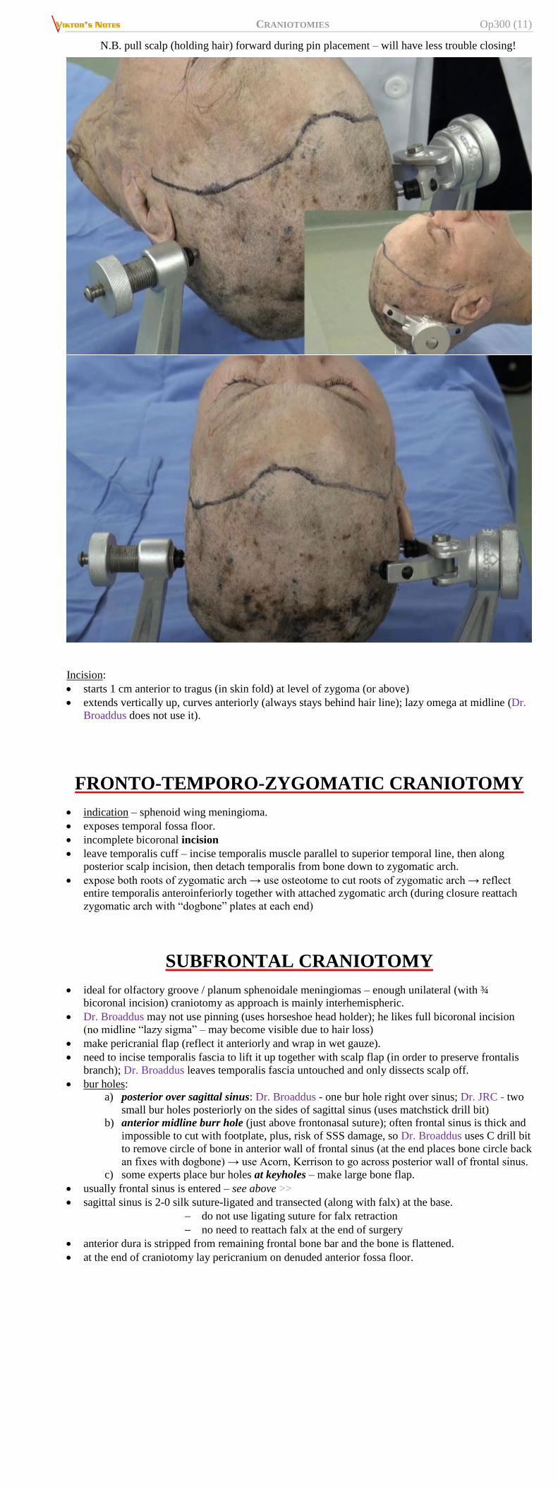
N.B. pull scalp (holding hair) forward during pin placement – will have less trouble closing!
Incision:
starts 1 cm anterior to tragus (in skin fold) at level of zygoma (or above)
extends vertically up, curves anteriorly (always stays behind hair line); lazy omega at midline (Dr.
Broaddus does not use it).
FRONTO-TEMPORO-ZYGOMATIC CRANIOTOMY
indication – sphenoid wing meningioma.
exposes temporal fossa floor.
incomplete bicoronal incision
leave temporalis cuff – incise temporalis muscle parallel to superior temporal line, then along
posterior scalp incision, then detach temporalis from bone down to zygomatic arch.
expose both roots of zygomatic arch → use osteotome to cut roots of zygomatic arch → reflect
entire temporalis anteroinferiorly together with attached zygomatic arch (during closure reattach
zygomatic arch with “dogbone” plates at each end)
SUBFRONTAL CRANIOTOMY
ideal for olfactory groove / planum sphenoidale meningiomas – enough unilateral (with ¾
bicoronal incision) craniotomy as approach is mainly interhemispheric.
Dr. Broaddus may not use pinning (uses horseshoe head holder); he likes full bicoronal incision
(no midline “lazy sigma” – may become visible due to hair loss)
make pericranial flap (reflect it anteriorly and wrap in wet gauze).
need to incise temporalis fascia to lift it up together with scalp flap (in order to preserve frontalis
branch); Dr. Broaddus leaves temporalis fascia untouched and only dissects scalp off.
bur holes:
a) posterior over sagittal sinus: Dr. Broaddus - one bur hole right over sinus; Dr. JRC - two
small bur holes posteriorly on the sides of sagittal sinus (uses matchstick drill bit)
b) anterior midline burr hole (just above frontonasal suture); often frontal sinus is thick and
impossible to cut with footplate, plus, risk of SSS damage, so Dr. Broaddus uses C drill bit
to remove circle of bone in anterior wall of frontal sinus (at the end places bone circle back
an fixes with dogbone) → use Acorn, Kerrison to go across posterior wall of frontal sinus.
c) some experts place bur holes at keyholes – make large bone flap.
usually frontal sinus is entered – see above >>
sagittal sinus is 2-0 silk suture-ligated and transected (along with falx) at the base.
do not use ligating suture for falx retraction
no need to reattach falx at the end of surgery
anterior dura is stripped from remaining frontal bone bar and the bone is flattened.
at the end of craniotomy lay pericranium on denuded anterior fossa floor.
CRANIOTOMIES Op300 (12)
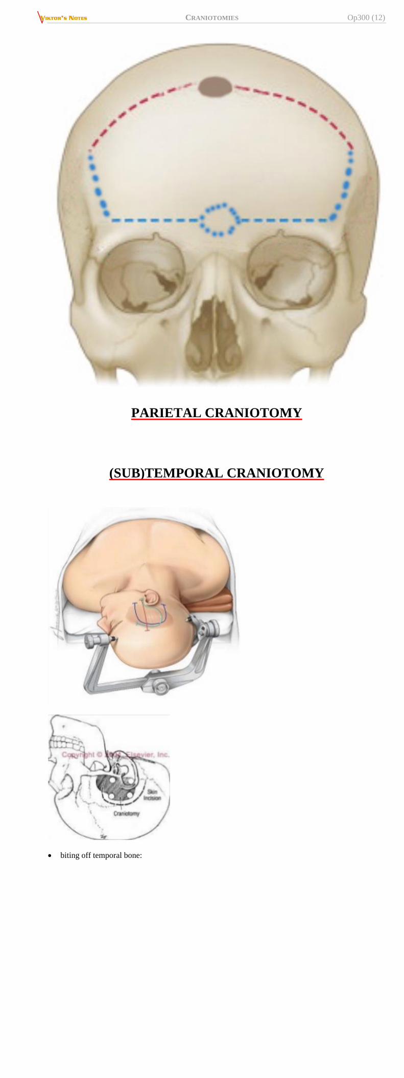
PARIETAL CRANIOTOMY
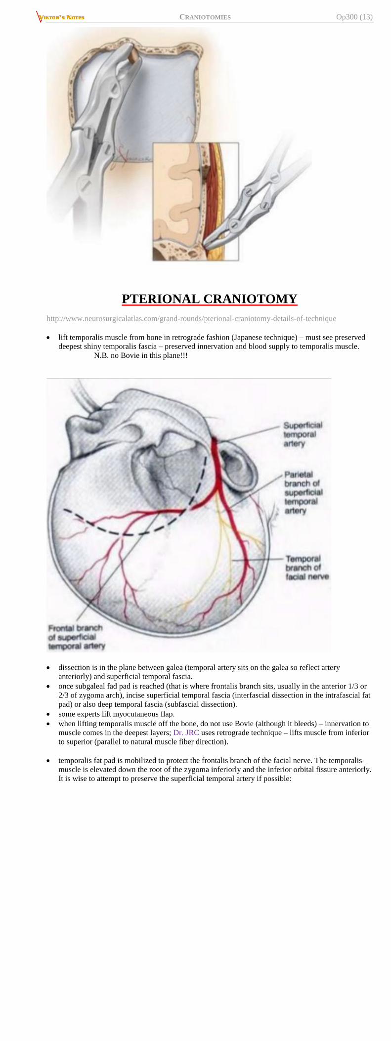
(SUB)TEMPORAL CRANIOTOMY
biting off temporal bone:
CRANIOTOMIES Op300 (13)
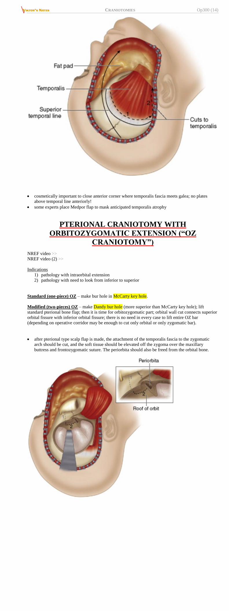
PTERIONAL CRANIOTOMY
http://www.neurosurgicalatlas.com/grand-rounds/pterional-craniotomy-details-of-technique
lift temporalis muscle from bone in retrograde fashion (Japanese technique) – must see preserved
deepest shiny temporalis fascia – preserved innervation and blood supply to temporalis muscle.
N.B. no Bovie in this plane!!!
dissection is in the plane between galea (temporal artery sits on the galea so reflect artery
anteriorly) and superficial temporal fascia.
once subgaleal fad pad is reached (that is where frontalis branch sits, usually in the anterior 1/3 or
2/3 of zygoma arch), incise superficial temporal fascia (interfascial dissection in the intrafascial fat
pad) or also deep temporal fascia (subfascial dissection).
some experts lift myocutaneous flap.
when lifting temporalis muscle off the bone, do not use Bovie (although it bleeds) – innervation to
muscle comes in the deepest layers; Dr. JRC uses retrograde technique – lifts muscle from inferior
to superior (parallel to natural muscle fiber direction).
temporalis fat pad is mobilized to protect the frontalis branch of the facial nerve. The temporalis
muscle is elevated down the root of the zygoma inferiorly and the inferior orbital fissure anteriorly.
It is wise to attempt to preserve the superficial temporal artery if possible:
CRANIOTOMIES Op300 (14)
cosmetically important to close anterior corner where temporalis fascia meets galea; no plates
above temporal line anteriorly!
some experts place Medpor flap to mask anticipated temporalis atrophy
PTERIONAL CRANIOTOMY WITH
ORBITOZYGOMATIC EXTENSION (“OZ
CRANIOTOMY”)
NREF video >>
NREF video (2) >>
Indications
1) pathology with intraorbital extension
2) pathology with need to look from inferior to superior
Standard (one-piece) OZ – make bur hole in McCarty key hole.
Modified (two-pieces) OZ – make Dandy bur hole (more superior than McCarty key hole); lift
standard pterional bone flap; then it is time for orbitozygomatic part; orbital wall cut connects superior
orbital fissure with inferior orbital fissure; there is no need in every case to lift entire OZ bar
(depending on operative corridor may be enough to cut only orbital or only zygomatic bar).
after pterional type scalp flap is made, the attachment of the temporalis fascia to the zygomatic
arch should be cut, and the soft tissue should be elevated off the zygoma over the maxillary
buttress and frontozygomatic suture. The periorbita should also be freed from the orbital bone.
CRANIOTOMIES Op300 (15)
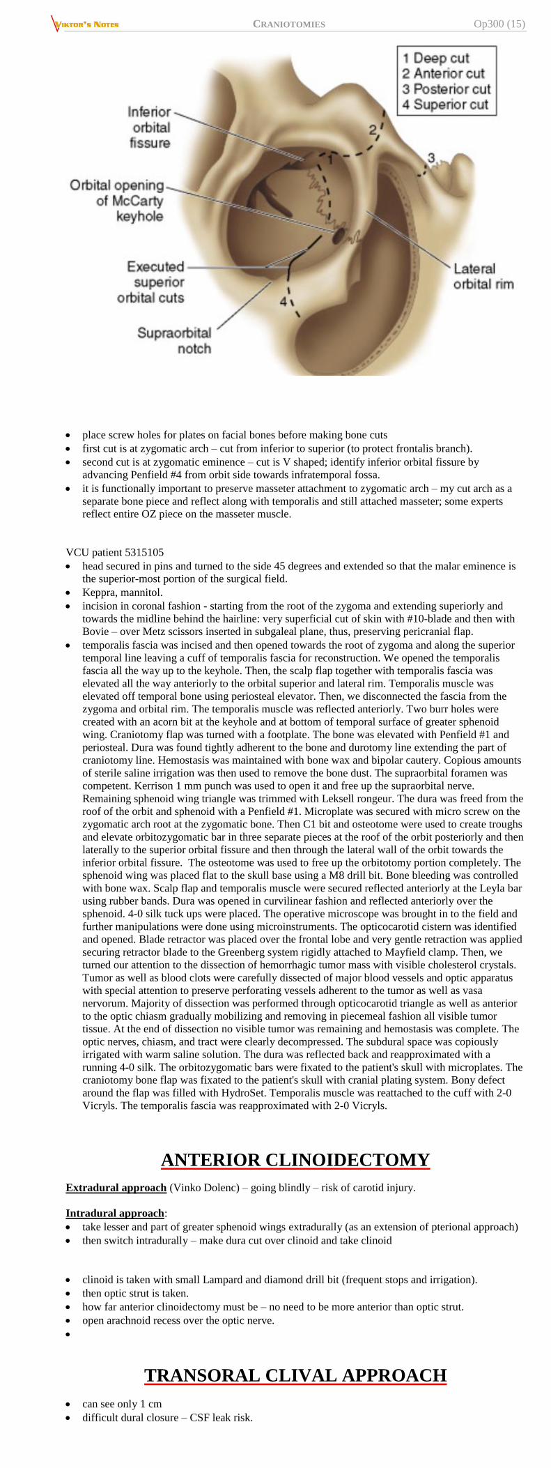
place screw holes for plates on facial bones before making bone cuts
first cut is at zygomatic arch – cut from inferior to superior (to protect frontalis branch).
second cut is at zygomatic eminence – cut is V shaped; identify inferior orbital fissure by
advancing Penfield #4 from orbit side towards infratemporal fossa.
it is functionally important to preserve masseter attachment to zygomatic arch – my cut arch as a
separate bone piece and reflect along with temporalis and still attached masseter; some experts
reflect entire OZ piece on the masseter muscle.
VCU patient 5315105
head secured in pins and turned to the side 45 degrees and extended so that the malar eminence is
the superior-most portion of the surgical field.
Keppra, mannitol.
incision in coronal fashion - starting from the root of the zygoma and extending superiorly and
towards the midline behind the hairline: very superficial cut of skin with #10-blade and then with
Bovie – over Metz scissors inserted in subgaleal plane, thus, preserving pericranial flap.
temporalis fascia was incised and then opened towards the root of zygoma and along the superior
temporal line leaving a cuff of temporalis fascia for reconstruction. We opened the temporalis
fascia all the way up to the keyhole. Then, the scalp flap together with temporalis fascia was
elevated all the way anteriorly to the orbital superior and lateral rim. Temporalis muscle was
elevated off temporal bone using periosteal elevator. Then, we disconnected the fascia from the
zygoma and orbital rim. The temporalis muscle was reflected anteriorly. Two burr holes were
created with an acorn bit at the keyhole and at bottom of temporal surface of greater sphenoid
wing. Craniotomy flap was turned with a footplate. The bone was elevated with Penfield #1 and
periosteal. Dura was found tightly adherent to the bone and durotomy line extending the part of
craniotomy line. Hemostasis was maintained with bone wax and bipolar cautery. Copious amounts
of sterile saline irrigation was then used to remove the bone dust. The supraorbital foramen was
competent. Kerrison 1 mm punch was used to open it and free up the supraorbital nerve.
Remaining sphenoid wing triangle was trimmed with Leksell rongeur. The dura was freed from the
roof of the orbit and sphenoid with a Penfield #1. Microplate was secured with micro screw on the
zygomatic arch root at the zygomatic bone. Then C1 bit and osteotome were used to create troughs
and elevate orbitozygomatic bar in three separate pieces at the roof of the orbit posteriorly and then
laterally to the superior orbital fissure and then through the lateral wall of the orbit towards the
inferior orbital fissure. The osteotome was used to free up the orbitotomy portion completely. The
sphenoid wing was placed flat to the skull base using a M8 drill bit. Bone bleeding was controlled
with bone wax. Scalp flap and temporalis muscle were secured reflected anteriorly at the Leyla bar
using rubber bands. Dura was opened in curvilinear fashion and reflected anteriorly over the
sphenoid. 4-0 silk tuck ups were placed. The operative microscope was brought in to the field and
further manipulations were done using microinstruments. The opticocarotid cistern was identified
and opened. Blade retractor was placed over the frontal lobe and very gentle retraction was applied
securing retractor blade to the Greenberg system rigidly attached to Mayfield clamp. Then, we
turned our attention to the dissection of hemorrhagic tumor mass with visible cholesterol crystals.
Tumor as well as blood clots were carefully dissected of major blood vessels and optic apparatus
with special attention to preserve perforating vessels adherent to the tumor as well as vasa
nervorum. Majority of dissection was performed through opticocarotid triangle as well as anterior
to the optic chiasm gradually mobilizing and removing in piecemeal fashion all visible tumor
tissue. At the end of dissection no visible tumor was remaining and hemostasis was complete. The
optic nerves, chiasm, and tract were clearly decompressed. The subdural space was copiously
irrigated with warm saline solution. The dura was reflected back and reapproximated with a
running 4-0 silk. The orbitozygomatic bars were fixated to the patient's skull with microplates. The
craniotomy bone flap was fixated to the patient's skull with cranial plating system. Bony defect
around the flap was filled with HydroSet. Temporalis muscle was reattached to the cuff with 2-0
Vicryls. The temporalis fascia was reapproximated with 2-0 Vicryls.
ANTERIOR CLINOIDECTOMY
Extradural approach (Vinko Dolenc) – going blindly – risk of carotid injury.
Intradural approach:
take lesser and part of greater sphenoid wings extradurally (as an extension of pterional approach)
then switch intradurally – make dura cut over clinoid and take clinoid
clinoid is taken with small Lampard and diamond drill bit (frequent stops and irrigation).
then optic strut is taken.
how far anterior clinoidectomy must be – no need to be more anterior than optic strut.
open arachnoid recess over the optic nerve.
TRANSORAL CLIVAL APPROACH
can see only 1 cm
difficult dural closure – CSF leak risk.
CRANIOTOMIES Op300 (16)
TRANSPETROSAL APPROACHES TO POSTERIOR
FOSSA
http://www.medscape.com/viewarticle/511118
resection of petrous temporal bone to various degrees provides different levels of access to lesions
of posterior fossa (cerebellopontine angle, petroclival region).
Variants of transpetrosal approaches can be classified:
A. Anterior transpetrosal approaches
B. Posterior transpetrosal approaches
ANTERIOR (MEDIAL) TRANSPETROSAL APPROACHES (KAWASE, S. ANTERIOR PETROSECTOMY)
- extensions of basic middle fossa approach.
designed to preserve hearing - spare lateral petrous bone.
involve resection of medial petrous bone to various degrees.
involve resection of bone within Kawase rhomboid and division of tentorium to provide exposure
of posterior fossa.
GSPN (easy to identify) is right above and parallel to petrous ICA
http://operativeneurosurgery.com/doku.php?id=kawase_approach
POSTERIOR TRANSPETROSAL (PRESIGMOID) APPROACHES
- retrolabyrinthine, translabyrinthine, and transcochlear.
based on the standard mastoidectomy and involve resection of petrous bone to various degrees -
progressively increased exposure anteriorly, but comes at expense of hearing in translabyrinthine
approach and of hearing and facial strength in transcochlear approach.
subarcuate artery (branch of AICA) – feeds bone of labyrinth – OK to sacrifice.
RETROLABYRINTHINE (PRESIGMOID) APPROACH
Used sources:
R. Jandial “Core Techniques in Operative Neurosurgery” (2011), procedure 20
Indications
• The retrolabyrinthine approach is a hearing-preserving presigmoid approach that uses a
mastoidectomy and skeletonization of the sigmoid sinus to expose the presigmoid dura behind
the semicircular canals.
• The principal appeal of this approach is its ability to expose widely the posterior petrous face
and cisternal portions of cranial nerves VII and VIII with a minimal degree of cerebellar
retraction.
• The retrolabyrinthine approach additionally is used to identify and expose the superior petrosal
sinus, as a first step for division of the tentorium.
Contraindications
• This approach is unable to access the internal auditory canal or petrous apex directly because of
the interposition of the labyrinthine and cochlear structures between the surgeon and these
regions.
Planning and positioning
• The patient generally is placed in a semilateral position on the operating table, with a bump
under the ipsilateral shoulder.
• The head is placed in a Mayfield head holder with two pins placed in the occiput just off
midline. The single pin is placed in the ipsilateral forehead, lateral to the mid-pupillary line
ideally behind the hairline.
• After pinning, the head is usually positioned such that the region just behind the pinna just
superior to the mastoid process is the highest point on the patient's head. With adequate
ipsilateral shoulder elevation, this position is achieved by a slight amount of contralateral head
rotation, minimal neck flexion, and head elevation.
Look for Trawman’s triangle
CRANIOTOMIES Op300 (17)
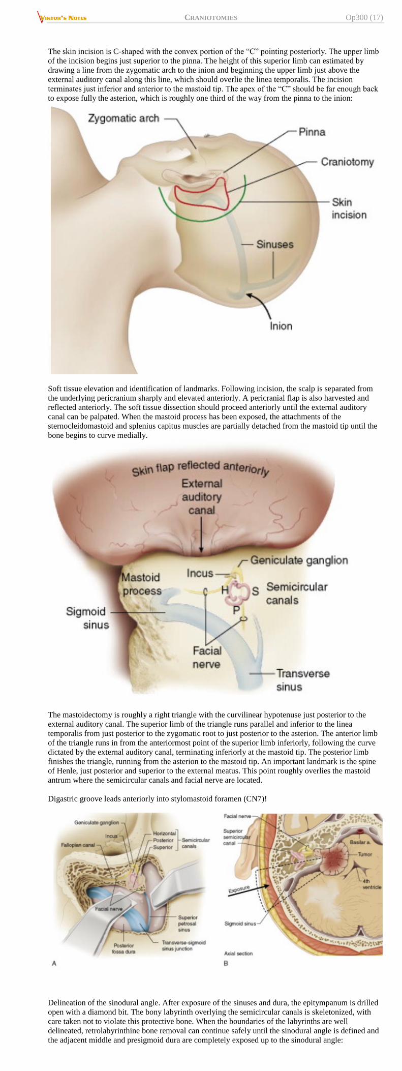
The skin incision is C-shaped with the convex portion of the “C” pointing posteriorly. The upper limb
of the incision begins just superior to the pinna. The height of this superior limb can estimated by
drawing a line from the zygomatic arch to the inion and beginning the upper limb just above the
external auditory canal along this line, which should overlie the linea temporalis. The incision
terminates just inferior and anterior to the mastoid tip. The apex of the “C” should be far enough back
to expose fully the asterion, which is roughly one third of the way from the pinna to the inion:
Soft tissue elevation and identification of landmarks. Following incision, the scalp is separated from
the underlying pericranium sharply and elevated anteriorly. A pericranial flap is also harvested and
reflected anteriorly. The soft tissue dissection should proceed anteriorly until the external auditory
canal can be palpated. When the mastoid process has been exposed, the attachments of the
sternocleidomastoid and splenius capitus muscles are partially detached from the mastoid tip until the
bone begins to curve medially.
The mastoidectomy is roughly a right triangle with the curvilinear hypotenuse just posterior to the
external auditory canal. The superior limb of the triangle runs parallel and inferior to the linea
temporalis from just posterior to the zygomatic root to just posterior to the asterion. The anterior limb
of the triangle runs in from the anteriormost point of the superior limb inferiorly, following the curve
dictated by the external auditory canal, terminating inferiorly at the mastoid tip. The posterior limb
finishes the triangle, running from the asterion to the mastoid tip. An important landmark is the spine
of Henle, just posterior and superior to the external meatus. This point roughly overlies the mastoid
antrum where the semicircular canals and facial nerve are located.
Digastric groove leads anteriorly into stylomastoid foramen (CN7)!
Delineation of the sinodural angle. After exposure of the sinuses and dura, the epitympanum is drilled
open with a diamond bit. The bony labyrinth overlying the semicircular canals is skeletonized, with
care taken not to violate this protective bone. When the boundaries of the labyrinths are well
delineated, retrolabyrinthine bone removal can continue safely until the sinodural angle is defined and
the adjacent middle and presigmoid dura are completely exposed up to the sinodural angle:

CRANIOTOMIES Op300 (18)
Delineation of the fallopian canal. Adequate exposure in this approach requires that the presigmoid
dura be exposed down to the jugular bulb. The vertical portion of the facial nerve overlies the jugular
bulb and dura in this region. The fallopian canal is identified anterior and inferior to the bony
labyrinths in the epitympanum. When identified, the canal is skeletonized with a diamond bit,
particularly on its deep surface in a rostral-to-caudal direction, until its relationship to the jugular bulb
is known. Bony removal can be continued as far anteroinferior as possible underneath the facial nerve.
Dural incision. The dura is opened in a C-shaped fashion around the epitympanum with the base
centered around the labyrinths. It is important that the endolymphatic sac is identified and included
with the dural flap so that endolymphatic flow is not disrupted. Depending on the goals of surgery, the
superior petrosal sinus can be sacrificed as part of a tentorial division, and the middle fossa dura can be
opened to provide visualization of the petrous apex, tentorial incisura, or middle fossa floor:
Tips from the masters
• Numerous mastoid emissary veins can be encountered during this dissection, and when
bleeding from one of these veins is encountered, it is best to continue dissection until well
beyond these veins before attempting to control the bleeding with bone wax.
• The drilling should start with a large cutting bur and proceed to diamond burs as critical
structures such as the facial nerve and sigmoid sinus are encountered.
• The largest burs possible for a given drilling task should be used because these are less likely to
pierce a small hole in a vessel or nerve than a small bur.
Pitfalls
Care should be taken not to cause kinking of the contralateral jugular vein with the head position.
Care should be taken to avoid entering the external auditory canal during soft tissue dissection.
TEMPOROPOLAR (HALF-AND-HALF) APPROACH
TO THE BASILAR ARTERY AND THE
RETROSELLAR SPACE
Used sources:
R. Jandial “Core Techniques in Operative Neurosurgery” (2011), procedure 18
INDICATIONS
Transsylvian approaches enter the parasellar cisterns on a superior-to-inferior trajectory, forcing
the surgeon to work past the carotid artery through the opticocarotid or carotid-oculomotor
triangles to access this region, making access of the mid-basilar and interpeduncular cisterns
difficult.
CRANIOTOMIES Op300 (19)
Although the subtemporal approach provides a good view of the basilar artery at the level of the
tentorium, it is limited in its rostral visualization, which can be necessary for high-riding basilar
apex aneurysms or tumors with significant superior extension. Also, the flat trajectory of this
approach limits the ability to see the retrosellar space.
The temporopolar approach combines these approaches largely through microsurgical
mobilization of the temporal lobe, which is retracted posteriorly and laterally to add the exposure
of the tentorial incisura to the visualization obtained with a transsylvian approach.
CONTRAINDICATIONS
Laterally projecting posterior communicating artery or middle cerebral artery aneurysms because
these might be attached to the temporal lobe and rupture with retraction.
PROCEDURE
patient is positioned supine, head is pinned similar to the orbitozygomatic approach (malar
eminence the highest point in the field).
craniotomy is identical to the orbitozygomatic approach.
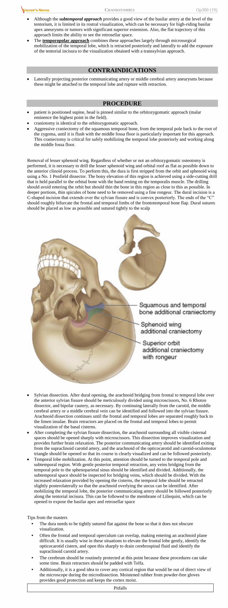
Aggressive craniectomy of the squamous temporal bone, from the temporal pole back to the root of
the zygoma, until it is flush with the middle fossa floor is particularly important for this approach.
This craniectomy is critical for safely mobilizing the temporal lobe posteriorly and working along
the middle fossa floor.
Removal of lesser sphenoid wing. Regardless of whether or not an orbitozygomatic osteotomy is
performed, it is necessary to drill the lesser sphenoid wing and orbital roof as flat as possible down to
the anterior clinoid process. To perform this, the dura is first stripped from the orbit and sphenoid wing
using a No. 1 Penfield dissector. The bony elevation of this region is achieved using a side-cutting drill
that is held parallel to the orbital bone with the hand resting on the temporalis muscle. The drilling
should avoid entering the orbit but should thin the bone in this region as close to this as possible. In
deeper portions, thin spicules of bone need to be removed using a fine rongeur. The dural incision is a
C-shaped incision that extends over the sylvian fissure and is convex posteriorly. The ends of the “C”
should roughly bifurcate the frontal and temporal limbs of the frontotemporal bone flap. Dural sutures
should be placed as low as possible and sutured tightly to the scalp
Sylvian dissection. After dural opening, the arachnoid bridging from frontal to temporal lobe over
the anterior sylvian fissure should be meticulously divided using microscissors, No. 6 Rhoton
dissector, and bipolar cautery, as necessary. By continuing laterally from the carotid, the middle
cerebral artery or a middle cerebral vein can be identified and followed into the sylvian fissure.
Arachnoid dissection continues until the frontal and temporal lobes are separated roughly back to
the limen insulae. Brain retractors are placed on the frontal and temporal lobes to permit
visualization of the basal cisterns.
After completing the sylvian fissure dissection, the arachnoid surrounding all visible cisternal
spaces should be opened sharply with microscissors. This dissection improves visualization and
provides further brain relaxation. The posterior communicating artery should be identified exiting
from the supraclinoid carotid artery, and the arachnoid of the opticocarotid and carotid-oculomotor
triangle should be opened so that its course is clearly visualized and can be followed posteriorly.
Temporal lobe mobilization. At this point, attention should be turned to the temporal pole and
subtemporal region. With gentle posterior temporal retraction, any veins bridging from the
temporal pole to the sphenoparietal sinus should be identified and divided. Additionally, the
subtemporal space should be inspected for bridging veins, which should be divided. With the
increased relaxation provided by opening the cisterns, the temporal lobe should be retracted
slightly posterolaterally so that the arachnoid overlying the uncus can be identified. After
mobilizing the temporal lobe, the posterior communicating artery should be followed posteriorly
along the tentorial incisura. This can be followed to the membrane of Liliequist, which can be
opened to expose the basilar apex and retrosellar space
Tips from the masters
• The dura needs to be tightly sutured flat against the bone so that it does not obscure
visualization.
• Often the frontal and temporal operculum can overlap, making entering an arachnoid plane
difficult. It is usually wise in these situations to elevate the frontal lobe gently, identify the
opticocarotid cistern, and open this sharply to drain cerebrospinal fluid and identify the
supraclinoid carotid artery.
• The cerebrum should be routinely protected at this point because these procedures can take
some time. Brain retractors should be padded with Telfa.
• Additionally, it is a good idea to cover any cortical region that would be out of direct view of
the microscope during the microdissection. Moistened rubber from powder-free gloves
provides good protection and keeps the cortex moist.
Pitfalls
CRANIOTOMIES Op300 (20)
It should never be necessary to take a vein crossing the fissure because these can always be
separated to one lobe or another through careful dissection.
Bailout options
• If more posterior exposure of the ambient cistern is needed, the temporal lobe can be retracted
upward, converting this approach to a subtemporal approach.
POSTERIOR FOSSA CRANIECTOMY / MIDLINE
SUBOCCIPITAL CRANIOTOMY
Panaudota literatūra:
R. Jandial “Core Techniques in Operative Neurosurgery” (2011), procedure 5
INDICATIONS
1. Cerebellar stroke
2. Chiari malformations (symptomatic, large syrinx)
3. Tumors
4. Vascular lesions (aneurysms, cavernous malformations, AVMs)
5. Infections
if lesions extend rostral to tentorium, consider combined supracerebellar and supratentorial
approach.
if lesion extends from posterior fossa to middle fossa, consider combined middle and posterior
fossa approach.
PREOP
DDEEXXAAMMEETTHHAASSOONNEE, MMAANNNNIITTOOLL (not for Chiari, except dex if opening dura and manipulating tonsils)
consider placing lumbar drain / EVD (e.g. occipital)
- to prevent postoperative hydrocephalus by draining CSF with debris and blood;
- to protect dural closure.
POSITION
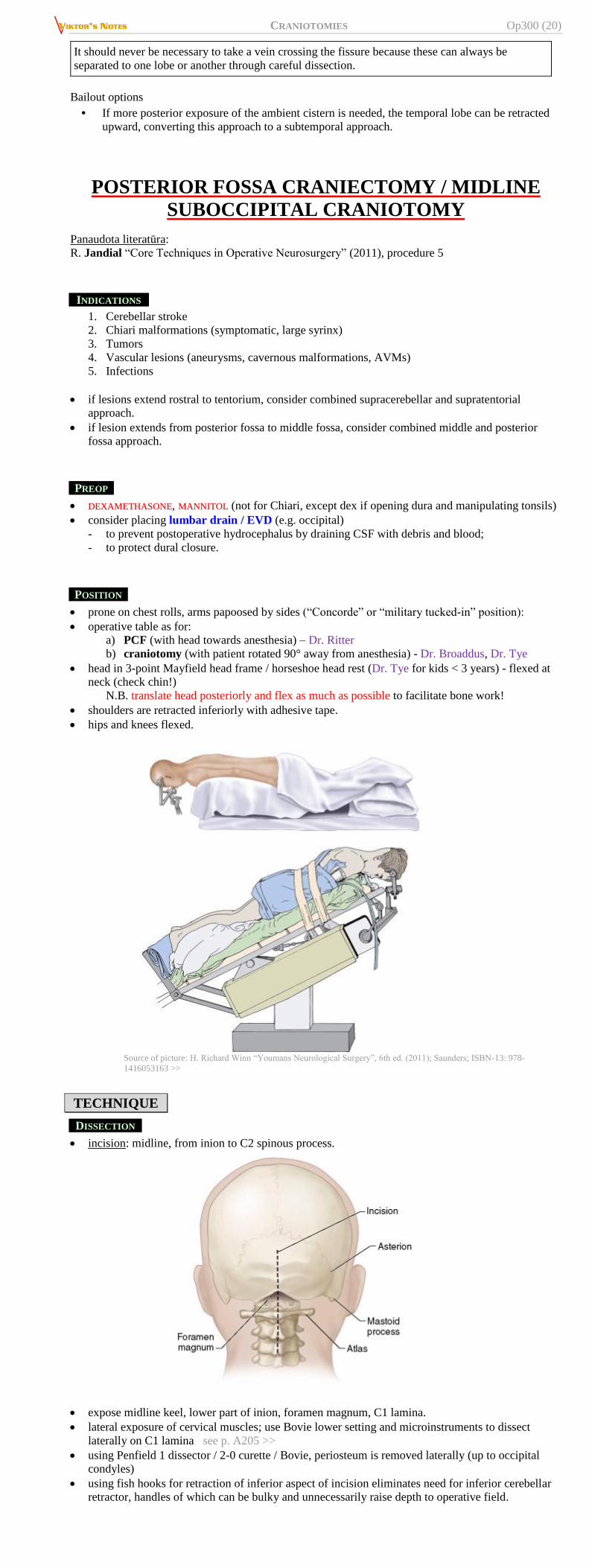
prone on chest rolls, arms papoosed by sides (“Concorde” or “military tucked-in” position):
operative table as for:
a) PCF (with head towards anesthesia) – Dr. Ritter
b) craniotomy (with patient rotated 90° away from anesthesia) - Dr. Broaddus, Dr. Tye
head in 3-point Mayfield head frame / horseshoe head rest (Dr. Tye for kids < 3 years) - flexed at
neck (check chin!)
N.B. translate head posteriorly and flex as much as possible to facilitate bone work!
shoulders are retracted inferiorly with adhesive tape.
hips and knees flexed.
Source of picture: H. Richard Winn “Youmans Neurological Surgery”, 6th ed. (2011); Saunders; ISBN-13: 978-
1416053163 >>
TECHNIQUE
DISSECTION
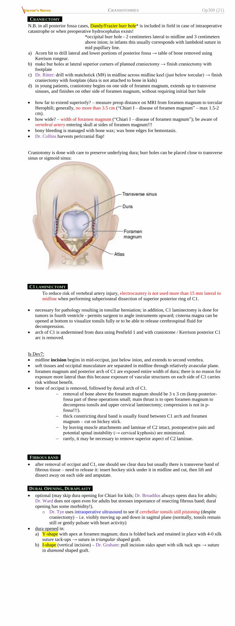
incision: midline, from inion to C2 spinous process.
expose midline keel, lower part of inion, foramen magnum, C1 lamina.
lateral exposure of cervical muscles; use Bovie lower setting and microinstruments to dissect
laterally on C1 lamina see p. A205 >>
using Penfield 1 dissector / 2-0 curette / Bovie, periosteum is removed laterally (up to occipital
condyles)
using fish hooks for retraction of inferior aspect of incision eliminates need for inferior cerebellar
retractor, handles of which can be bulky and unnecessarily raise depth to operative field.
CRANIOTOMIES Op300 (21)
CRANIECTOMY
N.B. in all posterior fossa cases, Dandy/Frazier burr hole* is included in field in case of intraoperative
catastrophe or when preoperative hydrocephalus exists!
*occipital burr hole - 2 centimeters lateral to midline and 3 centimeters
above inion; in infants this usually corresponds with lambdoid suture in
mid pupillary line.
a) Acorn bit to drill lateral and lower portions of posterior fossa → table of bone removed using
Kerrison rongeur.
b) make bur holes at lateral superior corners of planned craniectomy → finish craniectomy with
footplate
c) Dr. Ritter: drill with matchstick (M8) in midline across midline keel (just below torcular) → finish
craniectomy with footplate (dura is not attached to bone in kids)
d) in young patients, craniotomy begins on one side of foramen magnum, extends up to transverse
sinuses, and finishes on other side of foramen magnum, without requiring initial burr hole
how far to extend superiorly? – measure preop distance on MRI from foramen magnum to torcular
Herophili; generally, no more than 3.5 cm (“Chiari I – disease of foramen magnum” – max 1.5-2
cm).
how wide? – width of foramen magnum (“Chiari I – disease of foramen magnum”); be aware of
vertebral artery entering skull at sides of foramen magnum!!!
bony bleeding is managed with bone wax; wax bone edges for hemostasis.
Dr. Collins harvests pericranial flap!
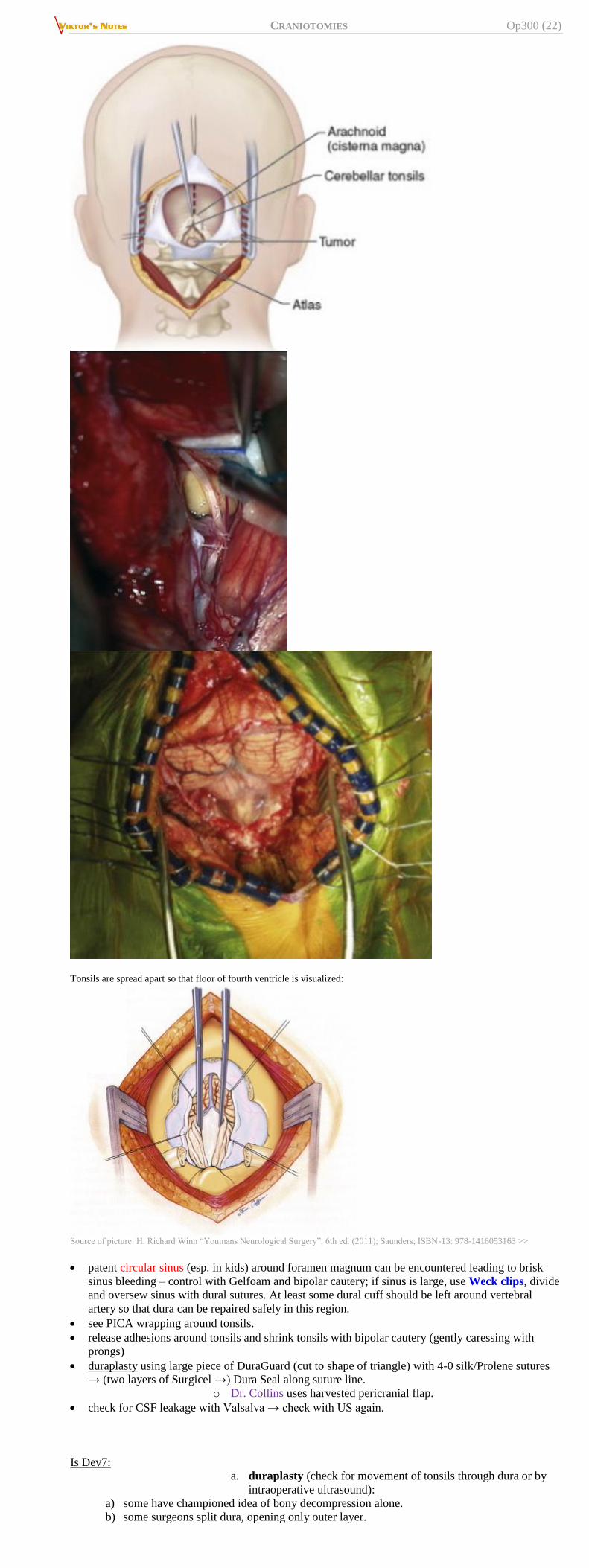
Craniotomy is done with care to preserve underlying dura; burr holes can be placed close to transverse
sinus or sigmoid sinus:
C1 LAMINECTOMY
To reduce risk of vertebral artery injury, electrocautery is not used more than 15 mm lateral to
midline when performing subperiosteal dissection of superior posterior ring of C1.
necessary for pathology resulting in tonsillar herniation; in addition, C1 laminectomy is done for
tumors in fourth ventricle - permits surgeon to angle instruments upward; cisterna magna can be
opened at bottom to visualize tonsils fully or to be able to release cerebrospinal fluid for
decompression.
arch of C1 is undermined from dura using Penfield 1 and with craniotome / Kerrison posterior C1
arc is removed.
Is Dev7:
midline incision begins in mid-occiput, just below inion, and extends to second vertebra.
soft tissues and occipital musculature are separated in midline through relatively avascular plane.
foramen magnum and posterior arch of C1 are exposed entire width of dura; there is no reason for
exposure more lateral than this because exposure of vascular structures on each side of C1 carries
risk without benefit.
bone of occiput is removed, followed by dorsal arch of C1.
removal of bone above the foramen magnum should be 3 x 3 cm (keep posterior-
fossa part of these operations small; main thrust is to open foramen magnum to
decompress tonsils and upper cervical laminectomy; compression is not in p-
fossa!!!).
thick constricting dural band is usually found between C1 arch and foramen
magnum – cut on hickey stick.
by leaving muscle attachments and laminae of C2 intact, postoperative pain and
potential spinal instability (→ cervical kyphosis) are minimized.
rarely, it may be necessary to remove superior aspect of C2 laminae.
FIBROUS BAND
after removal of occiput and C1, one should see clear dura but usually there is transverse band of
fibrous tissue – need to release it: insert hockey stick under it in midline and cut, then lift and
dissect away on each side and amputate.
DURAL OPENING, DURAPLASTY
optional (may skip dura opening for Chiari for kids; Dr. Broaddus always opens dura for adults;
Dr. Ward does not open even for adults but stresses importance of resecting fibrous band; dural
opening has some morbidity!).
o Dr. Tye uses intraoperative ultrasound to see if cerebellar tonsils still pistoning (despite
craniectomy) – i.e. visibly moving up and down in sagittal plane (normally, tonsils remain
still or gently pulsate with heart activity)
dura opened in:
a) Y-shape with apex at foramen magnum; dura is folded back and retained in place with 4-0 silk
suture tack-ups → suture in triangular shaped graft.
b) I-shape (vertical incision) – Dr. Graham: pull incision sides apart with silk tuck ups → suture
in diamond shaped graft.
CRANIOTOMIES Op300 (22)
Tonsils are spread apart so that floor of fourth ventricle is visualized:
Source of picture: H. Richard Winn “Youmans Neurological Surgery”, 6th ed. (2011); Saunders; ISBN-13: 978-1416053163 >>
patent circular sinus (esp. in kids) around foramen magnum can be encountered leading to brisk
sinus bleeding – control with Gelfoam and bipolar cautery; if sinus is large, use Weck clips, divide
and oversew sinus with dural sutures. At least some dural cuff should be left around vertebral
artery so that dura can be repaired safely in this region.
see PICA wrapping around tonsils.
release adhesions around tonsils and shrink tonsils with bipolar cautery (gently caressing with
prongs)
duraplasty using large piece of DuraGuard (cut to shape of triangle) with 4-0 silk/Prolene sutures
→ (two layers of Surgicel →) Dura Seal along suture line.
o Dr. Collins uses harvested pericranial flap.
check for CSF leakage with Valsalva → check with US again.
Is Dev7:
a. duraplasty (check for movement of tonsils through dura or by
intraoperative ultrasound):
a) some have championed idea of bony decompression alone.
b) some surgeons split dura, opening only outer layer.

CRANIOTOMIES Op300 (23)
c) in more severe forms of tonsillar herniation, intradural approach and dural grafting are
needed
dura is opened in midline in a "Y" shaped incision; excise triangular top flap.
tonsils are gently separated to inspect for veils covering outlets of fourth ventricle -
this re-establishes free flow of CSF from foramen of Magendie.
consider visualization of choroid plexus of fourth ventricle and free flow of CSF
into subarachnoid space as evidence of adequate decompression.
if CSF egress is limited, extrapial coagulation of one or both tonsillar tips shrinks
tonsils sufficiently to restore CSF flow.
if free CSF egress from fourth ventricle is not achieved after lysis of subarachnoid
adhesions and tonsil coagulation → place stent.
CEREBELLUM INTERVENTION
a) intracerebellar hematoma evacuated, necrotic tissue debrided to wide margin until normal
tissue is encountered.
b) tonsils slowly dissected free from surrounding arachnoid tissue, and elevated into field →
reduction of tonsillar size with bipolar cauterization ideally of ventral surface so it will not
form adhesions on dorsal surface (tonsils should ascend above foramen magnum); if still
suboptimal → tonsillar subpial resection (to visualize obex and floor of 4th ventricle) with
pia coagulated with bipolar cauterization and closed with 6-0 Prolene.
CLOSURE
± bone may be replaced using 2-0 PDS sutures.
± medium-sized Hemovac.
water-tight closure of fascia.
3-0 Monocryl or nylon running for skin.
POSTOPERATIVELY
Incl. complications
- see p. Dev7 >>
CSF leak – see p. S64 >>
LATERAL SUBOCCIPITAL CRANIOTOMY
Used resources
R. Nader “Neurosurgery Tricks of the Trade – CRANIAL“ (2014), ch. 13
INDICATIONS
1. Exposure of the lateral cerebellar hemisphere, anterolateral brainstem, posterior aspect of
petrous bone, craniovertebral junction, and upper cervical cord.
2. Building block for more extensive procedures, such as the transcondylar, far lateral, extreme
lateral, and posterior petrosal approach.
3. Provides vertebral artery (VA) control, VA can be mobilized from the vertebral sulcus and
foramen transversarium of C1.
4. Compared with the pre sigmoid approaches. it can provide access to lesions below the jugular
tubercle.
5. It provides access to the lateral and sometimes ventral brainstem and cerebellum with minimal
retraction.
ANESTHESIA
be ready for venous air embolism
for lesions in the region of the jugular foramen, consider temporary cardiac pacing.
TECHNIQUE

CRANIOTOMIES Op300 (24)
lateral oblique positioning, head flexed until nuchal muscles become firm
J-shaped or "hockey stick" incision; starts at upper mastoid; leave muscle insertion cuff at superior
nuchal line for later closure.
VA lays in the floor of the suboccipital triangle (superior oblique, inferior oblique, and rectus
capitis posterior major muscles); transverse process of C1 vertebra can be used as a localizing
landmark for the VA.
(A) Suboccipital triangle with vertebral artery and Cl transverse process located within the triangle. (B) Once
the craniotomy Is completed. the foramen magnum Is opened with Kerrison rongeurs after the muscles of the
suboccipital triangle have been reflected medially and the vertebral artery has been exposed. RM, rectus major
muscle; Rm, rectus minor muscle; SOM, superior oblique muscle; IOM, inferior oblique muscle; LS, levator
scapula; VA, vertebral artery.
craniotomy can be done in two parts:
i. bone flap next to transverse sigmoid junction
ii. bone resected below the level of the craniotomy + C1
laminectomy.
whenever drilling of the posterior part of the occipital condyle is necessary, subperiosteal
dissection with the use of curettes will prevent injury to the VA
VA injury can by minimized by avoiding rotation of the head once the patient is placed In the park
bench position.
Exposun! down to the 0 vertebra Is shown. A suboccipital craniotomy Is being perfonned and the
vertebral artery has been exposed. A curvilinear durallnclslon Is perfunned with care taken to preserve
the entry of the vertebral artery Into the Intradural space.

CRANIOTOMIES Op300 (25)
When the procedure is combined with a posterior petrOSectomy, the sigmoid sinus can be
skeletonized and on occasion divided {if the dominant sinus is contralateral or in certain cases of
codominance). Prior to pennanently occluding the sinus, the pressure of the superior stump should be
measured by placing a butterfly needle in the lumen and connecting it to a manometer. The VA can be
mobilized if necessary. This can be achieved by continuing the subperiosteal dissection at the posterior
arch of Cl up to the foramen transversarlum. which can be opened, the VA mobilized and followed
toward its entrance to the dura.ural space.
The dura is opened at the level of the cisterna magna to allow cerebrospinal fluid (CSF) egression and
subsequent relaxation of the brain surface. The incision extends vertically and posterolat erally parallel
to the lateral edge of the craniotomy
POSTOPERATIVELY
NPO until speech therapist evaluates swallowing
RETROSIGMOID (RETROMASTOID) CRANIOTOMY
term “suboccipital craniotomy” is still used, even though craniotomy now used rarely
extends inferior to attachment of cervical muscles at uncal line
Used resources
R. Jandial “Core Techniques in Operative Neurosurgery” (2011), procedure 6
Pending resources:
http://www.neurosurgicalatlas.com/grand-rounds/retromastoid-craniotomy-expanding-the-operative-
corridor
INDICATIONS
CPA angle lesions (acoustic neuroma, MVD for trigeminal neuralgia, etc)
PROCEDURE
LOCATION OF ASTERION
N.B. in many cases asterion is above of transverse-sigmoid sinus junction – be careful when drilling
bur hole!
a. 1 cm above and 4 cm posterior to external
auditory canal (Dr. Graham)
b. Intersection of:
vertical line through mastoid groove
line from inion-root of zygoma
POSITION
a) lateral decubitus (“park bench”); neck is flexed (chin tuck) and vertex is tilted towards floor
roll (towel or gel) under axilla!
upper shoulder is also tilted anteriorly (to give more space)
b) semisitting position with neck flexed and face rotated away from side of surgery (ipsilateral
tentorium parallel to floor).
ELECTROPHYSIOLOGICAL MONITORING
1) bilateral auditory potentials (BAER)
2) EMG of facial muscles.
PIN PLACEMENT
Single pin – just above superior temporal line, behind hairline
Two pins – pin is just above pinna, avoid suboccipital muscles
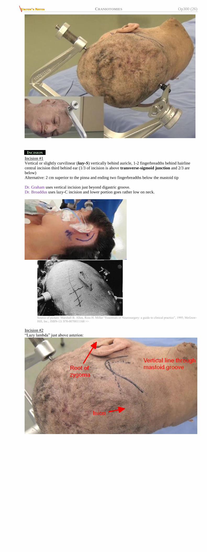
CRANIOTOMIES Op300 (26)
INCISION
Incision #1
Vertical or slightly curvilinear (lazy-S) vertically behind auricle, 1-2 fingerbreadths behind hairline
central incision third behind ear (1/3 of incision is above transverse-sigmoid junction and 2/3 are
below)
Alternative: 2 cm superior to the pinna and ending two fingerbreadths below the mastoid tip
Dr. Graham uses vertical incision just beyond digastric groove.
Dr. Broaddus uses lazy-C incision and lower portion goes rather low on neck.
Source of picture: Marshall B. Allen, Ross H. Miller “Essentials of Neurosurgery: a guide to clinical practice”, 1995; McGraw-
Hill, Inc.; ISBN-13: 978-0070011168 >>
Incision #2
“Lazy lambda” just above asterion:
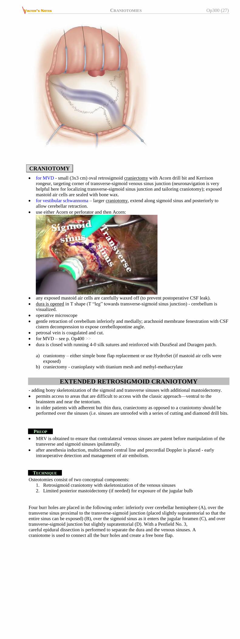
CRANIOTOMIES Op300 (27)
CRANIOTOMY
for MVD - small (3x3 cm) oval retrosigmoid craniectomy with Acorn drill bit and Kerrison
rongeur, targeting corner of transverse-sigmoid venous sinus junction (neuronavigation is very
helpful here for localizing transverse-sigmoid sinus junction and tailoring craniotomy); exposed
mastoid air cells are sealed with bone wax.
for vestibular schwannoma – larger craniotomy, extend along sigmoid sinus and posteriorly to
allow cerebellar retraction.
use either Acorn or perforator and then Acorn:
any exposed mastoid air cells are carefully waxed off (to prevent postoperative CSF leak).
dura is opened in T shape (T “leg” towards transverse-sigmoid sinus junction) - cerebellum is
visualized.
operative microscope
gentle retraction of cerebellum inferiorly and medially; arachnoid membrane fenestration with CSF
cistern decompression to expose cerebellopontine angle.
petrosal vein is coagulated and cut.
for MVD – see p. Op400 >>
dura is closed with running 4-0 silk sutures and reinforced with DuraSeal and Duragen patch.
a) craniotomy – either simple bone flap replacement or use HydroSet (if mastoid air cells were
exposed)
b) craniectomy - cranioplasty with titanium mesh and methyl-methacrylate
EXTENDED RETROSIGMOID CRANIOTOMY
- adding bony skeletonization of the sigmoid and transverse sinuses with additional mastoidectomy.
permits access to areas that are difficult to access with the classic approach—ventral to the
brainstem and near the tentorium.
in older patients with adherent but thin dura, craniectomy as opposed to a craniotomy should be
performed over the sinuses (i.e. sinuses are unroofed with a series of cutting and diamond drill bits.
PREOP
MRV is obtained to ensure that contralateral venous sinuses are patent before manipulation of the
transverse and sigmoid sinuses ipsilaterally.
after anesthesia induction, multichannel central line and precordial Doppler is placed - early
intraoperative detection and management of air embolism.
TECHNIQUE
Osteotomies consist of two conceptual components:
1. Retrosigmoid craniotomy with skeletonization of the venous sinuses
2. Limited posterior mastoidectomy (if needed) for exposure of the jugular bulb
Four burr holes are placed in the following order: inferiorly over cerebellar hemisphere (A), over the
transverse sinus proximal to the transverse-sigmoid junction (placed slightly supratentorial so that the
entire sinus can be exposed) (B), over the sigmoid sinus as it enters the jugular foramen (C), and over
transverse-sigmoid junction but slightly supratentorial (D). With a Penfield No. 3,
careful epidural dissection is performed to separate the dura and the venous sinuses. A
craniotome is used to connect all the burr holes and create a free bone flap.

CRANIOTOMIES Op300 (28)
Process of the limited posterior mastoidectomy begins with a cutting bur but then transitions to a
diamond bur as the veil of blue to visualize through a thin eggshell rim of bone.
mastoid emissary vein may be encountered;
SUPRACEREBELLAR INFRATENTORIAL
APPROACH
http://www.neurosurgicalatlas.com/grand-rounds/supracerebellar-approach-expanding-the-operative-
corridor
Panaudota literatūra:
R. Jandial “Core Techniques in Operative Neurosurgery” (2011), procedure 10
Skaityk:
Badie: 206-214, 42-54
INDICATIONS
- lesions of:
1) pineal region
2) posterior third ventricle
3) posterior mesencephalon
CONTRAINDICATIONS
- steeply angled tentorium (H: occipital transtentorial approach)
PREOPERATIVE
preoperative bubble cardiac Doppler study - to rule out any possible cardiac shunting or patent
foramen ovale.
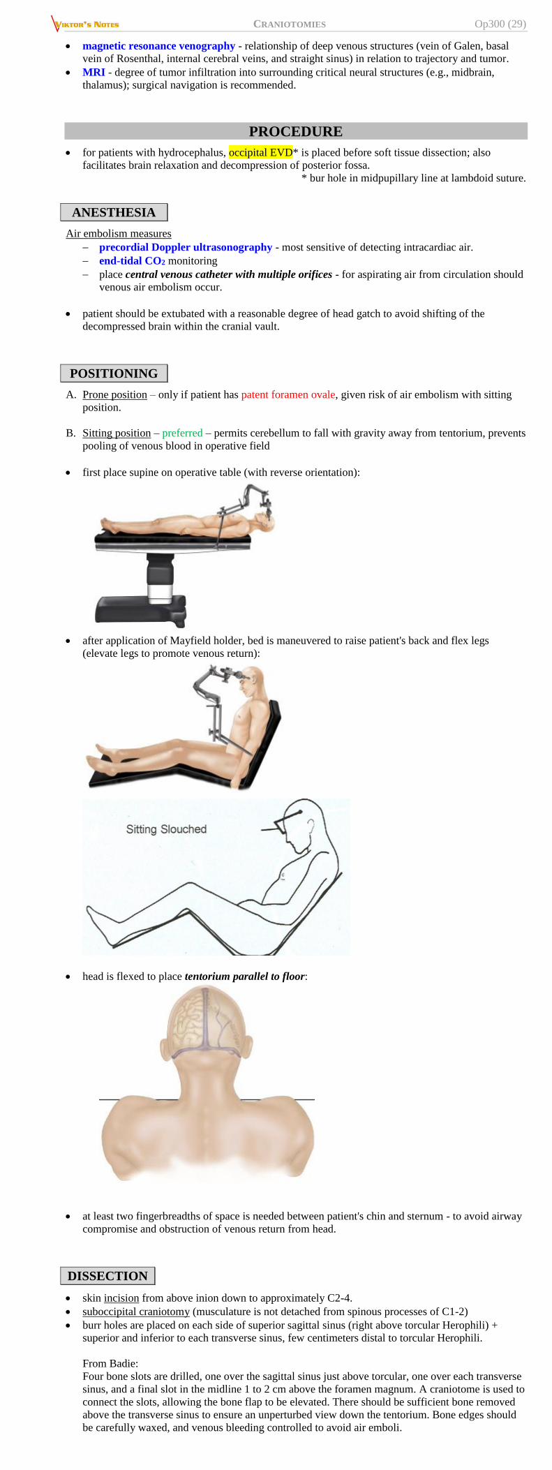
CRANIOTOMIES Op300 (29)
magnetic resonance venography - relationship of deep venous structures (vein of Galen, basal
vein of Rosenthal, internal cerebral veins, and straight sinus) in relation to trajectory and tumor.
MRI - degree of tumor infiltration into surrounding critical neural structures (e.g., midbrain,
thalamus); surgical navigation is recommended.
PROCEDURE
for patients with hydrocephalus, occipital EVD* is placed before soft tissue dissection; also
facilitates brain relaxation and decompression of posterior fossa.
* bur hole in midpupillary line at lambdoid suture.
ANESTHESIA
Air embolism measures
precordial Doppler ultrasonography - most sensitive of detecting intracardiac air.
end-tidal CO2 monitoring
place central venous catheter with multiple orifices - for aspirating air from circulation should
venous air embolism occur.
patient should be extubated with a reasonable degree of head gatch to avoid shifting of the
decompressed brain within the cranial vault.
POSITIONING
A. Prone position – only if patient has patent foramen ovale, given risk of air embolism with sitting
position.
B. Sitting position – preferred – permits cerebellum to fall with gravity away from tentorium, prevents
pooling of venous blood in operative field
first place supine on operative table (with reverse orientation):
after application of Mayfield holder, bed is maneuvered to raise patient's back and flex legs
(elevate legs to promote venous return):
head is flexed to place tentorium parallel to floor:
at least two fingerbreadths of space is needed between patient's chin and sternum - to avoid airway
compromise and obstruction of venous return from head.
DISSECTION
skin incision from above inion down to approximately C2-4.
suboccipital craniotomy (musculature is not detached from spinous processes of C1-2)
burr holes are placed on each side of superior sagittal sinus (right above torcular Herophili) +
superior and inferior to each transverse sinus, few centimeters distal to torcular Herophili.
From Badie:
Four bone slots are drilled, one over the sagittal sinus just above torcular, one over each transverse
sinus, and a final slot in the midline 1 to 2 cm above the foramen magnum. A craniotome is used to
connect the slots, allowing the bone flap to be elevated. There should be sufficient bone removed
above the transverse sinus to ensure an unperturbed view down the tentorium. Bone edges should
be carefully waxed, and venous bleeding controlled to avoid air emboli.
CRANIOTOMIES Op300 (30)
craniotome is used to connect burr holes to create bone flap.
craniectomy is possible option, but craniotomy is preferred to reduce postoperative pain,
fluid collections, and aseptic meningitis
if there is evidence of preoperative tonsillar descent, foramen magnum can be removed in addition
to C1 laminectomy.
dural incision - semilunar or cruciate - based on transverse sinuses and torcular Herophili and
reflected superiorly with tenting sutures (be cognizant of retraction placed on venous sinus when
reflecting dural flap!):
if angle of tentorium is too steep, craniotomy can be extended for occipital transtentorial approach,
or tentorium can be cut and retracted via supracerebellar approach.
arachnoid adhesions and bridging veins between cerebellum and tentorium are divided to open
supracerebellar infratentorial corridor.
bridging veins should be divided close to cerebellum to prevent retraction of inaccessible
bleeding sources back into tentorium.
as this process of dissection proceeds, cerebellum falls with gravity, and Greenberg
retractor can be placed on tentorium if necessary.
thickened arachnoid overlying pineal gland and quadrigeminal cistern is exposed and sharply
dissected open.
precentral cerebellar vein is visualized (draining into vein of Galen) – it is the only deep
venous structure that should be cauterized and divided:
normal anatomy when exposure is achieved and cerebellar retraction occurs (N.B. vascular
structures and neural structures may be shifted to nonanatomic positions by tumor)
COMPLICATIONS
AIR EMBOLISM
3) cover torcular Herophili sinus area with wet laparotomy pads
4) flood field with saline
5) lower patient's head.
6) use central venous catheter to retrieve any large emboli
FAR-LATERAL SUBOCCIPITAL APPROACH
NREF video >>
Rhoton video: http://rhoton.ineurodb.org/?page=23441

CRANIOTOMIES Op300 (31)
Used sources:
R. Jandial “Core Techniques in Operative Neurosurgery” (2011), procedure 17
INDICATIONS
suboccipital approach with C1 laminectomy provides adequate visualization of approximately 270
degrees of the circumference around the medulla; it does not provide access to the 90 degrees
anterior to the medulla, however, because the visual angle needed to see this region is obscured by
the occipital condyle, which must be drilled to allow access along this visual trajectory.
allows good proximal vascular control (on vertebral arteries); good approach for PICA aneurysms.
muscular bulk in the midline approach performed in a conventional suboccipital craniectomy
effectively limits the surgeon's ability to dissect safely laterally enough to visualize the extracranial
vertebral artery and to drill away the posterior occipital condyle.
limits of this approach are the ventral clivus and brainstem above the pontomedullary junction.
it is like retrosigmoid approach just more inferior.
PROCEDURE
positioning is the most complex of any common neurosurgical procedure.
table at least 120 degrees away from the anesthesia team
three-quarter prone position with the contralateral shoulder down
superior (ipsilateral) shoulder is in mild flexion on an arm rest in mild flexion. The contralateral
arm is draped off the edge of the bed and placed in a shoulder sling, which is secured to the edge of
the bed with towel clamps
two Mayfield pins placed just behind the contralateral occiput. The single pin is placed in the
ipsilateral frontal bone, above the superior temporal line. After pinning, the head is slightly flexed,
rotated toward the contralateral shoulder, and elevated slightly.
theoretical goal of head positioning is to place the posteromedial portion of the ipsilateral occipital
condyle at the highest point in the room. By doing so, the corridor of attack just medial to the
condyle is placed basically vertical, maximizing the retraction obtained by gravity.
it is important to check motor and sensory evoked potentials before and after positioning and to
adjust the positioning if there are adverse changes from baseline. We have done these cases with
the patient awake in some instances when positioning without neuromonitoring changes was
impossible while the patient was asleep
Incision
hockey stick–shaped, consisting of three unequal-length limbs that are roughly perpendicular to
each other. The long limb of the incision is midline and begins just below the spinous process of
C3 and extends to just above the inion. The horizontal incision extends laterally from just above
the inion to just above the mastoid tip. The short limb of the incision begins just below the mastoid
tip and extends upward to meet the horizontal limb. This incision parallels the transverse and
sigmoid sinuses and provides the ability to fold the myocutaneous flap laterally enough to expose
the entire hemiocciput and the arch of C1 out to the tip of the transverse process.
big mastoid emissary vein is likely – will bleed a lot.
if needed now can dissect occipital artery from inside of flap (alternative strategy to find occipital
artery in the more superficial layer is more difficult); find the origin of artery first – medial to
digastric groove.

CRANIOTOMIES Op300 (32)
soft tissue dissection is performed with a combination of periosteal dissectors and monopolar
cautery to expose three key landmarks in their entirety. The hemiocciput should be cleared of soft
tissue down to the foramen magnum; once muscle attachments are taken, switch Bovie to
periosteal to finalize towards foramen magnum Also, the mastoid process should be exposed down
to the point where the mastoid tip begins to curve medially and anteriorly until the mastoid curves
anteriorly.
Finally, the lamina of C1 should be exposed laterally until the tip of the C1 transverse process can
be palpated under the superior and inferior oblique muscles of the suboccipital triangle.
Sulcus arteriosus – where vertebral artery along with C1 root are travelling
It is important to remain oriented to the spinal midline; this can be confusing because of the degree
of head rotation in this position, which is much greater than the typical suboccipital approach. A
loss of one's sense of midline not only can increase blood loss owing to muscle dissection, but also
can lead to inappropriate trajectories toward critical structures such as the vertebral artery. For this
reason, we begin this approach by finding the intramuscular septum early and cautiously
identifying the spinal midline and exposing the C1-3 hemilamina from medial to lateral until the
C1 transverse process can be palpated.
During muscle dissection, it is possible to encounter large muscular branches of the vertebral
artery.
Identification and mobilization of the vertebral artery. After reflecting the scalp flap with the
inferior and superior oblique muscles laterally and posteriorly, the interlaminar and perivascular
venous plexus is slowly controlled with bipolar cautery and direct pressure and divided with
microscissors. Through this process, the course of the vertebral artery is delineated and prepared
for mobilization. The posterior bony portion of the foramen transversarium is removed with a
diamond bit drill to free the vertebral artery posteriorly. Multiple periosteal attachments that tether
the vertebral artery into the foramen superiorly and inferiorly may be present; these should be
sharply divided. The vertebral artery is mobilized away from the occipital condyle with a vessel
loop and protected.
extradural origin of PICA – such PICA always enters dura next to VA.
bone flap involves three bone cuts that viewed from above remove a J-shaped plate of
hemioccipital bone. The medial vertical limb of the bone flap extends upward from the foramen
magnum to just shy of the transverse sinus, which is just lateral to midline. The lateral vertical limb
begins just inferomedial to the asterion and extends inferiorly and medially in a curvilinear fashion
to reach the foramen magnum as lateral as possible. The horizontal limb connects the upper
portions of each vertical limb at right angles and roughly parallels the transverse sinus.

CRANIOTOMIES Op300 (33)
Although it is possible to use the foramen magnum as the sole entry point for the side-cutting bur,
we prefer to add two burr holes. One burr hole is placed inferolateral to the inion and torcular
Herophili and one is placed inferomedial to the asterion to help dissect a clear epidural plane to
turn the bone flap in, preventing injury to the venous sinuses.
thick dural attachments at the foramen magnum are bluntly separated from the bony rim of the
foramen magnum because dural tears in this region risk injuring the circular sinus.
C1 hemilaminectomy is necessary to lengthen the dural incision to achieve the desired exposure in
this approach. The hemilaminectomy is performed either piecemeal or using a side-cutting bur with
a footplate.
hemilaminectomy of C2 and C3 can further improve visualization.
It is wise to have a set of permanent and temporary aneurysm clips on the field throughout the case
if needed to address vertebral injury.
Retrosigmoid mastoidectomy. In contrast to the transpetrosal approaches, the goal of
mastoidectomy in the far-lateral approach is to expose the transverse and sigmoid sinuses from the
torcular Herophili to the beginning of the jugular bulb, defining the superior and lateral extent of
the dural incision. This is performed with a sequence of dural dissection away from the bone, bony
thinning with the drill, and removal with a Kerrison punch.
Drilling of occipital condyle – core out cancellous bone, then remove cortical shell (less venous
bleeding). Removal of the occipital condyle and associated lip of foramen magnum allows the
additional anterior visualization that this approach provides. Although the posterior half of the
condyle can be removed with relatively minimal adverse effects, additional condylar removal
provides increased visualization at the cost of decreased stability of the atlantooccipital joint.
Roughly 50% (8 mm) of condyle can be safely removed posteriorly before occipitocervical
fusion should be considered. Following this rule, hypoglossal canal is rarely seen. Before drilling,
the rerouted vertebral artery should be protected well away from the site of drilling
don’t drill too much of condyle → instability.
After the sigmoid sinus is exposed, it is essential that the remaining mastoid air cells are
aggressively obliterated with bone wax to prevent cerebrospinal fluid egress through the middle
ear.
Ideally, as much of the bone removal as possible should be performed as part of a bone flap
because replacement of bony surface is cosmetically superior and possibly prevents muscular
adhesion to the dura and suboccipital pain
dura is opened in a lazy J-shaped fashion from the transverse sigmoid junction curving medial and
inferiorly so that it crosses the foramen magnum just posteriorly to the intradural entry point of the
vertebral artery. The cervical dura should be opened in a linear and paramedian fashion down to at
least the upper edge of the C2 lamina. If a large circular sinus is encountered while crossing the
foramen magnum, this should be controlled with Weck clips, divided, and oversewn with dural
sutures. At least some dural cuff should be left around the vertebral artery so that the dura can be

CRANIOTOMIES Op300 (34)
repaired safely in this region. The dura should be reflected anteriorly with sutures placed as deeply
as possible to keep the dura flat against the bony surface of the drilled condyle
Supracondylar extension – through jugular tubercle (seen on medial wall of condylar part of occipital
bone); remember – CN11 is draped over jugular tubercle).
TRANSCONDYLAR APPROACH
Rhoton video: http://rhoton.ineurodb.org/?page=23441
Viktor’s Notes℠ for the Neurosurgery Resident
Please visit website at www.NeurosurgeryResident.net


