

VOT 74171
PRODUCTION OF ENZYMATIC GLUCOSE BIOSENSORS
DR. AZILA ABDUL AZIZ
PUSAT PENGURUSAN PENYELIDIKAN
UNIVERSITI TEKNOLOGI MALAYSIA
2006

ii
ACKNOWLEDGEMENTS
I would like to express my appreciation to all those who have assisted me,
whether directly or indirectly, in conducting this research. There are too many names
to list down. However, special thanks goes to the research assistants, Wong Fui Ling
and Norhana Mohd Jusoh for their dedications and tireless efforts in making this
research work a success.

iii
ABSTRACT
For this project, two types of glucose biosensors namely hydrogen peroxide-
based glucose biosensor and mediated glucose biosensor have been developed. The
performance of a glucose biosensor depends mostly on the immobilization method
and support materials that are being used. For hydrogen peroxide-based glucose
biosensor, selection of suitable materials for enzyme immobilization was done. Four
types of immobilization materials, including chemically-linked PVA, TMOS sol-gel,
alumina sol-gel, and freezed-thawed PVA cryogel, were used to immobilize glucose
oxidase (GOD) to determine the most appropriate material for GOD immobilization.
Generally the membranes had shown good sensitivity except for the chemically
cross-linked PVA. However, the main differences with the enzyme immobilization
methods were enzyme leakage and the values of Kmapp. Freeze-thawed PVA-GOD
membranes, which showed satisfactory sensitivity and adequate value of Kmapp was
chosen as the support material for immobilizing GOD. The enzyme leakage of this
type of membrane was improved by reducing enzyme loading. Even though this type
of sensor is very simple and easy to construct, it suffers from electrochemical
interferences from common electroactive species present in blood such as
acetaminophen. Thus, a selective inner layer based on permselectivity was studied.
pHEMA, at a cross-linking ratio of 0.043 which resulted in a permselectivity of 10,
successfully eliminated acetaminophen interference. Nafion membrane was used as
the outer membrane to protect the biosensor.
For the mediated based glucose biosensor development, the scopes of work
include the preparation of active layer, preparation of external layer and the
fabrication of glucose biosensor. Three methods of tethering a mediator to an
enzymatic membrane were studied to construct a non-leaking mediated glucose

iv
biosensor. The methods were immobilization of glucose oxidase (GOD) and
ferrocene redox polymer in cross-linked poly (vinyl alcohol) (CLPVA) with bovine
serum albumin (BSA) as a protein stabilizer, immobilization of ferrocene carboxylic
acid and glucose oxidase in a sol gel derived silica (SGS) matrix containing cross-
linked poly (vinyl alcohol) (CLPVA) and nafion, and lastly multilayered construction
of glucose oxidase and redox poly (allylamine) ferrocene utilizing layer-by-layer
covalent attachment. After evaluating the biosonser response amperometrically at
0.363V, the first method, which was immobilization of glucose oxidase and
ferrocene redox polymer in CLPVA with the addition of BSA was selected for the
fabrication of disposable glucose biosensor since this type of sensor provided good
responses over a wide range of concentration. Nafion was chosen as the external
layer and the works on the fabrication of the glucose biosensor are ongoing.

v
ABSTRAK
Untuk projek ini, kajian telah dijalankan atas dua jenis biosensor glukosa,
iaitu biosensor glukosa berdasarkan hidrogen peroksida dan biosensor glukosa
berdasarkan pengantara. Prestasi sesuatu biosensor glukosa banyak bergantung
kepada cara penyekatgerakan enzim dan jenis bahan sokongan yang digunakan.
Bagi biosensor glukosa berdasarkan hidrogen peroksida, pemilihan bahan yang
sesuai telah dilakukan. Empat jenis bahan penyekatgerak telah dikaji iaitu poli(vinil
alkohol) (PVA) disambung-silang secara kimia, , sol-gel (tetrametoksi)silane
(TMOS), sol-gel alumina, dan kryogel beku-cair PVA, untuk menentukan jenis
bahan yang paling sesuai bagi penyekatgerakan glukosa oksida (GOD). Secara
umum, membran-membran yang dihasilkan menunjukkan sensitiviti yang baik
kecuali PVA disambung-silang secara kimia. Walau bagaimanapun, perbezaan
utama antara cara-cara penyekatgerakan ialah kebocoran enzim dan nilai Kmapp.
Kryogel beku-cair PVA, yang menunjukkan sensitiviti yang memuaskan dan nilai
Kmapp yang memadai, telah dipilih sebagai bahan sokongan untuk menyekatgerak
GOD. Kebocoran enzim bagi membran jenis ini telah diperbaiki dengan
mengurangkan kuantiti enzim yang dimasukkan dalam proses penyekatgerakan.
Walaupun sensor yang berasaskan hydrogen peroksida tidak kompleks dan mudah
untuk dibina, ia menghadapi masalah gangguan elektrokimia daripada spesies
elektroaktif yang biasanya wujud dalam darah seperti asetaminofen. Oleh sebab itu,
suatu lapisan dalaman yang boleh menghalang gangguan elektrokimia berdasarkan
ketertelapan selektif telah dikaji. Poli(hidroksietil metakrilat) (pHEMA), dengan
nisbah sambung-silang 0.043 yang menunjukkan selektiviti 10, berjaya
menyingkirkan gangguan asetaminofen. Membran Nafion telah digunakan sebagai
lapisan luaran untuk melindungi biosensor tersebut.

vi
Bagi biosensor glukosa berdasarkan pengantara, skop kerja merangkumi
penyediaan lapisan aktif, lapisan luaran, dan pembinaan biosensor glukosa. Tiga
cara untuk mengikat pengantara ke membran yang mengandungi enzim telah dikaji
untuk membentuk sebuah biosensor glukosa tanpa kebocoran pengantara. Cara-cara
tersebut termasuk penyekatgerakan GOD dan polimer redoks ferrocene dalam PVA
(CLPVA) tersambung-silang dengan menggunakan bovine serum albumin (BSA)
sebagai agen penstabil protin, penyekatgerakan GOD dan asid karbosilik ferrocene
dalam matriks sol gel silika (SGS) yang mengandungi CLPVA dan nafion, dan yang
terakhirnya adalah multi-lapisan GOD dan redoks poli (allilamin) ferrocene
menggunakan pelekatan kovalen lapisan demi lapisan. Selepas menilai gerak balas
biosensor secara amperometrik pada 0.363V, cara pertama, iaitu penyekatgerakan
GOD dan polimer redoks ferrocene dalam CLPVA bersama BSA telah dipilih untuk
membentuk biosensor glukosa yang boleh dibuang selepas penggunaan
memandangkan sensor jenis tersebut menunjukkan gerak balas yang baik dalam julat
kepekatan yang luas. Nafion telah dipilih sebagai lapisan luaran dan kerja-kerja
pembinaan biosensor glukosa yang lengkap sedang dijalankan.
vii
TABLE OF CONTENTS
CHAPTER TITLE PAGE
TITLE i
ACKNOWLEDGEMENTS ii
ABSTRACT iii
ABSTRAK v
TABLE OF CONTENTS vii
LIST OF TABLES xi
LIST OF FIGURES xii
1 INTRODUCTION
1.1 Research Background 1
1.2 Objective 5
1.2 Scopes 5
2 LITERATURE REVIEW
2.1 Historical Overview of Biosensor Technology 7
2.2 Principle of Glucose Biosensor 8
2.3 Three Generations of Glucose Biosensor 10
2.3.1 First Generation of Glucose Biosensor 11
2.3.2 Second Generation of Glucose Biosensor 12

viii
2.3.3 Third Generation of Glucose Biosensor 14
2.4 Enzyme Immobilization 15
2.4.1 Adsorption 16
2.4.2 Entrapment 16
2.4.3 Covalent Coupling 20
2.4.4 Cross-linking 21
2.5 Methods of Tethering the Mediator to the Enzymatic 24
Membrane
2.5.1 Redox Polymer 26
2.5.2 Multilayer Systems 28
2.6 Permselective Layer for Hydrogen Peroxide- Based 30
Glucose Sensor
2.6.1 pHEMA 32
2.7 Protective Membrane for Mediator-Based 33
Glucose Biosensor
3 MATERIALS AND METHODS
3.1 Chemicals 35
3.2 Instrumentation 36
3.3 Methodology for Hydrogen Peroxide-Based 37
Glucose Sensor
3.3.1 General Casting Method of Free-standing 38
Membrane
3.3.2 Preparation of Chemically Cross-linked 39
PVA-GOD Membrane
3.3.3 Preparation of Freeze-thawed PVA-GOD 39
Membrane
3.3.4 Preparation of Alumina-PVA-GOD Membranes 40
3.3.5 Preparation of TMOS-PVA-GOD Membranes 40
3.3.6 Determination of Water Content 41
3.3.7 Determination of Enzyme Leakage 41
3.3.8 Determination of Apparent Enzyme Activity of 42

ix
Membrane
3.3.9 Enzyme Kinetics 42
3.3.10 Casting of pHEMA Membrane as Permselective 43
Layer
3.3.11 Permeability Analysis 43
3.3.12 Casting of Nafion Outer Membrane 44
3.4 Methodology for Mediator-Based Glucose Sensor 45
3.4.1 Immobilization of Glucose Oxidase and 46
Ferrocene Redox Polymer in Cross-linked
Poly (vinyl alcohol) with Bovine Serum
Albumin as Protein Stabilizer
3.4.2 Immobilization of Glucose Oxidase and 47
Ferrocene Carboxylic Acid in Composite
Silica Sol-gel (SGS) /Cross-linked
Poly (vinyl alcohol) (CLPVA)/Nafion Membrane
3.4.3 Multilayered Construction of Glucose 48
Oxidase and Poly(allylamine)ferrocene
3.4.4 Electrochemical Measurement 50
3.4.5 Ferrocene Leakage Detection 50
3.4.6 Enzyme Leakage Detection 51
3.4.7 Preparation of Nafion Protective Membrane 51
3.4.8 Fabrication of Glucose Sensor 51
4 RESULTS AND DISCUSSION
4.1 Hydrogen Peroxide-Based Glucose Sensor 52
4.1.1 Chemically cross-linked PVA-GOD Membrane 52
4.1.2 Freeze-thawed PVA-GOD Membrane 57
4.1.3 Alumina-PVA-GOD Sol-gel Derived Organic/ 59
Inorganic Membrane
4.1.4 TMOS-PVA-GOD Sol-gel Derived Organic/ 61
Inorganic Membrane
4.1.5 Overall Comparison of Performance of Different 63

x
Membranes
4.1.6 Permselectivity Analysis 64
4.1.7 Performance of Three Layers Biosensor 68
4.2 Mediator-Based Glucose Biosensor
4.2.1 Cyclic Voltammetry for Ferrocene 69
Carboxylic Acid
4.2.2 Glucose Oxidation 70
4.2.3 Immobilization Methods for Mediated Biosensor 71
5 CONCLUSIONS AND RECOMMENDATIONS
5.1 Conclusions for Hydrogen Peroxide-Based 85
Glucose Biosensor
5.2 Conclusions for Mediator-Based Glucose 86
Biosensor
5.3 Recommendations 88
REFERENCES 90

xi
LIST OF TABLE
TABLE TITLE
PAGE
3.1 Different preparation conditions of freeze-thawed PVA-GOD membranes 40
4.1 Water content of membranes clamped for different period 53
4.2 Freeze-thawed PVA-GOD prepared at different conditions 57
4.3 Permeability performance of pHEMA membranes at different 66
cross-linking ratio

xii
LIST OF FIGURES
FIGURE TITLE PAGE
2.1 Evolution of home blood glucose monitoring technology 7
2.2 Schematic representation of possible glucose biosensor construction 9
2.3 The differences between oxygen electrode based sensors and 12
mediator based sensor.
2.4 The electrical ‘wiring’ of an oxidative redox-enzyme via a 13
diffusional electron-transfer mediator shuttling between the
enzyme reaction centre and the electrode
2.5 A ferrocene mediated biosensor for glucose 14
2.6 Structure of (a) Tetramethoxysilane (b) Aluminium isopropoxide 19
2.7 Cross-linking of enzyme with glutaraldehyde 22
2.8 Aldol condensation of glutaraldehyde 22
2.9 Freeze-thawed crystallite structure 23
2.10 Ferrocene containing cross-linked polyallylamine 27
2.11 Cyclic voltammograms of the electrode modified with 28
ferrocene-containing cross-linked polyallylamine containing
glucose oxidase in the polymer matrix
3.1 Flow chart of research methodology of hydrogen peroxide-based glucose 37
biosensor
3.2 General casting method of Free-standing membrane 38
3.3 Methodology flow chart of mediator-based glucose biosensor flow chart 45
4.1 Comparison of leaking profile of membranes immobilized at different 54
temperature
4.2 Apparent enzyme activities for membranes immobilized at different 55
temperature

xiii
4.3 Comparison of current response of chemically cross-linked PVA-GOD 56
membranes with different concentration ratio of lysozyme upon 5mM
glucose
4.4 Comparison of leaking profile of freeze-thawed PVA membranes at 58
different conditions
4.5 Comparison of current response of freeze-thawed PVA-GOD membranes 59
at different conditions upon 5mM glucose
4.6 Leaking profile of alumina-PVA-GOD composite membrane 60
4.7 Current response of Alumina-PVA-GOD composite membrane upon 61
addition of 5mM glucose
4.8 Comparison of leaking profile of TMOS-PVA membranes D 62
(without cross-linker) and membranes E (with cross-linker) (3-
glycidoxydiethoxysilane)
4.9 Comparison of current response of TMOS-PVA-GOD membrane 63
at different conditions upon addition of 5mM glucose
4.10 Water content of pHEMA permselective layer at different cross-linking 65
ratio
4.11 Typical Koutecky-Levich plot of acetaminophen and hydrogen peroxide 65
through pHEMA membrane
4.12 Permeability of acetaminophen at different cross-linking ratio 66
4.13 Permeability of hydrogen peroxide at different cross-linking ratio 67
4.14 Selectivity of pHEMA membranes at different cross-linking ratio 67
4.15 Amperometric current response with injection of 5mM glucose and 68
0.2mM acetaminophen
4.16 Height of the current peaks correspond to concentration of 69
ferrocene carboxylic acid
4.17 Linear sweep voltammograms for ferrocene carboxylic acid 70
4.18 Leaking profile for CLPVA-GOD/Fc membrane with different 71
GOD and BSA loading a) 1:1 (weight ratio of GOD: BSA)
4.19 Leaking profile for CLPVA-GOD/Fc membrane with different 72
GOD and BSA loading b) 1:3 (weight ratio of GOD: BSA)
4.20 Typical glucose calibration curves for CLPVA-GOD/Fc 73
membranes with different GOD and BSA loading a) 1:1 b) 1:3
(weight ratio of GOD: BSA)

xiv
4.21 Double–reciprocal (Lineweaver Burke) plots of 74
CLPVA-GOD/Fc membranes with different GOD and BSA
loading a) 1:1 b) 1:3 (weight ratio of GOD: BSA in mg)
4.22 Stability of CLPVA-GOD/Fc membranes with different GOD 75
and BSA loading a) 1:1 b) 1:3 (weight ratio of GOD: BSA)
4.23 Enzyme leaking profile for SGS-CLPVA/nafion membranes 76
4.24 Ferrocene leaking profile for SGS-CLPVA/nafion membranes 77
4.25 Typical current response of SGS-CLPVA/nafion membranes 78
4.26 Typical calibration curves for SGS-CLPVA/nafion membranes 79
4.27 Double–reciprocal (Lineweaver Burke) plots of 79
SGS-CLPVA/nafion membranes
4.28 Stability of SGS-CLPVA/nafion membranes 81
4.29 Typical current responses of multilayered membranes 82
4.30 Typical calibration curves for multilayed membranes 83
4.31 Double –reciprocal (Lineweaver Burke) plot of multilayered 83
membranes

CHAPTER 1
INTRODUCTION
1.1 Research Background
Diabetes mellitus is a group of metabolic diseases characterized by high
blood glucose levels, which result from defects in insulin secretion, or action, or both
(Miller, 2003). In a person without diabetes, the body is able to regulate the amount
of glucose in the blood between 3.5 to 6.5 mM with the help of the hormone insulin.
Many people who are suffering from diabetes mellitus are not able to control their
blood glucose level. In diabetes, the auto regulation of glucose fails and the blood
glucose level of a diabetic sufferer may vary between 1 to 30 mM. The consequences
of poor glucose regulation are at best, long term damage to organs from too much
glucose (hyperglycemia), coma or death caused by too little glucose reaching the
(hypoglycemia).
There are two major types of diabetes mellitus. Type 1 diabetes sometimes
referred to as juvenile diabetes or insulin dependent diabetes mellitus (IDDM),
usually strikes children and young adults. The insulin producing islet cells in the
pancreas are destroyed by the diabetics’ own immune system. These type 1 diabetics
usually lose all insulin-producing capabilities and must inject themselves with insulin
before each meal to allow their bodies to utilize glucose from the food. The type 2

2
diabetes or also referred as non insulin dependent diabetes mellitus (NIDDM) is very
complex and usually strikes older people. Type 2 diabetics can usually increase their
glucose regulation by losing weight and are initially treated with diet control and
with drugs that help the body metabolizes glucose. Type 2 diabetics over time may
need to start using insulin injections to maintain glucose regulation (Henning and
Cunningham, 1998).
The findings of Diabetes Control and Complications Trial (DCCT) and
United Kingdom Prospective Diabetes Study (UKPDS) clearly show that intensive
control of elevated levels of blood glucose in patients with diabetes mellitus
decreases the complications of nephropathy, neuropathy, retinopathy, and may
reduce the occurrence and severity of large blood vessel diseases [Miller, 2003].
Tightly controlled blood glucose level means achieving fasting glucose level
between 70-120 mg/dL, and glucose level of less than 180 mg/dL after meals.
Studies in intensively treated type I patients have shown a decrease of diabetic eye
disease by 76%, kidney disease by 54%, and nerve disease by 60%. In patients with
type II diabetes mellitus, intensive blood glucose control shows similar beneficial
effects on the eyes, kidneys, nerves and blood vessels (Miller, 2003).
Since the first publication on a glucose biosensor (Clark and Lyons, 1962),
the detection of glucose has attracted a high degree of interest due to its biological
importance (Liu et al., 2004). A biosensor is a sensor that is based on the use of
biological material for its sensing function. The bio-component specifically reacts or
interacts with the analyte of interest resulting in a detectable chemical or physical
change. The amperometric glucose biosensor represents the most successful
commercial biosensor development to date. Amperometric biosensors based on
enzymes are interesting due to their high sensitivities, excellent selectivities,
simplicity, low cost and rapid response.
The most frequently used enzymatic methods for glucose determination
employ glucose oxidase (GOD), due to its high selectivity towards ß-D-glucose.
GOD happens to be easy and cheap to obtain; secondly, it is one of the most robust
enzymes around (it withstands greater extremes of pH, ionic strength, temperature
than many other enzymes), thus allowing less stringent conditions during the
3
manufacturing process and also relatively care-free storage and use by the home-user
of the biosensor) and thirdly, the concentration range of glucose with which GOD
reacts optimally happens to coincide with the range of concentrations encountered in
human blood. The other, less coincidental factor is that the glucose-test market has
always been and looks set to remain the largest single market for home-diagnostics
and biosensors.
Three general strategies are used for the electrochemical sensing of glucose,
all of which use immobilized glucose oxidase, an enzyme that catalyzes the oxidation
of glucose to gluconic acid with the production of hydrogen peroxide. The first
detection scheme measures oxygen consumption; the second measures the hydrogen
peroxide produced by the enzyme reaction; and a third uses a diffusable or
immobilized mediator to transfer the electrons from the glucose oxidase to the
electrode.
Among the amperometric biosensors, the peroxide based glucose biosensor is
the simplest. There are three membrane layers in a peroxide based amperometric
glucose biosensor which are the outer layer, the active layer, and the inner layer. The
electro oxidation of hydrogen peroxide requires high potential that results in
oxidation of easily oxidable substances in blood simultaneously, thus adding to the
electrical signal and giving a non-accurate reading of measured glucose
concentration. An interfering molecule is a species that is electroactive at the
operating potential of the amperometric sensor. This includes ascorbic acid, urate,
and acetaminophen.
In the efforts to minimize the interference effects, a selective layer is often
placed between the enzymatic active layer and the electrode to filter out and
interfering species. A permselective membrane can be used as the inner membrane
of the sensor. A permselective membrane restricts the passage of larger molecular
weight species based on MWCO (molecular weight cut off) (Kermis et al., 2003). On
the other hand, the permselective membranes may lead to the diffusional constraints
to analyte, while excluding the interference species (Poyard et al.,1998). Therefore,
studying the characteristics of the permselective layer is required to optimize the
function of this selective layer.

4
However, the use of an artificial electron acceptor or mediator to replace the
natural acceptor oxygen in the oxidation of glucose by glucose oxidase is also a
preferable approach that has been explored to overcome tissue oxygen dependence.
In addition, the oxidation of the reduced mediator occurs at low potential thus
reducing the sensitivity of the sensor to interfering substances. Claremont et al. 1986
were the first who reported an implantable amperometric ferrocene-modified glucose
sensor. However, the initial promise exhibited by mediator based glucose sensors for
in vivo applications, has failed to materialize.
The main problem remains the limited long-time-use stability of mediated
glucose sensors, which has been attributed to the leaching of the mediator. In
addition, the loss of mediator is a particularly important issue for implantable sensors
because of the inherent toxic effect of the mediators used. Therefore, in order to
develop a stable implantable mediated glucose sensor, a suitable immobilization
method should be investigated to avoid the leaking of mediator as well as the
enzyme. However, for disposable mediated glucose biosensor for home monitoring,
the issues of stability and leakage are not as crucial. What is more important is an
immobilization method that results in high sensitivity of the sensor and adequate
kinetics to extend the detection limit of the sensor.
In this work, for both hydrogen peroxide based glucose and mediated glucose
biosensor, various immobilization methods were investigated to determine which one
was the most stable and could able to retain enzyme with good responses over a wide
range of concentration. Besides, for hydrogen peroxide-based glucose sensor, the
characteristic of the permselective layer was studied in order to develop an
interference-free hydrogen peroxide-based glucose biosensor. Furthermore, for
mediated glucose sensor, selection of the appropriate method to tether the mediator
to the enzymatic membrane was important in order to develop a non leaking
mediator based biosensor. The methods involved in this research part were
immobilization of GOD and ferrocene redox polymer in cross-linked poly (vinyl
alcohol) (CLPVA) with bovine serum albumin (BSA) as a protein stabilizer,
immobilization of ferrocene carboxylic acid and GOD in a sol gel derived silica
(SGS) matrix containing cross-linked poly (vinyl alcohol) (CLPVA) and nafion, and

5
lastly multilayered construction of GOD and redox poly(allylamine) ferrocene
utilizing layer-by-layer covalent attachment.
1.2 Objective
The objectives of this work are as follows:
(i) To develop an interference-free hydrogen peroxide-based glucoe
biosensor
(ii) To develop a practical and stable mediated amperometric glucose
sensor
1.3 Scopes
To achieve objective (i), the following specific areas were investigated:
(i) Selection of the appropriate methods of immobilizing GOD. The
enzymatic membranes developed were chemically cross-linked
poly(vinyl alcohol) (PVA)-glucose oxidase (GOD) membranes,
freeze-thawed poly(vinyl alcohol) (PVA)-glucose oxidase (GOD)
membranes, tetramethoxysilane (TMOS) sol-gel -glucose oxidase
(GOD) membranes , and alumina sol-gel -glucose oxidase (GOD)
membranes.
(ii) Determination of the optimum cross-linking density of poly(2-
hydroxyethyl methacrylate) (pHEMA) membrane for it to
successfully perform as a selective inner membrane.
(v) Testing of a complete lab-scale sized peroxide-based glucose
biosensor.

6
To achieve objective (ii), the following specific areas were investigated:
(i) Selection of the appropriate methods of immobilizing GOD to the
polymer to construct a non-leaking mediated glucose sensor. Three
methods were studied:
a) Immobilization of glucose oxidase and ferrocene redox polymer in
cross-linked poly (vinyl alcohol) with bovine serum albumin as
protein stabilizer
b) Immobilization of glucose oxidase/ferrocene carbozylic acid in
composite silica sol gel (SGS) /cross-linked poly (vinyl alcohol)
(CLPVA)/nafion membrane
c) Multilayered construction of glucose oxidase and poly(allylamine)
ferrocene
(ii) Preparation of a nafion protective membrane
(iii) Fabrication of the mediator-based glucose sensor

7
CHAPTER 2
LITERATURE REVIEW
2.1 Historical Overview of Biosensor Technology
Figure 2.1 Evolution of home blood glucose monitoring technology
The first prototype biosensor was an enzyme electrode reported in 1962,
which utilized immobilized GOD on a Clark pO2 electrode for measuring the
concentration of glucose in solution. This prototype enzyme electrode later served as
the basis for the development of the first commercialized enzyme electrode and
glucose analyzer (Taylor, 1991]. The first step towards commercial exploitation was
that taken by the Yellow Springs Instrument Company in the seventies. YSI- in
close collaboration with Clark- developed a series of laboratory-scale glucose
sensors. Much work was invested in finding suitable membranes that rendered the
GOD-platinum electrode technique reproducible and accurate.
2000+ Least/
Noninvasive & Long Term
Late 1990s Painless Blood
Late 1980s Minimum Procedure
Blood
Late 1970sBlood
Late 1960s Urine

8
The key research that lead to the next generation of home-testing glucose
sensors was performed in the early 80's by H.A.O.Hill and I.J. Higgins and their
respective colleagues at the University of Oxford and the Cranfield Institute of
Technology (Newmann, 2005). The oxidized form of the mediator reacted with
reduced GOD instead of oxygen and thus reduced mediator is formed instead of
hydrogen peroxide. The reduced mediator is then re-oxidized at the electrode, giving
a current signal and regenerating the oxidized form of the mediator. This eliminates
the problem with variable oxygen concentrations in the sample and partially
eliminates electrochemical interference. The commercial reality of the mediated
sensor came with the foundation of Genetics International (later to change name to
Medisense) and the launch of the pen-sized Exactech glucose sensors in 1987. The
system consists of small, disposable, single-use glucose-sensitive electrodes (based
on a mixture of GOD and mediator in a conductive carbon-paste binder) and the
corresponding pen-sized (later pocket-calculator-sized) meter containing the
electronics and an LCD display (Newmann, 2005).
2.2 Principle of Glucose Biosensor
Biosensors are a class of extremely sensitive and selective sensors that
convert a biological action into an electrical signal to detect or quantitatively
determine a specific compound. This technology is the creative synergistic
combination of biotechnology, biochemistry, membrane technology and
microelectronics. Biosensors are analytical devices that incorporating a biological or
a biomimic material, such as tissue, microorganisms, organelles, cell receptors,
enzymes, antibodies, nucleic acids etc., which recognizes the analyte, and is
intimately associated with or integrated within a physicochemical transducer or
transducing microsystem, that translates the recognition event into a signal. The
usual aim of a biosensor is to produce either discrete or continuous digital electronic
signals which are proportional to a single analyte or a related group of analytes
(Eggins, 1996). In a glucose biosensor, glucose oxidase enzymes are employed as

9
the biological components of the sensors for molecular recognition. In the context of
glucose biosensor, the analyte involved is glucose and it is available in the blood.
Figure 2.2 shows the schematic representation of possible glucose biosensor
construction.
Figure 2.2 Schematic representation of possible glucose biosensor construction
Glucose biosensors are based on the fact that the enzyme glucose oxidase
catalyses the oxidation of glucose to gluconic acid. Glucose and oxygen would
diffuse into the enzyme layer from the sample site and the consequent depletion of
oxygen would provide a measurement of the glucose concentration. The most
common strategies for glucose detection can be partitioned into the following groups:
those employing glucose oxidase; those using a dehydrogenase enzyme or those
relying on an inorganic catalyst for oxidation of glucose or fluorescence due to the
combination of fluorescein and glucose. The first article describing an immobilized
enzyme electrode was due to Updike & Hicks in 1967. They immobilized the
enzyme glucose oxidase in a polyacrylamide gel at an oxygen electrode.
Since three decades, the search for an ideal glucose biosensor continues to be
one of the main motivations in this research field. The refinement of electrochemical
approaches for glucose sensitivity has occupied many research groups. Every year
there are lots of papers in glucose biosensor published. Most papers found by the
literature search appear to use glucose oxidase to oxidize one of the anomers of
glucose ( Niu and Lee, 2002; Zhang et al., 2004; Yoon et al., 2000; Hodak et al.,
1997; Koide and Yokoyama, 1999). Today, glucose sensor research is a relative

10
mature and well-worked research field. The majority of sensors is based on
electrochemical principles and employ enzymes as biological component for
molecular recognition. A successful biosensor must possess at least some of the
following beneficial features:
1. The biocatalyst must be highly specific for the purpose of the analyses, be
stable under normal storage conditions and show good stability over a large
number of assays (i.e. much greater than 100).
2. The reaction should be as independent of such physical parameters as stirring,
pH and temperature as is manageable.
3. The response should be accurate, precise, reproducible and linear over the
useful analytical range, without dilution or concentration. It should also be
free from electrical noise.
4. If the biosensor is to be used for invasive monitoring in clinical situations, the
probe must be tiny and biocompatible, having no toxic or antigenic effects
5. The complete biosensor should be cheap, small, portable and capable of being
used by semi-skilled operators.
6. There should be a market for the biosensor
2.3 Three Generations of Glucose Biosensor
Sometimes these three modes of oxidation are referred to as first, second and
third generation biosensors. First generation is oxygen electrode based sensors and
second generation is mediator-based sensor. Meanwhile, the third generation is
directly coupled enzyme electrode. However there is some evidence that the mode
of action of conducting salt electrodes is really the same as that of a mediator, so that
the third generation description may not be strictly accurate (Eggins, 1996). The
advantages of the mediated sensor are numerous. The reaction of GOD with
mediator is much better defined because of non-dependence on ambient oxygen.
Therefore, there is no need to worry about variable oxygen concentrations in blood.

11
Secondly, mediators can be re-oxidized at an electrode at less extreme
potentials than are necessary for hydrogen peroxide. This partially eliminates
electrochemical interference that always occurred when H2O2 detection method is
used. Working at such high potentials will increase the risk of interference from
easily oxidizable compounds. The hydrogen peroxide method is very sensitive to
many common interfering species present in the blood such as uric acid, vitamin C
and paracetamol. These substances will break down electrochemically and thus give
interfering signals. Besides mediated biosensors offer other advantages of increased
linear response and perhaps an extended biosensor lifetime, because hydrogen
peroxide is not being generated, which can contribute to the deactivation of the
enzyme (Reynolds et. al., 1992).
2.3.1 First Generation Glucose Biosensor
Glucose biosensors are generally based on the enzyme glucose oxidase. This
enzyme catalyzes the oxidation of β -D-glucose by molecular oxygen producing
gluconolactone and hydrogen peroxide. The detail reactions involved are as shown
below:
β-D-glucose + GOD(FAD) → Glucono-δ-lactone + GOD(FADH2)
GOD(FADH2) + O2 → GOD(FAD) + H2O2
Glucono-δ-lactone + H2O2 → Gluconic acid
β-D-glucose + O2 + H2O → Gluconic acid + H2O2
The very simple first generation glucose sensor, which generates hydrogen
peroxide in the presence of oxygen and glucose are the most widely used. The signal
is due to the oxidation of the hydrogen peroxide at a catalytic (usually platinum)
anode.
H2O2 O2 + 2H+ + 2e-
The most important advantage of the hydrogen peroxide electrode based sensors is
their ease of fabrication and the possibility of constructing them in small sizes even
700 mV vs Ag/AgCl
GOD
12
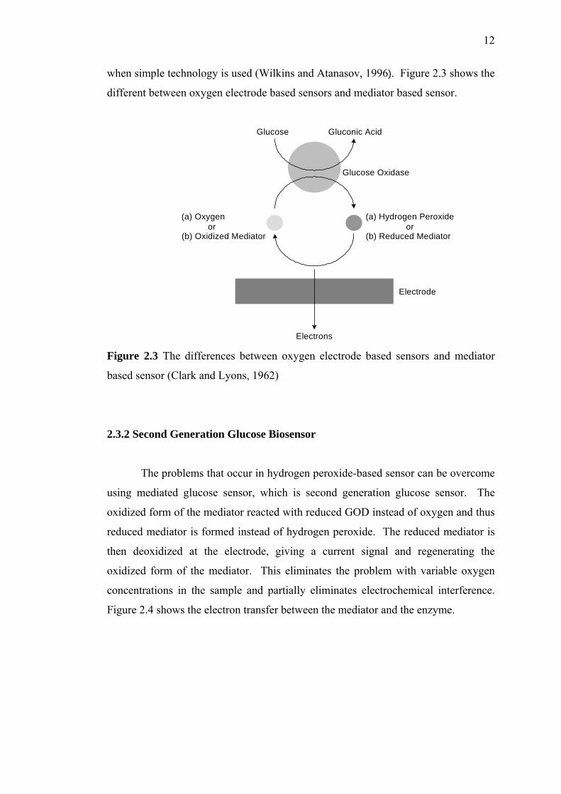
when simple technology is used (Wilkins and Atanasov, 1996). Figure 2.3 shows the
different between oxygen electrode based sensors and mediator based sensor.
Gluconic AcidGlucose
(a) Hydrogen Peroxideor
(b) Reduced Mediator
(a) Oxygenor
(b) Oxidized Mediator
Glucose Oxidase
Electrode
Electrons Figure 2.3 The differences between oxygen electrode based sensors and mediator
based sensor (Clark and Lyons, 1962)
2.3.2 Second Generation Glucose Biosensor
The problems that occur in hydrogen peroxide-based sensor can be overcome
using mediated glucose sensor, which is second generation glucose sensor. The
oxidized form of the mediator reacted with reduced GOD instead of oxygen and thus
reduced mediator is formed instead of hydrogen peroxide. The reduced mediator is
then deoxidized at the electrode, giving a current signal and regenerating the
oxidized form of the mediator. This eliminates the problem with variable oxygen
concentrations in the sample and partially eliminates electrochemical interference.
Figure 2.4 shows the electron transfer between the mediator and the enzyme.

13
Figure 2.4 The electrical ‘wiring’ of an oxidative redox-enzyme via a diffusional
electron-transfer mediator shuttling between the enzyme reaction centre and the
electrode. R: reduced mediator, R+: oxidized mediator (Degani and Heller, 1987)
There are three main steps in the reaction of the mediators with glucose
oxidase. The first one is the diffusion of the substrate from the bulk solution to the
surface of the enzyme. And the second one is the transfer of the electron from the
reaction centre of the enzyme to the mediator. Finally is the transport of the electron
from the mediators to the electrode. The distance between the electrode and the
reaction centre of enzyme will influence the membrane response time. Ferrocene
derivatives, organic dyes, ferricyanide, Ru-complexes and other electrochemically
active substrates have been employed as diffusional mediator and for the electrical
activation of soluble redox-enzymes lacking direct electrical contact with the
conductive support (Bartlett et al, 1991).
A ferrocene is a typical mediator for enzyme biosensor (Kase and Muguruma,
2004). Ferrocene fit all criteria of a good mediator such as no reaction with oxygen,
stable in both the oxidized and reduced forms, independent of pH, show reversible
electron transfer kinetics and react rapidly with the enzyme (Eggins, 1996). Besides,
in order to successfully mediate an enzyme reaction a potential mediator must posses
the following attributes such as low redox potential, reversible electrochemistry, fast
electron transfer kinetics and good stability. There are many ways in which
mediators can be incorporated into biosensors. In a biosensor, both the enzyme and
the ferrocene must be immobilized on the electrode. For glucose, the operation of
mediator is as follows:

14
Glucose + GODOX Gluconolactone + GODRED + 2H+
GODRED + 2Fc+ GODOX + 2Fc
2Fc – 2e- 2Fc+
The actual oxidation of the glucose is carried out by the FAD component of
the glucose oxidase, which is converted into FADH2. The FADH2 is reoxidized to
the FAD by the mediator, Fc+ . Then, the Fc is reoxidized to Fc+ directly at an
electrode. The current flowing through the electrode is an amperometric measure of
the glucose concentration. This is better shown in the cyclic diagram in figure 2.5.
Figure 2.5 A ferrocene mediated biosensor for glucose
2.3.3 Third Generation Glucose Biosensor
The third generation is directly coupled enzyme electrode. It may be strange
that a mediator is needed to couple an enzyme to an electrode. It is not possible to
reduce an enzyme directly on the electrode because the proteins tend to be denatured
on electrode surfaces. A better solution was developed by Albery and Cranston
(1987) and Bartlett (1987) using organic conducting salt electrodes. Tetrathiafulvane
(TTF) is reversibly oxidized, and tetracyanoquinodimethane (TCNQ) is similarly
reversibly reduced. These conducting salts can be built into electrodes in three ways
which are as single crystals as pressed pellet or a paste with graphite powder (Albery
and Cranston, 1987; Bartlett, 1987). Recently, immobilization techniques have been
developed to wire an enzyme directly to an electrode, facilitating rapid electron

15
transfer and hence high current densities. In general they involve an in situ
polymerization process using redox polymer.
2.4 Enzyme Immobilization
Enzymes are biocatalytically active entities upon which the metabolisms of
all living organisms are based. They speed up biochemical reactions by lowering the
energy of activation, without themselves appearing in the reaction products. The
catalytic action of enzymes involves their ability to alter the distribution of charges
on the compound to be converted, thus bringing about a lowering of the energy of
activation. Furthermore, they are highly specific, thus side reactions can be avoided
by employing enzymatic breakdown. A biocatalyst is termed “immobilized” if its
mobility has been restricted by chemical or physical means. This limitation of
mobility may be achieved by widely differing methods, such as trapping in the
network of a polymer matrix or by membrane confinement.
Immobilization of an enzyme results in a considerable change in the
microenvironment of the enzyme and may affect the properties of the enzyme, as
well as changes in the physical and kinetic properties. These changes may affect
their usefulness in biochemical analysis. With immobilized enzymes the measured
reaction rate depends not only on the substrate concentration and the kinetic
constants KM and Vmax, but also on immobilization effects. These effects are due to
the following alterations of the enzyme by the immobilization process. A variety of
immobilization methods have been used in the development of successful biosensors.

16
2.4.1 Adsorption
Adsorption of enzymes onto insoluble supports is a very simple method of
wide applicability and capable of high enzyme loading, which is about one gram per
gram of matrix. Simply mixing the enzyme with a suitable adsorbent under
appropriate conditions of pH and ionic strength, followed by a sufficient incubation
period, and finally washing off weakly bound or unbound enzyme will produce the
immobilized enzyme in directly usable form. The driving force causing this binding
is usually due to a combination of hydrophobic effects and the formation of several
salt links per enzyme molecule. The particular choice of adsorbent depends
principally upon minimizing leakage of the enzyme during use. Although the
physical links between the enzyme molecules and the support are often very strong,
they may be reduced by many factors including the introduction of the substrate.
Binding forces should not be weakened during use by inappropriate changes in pH or
ionic strength (Chaplin, 1990).
2.4.2 Entrapment
By matrix entrapment the enzymes are embedded in natural or synthetic
polymers, mostly of a gel-like structure. In order for the entrapped enzyme to fulfill
its catalytic function it is essential that the substrates and products of the reaction are
able to traverse the matrix. At the same time, the pores of the matrix should not be
so large that the enzyme itself can escape. Entrapment is a convenient method for
use in processes involving low molecular weight substrates and products. Amounts
in excess of 1g of enzyme per gram of gel or fiber may be entrapped. The advantage
of entrapping method is that enzymes are not subjected to serious modification, and
immobilization eliminates the effect of proteases and enzyme inhibitors of high
molecular weight. However, the difficulty which large molecules have in
approaching the catalytic sites of entrapped enzymes precludes the use of entrapped
enzymes with high molecular weight substrates. The entrapment process may be a
purely physical caging or involve covalent binding (Chaplin, 1990).

17
The natural polymers that are used for entrapment of enzyme usually lead to
relatively soft products. Subsequent hardening procedures, such as treatment with
glutaraldehyde are required. Synthetic polymers produced by polycondensation or
polymerization are frequently used for entrapping enzymes. The network of the
polymer can be made dense enough to retain the enzyme molecules. One of the
deficiencies of this method of immobilization is that the enzyme may slowly leak out
of gel matrix. This leakage is more pronounced with gels that have high water
content. Further linking procedures which provides additional cross-linking are
needed for enzyme because the mesh diameter is too large to retain single enzyme
molecules (Chaplin, 1990).
2.4.2.1 Sol-gel
An interesting recent entrapment procedure used is the sol gel method. Sol
gels are chemically inert, can resist swelling, are processed at low-temperatures, and
have tuneable porosity. Over 80% of GOD remained active in sol-gels and the
amperometric response agreed well with theoretical predictions (Audebert, 1993).
Sol-gel is a low-temperature process that involves the hydrolysis and
polycondensation of suitable precursors to form ceramic materials (Wu et al., 1999).
The low temperature gel synthesis facilitates the encapsulation of biorecognition
elements within the gel, by adding the biological compound to the reaction mixture
at the onset of polymerization. The porous inorganic sol-gel matrix possesses
physical rigidity, chemical inertness, high photochemical, biodegradational, tuneable
porosity, and experiences negligible swelling in both aqueous and organic solutions
(Liu et al., 1999)
The sol-gel process involved the initial hydrolysis and polycondensation of
alkoxides in localized regions, leading to the formation of colloidal particles, which
is called sol. As the interconnection between these particles increases, the viscosity
of the sol starts to increase and this leads to the formation of the porous gel, which is
used as enzyme encapsulation matrix (Wu et al., 1999). When dried near room
temperature, the dried sol-gel matrix provides an aqueous environment inside the
pores, which host the enzymes (Gudeman and Peppas, 1995). Due to the porous

18
nature of the matrix, an analyte can interact easily with immobilized enzyme (Lilis et
al., 2000).
The properties of the porous sol-gel matrix are affected by various process
factors (Wu et al.,1999). Rapid hydrolysis occurs under basic condition, which gives
rise to a more particulate sol-gel, with a larger average pore size (that able to give
higher initial enzyme activity), but results in a brittle and easily cracked film upon
drying at room temperature (Lilis et al., 2000). Cracking occurs due to capillary
stresses generated by evaporation of water and solvent molecules from the porous
network (Lilis et al., 2000). Slower hydrolysis occurs under acidic condition
creating a polymeric gel with a smaller average pore size, which may lead to
diffusional restraints in the sol-gel matrix, resulting in a lower initial enzyme activity
but more rigid enzyme layer (Lilis et al., 2000). Under acidic condition, aprotic
solvents such as dioxane promote initial hydrolysis while protic solvents such as
ethanol retard initial hydrolysis (Lilis et al., 2000).
In a typical procedure, tetramethoxysilane (or tetraethoxysilane) is mixed
with water in a mutual solvent such as methanol followed by the addition of suitable
catalyst. As the sol becomes interconnected, a macroscopically rigid, hydrated gel is
formed. Specific reagents such as proteins, organic dyes, and redox species can be
trapped into this optically transparent, stable host matrix by simply adding them to
the sol prior to its gelation. These materials have been used in numerous applications
including solid-state electrochemical devices, chemical sensors, catalysts, and
nonlinear and optic applications (Howells et al., 2000). An R value, which is the
water/alkoxide ratio, of 1:3.7 was seen to be optimal (Lilis et al., 2000). Higher R
value causes increase in the rate of hydrolysis resulting in a more particulate gel.

19
The utmost important point is the extent to which the entrapped reagents
maintain their chemical and physical properties when immobilized in this solid host.
The silica gel matrix is not a completely inert support while stable. The surface of
the pore walls contain several kinds of functional groups including siloxane (SiOSi),
silanol (SiOH), siloxide (SiO-), and possibly unreacted silicon alkoxide groups
(SiOCH3). Furthermore, the walls will be negatively charged with pI of silica is
approximately 2 under most condition. The degree of surface interactions between
an entrapped dopant and the walls of the silica host and the extent of surface
confinement can strongly affect the rotational and translational mobility of the
entrapped guest and impact the overall performance of sol-gel-based devices. The
size, charge, and functionality of the entrapped species as well as the average pore
size, pore connectivity, tortuosity, and interfacial polarity of the pore walls are
important variables that need to be considered (Howells et al., 2000).
As mentioned earlier, two types of alkoxides will be applied, the silica
alkoxide, tetramethoxysilane (TMOS) (C4H12O4Si) (Chen et al, 2002, Wu et al.,
1999; Sapsford, and Ligler, 2004; Wolfbeis et al., 2000), and metal alkoxides,
alumina (Aluminium isopropoxide) (Al[OCH(CH3)2]3) (Liu et al., 1999; Wei et al.,
2001; Chen et al., 2002) are of interest.
(a) (b)
Figure 2.6 Structure of (a) Tetramethoxysilane (b) Aluminium isopropoxide
The process is based on the inorganic polymerization of silica alkoxide
Si(OR)4 for which the hydrolysis and condensation concerted reactions are known to
be relatively slow. The need for a catalyst is due to the lower reactivity of silicon
alkoxide as compared to the reactivity of other metal alkoxides such as aluminum,
titanium, zirconium (Griesmar et al., 2003). Different alkoxides may give different
properties to the resulted sol-gel matrix.
OCH3
H3CO Si OCH3
OCH3
OCH(CH3)2
Al
(CH3)2HCO OCH(CH3)2

20
2.4.3 Covalent Coupling
The formation of covalent bonds between enzyme and an insoluble support is
the most frequently used techniques. This technique consists of forming a covalent
bond between one or more of the enzyme’s amino acid residues and a functional
group on the insoluble support (Saburo and Tanaka, 1990). The strength of binding
is very strong and very little leakage of enzyme from the support occurs. The
usefulness of the various functional groups for covalent link formation depends on
their availability and reactivity (nucleophilic). The reactivity of the protein side-
chain nucleophiles is determined by their state of protonation, which is the charge
status, and roughly follows the following relationship where the charges may be
estimated from the pKa values of the ionizing groups and the pH of the solution.
The functional groups of enzymes that can be utilized for covalent attachment
include (Saburo and Tanaka, 1990):
(a) the ε-amino groups of lysine and arginine, and α-amino groups of the
polypeptide chains
(b) the ε-carboxyl groups of the aspartate and glutamate residues and the
α-carboxyl groups of the chains
(c) the hydroxyl groups of the serine and threonine residues
(d) the aromatic ring of the tyrosine residues
(e) the imidazole ring of histidine
(f) the indole ring of tryptophan
(g) the sulfhydryl groups of the cysteine residues
Lysine residues are found to be the most generally useful groups for covalent
bonding of enzymes to insoluble supports due to their widespread surface exposure
and high reactivity. They also appear to be only very rarely involved in the active
sites of enzymes (Chaplin, 1990). The amino groups of a protein can react with a
large number of functional reagents such as acylating and alkylating agents,
-S- > -SH> -O- > -NH2 >-COO- > -OH >> -NH3+

21
aldehydes, diazonium salts, and isocyanates. Compared to the amino groups, the
carboxyl groups of proteins are much less reactive groups. Covalent coupling will be
quite generally applied, even if little is known about the structure or active site of the
enzyme (Saburo and Tanaka, 1990).
2.4.4 Cross-linking
In many cases the immobilization of enzymes has been achieved by cross-
linking the enzyme molecules to each other or to some functional groups on a carrier
matrix. The result is a coupling one enzyme molecule to another, thus forming large
matrices of enzyme molecules. The cross-linking is accomplished with bifunctional
reagents, which may either contain two identical functional groups or two different
functional groups (Saburo and Tanaka, 1990). Of these reagents, glutaraldehyde is
by far the most widely used. Glutaraldehyde is used to cross-link enzymes or link
them to supports. It is particularly useful for producing immobilized enzyme
membranes for use in biosensors by cross-linking the enzyme plus a non-catalytic
diluent protein within a porous sheet. Carbodiimides are very useful bifunctional
reagents as they allow the coupling of amines to carboxylic acids. Careful control of
the reaction conditions and choice of carbodiimide allow a great degree of selectivity
in this reaction. The use of trialkoxysilanes allows inert materials as glass to be
coupled to enzymes (Chaplin, 1990).
2.4.4.1 Chemically Cross-linked Poly(vinyl alcohol)
Chemically cross-linked PVA involves the cross-linking of glucose oxidase
and the support by using a bifunctional cross-linking agent. As discussed before, the
most commonly employed bifunctional reagent for cross-linking is glutardialdehyde,
simply called glutaraldehyde. The reaction aldehyde groups at the two ends of the
glutaraldehyde react with free amino groups (ε-amino groups, N-terminal amino
groups) of enzymes.

22
Figure 2.7 Cross-linking of enzyme with glutaradehyde (Hartmeier, 1986)
The cross-linking method is to stabilize the immobilized enzyme and to
minimize enzyme leaking from the matrix. At the same time, the access to the
substrate binding sites cannot be block by direct reaction with the sites or by burying
the sites under excess cross-linking rope. Cross-linking bridges can be formed by
two mechanism, include the slow cross-linking, and the fast cross-linking. The slow
cross-linking involves aldol condensation between two or more α,β-unsaturated
aldehydes. Figure 2.8 shows the aldol condensation equations. The condensation
products of glutaraldehyde provided the “glue” for cross-linking in the sense that
nucleophiles add to α,β-unsaturated aldehydes irreversibly.
2 O=HCH2CH2CH2CH=O ↔ O=HCH2CH2CCH=O
O=HCH2CH2CH2CH
Figure 2.8 Aldol condensation of glutaradehyde
The fast cross-linking process requires glutaradehyde and other precursors,
containing amine groups. When the latter are added to glutaraldehyde solutions, a
complex set of pyrimidine products in a range of sizes are rapidly generated. These
products are the cross-linking bridges and thus provide structural “glue” for cross-
linking (Johnson, 1993).

23
2.4.4.2 Freeze-thawed PVA
The chemically cross-linked PVA modifies the immobilized enzyme
drastically and leads to conformational changes and thus results in loss of enzyme
activity (Braun, 1976). Physically cross-linked PVA may be a good choice of
immobilizing the enzyme. While minimizing the chemically cross-linked PVA
problem, at the same time maintaining the good properties of PVA. The exposition
of aqueous PVA solutions to several freezing-thawing cycles leads to reinforced gels
owing to a densification of the macromolecular structure (Chen et al., 2002) which is
function of the cycling time and temperatures. After the freezing-thawing process,
fine crystallites are formed due to the slow heat treatment. The chains are physically
cross-linked by semipermanent entanglements, molecular associations or crystalline
(Doretti et al., 1997). Formations of crystallites serve as physical cross-links to
render the material insoluble in water.
Some characteristics of the physically cross-linked PVA gels include high
degree of swelling in water, a rubbery and elastic nature, and high mechanical
strength because the mechanical load can be distributed along the crystallites of the
three-dimensional structure (Chen et al., 2002). The properties of gel may depend on
the molecular weight of the polymer. the concentration of the aqueous PVA solution,
the temperature and time of freezing and thawing, and the number of freezing-
thawing cycles (Chen et al., 2002).
Figure 2.9 Freeze-thawed crystallite structure. A double layer of molecules is held
together by hydroxyl bonds while weaker van der Waals forces operate between the
double layers. A folded chain structure of PVA chains leads to small order regions
(crystallites), scattered in unordered, amorphous polymer matrix (Peppas et al.,
1985)

24
The freezing-thawing method us characterized by the absence of chemical
cross-linking agents that could compromise its biocompatibility or of physical
agents, such as γ radiation that could deactivate the biological substrates, due to
damage caused mostly by the indirect effect of water radiolysis (Doretti et al., 1997).
Generally, physical entrapment of enzyme molecules in polymeric membranes is one
of the most advantageous methods because it is rapid and simple, and the retained
activity is high (Doretti et al., 1998). The novel networks are of significant interest
in the biomedical field because they are nontoxic for organisms, contain no
impurities, and their water content matches that of biological tissue (Doretti et al.,
1997). In this work, the parameters that could affect the enzymatic layer of a
glucose biosensor, such as freezing-thawing cycle and PVA concentration will be
evaluated.
2.5 Methods of Tethering the Mediator to the Enzymatic Membrane
Various methods of tethering the mediator to the enzymatic membrane for the
second generation sensor have been reported. Cross-linking of the enzyme and the
redox polymer using glutaraldehyde was reported by Koide and Yokoyama, 1999.
Redox hydrogel polyallylamine ferrocene was prepared by crosslinking
polyallylamine hydrochloride with glutaraldehyde and attaching the ferrocene
covalently. Amino group of cross linked polyallylamine and carboxyl group of
ferrocene carboxylic acid was activated by using carbodiimide reagents.
The use of a load protein like albumin improves enzymatic activity because
of the better mass distribution of the various protein, but it does not alter the
mechanical properties of the membrane produced. Koide and Yokoyama, 1999
suggested that BSA addition prevented the polymer matrix from over-swelling.
There were decrease in the redox response resulted from the electrode without BSA.
These results indicate that such a decrease in the redox response resulted from
swelling of the polymer protein hydrogel. It was because the distance between the

25
redox site of the polymer was extended. Therefore, the electron transfer rate among
neighbouring redox redox sites decreased. Stable cyclic voltammograms were
obtained for electrode with a greater BSA content than the ratio of redox polymer.
For ferocene mediator, the leaching problem is less severe if electroactive or
ion exchange polymers, such as nafion, are used to contain the mediator. In a simple
Nafion–ferrocene film, where entrapment is provided by nafion only, the oxidized
and the reduced forms of ferrocene are believed to interact differently with the
hydrophilic and hydrophobic phases of nafion (Niu and Lee, 2002). Thus, the use of
polyelectrolytes (PE) incorporated SGS to fabricate reagentless mediator-based
enzyme was firstly reported by Niu and Lee, 2002. SGS-PE membrane is an
excellent matrix for the immobilization of enzyme and mediator in the development
of mediated reagentless biosensors. The electrode is fabricated by casting in
sequence of Nafion- ferrocene solution, enzyme solution and PE loaded silica sol.
Weakly held species as well as leached ferrocene derivatives from the inner Nafion
mediator layer will be retained by the outer PE-SGS network layer. The presence of
hydrophilic PVA and the relatively hydrophobic network of sol gel silica will modify
the environmental for ferrocene carboxylic acid retention. The result is a
consolidation of the effects of polymer, ionomer and sol gel network.
The simultaneous presence of the polyelectrolyte and sol–gel silica has
greatly improved the selectivity and stability of the sensors. High stability originated
from the effective entrapment of mediator and enzyme by the three-dimensional
interpenetrating network of the PE–SGS matrix. The co-operative effect from the
hydroxyl groups of PVA and the sol–gel environment sustain the rotational freedom
for the enzyme molecules to adopt the active configuration typical under
physiological conditions. The active matrix environment prolongs the life span of
the enzyme to result in high sensitivity. Biosensors based on PE–SGS
immobilization is simple to fabricate, work under lower operating potentials, and
provide good responses over a wide range of concentrations.

26
2.5.1 Redox Polymer
A promising strategy in biosensor design is the immobilization of both
enzyme and mediator, which generally require polymeric material. One of the
approaches to the electrical contacting of polymer-bound enzymes involves the use
of polymers that are functionalized with redox-units. The advantages of using the
redox polymer are several with the main advantage is more stable biosensors since
leaking of mediator from the electrode is minimized and higher and faster responses
are observed due to proximity between the enzyme and the mediator (Rondeau et al.,
1999).
Polyelectrolytes represent the best choice for the optimization of interactions
with enzymes and electrodes. Hydrophilic, charged, flexible chains of
polyelectrolytes can easily surround protein molecules, and even penetrate inside the
protein matrix, providing good contact between the protein structures and polymer
backbone. Each unit of a polyelectrolyte is weakly adsorbed on an electrode surface,
but the cooperative effect of the entire polymer chain leads to strong adsorption,
while some parts of the chain remain unattached, providing binding domains for
protein molecules. Three-dimensional redox polyelectrolyte networks that
electrically connect enzyme redox centers to electrodes have been formed in several
systems, of which enzyme ‘wired’ hydrophilic epoxy cements are an excellent
example.
A popular approach has been made to polymerize 4 vinylpyridine on the
surface (Lyons, 1991). A similar approach has been used with polypyrroles, poly N
methylenepyrroles and polythiophenes (Grimshaw and Perera, 1990) using mainly
covalently attached quinines as the redox group. In this case, the polymeric chain
consists of a poly(vinylpyridine) backbone of which approximately one-sixth of the
pyridine units are complexed to [Os(bpy)2Cl]2+ and about one-fifth of the pyridines
have been reacted with 2-bromoethylamine to form pyridinium-N-ethylamine
polycationic domains. This redox polyelectrolyte interacts with enzymes easily and
‘wires’ their redox centers by penetrating into the protein shell (e.g. of lactate
oxidase, glycero-3-phosphate oxidase, or cellobiose oxidase) (Heller, 1992; Heller
and Khatakis, 1992). Although negatively charged enzymes can strongly interact

27
with this polycationic polymer even without crosslinking, crosslinking with the
water-soluble diepoxide poly(ethylene glycol) diglycidyl ether can further stabilize
the system.
A similar positively-charged copolymer of allylamine and ferrocene-
functionalized acrylic acid can interact with negatively charged proteins and be
crosslinked with glutaric dialdehyde in the presence of GOD to yield stable
electrically ‘wired’ biocatalytic matrices (Koide and Yokoyama. 1999; Calvo et. al,
1994). Figure 2.10 shows the ferrocene containing crosslinked polyallylamine.
Figure 2.10 Ferrocene containing cross-linked polyallylamine ((Koide and
Yokoyama, 1999)
These enzyme electrodes also demonstrate an electrocatalytic current for
glucose oxidation. Koide and Yokoyama, 1999 have investigated a cross-linked
redox polymer that can be prepared readily and characterized mediated enzyme
electrode using this redox polymer. Polyallylamine was cross-linked with
glutaraldehyde and modified successively with ferrocene carboxylic acid (Koide and
Yokoyama, 1999). Figure 2.11 shows the cyclic voltammograms of the electrode
modified with ferrocene-containing crosslinked polyallylamine containing glucose
oxidase. Successive additions of glucose at a fixed oxidative potential result in
increases in the current.

28
Figure 2.11 Cyclic voltammograms of the electrode modified with ferrocene-
containing cross-linked polyallylamine containing glucose oxidase in the polymer
matrix: (a) in the absense of glucose, (b) with glucose, 1 mM, and (c) with glucose, 3
mM. Potential scan rate 5 mV s–1. Inset: amperometric responses of the enzyme
electrode (at 0.6 V) upon successive additions of glucose. Numbers show glucose
concentration in mM
2.5.2 Multilayer Systems
Enzymes deposited in ordered monolayers and multilayer systems have been
described using different assembling techniques for enzyme immobilization such as
Langmuir-Blodget, self-assembled monolayers, step by steps electrostatic adsorption
of alternate multilayers, antigen-antibody interaction, avidin-biotin interaction,
surfactant films and electrostatic adsorption of hyperbranched polyelectrolytes
(Calvo et al., 2001). The enzyme content in monolayers is low, however, and
electrical contact in the presence of a diffusional mediator does not usually result in a
detectable amperometric response. Thus, an increase of the enzyme content is
essential to obtain the detectable current when diffusional mediators are applied. The
stepwise deposition of a multilayer assembly results in the increase of the enzyme
content, resulting in a significantly larger current.

29
Layered construction of proteins into organized systems has attracted
considerable attention in recent years due to its potential application in the areas of
bioelectronic and biooptical devices, biosensors, etc. There have been a number of
approaches for constructing multilayer protein films on the surface of solid matrices,
including a layer-by-layer deposition of proteins on the surface of an electrode
through a coupling reagent and consecutive adsorption of positively and negatively
charged polyelectrolytes and proteins on a solid surface through an electrostatic force
of attraction. Above method have proven to be effective and successful ways to
fabricate multilayer thin films containing proteins. However, these procedures are
complex and somewhat tedious, and the latter are not stable enough (Zhang et al,
2004).
Multilayered construction of glucose oxidase and redox poly(allylamine)
ferrocene utilizing layer-by-layer covalent attachment has been reported by (Zhang et
al, 2004, Yoon et al., 2000 and (Hodak et al., 1997). In that method, glucose oxidase
iwas immobilized on a cystamine modified gold (Au) electrode by layer-by-layer
covalent attachment of periodate-oxidized glucose oxidase and poly(allylamine)
ferrocene complex (PAA-Fc). The key to produce the multilayer is by covalent
bonding through the formation of Schiff base bonds between aldehyde groups of
periodate-oxidized GOD and amino groups of PAA-Fc on a gold electrode. In
addition the formation of Schiff bond is also applied in the preparation of
polyallylamine ferrocene using ferrocene carboxaldehyde and polyallylamine
hydrochloride.
As it is well known that the reaction between amino group and carbaldehyde
group easily proceeds in a moderate condition. It is not necessary to introduce other
material or energy to the system and avoid the contamination and deactivation of the
enzyme (Zhang et al., 2004). The method has proven to be an efficient and
experimentally simple way to produce complex layered enzyme structure with
precise control of layer composition and thickness (Zaborsky et al., 1974, Yoon et
al., 2000a, Yoon et al., 2000b and Zhang et al., 2004).

30
The deposition of variable numbers of the enzyme layers also allows the
tuning of the enzyme electrode amperometric output by the control of the number of
layers. The enzyme content of monolayer assemblies may also be increased by the
application of rough electrode surfaces. Treatment of Au surfaces with Hg results in
a roughening of the conductive support by the generation and dissolution of an Au-
amalgam. Typically, Au surfaces with initial roughness factor of 1.2-1.5 can be
roughened to exhibit a roughness factor of 15-25. Multilayers of GOD were linked to
smooth and rough Au electrodes by coupling to cystamine-functionalized surfaces,
and ferrocene monocarboxylic acid was applied as a diffusional mediator to contact
the enzymes.
2.6 Permselective Layer for Hydrogen Peroxide-Based Glucose Sensor
The amperometric detection of hydrogen peroxide via electro-oxidation
requires high over potential (+700mV) and may cause interferences including
ascorbic acid, urate acid, and acetaminophen in biological fluids due to easily
oxidable substances presented in the fluid at the measuring potential (Sirat et al.,
1992). In order to minimize the interference effects, five approaches have been
adopted.
The first strategy is to replace the natural electron acceptor, which is oxygen,
with redox mediators that are able to transfer electrons from the GOD reduced active
sites to the electrode surface at lower potentials. However, the mediators for in vivo
use is limited due to the leaching of the mediator from the electrode, the sensitivity to
oxygen, and the catalytic oxidation of electroactive interference by the mediator
(Poyard et al., 1998).The second approach is to prepare a bienzyme glucose sensor
combining GOD with a wired peroxidase [8] or to incorporate an interference-
removing enzyme (Wan et al., 1990).
31
The third alternative is to apply a permselective membrane, which can
exclude interferent species through molecular size or charge effects (Palmisano et al.,
1993; Groon and Luong, 1993). However, such permselective membranes lead to
the diffusional constraints, which result in low analyte sensitivity [8]. Another two
methods are the development of optical-based sensors (Gunasingham and Tan, 1992)
and by applying differential measurements (Vincke et al., 1985).
A variety of materials have been used for this purpose including Nafion
(Harrison et al., 1988; Hu and Wilson, 1997; Matsumoto et al., 1998), cellulose
acetate (Zhang et al., 1994), silane film (Jung and Wilson, 1996), alternately
adsorbed polyions films (Mizutani et al., 1998), and electropolymerized membranes
(Bartlett and Cooper, 1993; Cosnier, 1997). The issue with the deposition of Nafion
and other conventional polymers on the electrode surface is difficult to control so as
to produce thin, homogeneous, reproducible, and strongly adhesive films by coating
methods. Electropolymerized films are attractive for its ability to carefully control
deposition conditions even with complex electrode shapes (Emr and Yacynych,
1995; Jung and Wilson, 1996) but the main problem is the maintenance of high
permselectivity with repeated use (Barlett and Cooper, 1993; Cosnier, 1997; Christie
et al., 1993; Eddy et al., 1995).
Permselectivity membrane using poly(2-hydroxyethyl methacrylate)
(pHEMA) has been demonstrated in conjunction with optical glucose affinity sensors
(Kumar and Chaudhari) pHEMA also prepared together with a redox hydrogel,
polypyrrole, in clinically important biosenors, for its biocompatility and high degree
of swelling (Brahim et al., 2002).
Another method demonstrated was to utilize selective electroanalysts. Metal
based electrodes, such as palladium (Sampath and Lev, 1996), ruthenium (Wang and
Pamidi, 1997), and iridium (Wang et al, 1997; Tian and Zhu, 2002), exhibits a strong
and preferential electrocatalytic action towards the enzymatically produced hydrogen
peroxide, while display no response to coexisting oxidizable substances (Tian and
Zhu, 2002).

32
Two of the main interference substances, which are urate acid and ascorbic
acid are charged molecules and thus can be excluded by ionic charge. While
acetaminophen, a neutral interference molecules, has to be excluded by molecular
weight. In this work, the third alternative is in concern and poly (2-hydroxyethyl
methacrylate) (pHEMA) is chosen to be the permselective layer due to its specific
characteristics.
2.6.1 pHEMA
Poly (2-hydroxyethyl methacrylate) (pHEMA) was firstly prepared for
biological use by Wichterle and Lim. Its well-tolerated safety, good
biocompatibility, non-toxicity, and non-antigenic properties contribute for its wide
applications in biomedical field (Hsiue et al., 2001). Moreover, pHEMA is a kind of
hydrogel, which is a class of polymeric material. It has the ability to hold substantial
amount of water, showing soft and rubbery-like consistency and low interfacial
tension (Kudela, 1976). These structural features dominate its surface properties,
permselectivity, and permeability that gives pHEMA their unique and interesting
properties and similarity of their physical properties to those of living tissue (Seidel
and Malmonge, 2000). The physical properties of the pHEMA can be adjusted
according to a specific application since it can be fabricated and easily altered in
various geometric forms (Seidel and Malmonge, 2000).
The bulk polymerization of HEMA can result in a glassy and transparent
material in dense form that is considered non porous (Chirilla et al.). In opposite,
the solution polymerization of this monomer allows the formation of porous
structures by deciding the type and amount of diluent used. For water amount less
than 55.7%, the solution polymerization technique led to formation of non-porous
hydrogel (Seidel and Malmonge, 2000).
Dense hydrogels show the behaviour of a rigid and fragile material when
dried and and elastomeric consistence when swelled in water. Generally dense
hydrogels show an amorphous structure since it is very difficult the arrangement of

33
the macromolecules and formation of crystallites is in the presence of crosslinks
(Seidel and Malmonge, 2000).
The non-porous pHEMA membrane has appropriate permselectivity and
biocompatibility (Peppas et al., 1985). The diffusion in pHEMA membrane can be
investigated based on two factors, which are the cross-linking density of the network
or mesh size, and the degree of swelling (Peppas et al., 1985). The mesh size of the
hydrogel is varied through different cross-linking ratio and in order to exclude larger
species while maintaining the small solute permeability (Peppas et al., 1985).
2.7 Protective Membrane for Mediator-Based Glucose Biosensor
Finally, the electrode is coated with a protective layer that renders the sensor
response to be limited by mass transfer rather than kinetically controlled and which
also provides a biocompatible interface with the surrounding environment. Another
role of the outer membrane is to protect the enzyme layer and preserve the enzyme
activity. Outer membrane is not an option when a sensor is continuously used in
biological fluids. If amperometric enzyme based biosensors are to function
successfully in vivo, they need biocompatible outer membranes that can prevent
fouling by proteins in physiological fluids. The outer membrane is especially
important for in vivo measurement because of its ability to make the enzymatic
reaction essentially independent of the oxygen partial pressure over a wide range
while excluding erythrocytes, tissue, catalase and other oxidizable interfering
substances at the electrodes.
The outer layer is applied to control glucose fluxes in order to optimize
linearity of sensor response and minimize dependence on oxygen tension. A variety
of different polymer coatings were employed in order to attempt to extend the linear
range of the prepared sensor. The stability of the sensors life’s time is recognized as
one of the most important factors with respect of their practical application. Ihab et

34
al., 1995, investigated different concentrations of polyurethane, polyvinylchloride
and cellulose acetate coating solutions. The polymer coatings were obtained by
dipping the face of the sensor in the polymer solution for 20 s.
Increasing the polymer coating solution concentrations extends the linear
range of the sensor response further, but is accompanied by a corresponding decrease
in the sensor sensitivity. This can be explained by assuming that increasing the
polymer coating concentration produces a thicker or less porous coating. Increasing
the coating thickness or decreasing its porosity limits the flux of glucose and thus
reducing the response of a given glucose concentration. Yang et al., 1998 have used
Nafion as external diffusion and additional interfering eliminating layer, which
extend the linear range of the sensor response to over 25mM glucose concentration.
Nafion is a negatively charged polymer. It can be used to reduce the
diffusion of negatively charged ions such as ascorbic acid and uric acid to the
catalytic electrode surface. The use of Nafion layer casted from solutions of the
ionomer as an external diffusion control membrane is an attractive approach for
reducing the interferences caused by small neutral or cationic electroactive molecule
present in biological media. The reproducibility of permeability of this layer in
whole blood and the ease of the film preparation are important features of this
material.
Also what is significant is the fact that Nafion has many of the features that
have been suggested as desirable for biocompatibility. These include having both
hydrophilic and hydrophobic properties and being chemically inert. Another
advantage of choosing Nafion as an external membrane is the resulted fast sensor
response. The response time (estimated as time to reach 95% of the steady-state
value of the current signal) is about 10s. The shorter stabilization period required
following electrode polarization (1–3 min) and low background current (10–40 nA)
are also advantageous. This is achieved as a result of Nafion as the coating
membrane which effectively limit the access of the larger molecules and anions to
the electrode (Yang et al., 1998).

35
CHAPTER 3
MATERIALS AND METHODS
3.1 Chemicals
Glucose oxidase (E.C. 1.1.3.4) from Aspergillus niger were purchased from
Sigma (England). Hydroxyehtyl methacrylate (HEMA), (3-
glycidoxypropyldimethoxy) silane (3-GPDES), Aluminium isopropoxide 98+%,
ethylene glycol dimethacrylate (EGDMA), 2,2-dimethoxy-2-phenylacetophenone
(DMPP), ferrocene carboxylic acid (97%) and ferrocene carboxaldehyde (98%) were
purchased from Aldrich (Germany). Cystamine dihydrochloride (98%) were
purchased from Aldrich (China). Peroxidase horseradish (E.C. 1.11.1.7, type VI
from Horseradish), glucose (corn sugar, 99.5%), sodium borohydride (98%), etylene
glycol, poly (allylamine hydrochloride) (Average MW CA:70 000), triethlamine,
sodium m-periodate, HEPES ( 99.5%, pH 6.8-8.2 ), bovine serum albumin (BSA),
polyvinyl alcohol ( PVA, Average MW 70 000-100 000), glutaraldehyde, lysozyme
(Grade 1 from chicken egg, 58,100 units/mg solid, and N-cyclohexyl-N%-(2-
morpholinoethyl) carbodiimide metho-p-toluenesulfonate were purchased from
Sigma (USA). Nafion solution (5% in a mixture of lower aliphatic alcohols and
water) were bought from Fluka (USA). Tetramethylorthosilicate (TMOS), kalium
di-hydrogen phosphate, di-kalium hydrogen phosphate, kalium chloride, acetic acid,

36
methanol, sulfuric acid and hydrochloric acid were purchased from Merck
(Germany).
3.2 Instrumentation
Electrochemical measurements were carried out using three-electrode
configuration. The working electrode (WE) used was a gold or platinum electrode.
A platinum auxiliary electrode was used as the counter electrode (CE). A Ag/AgCl/
KCl was employed as the reference electrode (RE). Electrochemical measurement
was carried out in normal or deoxygenated phosphate buffer solution with a
computer controlled potentiostat (Metrohm µAutolab Type III).

37
3.3 Methodology for Hydrogen Peroxide-Based Glucose Sensor
STAGE I Enzyme Immobilization Layer
Four Different Support Maerials
Chemically TMOS sol-gel Alumina sol-gel Freeze-thawed Cross-linked PVA PVA
Determination and Comparison of Enzyme Activities
STAGE II Permselective Layer
Synthesis of pHEMA Membranes
Permeability Measurement
STAGE III Determination of Overall Performance of Three Layers with
Additonal Nafion Outer Layer
Figure 3.1 Flow chart of research methodology of hydrogen peroxide-based glucose
biosensor

38
3.3.1 General Casting Method of Free-standing Membrane
15 µlGOD
T h e s o l u t i o n w asc l a m p e d w i t h g l a s ss l i d e f o r p r e s c r i b e do f t i m e
T h e s l i d e s w i t h t h em em b r an e w e r e s o ak edi n 2 5 m L o f 0 . 1 Mp h o s p h a t e b u f f e r p H 6 . 0
90 µl supportmaterial solution
(I)
80 µlmixture solution
(II)
(IV)
(III)
(V)
Th e me mb ran e an d w asu n c l a mp ed a n d wa ss t o r e d i n t h e p h o s p h a t eb u f f e r p H 6 . 0
Figure 3.2 General casting method of free-standing membrane
To prepare 280mg/ml GOD aqueous solution, 70mg of GOD lyophilized
powder was dissolved in 250µl distilled water. The solution was then stored at 4ºC
for later use. The general membrane casting procedures were as shown in figure 3.1.
An amount of 280mg/ml GOD solution was added to the support material solution at
a volume ratio of 6:1. The final enzyme loading was 40mg/ml. Then, an aliquot of
the mixture was transferred quickly onto a glass slide and air-dried for 10 minutes
before it was covered with another glass slide. The two glass slides were clamped
together and left for a prescribed amount of time at room temperature. The thickness
of the membrane was controlled with 3 layers of aluminium spacer tape which were
stuck on the first glass slide. Following the gelation of the membrane, the slides
39
were soaked in 0.1 M phosphate buffer pH 6.0. The slides were then unclamped and
the membrane layer was stripped carefully from the slide. Finally, the membrane
was washed and stored in phosphate buffer at 4°C for further analysis.
3.3.2 Preparation of Chemically Cross-linked PVA-GOD Membrane (Abdul-
Aziz, 2001)
This type of enzymatic layer was prepared by entrapment of GOD in 10%
PVA (w/v) aqueous solution using the methods established by Abdul-Aziz (2001)
with modifications. The PVA solution was prepared by adding 5 g of PVA powder
in 50 ml distilled water. Then, the solution was heated up and stirred at a
temperature of approximately 90°C. Subsequently, the well-dissolved solution was
filtered and kept at 4°C. 10 wt% aqueous PVA solution was mixed with 10% acetic
acid, 50% methanol, and 10% sulfuric acid, in a volume ratio of 5:3:2:1. An
appropriate amount of glutaraldehyde was added to this mixture to make up a
solution with a cross-linking ratio, CR of 0.06 where CR is defined as the moles of
glutaraldehyde per moles of PVA repeat unit. Following step III to IV as depicted in
figure 3.2, the two glass slides were clamped together, and left for 1-8 days at 4°C
and 24 hours at 25°C.
3.3.3 Preparation of Freeze-thawed PVA-GOD Membrane (Hickey and
peppas, 1995)
The preparation of the freeze-thawed PVA-GOD membranes was as
described in the general free-standing casting method. The preparations was based
on the methods established by Hickey and Peppas (1995) with modifications. The
mixture solution of PVA and GOD was pipetted onto the plates and maintained at -
20°C for 12 hours to induce crystallization. Following the freezing process, it was
allowed to thaw at 25°C for 12 hours. The freezing and thawing cycle, n, was

40
repeated for 5 times. The preparation conditions were varied as shown in Table 3.1
to investigate the effect on the enzyme leakage and apparent enzyme activity.
Table 3.1 Different preparation conditions of freeze-thawed PVA-GOD membranes
Notation Period (h) / process TFreezing (°C) TThawing (°C) No of cycle, n
A 12 -20 25 5
B 6 -20 25 5
C 12 -20 4 5
3.3.4 Preparation of Alumina-PVA-GOD Membranes (Chen et al., 2002)
Alumina sols were prepared according to the method established by Chen et
al. (2002) with modifications. Appropriate amount of Al(i-PrO)3 was added to
deionized water at 80°C and was stirred for 1h. Then, 1M of HCl was added into the
mixture. The molar ratio of Al(i-PrO)3: water: HCl was 1:100:0.07. The mixture
was then heated to 90°C and kept under reflux condition for 24h. Later, the clear sol
was decanted and stored at 4°C. Prior to membrane casting, the sol was dried at
100°C for 5h to evaporate part of the water and alcohol. 10% PVA solution was
mixed with the resulting alumina sol (1:2 v/v) to prepare the casting solution.
Enzyme immobilization was done following the general method in section 3.3.1. In
this case, polystyrene petri dish was used instead of glass slide to facilitate the
peeling of the membrane from the support.
3.3.5 Preparation of TMOS-PVA-GOD Membranes (Wu et al., 1999)
This type of enzymatic layer was prepared using the methods established by
Wu et al. (1999) with modifications A defined amount of TMOS, acidic water,
methanol, and pre-calculated volume of (3-glycidoxypropyldimethylethoxy)silane
were mixed, and stirred for 30min at 300rpm. Then, a defined amount of aqueous
acid was added into the mixture, and stirred for 1h to obtain a transparent sol
solution. 120µL of sol solution was mixed with 20µL of GOD solution. After that,
41
an aliquot of the mixture was pipetted onto the substrate surface, and spread evenly.
Gelation occurred within 5min and the ensuing membranes were sealed with
parafilm and left to age for 24 hours.
3.3.6 Determination of Water Content
The weight of the swollen membrane was recorded until equilibrium
hydration was achieved. Subsequently, the membrane was left to dry at
approximately 55ºC in the oven in order to remove residual water in the membranes.
The weight of the dehydrated membrane was recorded until constant weight was
obtained. Consequently, the water content, H, was determined according to equation
(3.1):
(Ww – Wd) H = × 100 (3.1) Ww
Buffer solutions that were used to store the ensuing membranes were changed
every 6 hours for the first day, 12 hours for the second day, and every 24 hours
thereafter. These washing solutions were collected and analyzed for the amount of
enzyme released from the membrane into the solutions.
3.3.7 Determination of Enzyme Leakage
The free enzyme activity assay was performed using GOD-HRP coupling
colourimetric method as described in the Worthington Enzyme Manual. To prepare
the chromogen solution, 0.1 ml of 1% o-dianisidine was diluted in 12 ml of 0.1 M
phosphate buffer pH 6.0. Then, 150 µl of 18% D-glucose solution and 50 µl of 200
µg/ml HRP were added to 1.25 ml of chromogen solution. The glucose solution was
prepared 24 hours prior to use for mutarotation at room temperature. 50 µl of diluted
enzyme (for the construction of calibration curve) or washing solution was then
added to the mixture and the reaction was allowed to proceed for 10 minutes at room
temperature. Upon termination of the reaction, 100 µl of 4 M HCl was added

42
(Horner, 1997). The formation of the colour was evaluated by reading the
absorbance value at 450 nm and compared to the standard GOD calibration curve.
3.3.8 Determination of Apparent Enzyme Activity of Membrane
The apparent enzyme activity was carried out with amperometric method
using a conventional three-electrode potentiostat system from Metrohm, Netherlands.
The setup consisted of an Ag/AgCl reference electrode and a platinum sheet counter
electrode. A platinum disk electrode with a surface area of 3.14 mm2 was employed
as the working electrode throughout the work.
Prior to every electrochemical measurement, the working electrode surface
was polished with alumina powder on a polishing cloth supplied by Metrohm. Then,
the enzyme-immobilized layer was secured tightly onto the working electrode
surface with gauze and rubber ring before immersing into a cell with 10 ml of 0.1 M
phosphate buffer pH 6.0 at room temperature under constant stirring.
A constant potential, +700 mV versus Ag/AgCl, was applied to the electrode.
After the background current baseline was stable, 526 µl of 100 mM glucose solution
was injected into the cell to give a final concentration of 5 mM. The current
response based on the oxidation of enzymatically liberated H2O2 at +700 mV was
recorded until steady state was reached.
3.3.9 Enzyme Kinetics
The modified kinetic parameters of the immobilized enzyme were determined
by electrochemical measurements as outlined in the previous section. 100 mM
glucose solution of different volumes was injected successively to obtain different
concentrations and the response at +700 mV was monitored.

43
3.3.10 Casting of pHEMA Membrane as Permselective Layer
HEMA monomer with 30 vol. % was mixed with ethylene glycol
methacrylate (EGDMA) at different cross-linking ratio. The photoinitiator, DMPP
(2,2-dimethoxy-2-phenylacetophenone) was added to the mixture solution in a vial
to achieve a final concentration of 1.6wt% (Lee et al., 2003)). An aliquot of the
mixture was transferred onto a clean glass slide or on the electrode surface and spin
coated at 150rpm for 1min. Then, the disc was placed under an UV light and
irradiated for 5 min under continuous purging with nitrogen gas. The pHEMA layer
was then soaked in 0.1M PBS pH6.7 for 48h to hydrate the layer. The water content
of pHEMA membranes were determined as described in section 3.3.6.
3.3.11 Permeability Analysis (Abdul-Aziz, 2001)
Determination of permeability was performed with a rotating disc electrode
(RDE) system. Prior to voltammetry experiments, the membrane-electrode surface
was conditioned by applying a constant potential of 700mV vs Ag/AgCl electrode
while rotating at 100rpm. 10mM acetaminophen solution was prepared and was
saturated with nitrogen before run. 10mL buffer into the cell and voltammograms of
the background current was recorded at rotation speeds from 100-400rpm by
scanning from 200mV-1100mV (vs Ag/AgCl) at a scan rate of 1mV/s.
Acetaminophen was added to the buffer solution to achieve a final concentration of
0.5mM. Experiments of hydrogen peroxide were performed with the same
procedure. Koutecky-Levich graph was constructed by plotting 1/ilim vs 1/ω1/2. The
permeability (αDm) of the diffusing species through the pHEMA membrane was
calculated from the intercept of the plot according to the following equation.

44
2/16/13/2
lim
162.0
11ωνα
bdlbm
m
CnFADCDnFAd
i −+=
where
ilim ~ limiting current
dm ~ membrane thickness
n ~ number of electrons involved
F ~ Faraday constant
αDm ~ Permeability of the diffusing species through the membrane
Ddl ~ Diffusion coefficient of the diffusing species in solution
ν ~ Liquid viscosity
Cb ~ Concentration of diffusing species
ω ~ Rotating speed
3.3.12 Casting of Nafion Outer Membrane
5 wt% nafion was diluted to 2 wt% with mixture of water and methanol at a
ratio of 1:9. Then,10 µL of the solution was deposited on the surface of enzymatic
layer and spin coated at 300rpm for 2 min. The ensuing membrane was let dry in the
air for 15min.

45
3.4 Methodology for Mediator-Based Glucose Sensor
Methodology Flow Chart
Figure 3.3 Methodology flow chart of mediator-based glucose biosensor
Membrane Preparation with Three Different Immobilization Methods
Multilayered construction of glucose oxidase and poly(allylamine) ferrocene
Immobilization of glucose oxidase and ferrrocene redox polymer in cross-linked poly (vinyl alcohol) with bovine serum albumin as protein stabilizer
Immobilization of glucose oxidase/ferrocene carbozylic acid in composite silica sol gel (SGS) /cross-linked poly (vinyl alcohol) (CLPVA)/nafion membrane
Electrochemical Measurement
Selection of the best method
Fabrication glucose sensor
Nafion protective membrane preparation.

46
3.4.1 Immobilization of Glucose Oxidase and Ferrrocene Redox Polymer in
Cross-linked Poly (vinyl alcohol) with Bovine Serum Albumin as Protein
Stabilizer
3.4.1.1 Synthesis of Poly(allylamine) Ferrocene (PAA-Fc).
Preparation of ferrocene-containing redox polymer was done according to
Koide and Yokoyama, 1999. 581 mg of polyallylamine hydrochloride and 5 mL of
20% glutaraldeheyde solution were dissolved in a HEPES buffer (50mM, pH6.8) to a
total volume of 25mL in a beaker and the solution was then left to gelate. The cross
linked gel was crushed through a sieve and freeze dried. 60mg of this polymer was
suspended in 50mL of HEPES buffer (50mM, pH6.8) containing 115mg of ferrocene
carboxylic acid. Water soluble carbodiimide was added drop wise during the first
hour. The reaction was allowed to proceed for 4 days. Small particles of the
ferrocene modified polyallylamine hydrogel were rinsed with a phosphate buffer
solution. These particles were enclosed in dialysis tubes containing phosphate
buffer. The outer buffer solution was stirred and exchanged many times with fresh
buffer. This dialysis procedure was carried out for 3 days.
3.4.1.2 Cross linking with PVA and BSA Addition
10% PVA stock solution was prepared by dissolving PVA in water and
heating the solution to 80–90 °C under stirring for about 30 minutes. Then, the 5%
PVA stock solution was mixed with 10% acetic acid as a buffer, 50% methanol as a
quencher, and 10% sulphuric acid as a catalyst in the volume ratio of 5: 3: 2: 1
(Abdul-Aziz, 2001). Appropriate amount of 2% glutaraldehyde was added to the
solution in order to obtain a cross-linking ratio of 0.06. Cross-linking ratio is defined
as the ratio of the moles of glutaraldehyde per moles of PVA repeat unit. Then,
polyallylamine ferrocene, BSA and GOD were added to the CLPVA solution and an
aliquot of the mixture was pipetted on a glass slide and air-dried for 20 minutes.
Then, it was covered with another glass slide and the two glass slides were clamped
together and left for 24hr at 4°C. The membranes obtained were swollen in
phosphate buffer at 4°C.

47
3.4.2 Immobilization of Glucose Oxidase/Ferrocene Carboxylic Acid in
Composite Silica Sol Gel (SGS) /Cross-linked Poly (vinyl alcohol)
(CLPVA)/Nafion Membrane.
3.4.2.1 Preparation of Nafion–ferrocene Carboxylic Acid (Nafion-FcA)
Ferrocene carboxylic acid solution in absolute alcohol was mixed with 2%
Nafion solution(diluted from 5% Nafion solution with absolute alcohol) in the
volume ratio of 5:1 (Niu and Lee, 2002). The ethanol content in the final mixture
was 91% (v/v). At such high ethanol content, the Nafion film cast on the electrode
surface should be stable and capable of good mediator retention.
3.4.2.2 Preparations of Cross-link with PVA (CLPVA) solution
10% PVA stock solution was prepared by dissolving PVA in water and
heating the solution to 80–90 °C under stirring for several hours. For preparation of
CLPVA solution, 10% PVA stock solution was mixed with 10% acetic acid, 50%
methanol, and 10% sulphuric acid in the volume ratio of 5: 3: 2: 1 (Abdul-Aziz,
2001). Later 2% glutaraldehyde was added in such a way that the cross-linking ratio
was 0.06. Cross linking ratio is defined as the ratio of the moles of glutaraldehyde
per moles of PVA repeat unit.
3.4.2.3 Preparation of SGS-CLPVA Solutions.
The TMOS stock sol gel was prepared by mixing TMOS, 50% methanol,
hydrochloric acid (HCl) and water in the mole ratio of (1: 3: 0.0013: 3.7) at 4ºC for 2
hours, based on 3.7 as water/silicate mole ratio. The long mixing time was to make
sure that the reaction occurred completely. Since TMOS and water were immiscible,
TMOS was initially mixed with methanol under constant stirring at room
temperature, followed by water and hydrochloric acid. Freshly prepared TMOS was
stored in fridge when not in use.
Then, the TMOS sol gel solution was mixed with the CLPVA solution in a
volume ratio of 1:4 (Cajlakovic et. al., 2001). Three – dimensional network

48
formation could be achieved using composition of PVA / TMOS equals to 80-90 /
20-90% to result in crack-free film (Cajlakovic et. al., 2001). Finally, nafion was
added to the mixed silica sol solution based on 1:1 of optimal weight ratio of nafion
and PVA (Shao et al., 2002). The performance of the membrane would be optimal by
using this dry wieght ratio (Shao et al., 2002). The mixture was then stirred using
vortex mixer until homogenus. The silica sol will only be prepared immediately
before the fabrication of sensors.
3.4.2.4 Casting of SGS-CLPVA/nafion Membranes
Two types of membranes with different GOD concentrations, 40mg/mL and
20mg/mL, were fabricated separately by casting the following solutions in sequence:
36µL Nafion–FcA solution, 54µL of respective GOD aqueous solution, and 36µL
SGS-CLPVA solution. Every layer was dried under ambient condition after each
casting before storage in a refrigerator at 4 °C overnight. The enzymatic membranes
were kept at 4 °C in the refrigerator when not in use.
3.4.3 Multilayered Construction of Glucose Oxidase and
Poly(allylamine)ferrocene
3.4.3.1 Preparations of Surface Carbaldehyde Groups
Carbohydrate groups on the peripheral surface of the glucose oxidase
molecule was oxidized with periodate to carbaldehydes according to established
procedure (Yoon et al., 1997). 20µM GOD solution in 5mL of 0.1 M phosphate
buffer solution (pH 6.8) was stirred slowly with 30 mg of sodium metaperiodate for
1 h at 4 °C in the dark. The reaction was stopped with the addition of 25mM of
ethylene glycol for 30min at 25 °C. The product was purified by membrane dialysis
against water and freeze-dried.

49
3.4.3.2 Synthesis of Poly(allylamine)ferrocene (PAA-Fc).
Poly(allylamine) ferrocene (PAA-Fc) used was synthesized according to the
method established by Zhang et al 2004 with modifications. 16mg ferrocene
carboxaldehyde was dissolved in 10mL methanol and was added drop wise within an
hour to 60mL of anhydrous methanolic solution of 80 mg of poly(allyamine)
containing 0.52mL of triethylamine. The mixture was stirred for another hour at
room temperature. Then sodium borohydride was carefully added in portions at 0 °C,
and the stirring was continued for 90 min. Finally the mixture the mixture was dried
at 35˚C and the residue was extracted with distilled water (Hodak et al., 1997). The
aqueous solution was further purified by membrane dialysis against water.
3.4.3.3 Production of Covalently Linked Enzyme Multilayer Films
Prior to the construction of enzyme multilayer films, amino functionalities
was introduced on the glass slide by pipetting 40µL aqueous solution of cystamine
dihydrochloride (10mM, 2h). Then, 40µL of periodate-oxidized GOD solution in
0.1M phosphate buffer (pH 6.8) was added to the modified glass slide containing
amino groups at room temperature. The GOD/PAA-Fc bilayer was formed by
pipetting 40µL PAA-Fc solution in 0.1 M phosphate buffer on the resulting enzyme
monolayer. A covalently attached enzyme multilayer film was fabricated by
repeating the last two steps in a cyclic fashion. Then, it was followed by rinsing with
buffer solution. Schiff bond was reduced by dipping the membrane in 5mM solution
of cyanoborohydride at 4 ºC in the dark for 30 minute. The remaining carbaldehyde
on the periphery was blocked with 10mM ethanolamine (pH 9.5, will be titrated with
concentrated HCL) for 30 minute to avoid self-polymerization (Yoon et al., 2000). .

50
3.4.4 Electrochemical Measurement
Electrochemical measurement was carried out for all three types of enzyme
electrodes separately. Before use, the electrode was rinsed with doubly distilled
water, and immersed in the 0.1 M phosphate buffer (pH 7.0) until a stable
electrochemical response is produced by the immobilized ferrocene. Generally,
freshly prepared biosensors could attain a stable electrochemical response after 5–
10 min of rinsing. Glucose stock solutions were allowed to mutarotate at room
temperature overnight before use. The electrolyte solutions were deoxygenated with
nitrogen bubbling for 90 min before each voltammetric run. The amperometric
studies were run at 363 mV vs Ag/AgCl. All experiments were performed at a
temperature of 25±1 °C and under nitrogen atmosphere, unless otherwise specified.
3.4.4.1 Kinetics Properties of Ferrocene Based Membrane
For kinetic studies and response time studies, the amperometric studies were
run at 363 mV vs Ag/AgCl. Following background current stabilization, an
appropriate amount of the stock 0.1M glucose solution was injected into the cell to
give a pre-selected concentration.
3.4.4.2 Stability of Ferrocene Based Membrane
To investigate the effect of storage on the stability of the membrane, all
membranes were prepared and than stored in buffer at 4ºC for different time periods.
The electrochemical response of the membranes to 5mM glucose solution was then
measured.
3.4.5 Ferrocene Leakage Detection
Leakage of ferrocene derivatives mediator was measured electrochemically. The
washing solution was subjected to cyclic potentials from 600mV to -100mV with
scan rate 10mVs-1. The concentration of the mediator was determined using a
calibration curve.

51
3.4.6 Enzyme Leakage Detection
Leakage of enzyme was measured colorimetrically. The chromogen solution
was prepared by diluting 0.1 mL of 1% O-dianisidine in 12 mL of 0.1 M phosphate
buffer, pH 6.7. Then, 150µL of 18% aqueous glucose solution and 50µL of
200µg/mL peroxidase solution were added to 1.25 mL of the chromogen solution.
The mixture was then placed in a water bath at 25˚C for temperature equilibration.
Then, 50µL of the washing solution was added to the mixture. The reaction was
allowed to proceed for 5 minutes before 100 µL of 4 M HCL was added to stop the
reaction. The amount of colour formed was measured by reading the absorbance
value at 450nm (Abdul-Aziz, 2001).
3.4.7 Preparation of Nafion Protective Membrane
The nafion layer preparation was based on the description in section 3.3.12.
3.4.8 Fabrication of Glucose Sensor
This part of the research has not been completed yet. Empty strips with three-
electrode configuration will be used in the fabrication of mediated glucose biosensor.
The electrodes are a working electrode, a counter electrode and reference electrode.
Prepared enzymatic membrane solution will be coated onto the empty strips. After
that, the enzyme strips will be connected to the potentiostat for electrochemical
measurements.
52
CHAPTER 4
RESULT AND DISCUSSION
4.1 Hydrogen Peroxide-Based Glucose Sensor
4.1.1 Chemically cross-linked PVA-GOD Membrane
In this work, the effects of immobilization temperature, and the content of
protein-based stabilizing agent (PBSA) have been investigated.
4.1.1.1 Temperature of Immobilization
In order to compare the apparent enzyme activity of membranes immobilized
at different temperatures, one of the conditions of the membranes that must be fixed
was the equivalent cross-linking density, which was evaluated by its water content at
both temperatures. Different duration of membrane clamping process that allows the
cross-linking reaction and dehydration or vaporization of water and other volatile
components contained in the membrane mixture solution would affect the water
content of a membrane and thus the cross-linking density. Membranes that are
immobilized at higher temperature would have lower water content relative to
membranes that are immobilized at lower temperature. In this work, water content
for membranes prepared at 25°C was compared to water content for membranes
prepared at 4°C with different clamping period.

53
Table 4.1 Water content of membranes clamped for different period
Temperature (°C) Clamping Period (Day) Water content, H (%) % Difference
25 1 76.73±3.07 -
4 1 81.07±1.73 5.66
2 84.33±2.00 9.91
3 86.86±1.57 13.20
4 85.66±1.98 11.65
5 86.69±2.29 12.99
6 81.62±5.98 6.38
7 77.18±1.63 0.59
8 74.39±1.07 3.05
From the result obtained, the membranes immobilized at 25°C had the lowest
water content, which was 76.73%. There was no significant difference in water
content for membranes clamped for 2-6 days, which ranged from 81.0%-86.0%.
While lower water content was observed for membranes clamped for 7-8 days. This
is expected, as water content is inversely proportional to the period of dehydration of
immobilized membranes. From the percent difference in water content of GOD-
PVA membranes immobilized at 4°C, it was clear that GOD-PVA membranes
dehydrated for 7 days had identical water content with the GOD-PVA membranes
immobilized at 25°C
However, water content for membranes immobilized at 4°C, which were
clamped for 1 day, had 81.07% of water content, which was an unexpectedly low
value. It should be pointed here that the membranes that were clamped for 1 day
were only partially gelled and very watery. Therefore the weights of the fully
swollen membrane obtained were less than a completely gelled membrane and thus
might result in inaccurate water content determinations.
Thus, it was concluded that membranes immobilized at 4°C have to be
clamped for 7 days for dehydration in order to have comparative cross-linking
density with membranes immobilized at 25°C and left dehydrated for 1 day.

54
In this study, the effect of immobilization temperature on the ability of the
PVA-GOD membranes to retain the immobilized enzyme was investigated. Washing
solutions were collected at certain period for enzyme activity determination.
Figure 4.1 shows that the enzyme activities of the washing solutions for the
PVA-GOD membranes demonstrate a declining profile for the whole period of
investigation both for membranes immobilized at 4°C and 25°C.
0.0010.0020.0030.0040.0050.0060.0070.0080.0090.00
100.00
0 10 20 30 40Time (hours)
GO
D A
ctiv
ity (m
U) 4 DegC 25 DegC
Figure 4.1 Comparison of leaking profile of membranes immobilized at different
temperature
As expected, the enzymes activities reached zero within 18 hours for
membranes immobilized at both temperatures. This clearly states that the chemically
cross-linked PVA is effective in retaining the enzyme within the matrix.
Colourimetrical enzyme assay based on the oxidation of o-dianisidine
through a peroxidase-coupled system had been performed on the PVA-GOD
membranes to investigate and compare the apparent enzyme activity of membranes
immobilized at 4°C and 25°C (L. Doretti et al., 1997). Furthermore, the stability of
the repeated-use PVA-GOD membranes was examined as well since the decay of
apparent enzyme activity over time and limited lifetime of the enzyme layer of a
biosensor have been reported (L. Doretti et al., 1996, Azila Abdul Aziz, 2001). The
enzyme activity of the membranes was tested at 5 days after the membranes were
unclamped and until 22 storage days.

55
The comparison of apparent enzyme activity for membranes immobilized at
both temperatures is represented in Figure 4.2.
0.000
0.100
0.200
0.300
0.400
0.500
0.600
0.700
5 8 11 15 22
Time (Days)
App
aren
t Enz
yme
Act
ivity
(m
U)
T=4 DegCT=25 DegC
Figure 4.2 Apparent enzyme activities for membranes immobilized at different
temperature
Membranes fabricated at 4°C showed initial decline in apparent enzyme
activity followed by stabilization until day 22. This demonstrates that there was slow
deterioration and denaturation of the immobilized GOD in the chemically cross-
linked PVA membrane. Hydrogen peroxide generated during the enzymatic reaction
was observed to be retained on the membranes surfaces, which can result in
poisoning of the membranes (C.E. Hall and E.A.H. Hall, 1993). As mentioned
before, the retention of hydrogen peroxide could be a result of high mass transfer
resistance to substrates and products imposed by the extensive cross-linking imposes
mass transfer resistance. The diffusional resistance encountered by the product
molecules results in the product to accumulate near the center of the gel to an
undesirable high level, leading to product inhibition for the enzyme.
The activity of the membranes formed at 25°C seemed to be stable
throughout the 22 days and did not show the same initial declining profile in
apparent enzyme activity such as that demonstrated by PVA-GOD membranes been
cast at 4°C.

56
In conclusion, it can be said that membranes immobilized at 4°C and 25°C
with comparable cross-linking density showed comparable long term activities.
However, membranes formed at 25°C seemed to be more stable than the membranes
formed at 4°C.
4.1.1.2 Content of Protein-based Stabilizing Agent
It is well known that glutaraldehyde is a very effective cross-linking agent in
immobilizing enzyme. However, as compensation, it causes serious conformational
changes to the enzyme and decreases the sensitivity as well as the operational life of
the enzyme (Broun, 1976). One of the solutions for this obstacle is the addition of a
protein-based stabilizing agent. The choice of the stabilizing agent must be inert to
the biological reaction involved, and provide complimentary surface (Chang and
Mahoney1995). Aside from bovine serum albumin (BSA), lysozyme has been found
to be efficient for this purpose (Gouda et al, 2002).
0.00000.00050.00100.00150.00200.00250.00300.00350.00400.00450.0050
0 10 20 30 40 50 60
Time (day)
I (A
)
GOD:PBSA:1:2 no PBSA GOD:PBSA:1:0.5 GOD:PBSA:1:1
Figure 4.3 Comparison of current response of chemically cross-linked PVA-GOD
membranes with different concentration ratio of lysozyme upon 5mM glucose
The comparison of apparent enzyme activity of chemically cross-linked
PVA-GOD incorporated with different ratio of lysozyme is shown in Figure 4.3. The
current was generated from the electrochemical oxidation of hydrogen peroxide at

57
the surface of the platinum working electrode at 700mV. It can be clearly seen that
lysozyme at concentration ratio of 0.5 to GOD concentration has revealed the highest
current response. This might be attributed to the formation of intermolecular bonds
between GOD and the stabilizing protein. As a result, the linkages functioned as a
protective barrier to the enzyme and intramolecular bonds between GOD and
glutaraldehyde could be avoided (Gouda et al, 2002). On the other hand, higher ratio
of lysozyme may impart a diffusive obstruction to the substrate and thus did not able
to improve the current response towards 5mM glucose.
4.1.2 Freeze-thawed PVA-GOD Membrane
PVA prepared from freeze-thawing method has been studied extensively for
drug delivery. In this work, the period of freeze and thaw process was investigated
for the feasibility as an immobilization support. The effect of thawing temperature
was also evaluated.
Table 4.2 Freeze-thawed PVA-GOD prepared at different conditions
Notation Period(h)/process TFreezing (°C) TThawing (°C)
A 12 -20 25
B 6 -20 25
C 12 -20 4
The comparison of the enzyme leaking for freeze-thawed PVA-GOD
membranes is as in Figure 4.4.

58
0.050.0
100.0150.0200.0250.0300.0350.0400.0
0 100 200 300 400Time (Hours)
GO
D A
ctiv
ity (U
)
ABC
(a)
0.0
5.0
10.0
15.0
20.0
25.0
30.0
0 100 200 300 400Time (Hours)
GO
D A
ctiv
ity (U
)
ABC
(b)
Figure 4.4 Comparison of leaking profile of freeze-thawed PVA membranes at
different conditions. (a) Enzyme leakage from 0 hour (b) A clearer picture of enzyme
leakage omitting the 0 hour leakage
Compared to PVA-GOD membranes that were cross-linked with a
bifunctional agent, enzyme leakage from freeze-thawed PVA had taken more than 2
weeks. From the freeze and thaw process, the PVA-GOD membrane was cross-
linked with a physical means while entrapping GOD within its matrix. The
insufficient capacity of the matrix to effectively retain the enzyme loaded might be
the main explanation for the inadequate immobilization. Among the 3 types of the
membranes, membrane A has shown the highest leaking while membrane C showed
the least. It is suspected that enzyme leaked out during the thawing process at 25°C
for membrane A and has shown an excessive leaking after the membranes were

59
unclamped. As for membrane B, unexpectedly the short hours of freezing and
thawing process might resulted in a looser matrix and the enzyme leaked out slowly
throughout the leaking period.
From the aspect of apparent enzyme activity, membrane A distinctly shows a
lower current response towards 5 mM glucose in 60 days of storage period as shown
in Figure 4.5. This might be due to longer period of exposure of membrane A to
higher temperature that may denature the immobilized enzyme.
0.00
0.50
1.00
1.50
2.00
2.50
3.00
0 10 20 30 40 50 60 70Time (day)
Cur
rent
(A
)
A B C
Figure 4.5 Comparison of current response of freeze-thawed PVA-GOD membranes
at different conditions upon 5mM glucose
4.1.3 Alumina-PVA-GOD Sol-gel Derived Organic/Inorganic Membrane
In this work, in the beginning only alumina was used to cast the sol-gel
material. Free-standing and crack-free alumina-GOD membranes were obtained. An
obvious problem with sol-gel derived material is the brittleness of the matrix formed
(Xu et al., 2006). As low mechanical strength was observed in alumina membranes,
a binder was required to strengthen the matrix. With the compatibility of alumina sol
and PVA which both possessed hydroxyl group, PVA appeared to be a good choice
of the binder (Yang et al., 1996). Addition of PVA into the casting solution was able
to reduce the surface tension of the membranes and thus avoid cracking (Yang et al.,
1996). This allowed the membranes to be peeled off from the support easily.

60
Figure 4.6 shows that the enzyme activities of the washing solutions for the
alumina-PVA-GOD membranes which decreased with time.
0.0
20.0
40.0
60.0
80.0
100.0
120.0
140.0
0.0 200.0 400.0 600.0Time (Hours)
Enzy
me
Act
ivity
(U)
Figure 4.6 Leaking profile of alumina-PVA-GOD composite membrane
However, the enzyme leaked out for more than 16 days. A very small
amount of enzyme activity still can be observed until day 25. Moreover, for the first
ten days high leakage was shown. The amount of enzyme leakage and the leaking
period were so much higher compared to chemically cross-linked PVA-GOD
membranes with the same concentration of GOD immobilized as reported before.
The results might be attributed to high concentration of enzyme added to the
membranes, which exceeded the immobilization capacity of the membranes. Without
a cross-linker such as glutaraldehyde, the enzymes were merely adsorbed within the
matrix. Though this will contribute to the retention of the apparent enzyme activities,
it resulted in poor retention of the enzymes. Compared to freeze-thawed PVA
membrane, the enzyme leakage was lower in amount but dragged for a longer period.
Lower concentration of enzyme might be employed to investigate the immobilization
ability of such material. Besides, addition of an appropriate coupling agent such as
(3-aminopropyl)trimethoxysilane might be able to enhance the matrix formation as
well as to increase the enzyme immobilization capacity (Chen et al., 2002).
Long term stability of the alumina-PVA-GOD membranes was investigated
to determine the shelf life of the sensor. The apparent enzyme activities of the
membranes were tested for 65 storage days.

61
0.000
0.400
0.800
1.200
1.600
28 33 45 65Time (Day)
I (A
)
Figure 4.7 Current response of Alumina-PVA-GOD composite membrane upon
addition of 5mM glucose
From Figure 4.7, it was shown that the alumina-PVA-GOD membranes were
stable over a period of 60days. This agrees with the study reported by Chan et al.
For a period of 65 days, it was observed that the apparent enzymatic activities of the
membranes immobilized were quite high compared to the chemically cross-linked
PVA-GOD membranes. This can be attributed to the relatively mild immobilization
condition. Though the leaking problem with alumina-PVA membranes was more
serious, the large amount of the remaining enzymes immobilized within the alumina
sol-gel matrix was still able to give higher response. As compared to freeze-thawed
PVA-GOD membrane, it is observed that the current response of alumina-PVA sol-
gel membrane was relatively lower.
4.1.4 TMOS-PVA-GOD Sol-gel Derived Organic/Inorganic Membrane
By using a different precursor, tetramethoxysilane (TMOS), the effect of
cross-linker was investigated in the effort to reduce enzyme leakage. PVA was also
incorporated for the purpose of increasing mechanical strength. An optimum volume
ratio of cross-linker to silane of 0.6 has been reported (Jian Wu et al., 1999) based on
the current response. In this work, the same ratio was investigated on the effect of
enzyme leakage.

62
0.0
100.0
200.0
300.0
400.0
0 100 200 300 400Time (Hour)
GO
D A
ctiv
ity(
U) D
E
(a)
0.0
10.0
20.0
30.0
40.0
50.0
0 100 200 300 400Time (Hour)
GO
D A
ctiv
ity(
U) D
E
(b)
Figure 4.8 Comparison of leaking profile of TMOS-PVA membranes D (without
cross-linker) and membranes E (with cross-linker) (3-glycidoxydiethoxysilane) (a)
Enzyme leakage from 0 hour (b) A clearer picture of enzyme leakage omitted 0 hour
leakage
Figure 4.8 shows the comparison of the leaking profile of TMOS-GOD
membranes with addition of a cross-linker, (3-
glycidoxypropyl)dimethylethoxysilane. The cross-linker was included in the sol-gel
system to covalently couple the backbone of the sol-gel matrix through a
condensation reaction. The hydroxy group of the cross-linker can also react with the
enzyme amine group (Jian Wu et al., 1999). Thus, it was expected the enzyme not
only be physically entrapped but also covalently coupled to the sol-gel matrix. In
addition, it was also expected that denser gel would be formed to reduce enzyme

63
leaking. However, addition of the cross-linker did not show improvement in enzyme
retention even though higher ratios of cross-linker were investigated.
0.00
0.50
1.00
1.50
2.00
2.50
3.00
0 10 20 30 40 50 60 70Time (day)
Cur
rent
(A
)
wtihout cross-linker with cross-linker
Figure 4.9 Comparison of current response of TMOS-PVA-GOD membrane at
different conditions upon addition of 5mM glucose
From Figure 4.9, TMOS-PVA-GOD membrane without cross-linker has
shown higher response to 5mM glucose. The cross-linker, (3-
glycidoxypropyl)dimethylethoxysilane, might lead to the denaturation of the
immobilized enzyme at some extent similar to the characteristic shown by
glutaraldehyde although it was not as serious.
4.1.5 Overall Comparison of Performance of Different Membranes
From the four types of membranes with immobilized GOD, it can concluded
that chemically cross-linked PVA membrane has the highest ability in retaining the
enzyme within the matrix which stopped leaking within 1 day after being unclamped.
TMOS-PVA sol-gel leaked for 1 week while alumina-PVA and freeze-thawed PVA
leaked for about 15 days. From the aspect of biosensor response, however, the
lowest response was observed in chemically cross-linked PVA membrane. With the
optimum PBSA ratio of 0.5, the current response was approximately 4.5nA whilst
other membranes were able to achieve more than 1µA. In contrast, apparent
Michaelis-Menten constant, Kmapp, of the chemically cross-linked PVA was very

64
high, which was around 70-90 mM. Kmapp of sol-gel materials were less than 5mM.
In terms of response time, sol-gel derived materials in general have shown faster
response compared to PVA membranes. Overall, high loading of enzyme for
immobilization may attribute to the good stability (Pfeifer D., 1997). From the
advantages and disadvantages reviewed, freeze-thawed PVA-GOD membrane has
been selected as the best choice.
4.1.6 Permselectivity Analysis
With the risk of electrochemical interference commonly faced by hydrogen
peroxide-based biosensor, a permselective membrane fabricated from
poly(hydroxyethyl methacrylate) (pHEMA) was studied in order to eliminate the
interference. The electro-active substance which was represented by acetaminophen
in this work can be oxidized at 700mV, the potential where oxidation of hydrogen
peroxide took place, thus generating interfering current. At certain cross-linking
ratio, the passage of acetaminophen through the pHEMA membrane was expected to
be restricted.
As discussed previously, water content of a membrane revealed the cross-
linking density as well as the mesh size of the membrane. Using ethylene glycol
dimethacralyate (EGDMA) as cross-linker, pHEMA membrane was prepared with
30 vol.% of HEMA monomer at different cross-linking ratio to provide different
mesh size (Kermis et al., 2003). The membranes were rubbery after swelling and
were transparent. As shown in Figure 4.10, water content of the membrane
decreased with increased cross-linking ratio, which indicated that denser networks
were obtained.

65
0.05.0
10.015.020.025.030.035.040.045.050.0
0 0.02 0.04 0.06 0.08Cross-linking ratio
(mol EGDMA/mol HEMA)
Wat
er c
onte
nt, H
(%)
Figure 4.10 Water content of pHEMA permselective layer at different cross-linking
ratio
The analysis of permeability was performed according to Koutecky-Levich
plot with the following equation.
2/16/13/2
lim
162.0
11ωνα
bdlbm
m
CnFADCDnFAd
i −+=
A typical Koutecky-Levich plot was shown below.
0.0
2.0
4.0
6.0
8.0
10.0
12.0
14.0
0.000 0.050 0.100 0.150 0.200 0.250 0.300 0.350w-1/2 (rad/s)-1/2
I lim
-1 (
A-1
)
hydrogen peroxide acetaminophen
Figure 4.11 Typical Koutecky-Levich plot of acetaminophen and hydrogen peroxide
through pHEMA membrane

66
The diffusion characteristics of pHEMA membranes are shown in Table 4.3.
As shown in Figure 4.12, the permeability of acetaminophen dropped when cross-
linking ratio was increased.
Table 4.3 Permeability performance of pHEMA membranes at different cross-
linking ratio
Cross-linking
Ratio
Water Content,
H (%)
Phpx106
(cm2/s)
Pacx106
(cm2/s)
Selectivity,
σ
0.015 35.6 ± 0.33 7.85 ± 0.75 1.28 ± 0.23 5.5 ± 0.7
0.043 27.2 ± 0.53 5.43 ± 0.46 0.53 ± 0.01 10.2 ± 0.6
0.060 24.9 ± 0.61 3.63 ± 0.02 0.30 ± 0.01 12.1 ± 0.5
0.000.200.400.600.801.001.201.401.601.802.00
0 0.02 0.04 0.06 0.08
Cross-linking ratio (mol EGDMA/mol HEMA)
Perm
eabi
lity
( x10
6 cm
2 /s)
Figure 4.12 Permeability of acetaminophen at different cross-linking ratio
Although by decreasing mesh size of the network resulted in the diffusion of the
interfering molecule, the permeability of hydrogen peroxide also declined in the
same trend as depicted by Figure 4.13.

67
0.001.002.003.004.005.006.007.008.009.00
10.00
0 0.02 0.04 0.06 0.08
Cross-linking ratio (mol EGDMA/mol HEMA)
Perm
eabi
lity
( x10
6 cm
2 /s)
Figure 4.13 Permeability of hydrogen peroxide at different cross-linking ratio
To achieve the objective of rejecting acetaminophen while maintaining a
reasonable passage of hydrogen peroxide, selection of an optimum cross-linking ratio
was performed by calculating the permselectivity of hydrogen peroxide over
acetaminophen. Figure 4.14 shows the dependence of the selectivity of pHEMA
membrane to different cross-linking ratio.
Pperoxide αperoxideDperoxide σ = = Pacetaminophen αacetaminophenDacetaminophen
0.0
2.0
4.0
6.0
8.0
10.0
12.0
14.0
0 0.02 0.04 0.06 0.08Cross-linking ratio
(mol EGDMA/mol HEMA)
Sele
ctiv
ity, σ
Figure 4.14 Selectivity of pHEMA membranes at different cross-linking ratio

68
By increasing the cross-linking ratio from 0.015 to 0.043, the selectivity was
doubled. Further increment of cross-linker concentration resulted in only slight
improvement of selectivity. As a conclusion, pHEMA membrane cross-linked at the
ratio of 0.043 was selected.
4.1.7 Performance of Three Layers Biosensor
Freeze-thawed membrane type B was selected to be the enzymatic active
layer. PHEMA membrane with the cross-linking ratio of 0.043 was selected to be the
inner layer and a 2% Nafion layer was selected to be the outer layer. The complete
lab-scale biosensor was tested for performance analysis. As shown in Figure 4.14,
no significant current response of interference was shown with pHEMA membrane
CR=0.043.
( )
50.0 150.0 250.0 350.0 450.0 550.0 650.0 750.0 850.00.100u0.110u0.120u0.130u0.140u0.150u0.160u0.170u0.180u0.190u0.200u
t / s
i / A
Figure 4.15 Amperometric current response with injection of 5mM glucose and
0.2mM acetaminophen
5mMGlucose
0.2mM Acetaminophen

69
4.2 Mediator-Based Glucose Biosensor
4.2.1 Cyclic Voltammetry for Ferrocene Carboxylic Acid
For oxidation redox potential determination, ferrocene carboxylic acid
solution was subjected to cyclic potentials from 600mV to -100mV with scan rate
10mVs-1. Figure 4.16 shows cyclic voltammograms at different concentrations of
ferrocene carboxylic acid. Each cyclic voltammogram shows two peaks, one
corresponding to the reduction of the original substrate and the second corresponding
to the re-oxidation of the product back to the original substrate. The average of this
the two peak potentials equals the standard redox regardless of the concentration of
substrate or its diffusion coefficient or rates of electron transfer. The obtained
oxidation redox potential was 0.363V. This is the same value obtained by Calvo and
Danilowicz, 1997 for ferrocene/ferricinium couple in polyallylamine ferrocene
hydrogel polymer. Meanwhile, the reduction potential obtained was 0.278V. Thus,
the mean peak potential, E0 was 0.321V for ferrocene carboxylic acid.
-5.00E-06
-4.00E-06-3.00E-06
-2.00E-06
-1.00E-060.00E+00
1.00E-06
2.00E-063.00E-06
4.00E-06
5.00E-066.00E-06
7.00E-06
-0.15 -0.05 0.05 0.15 0.25 0.35 0.45 0.55 0.65
Ferrocene carboxylic acid concentration, mM
Cur
rent
, A
Figure 4.16 Height of the current peaks correspond to concentration of ferrocene
carboxylic acid a) 1.4 mM b)1.0mM c) 0.5mM d) 0.2mM e) 0.1mM f) 0.05mM
and g) 0 mM in phosphate buffer
a bc de f g

70
The heights of current peak corresponded to the concentration of ferrocene
carboxylic acid as seen in Figure 4.1. It can be seen that the anodic peak current
increased with increasing concentration of ferrocene carboxylic acid. Solution with
highest concentration of mediator gave highest current responds. Meanwhile,
solution with lowest concentration of mediator gave lowest current response.
Therefore, by doing cyclic voltammetry experiments to the solutions that were used
to wash the immobilized enzyme and ferrocene, the concentration of the leaking
ferrocene mediator, if any, can be detected using a calibration curve.
4.2.2 Glucose Oxidation
As shown in Figure 4.16, in the absence of glucose and enzyme, only the
electrochemical behavior of ferrocene carboxylic can be observed. With the addition
of glucose and glucose oxidase to ferrocene carboxylic acid in phosphate buffer,
enzymatic reaction will occur. Figure 4.17 shows linear sweep voltammograms for
ferrocene carboxylic acid with and without the addition of glucose and glucose
oxidase. Increase in current response was observed when 0.1M glucose and glucose
oxidase were added to ferrocene acid carboxylic in phosphate buffer solution.
0.00E+00
5.00E-07
1.00E-06
1.50E-06
2.00E-06
2.50E-06
3.00E-06
3.50E-06
0.00 0.10 0.20 0.30 0.40 0.50 0.60 0.70
Potential,E (V)
Cur
rent
,I (A
)
Figure 4.17 Linear sweep voltammograms for ferrocene carboxylic acid before (a),
and after (b) addition of glucose and GOD in 0.1M phosphate buffer, ph 7.0 at scan
rate 10mVs-1
b a

71
Before the addition of glucose and the enzyme, the current obtained was
1.91µA. After addition of glucose and GOD to ferrocene acid carboxylic solution,
the current response increased about 65.96% to 3.59µA. It showed that the current
signal was given by the reduced mediator that was re-oxidized at the electrode to
generate the oxidized form of the mediator.
4.2.3 Immobilization Methods for Mediated Biosensor
4.2.3.1 Immobilization of Glucose Oxidase and Ferrocene Redox Polymer in
Cross-linked Poly (vinyl alcohol) with Bovine Serum Albumin as Protein
Stabilizer
1) Retention of enzyme and mediator in membranes
To investigate the ability of the membranes to retain GOD and ferrocene
mediator, the washing solutions for the CLPVA-GOD/Fc membranes were assayed
for any sign of enzyme activity and also leakage of the mediator. Figure 4.18 and
4.19 show the leaking profiles of GOD and ferrocene for the CLPVA-GOD/Fc
membranes.
0
50
100
150
200
250
300
350
400
450
500
0 100 200 300 400 500
Time (hours)
Enz
yme
activ
ity (m
U)
0.000
0.005
0.010
0.015
0.020
0.025
0.030
0.035
0.040
0.045
Fero
cene
con
cent
ratio
n (m
M)
Enzyme
Ferrocene
Figure 4.18 Leaking profile for CLPVA-GOD/Fc membrane with different GOD
and BSA loading a) 1:1 (weight ratio of GOD: BSA)

72
0
50
100
150
200
250
300
350
400
0 100 200 300 400 500Time (hours)
Enzy
me
activ
ity (m
U)
0.000
0.005
0.010
0.015
0.020
0.025
Ferr
ocen
e co
ncen
tratio
n (m
M)
EnzymeFerrocene
Figure 4.19 Leaking profile for CLPVA-GOD/Fc membrane with different GOD
and BSA loading b) 1:3 (weight ratio of GOD: BSA)
As shown in Figure 4.18 and 4.19, the leaking of enzyme as well as mediator
decreased with time. No sign of enzyme activity was observed in the washing
solutions after 15 days for membranes with the weight ratio of 1:3 (GOD: BSA),
which was 1 day earlier compared to membranes with the weight ratio of 1:1 (GOD:
BSA). Meanwhile, leakage of ferrocene from membranes with the weight ratio 1:3
(GOD: BSA) stopped after 11days, which was 2 days later than the membranes with
the weight ratio of 1:1 (GOD: BSA).The retention of enzyme and mediator in the
membranes were very poor although CLPVA was applied as a solid support. For
both membranes, the leakage of ferrocene stopped earlier compared to the enzyme.
However, the leaking of ferrocene should not have occurred since ferrocene was
covalently attached to the polyallylamine hydrogel. The leaking might be due to high
concentration of enzyme as well as ferrocene redox polymer that might have
exceeded the immobilization capacity of the membranes. The excess enzymes and
mediator were not immobilized within the solid support and leached out easily from
the membrane.

73
2) Kinetics properties of the membranes
The response time to arrive at 95% at the steady state current for CLPVA-
GOD/Fc membranes with 1:1 and 1:3 (weight ratio of GOD: BSA) were
approximately, 262s and 443s, respectively. Both membranes were quite thick, with
160 microns and 221 microns respectively, thus the distance between the electrode
and the reaction center of the enzyme was large. As a result, the time required to
reach 95% of the steady state current was relatively long. Typical calibration curves
for CLPVA-GOD/Fc membranes at an applied potential of 360mV in aerated
condition is shown in Figure 4.20. It shows the current – time response of CLPVA-
GOD/Fc membranes for successive additions of 0.1M glucose stock solution in
10mL phosphate buffer.
Figure 4.20 Typical glucose calibration curves for CLPVA-GOD/Fc membranes
with different GOD and BSA loading a) 1:1 b) 1:3 (weight ratio of GOD: BSA)
The ferrocene functioned well as mediator, shuttling electrons between GOD
and the platinum electrode. Current increased stepwise with addition of glucose.
Michaelis-Menten kinetics were determined from steady state currents and the
electrochemical version of the Lineweaver-Burk equation. Figure 4.21 shows the
Lineweaver Burke plot for this membrane for enzyme kinetics study.
0 5 10 15 20 250.00
0 .02
0 .04
0 .06
0 .08
(b)
(a)
Glucose (mM)
Cur
rent
(µA)

74
y = 159.65x + 8.1264R2 = 0.9924
y = 276.15x + 12.858R2 = 0.9968
0
10
20
30
40
50
60
70
80
0.00 0.05 0.10 0.15 0.20 0.25
1/[Glucose] (1/mM)
1/C
urre
nt (1
/uA)
(b)
(a)
Figure 4.21 Double–reciprocal (Lineweaver Burke) plots of CLPVA-GOD/Fc
membranes with different GOD and BSA loading a) 1:1 b) 1:3 (weight ratio of GOD:
BSA in mg)
The apparent Michaelis-Menten constant, Kmapp for membranes with weight
ratio (GOD: BSA) 1:1 and 1:3 were approximately, 21.48mM and 25.45mM,
respectively. Meanwhile, the corresponding maximum current, Imax for both cases
were 0.08µA and 0.16µA respectively. The membranes with higher BSA gave higher
current response towards glucose. BSA stabilized the enzymes, creating a ‘biological
like’ environment. Albumin improves enzymatic activity because of better mass
distribution of the various proteins without altering the mechanical properties of the
membrane. BSA could also prevent the polymer matrix from over-swelling (Koide
and Yokoyama, 1999), which could extend the distance between the redox sites of
the polymer. As the distance increased the electron transfer rate among neighbouring
redox sites would decrease.
The apparent Michaelis-Menten constants, Kmapp were 21.48mM and
25.45mM respectively for membranes with weight ratio (GOD: BSA) of 1:1 and 1:3.
These values were larger than the Kmapp of glucose oxidase in solution that has been
reported to be approximately 12.43mM and 15.94mM at temperature 25°C and 30°C,
respectively (Liu et al., 1996). Besides, the Kmapp of glucose oxidase in solution also

75
has been reported to be approximately 20mM (Wilson and Turner, 1992) Generally,
the Kmapp of an immobilized enzyme will be larger than that of the free enzyme in
solution due to the effect of the diffusion of substrate to the active sites (Abdul-Aziz,
2001). In this work, membranes with high loading of BSA had lower Kmapp. The low
Kmapp suggested that the enzyme had a high affinity for the substrate (Shuler and
Kargi, 2002).
3) Stability of CLPVA-GOD/Fc membranes
Stability of CLPVA-GOD/Fc membranes was investigated to determine the
shelf life of the sensors. The current outputs of the membranes to 5mM glucose at
certain period were measured. Figure 4.22 shows the effect of storage time on the
stability of CLPVA-GOD/Fc membranes.
0.000
0.005
0.010
0.015
0.020
0.025
0.030
17 30 45 60Time (Days)
Cur
rent
(uA
) (a)(b)
Figure 4.22 Stability of CLPVA-GOD/Fc membranes with different GOD and BSA
loading a) 1:1 b) 1:3 (weight ratio of GOD: BSA)
As shown in Figure 4.22, the membranes retained approximately only
38.87% and 66.00% of the initial current after 1 month, for membranes with weight
ratio (GOD: BSA) 1:1 and 1:3, respectively. Then, after 2 month, only 3.5% and
9.7% of the initial current remained, respectively for both membranes. The stability
of CLPVA-GOD/Fc membranes was not good. This could be due to the deterioration

76
of the immobilized GOD or problems with the mediator. Brooks et al., however,
reported that the loss of activity of ferrocene glucose sensors was more strongly
influenced by the loss of enzyme by denaturation or detachment (Brooks et al.,
1984). The addition of extra ferrocene to spent electrodes did not affect activity but
the addition of more glucose oxidase rejuvenated the sensitivity to glucose. Thus,
stability of CLPVA-GOD/Fc membranes could be improved if the immobilization
process was more effective.
4.2.3.2 Immobilization of Glucose Oxidase/Ferrocene Carboxylic Acid in
Composite Silica Sol Gel (SGS) /Cross-linked Poly (vinyl alcohol)
(CLPVA)/Nafion Membrane
1) Retention of enzyme and mediator in membranes
Two types of enzymatic membranes were prepared. One contained 40mg/mL
GOD and the other contained 20mg/mL GOD. To investigate the ability of the
membranes to retain GOD and ferrocene mediator, the washing solutions for the
SGS-CLPVA/nafion membranes were assayed for any sign of enzyme activity and
also leakage of the mediator. Figure 4.23 and 4.24 show the enzyme and ferrocene
leaking profile for SGS-CLPVA/nafion membranes.
0
200
400
600
800
1000
1200
1400
1600
1800
0 100 200 300 400
Time (hours)
Glu
cose
oxi
dase
(mU
) GOD - 40mg/ml
GOD - 20mg/ml
Figure 4.23 Enzyme leaking profile for SGS-CLPVA/nafion membranes

77
0
0.01
0.02
0.03
0.04
0.05
0 50 100 150 200
Time (hours)
Ferro
cene
(mM
)
GOD - 20mg/ml
GOD - 40mgml
Figure 4.24 Ferrocene leaking profile for SGS-CLPVA/nafion membranes
As shown in Figure 4.23 and 4.24, the leaking of enzyme as well as mediator
decreased with time for the two types of membranes with different GOD
concentrations. No sign of enzyme activity was observed in the washing solutions
after 12 days for both types of membranes. Meanwhile, leakage of ferrocene from
membranes with 40mg/mL of GOD stopped after 2 days, which was 1 day earlier
than the membranes with 20 mg/mL of GOD. For both membranes, the leakage of
ferrocene stopped earlier compared to the enzyme. With high ethanol content, the
Nafion film cast should be stable and capable of good mediator retention (Niu and
Lee, 2002). However, if there were weakly held species as well as leached ferrocene
derivatives from the inner Nafion mediator layer, they will be retained by the outer
SGS-CLPVA network layer. However, the leaking of the enzyme still occurred for a
long period for both of membranes. As shown in Figure 1, by reducing the enzyme
concentration, the amount of leached enzyme was reduced instead of the leaking
period. The leaking of enzyme might be due to the possibility that the enzyme
concentration might have exceeded the immobilization capacity of the membranes.
The excess enzymes were not immobilized within the solid support and leached out
easily from the membrane.

78
2) Kinetics properties of the membranes
Figure 4.25 show the typical current response towards 5mM glucose solution.
0.0E+00
1.0E-07
2.0E-07
3.0E-07
4.0E-07
5.0E-07
6.0E-07
7.0E-07
0 100 200 300 400 500 600 700
Time (s)
Cur
rent
(A)
GOD - 20mg/mLGOD - 40mg/mL
Figure 4.25 Typical current response of SGS-CLPVA/nafion membranes
As shown in Figure 4.25, the response time to arrive at 95% at the steady
state current for membranes with GOD concentration of 40mg/mL and 20mg/mL
were approximately, 87s and 73s, respectively. The response time for the two
membranes was almost same. Both membranes were quite thin, 82 microns and 76
microns respectively, thus the distance between the electrode and the reaction center
of the enzyme was small. As a result, the time required to reach 95% of the steady
state current was relatively short. However, the contact between the redox site and
reaction center of enzyme must be improved to get shorter response time of around
10s-20s.
Imax is the current at very high and saturated concentrations of substrate.
Under these conditions, every enzyme molecule will have substrate attached to it and
will be interacting with it to convert it to product as fact as it can. Imax for the
membrane with 40mg/mL GOD was 0.51µA higher than Imax for membrane with
20mg/mL GOD. It shows that in this case Imax depended on enzyme concentration.
Sato and Okuma (2006) reported that current response was found to increase with the
amount of enzyme, but it would be constant after reaching a maximum unit of GOD.
This effectively says that in the presence of sufficient amounts of GOD, the response

79
current is independent of the amount of GOD. Figure 4.26 shows typical calibration
curves for both types of membrane for kinetics study.
Figure 4.26 Typical calibration curves for SGS-CLPVA/nafion membranes
Michaelis-Menten kinetics were determined from steady state currents and the
electrochemical version of the Lineweaver-Burk equation. Figure 4.27 shows the
Lineweaver Burke plot for this membrane for enzyme kinetics study.
y = 3.7148x + 1.3145R2 = 0.9846
y = 2.907x + 0.6509R2 = 0.9945
0.0
1.0
2.0
3.0
4.0
5.0
6.0
0.00 0.25 0.50 0.75 1.00 1.251/Glucose (1/mM)
1/C
urre
nt (u
A)
GOD - 40mg/mL
GOD - 20mg/mL
Figure 4.27 Double–reciprocal (Lineweaver Burke) plots of SGS-CLPVA/nafion
membranes
0 1 2 3 4 5 6 70.0
0.2
0.4
0.6
0.8GOD - 40mg/mlGOD - 20mg/ml
Glucose (mM)
Cur
rent
(uA)

80
The corresponding maximum current, Imax, for both cases was 1.23µA and
0.72µA, respectively. The apparent Michaelis-Menten constant, Kmapp for
membranes with GOD concentration of 40mg/mL and 20mg/mL was approximately,
3.80mM and 3.08mM, respectively. The Kmapp obtained for both types of membranes
were quite low and with only 0.72mM differences between the two of them. The
high sensitivity and the small Kmapp mean that the immobilized glucose oxidase
possessed a higher enzymatic activity. Kmapp is independent of enzyme concentration.
The Kmapp value depends on the strength of the bonds between enzyme and substrate.
If these bonds are strong, the Kmapp will be low, indicating that the immobilized
enzyme retained its bioactivity and possessed high biological affinity to glucose. The
high degree of affinity of the enzyme to the substrate may be explained by a
favorable change in the structural organization of the enzyme due to the
immobilization procedure (Arica et al., 1995). Consequently, the active sites of the
enzymes could be more readily available for enzymatic interactions.
3) Stability of SGS-CLPVA/nafion membranes
The stability of SGS-CLPVA/nafion membranes was investigated to
determine the shelf life of the sensors. The current outputs of the membranes when
subjected to 5mM glucose at certain periods were measured. As shown in Figure
4.28, after 1 month, the membranes containing 40mg/mL and 20mg/mL GOD
retained approximately 82.30% and 95.50% of the initial activities, respectively.
After 2 months, only 59.50% of the activities of the membranes with 40mg/mL of
GOD remained. On the other hand, the membranes with 20mg/mL GOD were still
quite stable with 83.60% of the initial activity remained.

81
0.00
0.10
0.20
0.30
0.40
0.50
0.60
0.70
0.80
15 30 50 60Time (Days)
Cur
rent
(uA)
GOD - 40mg/mL
GOD - 20mg/mL
Figure 4.28 Stability of SGS-CLPVA/nafion membranes
As shown in Figure 4.28, the stability of membranes was quite good. This
could be due to the excellent SGS-CLPVA/nafion matrix. CLPVA was applied as a
solid support due to the ability to form very homogenous films with high quality. The
presence of hydrophilic PVA and the relatively hydrophobic network of sol gel silica
will modify the environment for ferrocene carboxylic acid retention. SGS was used
to increase the encapsulation capacity for the enzyme and mediator. The presence of
nafion, a negatively charged polymer, not only prevented the cracking of pure sol-gel
derived silica film but also improved the sensitivity and stability of the
enzyme/mediator membrane by minimizing the leaching of the mediator. The result
is a consolidation of the effects of polymer, ionomer and sol gel network.
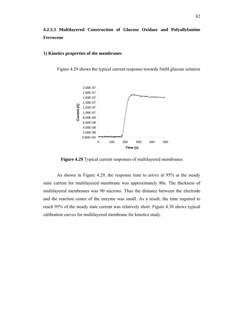
82
4.2.3.3 Multilayered Construction of Glucose Oxidase and Polyallylamine
Ferrocene
1) Kinetics properties of the membranes
Figure 4.29 shows the typical current response towards 5mM glucose solution
0.00E+002.00E-084.00E-086.00E-088.00E-081.00E-07
1.20E-071.40E-071.60E-071.80E-072.00E-07
0 100 200 300 400 500
Time (s)
Cur
rent
(A)
Figure 4.29 Typical current responses of multilayered membranes
As shown in Figure 4.29, the response time to arrive at 95% at the steady
state current for multilayered membrane was approximately 80s. The thickness of
multilayered membranes was 90 microns. Thus the distance between the electrode
and the reaction center of the enzyme was small. As a result, the time required to
reach 95% of the steady state current was relatively short. Figure 4.30 shows typical
calibration curves for multilayered membrane for kinetics study.

83
Figure 4.30 Typical calibration curves for multilayered membranes
The ferrocene functioned well as mediator, shuttling electrons between GOD
and the platinum electrode. The current increased stepwise with addition of glucose.
Michaelis-Menten kinetics were determined from steady state currents and the
electrochemical version of the Lineweaver-Burk equation. Figure 4.31 shows the
Lineweaver Burke plot for this membrane for enzyme kinetics study.
y = 8.4185x + 4.707R2 = 0.9951
0.00
1.00
2.00
3.00
4.00
5.00
6.00
7.00
0 0.05 0.1 0.15 0.2 0.25
1/ [Glucose] (1/mM)
1/ C
urre
nt (u
A)
Figure 4.31 Double –reciprocal (Lineweaver Burke) plot of multilayered membranes
The corresponding maximum current, Imax, and apparent Michaelis-Menten
constant, Kmapp for membranes were 0.21µA and 1.79mM respectively. The Km
app
obtained was very low. The high sensitivity and the small Kmapp mean that the
immobilized glucose oxidase possessed a higher enzymatic activity. Kmapp is
independent of enzyme concentration. The Kmapp value depends on the strength of the
0.0 2.5 5.0 7.5 10.0 12.5 15.0 17.50.00
0.05
0.10
0.15
0.20
Glucose (mM)
Cur
rent
(µA)

84
bonds between enzyme and substrate. If these bonds are strong, the Kmapp will be
low, indicating that the immobilized enzyme retained its bioactivity and possessed
high biological affinity to glucose. The high degree of affinity of the enzyme to the
substrate may be explained by a favorable change in the structural organization of the
enzyme due to the immobilization procedure (Arica et al., 1995).

85
CHAPTER 5
CONCLUSION AND RECOMMENDATIONS
5.1 Hydrogen Peroxide-Based Glucose Biosensor
For the selection of a suitable support material for the immobilization of
glucose oxidase, four types of matrixes had been investigated. The selection was
mainly based on the current response, enzyme leakage, long term stability, kinetic
parameter, response time and sensitivity. Chemically cross-linked PVA-GOD
membrane has shown the shortest leaking period, which indicated that the ability in
retaining the enzyme within the matrix was high. The apparent Michaelis-Menten
constant, Kmapp, shown was the highest too. However, its current response as well as
sensitivity were very much lower compared to the others even when lysozyme was
incorporated as a stabilizing agent. On the other hand, even though freeze-thawed
PVA-GOD membrane leaked for a considerable longer period, approximately 2
weeks, it showed satisfactory current response. Besides, even though its Kmapp was
low, it was still acceptable especially when combined with an appropriate external
membrane. Both types of organic-inorganic sol-gel derived materials,
tetramethoxysilane (TMOS), and alumina were brittle and PVA was added during
membrane fabrication in order to increase the mechanical strength. The enzyme
leakage was a problem with the mild immobilization condition and Kmapp was lower
than freeze-thawed PVA membrane. However, both of them had shown good
sensitivity and shorter response time as compared to PVA. In general, the
86
membranes were stable over a period of 60days. With an adequate level of
sensitivity and Kmapp, freeze-thawed PVA-GOD membrane has been chosen as the
support material for GOD immobilization. The enzyme leakage of this type of
membrane has been overcome by reducing the enzyme loading. Moreover, the Kmapp
can still be improved with a proper outer membrane.
To eliminate the interference suffered by hydrogen peroxide-based biosensor,
an inner membrane fabricated from poly(hydroxyethyl methacrylate) (pHEMA) was
studied to restrict the passage of acetaminophen, a model of an interfering molecule.
This was done by controlling the mesh size of the resulted network. A pHEMA
membrane with the cross-linking ratio of 0.043 was found to successfully obstruct
the diffusion of acetaminophen at a selectivity of 10. Significant reduction of
acetaminophen response was shown while maintaining reasonable diffusion of
hydrogen peroxide.
5.2 Mediator-Based Glucose Biosensor
For the first method, immobilization of glucose oxidase and ferrocene redox
polymer in CLPVA with the addition of BSA has been done. A membrane with
greater BSA content gave higher current response with larger Kmapp. For both
membranes, the large Km were obtained but with low current responses. However,
retention of enzyme and mediator as well as the membrane stability were very poor.
Therefore, further study must be done to improve the retention of enzyme and
mediator as the CLPVA, which had been shown to be an excellent retainer of GOD
(Abdul-Aziz, 2001) was not able to retain both GOD and ferrocene redox polymer
effectively. This would ultimately influence the stability of the membranes.
For the second method, immobilization of glucose oxidase and ferrocene
carboxylic acid in SGS-CLPVA/nafion was done. The immobilization technique
resulted in an enzyme/mediator membrane that was simple to cast, resulted in

87
minimal mediator losses and very stable at lower operating potentials. A membrane
with greater GOD concentration gave higher current response. However, Kmapp was
independent of enzyme concentration. Furthermore, the low value of Kmapp might
eventually decrease the detection limit of the biosensor. However, SGS-
CLPVA/nafion is a good matrix for the immobilization of mediator as well as an
enzyme. The co-operative effect from the hydroxyl groups of PVA and the sol–gel
environment is hoped to sustain the rotational freedom for the enzyme molecules to
adopt the active configuration typical under physiological conditions. The active
matrix environment prolongs the life span of the enzyme to result in high sensitivity.
The simultaneous presence of the sol–gel silica will greatly increase the selectivity
and stability of the sensors.
Lastly, for the third method, multilayered construction of glucose oxidase and
polyallylamine ferrocene has been done. Ferrocene was covalently bound to the
pollyalylamine by aldehyde and amino groups. Layer-by-layer covalent attachment
of periodate-oxidized glucose oxidase and poly(allylamine) ferrocene complex
(PAA-Fc) did not allow the loss of ferrocene into solution. However, the current
response and the Km were quite low. Thus the low value of Kmapp might eventually
decrease the detection limit of the biosensor.
After comparing the three almost totally different approaches, the first
method, which was the immobilization of glucose oxidase and ferrocene redox
polymer in CLPVA with the addition of BSA was selected for the fabrication of
disposable glucose biosensor, based on the Km value. Although the current response
was low, the Km value obtained for this method was very high compare to the other
method. By attaching the developed nafion external layer to the enzyme layer, this
mediated glucose sensor will provide good responses over a wide range of
concentration. However, implantable glucose biosensor could not be constructed
using this method since the mediator and enzyme retention as well as the stability
was not very good. But, the leaking was not a problem for disposable glucose sensor.
In the future, extensive study should be done on the external layer. Instead of
using nafion, as in this study, many other polymer coatings can be use as an outer
layer, such as polyurethane, polyvinylchloride, polycarbonates and cellulose acetate

88
coating solutions. By increasing the polymer coating solution concentrations, the
linear range of the sensor response can be extended (Yang et al., 1998). Therefore,
hopefully, with the new external layer, Km value for the second and the third method
could be improved. It is important since those two methods display better membranes
stability and mediator retention.
5.3 Recommendations
For hydrogen peroxide-based glucose biosensor, it is recommended that a
more complete model should be investigated on each material including the
optimization of each component’s concentration in a membrane, such as the cross-
linker and solvents used in membrane casting. Besides, a proper study on optimum
enzyme loading for each type of membrane should be performed to overcome
enzyme leakage problem. On the other hand, other types of outer membrane such as
polycarbonate should be investigated in the effort to enhance the kinetic parameters
of the immobilized enzyme.
One factor that can be studied in term of immobilization is diffusional
limitation in immobilized enzyme systems. Diffusional resistances may be observed
at different levels in immobilized enzyme. These resistances vary depending on the
nature of the support material, hydrodynamical conditions surrounding the support
material and the distribution of the enzyme inside or on the surface of the material.
Whether diffusion resistance has a significant effect on the rate of enzymatic reaction
depends on the relative rate of the reaction rate and diffusion rate, which is
characterized by the Damkohler number (Da).
Da= maximun rate of reaction / maximun rate of diffusion
= Vm / kL [Sb]
where kL is the mass transfer coefficient (cm/s) and [Sb] is the substrate concentration
in bulk liquid (g/cm3).

89
The rate of enzymatic conversion may be limited by diffusion of the substrate
or reaction, depending on the value of the Damkohler number. If Da >> 1, the
diffusion rate is limiting. For Da << 1, the reaction rate is limiting, and for Da ~ 1,
the diffusion and reaction resistances are comparable. Diffusion and enzymatic
reactions may be simultaneous, with the enzymes entrapped in a solid matrix, or may
be two consecutive phenomena for adsorbed enzymes.

90
REFERENCES
Albery, W. J. and Cranston, D. H. (1987). In: Turner, A.P.F., Karube, I. and Wilson,
G.S. eds. Biosensors: Fundamentals and Applications. Oxford: Oxford
University Press. 180-210.
Arica, M. Y., Hasirci, V. and Aleeddonoglu, N. (1995). Covalent Immobilization of
a-Amylase onto pHEMA Microsperes; Preparation and Application to Fixed
Bed Reactor. Biomaterials. 15: 761-768.
Audebert, P., Demaille, C. and Sanchez, C. (1993). Electrochemical Probing of The
Activity of GOD Embedded Sol Gel Matrixes. Chemistry of Materials. 5(7):
911- 913
Azila Abdul Aziz (2001). Amperometric Glucose Biosensors: Systematic Material
Selection and Qualitative Analysis of Performance. The John Hopkins
University, Baltimore, Maryland: Ph.D. Thesis.
Bartlett, P. N. (1987). The Use of Electrochemical Methods in The Study of
Modified Electrodes. In: Turner, A.P.F., Karube, I. and Wilson, G.S. eds.
Biosensors: Fundamentals and Applications. Oxford: Oxford University
Press. 211-146.
Bartlett, P. N., Tebbutt, P. and Whitaker, R.C. (1991). Prog. Reaction Kinetics. 16: 55-155.

91
Boguslavsky, L., Kalash, H., Xu, Z., Beckles, D., Geng, L, Skotheim, J.,
Laurinavicius, V., Lee, H.S. (1995). Anal. Chim. Acta.311: 15.
Brahim, S., Narinesingh, D., and Guiseppi-Elie, A. (2002). Polypyrrole-hydrogel
Composites for the Construction of Clinically Important Biosensors.
Biosensors and Bioelectronics. 17: 53-59.
Brooks, S. L., Ashby, R. E., Turner, A. P. F., Calder, M. R. and Clarke, D. I. 1984. Development of an On-line Glucose Sensor for Fermentation Monitor. Biosensors. 3: 45-56
Cajlakovic, M., Lobnik, A. and Werner, T. (2001). Stability of New Optical pH Sensing Material Based on Crosslinked Poly(vinyl alcohol) Coploymer. Analytica Chimica Acta. 207-213.
Calvo, E. J., Danilowicz, C. and Diaz, L. (1994). J. Electroanal. Chem. 369: 279-282;
Calvo. E.J. and Danilowicz C. (1997). Amperometric Enzyme Electrodes. Journal of The Brazilian Chemical Society. 6: 563-574.
Calvo, E. J., Etchenique, R., Pietrasantha, L. and Wolosiuk, A. (2001). Layer by layer Self Assembly of Glucose Oxidase and Os(Bpy)2CIPyCH2NH-polyallylamine Bioelectrode. Anal. Chem. 73: 1161-1168.
Chaplin, M. (1990). Enzyme Technology. United Kingdom: Cambrigde University
Press.
Chen, X. H., Hu, Y. B., and Wilson G. S. (2002). Glucose Microbiosensor Based on
Alumina Sol-gel Matrix/Electropolymerized Composite Membrane.
Biosensors & Bioelectronics. 17: 1005-1013.
Clark, L.C., Lyons, C. (1962). Electrode Systems for Continuous Monitoring in
Cardiovascular Surgery. Ann. New York Acad. Sci. 102: 29–45.
Degani, Y. and Heller, A. (1987). J. Phys. Chem. 91: 1285-1289.

92
Doretti, L., Ferrara, D., Gattolin, P., and Lora, S. (1997). Amperometric Biosensor
with Physically Immobilized Glucose Oxidase on a PVA Cryogel Membrane.
Talanta. 44: 859-866.
Doretti, L., Ferrara, D., Gattolin, P., and Lora, S., Schiavon, F. and Veronese, F. M.
(1998). PEG-Modified Glucose Oxidase Immobilized on a PVA Cryogel
Membrane for Amperometric Biosensor Applications. Talanta. 45: 891-898.
Eggins, B. R. (1996). Biosensors: An Introduction. England: John Wiley & Sons Ltd
and B.G. Teubner.
Griesmar, P., Ponton, A., Serfaty, S., Senouci, B., Gindre, M., Gouedard, G., Warlus,
S. (2003). Kinetic study of silicon alkoxides gelation by acoustic and
rheology investigations. Journal of Non-Crystalline Solids. 319: 57–64.
Gudeman, L.F., and Peppas N.A. (1995). Journal of Membrane Science. 107: 239.
Hartmeier, W. (1986). Immobilized Biocatalyst, An Introduction. New York:
Springer-Verlag Berlin Heidelberg. 3-68.
Henning, T. P. and Cunningham, D. D. (1998). Biosensor for Personal Diabetes
Management. In: Ramsay, G. ed. Commercial Biosensors: Application to
Clinical, Bioprocess and Environment Sample. Canada: John Wiley & Sons.
5.
Hickey, A. S. and Peppas N. A. (1995). Mesh Size and Diffusive Characteristics of
Semicrystalline Poly(vinyl alcohol) Membranes Prepared by
Freezing/Thawing Techniques. Journal of Membrane Science. 107: 229-237.
Hodak, J., Etchenique, R., Calvo, E.J., Singhal, K., Bartlett, P.N. (1997). Layer-by-
layer Self-assembly of Glucose Oxidase with a Poly(allylamine)ferrocene
Redox Mediator. Langmuir. 13: 2708–2716.

93
Horner R.D. (1997). Biochemistry Laboratory Workbook. Dept. of Biology, Johns
Hopkins University. Unpublished.
Howells, A. R., Zambrano, P. J,.and Collison, M. M. (2000). Diffusion of Redox
Probes in Hydrated Sol-Gel-Derived Glasses. Analytical Chemistry. 72:
5265-5271.
Hsiue G. H., Guu, J. A., Cheng, C. C. (2001). Poly(2-hydroxyethyl methacrylate)
film as a drug delivery system for pilocarpine. Biomaterials. 22: 1763-1769.
Johnson, T. J. .A. (1993). Glutaraldehyde Cross-Linking: Fast and Slow Modes.
Forth Chemical Congress of North America. New York: American Chemical
Society. 283-295
Kase, Y. and Muguruma, H. (2004). Amperometric Glucose Biosensor Based on
Mediated Electron Transfer between Immobilized Glucose Oxidase and
Plasma-polymerized Thin Film of Dimethylaminomethylferrocene on
Sputtered Gold Electrode. Analytical Sciences. 20: 1143-1146.
Katakis, I. and Heller, A. (1992). Anal. Chem. 64: 1008-1013.
Kermis, H.R., Govind Rao, Timothy A. Barbari (2003). Transport Properties of
PHEMA Membranes for Optical Glucose Affinity Sensors. Journal of
Membrane Science. 212: 75-86.
Koide, S., and Yokoyama, K. (1999). Electrochemical Characterization of an
Enzyme Electrode Based on a Ferrocene Containing Redox Polymer. Journal
of Electroanalytical Chemistry. 468: 193-201.
Kudela (1976). V. Hydrogels. In: Jacqueline IK, eds (1976). Encyclopedia of
Polymer Science and Engineering. Wiley Interscience. 783-807.

94
Kumar, H. V., and Chaudhari. A. Proteins Immobilized at the Galleries of Layered
α-Zirconium Phosphate: Structure and Activity Studies. University of
Connecticut.
Lee, Y. H, The, H. F., Loh, H. C., and Musa, A. (2003). A Hydrogen Ion-Selective
Sensor Based on Non-plasticised Methacrylic-acrylic Membranes. Sensors. 2:
339-346.
Lillis, B., Grogan, C., Berney, H., and Lane, W.A. (2000). “Investigation into
Immobilization of Lactate Oxidase to Improve Stability.” Sensors and
Actuators B. 68: 109-114.
Liu, Y., Zhang X., Liu, H. and Deng, J. (1996). Immobilization of Glucose Oxidase
onto the Blend Membrane of Poly(vinyl alcohol) and Regenerated Silk
Fibroin: Morphology and Application to Glucose Biosensor. Journal of
Biotechnology. 46: 131-138.
Liu, Z. J., Liu, B. H., Zhang, M., Kong, J. L., and Deng, J. Q. (1999). Al2O3 Sol-gel
Derived Amperometric Biosensor for Glucose. Analytica Chimica Acta. 392:
135-141.
Lyons, M. E. G. (1991). Electrochemistry-developing Technologies and Application.
Annu. Rep. Prog. Chem. Sect. C. 88: 135.
Miller, V. G. (2003). Diabetes Mellitus: Care & Control-A review and current
update.
Newman, J. D., Turner, A. P. .F. (2005). Home Blood Glucose Biosensors: A
Commercial Perspective. Biosensors and Bioelectronics. 20: 2435-2453.
Niu, J. and Lee, J. L. (2002). Reagentless Mediated Biosensors Based on
Polyelectrolyte and Sol-gel Derived Silica Matrix. Sensors and Actuaturs B.
82: 250-258.

95
Peppas NA, Moynihan HJ, Lucht LM (1985). The Structure of Highly Crosslinked
Poly(2-hydroxyethyl methacrylate) Hydrogels. J Biomed Mater Res. 19: 397-
411.
Poyard, S., Jaffrezic-Renault, N., Martelet, C., Cosnier, S., and Labbe, P. (1998).
Optimization of Inorganic/Bio-organic Matrix for the Development of New
Glucose Biosensor Membranes. Analytica Chimica Acta. 364: 165-172.
Reynolds, E. R., Geise, J. R. and Yacynych, A. M. (1992). In : Biosensor &
Chemical Sensors : Optimization Performance Through Polymeric Material.
Edelman, P. G and Joseph Wang. USA: Library of Congress. 106.
Rondeau, A., Larson, N., Boujtita, M., Gorton, L. and Murr, N. E. (1999). The
Synergetic Effect of Redox Mediators and Peroxidase in Bienzymatic
Biosensor for Glucose Assays in FIA. Analusis. 27:649-656.
Saburo Fukui, and Atfuo Tanaka (1990). Immobilization. In: Biocatalyst Design.
New York: VCH Publisher. 63-74.
Sampath, S., and Lev, O. (1996). Inert Metal-modified, Composite Ceramic-carbon,
Amperometric Biosensors: Renewable, Controlled Reactive Layer. Analytical
Chemistry. 68:2015-2021.
Sapsford, K. E., and Ligler, F. S. (2004). Real-time Analysis of Protein Adsorption
to a Variety of Thin Films. Biosensors and Bioelectronics. 19: 1045–1055.
Sato, N. and Okuma, H. (2006). Amperometric Simultaneous Sensing System for D-glucose and L-lactate Based On Enzyme-modified Bilayer Electrodes. Analytica Chimica Acta. 565: 250-254.
Seidel, J. M., and Malmonge, S. M. (2000). Synthesis of PolyHEMA for using as
Biomaterials. Bulk and Solution Radical-initiated Polymerization
Techniques. Materials Research. 3: 79-83.
96
Shao, Z. G., Wang, X. and Hsing, I. M. (2002). Composite Nafio/Poly (vinyl
Alcohol) Membranes for the Direct Methanol Fuel Cell. Journal of
Membrane Science. 210: 147-153.
Shuler, M. L. and Kargi, F. (2002). Bioprocess Engineering Basic Concepts. 2nd ed.
USA: Prentice Hall.
Sirat, D. M., Velhot, G., and Reach, G. (1992). Biosensors and Bioelectronics. 7:
345.
Taylor, R. F. (1991). Introduction: The Current Status of Immobilized Protein
Technology. In: Protein Immobilization. Volume 14. New York: Marcel
Dekker, Inc. 1-9.
Tian, F.M. and Zhu, G.Y. (2002). Sol-gel Derived Iridium Composite Glucose
Biosensor. Sensors and Actuators B. 86: 266-270.
Updike, S. J. and Hicks, G. P. (1967). The Enzyme Electrode. Nature. 214: 968-988.
Von Worthington ed. (1972). Worthington Enzyme Manual: Enzymes, Enzyme
Reagents, and Related Biochemicals. Freehold, N.J.: Worthington
Biochemical Corporation.
Wang, J., and Pamidi, P.V.A. (1997). Sol-gel Derived Metal Dispersed Carbon
Composite Amperometric Biosensors. Electroanalysis. 9: 52-55.
Wang, J., Rivas, D., and Chicharro M. (1997). Glucose Microsensor Based on
Electrochemical Deposition of Iridium and Glucose Oxidase onto Carbon
Fiber Electrodes. Journal of Electroanalytical Chemistry. 439: 55-61.
Wei, Q., Wang, D. W., Zhang, S. G., and Chen, C. S. (2001). Preparation and
Characterization of Sol-gel Derived Unsupported Al2O3-SiO2 Composite
Membrane. Journal of Alloys and Compounds. 325: 223-229.

97
Wichterle O, and Lim D (1960). Hydrophilic Gels for Biological Use. Nature. 185:
117.
Wilkins, E. and Atanasov, P. (1996). Glucose Monitoring: State of the Art and
Future Possibilities. Med. Eng. P/y. 18:.273-288.
Wilson, R. and Turner, A. P. F. (1992). Glucose Oxidase – and Ideal Enzyme.
Biosensor and Bioelectronics. 7:162-185.
Wolfbeis, O. S., Ines Oehme, Natalya Papkovskaya 1, Ingo Klimant (2000). Sol–gel
Based Glucose Biosensors Employing Optical Oxygen Transducers, and a
Method for Compensating for Variable Oxygen Background. Biosensors &
Bioelectronics. 15: 69–76.
Wu, J., Jan Suls, and Willy Sansen (1999). Amperometric Glucose Sensor with
Enzyme Covalently Immobilized by Sol-Gel Technology. Analytical
Sciences. 15: 1029-1032.
Yoldas, B. E. (1975). Alumina Sol Preparation from Alkoxides. Ceramic Bulletin 54:
289-290.
Yoon, H. C. and Kim, H. S. (2000a). Multilayered Assembly of Dendrimers With
Enzymes on Gold: Thickness-Controlled Biosensing Interface. Anal. Chem.
72:922-826.
Yoon, H. C., Hong, M. Y. and Kim, H.S. (2000b). Functionalization of a
Polyamidoamine Dendrimer with Ferrocenyls and its Application to the
Construction of a Reagentless Enzyme Electrode. Anal. Chem. 72: 4420-4427
Zaborsky, O. R. and Ogletree, J. (1974). The Immobilization of Glucose Oxidase via
Activation of its Carbohydtrate Residues. Biochem. Biophys. Res. Commun.
61: 210-216.
98
Zhang, S., Yang, W., Niu, Y. and Sun, Y. (2004). Multilayered Construction of
Glucose Oxidase and Poly(allylamine)ferrocene on Gold Electrodes by
Means of Layer-by-layer Covalent Attachment. Sensors and Actuaturs B.
101: 387-393.


