

X-ray Microtomography
National School on Neutron and X-ray Scattering August 14-28 2005
Group D
Jiong Hua
Vaibhav Kohli
Soonjoo Seo
Lu Zou
Paul J. Sideris

X-ray Computed Microtomography
Imaging tool using high flux at photon energies up to 100 keV. APS bending magnet source: 20 keV
High resolution, 3D images of various porous media
Characterization of fluid phases
Transport of multiphase flow

Why Microtomography?
Non-Invasive examination of the internal structure of objects
Meteorites, fossils valuable
Interplanetary dust particles, soil aggregates fragile
Diamonds, hard minerals time consuming
Studies of pore network : pore distribution, size, topography,
connectivity
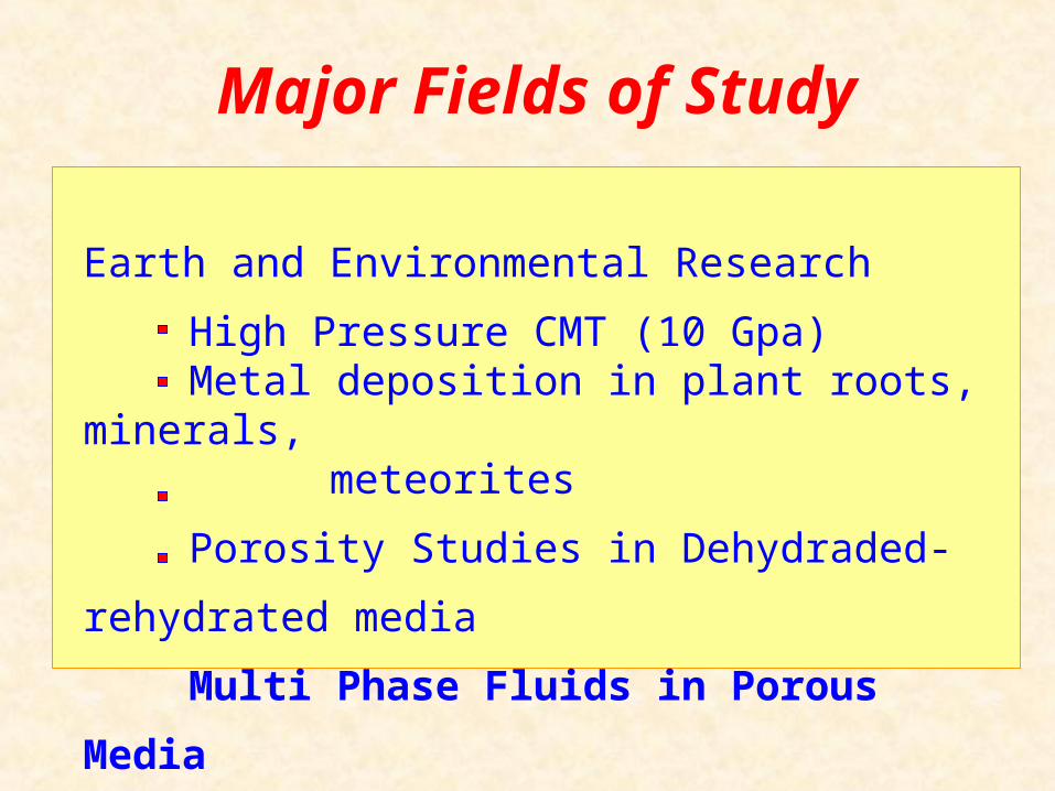
Major Fields of Study
Earth and Environmental Research
High Pressure CMT (10 Gpa) Metal deposition in plant roots, minerals,
meteorites
Porosity Studies in Dehydraded-rehydrated media
Multi Phase Fluids in Porous Media

Attenuation of X-rays
Greater Attenuation
X-rays Sample
x
I = I0e-x
I0: Incoming intensity of X-rayI : Outgoing intensity of X-ray: linear X-ray attenuation
xI = I0e-(kxk..
X-ray Computed Microtomography (CMT) gives 3D images of the X-ray
attenuation coefficient .

Experimental Setup
Microscope Objective
CCD Camera
Rotation Stage
Sample
x Translation Stage
YAG Scintillator
X-rays
Visible LightMirror
Microtomography at APS Beamline 13-BMVarious stages align the
sample with the X-ray beam and CCD camera
YAG converts X-rays to visible light for imaging

System Under Investigation
Material
Silica beads pack in a cylindrical tube
Two different fluids containing I and Cs
Goal of Study
What ions exist in the fluid?
Where are the two types of liquids located?
What are their domain sizes and distributions
within the tube?

Data Processing – Image Reconstruction
I’ (x’, y’, Θ)
I (x, y, z)

Data Processing – X-ray AbsorptionI absorption edge Cs absorption edge

Conclusions
• CMT allows the identification of Soltrol® and aqueous CsCl in porous media.
• Aqueous CsCl is found in smaller amounts around inner wall of tube.
• X-ray tomography is a powerful and efficient tool for studying multiphase fluids in solids.

Acknowledgements
National School of Neutron and X-ray Scattering
DOE
Mark Rivers - University of Chicago, APS 13-BM Clinton Willson - Louisiana State University


