
lable at ScienceDirect
International Biodeterioration & Biodegradation 65 (2011) 961e971
Contents lists avai
International Biodeterioration & Biodegradation
journal homepage: www.elsevier .com/locate/ ibiod
Diversity of culturable sodium dodecyl sulfate (SDS) degrading bacteria isolatedfrom detergent contaminated ponds situated in Varanasi city, India
Venkatesh Chaturvedi*, Ashok KumarSchool of Biotechnology, Banaras Hindu University, Varanasi 221 005, India
a r t i c l e i n f o
Article history:Received 2 February 2011Received in revised form4 July 2011Accepted 4 July 2011Available online 3 September 2011
Keywords:SDSBioremediationAlkyl sulfataseARDRAERIC-PCR16S rDNA sequencing
* Corresponding author. Tel.: þ91 542 2368331; faxE-mail address: venkatesh_chaturvedi@rediffmail.
0964-8305/$ e see front matter � 2011 Elsevier Ltd.doi:10.1016/j.ibiod.2011.07.005
a b s t r a c t
In the present investigation, an attempt has been made to isolate and identify SDS-degrading bacteriafrom different detergent contaminated ponds situated in Varanasi city, UP, India. Initial survey of pondsindicated that these ponds were contaminated with detergents. Employing enrichment technique inminimal medium (PBM) with SDS as a sole carbon source, a total of 24 isolates were recovered from7 detergent contaminated ponds. Studies on rates of SDS degradation indicated that the rate of SDSdegradation varied from 97.2% to 19.6% after 12h incubation under identical conditions. An estimation ofalkyl sulfatase activity indicated that the activity varied from 0.168 � 0.004 to 0.024 � 0.005 mmolSDS/mg protein/min. Molecular characterization of these isolates was performed on the basis of ARDRAand ERIC PCR, which indicated that these isolates were broadly divided in 8 groups. Some selectedisolates were identified on the basis of 16S rDNA sequencing. It was found that these isolates belonged toPseudomonas aeruginosa, Pseudomonas mendocina, Pseudomonas stutzeri, Pseudomonas alcaligenes, Pseu-domonas pseudoalcaligenes, Pseudomonas putida and Pseudomonas otitidis respectively. Among theseisolates P. aeruginosa, P. putida and P. otitidis have been previously shown to degrade and metabolize SDS,the rest of the isolates appear to be new.
� 2011 Elsevier Ltd. All rights reserved.
1. Introduction
In India, man-made ponds have been used as an alternate sourceof drinking water. However, these ponds are also employed forwashing of clothes and bathing purposes by washer men and localpeople (Prakash et al., 2009). Many ponds are situated in vicinity oftemples and are used for bathing purposes for people who visit thetemples for worshipping and also for disposal of wastes originatingfrom the temples (Sharma et al., 2009). In certain ponds, sewagewater and waste water from nearby cottage industries are alsodischarged. A detailed survey of few ponds located around the citywas made by Tyagi et al. (2006), which revealed that most of theponds are eutrophic. However, data related to amount of detergentspresent, if any in pond water is lacking. Furthermore, no attempthas been made to study detergent degrading bacteria. In a differentstudy, Ghose et al. (2009), reported the presence of high amount ofanionic detergents in surface and underground water in greaterKolkata, India.
Sodium dodecyl sulfate (SDS) is an anionic detergent widelyused in household products and in Industry (Karsa, 1992). It has
: þ91 542 2369693.com (V. Chaturvedi).
All rights reserved.
been reported that the presence of anionic detergent especially SDSin environment arises mainly from disposal of domestic andindustrial wastes (Fendinger et al., 1994). SDS is shown to be toxicto health and survival of aquatic animals (Ribelles et al., 1995;Rosety et al., 2001; Rocha et al., 2007). It was realized thatremoval of detergent from environment was a necessity (Singerand Tjeerdema, 1993). Certain bacteria such as Pseudomonas sp.,Bacillus cereus, Acinetobacter calcoaceticus and Pantoea agglomeranshave been isolated which utilize SDS as a carbon source for growth(Payne and Feisal,1963; Thomas andWhite,1989; Singh et al.,1998;Abboud et al., 2007). However, little if any, work has been done onmolecular diversity of SDS-degrading bacteria. Recent advances instudies pertaining to microbial ecology have led to the develop-ment of new approaches for the characterization of microbialcommunities (Zhou et al., 1997). Most common PCR-based genomicfingerprinting methods are Amplified Ribosomal DNA RestrictionAnalysis (ARDRA) (Porteous et al., 1994) and Repetitive (Rep) PCRi.e. Enterobacterial Repetitive Intergenic Consensus (ERIC)elements (Versalovic et al., 1991; Hulton et al., 1991). Although, PCRbased fingerprinting methods are suitable for characterization ofbacterial communities, however, these techniques cannot beemployed for the identification of bacteria (Ventura et al., 2001).16S rDNA sequence analysis is currently the most widely usedmethod for bacterial identification (Lawongsa et al., 2008). In the
V. Chaturvedi, A. Kumar / International Biodeterioration & Biodegradation 65 (2011) 961e971962
present study, an attempt has been made to survey various pondssituated in Varanasi city with a view to access the level of anionicdetergents in pond water. The next approach was to isolate andscreen the SDS-degrading bacteria present in pond water. Molec-ular diversity among various isolates was accessed by PCR basedtechniques such as ARDRA and ERIC-PCR. Identification of selectedisolates was performed on the basis of 16S rDNA sequencing.
2. Materials and methods
2.1. Chemicals and culture media
Sodium dodecyl sulfate (SDS), methylene blue and otherbiochemicals were purchased from SigmaeAldrich, MO, USA. Allother inorganic chemicals were of analytical grade and obtainedfrom SISCO Research Laboratories Pvt. Ltd. and HiMedia Pvt. Ltd.,Mumbai, India. Luria-Bertani (LB)medium contained (g/l): tryptone10.0, NaCl 5.0, yeast extract 5.0, pH 7.0. The Phosphate BufferedMedium (PBM) contained (g/l): K2HPO4 1.0, KH2PO4 1.0, NH4Cl 1.0,MgSO4.7H2O 0.20, NaCl 0.5 and CaCl2 0.02, pH 7.5. The medium alsocontained trace elements (1 ml of stock) having (g/l): FeCl3$6H2O0.24, CoCl2$6H2O 0.04, CuSO4$5H2O 0.06, MnCl2$4H2O 0.03,ZnSO4$7H2O 0.31 and Na2MoO4$2H2O 0.03. SDS (1 g/l) was addedseparately after autoclaving the medium.
2.2. Study sites
Detailed survey of various ponds located around Varanasi city,revealed that seven ponds namely, Assi (AS), Sundarpur (SP), Nir-alanagar (NN), Naipura (N), Kandwa (K), Pisachmochan (PM) andJawaharnagar (JN) were extensively used for bathing and washingpurposes and received either sewage water or industrial effluentsor both. All these ponds were found to retain water throughout theyear. However, the level of water decreases significantly duringsummer season.
2.3. Estimation of anionic detergent from pond water
Amount of anionic detergent in water samples was determinedby the methylene blue active substance (MBAS) assay (Hayashi,1975). Calibration curve was prepared by using standard solu-tions (based on weight) of pure SDS (SigmaeAldrich, USA).
2.4. Enrichment and Isolation of SDS-degrading bacteria
For the enrichment of SDS-degrading bacteria, 1 ml of pondwater was added to 100 ml sterilized PBM supplemented with SDS(1 g/l) in a culture flask and incubated at 30 �C and 120 rpm ina shaker (Orbitek LT, Scigenics Bioteck. Pvt. Ltd., Chennai). After 3e4days of growth, 1 ml culture was transferred to fresh PBM supple-mentedwith SDS (1 g/l). Repeated sub-culturing (at least 3e4 times)resulted in the enrichment of putative SDS-degrading bacteria.Spreading on solid PBM agar-plates containing SDS as a sole carbonsource, resulted in the formation of minute colonies. Based oncolony morphology different SDS-degrading bacteria were isolated.
2.5. Test for SDS degradation
Briefly, overnight grown cultures in LB medium were centri-fuged at 8000 rpm for 5 min at room temperature, washed withPBM and suspended in 100 ml of PBM containing SDS. Initial OD(at 600 nm) of each isolate was adjusted to approximately 0.025.The cultures were incubated at 30 �C with shaking at 120 rpm.Samples from each flask were removed at desired intervals, andassayed for residual SDS and growth (Ellis et al., 2002).
2.6. Preparation of crude cell extracts
Crude cell extracts were prepared following the method of Elliset al. (2002). Bacterial cells grown in PBM containing SDS as a solesource of carbon were harvested after mid logarithmic phase ofgrowth by centrifugation at 10,000 rpm for 20 min at 4 �C ina Sorvall RC-5B superspeed refrigerated centrifuge (Du PontInstruments, USA). Cell pellets were washed twice with 10 mMTriseHCl by centrifugation and suspended in 10 mM TriseHCl (pH7.5). The cells were ruptured by sonication for a total duration of3 min, consisting of intermittent sonication for 30 s on and 30 s off,operated at level 5 and a 25% duty cycle in a Branson Sonifier 450(Branson Ultrasonics Corp., USA). To minimize heat inactivation ofenzymes, samples were sonicated after placing eppendorf tubes inice box. Cell debris was removed by centrifugation at 12,000 rpmfor 10 min at 4 �C. The cell extracts were stored at �20 �C prior toassay for alkyl sulfatase activity.
2.7. Assay of alkyl sulfatase
Alkyl sulfatase activity in crude cell extracts was estimated asper the method of Ellis et al. (2002). Briefly, crude cell extract(10e50 ml) were incubated at 30 �C in 1 ml of 10 mM TriseHCl (pH7.5) containing SDS at a final concentration of 50 mg/ml. Samples(100 ml) were removed at fixed intervals and assayed for residualSDS by MBAS method as described earlier. One unit of enzymeactivity was defined as the amount of enzyme which converted1 mmol of SDS per minute under the assay conditions. The experi-ments were performed in triplicate and result was presented asmean � standard deviation.
2.8. Biochemical characterization of SDS-degrading isolates
All isolates were characterized by conventional biochemicaltests, i.e. Gram stain, motility, pigment production, growth, pres-ence of enzymes such as catalase, oxidase, urease, nitrate reductase,gelatin and starch hydrolysis (Shaw and Latty, 1982). Utilization ofcarbohydrates, amino acids was tested in PBM as described by(Ka€mpfer et al., 1991; Molin and Ternstrom, 1982).
2.9. Isolation of genomic DNA
Genomic DNA of all the isolates was extracted by DNeasy TissueKit (Qiagen, Gmbh, Hilden, Germany) according to the instructionsof manufacturer. The genomic DNA was electrophoresed on a 0.8%agarose gel in TAE buffer (40 mM Tris-acetate, 1 mM EDTA, pH-8.0)containing ethidium bromide (0.5 mg/ml) and visualized in a geldocumentation unit (Bio-Rad Laboratories, USA).
2.10. Amplification of full length 16S rDNA
16S rDNA (1.5 kb) was amplified using universal primer pair For50-AGA GTT TGA TYM TGG CTC AG -30, and Rev 50-CTA CGG CTA CCTTGT TAC GA -30. Amplification was performed in a final volume of50 ml. The PCR reaction mix included; 1.5 U of Taq DNA polymerase(Bangalore Genei), 1� PCR buffer with 1.5 mM MgCl2, 300 ng ofeach forward and reverse primers (Integrated DNA Technologies,USA), 125 mM of each dNTP’s (Bangalore Genei, India) and 50 ngtemplate DNA. Thermal cycle for the amplificationwas set as; 3minat 95 �C, 30 cycles of 1 min at 94 �C, 1 min at 50 �C and 2 min at72 �C followed by 5 min at 72 �C and storage at 4 �C. Amplificationwas performed in a PTC-100 Thermal Cyclar (MJ Research, Inc.,Walton, USA). The samples were analyzed by agarose (1.5%) gelelectrophoresis and visualized on gel documentation unit (Bio-RadLaborotories, USA).
V. Chaturvedi, A. Kumar / International Biodeterioration & Biodegradation 65 (2011) 961e971 963
2.11. Amplification of 16S rDNA (426 bp)
426 bp long 16S rDNA partial fragment was amplified usinguniversal primer pair; For 50-ACT GGC GGA CGG GTC AGT AA -30,and Rev 50-CGT ATT ACC GCG GCT GCT GG -30. Amplification wasperformed in a final volume of 50 ml. The PCR reaction mix included1.5 U of TaqDNA polymerase (Bangalore Genei), 1� PCR assay bufferwith 1.5 mM MgCl2, 300 ng of each forward and reverse primers(Integrated DNA Technologies, USA), 125 mM of each dNTP’s (Ban-galore Genei) and 50 ng template DNA. Thermal cycle for theamplification was set as; 3 min at 95 �C, 30 cycles of 1 min at 94 �C,1min at 50 �C and 1min 10 s at 72 �C followed by 5min at 72 �C andstorage at 4 �C. The amplified products were analyzed by agarose(3%) gel electrophoresis.
2.12. Amplified Ribosomal DNA Restriction Analysis (ARDRA)
16S rDNA gene (1.5 kb) amplified by PCR was subjected torestriction digestion using Taq1 and Rsa1 restriction enzymes, as perthe instructions of themanufacturer (New England Biolabs Ltd, UK).Restriction digestion was done in a final volume of 25 ml containing1� restriction enzyme buffer, 0.125 ml (1.25U) restriction enzymeand 15 ml PCR product. After mixing the reaction mixture carefully,samples were incubated for 3 h in a water bath preset at 37 �C.Reaction was terminated by heat inactivation at 70 �C for 20 min.The samples were analyzed by agarose (3%) gel electrophoresis andvisualized on gel documentation unit (Bio-Rad Laborotories, USA).
2.13. ERIC (Enterobacterial Repetitive Intergeneric Consensus) PCR
ERIC-PCRwas carried out in 40 ml reactionmixture containing,1�Taq DNA polymerase assay buffer, 125 mM each of dNTPs, 250 ng ofeachprimer (ERIC 1-50- ATGTAAGCTCCTGGGGATTCAC-30 andERIC2-50- AAGTAAGTGACTGGGGTGAGCG-30),1U TaqDNA polymerase(Bangalore Genie, India) and 50 ng of template DNA. Amplification ofinter-rep elements was done using thermal program set as initialdenaturation at 95 �C for 3min, followedby 35 cycles of denaturationat 94 �C for 1min, annealing at 52 �C for 1min and extension at 72 �Cfor 1 min 30s and a final extension at 72 �C for 5 min. The reactionmixture was stored at 4 �C until use. The amplified products wereanalyzed by agarose (3%) gel electrophoresis.
2.14. Similarity analysis and clustering
Data of biochemical characterization of SDS-degrading isolates,restriction pattern of 16S rDNA and ERIC-PCR pattern of all the SDS-degrading isolates was used for the construction of dendogramwith the help of NTSYSpc software 2.11a (Exeter Software, USA)(Rohlf, 1997). Every positive test of biochemical data was scored as1 and negative test was scored as 0. Similarly, the bands arisingfrom restriction digestion and those arising from amplification as inERIC-PCR were scored for each isolate as 1 (band present) or0 (band absent). A similarity matrix was generated from the pooledbinary data using SIMQUAL module of the NTSYSpc software. Dicecoefficient was used to derive similarity among isolates.
The similarity matrix thus generated was used for clusteranalysis by unweighted pair group method of arithmetic average(UPGMA) using sequential, agglomerative, hierarchial, nestedclustering module of NTSYSpc Software. The output data weregraphically presented as a phenetic tree.
2.15. 16S rDNA sequencing and identification of bacteria
Amplified full length (1.5 kb) and partial (426 bp) 16S rDNAfragments were purified by Invitrogen kit (Invitrogen Corp., USA)
following the instructions of manufacturer. The PCR product waseluted in Milli Q water and purity and concentration of DNA werechecked by reading OD at 230, 260 and 280 nm. The sequencingPCR reaction mix (DNA sequencing kit, Applied Biosystems, USA)included 8.0 mL Bigdye Terminator V3.0 cycle sequencing readyreaction mixture with ApliTaq, 10 ng purified PCR product and 3.2pmole forward primer (50-ACT GGC GGA CGGGTC AGTAA -30). Totalvolume was adjusted to 20 ml by adding deionised water. Thethermal cycle conditions were set as; initial denaturation at 98 �Cfor 1 min, 25 cycles of denaturation at 96 �C for 10 s, annealing at55 �C for 5s and extension at 60 �C for 4 min and hold at 4 �C.Ethanol precipitation was done for the removal of unused BigDyeTerminator present in the reaction mixture. After drying thesamples, 20 ml of TSR (template suppression reagent) was added,mixed well and heated for 2 min at 95 �C. Subsequently, thesamples were chilled on ice and after vortexing thoroughly,centrifuged briefly in a microcentrifuge. Samples were hold in iceuntil loading. Sequencing was performed in an ABI-PRISM 310Genetic Analyzer (Applied Biosystems, USA). PCR and directsequencing were performed at least twice to determine andconfirm the DNA sequences for each isolate. 16S rDNA sequencingwith partial 16S rDNA fragment (426 bp) was performed in fourisolates namely SDS1, SDS2, K1, and JN2. In 15 selected isolates, fulllength (1.5 kb) 16S rDNA fragment was directly sequenced. Thenucleotide sequences obtained from partial 16S rDNA fragmentwere in the range of 201e397 bp and sequences obtained from fulllength 16S rDNA fragment were in the range of 795e956 bp.
2.16. Nucleotide sequence accession numbers
Full length (1.5 kb) 16S rDNA fragments were directly sequencedusing forward primer. The nucleotide sequences obtained from fulllength 16S rDNA fragment were in the range of 795e956 bp. All thesequences were matched against nucleotide sequences present inGen Bank using BLASTn program (Altschul et al., 1990). Thenucleotide sequences of partial 16S rDNA of all the isolates weresubmitted to NCBI and accession numbers for all the strains havebeen obtained (Table 3).
2.17. Construction of phylogenetic trees based on 16SrDNA sequences
16S rDNA sequences of selected 15 SDS-degrading isolates and37 representative sequences of reference strains from NCB1 data-base were used to construct phylogenetic tree. The sequencesbelonging to isolates SDS1, SDS2, K1, and JN2 were very small ascompared to rest of the 15 isolates, so, these sequences were notincluded in the phylogenetic tree. Multiple alignment of sequenceswere carried out using ClustalW2 (version 2.0.10). Neighbor joiningtree was constructed in MEGA 4.0 (Saitou and Nie, 1987) usingbootstrapping at 1000 bootstraps trials with the two- parametersmodel of Kimura.
3. Results
3.1. Concentration of anionic detergent in pond water
Initial survey showed that due to human activities, the pondswere highly contaminated. It was desirable to study amount ofanionic detergent in pond water. From the data of Table 1, it isevident that amount of anionic detergent was at higher level in allthe ponds studied. The level of anionic detergent varied from 5.5 to9.5 mg/l. The highest level was observed in pond situated in Assi(9.5 mg/l) and lowest amount was observed in pond situated inNaipura (5.5 mg/l).
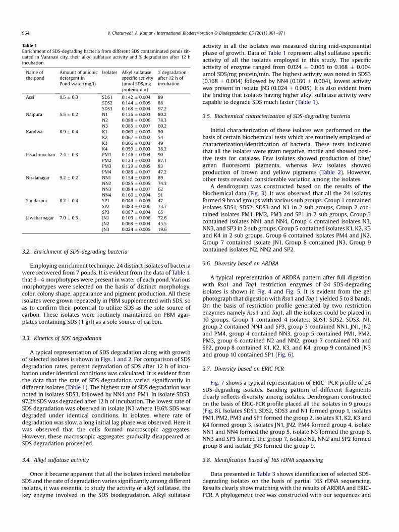
Table 1Enrichment of SDS-degrading bacteria from different SDS contaminated ponds sit-uated in Varanasi city, their alkyl sulfatase activity and % degradation after 12 hincubation.
Name ofthe pond
Amount of anionicdetergent inPond water(mg/l)
Isolates Alkyl sulfatasespecific activity(mmol SDS/mgprotein/min)
% degradationafter 12 h ofincubation
Assi 9.5 � 0.3 SDS1 0.142 � 0.004 89SDS2 0.144 � 0.005 88SDS3 0.168 � 0.004 97.2
Naipura 5.5 � 0.2 N1 0.136 � 0.003 80.2N2 0.088 � 0.006 78.3N3 0.085 � 0.007 60.2
Kandwa 8.9 � 0.4 K1 0.069 � 0.003 50K2 0.067 � 0.002 54K3 0.066 � 0.003 49K4 0.059 � 0.003 38.2
Pisachmochan 7.4 � 0.3 PM1 0.146 � 0.004 90PM2 0.124 � 0.003 87.1PM3 0.129 � 0.005 83PM4 0.088 � 0.007 47.2
Niralanagar 9.2 � 0.2 NN1 0.154 � 0.003 89NN2 0.085 � 0.005 74.3NN3 0.084 � 0.007 62NN4 0.160 � 0.004 91
Sundarpur 8.2 � 0.4 SP1 0.046 � 0.005 47SP2 0.083 � 0.006 73.7SP3 0.087 � 0.004 65
Jawaharnagar 7.0 � 0.3 JN1 0.103 � 0.006 72.6JN2 0.068 � 0.004 45.5JN3 0.024 � 0.005 19.6
V. Chaturvedi, A. Kumar / International Biodeterioration & Biodegradation 65 (2011) 961e971964
3.2. Enrichment of SDS-degrading bacteria
Employing enrichment technique, 24 distinct isolates of bacteriawere recovered from 7 ponds. It is evident from the data of Table 1,that 3e4 morphotypes were present inwater of each pond. Variousmorphotypes were selected on the basis of distinct morphology,color, colony shape, appearance and pigment production. All theseisolates were grown repeatedly in PBM supplemented with SDS, soas to confirm their potential to utilize SDS as the sole source ofcarbon. These isolates were routinely maintained on PBM agar-plates containing SDS (1 g/l) as a sole source of carbon.
3.3. Kinetics of SDS degradation
A typical representation of SDS degradation along with growthof selected isolates is shown in Figs. 1 and 2. For comparison of SDSdegradation rates, percent degradation of SDS after 12 h of incu-bation under identical conditions was calculated. It is evident fromthe data that the rate of SDS degradation varied significantly indifferent isolates (Table 1). The highest rate of SDS degradationwasnoted in isolates SDS3, followed by NN4 and PM1. In isolate SDS3,97.2% SDS was degraded after 12 h of incubation. The lowest rate ofSDS degradation was observed in isolate JN3 where 19.6% SDS wasdegraded under identical conditions. In isolates, where rate ofdegradation was slow, a long initial lag phase was observed. Here itwas observed that the cells formed macroscopic aggregates.However, these macroscopic aggregates gradually disappeared asSDS degradation proceeded.
3.4. Alkyl sulfatase activity
Once it became apparent that all the isolates indeed metabolizeSDS and the rate of degradation varies significantly among differentisolates, it was essential to study the activity of alkyl sulfatase, thekey enzyme involved in the SDS biodegradation. Alkyl sulfatase
activity in all the isolates was measured during mid-exponentialphase of growth. Data of Table 1 represent alkyl sulfatase specificactivity of all the isolates employed in this study. The specificactivity of enzyme ranged from 0.024 � 0.005 to 0.168 � 0.004mmol SDS/mg protein/min. The highest activity was noted in SDS3(0.168 � 0.004) followed by NN4 (0.160 � 0.004), lowest activitywas present in isolate JN3 (0.024 � 0.005). It is also evident fromthe finding that isolates having higher alkyl sulfatase activity werecapable to degrade SDS much faster (Table 1).
3.5. Biochemical characterization of SDS-degrading bacteria
Initial characterization of these isolates was performed on thebasis of certain biochemical tests which are routinely employed ofcharacterization/identification of bacteria. These tests indicatedthat all the isolates were gram negative, motile and showed posi-tive tests for catalase. Few isolates showed production of blue/green fluorescent pigments, whereas few isolates showedproduction of brown and yellow pigments (Table 2). However,other tests revealed considerable variation among the isolates.
A dendrogram was constructed based on the results of thebiochemical data (Fig. 3). It was observed that all the 24 isolatesformed 9 broad groups with various sub groups. Group 1 containedisolates SDS1, SDS2, SDS3 and N1 in 2 sub groups, Group 2 con-tained isolates PM1, PM2, PM3 and SP1 in 2 sub groups, Group 3contained isolates NN1 and NN4, Group 4 contained isolates N3,NN3, and SP3 in 2 sub groups, Group 5 contained isolates K1, K2, K3and K4 in 2 sub groups, Group 6 contained isolates PM4 and JN2,Group 7 contained isolate JN1, Group 8 contained JN3, Group 9contained isolates N2, NN2 and SP2.
3.6. Diversity based on ARDRA
A typical representation of ARDRA pattern after full digestionwith Rsa1 and Taq1 restriction enzymes of 24 SDS-degradingisolates is shown in Fig. 4 and Fig. 5. It is evident from the gelphotograph that digestionwith Rsa1 and Taq 1 yielded 5 to 8 bands.On the basis of restriction profile generated by two restrictionenzymes namely Rsa1 and Taq1, all the isolates could be placed in10 groups. Group 1 contained 4 isolates; SDS1, SDS2, SDS3, N1,group 2 contained NN4 and SP3, group 3 contained NN1, JN1, JN2and PM4, group 4 contained NN3, group 5 contained PM1, PM2,PM3, group 6 contained N2 and NN2, group 7 contained N3 andSP2, group 8 contained K1, K2, K3, and K4, group 9 contained JN3and group 10 contained SP1 (Fig. 6).
3.7. Diversity based on ERIC PCR
Fig. 7 shows a typical representation of ERICePCR profile of 24SDS-degrading isolates. Banding pattern of different fragmentsclearly reflects diversity among isolates. Dendrogram constructedon the basis of ERIC-PCR profile placed all the isolates in 9 groups(Fig. 8). Isolates SDS1, SDS2, SDS3 and N1 formed group 1, isolatesPM1, PM2, PM3 and SP1 formed the group 2, isolates K1, K2, K3 andK4 formed group 3, isolates JN1, JN2, PM4 formed group 4, isolateNN1 and NN4 formed the group 5, isolate N3 formed the group 6,NN3 and SP3 formed the group 7, isolate N2, NN2 and SP2 formedgroup 8 and isolate JN3 formed the group 9.
3.8. Identification based of 16S rDNA sequencing
Data presented in Table 3 shows identification of selected SDS-degrading isolates on the basis of partial 16S rDNA sequencing.Results clearly showmatching with the results of ARDRA and ERIC-PCR. A phylogenetic tree was constructed with our sequences and
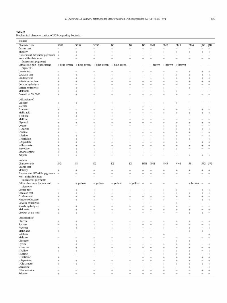
Table 2Biochemical characterization of SDS-degrading bacteria.
Isolates
Characteristic SDS1 SDS2 SDS3 N1 N2 N3 PM1 PM2 PM3 PM4 JN1 JN2Grams test e e e e e e e e e e e e
Motility þ þ þ þ þ þ þ þ þ þ þ þFluorescent diffusible pigments þ þ þ þ e e e e e e e e
Non- diffusible, nonfluorescent pigments
e e e e e e e e e e e e
Diffuasible non- fluorescentpigments
þ blue-green þ blue-green þ blue-green þ blue-green e e þ brown þ brown þ brown e e e
Urease test e e e e þ þ e e e þ þ þCatalase test þ þ þ þ þ þ þ þ þ þ þ þOxidase test þ þ þ þ þ e þ þ þ þ þ þNitrate reductase þ þ þ þ þ þ e e e þ þ þGelatin hydrolysis þ e þ e e e e e e e e e
Starch hydrolysis þ þ þ þ e e e þ e þ e e
Malonate þ þ þ þ e þ þ þ þ e e e
Growth at 5% NaCl þ þ þ þ þ þ þ þ þ e e e
Utilization ofGlucose þ þ þ þ e þ þ þ þ e e e
Sucrose e e e e e þ e e e e e e
Fructose þ e þ e e þ þ þ þ e þ e
Malic acid þ þ þ þ þ e þ þ þ þ þ þD-Ribose þ þ þ þ e þ e e e e e e
Maltose e e e e e e e e e e e e
Glycerol þ þ þ þ e e þ þ þ þ þ þGycine þ e þ e e þ þ þ þ e e e
L-Leucine þ e þ e e þ þ þ þ þ þ þL-Valine þ e þ e e þ þ þ þ þ e þL-Serine þ þ þ þ þ þ þ þ þ e e þL-Histidine þ þ þ þ þ þ þ þ þ þ e þL-Aspartate þ þ þ þ e þ þ þ þ e þ þL-Glutamate þ þ þ þ e þ þ þ þ þ þ þSarcocine þ þ þ þ e þ þ þ þ e e e
Ethanolamine þ þ þ þ þ þ e e e e e e
Adipate þ þ þ þ e e e e e þ þ þIsolatesCharacteristic JN3 K1 K2 K3 K4 NN1 NN2 NN3 NN4 SP1 SP2 SP3Grams test e e e e e e e e e e e e
Motility þ þ þ þ þ þ þ þ þ þ þ þFluorescent diffusible pigments e e e e e e e e e e e e
Non- diffusible, nonfluorescent pigments
e e e e e e e e e e e e
Diffuasible non- fluorescentpigments
e þ yellow þ yellow þ yellow þ yellow e e e e þ brown e e
Urease test e þ þ þ þ þ þ þ þ e þ þCatalase test þ þ þ þ þ þ þ þ þ þ þ þOxidase test þ þ þ þ þ þ þ e þ þ þ e
Nitrate reductase þ þ þ þ þ þ þ þ e e þ þGelatin hydrolysis e e e e e þ e e þ e e e
Starch hydrolysis e þ þ þ þ þ e e þ e e e
Malonate þ þ þ þ þ þ e þ þ þ e þGrowth at 5% NaCl þ þ þ þ þ e þ e e þ þ e
Utilization ofGlucose þ þ þ þ þ þ e þ þ þ e þSucrose e þ þ þ þ e e þ e e e þFructose e þ þ þ þ e þ þ e þ e þMalic acid þ e e e e e þ e e þ þ e
D-Ribose e e e e e e e þ e e e þMaltose e e e e e e e e e e e e
Glycogen e þ þ þ þ þ e e þ þ e e
Gycine e þ þ e e þ e þ þ þ e þL-Leucine þ þ þ þ þ þ e þ þ þ e þL-Valine þ þ þ þ þ þ e þ þ þ e þL-Serine e e e e e þ þ þ þ þ e þL-Histidine þ e e e e þ þ þ þ þ þ þL-Aspartate þ þ e þ þ þ e þ þ þ e þL-Glutamate þ þ þ þ þ þ þ þ þ þ þ þSarcocine e e e e e e e þ e þ e þEthanolamine e e e e e e þ þ e e þ þAdipate þ e e e e e e e e e e e
V. Chaturvedi, A. Kumar / International Biodeterioration & Biodegradation 65 (2011) 961e971 965
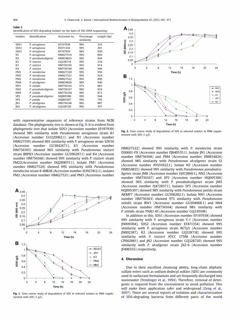
Table 3Identification of SDS-degrading isolates on the basis of 16S rDNA sequencing.
Isolates Identification Accession no. Percentagesimilarity
Length (bp)
SDS1 P. aeruginosa EF197938 99% 332SDS2 P. aeruginosa EF471354 99% 201SDS3 P. aeruginosa EF197939 98% 850N1 P. aeruginosa HM627519 99% 894N2 P. pseudoalcaligenes HM854825 99% 944K2 P. stutzeri GQ328718 99% 234K3 P. stutzeri HM756165 96% 914K4 P. stutzeri HM756166 99% 944PM1 P. mendocina HM627520 99% 956PM2 P. mendocina HM627521 99% 924PM3 P. mendocina HM627522 99% 899PM4 P. alcaligenes HM854826 98% 946NN1 P. otitidis HM756163 97% 883NN2 P. pseudoalcaligenes HM756167 96% 824NN4 P. otitidis HM756164 98% 873SP2 P. pseudoalcaligenes HQ005306 99% 944SP3 P. putida HQ005307 99% 795JN1 P. alcaligenes HM756168 98% 887JN2 P. alcaligenes GQ328720 99% 397
Fig. 2. Time course study of degradation of SDS in selected isolates in PBM supple-mented with SDS (1 g/l).
V. Chaturvedi, A. Kumar / International Biodeterioration & Biodegradation 65 (2011) 961e971966
with representative sequences of reference strains from NCBIdatabase. The phylogenetic tree is shown in Fig. 9. It is evident fromphylogenetic tree that isolate SDS3 (Accession number EF197939)showed 98% similarity with Pseudomonas aeruginosa strain R1(Accession number GU220062.1) and N1 (Accession numberHM627519) showed 99% similarity with P. aeruginosa strain SZH16(Accession number GU384267.1), K3 (Accession numberHM756165) showed 96% similarity with Pseudomonas stutzeristrain BRP83 (Accession number GU396287.1) and K4 (Accessionnumber HM756166) showed 99% similarity with P. stutzeri strainFM22(Accession number DQ289075.1). Isolate PM1 (Accessionnumber HM627520) showed 99% similarity with Pseudomonasmendocina strain 8-40B28 (Accession number EU927412.1), isolatesPM2 (Accession number HM627521) and PM3 (Accession number
Fig. 1. Time course study of degradation of SDS in selected isolates in PBM supple-mented with SDS (1 g/l).
HM627522) showed 99% similarity with P. mendocina strainDS0601-FX (Accession number FJ840535.1). Isolate JN1 (Accessionnumber HM756168) and PM4 (Accession number HM854826)showed 98% similarity with Pseudomonas alcaligenes strain S2(Accession number AY651922.1). Isolate N2 (Accession numberHM854825) showed 99% similarity with Pseudomonas pseudoalca-ligenes strain JM8 (Accession number FJ472860.1), NN2 (Accessionnumber HM756167) and SP2 (Accession number HQ005306)showed 96% similarity with P. pseudoalcaligenes strain JM5(Accession number FJ472857.1). Isolates SP3 (Accession numberHQ005307) showed 98% similarity with Pseudomonas putida strainAKMP7 (Accession number GU396282.1). Isolate NN1 (Accessionnumber HM756163) showed 97% similarity with Pseudomonasotitidis strain RW1 (Accession number GU204968.1) and NN4(Accession number HM756164) showed 98% similarity withP. otitidis strain TNAU 45 (Accession number GQ339108).
In addition to this, SDS1 (Accession number EF197938) showed99% similarity with P. aeruginosa strain T-1 (Accession numberJN000304), SDS2 (Accession number EF471354) showed 99%similarity with P. aeruginosa strain RCTy5 (Accession numberJN002387), K2 (Accession number GQ328718) showed 99%similarity with P. stutzeri ATCC 17588 (Accession numberCP002881) and JN2 (Accession number GQ328720) showed 99%similarity with P. alcaligenes strain JXZ-6 (Accession numberJF496555) respectively.
4. Discussion
Due to their excellent cleansing ability, long-chain aliphaticsulfate esters such as sodium dodecyl sulfate (SDS) are commonlyused in surfactant formulations and are frequently discharged intowastewater (Fendinger et al., 1994). Therefore, removal of deter-gents is required from the environment to avoid pollution. Thiswill make their application safer and widespread (Zeng et al.,2007). There are several reports of isolation and characterizationof SDS-degrading bacteria from different parts of the world
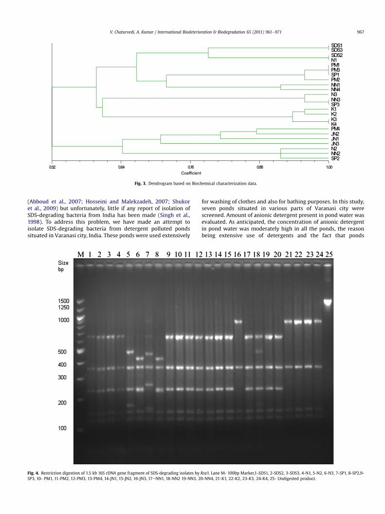
Fig. 3. Dendrogram based on Biochemical characterization data.
V. Chaturvedi, A. Kumar / International Biodeterioration & Biodegradation 65 (2011) 961e971 967
(Abboud et al., 2007; Hosseini and Malekzadeh, 2007; Shukoret al., 2009) but unfortunately, little if any report of isolation ofSDS-degrading bacteria from India has been made (Singh et al.,1998). To address this problem, we have made an attempt toisolate SDS-degrading bacteria from detergent polluted pondssituated in Varanasi city, India. These ponds were used extensively
Fig. 4. Restriction digestion of 1.5 kb 16S rDNA gene fragment of SDS-degrading isolates bySP3, 10- PM1, 11-PM2, 12-PM3, 13-PM4, 14-JN1, 15-JN2, 16-JN3, 17eNN1, 18-NN2 19-NN3, 2
for washing of clothes and also for bathing purposes. In this study,seven ponds situated in various parts of Varanasi city werescreened. Amount of anionic detergent present in pond water wasevaluated. As anticipated, the concentration of anionic detergentin pond water was moderately high in all the ponds, the reasonbeing extensive use of detergents and the fact that ponds
Rsa1. Lane M- 100bp Marker,1-SDS1, 2-SDS2, 3-SDS3, 4-N1, 5-N2, 6-N3, 7-SP1, 8-SP2,9-0-NN4, 21-K1, 22-K2, 23-K3, 24-K4, 25- Undigested product.
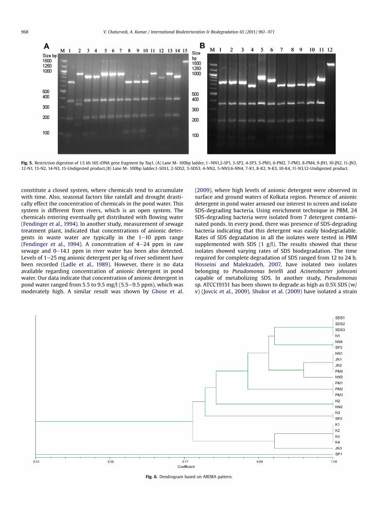
Fig. 5. Restriction digestion of 1.5 kb 16S rDNA gene fragment by Taq1. (A) Lane M- 100bp ladder, 1eNN1,2-SP1, 3-SP2, 4-SP3, 5-PM1, 6-PM2, 7-PM3, 8-PM4, 9-JN1, 10-JN2, 11-JN3,12-N1, 13-N2, 14-N3, 15-Undigested product.(B) Lane M- 100bp ladder,1-SDS1, 2-SDS2, 3-SDS3, 4-NN2, 5-NN3,6-NN4, 7-K1, 8-K2, 9-K3, 10-K4, 11-N3,12-Undigested product.
V. Chaturvedi, A. Kumar / International Biodeterioration & Biodegradation 65 (2011) 961e971968
constitute a closed system, where chemicals tend to accumulatewith time. Also, seasonal factors like rainfall and drought drasti-cally effect the concentration of chemicals in the pond water. Thissystem is different from rivers, which is an open system. Thechemicals entering eventually get distributed with flowing water(Fendinger et al., 1994). In another study, measurement of sewagetreatment plant, indicated that concentrations of anionic deter-gents in waste water are typically in the 1e10 ppm range(Fendinger et al., 1994). A concentration of 4e24 ppm in rawsewage and 0e14.1 ppm in river water has been also detected.Levels of 1e25 mg anionic detergent per kg of river sediment havebeen recorded (Ladle et al., 1989). However, there is no dataavailable regarding concentration of anionic detergent in pondwater. Our data indicate that concentration of anionic detergent inpond water ranged from 5.5 to 9.5 mg/l (5.5e9.5 ppm), which wasmoderately high. A similar result was shown by Ghose et al.
Fig. 6. Dendrogram based
(2009), where high levels of anionic detergent were observed insurface and ground waters of Kolkata region. Presence of anionicdetergent in pond water aroused our interest to screen and isolateSDS-degrading bacteria. Using enrichment technique in PBM, 24SDS-degrading bacteria were isolated from 7 detergent contami-nated ponds. In every pond, there was presence of SDS-degradingbacteria indicating that this detergent was easily biodegradable.Rates of SDS degradation in all the isolates were tested in PBMsupplemented with SDS (1 g/l). The results showed that theseisolates showed varying rates of SDS biodegradation. The timerequired for complete degradation of SDS ranged from 12 to 24 h.Hosseini and Malekzadeh, 2007, have isolated two isolatesbelonging to Pseudomonas betelli and Acinetobacter johnsonicapable of metabolizing SDS. In another study, Pseudomonassp. ATCC19151 has been shown to degrade as high as 0.5% SDS (w/v) (Jovcic et al., 2009). Shukor et al. (2009) have isolated a strain
on ARDRA pattern.
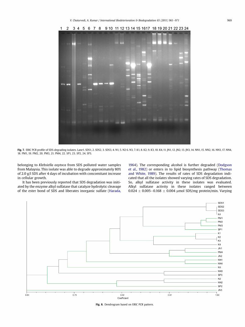
Fig. 7. ERIC PCR profile of SDS-degrading isolates. Lane1. SDS1, 2. SDS2, 3. SDS3, 4. N1, 5. N2 6. N3, 7. K1, 8. K2, 9. K3, 10. K4, 11. JN1, 12. JN2, 13. JN3, 14. NN1, 15. NN2, 16. NN3, 17. NN4,18. PM1, 19. PM2, 20. PM3, 21. PM4, 22. SP1, 23. SP2, 24. SP3.
V. Chaturvedi, A. Kumar / International Biodeterioration & Biodegradation 65 (2011) 961e971 969
belonging to Klebsiella oxytoca from SDS polluted water samplesfromMalaysia. This isolate was able to degrade approximately 80%of 2.0 g/l SDS after 4 days of incubation with concomitant increasein cellular growth.
It has been previously reported that SDS degradation was initi-ated by the enzyme alkyl sulfatase that catalyze hydrolytic cleavageof the ester bond of SDS and liberates inorganic sulfate (Harada,
Fig. 8. Dendrogram based
1964). The corresponding alcohol is further degraded (Dodgsonet al., 1982) or enters in to lipid biosynthesis pathway (Thomasand White, 1989). The results of rates of SDS degradation indi-cated that all the isolates showed varying rates of SDS degradation.So, alkyl sulfatase activity in these isolates was evaluated.Alkyl sulfatase activity in these isolates ranged between0.024 � 0.005e0.168 � 0.004 mmol SDS/mg protein/min. Varying
on ERIC PCR pattern.
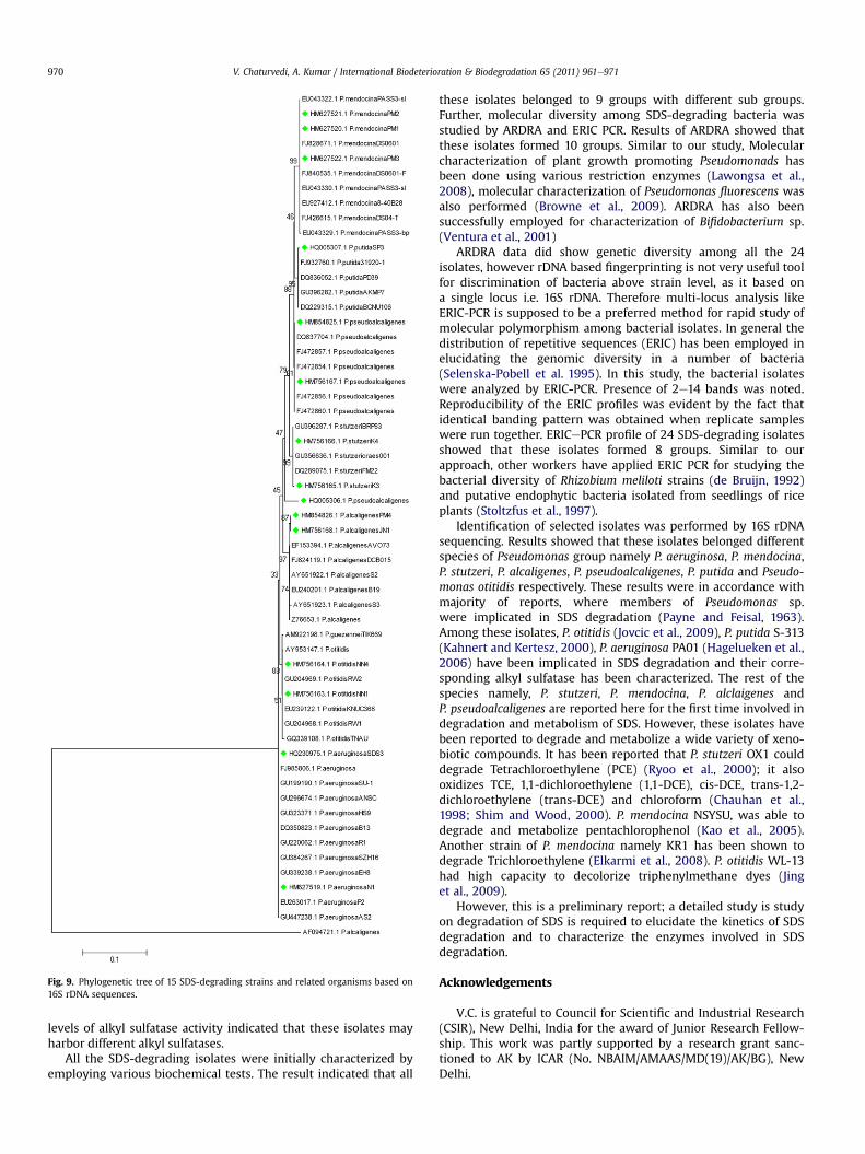
Fig. 9. Phylogenetic tree of 15 SDS-degrading strains and related organisms based on16S rDNA sequences.
V. Chaturvedi, A. Kumar / International Biodeterioration & Biodegradation 65 (2011) 961e971970
levels of alkyl sulfatase activity indicated that these isolates mayharbor different alkyl sulfatases.
All the SDS-degrading isolates were initially characterized byemploying various biochemical tests. The result indicated that all
these isolates belonged to 9 groups with different sub groups.Further, molecular diversity among SDS-degrading bacteria wasstudied by ARDRA and ERIC PCR. Results of ARDRA showed thatthese isolates formed 10 groups. Similar to our study, Molecularcharacterization of plant growth promoting Pseudomonads hasbeen done using various restriction enzymes (Lawongsa et al.,2008), molecular characterization of Pseudomonas fluorescens wasalso performed (Browne et al., 2009). ARDRA has also beensuccessfully employed for characterization of Bifidobacterium sp.(Ventura et al., 2001)
ARDRA data did show genetic diversity among all the 24isolates, however rDNA based fingerprinting is not very useful toolfor discrimination of bacteria above strain level, as it based ona single locus i.e. 16S rDNA. Therefore multi-locus analysis likeERIC-PCR is supposed to be a preferred method for rapid study ofmolecular polymorphism among bacterial isolates. In general thedistribution of repetitive sequences (ERIC) has been employed inelucidating the genomic diversity in a number of bacteria(Selenska-Pobell et al. 1995). In this study, the bacterial isolateswere analyzed by ERIC-PCR. Presence of 2e14 bands was noted.Reproducibility of the ERIC profiles was evident by the fact thatidentical banding pattern was obtained when replicate sampleswere run together. ERICePCR profile of 24 SDS-degrading isolatesshowed that these isolates formed 8 groups. Similar to ourapproach, other workers have applied ERIC PCR for studying thebacterial diversity of Rhizobium meliloti strains (de Bruijn, 1992)and putative endophytic bacteria isolated from seedlings of riceplants (Stoltzfus et al., 1997).
Identification of selected isolates was performed by 16S rDNAsequencing. Results showed that these isolates belonged differentspecies of Pseudomonas group namely P. aeruginosa, P. mendocina,P. stutzeri, P. alcaligenes, P. pseudoalcaligenes, P. putida and Pseudo-monas otitidis respectively. These results were in accordance withmajority of reports, where members of Pseudomonas sp.were implicated in SDS degradation (Payne and Feisal, 1963).Among these isolates, P. otitidis (Jovcic et al., 2009), P. putida S-313(Kahnert and Kertesz, 2000), P. aeruginosa PA01 (Hagelueken et al.,2006) have been implicated in SDS degradation and their corre-sponding alkyl sulfatase has been characterized. The rest of thespecies namely, P. stutzeri, P. mendocina, P. alclaigenes andP. pseudoalcaligenes are reported here for the first time involved indegradation and metabolism of SDS. However, these isolates havebeen reported to degrade and metabolize a wide variety of xeno-biotic compounds. It has been reported that P. stutzeri OX1 coulddegrade Tetrachloroethylene (PCE) (Ryoo et al., 2000); it alsooxidizes TCE, 1,1-dichloroethylene (1,1-DCE), cis-DCE, trans-1,2-dichloroethylene (trans-DCE) and chloroform (Chauhan et al.,1998; Shim and Wood, 2000). P. mendocina NSYSU, was able todegrade and metabolize pentachlorophenol (Kao et al., 2005).Another strain of P. mendocina namely KR1 has been shown todegrade Trichloroethylene (Elkarmi et al., 2008). P. otitidis WL-13had high capacity to decolorize triphenylmethane dyes (Jinget al., 2009).
However, this is a preliminary report; a detailed study is studyon degradation of SDS is required to elucidate the kinetics of SDSdegradation and to characterize the enzymes involved in SDSdegradation.
Acknowledgements
V.C. is grateful to Council for Scientific and Industrial Research(CSIR), New Delhi, India for the award of Junior Research Fellow-ship. This work was partly supported by a research grant sanc-tioned to AK by ICAR (No. NBAIM/AMAAS/MD(19)/AK/BG), NewDelhi.
V. Chaturvedi, A. Kumar / International Biodeterioration & Biodegradation 65 (2011) 961e971 971
References
Abboud, M.M., Khleifat, K.M., Batarseh, M., Tarawneh, K.A., Al-Mustafa, A.,Al-Madadhah, M., 2007. Different optimization conditions required forenhancing the biodegradation of linear alkyl benzo sulfonate and sodiumdodecyl sulfate surfactants by novel consortium of Acinetobacter calcoaceticusand Pantoea agglomerans. Enzymes and Microbial Technology 41, 432e439.
Altschul, S.F., Gish, W., Miller, W., Myers, E.W., Lipman, D.J., 1990. Basic localalignment search tool. Journal of Molecular Biology 215, 403e410.
Browne, P., Rice, O., Miller, S.H., Burke, J., Dowling, D.N., Morrissey, J.P., O’Gara, F.,2009. Superior inorganic phosphate solubilization is linked to phylogeny withinthe Pseudomonas fluorescens complex. Applied Soil Ecology 43, 131e138.
Chauhan, S., Barbieri, P., Wood, T.K., 1998. Oxidation of trichloroethylene,1,1-dichloroethylene, and chloroform by toluene/oxylene monooxygenase fromPseudomonasstutzeriOX1.AppliedandEnvironmentalMicrobiology64,3023e3024.
de Bruijn, F., 1992. Use of repetitive (repetitive extragenic palindromic and entero-bacterial repetitive intergeneric consensus) sequences and the polymerase chainreaction to fingerprint the genomes of Rhizobium meliloti isolates and other soilbacteria. Applied and Environmental Microbiology 58, 2180e2187.
Dodgson, K.S., White, G.F., Fitzgerald, J.W., 1982. Sulfatases of Microbial Origin. CRCPress, Boca Raton, FL.
Elkarmi, A., Abu-Elteen, K., Khader, M., 2008. Modeling the biodegradation effi-ciency and growth of Pseudomonas alcaligenes utilizing 2,4-dichlorophenol asa carbon source Pre- and Post-exposure to UV radiation. Journal of Environ-mental Sciences 1, 7e11.
Ellis, A.J., Hales, S.G., Ur-Rehman, N.G.A., White, G.F., 2002. Novel alkyl sulfatasesrequired for biodegradation of the branched primary alkyl sulfate surfactant2-butyloctyl sulfate. Applied and Environmental Microbiology 68, 31e36.
Fendinger, N.J., Versteg, D.J., Weeg, E., Dyer, S., Rapaport, R.A., 1994. Environmentalbehavior and fate of anionic surfactants. In: Baker, L.A. (Ed.), EnvironmentalChemistry of Lakes and Reservoirs. American Chemical Society, Washington DC,USA, pp. 527e557.
Ghose, N.C., Saha, D., Gupta, A., 2009. Synthetic detergents (Surfactants) andOrganochlorine Pesticide Signatures in surface water and Groundwater ofgreater Kolkata, India. Journal of Water Resource and Protection 4, 290e298.
Hagelueken, G., Adams, T.M., Wiehlmann, L., Widow, U., Kolmar, H., Tummler, B.,Heinz, D.W., Schubert, W., 2006. The crystal structure of SdsA1, an alkyl sulfa-tase from Pseudomonas aeruginosa, defines a third class of sulfatase. Proceed-ings of National Academy of Sciences (USA) 103, 7631e7636.
Harada, T., 1964. The formation of sulfatases by Pseudomonas aeruginosa. Biochimicaet Biophysica Acta 81, 193e196.
Hayashi, K., 1975. A rapid determination of sodium dodecyl sulfate with methyleneblue. Analytical Biochemistry 67, 503e506.
Hosseini, F., Malekzadeh, F., Amir Mozafari, N., Ghaemi, N., 2007. Biodegradation ofanionic surfactants by isolated bacteria from activated sludge. InternationalJournal of Environmental Science and Technology 4, 127e132.
Hulton, C.S.J., Higgins, C.F., Sharp, P.M., 1991. ERIC sequences: a novel family ofrepetitive elements in the genomes of Escherichia coli, Salmonella typhimuriumand other enterobacteria. Molecular Micrbiology 5, 825e834.
Jing,W., Byung-Gil, J., Kyoung-Sook, K., Young-Choon, L., Nak-Chang, S., 2009. Isolationand characterization of Pseudomonas otitidisWL-13 and its capacity to decolorizetriphenylmethane dyes. Journal of Environmental Sciences 21, 960e964.
Jovcic, B., Begovic, J., Lozo, J., Topisirovic, L., Kojic, M., 2009. Dynamic of sodiumdodecyl sulfate utilization and antibiotic susceptibility of strain Pseudomonassp. ATCC19151. Archives of Biologibal Sciences 61, 159e165.
Kahnert, A., Kertesz, M.A., 2000. Characterization of a sulfur-regulated oxygenativealkyl sulfatase from Pseudomonas putida S-313. Journal of Biological Chemistry41, 31661e31667.
Ka€mpfer, P., Steiof, M., Dott, W., 1991. Microbiological characterization of a fuel-oilcontaminated site including numerical identification of heterotrophic waterand soil bacteria. Microbial Ecology 21, 227e251.
Kao, C.M., Liu, J.K., Chen, Y.L., Chai, C.T., Chen, S.C., 2005. Factors affecting thebiodegradation of PCP by Pseudomonas mendocina NSYSU. Journal of HazardousMaterial 124, 68e73.
Karsa, D.R. (Ed.), 1992, The Industrial Application of Surfactants, vol. 3. Royal Societyof Chemistry, Cambridge, England Special Publication 107.
Ladle, M., House, W.A., Armitage, P.D., Farr, 1S., 1989. Faunal characteristics of a sitesubject to sewage plant discharge. Tenside Detergents 2, 159e168.
Lawongsa, P., Boonkerd, N., Wongkaew, S., O’Gara, F., Teaumroong, N., 2008. Molec-ular and phenotypic characterization of potential plant growth-promotingPseudomonas from rice and maize rhizospheres. World Journal of Microbiologyand Biotechnology 24, 1877e1884.
Molin, G., Ternstrom, A., 1982. Numerical taxonomy of psychrotrophic Pseudomo-nads. Journal of General Microbiology 128, 1249e1264.
Payne, W.J., Feisal, V.E., 1963. Bacterial utilization of sodium dodecyl sulfate anddodecyl benzene sulfonate. Applied Microbiology 11, 339e344.
Porteous, L.A., Armstrong, J.L., Seidler, R.J., Watrud, L.S., 1994. An effectivemethod to extract DNA from environmental samples for polymerase chainreaction amplification and DNA fingerprint analysis. Current Microbiology 29,301e307.
Prakash, S., Lawton, L.A., Edwards, C., 2009. Stability of toxigenic Microcystisblooms. Harmful Algae 8, 377e384.
Ribelles, A., Carrasco, C., Rosety, M., Aldana, M., 1995. A histochemical study of thebiological effects of sodium dodecyl sulfate on the intestine of giltheadseabream, Sparus aurata. Ecotoxicology And Environmental Safety 32,131e138.
Rohlf, F.J., 1997. NTSYS-PC 2.1. Numerical taxonomy and multivariate analysissystem. Exeter Software, New York.
Rocha, A.J., Gomes, V., Ngan, P.V., Passos, M.J., Furia, R.R., 2007. Effects of anionicsurfactant and salinity on the bioenergetics of juveniles of Centropomus paral-lelus (Poey). Ecotoxicology and Environment Safety 3, 397e404.
Rosety, M., Ordonez, F.J., Rosety, R.M., Rosety, J.M., Rosety, I., Carrasco, C., Ribelles, A.,2001. Comparative study of the acute toxicity of anionic surfactants alkylbenzene sulfonate (ABS) and sodium dodecyl sulfate (SDS) on gilthead, Sparusaurata L., eggs. Histology and. Histopathology 4, 1091e1095.
Ryoo, D., Shim, H., Canada, K., Barbieri, P., Wood, T.K., 2000. Aerobic degradation oftetrachloroethylene by toluene-o-xylene monooxygenase of Pseudomonasstutzeri OX1. Nature Biotechnology 18, 775e778.
Saitou, N., Nie, M., 1987. The neighbour-joining method: a new method for recon-structing phylogenetic trees. Molecular Biolaogy and Evolution 4, 406e425.
Selenska-Pobell, S., Evguenieva-Kackenberg, E., Schickerath, O., 1995. Random andrepetitive primer amplified polymorphic DNA analysis of five soil and twoclinical isolates of Rahnella aquatilis. Systematic and Applied Microbiology 18,425e438.
Sharma, N.K., Mohan, D., Rai, A.K., 2009. Predicting Phytoplankton growth anddynamics in relation to physico-chemical characteristics of water body. WaterAir and Soil Pollution 202, 325e333.
Shaw, B.G., Latty, J.B., 1982. A numerical taxonomic study of Pseudomonas strainsfrom spoiled meat. Journal of Applied Bacteriology 52, 219e228.
Shim, H., Wood, T.K., 2000. Aerobic degradation of mixtures of chlorinatedaliphatics by cloned toluene-o-xylene monooxygenase and toluene o-mono-oxygenase in resting cells. Biotechnology and Bioengineering 70, 693e698.
Shukor, M.Y., Hussain, W.S.W., Rahman, M.F.A., Shamaan, N.A., Syed, M.A., 2009.Isolation and characterization of an SDS-degrading Klebsiella oxytoca. Journal ofEnvironmental Biology 30, 129e134.
Singer, M.M., Tjeerdema, R.S., 1993. Fate and effects of the surfactant sodiumdodecyl sulfate. Reviews of Environmental Contamination and Toxicology 133,95e149.
Singh, K.L., Kumar, A., Kumar, A., 1998. Bacillus cereus capable of degrading SDSshows growth with a variety of detergents. World Journal of Microbiology andBiotechnology 14, 777e779.
Stoltzfus, J.R., So, R., Malarvizhi, P.P., Ladha, J.K., de Bruijn, F.J., 1997. Isolation ofendophytic bacteria from rice and assessment of their potential for supplyingrice with biologically fixed nitrogen. Plant Soil 194, 25e36.
Thomas, O.R.T., White, G.F., 1989. Metabolic pathway for the biodegradation ofsodium dodecyl sulfate by Pseudomonas sp. C12B. Biotechnology and AppliedBiochemistry 11, 318e327.
Tyagi, M.B., Singh, D.P., Kumar, A., Jha, P.N., Sinha, R.P., Kumar, A., 2006. Hepato-toxicity of Microcystis aeruginosa strains growing as blooms in certain eutrophicponds. EXCLI Journal 5, 66e78.
Ventura, M., Elli, M., Reniero, R., Zink, R., 2001. Molecular microbial analysis ofBifido bacterium isolates from different environments by the species-specificamplified ribosomal DNA restriction analysis (ARDRA). FEMS MicrobialEcology 36, 113e121.
Versalovic, J., Koeuth, T., Lupski, J.R., 1991. Distribution of repetitive DNA sequencesin eubacteria and application to fingerprinting of bacterial genomes. NucleicAcids Research 19, 6823e6831.
Zeng, G., Fu, H., Zhong, H., Yuan, X., Fu, M., Wan, W., Huang, G., 2007. Co-degra-dation with glucose of four surfactants, CTAB, Triton X-100, SDS, and rham-nolipid, in liquid culture media and compost matrix. Biodegradation 18,303e310.
Zhou, J., Davey, M.E., Figueras, J.B., Rivkina, E., Gilichinsky, D., Tiedje, J.M., 1997.Phylogenetic diversity of a bacterial community determined from Siberiantundra soil DNA. Microbiology 143, 3913e3919.


