

Susceptibility of Candida albicans to photodynamic therapy in amurine model of oral candidosisEwerton Garcia de Oliveira Mima, DDS, MSc, PhD,a Ana Cláudia Pavarina, DDS, MSc, PhD,b
Lívia Nordi Dovigo, DDS, MSc,c Carlos Eduardo Vergani, DDS, MSc, PhD,b
Carlos Alberto de Souza Costa, DDS, MSc, PhD,d Cristina Kurachi, DDS, MSc, PhD,e andVanderlei Salvador Bagnato, MSc, PhD,e São Paulo, BrazilSÃO FRANCISCO UNIVERSITY, SÃO PAULO STATE UNIVERSITY, AND UNIVERSITY OF SÃO PAULO
Objective. In vivo studies of antimicrobial PDT in animal models of oral candidosis are scarce and the association ofporphyrin and LED light has not been evaluated for in vivo photoinactivation of Candida. In this study theeffectiveness of photodynamic therapy (PDT) on the inactivation of Candida albicans in vivo was evaluated.Study design. Seventy-one 6-week-old female Swiss mice were immunosuppressed, provided tetracycline to theirdrinking water, then orally swabbed with a suspension of C. albicans (107 CFU/mL). Four days after oral inoculation,PDT was performed on the dorsum of the tongue after topical administration of Photogem at 400, 500, or 1000 mg/Land followed 30 minutes later by illumination with LED light (305 J/cm2) at 455 or 630 nm (n � 5 each). Afterswabbing to recover yeast from the tongue, the number of surviving yeast cells was determined (CFU/mL) andanalyzed by ANOVA and Holm-Sidak tests (P � .05). Animals were humanely killed, and the tongues surgicallyremoved and processed for histological evaluation of presence of yeast and inflammatory reaction.Results. PDT resulted in a significant reduction in C. albicans recovered from the tongue (P � .001) when comparedwith mice from the positive control group. There was no difference between the concentrations of Photogem and LEDlight wavelengths used. Histological evaluation of the tongue revealed that PDT causes no significant adverse effects tothe local mucosa.Conclusion. PDT promoted significant reduction in the viability of C. albicans biofilm without harming the tongue
tissue. (Oral Surg Oral Med Oral Pathol Oral Radiol Endod 2010;109:392-401)Oral candidosis is the most common infection of theoral cavity and is caused by Candida species, the com-monest being Candida albicans.1,2 The predisposingfactors of oral candidosis include immunocompromisedstates, diabetes mellitus, dental prostheses, xerosto-mia,3,4 and prolonged use of antibiotics or immunosup-
This research was supported by São Paulo Council of Research(FAPESP – Grant No. 2005/02193-4 and 2005/03226-3, and CePOF –CEPID Program).aAssistant Professor, Dentistry School, São Francisco University–USF,Bragança Paulista, SP, Brazil.bAdjunct Professor, Department of Dental Materials and Prosthodon-tics, Araraquara Dental School, São Paulo State University–UNESP,Araraquara, SP, Brazil.cPostgraduate Student, Department of Dental Materials and Prosthodon-tics, Araraquara Dental School, São Paulo State University-UNESP,Araraquara, SP, Brazil.dAdjunct Professor, Department of Physiology and Pathology, Arara-quara Dental School, São Paulo State University–UNESP, Araraquara,SP, Brazil.eProfessor, Physics Institute of São Carlos, University of São Paulo–USP, São Carlos, SP, Brazil.Received for publication Jul 1, 2009; returned for revision Sep 25,2009; accepted for publication Oct 4, 2009.1079-2104/$ - see front matter© 2010 Mosby, Inc. All rights reserved.
doi:10.1016/j.tripleo.2009.10.006392
pressive drugs.5,6 With the advent of the human immu-nodeficiency virus (HIV) infection, increased attentionhas been paid to oral candidosis, because up to 90% ofHIV-infected individuals suffer from oral Candida in-fection.3
The widespread use of topical and systemic antifun-gal agents as conventional treatment for oral candidosishas resulted in the development of resistance in C.albicans.7 Although resistance of C. albicans to poly-enes is rare, several mechanisms of azole resistance8
have been reported, including changes in the cell wallor plasma membrane, which lead to impaired azoleuptake; overexpression of or mutations in the targetenzyme of azoles; and the efflux of drugs mediated bymembrane transport proteins.9 Resistance appears toincrease proportionally with the extend of previousexposure to the antifungal drugs.10 Moreover, becauseof the fungistatic rather than fungicidal effect ofazoles,7 the host defenses are essential for eradicatingthe infection.11 Therefore, in immunosuppressed pa-tients, the use of azole agents to treat oral candidosiscan be ineffective.
Thus, it is necessary to develop alternative therapiesfor the treatment of oral candidosis. A promising mo-
dality is photodynamic therapy (PDT), which uses a
OOOOEVolume 109, Number 3 Mima et al. 393
photosensitizing agent and an appropriate wavelengthof light. The interaction between the photosensitizer(PS) and light in the presence of oxygen producesreactive species, such as singlet oxygen and free radi-cals, which causes cell damage and death. In this sense,the mechanism of PDT for inactivating fungi differscompletely from that of antifungal agents. Owing to thenonspecific oxidizing agents, organisms resistant toconventional antifungal agents could be successfullykilled by PDT and it seems unlikely that resistance tosuch therapy will be developed. Although PDT is moreusually applied for treating cancer, several studies havereported that microorganisms, such as bacteria, viruses,and fungi, can be killed by PDT.12,13 It has been dem-onstrated that PDT is effective against oral species andmay not promote damage to host cells and tissues.14,15
However, most studies on cell damage are short-terminvestigations and safety studies have been performedover the short term. Kömerik et al.15 observed completeinactivation of Porphyromonas gingivalis in the max-illary molar region of rats after PDT, with no adverseeffects on the periodontal structures after 3 days andsignificant reductions in bone loss after 90 days. More-over, Zeina et al.16 detected no genotoxicity (immediateand delayed effects) on skin cells after a PDT protocolthat was effective for killing microbial species. Theseauthors concluded that there is a wide safety margin forPDT between microbial elimination and damage to hostcells.
In vitro investigations have shown that Candida spp.are susceptible to photoinactivation.17-24 Usually, dyes(toluidine blue and methylene blue) and porphyrins areused as PS combined with red laser light. However,light sources with simpler technology and lower costthan lasers, such as light-emitting diodes (LED), havebeen successfully applied in PDT.25,26 In addition,there are different colors of LED light, with radiationscovering almost the entire visible electromagnetic spec-trum. Investigations with the aim of confirming theeffectiveness of antimicrobial PDT in animal modelsare scarce. Only one in vivo study reported completeinactivation of C. albicans with topical methylene blueand red laser light in a murine model of oral candido-sis.27 Nevertheless, dyes have the undesirable effect ofstaining teeth, lips, tongue, buccal mucosa, estheticrestorations, and prosthetic surfaces. Therefore, a non-dye sensitizer, such as the porphyrins, would be pref-erable. A previous in vitro study showed that PDTmediated by Photogem and blue LED light resulted incomplete inactivation of planktonic suspensions andsignificant reduction in biofilm viability.28 However, invivo application of antimicrobial PDT using porphyrinsand LED light has not yet been well established and
animal models may provide outcomes more closelycorrelated to clinical situations. Thus, the aim of thepresent investigation was to contribute to in vivo anti-microbial PDT development by reporting on the pho-toinactivation of C. albicans in a murine model of oralcandidosis using a porphyrin in association with LEDlight sources of different wavelengths.
MATERIALS AND METHODSPhotosensitizer and light sources
The PS used in this study was a hematoporphyrinderivative produced in Moscow, Russia (Photogem;Limited Liability Company Photogem, Moscow, Rus-sia). Stock solutions of Photogem (pH 6.6) were pre-pared by dissolving the powder in sterile saline andkept in the dark instantly before use. The absorptionbands of Photogem are shown in Fig. 1.
Two handpieces with a blue (455 nm) or red (630nm) light-emitting diode (LED, LXHL–PR09,Luxeon III Emitter, Lumileds Lighting, San Jose,CA) were designed by the “Instituto de Física de SãoCarlos” (University of São Paulo, São Carlos, SP,Brazil). The output power of light delivered at theend of each handpiece (5 mm in diameter) was 200mW. The wavelengths of 455 nm (blue) and 630 nm(red) were chosen because the absorption bands ofPhotogem match these wavelengths. Although thehighest absorption of Photogem is close to 455 nm(see Fig. 1), the wavelength of 630 nm achieves ahigher penetration into the tissue.
Microorganisms and culture conditionA reference strain (ATCC 90028) of C. albicans
(ATCC, Rockville, MD) was evaluated. This strain wasmaintained in yeast-peptone-glucose (YEPD, 1.0%
Fig. 1. Absorption bands of Photogem and intensity of blue(455 nm) and red (630 nm) LED light.
yeast extract, 2.0% peptone, 2.0% glucose, 0.1 M cit-

OOOOE394 Mima et al. March 2010
rate-phosphate buffer pH 5.0) and glycerol medium at�70°C. The yeast was reactivated by cultivation inSabouraud Dextrose Agar (SDA, Acumedia Manufac-tures Inc., Baltimore, MD) containing 5 �g/mL genta-micin at 37°C for 48 hours before each experiment. Theyeast suspended in sterile saline (pH 5.3) was inocu-lated in 5 mL of Tryptic Soy Broth (TSB, pH 7.2,Acumedia Manufactures Inc., Baltimore, MD) andgrown aerobically at 37°C for 24 hours. Each culturewas harvested after centrifugation at 2000 rpm for 10minutes, washed twice with sterile distilled water, andresuspended in sterile saline (4.5 � 107 colony-formingunits [CFU]/mL).
Preparation of animals and oral inoculationThe research protocols for using mice and all the
animal experiments were approved by the Ethics Com-mittee for Animal Investigations (Araraquara DentalSchool, São Paulo State University). Seventy-one6-week-old female Swiss mice were used for all animalexperiments. The mice were kept in cages housing 5animals in a temperature-controlled room (23 � 2°C)with a 12:12-hour light/dark cycle. Standard mousechow and tap water were given ad libitum.
The methodology described by Takakura et al.29 wasused to induce oral candidosis in mice. The timeline ofevents used in this study can be seen in Fig. 2. Theanimals were immunosuppressed with 2 subcutaneousinjections of prednisolone (Depo-Medrol, LaboratóriosPfizer Ltda., Guarulhos, SP, Brazil) at a dose of 100mg/kg body weight 1 day before and 3 days after theinfection with Candida (days 0 and 4 in Fig. 2). Themice were given tetracycline hydrochloride (FarmáciaSanta Paula, Araraquara, SP, Brazil) in their drinkingwater at the concentration of 0.83 mg/mL beginning onday 0. On day 1, animals were anesthetized by anintramuscular injection with 50 �L of 2 mg/mL chlor-promazine chloride (Farmácia Santa Paula, Araraquara,SP, Brazil) in each femur. Small cotton pads (Cotton-baby, Higie-Plus Cottonbaby Ind. Com. Ltda., SãoJosé, SC, Brazil) were soaked in a C. albicans cellsuspension (4.5 � 107 CFU/mL) in such a way that theentire oral cavity of the anesthetized mice was swabbedto produce oral infections.
Photodynamic therapy and microbiologicalevaluation
On day 5 (4 days after Candida inoculation), micewere anesthetized by an intramuscular injection of100 mg/kg body weight ketamine (União QuímicaFarmacêutica Nacional S/A., Embu-Guaçu, SP, Brazil)and 10 mg/kg body weight xylazine (Produtos Veter-inários J. A. Ltda., Patrocínio Paulista, SP, Brazil).
Each animal was placed in a supine position on a pad ina device fitted with stainless steel wires that werelooped around the incisors to hold the mouth open.With mandible and cheeks retracted, the tongue wasgently taken out of the mouth as far as it would go, toexpose it without causing any injury to the tissue. Then,50 �L of Photogem at concentration of 400, 500, or1000 mg/L was pipetted onto the dorsum of the tongueand mice were kept in the dark for 30 minutes (pre-irradiation time). During this period, the tongue of eachanimal was kept in the oral cavity and the photosensi-tizer was not swallowed, as the animals were anesthe-tized. After this period, the tongue was gently taken outof the mouth again to expose it for illumination. Thesolution was not rinsed off or removed; it remained inthe oral cavity and the dorsum of the tongue wascompletely wet with Photogem. For illumination, theLED device was placed onto the dorsum of the tongue,which was illuminated for 20 minutes, resulting in atotal fluence of 305 J/cm2 (P�L� groups). Thereforethe P�L� groups corresponded to 6 combinationsof the 3 PS concentrations (400, 500, or 1000 mg/L)and the 2 wavelengths of LED light (455 or 630 nm)evaluated (6 groups). The effect of PS alone was testedby application of Photogem at the same concentrationsfor the same period of pre-irradiation time and irradi-ation but in the absence of light (P�L�, total of 3groups). The groups that received light only (blue orred) were exposed to the same LED dose mentionedpreviously (P�L�, 2 groups). The positive controlgroup did not receive any PS or light (P�L�). Anegative control group of animals was immunosup-pressed as described but did not receive C. albicansinoculation or any treatment. Each experimental groupconsisted of 5 animals. As mice from positive controlwere evaluated concurrently with the animals from allother experimental groups, in order to compare theresults, a higher number of animals in the positivecontrol group was tested (n � 7). Two additional micewere not immunosuppressed, and did not receive C.albicans inoculation or any treatment, and were con-sidered as overall control.
The occurrence of Candida infection was microbio-logically confirmed in the positive control group(P�L�) on day 5. The dorsum of the tongue wasswabbed for 1 minute with a cotton pad. The end of thecotton pad was then cut off and placed in a tubecontaining 1 mL sterile saline. After mixing on a vortexmixer for 1 minute to release C. albicans cells from theswab into the saline, duplicate 25-�L aliquots from the10-fold serial dilutions were spread over the surface ofSDA with 5 mg/L gentamicin. All plates were aerobi-cally incubated at 37°C for 48 hours. After incubation,the yeast colony counts of each plate were quantified
using a digital colony counter (CP 600 Plus, Phoenix
OOOOEVolume 109, Number 3 Mima et al. 395
Ind Com Equipamentos Científicos Ltda, Araraquara,SP, Brazil). The number of CFU/mL was determined.The same procedures of swabbing and plating sampleswere performed in animals from experimental groups(P�L�, P�L�, P�L�) after treatments. These ani-mals were not swabbed before treatment to avoid re-moving Candida cells from the tissue, which could
Fig. 2. Study design.
potentially interfere in the results, decreasing the
CFU/mL values. Sampling (recovery of C. albicans fromtongues of mice and yeast culture) was done at only onetime point, on day 5 immediately after PDT, in order toassess the yeast viability immediately after treatment.
Histopathological studyOn day 6, all mice from all groups were killed with
a lethal dose of ketamine. Tongues were surgically
OOOOE396 Mima et al. March 2010
removed, fixed in 10% formalin fixative solution at pH7.2 and embedded in paraffin. Five-micrometer-thickserial sections were cut, mounted on glass slides, andstained with periodic acid-Schiff and hematoxylin(PAS-H) stain for histopathological examination andfungal detection by light microscopy (Carl Zeiss 62774,Oberkochen, West Germany). Tissue reaction causedby C. albicans infection whether or not associated withthe PDT was examined by a pathologist blinded to theall groups of mice. A descriptive analysis of the histo-logical characteristics of the tissue with and withoutlocal inflammatory response of varied intensity wasperformed.
Characterization of LED light penetrationinto the tongue
Two additional animals were used for this evaluation.These mice were killed and tongues were surgically re-moved. Each tongue was cut into halves sagitally. EachLED handpiece was positioned perpendicularly to thedorsum of the tongue sample. A CCD (charge-coupleddevice) camera (DSC-F828 Cybershot Digital StyleCamera, Sony Corp, Tokyo, Japan) placed on the sideof the tissue recorded the whole image formed by thelight scattered within the tissue. The image was savedto a PC computer as a file.
Statistical analysisThe log10 (CFU/mL) data of C. albicans isolated
from the tongues of mice in the different groups were
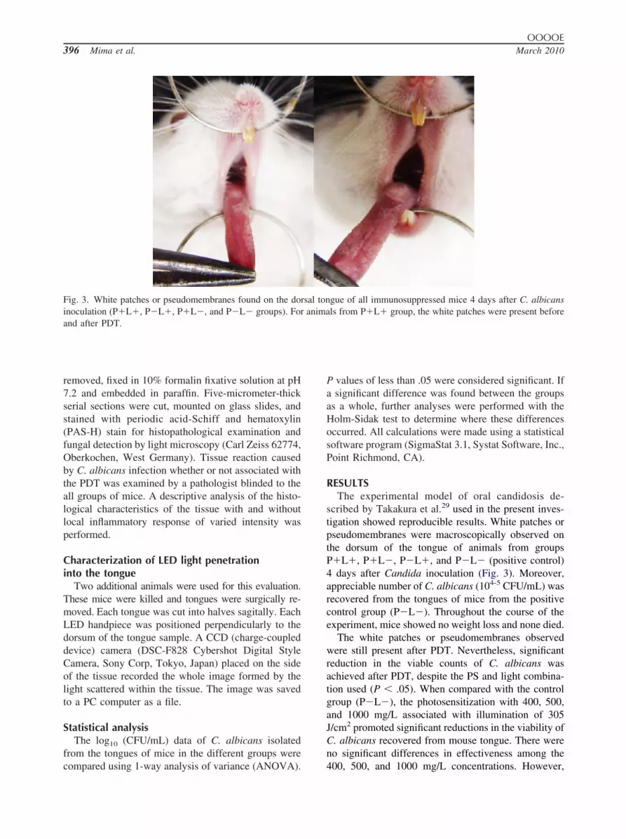
Fig. 3. White patches or pseudomembranes found on the doinoculation (P�L�, P�L�, P�L�, and P�L� groups). Forand after PDT.
compared using 1-way analysis of variance (ANOVA).
P values of less than .05 were considered significant. Ifa significant difference was found between the groupsas a whole, further analyses were performed with theHolm-Sidak test to determine where these differencesoccurred. All calculations were made using a statisticalsoftware program (SigmaStat 3.1, Systat Software, Inc.,Point Richmond, CA).
RESULTSThe experimental model of oral candidosis de-
scribed by Takakura et al.29 used in the present inves-tigation showed reproducible results. White patches orpseudomembranes were macroscopically observed onthe dorsum of the tongue of animals from groupsP�L�, P�L�, P�L�, and P�L� (positive control)4 days after Candida inoculation (Fig. 3). Moreover,appreciable number of C. albicans (104-5 CFU/mL) wasrecovered from the tongues of mice from the positivecontrol group (P�L�). Throughout the course of theexperiment, mice showed no weight loss and none died.
The white patches or pseudomembranes observedwere still present after PDT. Nevertheless, significantreduction in the viable counts of C. albicans wasachieved after PDT, despite the PS and light combina-tion used (P � .05). When compared with the controlgroup (P�L�), the photosensitization with 400, 500,and 1000 mg/L associated with illumination of 305J/cm2 promoted significant reductions in the viability ofC. albicans recovered from mouse tongue. There wereno significant differences in effectiveness among the
gue of all immunosuppressed mice 4 days after C. albicansls from P�L� group, the white patches were present before
rsal tonanima
400, 500, and 1000 mg/L concentrations. However,
L�).
OOOOEVolume 109, Number 3 Mima et al. 397
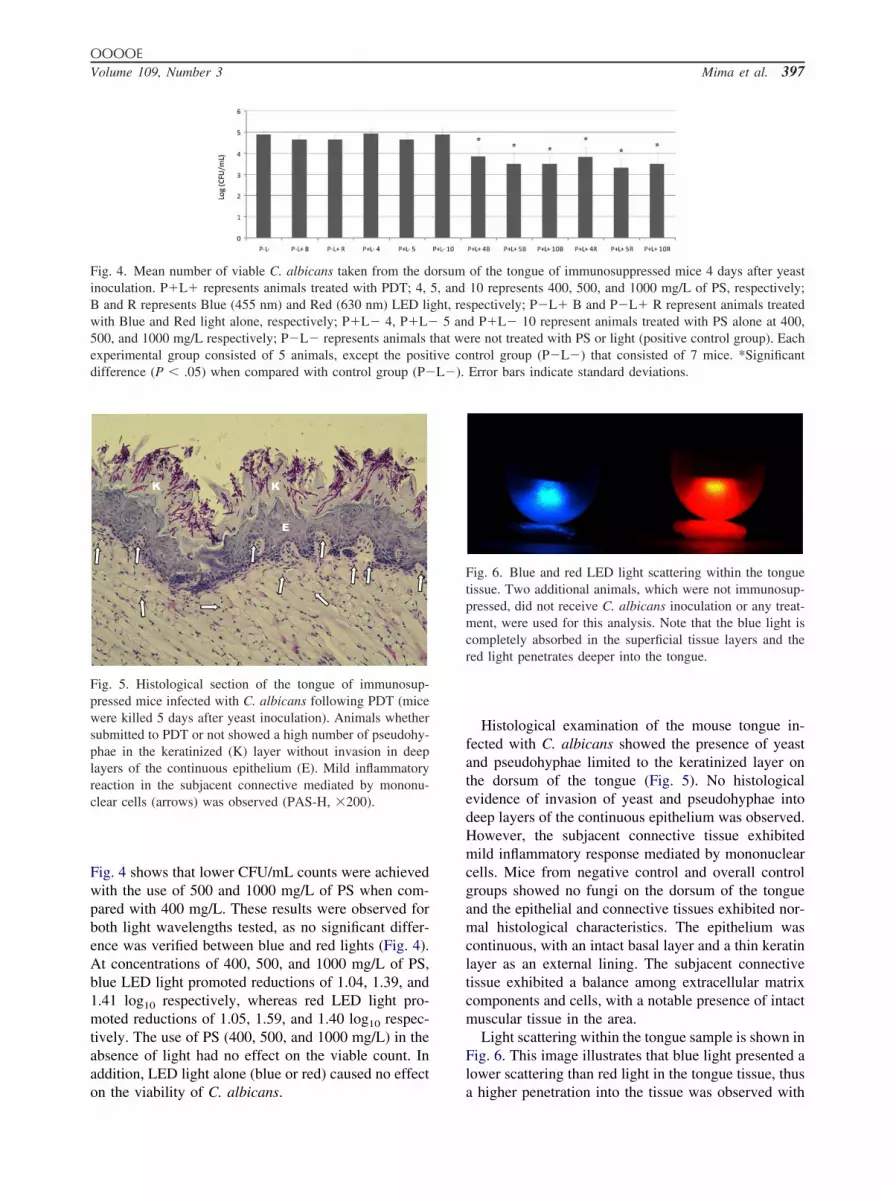
Fig. 4 shows that lower CFU/mL counts were achievedwith the use of 500 and 1000 mg/L of PS when com-pared with 400 mg/L. These results were observed forboth light wavelengths tested, as no significant differ-ence was verified between blue and red lights (Fig. 4).At concentrations of 400, 500, and 1000 mg/L of PS,blue LED light promoted reductions of 1.04, 1.39, and1.41 log10 respectively, whereas red LED light pro-moted reductions of 1.05, 1.59, and 1.40 log10 respec-tively. The use of PS (400, 500, and 1000 mg/L) in theabsence of light had no effect on the viable count. Inaddition, LED light alone (blue or red) caused no effect
Fig. 4. Mean number of viable C. albicans taken from the dinoculation. P�L� represents animals treated with PDT; 4,B and R represents Blue (455 nm) and Red (630 nm) LED liwith Blue and Red light alone, respectively; P�L� 4, P�L�500, and 1000 mg/L respectively; P�L� represents animalsexperimental group consisted of 5 animals, except the posidifference (P � .05) when compared with control group (P�
Fig. 5. Histological section of the tongue of immunosup-pressed mice infected with C. albicans following PDT (micewere killed 5 days after yeast inoculation). Animals whethersubmitted to PDT or not showed a high number of pseudohy-phae in the keratinized (K) layer without invasion in deeplayers of the continuous epithelium (E). Mild inflammatoryreaction in the subjacent connective mediated by mononu-clear cells (arrows) was observed (PAS-H, �200).
on the viability of C. albicans.
Histological examination of the mouse tongue in-fected with C. albicans showed the presence of yeastand pseudohyphae limited to the keratinized layer onthe dorsum of the tongue (Fig. 5). No histologicalevidence of invasion of yeast and pseudohyphae intodeep layers of the continuous epithelium was observed.However, the subjacent connective tissue exhibitedmild inflammatory response mediated by mononuclearcells. Mice from negative control and overall controlgroups showed no fungi on the dorsum of the tongueand the epithelial and connective tissues exhibited nor-mal histological characteristics. The epithelium wascontinuous, with an intact basal layer and a thin keratinlayer as an external lining. The subjacent connectivetissue exhibited a balance among extracellular matrixcomponents and cells, with a notable presence of intactmuscular tissue in the area.
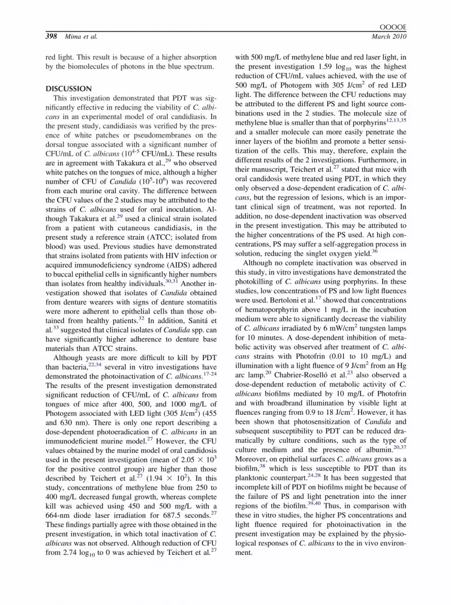
Light scattering within the tongue sample is shown inFig. 6. This image illustrates that blue light presented alower scattering than red light in the tongue tissue, thus
of the tongue of immunosuppressed mice 4 days after yeast10 represents 400, 500, and 1000 mg/L of PS, respectively;
spectively; P�L� B and P�L� R represent animals treatedd P�L� 10 represent animals treated with PS alone at 400,re not treated with PS or light (positive control group). Eachntrol group (P�L�) that consisted of 7 mice. *SignificantError bars indicate standard deviations.
Fig. 6. Blue and red LED light scattering within the tonguetissue. Two additional animals, which were not immunosup-pressed, did not receive C. albicans inoculation or any treat-ment, were used for this analysis. Note that the blue light iscompletely absorbed in the superficial tissue layers and thered light penetrates deeper into the tongue.
orsum5, andght, re
5 anthat wetive co
a higher penetration into the tissue was observed with
OOOOE398 Mima et al. March 2010
red light. This result is because of a higher absorptionby the biomolecules of photons in the blue spectrum.
DISCUSSIONThis investigation demonstrated that PDT was sig-
nificantly effective in reducing the viability of C. albi-cans in an experimental model of oral candidiasis. Inthe present study, candidiasis was verified by the pres-ence of white patches or pseudomembranes on thedorsal tongue associated with a significant number ofCFU/mL of C. albicans (104-5 CFU/mL). These resultsare in agreement with Takakura et al.,29 who observedwhite patches on the tongues of mice, although a highernumber of CFU of Candida (105-106) was recoveredfrom each murine oral cavity. The difference betweenthe CFU values of the 2 studies may be attributed to thestrains of C. albicans used for oral inoculation. Al-though Takakura et al.29 used a clinical strain isolatedfrom a patient with cutaneous candidiasis, in thepresent study a reference strain (ATCC; isolated fromblood) was used. Previous studies have demonstratedthat strains isolated from patients with HIV infection oracquired immunodeficiency syndrome (AIDS) adheredto buccal epithelial cells in significantly higher numbersthan isolates from healthy individuals.30,31 Another in-vestigation showed that isolates of Candida obtainedfrom denture wearers with signs of denture stomatitiswere more adherent to epithelial cells than those ob-tained from healthy patients.32 In addition, Sanitá etal.33 suggested that clinical isolates of Candida spp. canhave significantly higher adherence to denture basematerials than ATCC strains.
Although yeasts are more difficult to kill by PDTthan bacteria,22,34 several in vitro investigations havedemonstrated the photoinactivation of C. albicans.17-24
The results of the present investigation demonstratedsignificant reduction of CFU/mL of C. albicans fromtongues of mice after 400, 500, and 1000 mg/L ofPhotogem associated with LED light (305 J/cm2) (455and 630 nm). There is only one report describing adose-dependent photoeradication of C. albicans in animmunodeficient murine model.27 However, the CFUvalues obtained by the murine model of oral candidosisused in the present investigation (mean of 2.05 � 103
for the positive control group) are higher than thosedescribed by Teichert et al.27 (1.94 � 102). In thisstudy, concentrations of methylene blue from 250 to400 mg/L decreased fungal growth, whereas completekill was achieved using 450 and 500 mg/L with a664-nm diode laser irradiation for 687.5 seconds.27
These findings partially agree with those obtained in thepresent investigation, in which total inactivation of C.albicans was not observed. Although reduction of CFU
from 2.74 log10 to 0 was achieved by Teichert et al.27with 500 mg/L of methylene blue and red laser light, inthe present investigation 1.59 log10 was the highestreduction of CFU/mL values achieved, with the use of500 mg/L of Photogem with 305 J/cm2 of red LEDlight. The difference between the CFU reductions maybe attributed to the different PS and light source com-binations used in the 2 studies. The molecule size ofmethylene blue is smaller than that of porphyrins12,13,35
and a smaller molecule can more easily penetrate theinner layers of the biofilm and promote a better sensi-tization of the cells. This may, therefore, explain thedifferent results of the 2 investigations. Furthermore, intheir manuscript, Teichert et al.27 stated that mice withoral candidosis were treated using PDT, in which theyonly observed a dose-dependent eradication of C. albi-cans, but the regression of lesions, which is an impor-tant clinical sign of treatment, was not reported. Inaddition, no dose-dependent inactivation was observedin the present investigation. This may be attributed tothe higher concentrations of the PS used. At high con-centrations, PS may suffer a self-aggregation process insolution, reducing the singlet oxygen yield.36
Although no complete inactivation was observed inthis study, in vitro investigations have demonstrated thephotokilling of C. albicans using porphyrins. In thesestudies, low concentrations of PS and low light fluenceswere used. Bertoloni et al.17 showed that concentrationsof hematoporphyrin above 1 mg/L in the incubationmedium were able to significantly decrease the viabilityof C. albicans irradiated by 6 mW/cm2 tungsten lampsfor 10 minutes. A dose-dependent inhibition of meta-bolic activity was observed after treatment of C. albi-cans strains with Photofrin (0.01 to 10 mg/L) andillumination with a light fluence of 9 J/cm2 from an Hgarc lamp.20 Chabrier-Roselló et al.23 also observed adose-dependent reduction of metabolic activity of C.albicans biofilms mediated by 10 mg/L of Photofrinand with broadbrand illumination by visible light atfluences ranging from 0.9 to 18 J/cm2. However, it hasbeen shown that photosensitization of Candida andsubsequent susceptibility to PDT can be reduced dra-matically by culture conditions, such as the type ofculture medium and the presence of albumin.20,37
Moreover, on epithelial surfaces C. albicans grows as abiofilm,38 which is less susceptible to PDT than itsplanktonic counterpart.24,28 It has been suggested thatincomplete kill of PDT on biofilms might be because ofthe failure of PS and light penetration into the innerregions of the biofilm.39,40 Thus, in comparison withthese in vitro studies, the higher PS concentrations andlight fluence required for photoinactivation in thepresent investigation may be explained by the physio-logical responses of C. albicans to the in vivo environ-
ment.
OOOOEVolume 109, Number 3 Mima et al. 399
Effective results of photoinactivation of C. albicanshave also been reported using phenothiazine dyes, to-luidine blue O, and methylene blue (TBO and MBrespectively). The results obtained by Wilson andMia18 demonstrated photokilling of C. albicans by anumber of PS in association with light from low-powerlaser, with TBO and helium/neon gas laser being themost effective combination. These authors also verifiedphotosensitization of C. albicans under conditions re-sembling those that would be encountered in vivo (insaliva, serum, saline, and broth at a pH ranging from4.0 to 7.0).41 Jackson et al.19 found that the hyphal formof C. albicans was more susceptible to photoinactiva-tion, requiring a lower TBO concentration than theyeast form. The germ tube formation of C. albicans, atransition state from budding to hyphal cells and anessential phase to virulence, was also inhibited by PDTusing MB (0.027 to 13.37 mM) and laser light (28J/cm2).42 In contrast to bacterial species, C. albicanssensitized by 100 mg/L MB was killed by visible lightfrom a slide projector (42 mW/cm2) after only 20minutes of irradiation.34 Giroldo et al.43 verified inhi-bition of C. albicans CFUs to approximately 50% in thepresence of 50 mg/L MB and laser light irradiation of28 J/cm2. These authors also verified that cell deathpromoted by this combination can be related to damageto the plasma membrane of the yeast. However,Demidova and Hamblin22 showed that 5 �M of poly-L–lysine chlorine(e6) conjugate was more effective inkilling C. albicans than 50 �M of TBO and 200 �M ofrose bengal after illumination at fluences ranging from0 to 200 J/cm2. Although the effectiveness of MB as aPS was also verified in vivo,27 dyes have the undesir-able effect of staining teeth, lips, tongue, buccal mu-cosa, and prosthetic devices. For this reason, a nondyephotosensitizer would be more useful in the oral cavity.
Laser light sources are usually chosen to performPDT. However, their high cost makes the appliancesinaccessible to many institutions. Recently, alternativelight sources, such as LED, have been successfully usedin PDT.25,26,44 In the present investigation, LED wasused as a light source because of its ability to irradiatelarger areas than is possible with collimated laser light.Moreover, LED technology is simpler and has a lowercost than laser. The results of this study showed nosignificant difference between the LED light wave-lengths used (blue and red) for photoinactivation of C.albicans. It was an unexpected result, because the max-imum absorption band of Photogem is closer to 455 nm(blue) and 630 nm (red) matches the lowest Q-band.However, the chosen energy doses for both wave-lengths were effective even for the red LED. Probably,the deeper penetration of red light into the biofilm, and
therefore a higher-treated volume, may compensate itsweak absorption by Photogem. This may explain thelack of difference between blue and red LED lightsobserved in the present investigation.
Light propagation in biological tissues has been eval-uated using direct measurement and complex mathe-matical equations.45-47 Absorption and scattering coef-ficients of tissues are factors responsible for severaleffects of light-tissue interaction; however, the opticalproperties of the same tissue may differ among pa-tients.48 Despite these considerations, it is known thatlight penetration into the tissue is proportional to itswavelength when considering the UV-near infraredrange, i.e., the longer the wavelength, the deeper is thelight penetration.49-52 The shorter wavelengths are bet-ter absorbed by biological molecules, i.e., the greaterpart of light intensity is absorbed by the superficialtissue layers and as a consequence, lower penetration isobserved. Whereas, photons in the red and infraredrange are less absorbed by the biological chromophoresand a higher light penetration is achieved. In addition,noncoherent light sources, such as LED lights, wouldbe expected to achieve less optical penetration becauseof increased scattering.50 Light scattering can be seenas a type of light diffusion as the penetration proceedsalong the tissue. In the present investigation, it wasobserved that red LED light showed higher scatteringthan blue LED light in the tongue tissue.
Clinically, a 20-minute irradiation may be consid-ered too long. However, a previously conducted pilotstudy demonstrated that shorter times (2.45, 10.00, and15.00 minutes, which correspond to 37.5, 152.0, and229.0 J/cm2, respectively) were not effective whencombined with 500, 300, 100, and 50 mg/L of Photo-gem (data not shown). As light fluence is proportionalto exposure time and power output, increasing thepower output of the light source would result in thesame light fluence in a shorter exposure time. None-theless, a higher power output led to greater heatingof the light source, which may not be clinically safe. Onthe other hand, when topical or systemic antifungaldrugs are used, the medicament should be applied morethan once a day, for several days, weeks, and, some-times, months. The medicine should be taken for aslong as recommended by the professional, because ifthe drug is stopped too soon, the symptoms may return.Thus, in comparison with antifungal agents, 20 minutesof illumination during a PDT session may be clinicallyapplicable. Nonetheless, further clinical trials should beconducted to investigate whether the parameters foundin this investigation would be effective in humans.
The histological evaluation of the animals’ tongueswas performed after they were killed on day 6, whichcorresponds to 24 hours after PDT for the animals from
P�L�, P�L, and P�L� groups. This analysis showed
OOOOE400 Mima et al. March 2010
that PDT had no adverse effects on the adjacent tissue.This finding is in agreement with that demonstrated byKömerik et al.,15 who also verified no damage to peri-odontal tissues of rats submitted to PDT. Junqueira etal.53 described fewer epithelial alterations and lowerchronic inflammatory response in rats submitted toPDT and more intense lesions in rats treated only withlaser. In the present study, yeast and pseudohyphaewere observed only in the keratinized layer withoutinvasion of the epithelium and the inflammation in thesubjacent connective tissue was scored as mild for allmice infected with C. albicans, whether or not theywere submitted to PDT. Therefore, the inflammationobserved in the connective tissue may be associatedwith Candida infection but not with PDT. Nevertheless,the murine model of oral candidosis used in this inves-tigation has previously shown the destruction of severalepithelial layers.29 This finding could be attributed tothe strain used by these authors, as clinical strainsisolated from infections showed increased virulencefactors.32 The findings of the present investigation par-tially corroborate those of Teichert et al.,27 as theseauthors also observed yeast and pseudohyphae limitedto the keratinized layer, but lack of inflammatorychanges in mice not submitted to PDT and subepithelialinflammatory infiltrate and neutrophilic exocytosis af-ter PDT. Moreover, the different mouse strain, time ofassays, PS, and light source used may also explain thedivergence between the histological findings in thesestudies.
As regards the limitations of this study, differentlight fluences were not investigated, because it waspreviously found that shorter illumination times werenot effective, as was explained previously, and longerillumination times were considered inapplicable. More-over, the long-term effect of PDT was not evaluated.Sampling (recovery of C. albicans from tongues ofmice and yeast culture) was done at only one timepoint, immediately after PDT, to assess the yeast via-bility immediately after treatment. However, samplingcould be performed in different periods and a long-termeffect of PDT on the yeast viability would be evaluated.In the present investigation, decrease of C. albicanscounts was verified, but the macroscopic regression ofwhite patches or pseudomembranes was not assessed.The partial or total disappearance of these lesions couldhave been evaluated if the mice had been killed atdifferent intervals of time (longer periods than 24 hoursafter PDT).
In conclusion, the results of this study demonstratedthat Photogem-mediated PDT promoted significant re-duction in the viability of C. albicans biofilm withoutharming the tongue tissue. These results indicate the
critical importance of determining effective in vivoPDT parameters before clinical applications. However,the results cannot be extrapolated to a clinical situation,as the oral environment of humans is different (micro-biota and biofilm composition, salivary flux, food hab-its, and so forth). Further in vivo studies are still nec-essary to investigate the parameters required forcomplete inactivation of Candida biofilms and the con-sequence of a repeatable therapy. Clinical trials are alsorequired to evaluate the effect of PDT as a treatment oforal candidosis.
REFERENCES1. Totti MA, dos Santos EB, de Almeida OP, Koga-Ito CY, Jorge
AO. Oral candidosis by Candida albicans in normal and xeros-tomic mice. Braz Oral Res 2004;18:202-7.
2. Scully C, eL-Kabir M, Samaranayake LP. Candida and oralcandidosis: a review. Crit Rev Oral Biol Med 1994;5:125-57.
3. Egusa H, Soysa NS, Ellepola AN, Yatani H, Samaranayake LP.Oral candidosis in HIV-infected patients. Curr HIV Res 2008;6:485-99.
4. Samaranayake LP, MacFarlane TW, editors. Oral candidosis.London: Wright; 1990. p. 265.
5. Allen CM. Animal models of oral candidiasis. A review. OralSurg Oral Med Oral Pathol 1994;78:216-21.
6. Samaranayake YH, Samaranayake LP. Experimental oral candi-diasis in animal models. Clin Microbiol Rev 2001;14:398-429.
7. Rex JH, Rinaldi MG, Pfaller MA. Resistance of Candida speciesto fluconazole. Antimicrob Agents Chemother 1995;39:1-8.
8. Cannon RD, Lamping E, Holmes AR, Niimi K, Tanabe K, NiimiM, et al. Candida albicans drug resistance: another way to copewith stress. Microbiology 2007;153:3211-7.
9. Mishra NN, Prasad T, Sharma N, Payasi A, Prasad R, Gupta DK,et al. Pathogenicity and drug resistance in Candida albicans andother yeast species. A review. Acta Microbiol Immunol Hung2007;54:201-35.
10. Johnson EM, Warnock DW, Luker J, Porter SR, Scully C.Emergence of azole drug resistance in Candida species fromHIV-infected patients receiving prolonged fluconazole therapyfor oral candidosis. J Antimicrob Chemother 1995;35:103-14.
11. Hitchcock CA. Resistance of Candida albicans to azole antifun-gal agents. Biochem Soc Trans 1993;21:1039-47.
12. Donnelly RF, McCarron PA, Tunney MM. Antifungal photody-namic therapy. Microbiol Res 2008;163:1-12.
13. Konopka K, Goslinski T. Photodynamic therapy in dentistry. JDent Res 2007;86:694-707.
14. Paulino TP, Ribeiro KF, Thedei G Jr, Tedesco AC, CiancagliniP. Use of hand held photopolymerizer to photoinactivate Strep-tococcus mutans. Arch Oral Biol 2005;50:353-9.
15. Kömerik N, Nakanishi H, MacRobert AJ, Henderson B, SpeightP, Wilson M. In vivo killing of Porphyromonas gingivalis bytoluidine blue-mediated photosensitization in an animal model.Antimicrob Agents Chemother 2003;47:932-40.
16. Zeina B, Greenman J, Corry D, Purcell WM. Antimicrobialphotodynamic therapy: assessment of genotoxic effects on ker-atinocytes in vitro. Br J Dermatol 2003;148:229-32.
17. Bertoloni G, Reddi E, Gatta M, Burlini C, Jori G. Factorsinfluencing the hematoporphyrin-sensitized photoinactivation ofCandida albicans. J Gen Microbiol 1989;135:957-66.
18. Wilson M, Mia N. Sensitisation of Candida albicans to killing bylow-power laser light. J Oral Pathol Med 1993;22:354-7.
19. Jackson Z, Meghji S, MacRobert A, Henderson B, Wilson M.
Killing of the yeast and hyphal forms of Candida albicans using
OOOOEVolume 109, Number 3 Mima et al. 401
a light-activated antimicrobial agent. Lasers Med Sci 1999;14:150-7.
20. Bliss JM, Bigelow CE, Foster TH, Haidaris C. Susceptibility ofCandida species to photodynamic effects of photofrin. Antimi-crob Agents Chemother 2004;48:2000-6.
21. Lambrechts SA, Aalders MC, Van Marle J. Mechanistic study ofthe photodynamic inactivation of Candida albicans by a cationicporphyrin. Antimicrob Agents Chemother 2005;49:2026-34.
22. Demidova TN, Hamblin MR. Effect of cell-photosensitizer bind-ing and cell density on microbial photoinactivation. AntimicrobAgents Chemother 2005;49:2329-35.
23. Chabrier-Roselló Y, Foster TH, Pérez-Nazario N, Mitra S, Hai-daris CG. Sensitivity of Candida albicans germ tubes and bio-films to photofrin-mediated phototoxicity. Antimicrob AgentsChemother 2005;49:4288-95.
24. Donnelly RF, McCarron PA, Tunney MM, David Woolfson A.Potential of photodynamic therapy in treatment of fungal infec-tions of the mouth. Design and characterisation of a mucoadhe-sive patch containing toluidine blue O. J Photochem Photobiol B2007;86:59-69.
25. Zanin IC, Gonçalves RB, Junior AB, Hope CK, Pratten J. Sus-ceptibility of Streptococcus mutans biofilms to photodynamictherapy: an in vitro study. J Antimicrob Chemother 2005;56:324-30.
26. Peloi LS, Soares RR, Biondo CE, Souza VR, Hioka N, KimuraE. Photodynamic effect of light-emitting diode light on cellgrowth inhibition induced by methylene blue. J Biosci 2008;33:231-7.
27. Teichert MC, Jones JW, Usacheva MN, Biel MA. Treatment oforal candidiasis with methylene blue–mediated photodynamictherapy in an immunodeficient murine model. Oral Surg OralMed Oral Pathol Oral Radiol Endod 2002;93:155-60.
28. Dovigo LN, Pavarina AC, Mima EG, Giampaolo ET, VerganiCE, Bagnato VS. Fungicidal effect of photodynamic therapyagainst fluconazole-resistant Candida albicans and Candida gla-brata. Mycoses 2009 [Epub ahead of press].
29. Takakura N, Sato Y, Ishibashi H, Oshima H, Uchida K, Yamagu-chi H, et al. A novel murine model of oral candidiasis with localsymptoms characteristic of oral thrush. Microbiol Immunol2003;47:321-6.
30. Sweet SP, Cookson S, Challacombe SJ. Candida albicans iso-lates from HIV-infected and AIDS patients exhibit enhancedadherence to epithelial cells. J Med Microbiol 1995;43:452-7.
31. Schwab U, Milatovic D, Braveny I. Increased adherence ofCandida albicans to buccal epithelial cells from patients withAIDS. Eur J Clin Microbiol Infect Dis 1997;16:848-51.
32. Lyon JP, de Resende MA. Correlation between adhesion, en-zyme production, and susceptibility to fluconazole in Candidaalbicans obtained from denture wearers. Oral Surg Oral MedOral Pathol Oral Radiol Endod 2006;102:632-8.
33. Sanitá PV, Vergani CE, Giampaolo ET, Pavarina AC, MachadoAL. Growth of Candida species on complete dentures: effect ofmicrowave disinfection. Mycoses 2009;52:154-60.
34. Zeina B, Greenman J, Purcell WM, Das B. Killing of cutaneousmicrobial species by photodynamic therapy. Br J Dermatol 2001;144:274-8.
35. Wainwright M. Photodynamic antimicrobial chemotherapy(PACT). J Antimicrob Chemother 1998;42:13-28.
36. Sternberg ED, Dolphin D. Porphyrin-based photosensitizers foruse in photodynamic therapy. Tetrahedron 1998;54:4151-202.
37. Lambrechts SA, Aalders MC, Verbraak FD, Lagerberg JW,
Dankert JB, Schuitmaker JJ. Effect of albumin on thephotodynamic inactivation of microorganisms by a cationic por-phyrin. J Photochem Photobiol B 2005;79:51-7.
38. Douglas LJ. Candida biofilms and their role in infection. TrendsMicrobiol 2003;11:30-6.
39. Soukos NS, Mulholland SE, Socransky SS, Doukas AG. Pho-todestruction of human dental plaque bacteria: enhancement ofthe photodynamic effect by photomechanical waves in an oralbiofilm model. Lasers Surg Med 2003;33:161-8.
40. de Beer D, Srinivasan R, Stewart PS. Direct measurement ofchlorine penetration into biofilms during disinfection. Appl En-viron Microbiol 1994;60:4339-44.
41. Wilson M, Mia N. Effect of environmental factors on the lethalphotosensitization of Candida albicans in vitro. Lasers Med Sci1994;9:105-9.
42. Munin E, Giroldo LM, Alves LP, Costa MS. Study of germ tubeformation by Candida albicans after photodynamic antimicro-bial chemotherapy (PACT). J Photochem Photobiol B 2007;88:16-20.
43. Giroldo LM, Felipe MP, de Oliveira MA, Munin E, Alves LP,Costa MS. Photodynamic antimicrobial chemotherapy (PACT)with methylene blue increases membrane permeability in Can-dida albicans. Lasers Med Sci 2009;24:109-12.
44. Soares BM, da Silva DL, Sousa GR, Amorim JC, de ResendeMA, Pinotti M, et al. In vitro photodynamic inactivation ofCandida spp. growth and adhesion to buccal epithelial cells. JPhotochem Photobiol B 2009;94:65-70.
45. Maschesini R, Bertoni A, Andreola S, Mellari E, Sichirollo AE.Extinction and absorption coefficients and scattering phase func-tions of human tissue in vitro. Appl Opt 1989;28:2318-24.
46. Cheong WF, Prahl SA, Welch AJ. A review of the opticalproperties of biological tissues. IEEE J Quant Electr 1990;26:2166-85.
47. Arnfield MR, Mathew RP, Tulip J, McPhee MS. Analysis ofoptical coefficients using an approximate equation valid forcomparable absorption and scattering. Phys Med Biol 1992;37:1219-30.
48. Star WM. Light dosimetry in vivo. Phys Med Biol 1997;42:763-87.
49. Stolik S, Delgado JA, Pérez A, Anasagasti L. Measurement ofthe penetration depths of red and near infrared light in human “exvivo” tissues. J Photochem Photobiol B 2000;57:90-3.
50. Alexiades-Armenakas M. Laser-mediated photodynamic ther-apy. Clin Dermatol 2006;24:16-25.
51. Wei HJ, Xing D, Wu GY, Jin Y, Gu HM. Optical properties ofhuman normal bladder tissue at five different wavelengths oflaser and their linearly polarized laser irradiation in vitro. Spec-troscopy and Spectral Analysis 2004;24:1039-41.
52. Taroni P, Pifferi A, Torricelli A, Comelli D, Cubeddu R. In vivoabsorption and scattering spectroscopy of biological tissues. Pho-tochem Photobiol Sci 2003;2:124-9.
53. Junqueira JC, da Silva Martins J, Faria RL, Colombo CE, JorgeAO. Photodynamic therapy for the treatment of buccal candidi-asis in rats. Lasers Med Sci 2009;24:877-84.
Reprint requests:
Ana Cláudia Pavarina, DDS, MSc, PhDFaculdade de Odontologia de Araraquara-UNESPRua Humaitá, n° 1680CEP: 14801–903, Araraquara, SP, Brazil
[email protected]


