Brucella abortus Strain RB51 Outer Membrane Vesicles as a Vaccine Against Brucellosis in a Murine Model by Clifton Clark Cassidy II Thesis submitted to the faculty of the Virginia Polytechnic Institute and State University In partial fulfillment of the requirements for the degree of Master of Science in Biomedical and Veterinary Sciences Stephen M. Boyle, Chairman Nammalwar Sriranganathan Isis K. Mullarky February 3, 2010 Blacksburg, VA Keywords: Brucella abortus, strain RB51, outer membrane vesicles, blebs, vaccine, mice, OMV, malaria, FRAP, adjuvant
Transcript
Brucella abortus Strain RB51 Outer Membrane Vesicles as a Vaccine Against Brucellosis in a Murine Model
by
Clifton Clark Cassidy II
Thesis submitted to the faculty of the Virginia Polytechnic Institute and State University
In partial fulfillment of the requirements for the degree of
Brucella abortus Strain RB51 Outer Membrane Vesicles as a Vaccine Against Brucellosis in a Murine Model
by
Clifton Clark Cassidy II
Abstract Brucella abortus is a zoonotic agent that primarily infects cattle and causes
brucellosis. B. abortus strain RB51 is a live, attenuated vaccine licensed for cattle.
However, there is no available vaccine to prevent human brucellosis. Outer membrane
vesicles have been tested as potential vaccines to prevent diseases caused by bacterial
species. OMV are constantly released from Gram-negative bacteria. They are comprised
principally of the outer membrane components and periplasmic proteins from the
bacterial cell envelope. The research in this thesis examined the adjuvant property of
non-replicative, metabolically active irradiated strain RB51 and the protective ability of
OMV derived from strain RB51. Irradiated B. abortus strain RB51 was assessed for its
ability to act as an adjuvant to induce protection against malaria. It was found that
irradiated B. abortus strain RB51 administered along with fasciclin related adhesive
protein (FRAP) to mice induced a protective immune response and a significant decrease
in parasitemia after challenge with Plasmodium berghei. Strain RB51 and strain RB51
over-producing Cu/Zn superoxide dismutase (Cu/Zn SOD) were used to produce OMV.
Western blotting and SDS-PAGE gel staining confirmed the presence of OMV and the
over-production of Cu/Zn SOD. OMV were delivered to mice using an intraperitoneal
route and, in some cases, with aluminum hydroxide adjuvant. The immune response was
assessed by antibody isotyping with respect to OMV and measuring splenic clearance
(i.e. protection) from a B. abortus strain 2308 challenge. The results demonstrate that
iii
OMV from B. abortus strain RB51 or strain RB51 over producing Cu/Zn SOD produced
a Th1 polarized immune response as measured by specific OMV antibodies and
cytokines but no statistically significant protection was observed.
iv
Dedication I would like to dedicate this thesis to my parents, Mr. Clifton Cassidy and Mrs.
Cindy Cassidy. Nothing I have done in my life would have been possible without them.
They have always pushed me to be my best and to succeed in life. This work is a product
of the faith and hope that they have always had in me.
v
Acknowledgements
I would like to begin by thanking the Virginia-Maryland Regional College of
Veterinary Medicine, and in particular Dr. Roger Avery, Cindy Booth, and Becky Jones,
for giving me the opportunity to come to this school and do my graduate work. I would
also like to thank them for supporting me financially and emotionally throughout my
graduate career.
Dr. Boyle, I would like to sincerely thank you for everything that you have done
for me. From the first day that I began working, you have always fostered my curiosity
and encouraged me to think on my own. Under your direction, my scientific curiosity
has blossomed and that is something that I will always carry with me through my many
endeavors in life. Thank you for everything that you have done for me.
Dr. Nathan, I have always been delighted to work along side with you. You bring
a youthful joy to everything that you do in life and work. No matter what the question
has been, your door has always been open to me. I want to thank you from the bottom of
my heart for all of these things and for allowing me to work for you.
Dr. Mullarky, I have enjoyed having you on my committee as well as having the
privilege to have you as an instructor. You always ask me questions and cause me to
think of every aspect of the work that I am doing. It is for this, among many other things,
that I want to truly thank you. You have helped instill the instinct in me to always ask
questions, even if I believe that I already know the answer.
vi
I truly want to think all of the people in my committee, as you have all been
wonderful in guiding and directing me through my graduate career. I truly believe that
you have helped prepare me for my next endeavor and the rest of my life.
I would like to thank Gade Kimsawatde for always being supportive of me. She
is always there when I need help or advice. Gade is truly my support and I do not believe
I would have been able to complete this work with out her. For that, I am truly thankful
to Gade.
I would like to thank Nancy Tenpenny and Kay Carlson for always helping me
with anything that I have ever asked of them. CMMID would truly fall apart if it were
not for these wonderful ladies. They always do more than what is expected of them and
never hesitate to help someone in need. Thank you both from the bottom of my heart.
I would like to thank all of the wonderful people that I have met and became
friends with during my graduate career at VMRCVM. I would like to thank, in
particular, Parthiban Rajasekaran, Andrew Herbert, Brent Sanford, Courie Cohen,
Vrushali Chavan, Nathan Beach, Laura Cordoba and Carmine Graniello for being
wonderful friends and making my graduate career and life in Blacksburg much better. I
am indebted to all of you.
Last but certainly not least, I want to thank my parents. My parents are, and
always have been, the driving force in my life. They have been with me every step of the
way. I could not ask or hope for better role models than I have been given. My life is
continually improved and enriched by my parents. Words cannot express how much I
owe to my parents. I love them with all of my heart and I truly am the luckiest person in
the world to have them in my life. Thank you Mom and Dad!
vii
Table of Contents Abstract.......................................................................................................................................ii Dedication................................................................................................................................. iv Acknowledgements................................................................................................................. v List of Figures........................................................................................................................... ix Chapter 1: Literature Review .............................................................................................1
OMV Background.............................................................................................................................................1 OMV Vaccines....................................................................................................................................................3 Malaria Prevalence and Symptoms .........................................................................................................4 Brucella Etiology and Pathogenesis.........................................................................................................5 Brucella Immunity...........................................................................................................................................6 Brucella as an Adjuvant.................................................................................................................................7 Brucella Vaccines .............................................................................................................................................8 Perspective .........................................................................................................................................................9
Chapter 2: Brucella abortus as an Adjuvant for the FRAP Protein of Plasmodium falciparum...................................................................................................... 11 Introduction..................................................................................................................................... 11 Materials and Methods ................................................................................................................. 13 Irradiation........................................................................................................................................................13 Vaccines: subunit and strain construction .......................................................................................13 Vaccine immunizations..............................................................................................................................14 ELISA assay for antibody titrations ......................................................................................................14 Challenge of mice with Plasmodium berghei and assessment of protection.......................15 Statistical Analysis .......................................................................................................................................15
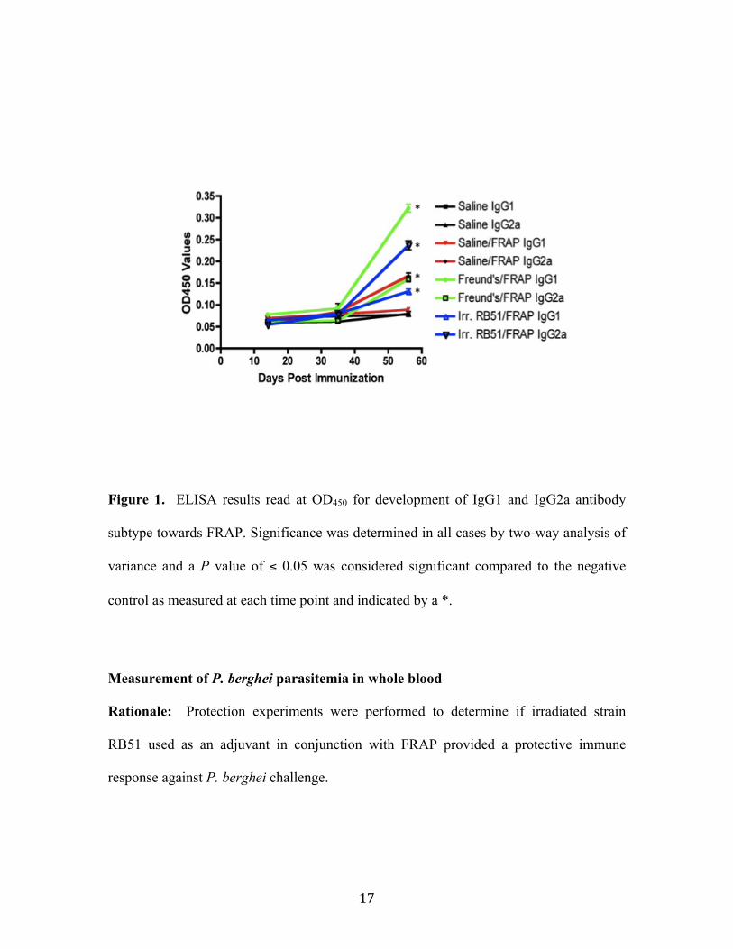
Results................................................................................................................................................ 16 Serologic Response ......................................................................................................................................16 Measurement of P. berghei parasitemia in whole blood .............................................................17
Discussion......................................................................................................................................... 20 Chapter 3: Outer Membrane Vesicle Based Vaccines .............................................. 23 Introduction..................................................................................................................................... 23 Methods and Materials ................................................................................................................. 25 Isolation of OMV............................................................................................................................................25 Electron Microscopy ...................................................................................................................................25 Immunoblot assay........................................................................................................................................26 Vaccine strain construction .....................................................................................................................26 Vaccine immunization ................................................................................................................................27 Challenge of mice with Brucella abortus 2308 and collection of tissues..............................27 ELISA assay for antibody titrations ......................................................................................................27 Mixed Splenocyte culture..........................................................................................................................28 Cytokine Quantitation assays..................................................................................................................29 Statistical Analysis .......................................................................................................................................29
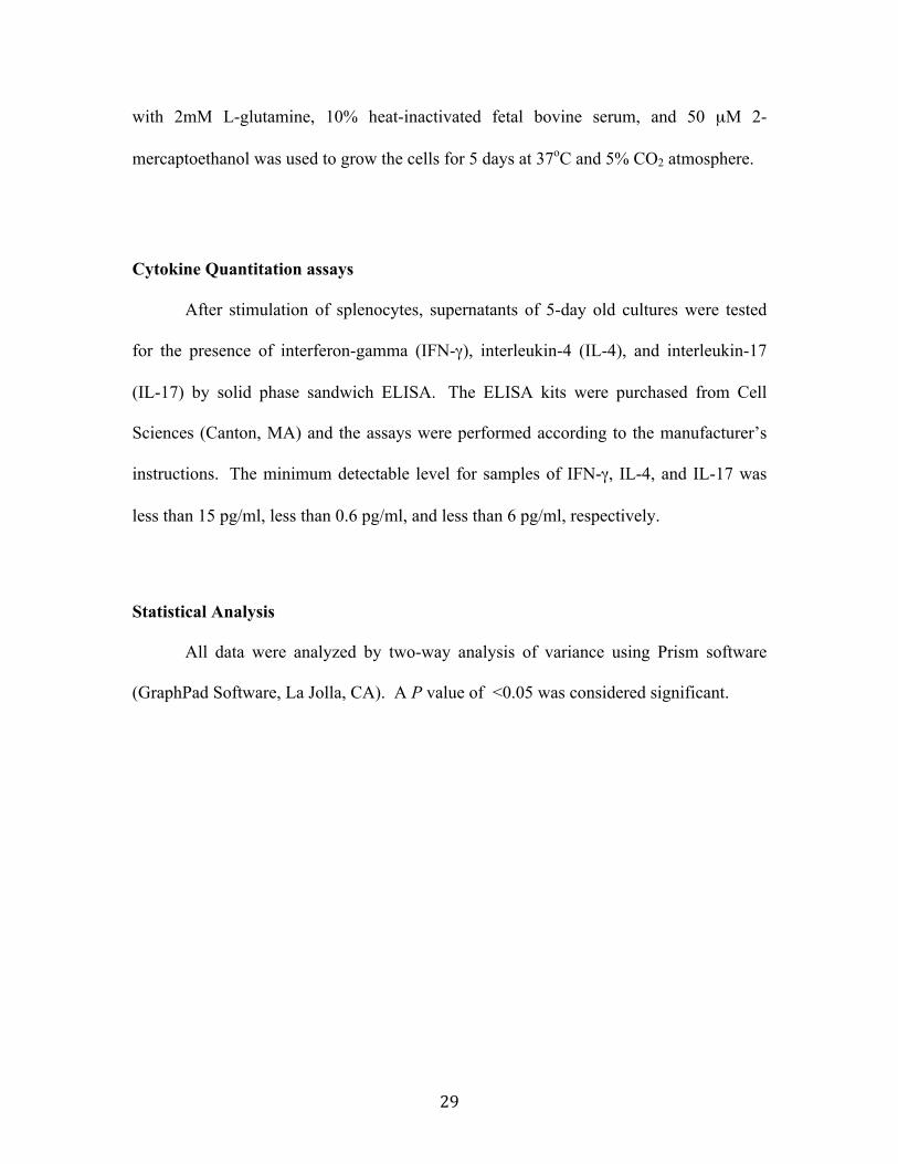
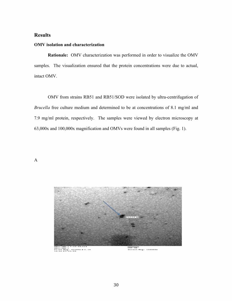
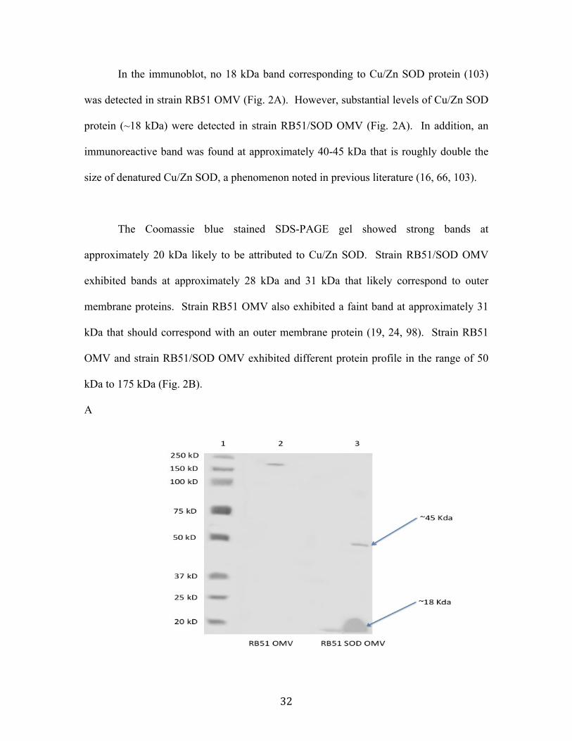
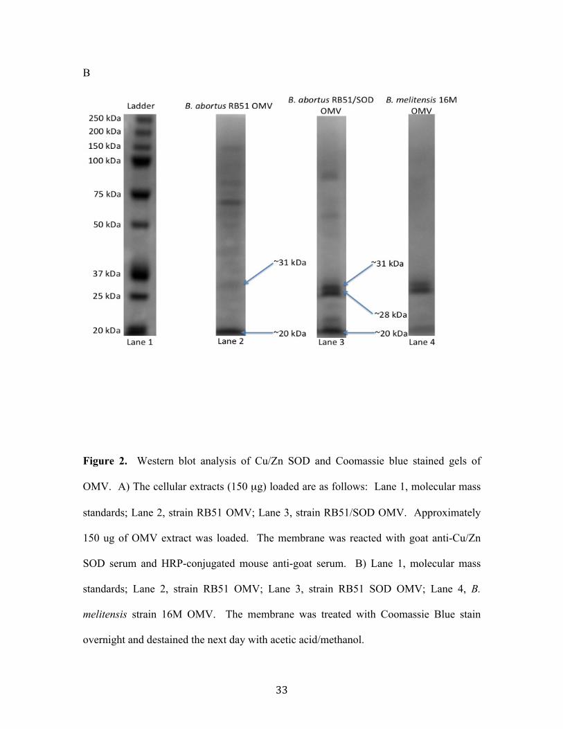
Results................................................................................................................................................ 30 OMV isolation and characterization .....................................................................................................30 Immunodetection of Cu/Zn SOD in OMV from strain RB51 ......................................................31 Serologic Response ......................................................................................................................................34
viii
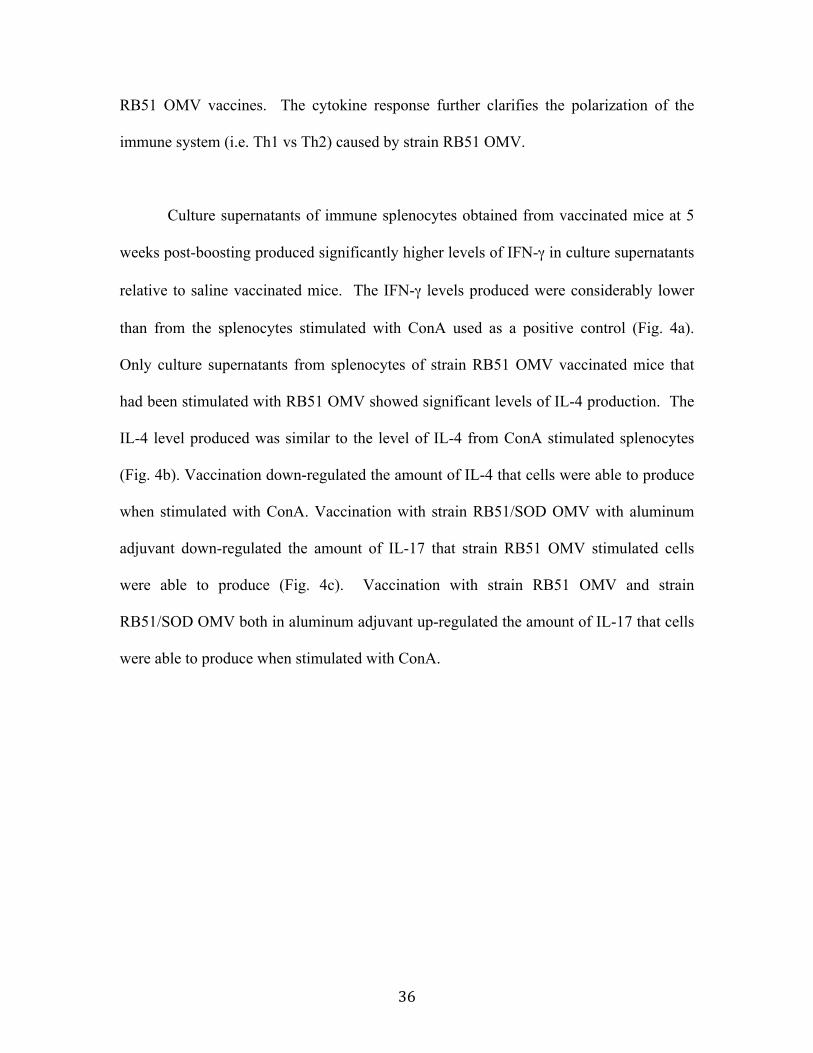
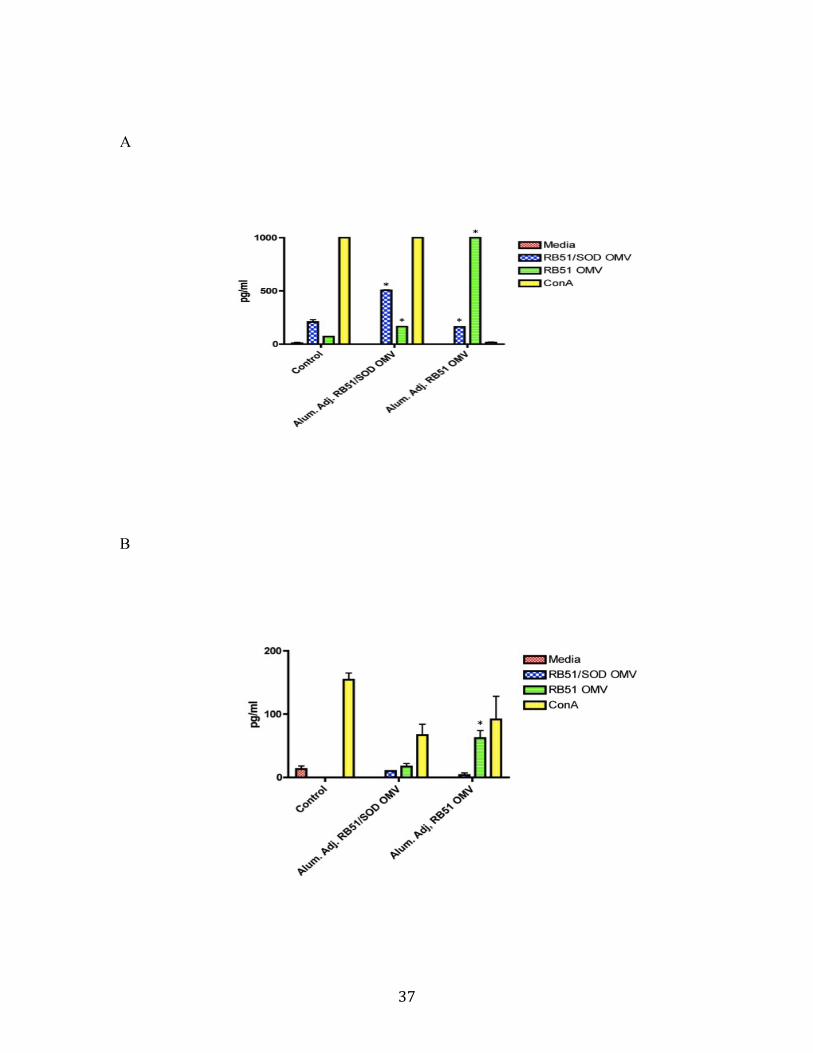
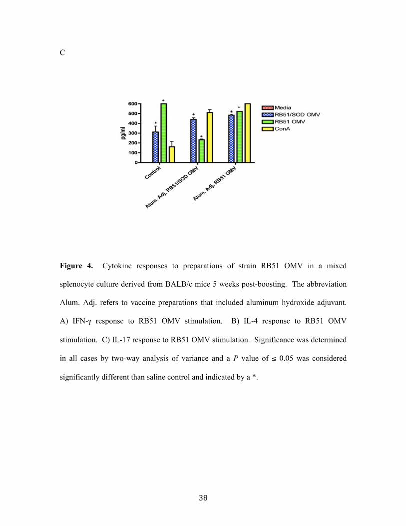
Cytokine Production in Mixed Splenocyte Cultures ......................................................................35 Measurement of B. abortus 2308 CFU in Spleens...........................................................................39
Discussion......................................................................................................................................... 41 Tables ....................................................................................................................................... 49 Table 1: List of vaccines used in the study presented in Chapter 2.............................. 49 Table 2: List of vaccines used in the study presented in Chapter 3.............................. 49
production than the B. abortus extract stimulated splenocytes when compared to positive
controls (100). However, even with the increase in IFN-γ, protection was not achieved in
this study.
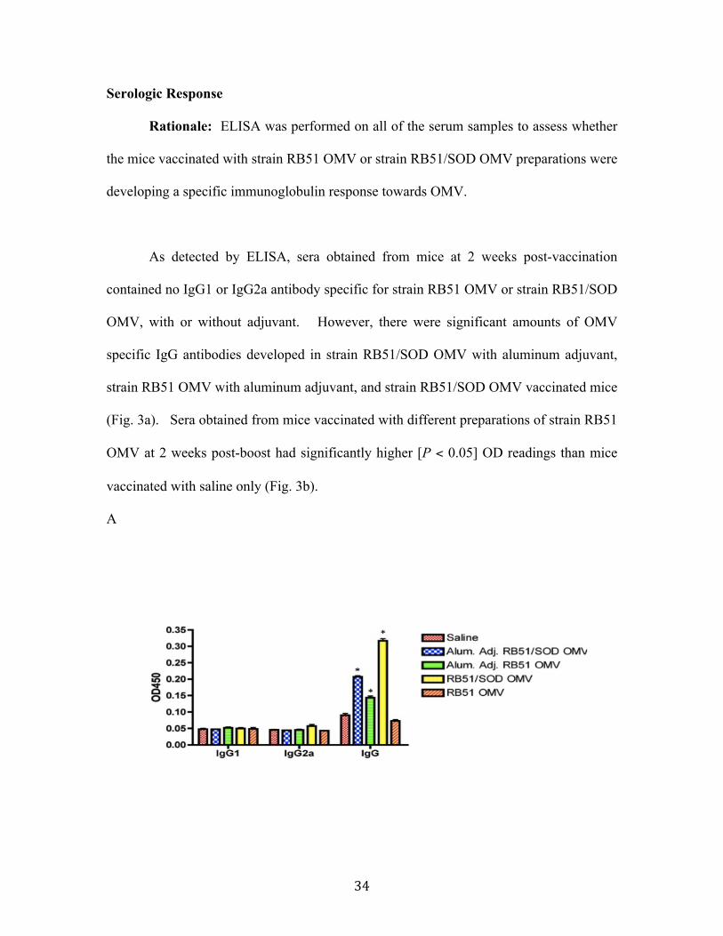
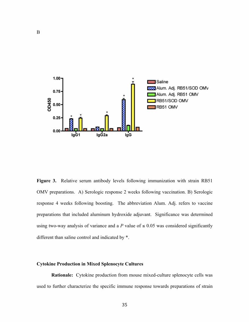
Strain RB51 OMV vaccinated mice did not exhibit an immunoglobulin response
indicating a Th1 response (i.e. IgG2a) that was specific to RB51 OMV (47); however,
46
they were able to induce a very strong IFN-γ response in primary, mixed splenocyte
culture. The data indicate that even though strain RB51 OMV may not induce a strong
immunoglobulin response, they do elicit a very strong cytokine based, Th1 directed
response. Both strain RB51 OMV and strain RB51/SOD OMV stimulate a strong Th1
response that should help the host clear a B. abortus 2308 infection (72, 82, 104).
Strain RB51 OMV stimulated splenocytes produced significant levels of IL-4
indicative of a Th2 response (95). Strain RB51 OMV vaccinated mice showed no trend
of decrease in splenic cfu following a challenge with smooth strain 2308. Strain
RB51/SOD OMV stimulated splenocytes had little to no detectable levels of IL-4 and did
provide protection against smooth strain 2308 in the mouse model.
IL-17 production, which induces a Th17 immune response, is a pro-inflammatory
response that has been shown to be important in fighting infection in aged mice, allergy
related disorders, and autoimmunity disorders (3, 37). Strain RB51 OMV and strain
RB51/SOD OMV stimulated splenocytes showed significant increases in IL-17
production. In the unvaccinated control splenocytes there was a significant increase in
the amount of IL-17 produced when stimulated with strain RB51 OMV or strain
RB51/SOD OMV. However, vaccination with strain RB51/SOD OMV down-regulated
the amount of IL-17 that strain RB51 OMV stimulated splenocytes were able to produce.
Strain RB51 OMV vaccination did not have the same effect on strain RB51/SOD OMV
stimulate splenocytes. Vaccination with either strain RB51 OMV with aluminum
adjuvant or strain RB51/SOD OMV with aluminum adjuvant did not have a positive
47
effect on the amount of IL-17 produced when splenocytes were stimulated with strain
RB51 OMV and strain RB51/SOD OMV. It would be interesting to repeat the
experiment and stimulate with different purified outer membrane proteins and other
purified proteins, such as Cu/Zn SOD, to determine which components of the OMV were
causing this non-specific response. More importantly, it would be useful to determine if
the OMV vaccines protect aged mice against a Brucella challenge (37) to determine if
Brucella based OMV vaccines could potentially be effective in the elderly human
population.
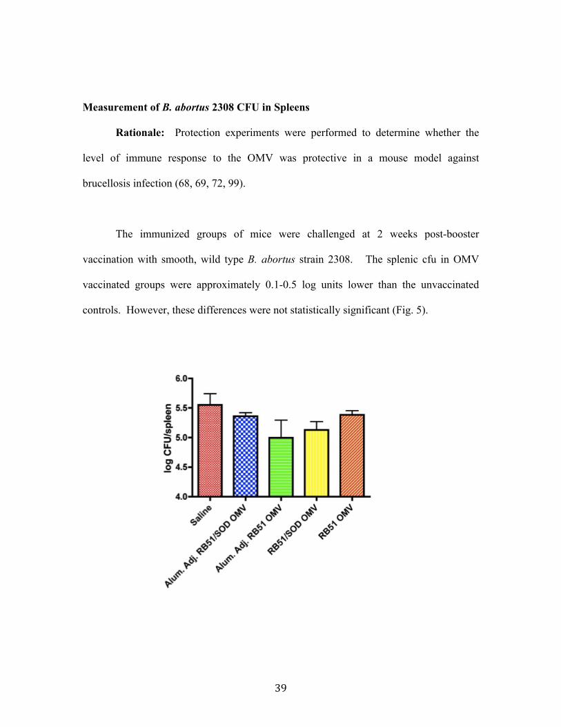
Clearance studies, as indicated by splenic CFU, suggested a protective trend in
mice vaccinated with strain RB51 OMV in aluminum hydroxide adjuvant as well as in
mice vaccinated with strain RB51/SOD OMV. However, neither of the decreases in
splenic CFU was found to be statistically significant. Protection could potentially be
achieved by the use of more OMV during vaccination or additional boosting.
Alternatively, the route of immunization could be varied. In unpublished studies, OMV
from B. melitensis given intramuscularly and boosted, induced protection in mice (A.
Contreras, Mexico, personal communication).
The assay used to determine protein concentration (Bio-Rad, Hercules, CA)
suggested a much higher concentration than what was observed after running an SDS-
PAGE containing extracts of strain RB51 OMV or strain RB51/SOD OMV. Thus the
small amount of actual OMV present in the vaccine preparations could account for the
lack of protection observed in immunized mice. The amount of OMV delivered was
48
sufficient to produce an immune response in terms of higher amounts of serum
immunoglobulins and cytokines, but not sufficient to produce a protective immune
response (68, 82).
Repeating the protection studies with a known concentration of strain RB51 OMV
and strain RB51/SOD OMV would be a very important next step. A more suitable assay
to determine the protein concentration of OMV should be identified. Alternative routes
of inoculation should also be considered as they could affect the type and quality of
immune response that is stimulated (unpublished data, A. Contreras). In this study,
intraperitoneal vaccination was chosen because of previously determined models for
Brucella spp. vaccination and challenge protocols (72, 101, 103) using attenuated strains.
However, strain RB51 OMV are not the same vaccine as a live, attenuated strain RB51
vaccine and may require a different route of administration in order to be effective.
In conclusion, the studies presented in this thesis indicated for the first time that
B. abortus strain RB51 derived OMV are capable of producing an immune response in
mice. A correlation existed between vaccination with strain RB51 derived OMV and a
trends towards protection, although the protection levels were not significant. Future
research should address repeating the protection studies with the aforementioned
suggestions in order to determine the protective capabilities of strain RB51 derived
OMV.
49
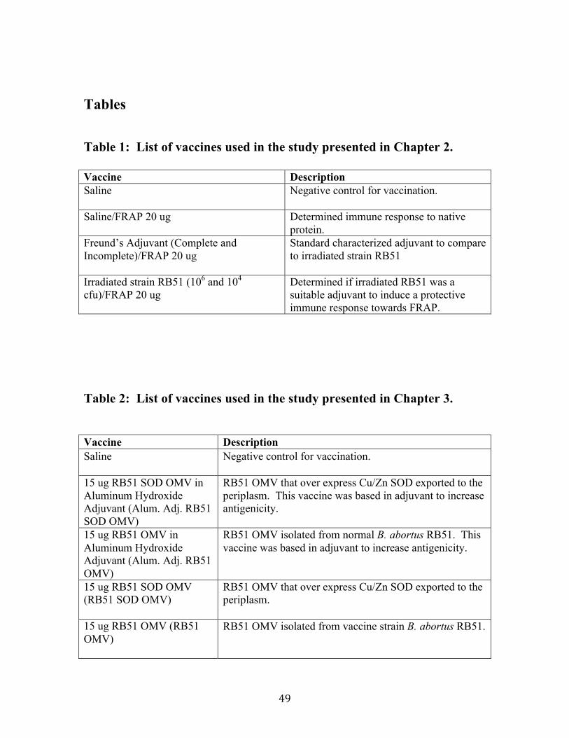
Tables
Table 1: List of vaccines used in the study presented in Chapter 2. Vaccine Description Saline
Negative control for vaccination.
Saline/FRAP 20 ug
Determined immune response to native protein.
Freund’s Adjuvant (Complete and Incomplete)/FRAP 20 ug
Standard characterized adjuvant to compare to irradiated strain RB51
Irradiated strain RB51 (106 and 104 cfu)/FRAP 20 ug
Determined if irradiated RB51 was a suitable adjuvant to induce a protective immune response towards FRAP.
Table 2: List of vaccines used in the study presented in Chapter 3.
Vaccine Description Saline
Negative control for vaccination.
15 ug RB51 SOD OMV in Aluminum Hydroxide Adjuvant (Alum. Adj. RB51 SOD OMV)
RB51 OMV that over express Cu/Zn SOD exported to the periplasm. This vaccine was based in adjuvant to increase antigenicity.
15 ug RB51 OMV in Aluminum Hydroxide Adjuvant (Alum. Adj. RB51 OMV)
RB51 OMV isolated from normal B. abortus RB51. This vaccine was based in adjuvant to increase antigenicity.
15 ug RB51 SOD OMV (RB51 SOD OMV)
RB51 OMV that over express Cu/Zn SOD exported to the periplasm.
15 ug RB51 OMV (RB51 OMV)
RB51 OMV isolated from vaccine strain B. abortus RB51.
50
References: 1. Abraham, A. M., R. Kannangai, and G. Sridharan. 2008. Nanotechnology: a
new frontier in virus detection in clinical practice. Indian J Med Microbiol 26:297-301.
2. Adams, L. G. 2002. The pathology of brucellosis reflects the outcome of the battle between the host genome and the Brucella genome. Vet Microbiol 90:553-61.
3. Aggarwal, S., and A. L. Gurney. 2002. IL-17: prototype member of an emerging cytokine family. J Leukoc Biol 71:1-8.
4. Al-Tawfiq, J. A. 2008. Therapeutic options for human brucellosis. Expert Rev Anti Infect Ther 6:109-20.
5. Alaniz, R. C., B. L. Deatherage, J. C. Lara, and B. T. Cookson. 2007. Membrane vesicles are immunogenic facsimiles of Salmonella typhimurium that potently activate dendritic cells, prime B and T cell responses, and stimulate protective immunity in vivo. J Immunol 179:7692-701.
6. Andersen, S. R., G. Bjune, J. Lyngby, K. Bryn, and E. Jantzen. 1995. Short-chain lipopolysaccharide mutants of serogroup B Neisseria meningitidis of potential value for production of outer membrane vesicle vaccines. Microb Pathog 19:159-68.
7. Araj, G. F. 1999. Human brucellosis: a classical infectious disease with persistent diagnostic challenges. Clin Lab Sci 12:207-12.
8. Arigita, C., W. Jiskoot, J. Westdijk, C. van Ingen, W. E. Hennink, D. J. Crommelin, and G. F. Kersten. 2004. Stability of mono- and trivalent meningococcal outer membrane vesicle vaccines. Vaccine 22:629-42.
9. Arigita, C., T. Luijkx, W. Jiskoot, M. Poelen, W. E. Hennink, D. J. Crommelin, P. Ley, C. Els, and G. F. Kersten. 2005. Well-defined and potent liposomal meningococcal B vaccines adjuvated with LPS derivatives. Vaccine 23:5091-8.
10. Balsalobre, C., J. M. Silvan, S. Berglund, Y. Mizunoe, B. E. Uhlin, and S. N. Wai. 2006. Release of the type I secreted alpha-haemolysin via outer membrane vesicles from Escherichia coli. Mol Microbiol 59:99-112.
11. Beckett, F. W., and S. C. MacDiarmid. 1985. The effect of reduced-dose Brucella abortus strain 19 vaccination in accredited dairy herds. Br Vet J 141:507-14.
12. Beveridge, T. J. 1999. Structures of gram-negative cell walls and their derived membrane vesicles. J Bacteriol 181:4725-33.
13. Beveridge, T. J., and J. L. Kadurugamuwa. 1996. Periplasm, periplasmic spaces, and their relation to bacterial wall structure: novel secretion of selected periplasmic proteins from Pseudomonas aeruginosa. Microb Drug Resist 2:1-8.
14. Boigegrain, R. A., I. Salhi, M. T. Alvarez-Martinez, J. Machold, Y. Fedon, M. Arpagaus, C. Weise, M. Rittig, and B. Rouot. 2004. Release of periplasmic proteins of Brucella suis upon acidic shock involves the outer membrane protein Omp25. Infect Immun 72:5693-703.
15. Boutriau, D., J. Poolman, R. Borrow, J. Findlow, J. D. Domingo, J. Puig-Barbera, J. M. Baldo, V. Planelles, A. Jubert, J. Colomer, A. Gil, K. Levie, A.
51
D. Kervyn, V. Weynants, F. Dominguez, R. Barbera, and F. Sotolongo. 2007. Immunogenicity and safety of three doses of a bivalent (B:4:p1.19,15 and B:4:p1.7-2,4) meningococcal outer membrane vesicle vaccine in healthy adolescents. Clin Vaccine Immunol 14:65-73.
16. Bricker, B. J., L. B. Tabatabai, B. A. Judge, B. L. Deyoe, and J. E. Mayfield. 1990. Cloning, expression, and occurrence of the Brucella Cu-Zn superoxide dismutase. Infect Immun 58:2935-9.
17. Cardoso, P. G., G. C. Macedo, V. Azevedo, and S. C. Oliveira. 2006. Brucella spp noncanonical LPS: structure, biosynthesis, and interaction with host immune system. Microb Cell Fact 5:13.
18. Carvalho Neta, A. V., J. P. Mol, M. N. Xavier, T. A. Paixao, A. P. Lage, and R. L. Santos. 2010. Pathogenesis of bovine brucellosis. Vet J 184:146-55.
19. Cassataro, J., K. Pasquevich, L. Bruno, J. C. Wallach, C. A. Fossati, and P. C. Baldi. 2004. Antibody reactivity to Omp31 from Brucella melitensis in human and animal infections by smooth and rough Brucellae. Clin Diagn Lab Immunol 11:111-4.
20. Chen, D. J., N. Osterrieder, S. M. Metzger, E. Buckles, A. M. Doody, M. P. DeLisa, and D. Putnam. 2010. Delivery of foreign antigens by engineered outer membrane vesicle vaccines. Proc Natl Acad Sci U S A 107:3099-104.
21. Chitcholtan, K., M. B. Hampton, and J. I. Keenan. 2008. Outer Membrane Vesicles Enhance the Carcinogenic Potential of Helicobacter Pylori. Carcinogenesis.
22. de Kleijn, E. D., R. de Groot, J. Labadie, A. B. Lafeber, G. van den Dobbelsteen, L. van Alphen, H. van Dijken, B. Kuipers, G. W. van Omme, M. Wala, R. Juttmann, and H. C. Rumke. 2000. Immunogenicity and safety of a hexavalent meningococcal outer-membrane-vesicle vaccine in children of 2-3 and 7-8 years of age. Vaccine 18:1456-66.
23. Delpino, M. V., J. Cassataro, C. A. Fossati, F. A. Goldbaum, and P. C. Baldi. 2006. Brucella outer membrane protein Omp31 is a haemin-binding protein. Microbes Infect 8:1203-8.
24. Estein, S. M., J. Cassataro, N. Vizcaino, M. S. Zygmunt, A. Cloeckaert, and R. A. Bowden. 2003. The recombinant Omp31 from Brucella melitensis alone or associated with rough lipopolysaccharide induces protection against Brucella ovis infection in BALB/c mice. Microbes Infect 5:85-93.
25. Francis, S. E., D. J. Sullivan, Jr., and D. E. Goldberg. 1997. Hemoglobin metabolism in the malaria parasite Plasmodium falciparum. Annu Rev Microbiol 51:97-123.
26. Fukasawa, L. O., R. P. Schenkman, C. T. Perciani, S. M. Carneiro, W. O. Dias, and M. M. Tanizaki. 2006. Optimization of the conjugation method for a serogroup B/C meningococcal vaccine. Biotechnol Appl Biochem 45:141-6.
27. Glynn, M. K., and T. V. Lynn. 2008. Brucellosis. J Am Vet Med Assoc 233:900-8.
28. Godefroid, M., M. V. Svensson, P. Cambier, S. Uzureau, A. Mirabella, X. De Bolle, P. Van Cutsem, G. Widmalm, and J. J. Letesson. Brucella melitensis 16M produces a mannan and other extracellular matrix components typical of a biofilm. FEMS Immunol Med Microbiol.
52
29. Godfroid, J., A. Cloeckaert, J. P. Liautard, S. Kohler, D. Fretin, K. Walravens, B. Garin-Bastuji, and J. J. Letesson. 2005. From the discovery of the Malta fever's agent to the discovery of a marine mammal reservoir, brucellosis has continuously been a re-emerging zoonosis. Vet Res 36:313-26.
30. Golding, B., N. Eller, L. Levy, P. Beining, J. Inman, N. Matthews, D. E. Scott, and H. Golding. 2002. Mucosal immunity in mice immunized with HIV-1 peptide conjugated to Brucella abortus. Vaccine 20:1445-50.
31. Golding, B., J. Inman, P. Highet, R. Blackburn, J. Manischewitz, N. Blyveis, R. D. Angus, and H. Golding. 1995. Brucella abortus conjugated with a gp120 or V3 loop peptide derived from human immunodeficiency virus (HIV) type 1 induces neutralizing anti-HIV antibodies, and the V3-B. abortus conjugate is effective even after CD4+ T-cell depletion. J Virol 69:3299-307.
32. Gonzalez, S., E. Caballero, Y. Soria, K. Cobas, M. Granadillo, and R. Pajon. 2006. Immunization with Neisseria meningitidis outer membrane vesicles prevents bacteremia in neonatal mice. Vaccine 24:1633-43.
33. Gonzalez-Smith, A., R. Vemulapalli, E. Andrews, and A. Onate. 2006. Evaluation of Brucella abortus DNA vaccine by expression of Cu-Zn superoxide dismutase antigen fused to IL-2. Immunobiology 211:65-74.
34. Gorringe, A. R., S. Taylor, C. Brookes, M. Matheson, M. Finney, M. Kerr, M. Hudson, J. Findlow, R. Borrow, N. Andrews, G. Kafatos, C. M. Evans, and R. C. Read. 2009. Phase I safety and immunogenicity study of a candidate meningococcal disease vaccine based on Neisseria lactamica outer membrane vesicles. Clin Vaccine Immunol 16:1113-20.
35. Greenwood, B. M., D. A. Fidock, D. E. Kyle, S. H. Kappe, P. L. Alonso, F. H. Collins, and P. E. Duffy. 2008. Malaria: progress, perils, and prospects for eradication. J Clin Invest 118:1266-76.
36. Haneberg, B., R. Dalseg, E. Wedege, E. A. Hoiby, I. L. Haugen, F. Oftung, S. R. Andersen, L. M. Naess, A. Aase, T. E. Michaelsen, and J. Holst. 1998. Intranasal administration of a meningococcal outer membrane vesicle vaccine induces persistent local mucosal antibodies and serum antibodies with strong bactericidal activity in humans. Infect Immun 66:1334-41.
37. High, K. P., R. Prasad, C. R. Marion, G. G. Schurig, S. M. Boyle, and N. Sriranganathan. 2007. Outcome and immune responses after Brucella abortus infection in young adult and aged mice. Biogerontology 8:583-93.
38. Hou, V. C., O. Koeberling, J. A. Welsch, and D. M. Granoff. 2005. Protective antibody responses elicited by a meningococcal outer membrane vesicle vaccine with overexpressed genome-derived neisserial antigen 1870. J Infect Dis 192:580-90.
39. Jani, D., R. Nagarkatti, W. Beatty, R. Angel, C. Slebodnick, J. Andersen, S. Kumar, and D. Rathore. 2008. HDP-a novel heme detoxification protein from the malaria parasite. PLoS Pathog 4:e1000053.
40. Kadurugamuwa, J. L., and T. J. Beveridge. 1995. Virulence factors are released from Pseudomonas aeruginosa in association with membrane vesicles during normal growth and exposure to gentamicin: a novel mechanism of enzyme secretion. J Bacteriol 177:3998-4008.
53
41. Keenan, J. I., R. A. Allardyce, and P. F. Bagshaw. 1998. Lack of protection following immunisation with H. pylori outer membrane vesicles highlights antigenic differences between H. felis and H. pylori. FEMS Microbiol Lett 161:21-7.
42. Keenan, J. I., S. G. Rijpkema, Z. Durrani, and J. A. Roake. 2003. Differences in immunogenicity and protection in mice and guinea pigs following intranasal immunization with Helicobacter pylori outer membrane antigens. FEMS Immunol Med Microbiol 36:199-205.
43. Khandelwal, P., and N. Banerjee-Bhatnagar. 2003. Insecticidal activity associated with the outer membrane vesicles of Xenorhabdus nematophilus. Appl Environ Microbiol 69:2032-7.
44. Ko, J., and G. A. Splitter. 2003. Molecular host-pathogen interaction in brucellosis: current understanding and future approaches to vaccine development for mice and humans. Clin Microbiol Rev 16:65-78.
45. Lapham, C., B. Golding, J. Inman, R. Blackburn, J. Manischewitz, P. Highet, and H. Golding. 1996. Brucella abortus conjugated with a peptide derived from the V3 loop of human immunodeficiency virus (HIV) type 1 induces HIV-specific cytotoxic T-cell responses in normal and in CD4+ cell-depleted BALB/c mice. J Virol 70:3084-92.
46. Lapinet, J. A., P. Scapini, F. Calzetti, O. Perez, and M. A. Cassatella. 2000. Gene expression and production of tumor necrosis factor alpha, interleukin-1beta (IL-1beta), IL-8, macrophage inflammatory protein 1alpha (MIP-1alpha), MIP-1beta, and gamma interferon-inducible protein 10 by human neutrophils stimulated with group B meningococcal outer membrane vesicles. Infect Immun 68:6917-23.
47. Leclerq, S., J. S. Harms, G. M. Rosinha, V. Azevedo, and S. C. Oliveira. 2002. Induction of a th1-type of immune response but not protective immunity by intramuscular DNA immunisation with Brucella abortus GroEL heat-shock gene. J Med Microbiol 51:20-6.
48. Lewis, S., M. Sadarangani, J. C. Hoe, and A. J. Pollard. 2009. Challenges and progress in the development of a serogroup B meningococcal vaccine. Expert Rev Vaccines 8:729-45.
49. Lindmark, B., P. K. Rompikuntal, K. Vaitkevicius, T. Song, Y. Mizunoe, B. E. Uhlin, P. Guerry, and S. N. Wai. 2009. Outer membrane vesicle-mediated release of cytolethal distending toxin (CDT) from Campylobacter jejuni. BMC Microbiol 9:220.
50. MacKenzie, J. J., N. D. Gomez, S. Bhattacharjee, S. Mann, and K. Haldar. 2008. A Plasmodium falciparum host-targeting motif functions in export during blood stage infection of the rodent malarial parasite Plasmodium berghei. PLoS ONE 3:e2405.
51. Mahajan, B., D. Jani, R. Chattopadhyay, R. Nagarkatti, H. Zheng, V. Majam, W. Weiss, S. Kumar, and D. Rathore. 2005. Identification, cloning, expression, and characterization of the gene for Plasmodium knowlesi surface protein containing an altered thrombospondin repeat domain. Infect Immun 73:5402-9.
54
52. Mantur, B. G., and S. K. Amarnath. 2008. Brucellosis in India - a review. J Biosci 33:539-47.
53. Mantur, B. G., S. K. Amarnath, and R. S. Shinde. 2007. Review of clinical and laboratory features of human brucellosis. Indian J Med Microbiol 25:188-202.
54. Martin, S. L., R. Borrow, P. van der Ley, M. Dawson, A. J. Fox, and K. A. Cartwright. 2000. Effect of sequence variation in meningococcal PorA outer membrane protein on the effectiveness of a hexavalent PorA outer membrane vesicle vaccine. Vaccine 18:2476-81.
55. Martin-Martin, A. I., P. Caro-Hernandez, P. Sancho, C. Tejedor, A. Cloeckaert, L. Fernandez-Lago, and N. Vizcaino. 2009. Analysis of the occurrence and distribution of the Omp25/Omp31 family of surface proteins in the six classical Brucella species. Vet Microbiol 137:74-82.
56. Mashburn-Warren, L., R. J. McLean, and M. Whiteley. 2008. Gram-negative outer membrane vesicles: beyond the cell surface. Geobiology 6:214-9.
57. Mayrand, D., and D. Grenier. 1989. Biological activities of outer membrane vesicles. Can J Microbiol 35:607-13.
58. McBroom, A. J., A. P. Johnson, S. Vemulapalli, and M. J. Kuehn. 2006. Outer membrane vesicle production by Escherichia coli is independent of membrane instability. J Bacteriol 188:5385-92.
59. McBroom, A. J., and M. J. Kuehn. 2007. Release of outer membrane vesicles by Gram-negative bacteria is a novel envelope stress response. Mol Microbiol 63:545-58.
60. McCaughey, W. J., and D. A. Purcell. 1973. Brucellosis in bulls. Vet Rec 93:336-7.
61. Mirlashari, M. R., E. A. Hoiby, J. Holst, and T. Lyberg. 2001. Outer membrane vesicles from Neisseria meningitidis: effects on cytokine production in human whole blood. Cytokine 13:91-7.
62. Mirlashari, M. R., and T. Lyberg. 2003. Expression and involvement of Toll-like receptors (TLR)2, TLR4, and CD14 in monocyte TNF-alpha production induced by lipopolysaccharides from Neisseria meningitidis. Med Sci Monit 9:BR316-24.
63. Naess, L. M., E. Rosenqvist, E. A. Hoiby, and T. E. Michaelsen. 1996. Quantitation of IgG subclass antibody responses after immunization with a group B meningococcal outer membrane vesicle vaccine, using monoclonal mouse-human chimeric antibodies as standards. J Immunol Methods 196:41-9.
64. Ochoa-Reparaz, J., B. Sesma, M. Alvarez, M. Jesus Renedo, J. M. Irache, and C. Gamazo. 2004. Humoral immune response in hens naturally infected with Salmonella Enteritidis against outer membrane proteins and other surface structural antigens. Vet Res 35:291-8.
65. Oliveira, S. C., Y. Zhu, and G. A. Splitter. 1994. Recombinant L7/L12 ribosomal protein and gamma-irradiated Brucella abortus induce a T-helper 1 subset response from murine CD4+ T cells. Immunology 83:659-64.
66. Onate, A. A., R. Vemulapalli, E. Andrews, G. G. Schurig, S. Boyle, and H. Folch. 1999. Vaccination with live Escherichia coli expressing Brucella abortus Cu/Zn superoxide dismutase protects mice against virulent B. abortus. Infect Immun 67:986-8.
55
67. Oster, P., J. O'Hallahan, I. Aaberge, S. Tilman, E. Ypma, and D. Martin. 2007. Immunogenicity and safety of a strain-specific MenB OMV vaccine delivered to under 5-year olds in New Zealand. Vaccine 25:3075-9.
68. Pasquevich, K. A., S. M. Estein, C. Garcia Samartino, A. Zwerdling, L. M. Coria, P. Barrionuevo, C. A. Fossati, G. H. Giambartolomei, and J. Cassataro. 2009. Immunization with recombinant Brucella species outer membrane protein Omp16 or Omp19 in adjuvant induces specific CD4+ and CD8+ T cells as well as systemic and oral protection against Brucella abortus infection. Infect Immun 77:436-45.
69. Pasquevich, K. A., S. M. Estein, C. Garcia Samartino, A. Zwerdling, L. M. Coria, P. Barrionuevo, C. A. Fossati, G. H. Giambartolomei, and J. Cassataro. 2008. Immunization with recombinant Brucella spp. outer membrane proteins Omp16 or Omp19 in adjuvant induces specific CD4+ and CD8+ T cells as well as systemic and oral protection against Brucella abortus infection. Infect Immun.
70. Peeters, C. C., I. J. Claassen, M. Schuller, G. F. Kersten, E. M. van der Voort, and J. T. Poolman. 1999. Immunogenicity of various presentation forms of PorA outer membrane protein of Neisseria meningitidis in mice. Vaccine 17:2702-12.
71. Punturieri, A., P. Copper, T. Polak, P. J. Christensen, and J. L. Curtis. 2006. Conserved nontypeable Haemophilus influenzae-derived TLR2-binding lipopeptides synergize with IFN-beta to increase cytokine production by resident murine and human alveolar macrophages. J Immunol 177:673-80.
72. Rajasekaran, P., M. N. Seleem, A. Contreras, E. Purwantini, G. G. Schurig, N. Sriranganathan, and S. M. Boyle. 2008. Brucella abortus strain RB51 leucine auxotroph as an environmentally safe vaccine for plasmid maintenance and antigen overexpression. Appl Environ Microbiol 74:7051-5.
73. Ramamoorthy, S., N. Sanakkayala, R. Vemulapalli, N. Jain, D. S. Lindsay, G. S. Schurig, S. M. Boyle, and N. Sriranganathan. 2007. Prevention of vertical transmission of Neospora caninum in C57BL/6 mice vaccinated with Brucella abortus strain RB51 expressing N. caninum protective antigens. Int J Parasitol 37:1531-8.
74. Rankin, J. E. 1965. Brucella Abortus in Bulls: A Study of Twelve Naturally-Infected Cases. Vet Rec 77:132-5.
75. Rathore, D. 2007. Targeting parasite-mediated host hemoglobin degradation in malaria. IDrugs 10:877-80.
76. Rathore, D., T. F. McCutchan, M. Sullivan, and S. Kumar. 2005. Antimalarial drugs: current status and new developments. Expert Opin Investig Drugs 14:871-83.
77. Roberts, R., G. Moreno, D. Bottero, M. E. Gaillard, M. Fingermann, A. Graieb, M. Rumbo, and D. Hozbor. 2008. Outer membrane vesicles as acellular vaccine against pertussis. Vaccine 26:4639-46.
78. Rosas, G., G. Fragoso, N. Ainciart, F. Esquivel-Guadarrama, A. Santana, R. J. Bobes, O. Ramirez-Pliego, A. Toledo, C. Cruz-Revilla, G. Meneses, P. Berguer, F. A. Goldbaum, and E. Sciutto. 2006. Brucella spp. lumazine
56
synthase: a novel adjuvant and antigen delivery system to effectively induce oral immunity. Microbes Infect 8:1277-86.
79. Rosenqvist, E., E. A. Hoiby, G. Bjune, K. Bryn, O. Closs, B. Feiring, A. Klem, H. Nokleby, and L. O. Frolm. 1991. Human antibody responses after vaccination with the Norwegian group B meningococcal outer membrane vesicle vaccine: results from ELISA studies. NIPH Ann 14:169-79; discussion 180-1.
80. Rouppe van der Voort, E., M. Schuller, J. Holst, P. de Vries, P. van der Ley, G. van den Dobbelsteen, and J. Poolman. 2000. Immunogenicity studies with a genetically engineered hexavalent PorA and a wild-type meningococcal group B outer membrane vesicle vaccine in infant cynomolgus monkeys. Vaccine 18:1334-43.
81. Samartino, L. E., and F. M. Enright. 1996. Brucella abortus differs in the multiplication within bovine chorioallantoic membrane explants from early and late gestation. Comp Immunol Microbiol Infect Dis 19:55-63.
82. Sanakkayala, N., A. Sokolovska, J. Gulani, H. Hogenesch, N. Sriranganathan, S. M. Boyle, G. G. Schurig, and R. Vemulapalli. 2005. Induction of antigen-specific Th1-type immune responses by gamma-irradiated recombinant Brucella abortus RB51. Clin Diagn Lab Immunol 12:1429-36.
83. Sauret, J. M., and N. Vilissova. 2002. Human brucellosis. J Am Board Fam Pract 15:401-6.
84. Schild, S., E. J. Nelson, A. L. Bishop, and A. Camilli. 2009. Characterization of Vibrio cholerae outer membrane vesicles as a candidate vaccine for cholera. Infect Immun 77:472-84.
85. Schild, S., E. J. Nelson, and A. Camilli. 2008. Immunization with Vibrio cholerae outer membrane vesicles induces protective immunity in mice. Infect Immun 76:4554-63.
86. Schurig GG, R. I. R., Bagchi T, et al. 1991. Biological properties of RB51; a stable rough strain of Brucella abortus. Veterinary Microbiology 28:171-188.
87. Schurig, G. G., N. Sriranganathan, and M. J. Corbel. 2002. Brucellosis vaccines: past, present and future. Vet Microbiol 90:479-96.
88. Scott, D. E., H. Golding, L. Y. Huang, J. Inman, and B. Golding. 1998. HIV peptide conjugated to heat-killed bacteria promotes antiviral responses in immunodeficient mice. AIDS Res Hum Retroviruses 14:1263-9.
89. Shang, E. S., C. I. Champion, X. Y. Wu, J. T. Skare, D. R. Blanco, J. N. Miller, and M. A. Lovett. 2000. Comparison of protection in rabbits against host-adapted and cultivated Borrelia burgdorferi following infection-derived immunity or immunization with outer membrane vesicles or outer surface protein A. Infect Immun 68:4189-99.
90. Silva, D. G., P. D. Cooper, and N. Petrovsky. 2004. Inulin-derived adjuvants efficiently promote both Th1 and Th2 immune responses. Immunol Cell Biol 82:611-6.
91. Silva Junior, F. C., C. A. Gioia, J. M. Oliveira, S. C. Cruz, C. E. Frasch, and L. G. Milagres. 2007. Differential capacities of outer membrane proteins from Neisseria meningitidis B to prime the murine immune system after vaccination. Scand J Immunol 65:1-7.
57
92. Stevens, M. G., S. C. Olsen, G. W. Pugh, Jr., and M. V. Palmer. 1994. Immune and pathologic responses in mice infected with Brucella abortus 19, RB51, or 2308. Infect Immun 62:3206-12.
93. Stinnett, J. D., H. E. Gilleland, Jr., and R. G. Eagon. 1973. Proteins released from cell envelopes of Pseudomonas aeruginosa on exposure to ethylenediaminetetraacetate: comparison with dimethylformamide-extractable proteins. J Bacteriol 114:399-407.
94. Stinnett, J. D., L. F. Guymon, and R. G. Eagon. 1973. A novel technique for the preparation of transport-active membrane vesicles from Pseudomonas aeruginosa: observations on gluconate transport. Biochem Biophys Res Commun 52:285-90.
95. Street, N. E., J. H. Schumacher, T. A. Fong, H. Bass, D. F. Fiorentino, J. A. Leverah, and T. R. Mosmann. 1990. Heterogeneity of mouse helper T cells. Evidence from bulk cultures and limiting dilution cloning for precursors of Th1 and Th2 cells. J Immunol 144:1629-39.
96. Su, Z., M. Segura, and M. M. Stevenson. 2006. Reduced protective efficacy of a blood-stage malaria vaccine by concurrent nematode infection. Infect Immun 74:2138-44.
97. Ulanova, M., A. Tarkowski, M. Hahn-Zoric, and L. A. Hanson. 2001. The Common vaccine adjuvant aluminum hydroxide up-regulates accessory properties of human monocytes via an interleukin-4-dependent mechanism. Infect Immun 69:1151-9.
98. Uzureau, S., M. Godefroid, C. Deschamps, J. Lemaire, X. De Bolle, and J. J. Letesson. 2007. Mutations of the quorum sensing-dependent regulator VjbR lead to drastic surface modifications in Brucella melitensis. J Bacteriol 189:6035-47.
99. Vemulapalli, R., A. Contreras, N. Sanakkayala, N. Sriranganathan, S. M. Boyle, and G. G. Schurig. 2004. Enhanced efficacy of recombinant Brucella abortus RB51 vaccines against B. melitensis infection in mice. Vet Microbiol 102:237-45.
100. Vemulapalli, R., A. J. Duncan, S. M. Boyle, N. Sriranganathan, T. E. Toth, and G. G. Schurig. 1998. Cloning and sequencing of yajC and secD homologs of Brucella abortus and demonstration of immune responses to YajC in mice vaccinated with B. abortus RB51. Infect Immun 66:5684-91.
101. Vemulapalli, R., Y. He, S. M. Boyle, N. Sriranganathan, and G. G. Schurig. 2000. Brucella abortus strain RB51 as a vector for heterologous protein expression and induction of specific Th1 type immune responses. Infect Immun 68:3290-6.
102. Vemulapalli, R., Y. He, L. S. Buccolo, S. M. Boyle, N. Sriranganathan, and G. G. Schurig. 2000. Complementation of Brucella abortus RB51 with a functional wboA gene results in O-antigen synthesis and enhanced vaccine efficacy but no change in rough phenotype and attenuation. Infect Immun 68:3927-32.
103. Vemulapalli, R., Y. He, S. Cravero, N. Sriranganathan, S. M. Boyle, and G. G. Schurig. 2000. Overexpression of protective antigen as a novel approach to enhance vaccine efficacy of Brucella abortus strain RB51. Infect Immun 68:3286-9.
58
104. Vemulapalli, R., Y. He, N. Sriranganathan, S. M. Boyle, and G. G. Schurig. 2002. Brucella abortus RB51: enhancing vaccine efficacy and developing multivalent vaccines. Vet Microbiol 90:521-32.
105. Vemulapalli, R., N. Sanakkayala, J. Gulani, G. G. Schurig, S. M. Boyle, D. S. Lindsay, and N. Sriranganathan. 2007. Reduced cerebral infection of Neospora caninum in BALB/c mice vaccinated with recombinant Brucella abortus RB51 strains expressing N. caninum SRS2 and GRA7 proteins. Vet Parasitol 148:219-30.
106. Vinayak, S., D. Rathore, S. Kariuki, L. Slutsker, Y. P. Shi, L. Villegas, A. A. Escalante, and V. Udhayakumar. 2009. Limited genetic variation in the Plasmodium falciparum heme detoxification protein (HDP). Infect Genet Evol 9:286-9.
107. Wedege, E., and L. O. Froholm. 1986. Human antibody response to a group B serotype 2a meningococcal vaccine determined by immunoblotting. Infect Immun 51:571-8.
108. Williams, J. N., P. J. Skipp, H. E. Humphries, M. Christodoulides, C. D. O'Connor, and J. E. Heckels. 2007. Proteomic analysis of outer membranes and vesicles from wild-type serogroup B Neisseria meningitidis and a lipopolysaccharide-deficient mutant. Infect Immun 75:1364-72.
109. Zhang, Z. H., P. H. Jiang, N. J. Li, M. Shi, and W. Huang. 2005. Oral vaccination of mice against rodent malaria with recombinant Lactococcus lactis expressing MSP-1(19). World J Gastroenterol 11:6975-80.
110. Zhu, W., C. E. Thomas, C. J. Chen, C. N. Van Dam, R. E. Johnston, N. L. Davis, and P. F. Sparling. 2005. Comparison of immune responses to gonococcal PorB delivered as outer membrane vesicles, recombinant protein, or Venezuelan equine encephalitis virus replicon particles. Infect Immun 73:7558-68.