Page 1
© 2013 Pearson Education, Inc.
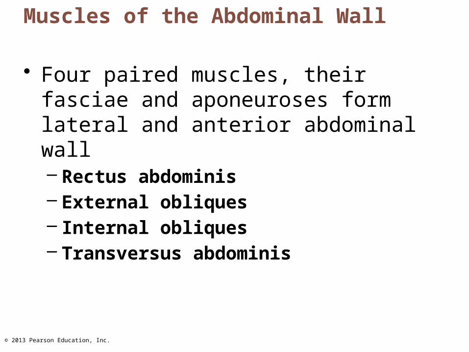
Muscles of the Abdominal Wall
• Four paired muscles, their fasciae and aponeuroses form lateral and anterior abdominal wall– Rectus abdominis – External obliques– Internal obliques– Transversus abdominis
Page 2
© 2013 Pearson Education, Inc.
Figure 10.12a Muscles of the abdominal wall.
Linea alba
Pectoralis major
Tendinous intersection
Rectus abdominis
Inguinal ligament(formed by free inferiorborder of the external oblique aponeurosis)
Aponeurosis of the external oblique
Transversus abdominis
Internal oblique
External oblique
Serratus anterior
Page 3
© 2013 Pearson Education, Inc.
Muscles of the Abdominal Wall
• Fascicles run at angles to one another, provide added strength
• All innervated by intercostal nerves• Actions of these muscles
– Lateral flexion and rotation of trunk– Help promote urination, defecation, childbirth,
vomiting, coughing, and screaming
Page 4
© 2013 Pearson Education, Inc.
PLAY A&P Flix™: Muscles that act on the shoulder joint and humerus: An overview (b)
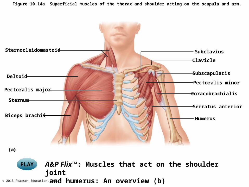
Figure 10.14a Superficial muscles of the thorax and shoulder acting on the scapula and arm.
Subclavius
Clavicle
Subscapularis
Pectoralis minor
Coracobrachialis
Serratus anterior
Humerus
Sternocleidomastoid
Deltoid
Pectoralis major
Sternum
Biceps brachii
Page 5
© 2013 Pearson Education, Inc.
PLAY A&P Flix™: Movement of the pectoral girdle
PLAY A&P Flix™: Muscles of the pectoral girdle (c)
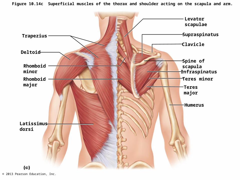
Superficial Muscles of the Posterior Thorax
• Posterior extrinsic shoulder muscles– Trapezius– Levator scapulae– Rhomboids (major and minor)
Page 6
© 2013 Pearson Education, Inc.
Figure 10.14c Superficial muscles of the thorax and shoulder acting on the scapula and arm.
Levatorscapulae
Supraspinatus
Clavicle
Spine ofscapulaInfraspinatus
Teres minor
Teresmajor
Humerus
Trapezius
Deltoid
Rhomboidminor
Rhomboidmajor
Latissimusdorsi
Page 7
© 2013 Pearson Education, Inc.
Muscles Crossing the Shoulder Joint
• Nine muscles cross shoulder joint; insert on and move humerus
• Some originate from scapula; others from axial skeleton
• Actions include flexion, extension, adduction
Page 8
© 2013 Pearson Education, Inc.
Figure 10.15a Muscles crossing the shoulder and elbow joints, causing movements of the arm and forearm, respectively.
Clavicle
Deltoid
Sternum
Pectoralismajor
Coracobrachialis
Triceps brachii:Lateral headLong headMedial head
Bicepsbrachii
Brachialis
Brachio-radialis
Anterior view
Page 9
© 2013 Pearson Education, Inc.
Figure 10.15b Muscles crossing the shoulder and elbow joints, causing movements of thearm and forearm, respectively.
Supraspinatus*
Spine of scapulaDeltoid (cut)
Greater tubercleof humerusInfraspinatus*
Teres minor*
Teres major
Triceps brachii: Lateral head Long head
Latissimus dorsi
Humerus
Olecranonof ulna
Anconeus
Posterior view
Page 10
© 2013 Pearson Education, Inc.
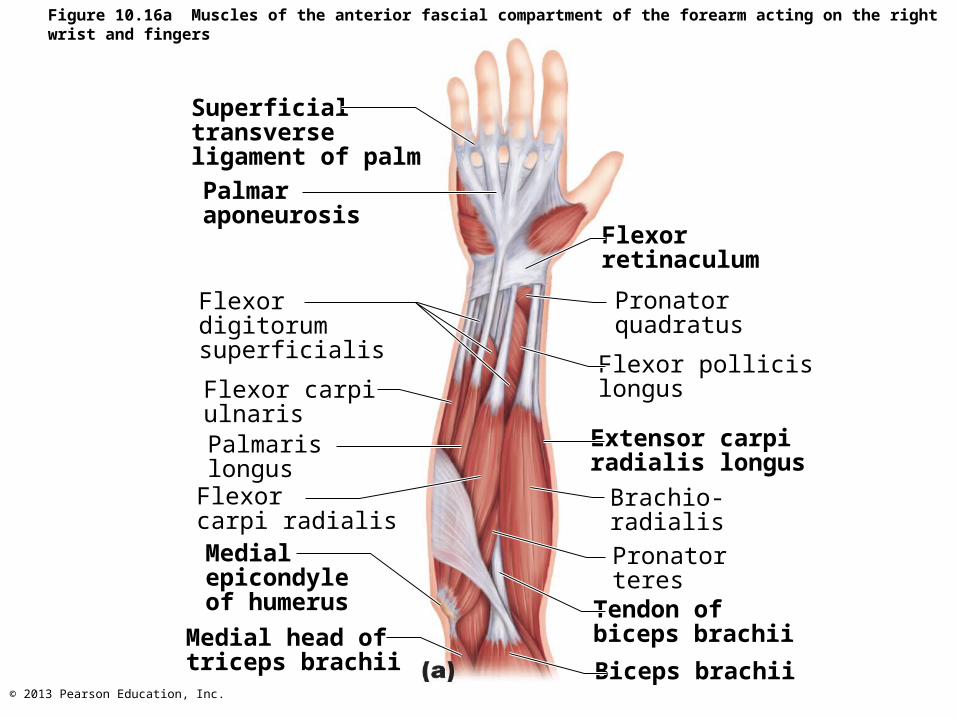
Figure 10.16a Muscles of the anterior fascial compartment of the forearm acting on the right wrist and fingers.
Superficialtransverseligament of palm
Palmaraponeurosis
Flexor digitorumsuperficialis
Flexor carpiulnarisPalmaris longusFlexor carpi radialis
Medial epicondyleof humerus
Medial head oftriceps brachii
Flexorretinaculum
Pronatorquadratus
Flexor pollicislongus
Extensor carpiradialis longus
Brachio-radialisPronatorteresTendon ofbiceps brachii
Biceps brachii
Page 11
© 2013 Pearson Education, Inc.
Figure 10.16c Muscles of the anterior fascial compartment of the forearm acting on the right wrist and fingers.
Tendon of flexordigitorum superficialis(cut)
Lumbricals
Tendon of flexorpollicis longus
Thenar muscles of thumb
Pronator quadratus
Flexor pollicis longus
Flexor digitorumprofundus
Tendon offlexor digitorumprofundus
Tendon of flexor carpi ulnaris (cut)
Supinator
Page 12
© 2013 Pearson Education, Inc.
Figure 10.17a Muscles of the posterior fascial compartment of the right forearm acting on the wrist and fingers.
Extensor expansion
Tendons of extensordigitorum
Extensor pollicislongusExtensor pollicisbrevisAbductorpollicis longus
Extensor digitorum
Extensor carpiradialis brevis
Extensor carpiradialis longus
Tendons of extensorcarpi radialis brevisand longus
Extensor indicis
Extensor digiti minimi
Extensor carpiulnaris
Flexor carpi ulnaris
Anconeus
Insertion oftriceps brachii
Brachioradialis
Page 13
© 2013 Pearson Education, Inc.
A&P Flix™: Muscles that act on the hip joint and femur:An overview
PLAY
Muscles Crossing Hip and Knee Joints
• Most anterior muscles flex femur at hip, extend leg at knee (foreswing of walking)
• Most posterior muscles extend thigh, flex leg (backswing of walking)
• Medial muscles all adduct thigh• All three groups enclosed by fascia lata
Page 14
© 2013 Pearson Education, Inc.
PLAY A&P Flix™: Anterior muscles that cross the hip joint
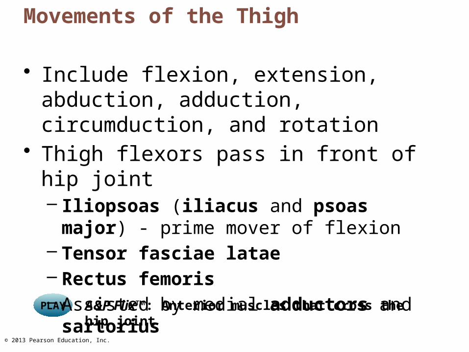
Movements of the Thigh
• Include flexion, extension, abduction, adduction, circumduction, and rotation
• Thigh flexors pass in front of hip joint– Iliopsoas (iliacus and psoas major) - prime
mover of flexion– Tensor fasciae latae– Rectus femoris– Assisted by medial adductors and sartorius
Page 15
© 2013 Pearson Education, Inc.
12th ribQuadratus lumborumPsoas minorIliac crestIliopsoas Psoas major
Iliacus
12th thoracicvertebra
5th lumbarvertebraAnterior superior
iliac spineTensor fasciae lataePectineus
Sartorius Adductor longus
Quadriceps femoris • Rectus femoris
GracilisAdductor magnus
• Vastus lateralis
• Vastus medialis Tendon of quadriceps femoris
Patella
Patellarligament
Figure 10.20a Anterior and medial muscles promoting movements of the thigh and leg.
Page 16
© 2013 Pearson Education, Inc.
Figure 10.21a Posterior muscles of the right hip and thigh.
Gluteus medius
Gluteusmaximus
Adductor magnus
GracilisIliotibial tract
Long headShort head
Bicepsfemoris
HamstringsSemitendinosusSemimembranosus
Page 17
© 2013 Pearson Education, Inc.
Figure 10.22a Muscles of the anterior compartment of the right leg.
Fibularis longus
Gastrocnemius
Tibia
Tibialis anterior
Extensor digitorum longusSoleusExtensor hallucis longus
Fibularis tertius
Superior and inferiorextensor retinaculaExtensor hallucis brevis
Extensor digitorumbrevis
Page 18
© 2013 Pearson Education, Inc.
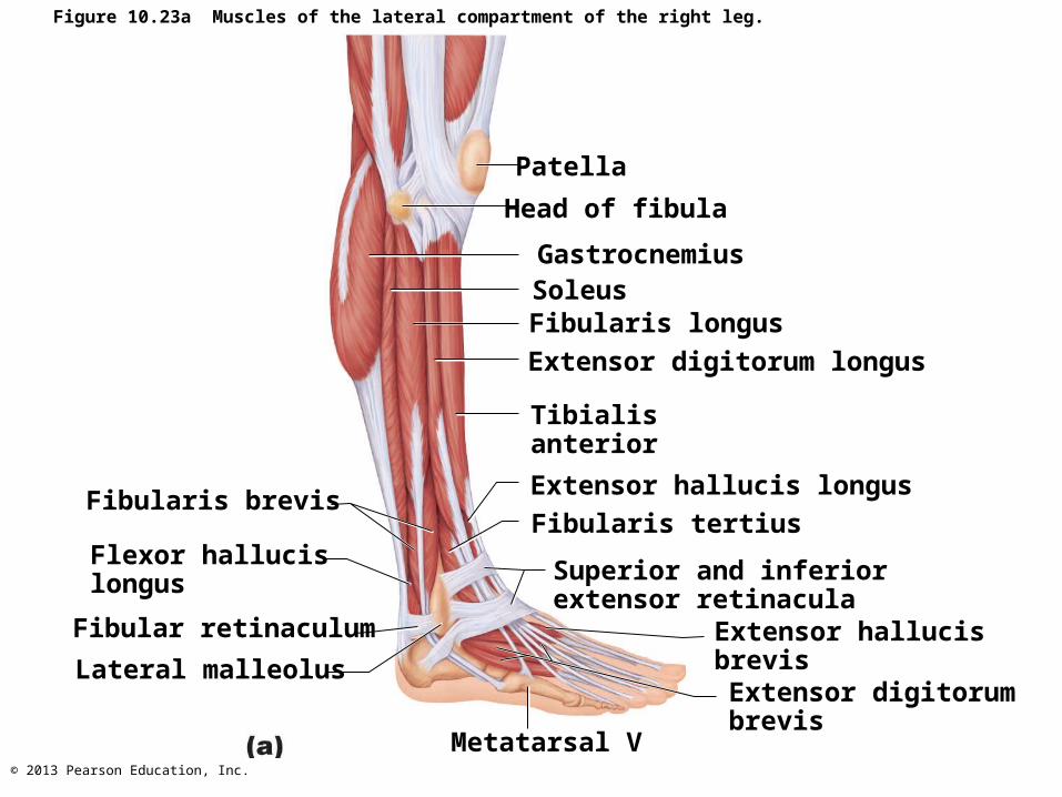
Figure 10.23a Muscles of the lateral compartment of the right leg.
Fibular retinaculum
Fibularis brevis
Flexor hallucislongus
Lateral malleolus
Patella
Head of fibula
GastrocnemiusSoleusFibularis longusExtensor digitorum longus
Tibialis anterior
Extensor hallucis longusFibularis tertius
Superior and inferiorextensor retinacula
Extensor hallucisbrevisExtensor digitorumbrevis
Metatarsal V
Page 19
© 2013 Pearson Education, Inc.
Muscles of the Posterior Compartment of the Leg
• Plantar flex ankle– Gastrocnemius– Soleus– Plantaris– Popliteus– Flexor digitorum longus– Flexor hallucis longus– Tibialis posterior
Page 20
© 2013 Pearson Education, Inc.
Figure 10.24a Muscles of the posterior compartment of the right leg.
Plantaris
Gastrocnemius Medial headLateral head
Tendon ofgastrocnemius
Calcanealtendon Medialmalleolus
Superficial view of the posterior leg.
Lateral malleolus
Calcaneus
Page 21
© 2013 Pearson Education, Inc.
Figure 10.24c Muscles of the posterior compartment of the right leg.
Gastrocnemiusmedial head(cut)
Flexor digitorumlongus
Tendon oftibialis posterior
MedialmalleolusCalcanealtendon (cut) Calcaneus
The triceps surae has been removed to show thedeep muscles of the posterior compartment.
Fibularis brevis
Flexor hallucislongus
Fibularislongus
FibulaTibialis posteriorSoleus (cut)Popliteus
Gastrocnemiuslateral head (cut)
Plantaris (cut)
Page 22
© 2013 Pearson Education, Inc.
Figure 10.22a Muscles of the anterior compartment of the right leg.
Fibularis longus
Gastrocnemius
Tibia
Tibialis anterior
Extensor digitorum longusSoleusExtensor hallucis longus
Fibularis tertius
Superior and inferiorextensor retinaculaExtensor hallucis brevis
Extensor digitorumbrevis


![Respiratory function of the rib cage muscles · laxation of expiratory muscles; mostly (>80%) due to the action of the abdominal, rather than the rib cage, expira tory muscles [16].](https://static.documents.pub/doc/80x56/60185e9ecadd0b1fe4741bb0/respiratory-function-of-the-rib-cage-muscles-laxation-of-expiratory-muscles-mostly.jpg)


