61
HAEMORRHAGIC DISEASE
| Date post: | 03-Dec-2015 |
| Category: |
Documents |
| Upload: | miqdadmohdsuberi |
| View: | 242 times |
| Download: | 1 times |

HAEMORRHAGIC DISEASE
HAEMORRHAGIC DISEASE

Suspected of hemorrhagic disease :
1. Spontaneous bleeding
2. Prolonged bleeding/massive 3. More than one site bleeding
Suspected of hemorrhagic disease :
1. Spontaneous bleeding
2. Prolonged bleeding/massive 3. More than one site bleeding
PATHOGENESISPATHOGENESIS
Hemostasis process :
- maintaining blood in a state of dilution - maintaining blood in vascular - to stop bleeding vascular damage

3 components of hemostasis:
HEMOSTASIS
VASCU
LAR
THROMBOCYTECLO
TTING
Disturbance one of components homeostasis bleeding

TRAUMA/INJURY
VASOCONSTRICTION
BLOOD
CLOTTING ADHESION OF THROMBCYTE
THROMBINE
FIBRINE
ADP/SEROTONINE
AGREGATION OF THROMBOCYTE
STABILE HAEMOSTATIC BLOCKAGE
+

DETECTION OF HAEMORRAGIC DISEASEDETECTION OF HAEMORRAGIC DISEASE
Step I - good history taking - physical examination
- Trauma: - History of trauma chronologically - Mild trauma bleeding - Severe spontaneous bleeding
- Quantity and duration of bleeding - Recurrent bleeding
Step I - good history taking - physical examination
- Trauma: - History of trauma chronologically - Mild trauma bleeding - Severe spontaneous bleeding
- Quantity and duration of bleeding - Recurrent bleeding

- trauma always bleeding Congenital hemorrhagic disease
- Deep tissue bleeding
( large hematom or hemarthrosis)
Congenital hemorrhagic disease
- Petechie not congenital hemorrhagic disease
- Congenital hemorrhagic disease usually coagulation disorder
- trauma always bleeding Congenital hemorrhagic disease
- Deep tissue bleeding
( large hematom or hemarthrosis)
Congenital hemorrhagic disease
- Petechie not congenital hemorrhagic disease
- Congenital hemorrhagic disease usually coagulation disorder
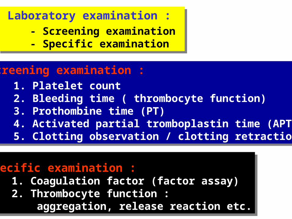
Laboratory examination :- Screening examination - Specific examination
Laboratory examination :- Screening examination - Specific examination
Screening examination : 1. Platelet count 2. Bleeding time ( thrombocyte function) 3. Prothombine time (PT) 4. Activated partial tromboplastin time (APTT) 5. Clotting observation / clotting retraction
Screening examination : 1. Platelet count 2. Bleeding time ( thrombocyte function) 3. Prothombine time (PT) 4. Activated partial tromboplastin time (APTT) 5. Clotting observation / clotting retraction
Specific examination : 1. Coagulation factor (factor assay) 2. Thrombocyte function : aggregation, release reaction etc.
Specific examination : 1. Coagulation factor (factor assay) 2. Thrombocyte function : aggregation, release reaction etc.

VASCULAR DISORDERVASCULAR DISORDER
Mostly : secondary vascular pupura :
- Immunology: Schöenlein-Henoch syndrome - Infection: Virus, Rickets, Bacteria - Drugs - Deficiency of Vit. C - Uremia
Congenital: - Hereditary hemorrhagic telangiectasia
(Osler-Weber-Rendu)
- Cutis hyperelastica (Ehler-danlos)
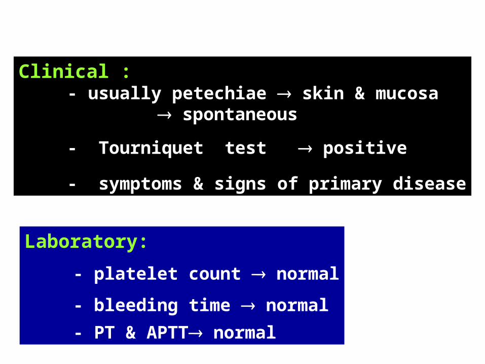
Laboratory:
- platelet count normal
- bleeding time normal
- PT & APTT normal
Clinical : - usually petechiae skin & mucosa
spontaneous
- Tourniquet test positive
- symptoms & signs of primary disease

SCHöENLEIN-HENOCH SYNDROME SCHöENLEIN-HENOCH SYNDROME
Incidence :- 3 -7 years of age- Male : female = 3 : 2
Etiology:
Immunologic Reaction:- Infection: beta hemolytic Streptococcal, Viral- Food : milk, egg, tomato, fish etc.- Drug : erythromycin, sulfa, penicillin, ect.- Insect bite
- Allergic Purpura - Anaphylactic Purpura

-Hemolytic Streptococcal Infection important
- 75% cases History of respiratory tract
infection 1-3 weeks before
- 50% cases positive throat swab culture
- 30% cases titer ASO
PATHOGENESISPATHOGENESIS
Immune complex :- vasculitis increase permeability - perivasculer inflammation

CLINICAL MANIFESTATIONCLINICAL MANIFESTATION
1. Skin involvement:
- erytema, maculopapuler
- petechie & echymosis
Distribution of lesion: symmetric:- extensor lower extremity- gluteus, hip- extensor arm elbow
2.Articular involvement:
- 75% case - polyarthralgia/polyarthritis non-migrants - periarthriculer swelling - especially knee & ankle - full recovery
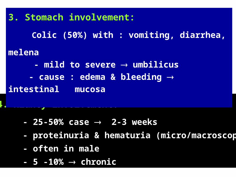
4. Kidney involvement:
- 25-50% case 2-3 weeks
- proteinuria & hematuria (micro/macroscopic)
- often in male
- 5 -10% chronic
3. Stomach involvement:
Colic (50%) with : vomiting, diarrhea,
melena - mild to severe umbilicus - cause : edema & bleeding intestinal mucosa

MANAGEMENTMANAGEMENT
self limiting symptomatic treatment
- Corticosteroid:- intestinal mucosa edema colic & invagination- arthricular involvement
- Bed rest avoid intracranial bleeding

- Good if no complication- Full recovery in 4 weeks- Residive
- Complication rare:- invagination, intestinal perforation- intracranial bleeding - renal failure
PROGNOSTICPROGNOSTIC

THROMBOCYTE DISORDERTHROMBOCYTE DISORDER
A. QUANTITATIVE DISORDER
1. Thrombocytopenia bleeding 2. Thrombocytosis thrombus formation
Normal:
platelet count 150.000 - 400.000/mm3
< 50.000/mm3 spontaneous bleeding

a. Production disorder:- Hypoproliferation: aplastic anemia, ATP - Ineffective thrombopoesis :
- Megaloblastic anemia- ANLL M7
b. Distribution disturbance:- Splenomegali: “pooling” thrombocyte- Lymphoma
c. Dilution:- Massive blood transfusion
THROMBOCYTOPENIA:THROMBOCYTOPENIA:

d. Abnormal destruction
- Non-immune: - DIC - Infection: DHF, sepsis
- Immune:- Idiopathic Thrombocytopenic Purpura (ITP)- Drugs: Kina, kinidin, sulfa, dilantin, ect. - Neonatal thrombocytopenia - Purpura post-transfusion
e. Abnormal consumption:- DIC, DHF

1. Adhesion disturbance
2. Aggregation anomaly Diphenydramin:
- prevent platelet aggregation
3. Disturbance of platelet release reaction Asetil salisilic ac.:
- distrub release of ADP - asetilasi platelet membrane
B. QUALITATIVE DISORDER= Trombastenia or thrombopati

IDIOPATHIC/IMMUNE THROMBOCYTOPENIC PURPURA (ITP)
IDIOPATHIC/IMMUNE THROMBOCYTOPENIC PURPURA (ITP)
Destruction of platelet shorter age immunologic mechanism:
- antibody (IgG) platelet
- C3 complement
- cellular immunity activation: macrophage & cytotoxic cell

KLASIFIKASIKLASIFIKASI
1. Acute ITP (85-90%): self limiting anak-anak
2. Chronic ITP (10-15%): dewasa
- Umur : 2 - 8 tahun
- 50% kasus : 1 - 6 minggu sebelumnya viral infection: ARTI, hepatitis, mumps, mononucleosus infectiosa, cytomegaloviral etc.)
ITP AkutITP Akut

- perdarahan kulit dan selaput lender peteki dan ekimosis melena, hematuri
- peradarahan alat dalam jarang
- thrombositopeni berat perdarahan otak
- tourniquet test is positive
Gejala klinis:Gejala klinis:

- thrombositopeni- hapusan darah:
bentuk trombosit abnormal, ukuran besar, terpisah-pisah
- retraksi bekuan berkurang
- waktu perdarahan memanjang
- PT & APTT normal
Gambaran darah:Gambaran darah:

Penting menyingkirkan:- aplastic anemia- leukemia
Megakariosit:- Jumlah normal atau meningkat- Morfologi: - immatur - sitoplasma lebih basofil
- kurang granulasi
Sum-sum tulang:Sum-sum tulang:

- Istirehat dan hindari trauma
- Kasus ringan tidak perlu pengobatan
- Kasus berat perdarahan luas/berat:- kortikosteroid- suspense trombosit tidak dianjurkan- blood transfusion (PRC): atas indikasi
Pengobatan ITP akutPengobatan ITP akut
- Sebahagian besar (85 - 90 %) sembuh- 10 - 15% kronis
Prognosis:Prognosis:
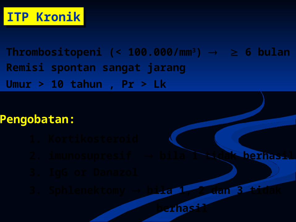
ITP KronikITP Kronik
- Thrombositopeni (< 100.000/mm3) 6 bulan- Remisi spontan sangat jarang
- Umur > 10 tahun , Pr > Lk
Pengobatan:
1. Kortikosteroid
2. imunosupresif bila 1 tidak berhasil
3. IgG or Danazol
3. Sphlenektomy bila 1, 2 dan 3 tidak
berhasil

GANGGUAN PEMBEKUAN
Komponen pembekuan:
1. sistem Pembekuan Darah mekanisme pembekuan darah
2. Sistem Pencegahan Pembekuan mencegah pembekuan intravascular darah tetap cair
3. Sistem Fibrinolytic melisiskan fibrin lumen pembuluh darahtetap terbuka
Komponen pembekuan:
1. sistem Pembekuan Darah mekanisme pembekuan darah
2. Sistem Pencegahan Pembekuan mencegah pembekuan intravascular darah tetap cair
3. Sistem Fibrinolytic melisiskan fibrin lumen pembuluh darahtetap terbuka

SISTEM PEMBEKUAN DARAHSISTEM PEMBEKUAN DARAH
International NameSynonym
I Fibrinogen
II Prothrombin
III Tissue factor, Tissue thromboplastin
IV Calcium (Ca)
V Proacelerin, Labile Factor
VII Proconvertin, Stable factor
VIII Antihemophilic Factor, AHF-A
Faktor-faktor pembekuan darah:

IX Plasma Thromboplastin Component (PTC), Christmas Factor, AHF-B
X Stuart Prower Factor
XI Plasma Thromboplastin Antecedent (PTA), AHF-C
XII Hageman Factor, AHF-D
XIII Fibrin Stabilizing factor (FSF)
Prekalikrein Fletcher Factor
Kininogen Fitzgerald factor
SISTEM PEMBEKUAN DARAHSISTEM PEMBEKUAN DARAH

Inhibitor pembekuan:
- Antithrombin III - C Protein & S Protein
- Alpha-2 macroglobulin
Inhibitor pembekuan:
- Antithrombin III - C Protein & S Protein
- Alpha-2 macroglobulin
Plasminogen system- plasmin:- Plasminogen- Plasminogen activator- Anti plasmin
Plasminogen system- plasmin:- Plasminogen- Plasminogen activator- Anti plasmin
SISTEM PENCEGAH PEMBEKUAN: SISTEM PENCEGAH PEMBEKUAN:
SISTEM FIBRINOLITIK:SISTEM FIBRINOLITIK:

1.Pembentukan activator protrombin (Protrombinase):
- Intrinsic- Ekstrinsic
2. Prothrombin trombin
3. Fibrinogen fibrin
1.Pembentukan activator protrombin (Protrombinase):
- Intrinsic- Ekstrinsic
2. Prothrombin trombin
3. Fibrinogen fibrin
Proses pembekuan darah:Proses pembekuan darah:

Kontak permukaan
XII XIIa
XI XIa
IX IXa
X Xa X
III +VII
V
F.Tr-3
Prothrombin Thrombin
Fibrinogen Fibrin
Fibrin polymer
XIII
PROTHROMBINASE
VIII
Ca++ Ca++
Ca++
Kerusakan jaringan
INTRINSIC
ExTRINSIC

1. Sistem pembekuan
2. Sistem pencegah pembekuan
3. Sistem fibrinolitik
GANGGUAN PEMBEKUANGANGGUAN PEMBEKUAN
Gangguan sistem/mekanisme pembekuan defisiensi satu atau lebih :
faktor pembekuan

1. Pembentukan berkurang:
- genetik/kongenital : hemophilia - Vit. K deficiency II, VII, IX & X, C Protein
- penyakit hati berat
1. Pembentukan berkurang:
- genetik/kongenital : hemophilia - Vit. K deficiency II, VII, IX & X, C Protein
- penyakit hati berat
2. PEMAKAIAN BERTAMBAH - Consumption coagulopathy Disseminated Intravascular Coagulation (DIC)
2. PEMAKAIAN BERTAMBAH - Consumption coagulopathy Disseminated Intravascular Coagulation (DIC)

1. Perdarahan trauma ringan, jarang spontan2. Jarang petechie3. Perdarahan sendi dan jaringan dalam
hematoma besar, ekimosis besar4. Perdarahan dari luka:
- tidak segera timbul - sering berulang - berlangsung lama (>48 jam) - merembes ( oozing )
Laboratorium:- PT & PTT: salah satu atau
keduanya - waktu perdarahan normal - observasi bekuan rapuh
Sifat-sifat gangguan pembekuan:Sifat-sifat gangguan pembekuan:

HEMOFILIAHEMOFILIA

HEMOFILIAHEMOFILIA
Penyakit perdarahn:
- Gangguan pembekuan Coagulation factors deficiency
- congenital, herediter
Hemophilia:Hemophilia A factor VIII deficiency
Hemophilia B factor IX deficiency

INSIDENINSIDEN
1 : 10.000 Hemofilia paling banyak
GENETIKA DAN PATOFISIOLOGIGENETIKA DAN PATOFISIOLOGI
- Factor VIII Gen X chromosome- Mutasi gen (substitusi dan delesi) gangguan sintesis faktor VIII
Penyakit diturunkan secara resesif Kromosom seks : X-linked

Male (Xh Y) affectedfemale (Xh X) carrier
Usually by marriage:
Normal father (X Y) Carrier mother (Xh X) Hemophilia almost entirely in boys
GENETICS AND PATHOPHYSIOLOGYGENETICS AND PATHOPHYSIOLOGY

F VIII: protein plasma are needed inprothrombin activator synthesis process
Women could be affected:- father = (Xh Y) & mother = (Xh X)- Inactive gene of VIII factor- Spontaneous mutation gene of VIII factor
F VIII deficiency coagulation cascade disturbance
GENETICS AND PATOPHYSIOLOGYGENETICS AND PATOPHYSIOLOGY

CLINICAL MANIFESTATION:CLINICAL MANIFESTATION:
Severe Hemophilia : F VIII < 1%spontaneous bleeding
hemarthrosis, muscle bleeding, gastrointestinal, hematuria & brain
Depends on F VIII levels
Moderate Hemophilia : F VIII 1 – 5 %bleeding after minor trauma
Mild Hemophilia : F VIII 6 – 25 %bleeding after major trauma,surgery

DIAGNOSISDIAGNOSIS
History:- History of repeated bleeding joints- Brothers with the same illness- Brothers from mother with the same illness
Physical examination:- hemarthrosis, hematoma, etc
Laboratory:- normal platelet & bleeding time - Prolonged PTT & normal PT- TGT & AHF assay F VIII deficiency

COMPLICATIONCOMPLICATION
hemophilia arthropathycontracture and paresis/paralysis of musclehemophilic pseudotumor
Formation antibody against F VIIIthrombosisITPViral hepatitis
Because of the disease:
Because of treatment:

TREATMENTTREATMENT
1. Stop the bleeding:Administration of F VIII:
- cryoprecipitate- F VIII concentrate (KOATE)
Bed rest Immobilizes bleeding area: cold compress, tampon
- Treatment of anemia & shock- synovectomy- joints & muscles rehabilitation
2. Correction of bleeding consequence:

3. Bleeding prevention:- prevention of trauma- addition of F VIII before surgery- contraindication: aspirin
TREATMENTTREATMENT

VITAMIN K DEFICIENCYVITAMIN K DEFICIENCY
Is found in:1. Hemorrhagic disease of the newborn (HDN)
2. Disorder of Vit. K absorption:- Biliary tract obstruction- Chronic diarrhea- Severe liver disease
3. Intestinal sterilization by antibiotics

HEMORRHAGIC DISEASE OF THE NEWBORN(HDN)
HEMORRHAGIC DISEASE OF THE NEWBORN(HDN)
Hemorrhagic disease in newborn baby due to:Deficiencies of factor II, VII, IX & X vitamin K
Physiology (normal):
Coagulation factor II,VII,IX & X:- decrease in newborn the lowest rate at 2 - 5 days of age
- increase at 7 – 14 days of age
Physiology (normal):
Coagulation factor II,VII,IX & X:- decrease in newborn the lowest rate at 2 - 5 days of age
- increase at 7 – 14 days of age
Etiology:
- Uncomplete colonization of intestinal flora the synthesis of vit K in gut is still low
- decrease of vit K in placenta
Etiology:
- Uncomplete colonization of intestinal flora the synthesis of vit K in gut is still low
- decrease of vit K in placenta

If decreasing of coagulation factor excessive HDN
May result from :1.Very low amounts of vitamin K storage 2.No synthesis of vit. K in gut sterile intestinal flora
3. Absorption of vit K in gut very low
4. Disorder of vitamin K metabolism:- Damaging of vit. K :
barbiturat, phenytoin, diazepam, INH, Rifampisin
- disturbance of vit.K usage by liver: dicumarol, salicylat

FUNCTION OF VITAMIN K
Protein (II, VII, IX & X)
CarboxylationVitamin K
Functional of coagulation factor (II, VII, IX & X)
The process were done in liver

Clinical manifestations:
Bleeding in various location:- gastrointestinal tract: melena- umbilical cord, skin, mucosa- cephalhematom, brain bleeding
Clinical manifestations:
Bleeding in various location:- gastrointestinal tract: melena- umbilical cord, skin, mucosa- cephalhematom, brain bleeding
Incidence: - Age: 2 - 5 days

BLOOD HEMOSTASIS BLOOD HEMOSTASIS
ABNORMAL/PROLONGED NORMAL
PT (Factor II, VII, X)
APTT (Factor II, IX, X)
Thrombotest, Normotest (F. II, VII, X)
Activity F. II, VII, IX, X
There are PIVKA II
Thrombin time (TT)
Fibrinogen
Activity F. V, VIII, XI
Antigen F. II,VII,IX,X
Platelet count & BT
Practical:HDN: bleeding manifestation in baby <12 weeks with : - Prolonged of PT & APTT - Normal platelet and BT

TREATMENTTREATMENT
HDN self limited
Bleeding can stop spontaneously
but needs long time
- Massive hemorrhagic- Continuous hemorrhagic- intracranial hemorrhagic
Threaten the newborns’ life
Needs immediate & the right treatment

HDN
Vit. K 1-2 mg im/timesVit. K 1-2 mg im/times
AnemiaAnemia
PRC transfPRC transf
Repeat Vit. K (3 times, every 6 hours)
Repeat Vit. K (3 times, every 6 hours)
-Continous bleeding or recurrent- Prolonged PTT
-Continous bleeding or recurrent- Prolonged PTT
Plasma or Fresh frozen plasma (FFP)
Plasma or Fresh frozen plasma (FFP)
Plasma or fresh frozen plasma (FFP)
Plasma or fresh frozen plasma (FFP)
-Continous bleeding or recurrent -Prolonged PTT
-Continous bleeding or recurrent -Prolonged PTT
Severe hemorrhagic shock
Severe hemorrhagic shock
20 ml/kgBW

PROPHYLAXISPROPHYLAXIS
Vitamin K 1 mg
High risk newborn :
- Premature
- Twins
- Assisted labor
- Asphyxia

DICDIC= = DISSEMINATED INTRAVASCULAR DISSEMINATED INTRAVASCULAR
COAGULATIONCOAGULATION
DICDIC= = DISSEMINATED INTRAVASCULAR DISSEMINATED INTRAVASCULAR
COAGULATIONCOAGULATION
- Intravascular coagulation spread
everywhere in blood vessel (systemic) pathologic activation of haemostatic mechanism
DIC:Defibrination syndrome = Consumption coagulopathy complication: many condition / disease
initiate DIC

- WIDE ENDOTHEL DAMAGE - TISSUE THROMBOPLASTIN CIRCULATION
- WIDE ENDOTHEL DAMAGE - TISSUE THROMBOPLASTIN CIRCULATION
WIDE ACTIVATION OFCOAGULATION PROCESS
WIDE ACTIVATION OFCOAGULATION PROCESS
INTRAVASCULARTROMBI-FIBRIN
INTRAVASCULARTROMBI-FIBRIN
USAGE:- COAGULATION FACTOR- PLATELET
USAGE:- COAGULATION FACTOR- PLATELET
DEFICIENCY- COAGULATION FACTOR- PLATELET
DEFICIENCY- COAGULATION FACTOR- PLATELET
HEMORAGEHEMORAGE
FIBRINOLISISFIBRINOLISIS
FDP FDP
COAGULATIONDISORDER
COAGULATIONDISORDER
BLOOD VESSELOCLUTION
BLOOD VESSELOCLUTION
ISCHEMIAISCHEMIA
MAHAMAHA

ETIOLOGY:ETIOLOGY:
- Massive vascular endothel damage- Tissue Factor (tromboplastin) circulation
1. Trauma:- burn, crush injury, heat stroke
2. Infection:- Viral: DHF, Variola- Bacterial: sepsis- Fungus: candidiasis
3. Metabolic:- Acidosis, alkalosis, ketosis- Hyperthermia, hypothermia
1. Trauma:- burn, crush injury, heat stroke
2. Infection:- Viral: DHF, Variola- Bacterial: sepsis- Fungus: candidiasis
3. Metabolic:- Acidosis, alkalosis, ketosis- Hyperthermia, hypothermia

4. Immunologic:
- Blood transfusion reaction (massive hemolisis)- Anaphylactic, Immune complex diseases.
5. Malignancy:- Leukemia (ANLL-M3)
6. Others:- Shock- Anoxia
4. Immunologic:
- Blood transfusion reaction (massive hemolisis)- Anaphylactic, Immune complex diseases.
5. Malignancy:- Leukemia (ANLL-M3)
6. Others:- Shock- Anoxia

DIAGNOSISDIAGNOSIS
Primary Severe Disease
Duration of illness with:- hemorrhage
- tissue/organ ischemia : acral necrosis
renal failure
Primary Severe Disease
Duration of illness with:- hemorrhage
- tissue/organ ischemia : acral necrosis
renal failure
CLINICAL:

- Blood smear microangiopathy:
burr cells, helmet cells
- Thrombocytopenia & prolonged bleeding
time
- PT, PTT & prolonged thrombin time
- coagulation factor Fibrinogen
- FDP (FSP)
- Blood smear microangiopathy:
burr cells, helmet cells
- Thrombocytopenia & prolonged bleeding
time
- PT, PTT & prolonged thrombin time
- coagulation factor Fibrinogen
- FDP (FSP)
LABORATORY

THERAPYTHERAPY
1. Treat etiology factor
2. Blockade process
3. Blood/plasma component substitution
1. Treat etiology factor
2. Blockade process
3. Blood/plasma component substitution


