Transworld Research Network 37/661 (2), Fort P.O. Trivandrum-695 023 Kerala, India Recent Advances in Pharmaceutical Sciences II, 2012: 1-13 ISBN: 978-81-7895-569-8 Editors: Diego Muñoz-Torrero, Diego Haro and Joan Vallès 1. Biofilms on rocks Mariona Hernández-Mariné 1 and Mónica Roldán Molina 2 1 Unitat de Botànica. Facultat de Farmàcia. Universitat de Barcelona. Av. Joan XXIII s/n. E-08028 Barcelona, Spain; 2 Servei de Microscòpia. Universitat Autònoma de Barcelona. Edifici C, Facultat de Ciències. E-08193, Bellaterra, Spain Abstract. Microorganisms group themselves into assemblies known as communities or biofilms, which are associated with surfaces. A matrix of self-segregated polymeric substances enhances their attachment. Communication between bacterial cells involves the production and detection of diffusible signal molecules, known as quorum sensing, which is an important regulatory mechanism of biofilm strategies. Biofilms thrive everywhere; in subaerial surfaces they can be driven by sunlight, with photosynthesizing components. A special case is those which colonize works of art, forming patinas and becoming involved in the degradation of colonized substrata. Knowledge of three- dimensional structure of the biofilm and the distribution of species concerned is crucial for managing and preventing uncontrolled colonization and for preserving cultural heritage sites. This paper describes their role in this degradation, some examples of biofilms and their resilience mechanisms. The methods used in their study when growing in monuments and caves are also discussed. Introduction Most microorganisms live attached to a surface rather than as single, suspended cells [1]. The aggregation of microorganisms attached to a surface Correspondence/Reprint request: Dr. Mariona Hernández-Mariné, Unitat de Botànica. Facultat de Farmàcia. Universitat de Barcelona. Av. Joan XXIII s/n. E-08028, Barcelona, Spain. E-mail: [email protected]
Transcript
Transworld Research Network 37/661 (2), Fort P.O. Trivandrum-695 023 Kerala, India
Recent Advances in Pharmaceutical Sciences II, 2012: 1-13 ISBN: 978-81-7895-569-8
Editors: Diego Muñoz-Torrero, Diego Haro and Joan Vallès
1. Biofilms on rocks
Mariona Hernandez-Mariné1 and Mónica Roldán Molina2 1Unitat de Botànica. Facultat de Farmàcia. Universitat de Barcelona. Av. Joan XXIII s/n. E-08028
Barcelona, Spain; 2Servei de Microscopia. Universitat Autonoma de Barcelona. Edifici C, Facultat de Ciencies. E-08193, Bellaterra, Spain
Abstract. Microorganisms group themselves into assemblies known as communities or biofilms, which are associated with surfaces. A matrix of self-segregated polymeric substances enhances their attachment. Communication between bacterial cells involves the production and detection of diffusible signal molecules, known as quorum sensing, which is an important regulatory mechanism of biofilm strategies. Biofilms thrive everywhere; in subaerial surfaces they can be driven by sunlight, with photosynthesizing components. A special case is those which colonize works of art, forming patinas and becoming involved in the degradation of colonized substrata. Knowledge of three-dimensional structure of the biofilm and the distribution of species concerned is crucial for managing and preventing uncontrolled colonization and for preserving cultural heritage sites. This paper describes their role in this degradation, some examples of biofilms and their resilience mechanisms. The methods used in their study when growing in monuments and caves are also discussed.
Introduction Most microorganisms live attached to a surface rather than as single, suspended cells [1]. The aggregation of microorganisms attached to a surface Correspondence/Reprint request: Dr. Mariona Hernández-Mariné, Unitat de Botànica. Facultat de Farmàcia. Universitat de Barcelona. Av. Joan XXIII s/n. E-08028, Barcelona, Spain. E-mail: [email protected]
Mariona Hernández-Mariné & Mónica Roldán Molina 2
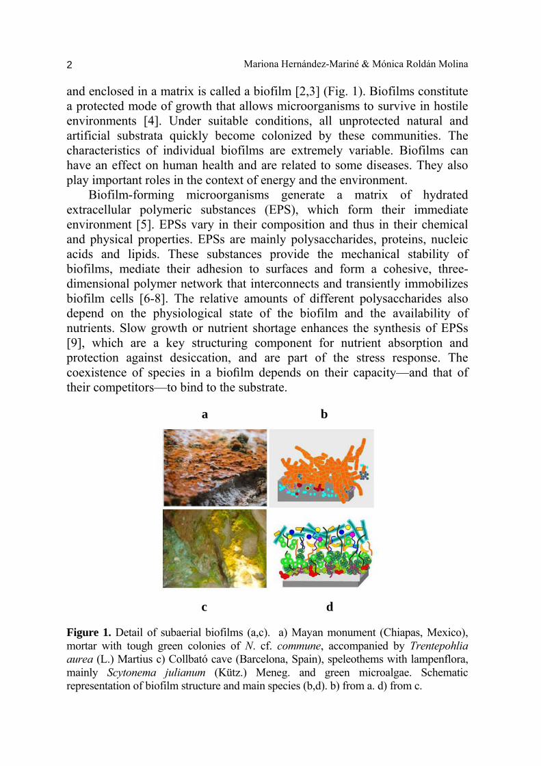
and enclosed in a matrix is called a biofilm [2,3] (Fig. 1). Biofilms constitute a protected mode of growth that allows microorganisms to survive in hostile environments [4]. Under suitable conditions, all unprotected natural and artificial substrata quickly become colonized by these communities. The characteristics of individual biofilms are extremely variable. Biofilms can have an effect on human health and are related to some diseases. They also play important roles in the context of energy and the environment. Biofilm-forming microorganisms generate a matrix of hydrated extracellular polymeric substances (EPS), which form their immediate environment [5]. EPSs vary in their composition and thus in their chemical and physical properties. EPSs are mainly polysaccharides, proteins, nucleic acids and lipids. These substances provide the mechanical stability of biofilms, mediate their adhesion to surfaces and form a cohesive, three-dimensional polymer network that interconnects and transiently immobilizes biofilm cells [6-8]. The relative amounts of different polysaccharides also depend on the physiological state of the biofilm and the availability of nutrients. Slow growth or nutrient shortage enhances the synthesis of EPSs [9], which are a key structuring component for nutrient absorption and protection against desiccation, and are part of the stress response. The coexistence of species in a biofilm depends on their capacity—and that of their competitors—to bind to the substrate. a b
c d
Figure 1. Detail of subaerial biofilms (a,c). a) Mayan monument (Chiapas, Mexico), mortar with tough green colonies of N. cf. commune, accompanied by Trentepohlia aurea (L.) Martius c) Collbató cave (Barcelona, Spain), speleothems with lampenflora, mainly Scytonema julianum (Kütz.) Meneg. and green microalgae. Schematic representation of biofilm structure and main species (b,d). b) from a. d) from c.
Biofilms on rocks 3
1. Biofilm development Formation of a microbial biofilm is a complex multistep developmental process that consists of several overlapping stages. Most of our information comes from bacterial biofilms for which the formation of a biofilm has been described as a sequence of events [10,11]: i) the first event is the substratum conditioning with the absorption of water, organic matter or dissolved organic macromolecules; ii) the second event is the arrival or mass transport of microorganisms to the area; iii) the third event is the adhesion of the microorganisms, which is a prerequisite for the formation of biofilms on surfaces. Initial attachment to a variety of materials and early biofilm formation depend on the surface and type and degree of roughness of the substratum. Irregular surfaces are preferential starting points for attachment because they provide niches in which microorganisms are protected. After making contact with a surface, bacteria become attached in a process that was formerly considered reversible. This process is frequently mediated by the presence of extracellular materials but is also accompanied by physiological changes that end in irreversible surface binding [12]; iv) the fourth event is the expansion of the biofilm, which involves the aggregation of cells into microcolonies that then grow and mature. The microorganisms produce and release materials mediated by the microorganisms themselves. Changes in the gene expression and formation of exopolymeric material are regulated by cell-to-cell signals [13]; v) the fifth event is the return to temporary motility in response to nutritional cues, so that biofilm cells are released in order to repeat the process. 2. Quorum-sensing Quorum-sensing (QS) regulates the communication, behavior and several cellular processes of the microorganisms, including biofilm formation. QS is a form of intercellular communication between single-cell organisms that allows them to act coordinately like multicellular organisms. Release of the QS signal compounds is dependent on the density of the population [14-15] and leads to changes in bacterial gene expression [2] and to significant changes in the phenotype [17]. QS signals synchronize with the growth stage of a culture and accumulate to a threshold level. This controls the switch from the behavior typical of single-cell organisms to that of cells within a colony or a biofilm [11, 18]. There are a number of different QS signaling systems employed by Gram-negative bacteria, such as oligopeptide autoinducers, furanones, triclosan or mixtures of fatty acids, but the most characterized is the component LuxR-N-acyl homoserine lactones (AHLs)
Mariona Hernández-Mariné & Mónica Roldán Molina 4
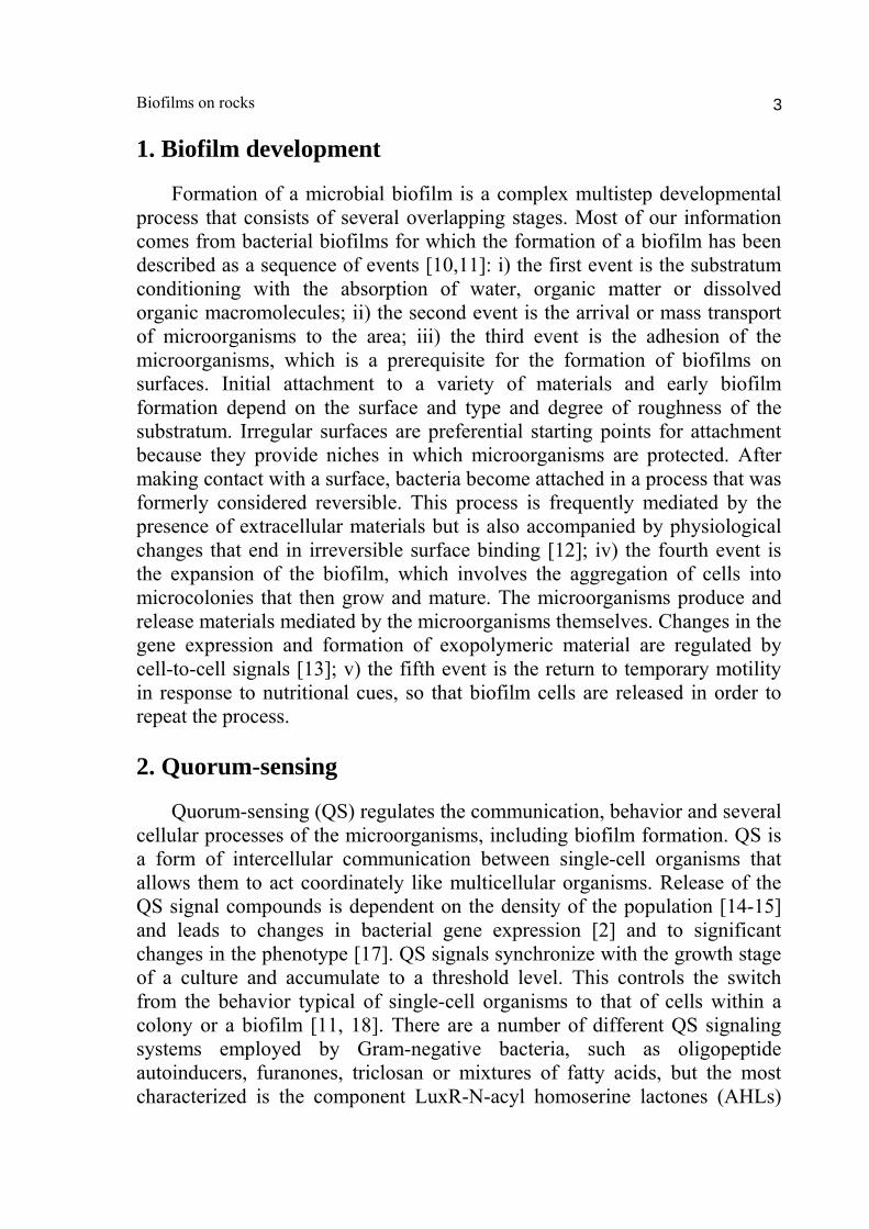
[19-20]. AHLs are produced by homologues of the AHL synthase LuxI from S-adenosyl methionine and the intracellular pool of acyl-carrier proteins [21]. AHLs differ in the length, degree of saturation and substitution of the acyl side chains. The autoinducer molecules bind to the appropriate transcription regulator(s) when the bacterial population reaches the quorum level (that is, the signal concentration reaches a threshold concentration high enough to facilitate binding to the receptor) [13]. Binding of the autoinducers is followed by activation or repression of target genes. Thus, quorum sensing allows bacteria to make a unified response. This benefits the population [22] because it enhances access to nutrients and more favorable environmental niches and improves action against competing bacteria and environmental stresses [11, 15] (Fig. 2). The bacterial species within a consortia respond to different QS signals [23,16]. There are few reports of AHL activity in cyanobacteria [24, 15], but this could be due to the lack of studies.
Figure 2. Communication between bacterial cells involves the production and detection of diffusible signal molecules known as quorum sensing (QS). Numbers in the flow chart represent different stages of the QS process. Arrows show the main direction of signal transport (based on Dobretsov et al. 2009). 3. Methods of study A multistep approach involving molecular, chemical and microscopy techniques is used to assess the composition, 3D structure and distribution of the biofilms.
Biofilms on rocks 5
Nonculture-based molecular methods allow community compositions and activities to be characterized in situ. These methods include DGGE, clone library analysis, quantitative PCR, and stable isotope probing that can be used to obtain the phylogeny, relative abundance and genetic activity of individual members of a biofilm community [25, 26]. In particular, functional genomic approaches provide important clues about phototrophic biofilm biology [27]. A number of techniques for detecting and identifying quorum sensing molecules or for monitoring the activity of these compounds have been described [15, 13, 28]. Approaches used for detecting and identifying AHLs include cell-based assays using AHL-specific bacterial biosensors, thin-layer chromatography, gas chromatography/mass spectrometry, and liquid chromatography. Bacterial biosensors cannot synthesize AHLs, but they can express specific compounds when exogenous AHL [a common one is Agrobacterium tumefaciens (Smith & Townsend) Conn] is added. Microscopy techniques include light microscopy (LM), scanning electron microscopy in back-scattered electron mode (SEM-BSE), energy dispersive X-ray microanalysis (EDX), X-ray diffraction (XRD), transmission electron microscopy (TEM) and confocal laser scanning microscopy (CLSM), which enables non-disruptive observation of live cells. Scanning electron microscopy (SEM) is useful for examining surface topology and the distribution of specimens as well as monitoring the interactions between biofilms and substrates (Fig. 3). However, conventional SEM requires samples to be imaged under vacuum, and biological material tends to be susceptible to dehydration. Sheaths and mucilaginous outer layers may become condensed or blur the surface of the specimens. Other types of SEM overcome some of these processing inconveniences. For example, environmental scanning electron microscopy (ESEM) allows hydrated samples to be observed without the need for coating them or further processing. Scanning microscopes can be coupled to backscattered electron imaging (BSE) (Figs. 3b and d) or energy dispersive spectroscopy (EDS). These two analytical techniques are mainly used to discriminate inorganic from biological substances or for the elemental analysis of a sample. Confocal scanning laser microscopy (CLSM) is a tool for 3-D localization of fluorescent organisms or items dyed with fluorescent labels, externally or inside the substrata. The technique provides an efficient way of determining the presence, the viability, the functionality and the spatial organization of specific organisms. It makes non-invasive optical sectioning possible by subtracting out-of-focus planes of the image. The surface and the in-depth structure of the sample can be examined by CLSM with minimal preparation and without disturbing the structure [29, 30].
Mariona Hernández-Mariné & Mónica Roldán Molina 6
a b
c d Figure 3. SEM-BSE images. Low magnification overview of biofilms colonizing the surface of subaerial biofilms. a) SEM image of a biofilm formed by diatoms (Diadesmis gallica W. Smith) and moss protonemata (Zuheros cave, Cordoba, Spain). b) SEM-BSE image of endolithic coccoid growth (Chroococcidiopsis sp.) through the intercrystalline porosity of a speleothem (Nerja cave, Malaga, Spain). c) SEM image of a biofilm formed by a single-celled chlorophyta (Muriella sp.) irregularly distributed on a dolomite surface (Zuheros cave, Córdoba, Spain). d) SEM-BSE image showing coccoid cyanobacteria (grey) entrapped with inorganic granules (in a bright shade of gray). The above described microscopy techniques complement each other, providing an efficient method for determining the presence and viability of biofilms, which allow designing control strategies and biofilm monitoring. 4. Subaerial biofilms Most earth-illuminated surfaces are covered by biofilms formed by subaerial or endolithic photosynthesis-based microbial ecosystems. Subaerial means at the surface, exposed to the air, and endolithic means into outer centimeters of rocks, close to the surface [31, 32]. Photosynthetic microorganisms only need light as a source of energy, and inorganic substances
Biofilms on rocks 7
a b
Figure 4. Confocal compound images of biofilm forming microorganisms from aerophytic biofilms: a) Field material of Chroococcidiopsis sp. b) General view of a cultured strain of Nostoc cf. commune. Color key: photosynthetic pigments (Chlorophyll a and/or phycobilins), magenta. EPS (labeled with ConA-Alexa 488), green. Chroococcidiopsis sp. sheaths, blue. Scale bar = 10 μm. to grow. Heterotrophic organisms are integral parts of the communities and use organic matter both as a source of energy and as a substrate to synthesize their own components. The EPSs and organic matter produced by phototrophs are often exploited by non-photosynthetic microorganisms, such as fungi or bacteria, which subsequently flourish. Reported examples include proteobacteria and actinobacteria, mainly in epilithic communities, and acidobacteria, actinobacteria and low GC firmicutes, mainly in endolithic communities [33-36]. Subaerial biofilms are composed of photosynthetic cyanobacteria, algae, lichens and mosses, and several kinds of heterotrophic bacteria are companions [36-40]. Sequences of bacteria and archaea on monuments around the world are phylogenetically related to sequences found on different surfaces and at different geographical locations [41]. Striking similarities have been observed in subsurface (hypogean) environments, such as caves [42, 43], mainly associated with crystal formation. The main biomass observed with optical and electronic examination usually corresponds to cyanobacteria, also known as blue-green algae, so named because these organisms have the characteristics of bacteria and photosynthetic pigments like algae, which makes them blue-green or dark in color. Cyanobacteria show entangled filaments or heterogeneous aggregates with air spaces between them. Cyanobacteria in subaerial biofilms resist environmental changes (e.g. extended droughts, high temperatures or prolonged solar exposure). They have
Mariona Hernández-Mariné & Mónica Roldán Molina 8
several well-known survival strategies related to desiccation [44, 45], which include using water retained within the substrata and the formation of protective, drought-resistant compounds [46]. Their persistence and success in terrestrial environments has been attributed to their ability to tolerate desiccation and to rapidly rehydrate and recover metabolic activity once favorable conditions have been reestablished [44, 47, 48]. Resilient species such as Chroococcidiopsis spp. (Fig. 4a) and Nostoc spp. (Fig. 4b) dominate certain communities in both hot and cold deserts due to their heat-tolerance and ability to recover after desiccation [48, 49, 50]. Here we report some examples of biofilms mainly made up of photosynthetic organisms. Mexican Mayan monuments have low diversity of species due to extreme environmental conditions (Fig. 1a). In a habitat dictated by alternating wet and dry seasons, a N. cf. commune (Fig. 4b) survives by varying its developmental stages. Its life cycle comprises two seasonally-determined stages -growth during the wet season and dormancy during the dry season- and two transitional stages -preparation for the dry season, and rehydration and recovery-. To survive the driest conditions, the biofilm reduces the number of cells inside thick sheathed colonies and synthesizes the pigment phycoerythrin, which increases cell tolerance against the detrimental effects of strong light [50]. In addition, some photosynthetic microorganisms cope with high solar irradiance by synthesizing UVR screens [51, 52]. Subaerial biofilms can also thrive in dim environments. In cave habitats and catacombs the most common stress factor is light shortage, followed by humidity, a lack of nutrients, and to a far lesser extent, temperature [53, 54]. The amount of light varies depending on the cave type, and also within the cave according to gradients that go from the mouth to the interior [55, 56]. From the mouth of the cave to the sampling point furthest inside the cave, the organisms are organized in mosaics or belts according to light gradients, relative humidity and temperature. The diversity of microalgal and cyanobacterial species decreases with decreasing light [56]. Coccoid forms are more abundant in illuminated areas and dripping sites, and form biofilms marked by vertical stratification [39]. Filamentous forms tend to be more diverse in darker or more humid locations [55]; Scytonema julianum can withstand strong environmental fluctuations [38], while other filamentous cyanobacteria such as Loriellopsis cavernicola Hernández-Mariné & Canals require long-term stability [56, 57]. This heterogeneous distribution reflects the different adaptation strategies used by microorganisms [54], and provides certain advantages for some of the constituent microorganisms in the biofilm structure.
Biofilms on rocks 9
a b
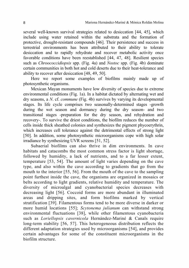
Figure 5. Confocal three-dimensional images of aerophytic biofilms: a) Projection showing inorganic calcareous material (white) (Nerja cave, Cordoba, Spain), plus spatial distribution of pigment autofluorescence (red) of single celled chlorophytes (Muriella sp.) distributed in a thin layer, penetrating the fissures and fractures. b) Extended projection of a biofilm dominated by Asterocapsa divina J. Komárek (Mayan monuments, Chiapas, México). Color key: photosynthetic pigments (Chlorophyll a and/or phycobilins), red. EPS (labeled with ConA-Alexa 488), green. Reflection from inorganic materials, white. 5. Biodeterioration Cultural heritage sites made of natural or artificial materials (e.g. rocks or stones and concrete or plaster) are governed by similar principles to any other terrestrial system, and are therefore susceptible to weathering. Subaerial biofilms colonize both the stone surface and the porous interior. Biofilm colonization of buildings or artwork is considered damaging, basically due to their chemical and physical activity [33, 58, 59]. Moreover, detectable colored patinas are considered dirty [42, 60]. Biodeterioration of exposed stone is primarily dependent on the availability of water and nutrients. Specific parameters, like porosity, permeability and architectural conditions, exposure and environmental factors at the site will determine the intensity and rate of biocorrosive attacks [61]. In many cases, these processes have been found to deteriorate the stone. Alterations are associated with repeated wetting and drying cycles that lead the organisms, which are attached to the rocks by EPSs, to expand and contract. In addition, the action of organic acids or metabolic products can enhances the weathering reactions and decrease the integrity of the mechanical properties of the natural or artificial materials. In spite of this, it is
difficult to determine whether photosynthetic biofilms will have a biodeterioration or bioprotection effect on the surface they are attached to because biofilms can be both beneficial and detrimental depending on the substrate and microorganisms involved. Thin superficial biofilms can cause discoloration of stone surfaces and mechanical and biochemical deterioration, or alternatively, they can protect the surface from weathering processes [54, 62]. a b
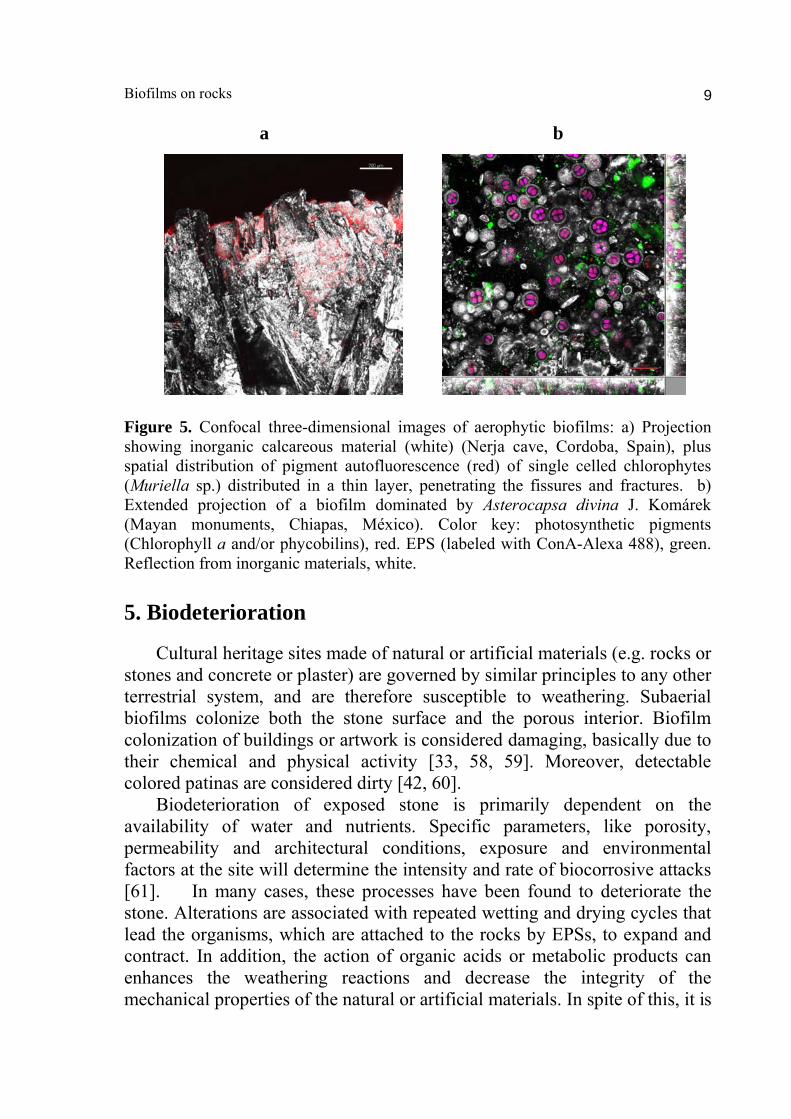
Figure 6. Optical micrographs: a) Discoloration on a roman fresco caused by a ubiquous development of actinobacteria and cyanobacteria forming a subaerial biofilm. b) Typical subaerial biofilm that develops in an area with run-out water (Vittoriosa, Malta). The black stain is mainly caused by actinobacteria, cyanobacteria and fungi.
6. Conclusion Biofilms are the default mode-of-life for many bacterial species and present remarkable complexity aimed at the protection of constituent microorganisms. Understanding factors that control the microorganisms as well as the effects of environmental factors on biofilm formation and control strategies will require further studies. Acknowledgements The authors are indebted to Servei de Microscòpia, Universitat Autònoma de Barcelona, and to Centres Científics i Tecnològics (CCiTUB), Universitat de Barcelona. Technical help of M. Ramírez is gratefully acknowledged. References 1. Zobel, C. E. J. 1943, Bacteriology, 46, 39. 2. Costerton, J. W., Stewart, P., Greenberg, E. P. 1999, Science, 284, 1318.
Biofilms on rocks 11
3. Warscheid, T., Braams, J. 2000, Int. Biodeter. Biodeg., 46, 343. 4. Hall-Stoodley, L. Costerton, J. W., Stoodley, P. 2004, Nat. Rev. Microbiol., 2, 95. 5. Flemming, H. -C., Wingender, J. 2010, Nat. Rev. Microbiol., 8, 623. 6. Christensen, B. E., Characklis, W. G. 1990, Physical properties of biofilms. In Biofilms, pp. 93-130. Edited by W. G. Characklis & K. C. Marshall.Wiley. New York. 7. Nielsen, P. H., Jahn, A., Palmgren, R. 1997, Water Sci. Technol., 36, 11. 8. Fleming, E. D., Castenholz, R. W. 2008, FEMS Microbiol. Ecol., 63, 301. 9. Sutherland, I. W. 2001. Trends Microbiol., 9, 222. 10. Hall-Stoodley, L. Costerton, J. W., Stoodley, P. 2004, Nat. Rev. Microbiol., 2, 95. 11. Dobretsov, S., Teplitski, M., Paul, V. 2009, Biofouling, 25, 413. 12. Davies, D. G., Parsek, M. R., Pearson, J. P., Iglewski, B. H., Costerton, J. W., Greenberg E. P. 1998, Science, 280, 295. 13. Annous, B. A., Fratamico, P. M., Smith, J. L. 2009, J. Food Sci., 74, R24. 14. Miller, M. B., Bassler, B. L. 2001, Annu. Rev. Microbiol., 55,165. 15. Sharif, D. I., Gallon, J., Smith, C. J., Dudley E. 2008, ISME J., 2, 1171. 16. Waters, C. M., Bassler, B. L. 2005, Annu. Rev. Cell Dev. Biol., 21, 319. 17. Shapiro, J. A. 1998, Annu. Rev. Microbiol., 52, 81. 18. Sauer, K., Camper, A. K., Ehrlich, G. D., Costerton, J. W., Davies, D. G. 2002, J. Bacteriol., 184, 1140. 19. Fuqua, C., Parsek, M. R., Greenberg, E. P. 2001, Annu. Rev. Genet., 35, 439. 20. Whitehead, N. A., Barnard, A. M., Slater, H., Simpson, N. J., Salmond, G. P. 2001, FEMS Microbiol. Rev., 25, 365. 21. Parsek, M. R., Val, D. L., Hanzelka, B. L., Cronan, J. E. J., Greenberg, E. P. 1999, Proc. Natl. Acad. Sci. USA, 96, 4360. 22. Smith, J. L., Fratamico, P. M., Novak, J. S. 2004. J. Food Prot., 67, 1053. 23. Chen, X., Schauder, S., Potie, N., Van Dorsselaer, A., Pelczer, I., Bassler, B. L., Hughson, F. M. 2002, Nature 415, 545. 24. Braun, E., Bachofen, R. 2004, Hydrobiologia, 522, 271. 25. Steunou, A. S., Bhaya, D., Bateson, M. M., Melendrez, M. C., Ward, D. M., Brecht, E., Peters, J. W., Kuhl, M., Grossman, A. R. 2006, Proc. Natl. Acad. Sci. USA, 103, 2398. 26. Roeselers, G., Zippel, B., Staal, M., van Loosdrecht, M., Muyzer, G. 2006, FEMS Microbiol. Ecol., 58,169. 27. Roeselers, G., van Loosdrecht, M. C. M., Muyzer, G. 2008, J. Appl. Phycol., 20, 227. 28. García-Aljaro, C., Vargas-Cespedes, G. J. Blanch A. R. 2011, J. Appl. Microbiol., 112, 383. 29. Neu, T. R., Kuhlicke, J. J., Lawrence, J. R. 2002, Appl. Environ. Microbiol., 68, 901. 30. Hernández-Mariné, M., Clavero, E., Roldán, M. 2003, Arch. Hydrobiol. Algol. Stud., 109, 229. 31 Walker, J. J., Pace, N. R. 2007, Annu. Rev. Microbiol., 61, 331. 32. Gorbushina, A. A., Broughton, W. J. 2009, Annu. Rev. Microbiol., 63, 431.
33. Wakefield, R., Jones, M., Wilson, M., Young, M., Nicholson, K., Urguhart, C. 1996, Aerobiologia, 12, 19. 34. Kumar, R., Kumar, A. V. 1999, Biodeterioration of stone in tropical environments. Research in Conservation, Getty Conservation Institute, Los Angeles, 88. 35. Caneva, G., Salvadori, O., Ricci, S., Ceschin, S. 2005, Plant Biosyst., 139, 295. 36. McNamara, C. J., Perry, I. V., Bearce, K. A., Hernández-Duque, G., Mitchell, R. 2006, Microb. Ecol., 51, 51. 37. Albertano, P., Hernandez-Mariné, M. 2001, Nova Hedwigia, 123, 225. 38. Ariño, X., Hernandez-Mariné, M., Saiz-Jimenez, C. 1997, Phycologia, 36, 366. 39. Hernández-Mariné, M., Roldán, M., Clavero, E., Canals, A., Ariño, X. 2001, Nova Hedwigia Beih., 123, 235. 40. Urzi, C., De Leo, F., Bruno, L., Albertano, P. 2010, Microb. Ecol., 60, 116. 41. Piñar, G., Ripka, K., Weber, J., Sterflinger, K. 2009, Int. Biodeter. Biodeg., 63, 851. 42. Laiz, L., Miller, A. Z., Jurado, V., Akatova, E., Sanchez-Moral, S., Gonzalez, J. M., Dionísio, A., Macedo, M.F., Saiz-Jimenez, C. 2009, Naturwissenschaften, 96, 71. 43. Cañaveras, J. C., Sanchez-Moral, S., Soler, V., Saiz-Jimenez, C. 2001, Geomicrobiol. J., 18, 223. 44. Potts, M. 2000, Chapter 17: Nostoc. In: Whitton, B. A., Potts, M. (Eds): The ecology of cyanobacteria: 465-504. Kluwer Academic Publishers, Dordrecht, 704 pp. 45. Wynn-Williams, D.D. 2000, In: Whitton, B. A., Potts, M. (Eds.). Kluwer Academic Publishers, Dordrecht, pp. 341-366. 46. Tamaru, Y., Takani, Y., Yoshida, T., Sakamoto, T. 2005, Appl. Environ. Microbiol., 71, 7327. 47. Dodds, W. K., Gudder, D. A., Mollenhauer, D. 1995, J. Phycol., 31, 2. 48. Fukuda, S., Yamakawa, R., Manabu, H., Yasuhiro, K., Koike, H., Satoh, K. 2008, Plant Cell Physiol., 49, 488. 49. Büdel, B., Darienko, T., Deutschewitz, K., Dojani, S., Friedl, T., Mohr, K. I, Salisch, M., Reisser, W., Weber, B. 2009, Microb. Ecol., 57, 229. 50. Ramírez, M., Hernández-Mariné, M., Mateo, P., Berrendero, E., Roldán, M. 2011, Fottea, 11, 73. 51. Garcia-Pichel. F., Castenholz, R.W. 1991, J. Phycol., 27, 395. 52. Fleming, E. D., Castenholz, R. W. 2007, Environ. Microbiol., 9, 1448. 53. Smith, T., Olson, R. 2007, Int. J. Speleol., 36, 105. 54. Roldán, M., Hernández-Mariné, M. 2009, Int. J. Speleol., 38, 41. 55. Vinogradova, O. N., Kovalenko, O. V., Wasser, S. P., Nevo, E., Weinstein- Evron, M., 1998, Isr. J. Plant Sci., 46, 229. 56. Roldán, M., Clavero, E., Canals, A., Gómez-Bolea, A., Ariño, X., Hernández- Mariné, M. 2004, Nova Hedwigia, 78, 329. 57. Lamprinou, V., Hernández-Mariné, M., Canals, T., Kormas, K.. Economou- Amilli, A., Pantazidou, A. 2011, Int. J. Syst. Evol. Microbiol., 61, 2907. 58. Gorbushina, A. A. 2007, Environ. Microbiol. 9, 1613. 59. Ortega-Calvo, J. J., Hernández-Mariné, M., Saiz-Jimenez, C. 1991, Int. Biodeterior., 28, 165.
Biofilms on rocks 13
60. Cappitelli, F., Abbruscato, P., Foladori, P., Zanardini, E., Ranalli, G., Principi, P., Villa, F., Polo, A., Sorlini, C. 2009, Microb. Ecol., 57, 633. 61. Warscheid, T., Braams, J. 2000, Int. Biodet. Biodeg., 46, 343. 62. Ramírez, M., Hernández-Mariné, M., Mateo, P., Berrendero, E., Roldán, M. 2011, Fottea, 11, 73.