Page 1
1
Cell Biology
Running title: Osmoregulation of Guard Cells by a Cation/H+ antiporter
Corresponding Author:
Dr. Heven Sze
Department of Cell Biology & Molecular Genetics
H. J. Patterson Hall
University of Maryland, College Park, MD 20742-5815, USA
E-mail: [email protected]
Phone: 301-405-1645
Fax: 301-314-9081
Plant Physiology Preview. Published on March 2, 2007, as DOI:10.1104/pp.106.092155
Copyright 2007 by the American Society of Plant Biologists
www.plantphysiol.orgon June 16, 2020 - Published by Downloaded from Copyright © 2007 American Society of Plant Biologists. All rights reserved.
Page 2
2
Participation of an Endomembrane Cation/H+ Exchanger AtCHX20
in Osmoregulation of Guard Cells
Senthilkumar Padmanaban1, Salil Chanroj1, June M. Kwak1, Xiyan Li1, John M. Ward2,
and Heven Sze1
1Department of Cell Biology & Molecular Genetics, H. J. Patterson Hall, University of
Maryland, College Park, MD 20742-5815, USA (SP, SC, JMK, XL, HS); 2Department Plant Biology, 250 Biological Sciences Center, 1445 Gortner Ave., University of
Minnesota, St. Paul, MN 55108, USA (JMW.)
www.plantphysiol.orgon June 16, 2020 - Published by Downloaded from Copyright © 2007 American Society of Plant Biologists. All rights reserved.
Page 3
3
Footnotes 1Supported in part by NSF Arabidopsis 2010 grant #IBN0209788 to HS, and #0209792 to JMW,
and by an NSF grant MCB-0614203 to JMK.
Corresponding author: [email protected] ; Fax 301-314-9081
www.plantphysiol.orgon June 16, 2020 - Published by Downloaded from Copyright © 2007 American Society of Plant Biologists. All rights reserved.
Page 4
4
Abstract
Guard cell movement is induced by environmental and hormonal signals that cause
changes in turgor through changes in uptake or release of solutes and water. Several transporters
mediating these fluxes at the plasma membrane have been characterized, however less is known
about transport at endomembranes. CHX20, a member of a poorly-understood cation/H+
exchanger gene family in Arabidopsis thaliana, is preferentially and highly expressed in guard
cells as shown by promoter::GUS activity and by whole-genome microarray. Interestingly, three
independent homozygous mutants carrying T-DNA insertions in CHX20 showed 35% reduction
in light-induced stomatal opening compared to wild type plants. To test the biochemical
function of CHX20, the cDNA was expressed in a yeast mutant which lacks Na+(K+)/H+
antiporters (∆nhx1∆nha1∆kha1) and plasma membrane Na+ pumps (∆ena1-4). Curiously,
CHX20 did not enhance tolerance of mutants to moderate Na+ or high K+ stress. Instead it
restored growth of the mutant on medium with low K+ at slightly alkaline pH, but had no effect
on growth at acidic pH. GFP-tagged CHX20 expressed in mesophyll protoplasts was localized
mainly to intracellular membranes, like endosomes. Furthermore, light-induced stomatal
opening of the Arabidopsis mutants was insensitive to external pH and was impaired at high
KCl. The results are consistent with the idea that in exchanging K+ for H+, CHX20 maintains K+
homeostasis and influences pH under certain conditions. Together these results provide genetic
and biochemical evidence that one CHX protein plays a critical role in osmoregulation through
K+ fluxes and possibly pH modulation of an active endomembrane system in guard cells.
www.plantphysiol.orgon June 16, 2020 - Published by Downloaded from Copyright © 2007 American Society of Plant Biologists. All rights reserved.
Page 5
5
Introduction
One of the most fascinating processes in most land plants is the ability to regulate gas
exchange and transpiration by the opening and closing of the stomatal aperture. The movement
of a pair of special epidermal cells, the guard cells, controls the size of the stomatal aperture and
so determines the extent of water loss via transpiration, and of CO2 uptake into the leaf for
photosynthetic carbon fixation. At the beginning of the day, light stimulates the opening of the
stomatal aperture of most plants by increasing solute concentration and decreasing water
potential thus attracting water into the guard cells (see reviews by Assmann, 1993; Schroeder et
al. 2001; Roelfsema & Hedrich, 2005). The increase in turgor pressure causes the guard cells to
swell and pushes the pair of cells apart increasing the aperture between the two cells. At dusk,
the aperture size decreases and becomes nearly closed at night thus reducing transpiration and
gas exchange. During drought, the amount of ABA reaching the guard cells can increase
triggering the efflux of ions and loss of water and turgor pressure, leading to closure of stomatal
aperture. Abscisic acid (ABA) also prevents light-induced stomatal opening (Schroeder et al
2001).
Although our knowledge of cellular signaling and osmoregulation in guard cells has
advanced significantly in the last decade, the osmotic changes driving guard cell movement have
focused mainly on the roles of plasma membrane-associated transporters, and signaling elements
regulating the transporters (Blatt 2000; Fan et al. 2004; Roelfsema & Hedrich, 2005). The
advances have been triggered by the ability to patch guard cell plasma membrane (PM), to study
transport across this membrane, and to analyze mutants. Light-induced stomatal opening starts
when light activates the PM H+-ATPase causing membrane hyperpolarization. K+ then enters
via inward-rectifying channels, and anions enter via predicted H+/Cl- and H+/NO3- symporters.
Ion, malate and sugar accumulation decreases the water potential, thus water is taken up
increasing turgor pressure. Several inward rectifying K+ channels (e.g. KAT1, KAT2, AKT1) in
stomatal opening have been identified at the molecular level (see review of Very and Sentenac,
2003; Fan et al. 2004). Nitrate is one counter-ion that balances K+ uptake via a H+-coupled NO3-
symporter (AtNRT1.1) (Guo et al. 2003). Stomatal closing begins when the membrane
depolarizes causing opening of outward-rectifying K+ channels. Dark-induced depolarization is
caused by deactivation of the PM H+-extrusion pump and by opening of anion efflux channels.
www.plantphysiol.orgon June 16, 2020 - Published by Downloaded from Copyright © 2007 American Society of Plant Biologists. All rights reserved.
Page 6
6
Loss of K+ and anions leads to a decrease in solute concentration, water efflux and loss of guard
cell turgor. GORK is suggested to be the major outward rectifying K+ channel (Hosy et al.
2003); however the molecular identity of the PM R-type and S-type anion channels is still
unclear. Genetic evidence suggests that AtMRP5 ABC transporter mediates anion efflux (Klein
et al. 2003).
Less well understood are the changes of intracellular compartments during guard cell
movement. As guard cells increase in volume, the size of vacuoles increases considerably
(Louget et al., 1990) indicating that the bulk of solutes entering guard cells accumulate in the
large vacuoles (MacRobbie, 1999) which is iso-osmotic with the cytosol. When stomata close,
guard cells are filled with numerous relatively small vacuoles. Many vacuolar transporters
identified in plant cells, are expressed in guard cells according to the Affymetrix 8k gene chip
results (Leonhardt et al., 2004). Endomembrane compartments, including vacuoles, are acidified
by electrogenic H+ pumping vacuolar-type ATPases (V-ATPase) and H+-pumping
pyrophosphotases (V-PPase) (Sze, 1985; Rea, 1993). Thus it is very likely that the vacuolar
membrane potential (∆ψvac) slightly positive inside the lumen relative to the cytosolic side, and a
∆pH acidic inside (lumen relative to the cytosol) could drive the accumulation of K+ into the
lumen via H+/cation antiporters. Anions, including Cl- and NO3-, were predicted to enter
vacuoles via anion-specific channels, as these anions rapidly dissipate the membrane potential
generated by the V-ATPase of intracellular vesicles (Sze, 1985); although recent evidence
showed that NO3- enters vacuoles through a H+ coupled NO3
- antiporter (ClC-a) at the vacuolar
membrane (De Angeli et al., 2006). VK channel activity previously characterized to function in
K+ release from vacuoles in response to elevated cytosolic Ca2+ (Ward and Schroeder, 1994) is
mediated by TPK1/KCO1 (Bihler et al. 2005). FV channels are inhibited by elevated cytosolic
Ca2+ and may modulate K+ uptake into vacuoles during stomatal opening (Pei et al. 1999). It is
unclear how most of these transporters are integrated with guard cell movement.
Here we provide genetic evidence for the role of a novel endomembrane transporter in
guard cell movement. CHX20 belongs to a large family of 28 cation/proton exchangers whose
functions are largely unknown (Sze et al., 2004). Functional expression of CHX20 in a salt-
sensitive yeast strain suggests it has a role in pH regulation and K+ transport. Intriguingly,
CHX20 is preferentially expressed in guard cells, and chx20 null mutants showed reduction in
www.plantphysiol.orgon June 16, 2020 - Published by Downloaded from Copyright © 2007 American Society of Plant Biologists. All rights reserved.
Page 7
7
light-induced stomatal opening. Together these results provide evidence that a member of the
CHX plays a critical role in osmoregulation of guard cells.
RESULTS
CHX20 cDNA isolation and predicted protein
To obtain CHX20 (At3g53720) cDNA, total RNA was extracted from rosette leaves of 3-
week-old Arabidopsis thaliana plants and the first strand cDNA was used to amplify the coding
sequence. The primers at the start and end of the ORF (X20Cf and X20Cr) (Sup Table I) were
designed based on the genomic sequence. A 2.5 kb fragment was amplified and its sequence
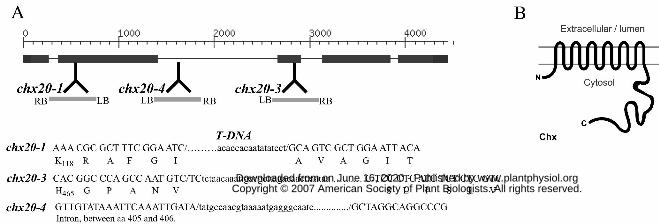
(AY926476) matched the coding sequence which is formed from five exons (Fig. 1A).
The predicted CHX20 protein of 842 residues has two domains:I) a hydrophobic domain
(434 residues) with 10-12 transmembrane spans at the amino half, and ii) a large hydrophilic
domain of 403 residues at the carboxylic end (Fig. 1B). The hydrophobic domain shows
extensive similarity (56.5% similarity, 33.6% identity; E-value of 1e-54) to the transmembrane
domain of yeast ScKHA1 protein, though the long carboxylic tail of the two proteins did not
align (10.6% identify; no E-value; Fig. 1 Sup). These results suggest that the transport activities
of AtCHX20 and yeast ScKHA1 are similar.
Function of AtCHX20 in yeast
To test the transport function of CHX20, the coding sequence was cloned in pYES-
DEST52 yeast expression vector under the GAL promoter. Yeast mutants with disrupted kha1
gene alone exhibited no obvious phenotype (Maresova and Sychrova, 2005), so we expressed
CHX20 in a yeast mutant (KTA 40-2). This strain lacks functional vacuolar and plasma
membrane-localized Na+/H+ antiporters, the plasma membrane Na+ pumps (∆nhx1, ∆nha1, and
∆ena1-4) as well as the putative K+/H+ exchanger (∆kha1) (Maresova and Sychrova, 2005).
Strain KTA40-2 is highly sensitive to salt and to high K+, so the transformant (KTA40-2-
CHX20) was tested for its ability to grow on moderate levels of Na+ and very high K+.
Surprisingly, mutant yeast expressing AtCHX20 was consistently more sensitive on media
containing 100 mM Na+ or 500 mM K+ at various pHs (Fig. 2A) than the mutant yeast harboring
the vector alone. KTA40-2 mutants grew as well as CHX20 transformants on standard SC
medium. These results suggest that CHX20 does not confer tolerance to salt stress.
www.plantphysiol.orgon June 16, 2020 - Published by Downloaded from Copyright © 2007 American Society of Plant Biologists. All rights reserved.
Page 8
8
Intriguingly, CHX20 enhanced KTA40-2 yeast mutant growth on slightly basic medium
with no added K+. At an external pH of 4.5 to 7.0, mutants grew relatively well with no added
K+. In fact, at acidic pH between 4.5-6.5 mutants grew consistently better than yeast
transformants carrying CHX20. Curiously, growth of mutants carrying the vector alone was
retarded at pHext 7.5; while transformants harboring CHX20 continued to grow as well as at pH
4.5 (Fig. 2B). Thus strains carrying CHX20 had an advantage when the external pH was 7.5,
suggesting that CHX20 conferred an ability to sustain growth at slightly basic pH.
We tested the effect of external K+ concentration on yeast growth at pH 7.5.
Transformants harboring CHX20 consistently grew better than KTA40-2 mutants as long as the
K+ level was kept low from ~0.4 mM up to 3 mM (Fig. 2C). When no exogenous K+ was added,
the agar medium contained about 0.4 mM K+. Increasing external KCl concentration beyond 25
mM decreased the beneficial effect of CHX20. Since K+ is required to sustain growth of all
cells, the enhanced growth of transformants at low K+ levels would suggest that CHX20 has a
role in acquiring K+ when the external pH is slightly alkaline, or in maintaining a suitable
cellular homeostasis for growth. This idea is supported by nearly similar growth exhibited by
yeast mutants carrying either vector alone or CHX20 when K+ is raised to 50 mM.
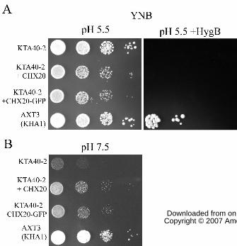
Yeast KHA1 was shown before to confer tolerance to hygromycin (Maresova &
Sychrova, 2005) as confirmed here in the AXT3 strain (Fig. 3A). This strain has a functional
wild-type KHA1, but lacks three Na transporters (ena1-4∆ nha1∆ nhx1∆). However, although
transformants expressing CHX20 grew well at pH 5.5, they showed no growth in the presence of
150 µM hygromycin B. CHX20 did promote growth of mutants grown on YNB medium at pH
7.5 similar to yeast KHA1 (Fig. 3B). These results suggest that CHX20 and KHA1 share
similar, but not identical activities.
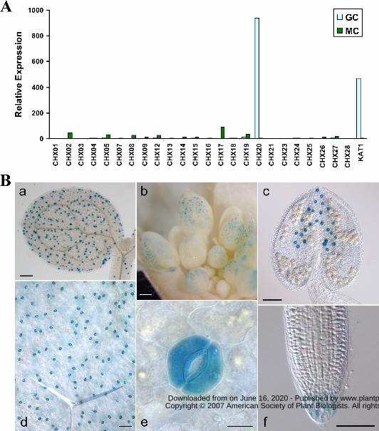
AtCHX20 is preferentially expressed in guard cells
Analyses of a guard cell transcriptome (Leonhardt et al., 2004; Kwak J, Leonhardt N,
Schroeder JI, unpublished data) revealed that only one member of the CHX gene family was
highly expressed in guard cells. CHX20 showed little or no expression in mesophyll cells,
whereas several other genes, such as CHX17, showed low to moderate expression (Fig. 4A).
Furthermore, CHX20 expression is particularly strong in guard cells as shown by the two-fold
www.plantphysiol.orgon June 16, 2020 - Published by Downloaded from Copyright © 2007 American Society of Plant Biologists. All rights reserved.
Page 9
9
increase in normalized relative expression of CHX20 compared to that of AtKAT1, a K+ channel
preferentially expressed in guard cells (Nakamura et al. 1995).
To verify the microarray results, CHX20 promoter-driven GUS activity was determined.
Arabidopsis (Col) plants were transformed with a construct containing a 2 kb region upstream of
the CHX20 open reading frame transcriptionally fused to the GUS reporter gene. T2 seeds were
collected from six independent transgenic lines and all six lines of CHX20::GUS analyzed gave
similar expression patterns. Striking GUS activity was observed in guard cells located in
expanded cotyledons and in hypocotyls of one-week old seedling (Fig. 4B-a). Three-week-old
rosette leaves (Fig. 4B-d and e), and cauline leaves also showed very high GUS staining in guard
cells. However, GUS staining was not detected in leaf pavement epidermal cells, or in
mesophyll cells. Interestingly, GUS activity was also detected in guard cells of floral organs,
including the sepal, anther (Fig. 4B-b and c) and carpel (not shown). GUS activity was not
detected in the differentiated cells of roots, though CHX20 expression was only observed in the
root cap of one-week old seedling (Fig. 4B-f), consistent with the microarray results of root cap
cells (Benfey P, personal comm.). Thus, analyses of both CHX20 promoter-GUS expression and
guard cell-specific transcriptome data clearly indicate a selective expression of CHX20 in guard
cells.
CHX20-GFP is localized to endomembrane
When transiently expressed in Arabidopsis mesophyll protoplasts, CHX20-GFP was
visualized at the periphery of the nucleus, and in the cytosol (Fig. 5A-f), suggesting it is
localized at the ER or in endomembranes. The CHX20-GFP signal was compared with those
from a soluble GFP, GFP tagged to an ER retention sequence (GFP-HDEL), or to markers, such
as sialyltransferase (ST)-GFP for trans Golgi, GFP-CPK9 for PM, and GFP-δTIP for vacuolar
membrane (Fig. 5A). Though CHX20-GFP appeared to be localized to endomembranes, its
pattern did not coincide entirely with any of the markers tested. To determine its location more
precisely, the CHX20-GFP and a Golgi marker, ST-RFP constructs were co-transfected into
mesophyll protoplasts. Of the cells that co-expressed both probes, the pattern of green
fluorescence-labeled structures for the most part did not overlap with that of the red fluorescence
(data not shown), indicating that CHX20 is not restricted to the trans-Golgi membrane.
www.plantphysiol.orgon June 16, 2020 - Published by Downloaded from Copyright © 2007 American Society of Plant Biologists. All rights reserved.
Page 10
10
Stably transformed plants expressing CaMV35S-driven CHX20-GFP also showed
perinuclear fluorescent signals in guard cells (Fig. 5C) whereas soluble free GFP appeared inside
the nucleus. Strong fluorescent signals were also detected inside the cytoplasm of cells
expressing CHX20-GFP relative to that expressing free GFP control. Together, the results
suggest that CHX20 is localized to a subpopulation of endomembranes though the protein does
not appear to be a fixed resident of either the ER, Golgi, vacuole or plasma membrane. We
postulated that CHX20 is associated with vesicles/membranes that traffick among various
subcellular membranes (Jurgen, 2004).
To test this idea, we examined the distribution of an endosome marker, Ara6-GFP (Ueda
et al. 2001) and of CHX20 at several focal planes. Fluorescent signals of these two proteins
were strikingly similar in several independent experiments. At the medial plane, the signal was
cytoplasmic and at or near the PM (Fig. 5B). At the sub-medial focal plane, fluorescent signals
were mostly cytoplasmic surrounding the plastids (not shown in Fig. 5B). At the peripheral focal
plane, CHX20-GFP signal included several punctate regions. This pattern was distinct from a
soluble ER marker that showed a reticulate pattern. The GFP-tagged CHX20 was functionally
active as shown by its ability to restore growth of KTA40-2 yeast at alkaline pH (Fig. 3B). Ara6,
a Rab5-related GTPase, is distributed on a subset of endosomes and is involved in regulating
vesicular transport (Ueda et al. 2001). These results suggest that an active CHX20 protein is
associated with endosomes.
Identification of chx20 null mutants
To determine the in planta function of CHX20, we obtained three independent T-DNA
insertional lines of Arabidopsis chx20. Two lines, chx20-1 and chx20-3 were identified in the
SIGnAL database (Alonso et al. 2003) and one line, chx20-4, was obtained from Genoplante
(France). To confirm the T-DNA insertion site and select homozygous lines, PCR-based
screening was performed using CHX20 specific primers and T-DNA primers (Supplemental
Table I). Sequencing of the PCR-amplified fragments confirmed that a T-DNA insertion was
located within exon 2 at coding sequence base 477 of chx20-1 mutant, inside third exon at 1299
coding sequence of chx20-3 and within the second intron of chx20-4 (Fig. 1A). We tested for
CHX20 transcripts in leaves of all the mutants. RT-PCR was performed using CHX20 gene-
specific primers located at either side of the T-DNA insertion using template cDNA reverse-
www.plantphysiol.orgon June 16, 2020 - Published by Downloaded from Copyright © 2007 American Society of Plant Biologists. All rights reserved.
Page 11
11
transcribed from total leaf RNA. No products were amplified indicating an absence of messages
in all 3 alleles (Fig. 6B). The chx20 mutants showed no obvious morphological or growth
differences compare to WT plants under standard growth conditions (Fig. 6A). Overall, the size
and shape of the guard cells were indistinguishable between mutants and wild-type plants.
Impaired stomatal opening in chx20 mutants
The highly specific expression of CHX20 in guard cells (Fig. 4) suggested that CHX20
plays a role in guard cell signaling and/or development. As we did not notice any developmental
defects in the chx20 knockout mutants, we tested whether the chx20 null mutants had any altered
stomatal movement. We first compared light-induced stomatal opening in mutants and wild-type
plants. Excised leaves of chx20-1, chx20-3 and chx20-4 mutants were first exposed to white
light for 3 h in a solution containing 5 mM KCl and 10 mM MES at pH 6.15. In all 3 mutants,
the stomata failed to open as widely as WT plants. The ratio of light-induced stomatal opening
per guard cell length in wild type plant and in mutants ranged from 0.072-0.076, and 0.042-
0.047, respectively. Thus stomatal opening was reduced by ~35% in chx20 mutants (Fig. 7A).
We reduced external KCl concentration in the opening solution to 0.1 and 1.0 mM. The aperture
size was reduced slightly in wild-type and mutant leaves exposed to 0.1 mM K+ (Fig. 7B),
implying that guard cell movement is limited at low K+ concentration. However, chx20 mutants
still showed ~35% reduction in light-induced stomatal opening irrespective of the external K+
concentration, indicating that the defect is not due to limited K+ level alone.
Using isolated epidermis, we found that light-induced stomatal opening was maximal at
pH 6.1 and at pH 7. At basic pH of 7.5 and 8.0, light-induced opening was decreased in wild-
type plants (Fig. 7C) consistent with inactivation by basic pH of inward-rectifying K+ channels
and activation of outward-rectifying K+ channels in Vicia fava guard cells (Ilan et al. 1994;
1996). However, mutants appeared to be insensitive to an acidic apolastic pH that stimulated
stomatal opening of wild-type guard cells. Thus the reduced stomatal aperture of chx20 mutants
was particularly apparent at pH 6.1 and 7.0. At pH 7.5 and 8.0, mutants showed reduced
stomatal aperture nearly similar to that of wild-type. Thus chx20 mutants appeared to be
unresponsive to pH regulation of guard cell movement.
To test if stomatal closure was affected, isolated epidermis of wild type and chx20 mutant
leaves were first exposed to white light for 3 h to induce stomatal opening, and then incubated in
www.plantphysiol.orgon June 16, 2020 - Published by Downloaded from Copyright © 2007 American Society of Plant Biologists. All rights reserved.
Page 12
12
1 µM ABA to induce closure. The decrease in stomatal aperture was measured at 30 min
intervals for 3 h. Although the aperture size of wild-type plants was larger than that of mutants
before ABA addition, the % closure of wild type was higher than that of mutants at all times
(Fig. 8). These results indicate that the chx20 mutants were responsive to ABA; however
mutants were delayed in stomatal closure compared to WT plants (Fig. 8). Results would
suggest that CHX20 might also participate in events leading to stomatal closure.
DISCUSSION
Here we have discovered a new transporter that participates in guard cell movement.
Nearly nothing is known about the roles of cation/proton antiporter (CPA) genes in guard cells,
though several members of the superfamily, including NHX1, are expressed there. The CHX
family was uncovered recently as a novel subfamily (Mäser et al 2001; Sze et al. 2004), though
the biochemical properties of this family remained uncharacterized until recently (Maresova &
Sychrova, 2006). Previous studies showed 18 CHXs are preferentially expressed in pollen, and 6
CHXs are highly expressed in roots and/or shoots (Cellier et al. 2004; Sze et al. 2004; Hall et al.
2006). However, discrepancy in the spatial expression of CHX23 mainly in either pollen (Sze et
al. 2004) or in sporophytic tissues (Song et al. 2004) raises concerns about unconfirmed results.
In this study, we find that CHX20 is preferentially expressed in guard cells using microarray and
promoter::Gus analyses. Therefore we tested the cellular function of AtCHX20 in yeast as a first
step to understand its role in guard cell movement.
Role of CHX20 in K+ acquisition and pH homeostasis in yeast
Curiously, instead of conferring tolerance to moderate Na+ stress or high K+, CHX20
consistently caused mutant KTA40-2 (∆ena1-4 ∆nha1 ∆nhx1 ∆kha1) to be more sensitive to salt.
In another salt-sensitive yeast mutant AXT3 (∆ena1-4 ∆nha1 ∆nhx1), expression of AtCHX20
also resulted in increased sensitivity to moderate Na+ stress and high K+; although AtNHX1 or
AtNHX2 conferred moderate tolerance to Na+ stress (data not shown) as shown before (Yokoi et
al., 2002). Furthermore, CHX20 was unable to confer hygromycin B tolerance. Thus AtCHX20
is functionally distinct from the vacuolar AtNHX1 that sequesters excess Na+ or K+ into vacuoles
and confers tolerance to high Na+ or K+; and to hygromycin B (Pardo et al., 2006).
www.plantphysiol.orgon June 16, 2020 - Published by Downloaded from Copyright © 2007 American Society of Plant Biologists. All rights reserved.
Page 13
13
Instead CHX20 function appears to be important particularly when K+ is depleted and
when the external pH is slightly alkaline. This is shown by improved growth of KTA40-2
expressing CHX20 at pH 7.5, and when [K+]ext was low (between 0.4 to 3 mM). Yeast growth
and budding depends on the continuous uptake and accumulation of osmotic solutes, like K+,
especially when the external medium is depleted of this cation (Rodriguez-Navarro 2000). The
mechanism for K+ uptake by CHX20 is not clear, though several observations are consistent with
the idea that CHX20 participates in K+ homeostasis through fluxes at intracellular compartments:
(i) CHX20 is mainly localized to endomembranes, possibly endosomes, in plant cells, (ii)
phylogenetic analysis showed that CHX20, is a cation-proton antiporter belonging to the CPA2
subfamily (Sze et al. 2004); and (iii) CHX20, like ScKHA1, enhanced yeast growth at basic pH
when K+ext concentration was low (Maresova and Sychrova, 2005). Interestingly, KHA1 is also
localized in yeast to endomembranes, possibly the Golgi. As endomembrane compartments, are
acidified by V-ATPase in plants (Sze et al. 1999) as in yeast (Kane, 2006), cation accumulation
into the lumen is most likely driven by the downhill influx of H+ into the cytosol. Thus one
working model is that CHX20 is an endomembrane K+/H+ antiporter. In preliminary
experiments, we did not detect an increase in K+ content in yeast expressing CHX20, indicating
that net changes in K+ is small.
How are endomembrane compartments acidified when yeast is exposed to a basic
medium? An important clue is provided by yeast vma (Vacuolar membrane H+-ATPase) mutants
which cannot survive at pH 7.5 whereas wild-type yeast can. However vma mutants grow at pH
5.5 (Nelson and Nelson, 1990), suggesting that mutants form acidic intracellular compartments
through endocytosis of the extracellular medium (Munn and Riezman, 1994) and/or by passive
uptake of acids (see Kane, 2006). The internalized compartment is apparently acidic enough to
activate and energize the H+-coupled transport of solutes needed to sustain vma mutant growth,
and to sort proteins in the secretory system. However, when the external medium is buffered at
an alkaline pH, endocytosis would result in endosomal compartments with little or no pH
gradient relative to the cytoplasm. Apparently the PM H+-ATPase (PMA1) alone is either
inactivated or unable to generate a proton electrochemical gradient to support cell growth when
the pHext is alkaline, thus vma mutants succumb. However, wild-type yeast with functional
VMA proteins sustains growth at pH 7.5, indicating that endomembrane compartments play a
vital role in pH regulation and homeostasis. Our results would indicate that in addition to a
www.plantphysiol.orgon June 16, 2020 - Published by Downloaded from Copyright © 2007 American Society of Plant Biologists. All rights reserved.
Page 14
14
vacuolar H+-pump, CHX20 has a role in sustaining growth at pH 7.5 when other K+(Na+)/H+
antiporters are absent. Thus CHX20 could fill a role in pH regulation.
What is the role of an endomembrane K+/H+ antiporter when external K+ is low? We
propose a model where CHX helps distribute cellular K+ when the external pH is alkaline. It is
well-known that when medium K+ is low or nearly depleted (< 0.1 mM), energy-dependent K+
uptake is needed to maintain [K+]cyt at millimolar levels (Rodriguez-Navarro, 2000). However
when the medium pH is slightly alkaline, the proton-motive force for K+-H+ symport at the PM is
reduced; and the alkalinization of the cytosol could inactivate the PM H+ pump. To counter the
reduced proton motive force at the PM, acidification of intracellular compartments by yeast
VMA could energize accumulation of K+ from the cytosol into internal compartments using a
K+/H+ antiporter. Due to the small volume of vesicles and internal compartments of the
endomembrane system, a proton electrochemical gradient (acidic in the lumen) forms rapidly
energizing K+ accumulation. Accumulated K+ can then be redistributed to the cytosol and other
compartments by release via cation channels and by vesicle trafficking. Our results and model
are consistent with genetic studies of a related protein, CHX17. K+ starvation induced increase
of CHX17 transcripts in wild-type plants; and K+ starvation caused a 20% decrease in K+ content
of chx17 mutant roots (Cellier et al., 2004).
To offset alkalization of the cytosol in yeast grown at pH 7.5, we suggest that antiporters,
like CHX20, neutralizes cytosolic pH by mediating K+ exchange for H+ release from acidified
compartments. The ability of CHX20-expressing KTA40-2 mutants to grow at pH 7.5, suggests
that CHX20 is active at basic pH. Fungi and plant V-ATPases show optimal activity in vitro at
slightly alkaline pH (7.0-8.0) when PM H+-ATPase is less active (Sze, 1985). Thus two
functions are proposed for CHX20: (i) H+/K+ antiport loads K+ into intracellular compartments
under conditions when mechanisms for active loading at the PM via H+/K+ symport are
compromised; and (ii) the cation/H+ exchange maintains cytosolic pH homeostasis when cells
are alkaline-stressed.
The lack of a growth phenotype in yeast expressing CHX20 at pH 7.5 when K+ext is
replete suggests other mechanisms take over to modulate K+ and pH homeostasis when K+ext is
high (25-50 mM). Conceivably, high external K+ could i) depolarize the cell membrane
potential; (ii) increase K+ influx into the cytosol and intracellular compartments, or both. With
sufficient K+ in the cell and intracellular compartments to support growth, the role of CHX20
www.plantphysiol.orgon June 16, 2020 - Published by Downloaded from Copyright © 2007 American Society of Plant Biologists. All rights reserved.
Page 15
15
may be shielded by other activities. Together, these results point to a role of CHX20 either in
acquiring K+ for cells under certain conditions, or setting a suitable cellular pH homeostasis or
both.
Model of CHX20 function in guard cell movement: Modulating vesicle trafficking
Our genetic studies demonstrate that CHX20 participates in guard cell movement, though
its role in mediating stomatal opening may involve multiple tasks. Based on functional studies
of yeast, it is reasonable to conclude that one role of CHX20 is to load guard cells with K+.
Stomatal aperture from chx20 mutants failed to fully open after light-induction. If CHX20 has a
major role in K+ loading, then the defect in opening might be minimized when K+ext is not
limiting. However, chx20 mutants were impaired in stomatal opening whether the K+ext was at
0.1 mM or at 10 mM, when K+ entry and content in cells in theory are not limited. These results
suggest that CHX20 fill other roles. We tested whether CHX20 activity might be revealed at
different pH’s as seen in KTA40-2 yeast. Stomatal opening was maximal at pH 6-7 and reduced
at pH 7.5-8.0 in wild-type leaves, consistent with activation and deactivation by acidic pHi of
inward and outward K+ channels, respectively, seen before (Ilan et al., 1994; 1996). Stomatal
opening in mutants however, failed to respond to acidic pH, suggesting that loss of CHX20
function could have interfered perhaps with pH homeostasis, and with the activation and/or
membrane trafficking of K+-inward rectifying channels.
Considering the large number of cation/proton antiporters in plants (Maser et al., 2001;
Pardo et al. 2006), it is striking that single chx20 mutants were impaired in stomatal opening.
The contribution of other CHXs appears to be minimal in guard cells; however cation/H+
antiporters, like NHXs, are highly expressed in shoots, roots (Yokoi et al., 2002), and guard cells
(Shi and Zhu, 2002; JM Ward, data not shown). Members of this family (NHX1-NHX8) are
localized to various membranes, including vacuole, prevacuolar compartment (PVC), Golgi or
plasma membrane PM (Venema et al 2003; Pardo et al 2006). If other endomembrane K+
(Na+)/H+ antiporters are unable to substitute for CHX20 function, then CHX20 occupies a
distinct functional niche. A simple model is that CHX20 function differs from other cation/H+
exchangers, because of: (i) differential endomembrane localization; (ii) different substrate
affinity and specificity (Km and Vmax); and (iii) differential modulation by pH and/or other
signals and different interacting partners.
www.plantphysiol.orgon June 16, 2020 - Published by Downloaded from Copyright © 2007 American Society of Plant Biologists. All rights reserved.
Page 16
16
Confocal laser scanning microscopy of guard cell vacuoles loaded with acridine orange
revealed striking changes in the size and shape of vacuolar compartments during guard cell
movement. Small vacuoles in cells surrounding closed stomata fuse with one another to form
large vacuoles as stomata open (Gao et al., 2005). During closure, the large vacuoles break up
into small vacuoles and undefined membrane structures. Moreover, pressure-induced guard cell
swelling have demonstrated an increase in the PM area based on membrane capacitance
measurements, and increased K+ conductance of both inward and outward rectifiers (Homann
and Thiel, 2002; Meckel et al. 2005). These changes indicate the incorporation of exocytic
vesicle with the PM. Guard cells undergo constitutive endocytosis and exocytosis even in turgid
cells as shown by the uptake of impermeant fluorescent FM4-64 dyes into vesicles beneath the
plasma membrane (Meckel et al., 2004). Although direct biophysical measurements are not yet
available to measure vacuolar dynamics, electron microscopy (e.g. Louget et al. 1990) and live
imaging support the idea that guard cells are active in vesicle/tubule budding from and fusion
with vacuoles (Gao et al. 2005).
One model is that endomembrane-associated CHX20 exchanges K+ for H+ and
accomplishes one or more purposes, including (i) bring in external K+ indirectly by
accumulating K+ in small intracellular compartments, (ii) regulate pH in the cytosol as well as in
the compartment lumen, and (iii) perhaps participate in some way to facilitate the membrane
dynamics, including vesicle budding, trafficking, and fusion events required to bring about
turgor and volume changes during guard cell movement (MacRobbie 1999; Pratelli et al. 2004).
The importance of pH for protein processing and sorting in the secretory and endocytic system is
well-recognized in yeast and in animal cells (Paroutis et al., 2004), and this function is most
likely conserved in plant cells (Sze et al, 1999). This idea is supported by a recent study showing
Arabidopsis H+-pumping V-ATPase is required for endocytic and secretory trafficking (Dettmer
et al. 2006).
This study provides the first genetic evidence for a role of an endomembrane CHX in
osmoregulation and in guard cell movement, and working ideas to further test CHX cellular
function. As the only CHX expressed in guard cells, CHX20 is an attractive model to understand
the function and regulation of other CHX proteins (Sze et al., 2004). The transmembrane
domains and the hydrophilic carboxylic tails of CHX members are highly conserved, suggesting
a role in integrating signals with osmoregulation and possibly membrane trafficking.
www.plantphysiol.orgon June 16, 2020 - Published by Downloaded from Copyright © 2007 American Society of Plant Biologists. All rights reserved.
Page 17
17
Significantly, this study highlights the active involvement of multiple transporters associated
with intracellular membranes, like endosomes, in signaling, osmoregulation, and movement.
MATERIALS AND METHODS
Plant Materials and Growth Conditions
All experiments were conducted with Arabidopsis thaliana ecotype Columbia (Col-0).
Wild type, mutant and transgenic plants were grown under the same conditions. Plants were
grown in Miracle-Gro potting soil (Stern’s Miracle-Gro, Port Washington, NY). Seeds in soil
were stratified at 4° C for 3 d and then plants were grown in controlled-environment chambers at
20°C under illumination of 150 µE m-2 s-1 with a 16-h photoperiod. Two weeks after
germination, plants were given Miracle-Gro plant food at 20 d intervals. To test for
promoter::Gus expression, transgenic seeds were grown under light (150 µE m-2 s-1) at 20°C on
plates containing 0.5 X Murashige and Skoog (1962) salts, and 1.0 % agar at pH 5.8.
cDNA cloning and DNA constructs
Promoter::GUS. The CHX20 promoter was transcriptionally fused to the GUS gene. A 2 kb
region upstream of CHX20 was amplified by PCR. Primers, CHX20-PF and CHX20-PR, have
Sal I and Bam HI restriction sites, respectively (see Supplemental Table I for all primers). The
amplified products were digested and fused with GUS in pRITA I plasmid. Clones were
confirmed by sequencing. The region containing the CHX20 promoter and GUS was subcloned
into the binary vector pMLBart using Not I and named CHX20::GUS (sup. Table II).
CHX20 cDNA. To isolate CHX20 cDNA, total RNA was isolated from leaves of wild-type A.
thaliana and first strand cDNA was synthesized using reverse transcriptase. Primers X20Cf and
X20Cr were used to amplify the cDNA by 25 cycles (94°C 30 s, 55°C 30 s and 72°C 90 s). The
forward and reverse primers contain attB1 and attB2 sequences (Table I Sup) for Gateway
recombination cloning. Gel purified PCR products were recombined with pDONR221 using BP
clonase according to manufacturer’s method (Invitrogen, Carlsbad, CA). Resulting clones were
sequenced using forward and reverse M13, S1, S2, and S3 primers. The correctly spliced clone
with longest ORF was named entry clone pECHX20.
To make a CHX20-GFP fusion construct, the CHX20 coding sequence from pECHX20
was recombined to the binary vector pK7FWG2 (Karimi et al., 2002) using LR clonase to give
www.plantphysiol.orgon June 16, 2020 - Published by Downloaded from Copyright © 2007 American Society of Plant Biologists. All rights reserved.
Page 18
18
an in frame fusion of eGFP at the C tail of CHX20 or pDCHX20-GFP. For expression in yeast,
CHX20 from pECHX20 was recombined into a yeast-E coli shuttle vector, pYES-DEST52, to
yield pDYES-CHX20.
Transformation of Plants
The binary vectors with CHX20 promoter::GUS or pDCHX20-GFP were introduced
stably into Arabidopsis using Agrobacterium tumefaciens-mediated floral dip (Clough and Bent,
1998). Transformants were selected on 0.5X MS plates containing kanamycin (50 µg mL-1) or
on soil by spraying with BASTA. T2 plants were analyzed for GUS expression or CHX20-GFP
fluorescence. pDCHX20-GFP or other GFP-tagged constructs (see Supplemental Methods and
Table II) were transiently expressed in onion epidermal cell or in Arabidopsis mesophyll
protoplast (Kovtun et al., 2000) and observed after 12-16 h.
Microscopy
GFP. The cells or tissues from transient and stable transformants were imaged for GFP
fluorescence using a Zeiss LSM 510 laser-scanning confocal microscope with a 10x dry 0.8-
numerical-aperture lens and a 63x 1.2-numerical-aperture water immersion lens (Carl Zeiss Inc.,
Thornwood, NY). The filter settings are Ex 488 nm/Em BP 510-530 nm for GFP, and Ex 488
nm /Em LP 570 nm for chlorophyll. Sometimes, optical sections of ~5 µm increments were
made to visualize the signal patterns at the medial to the peripheral plane. Images were
assembled in Photoshop (Adobe, San Jose CA).
GUS Staining. At least 6 independent transgenic lines were tested for GUS activity. Tissues
were incubated in a mixture containing 84 mM Na phosphate pH 7.0, 0.5 mM K-ferrocyanide,
0.5 mM K-ferricyanide, 0.5% Triton X-100, and 1.5 mM 5-bromo-4-chloro-3-indolyl-beta-D-
glucuronic acid (X-Gluc) at 37 C for 2 h. Samples were then fixed in 70% ethanol overnight to
clear chlorophyll. Photograph was taken under a Nikon stereoscopic zoom microscope
SMZ1000 or with differential interference contrast using a Nikon E600 microscope.
ATH1 GeneChip analysis
www.plantphysiol.orgon June 16, 2020 - Published by Downloaded from Copyright © 2007 American Society of Plant Biologists. All rights reserved.
Page 19
19
The ATH1 23k GeneChip experiment was performed with guard cell and mesophyll cell
RNA extracted from WT plants using methods described for the 8k chip (Leonhardt et al., 2004;
supplemental info.). Overall intensity normalization for the entire probe set was performed using
Affymetrix Microarray Suite 5.0. Using the GeneChip Suite 5.0 default parameters, the detection
P-value and the signal value were calculated for each probe set from each independent guard cell
and mesophyll cell hybridization.
Yeast strains and growth conditions
Yeast strains. Saccharomyces cerevisiae strains used in the study are (i) AXT3 (MATα, his3-11,
leu2-112, trp1-1, ade2-1, ura3-1, ena1∆::HIS3::ena4∆, nha1∆::LEU2, nhx1∆:: TRP1 in W303-
1B), (ii) KTA 40-2 (MATα ade2-1 can1-100 his3-11,15 leu2-3,112 trp1-1 ura3-1 mall0
ena1∆::HIS3::ena4∆ nha1∆::LEU2 nhx1∆::TRP1 kha1∆::kanMX) and (iii) LMB 01 (MATα
ade2-1 can1-100 his3-11,15 leu2-3,112 trp1-1 ura3-1 mall0 ena1∆::HIS3::ena4∆ nha1∆::LEU2
kha1∆::kanMX) (Quintero et al., 2000, Maresova and Sychrova, 2005). Yeast was transformed
with plasmid DNA using the lithium acetate method (Gietz et al., 1992), and the resulting
transformants were selected on SC medium minus URA (0.67% yeast nitrogen base, 2% glucose,
2% drop out mix, 2% agar).
Determination of growth. Fresh cells grown in liquid media were washed and suspended in
water and then adjusted to OD600 of 1.0 (1x). Ten-fold serial dilutions of the cells were prepared
with sterile water and 5 µl of each dilution was spotted on plates containing appropriate SC -
URA (0.67% yeast nitrogen base, 2% glucose or galactose, 2% drop out mix -URA, 2% agar) or
SDAP minus URA and adjusted to the desired pH. To reduce K+ and NH4+, a modified SDAP
medium was used. SDAP-URA medium consisted of 10 mM arginine-HCl (or arginine base),
2% (w/v) glucose or galactose, 2% drop out mix -URA, 2 mM MgSO4, 0.9 mM CaCl2, trace
minerals, vitamins, and 2% agar. Medium containing Arg-HCl was adjusted to pH 4.5 with
tartaric acid, and to pH 5.5-6.0 with 10 mM HEPES and Tris. For pH 7.0-7.5, medium contained
arginine base and 10 mM HEPES and was adjusted to desired pH with tartaric acid or Tris.
Plates were incubated at 30°C for 2 days and the relative growth of yeast was recorded using a
Nikon Coolpix995 digital camera.
For some experiments, 3 d yeast cells were cultured in liquid YNB medium containing
0.67% yeast nitrogen base without amino acids, 2% glucose, 0.01% adenine, 0.01% tryptophan,
www.plantphysiol.orgon June 16, 2020 - Published by Downloaded from Copyright © 2007 American Society of Plant Biologists. All rights reserved.
Page 20
20
and 10 mM MES adjusted to pH 5.5 with arginine base, and grown for 18 h at 30 oC. One ml
cultures were diluted to 6 ml with YNB medium without glucose, and then starved for 18 h at 30
C. Starved cells were washed with 6 ml water, pelleted, and suspended in water. Cell density
was normalized to OD600 of 0.2 and subjected to 10-fold serial dilution. Five µl aliquot was
spotted on modified YNB plates at pH 7.5 or 5.5. The YNB medium also contained 2% agar,
0.02% bromocresol purple (sigma cat# 860891), and 20 mM MES adjusted to either 5.5. or pH
7.5 with arginine base. Hygromycin B when added was 150 µg/ml (sigma cat# H7772). Plates
were incubated at 30 oC for 2-4 days.
Arabidopsis T-DNA mutant analyses
T-DNA-insertional mutants of chx20 (Alonso et al., 2003) were detected in the Salk
database (http://signal.salk.edu/cgi-bin/tdnaexpress). Homozygous mutants, Salk_031420
(chx20-1) and Salk_011726 (chx20-3) seeds, were first identified by PCR using LBa1 primer and
CHX20-specific primers: CHX20-1-LP, CHX20-1-RP, CHX20-3-LP, CHX20-3-RP, CHX20-
4F-P1, CHX20-4F-P2 (Table I sup). The site of insertion was verified by sequencing.
To detect CHX20 transcript, total RNA isolated from chx20-1, chx20-3 and chx20-4
mutant and WT plants was reverse transcribed. Primer sets (supplemental Table 1) ( i) F1 (from
-15 to 30 bp) and R1 (1300-1252 b), ii) F2 (1252-1300) and reverse primer R2 (3’-UTR), and iii)
F3 (1506-1541 bp) and R2 are expected to amplify products of 1230 bp, 1413 bp and 1158bp in
wild-type plants, respectively.
Guard cell movement
To test light-induced stomatal opening (Kwak et al., 2001), leaves were excised from 3-
week old wild-type and chx20 mutants. Leaves were separated into two batches and placed in
aluminum foil-covered containers in the opening solution (5 mM KCl and 10 mM MES-KOH at
pH 6.15) for 3 h. The dark-adapted leaves were then exposed to white light (~ 150 µE m-2 s-1) or
dark for 3 h at 20°C. Leaves were blended and filtered through 200 µm nylon mesh. Isolated
epidermis was observed under a microscope (Axiovert, 40 CFL; ZEISS), and 20 stomata were
measured for each condition. To study the effect of KCl concentration, isolated epidermal cells
from WT or chx20-3 were incubated in 10 mM MES-Tris at pH 6.15 without K+ for 3 h in the
dark; and then KCl was added to a final concentration of 0.1, 1 and 10 mM before exposure to 3
www.plantphysiol.orgon June 16, 2020 - Published by Downloaded from Copyright © 2007 American Society of Plant Biologists. All rights reserved.
Page 21
21
h light. To test pH, the pH 6.15 medium was buffered with 10 mM MES-Tris and that of pH 7.0
to 8.0 was adjusted with 10 mM HEPES-Tris. To test ABA induced stomatal closure, isolated
epidermal cells from WT or chx20-3 were incubated in opening solution with light for 3 h, then
ABA was added to 1 µM and stomatal pore size and guard cell length were measured at 30 min
intervals using Scion image analysis. The stomatal aperture was measured as the maximal width
between the inner cuticular lips.
Upon request, all novel materials described in this publication will be made available in a
timely manner for non-commercial research purposes, subject to the requisite permission from
any third-party owners of all or parts of the material. Obtaining permission from third parties is
the responsibility of the requestor.
ACKNOWLEDGEMENTS
We thank H. Sychrova (Institute of Physiology, Prague) for providing yeast strains
KTA40-2 and LMB01. Strain AXT3 was a gift from J.M. Pardo (Inst. Recursos Naturales y
Agrobiologia, Sevilla). F. Cellier and Genoplante provided mutant chx20-4. We gratefully
acknowledge J.I. Schroeder and N. Leonhardt for making the guard cell transcriptome available,
and to JY Lee (U. Delaware), Inhwan Hwang (Pohang U.), and N. Federoff (Penn St. U.), T.
Ueda (RIKEN, Japan) for GFP-tagged markers (see Sup Table II). HS thanks J. Sheen (Harvard
Medical School) for introducing the protoplast model, and R. Rao (J. Hopkins Univ.) and Kendal
Hirschi (Baylor College of Medicine) for suggestions.
www.plantphysiol.orgon June 16, 2020 - Published by Downloaded from Copyright © 2007 American Society of Plant Biologists. All rights reserved.
Page 22
22
LITERATURE CITED
Alonso JM et al (2003). Genome-wide insertional mutagenesis of Arabidopsis thaliana. Science
301: 653-657
Assmann SM (1993) Signal transduction in guard cells. Annu Rev Cell Biol 9: 345-375
Bihler H, Eing C, Hebeisen S, Roller A, Czempinski K, Bertl A. (2005) TPK1 is a vacuolar
ion channel different from the slow-vacuolar cation channel. Plant Physiol. 139:417-424
Blatt MR (2000) Cellular signaling and volume control in stomatal movements in plants. Annu
Rev Cell Dev Biol 16: 221–241
Cellier F, Conéjéro G, Ricaud L, Luu DT, Lepetit M, Gosti F, Casse F (2004)
Characterization of AtCHX17, a member of the cation/H+ exchangers, CHX family, from
Arabidopsis thaliana suggests a role in K+ homeostasis. Plant J 39:834-846.
Clough SJ, Bent AF (1998) Floral dip: a simplified method for Agrobacterium-mediated
transformation of Arabidopsis thaliana. Plant J 16: 735-743
De Angeli A, Monachello D, Ephritikhine G, Frachisse JM, Thomine S, Gambale F,
Barbier-Brygoo H (2006) The nitrate/proton antiporter AtCLCa mediates nitrate
accumulation in plant vacuoles. Nature 442:939-942
Dettmer J, Hong-Hermesdorf A, Stierhof YD, Schumacher K (2006) Vacuolar H+-ATPase
activity is required for endocytic and secretory trafficking in Arabidopsis. Plant Cell 18:
715-730.
Fan LM, Zhao Z, Assmann SM (2004) Guard cells: a dynamic signaling model. Curr Opin
Plant Biol 7: 537-546
Gao XQ, Li CG, Wei PC, Zhang XY, Chen J, Wang XC (2005) The dynamic changes of
tonoplasts in guard cells are important for stomatal movement in Vicia faba. Plant Physiol
139: 1207-1216
Gietz D, Jean AS, Woods RA, Schiestl RH (1992) Improved method for high efficiency
transformation of intact yeast. Nuc Acid Res 20: 1425
Guo FQ, Young J, Crawford NM (2003) The nitrate transporter AtNRT1.1 (CHL1) functions
in stomatal opening and contributes to drought susceptibility in Arabidopsis. Plant Cell 15:
107-117
Hall D, A. R. Evans, H. J. Newbury, and J. Pritchard (2006) Functional analysis of CHX21: a
putative sodium transporter in Arabidopsis. J. Exp. Bot 57: 1201 - 1210
www.plantphysiol.orgon June 16, 2020 - Published by Downloaded from Copyright © 2007 American Society of Plant Biologists. All rights reserved.
Page 23
23
Homann U, Thiel G (2002) The number of K+ channels in the plasma membrane of guard cell
protoplasts changes in parallel with the surface area. Proc Natl Acad Sci USA 99:10215-
10220
Hosy E, Vavasseur A, Mouline K, Dreyer I, Gaymard F, Poree F, Boucherez J, Lebaudy A,
Bouchez D, Very AA, Simonneau T, Thibaud JB, Sentenac H (2003) The Arabidopsis
outward K+ channel GORK is involved in regulation of stomatal movements and plant
transpiration. Proc Natl Acad Sci USA 100: 5549-5554
Ilan N, Schwartz A, Moran N (1994) External pH effects on the depolarization-activated K+
channels in guard cell protoplasts of Vicia faba. J Gen Physiol 103: 807-831
Ilan N, Schwartz A, Moran N (1996) External protons enhance the activity of the
hyperpolarization-activated K+ channels in guard cell protoplasts of Vicia faba. J Membr
Biol 154: 169-181
Jurgens G. (2004) Membrane trafficking in plants. Annu Rev Cell Dev Biol 20:481-504
Kane, PM (2006) The where, when and how of organelle acidification by the yeast vacuolar H+-
ATPase. Microbiol Mol Biol Rev 70: 177-191
Karimi M, Inze D, Depicker A (2002) GATEWAY vectors for Agrobacterium-mediated plant
transformation. Trends Plant Sci 7: 193-195
Klein M, Perfus-Barbeoch L, Frelet A, Gaedeke N, Reinhardt D, Mueller-Roeber B,
Martinoia E, Forestier C (2003) The plant multidrug resistance ABC transporter AtMRP5
is involved in guard cell hormonal signaling and water use. Plant J 33: 119-129
Kovtun Y, Chiu W-L, Tena G, Sheen J (2000) Functional analysis of oxidative stress-activated
MAPK cascade in plants. Proc Natl Acad Sci USA 97: 2940-2945
Kwak JM, Murata Y, Baizabal-Aguirre VM, Merrill J, Wang M, Kemper A, Hawke SD,
Tallman G, Schroeder JI (2001) Dominant negative guard cell K+ channel mutants reduce
inward-rectifying K+ currents and light-induced stomatal opening in Arabidopsis. Plant
Physiol 127: 473–485
Leonhardt N, Kwak JM, Robert N, Waner D, Leonhardt G, Schroeder JI (2004) Microarray
expression analyses of Arabidopsis guard cells and isolation of a recessive abscisic acid
hypersensitive protein phosphatase 2C mutant. Plant Cell 16: 596-615
Louget P, Coudret A, Couot-Gastelier J, Lasceve G (1990) Structure and ultrastructure of
stomata. Biochem Biophys Pflanzen 186: 273-289
www.plantphysiol.orgon June 16, 2020 - Published by Downloaded from Copyright © 2007 American Society of Plant Biologists. All rights reserved.
Page 24
24
MacRobbie EAC (1999) Vesicle trafficking: a role in trans-tonoplast ion movements? J Exp
Bot 50: 925-934
Maresova L, Sychrova H (2005) Physiological characterization of Saccharomyces cerevisiae
kha1 deletion mutants. Mol Microbiol 55: 588-600
Maresova L, Sychrova H (2006) Arabidopsis thaliana CHX17 gene complements the kha1
deletion phenotypes in Saccharomyces cerevisiae. Yeast 16:1167-71.
Maser P, Thomine S, Schroeder JI, Ward JM, Hirschi K, Sze H, Talke IN, Antmann A,
Maathius FL, Sanders D, Harper JF, Tchieu J, Gribskov M, Persans W, Salt DE, Kim
SA, Guerinot ML (2001) Phylogenetic relationships within cation-transporter families of
Arabidopsis thaliana. Plant Physiol 126: 1646-1667
Meckel T, Hurst AC, Thiel G, Homann U (2004) Endocytosis against high turgor: intact guard
cells of Vicia faba constitutively endocytose fluorescently labeled plasma membrane and
GFP-tagged K+-channel KAT1. Plant J 39: 182-193.
Meckel T, Hurst AC, Thiel G, Homann U (2005) Guard cells undergo constitutive and
pressure-driven membrane turnover. Protoplasma 226: 23-29
Munn AL, Riezman H (1994) Endocytosis is required for the growth of vacuolar H+-ATPase-
defective yeast: identification of six new END genes. J Cell Biol 127: 373-386
Murashige T, Skoog F (1962) A revised medium for rapid growth and bioassays with tobacco
tissue cultures. Physiol Plant 15: 473-497
Nakamura RL, McKendree WL Jr, Hirsch RE, Sedbrook JC, Gaber RF, Sussman MR
(1995) Expression of an Arabidopsis potassium channel gene in guard cells. Plant Physiol
109: 371-374
Nelson H, Nelson N (1990) Disruption of genes encoding subunits of yeast vacuolar H+-ATPase
causes conditional lethality. Proc Natl Acad Sci USA 87: 3503-3507
Pardo JM, Cubero B, Leidi EO, Quintero FJ (2006) Alkali cation exchangers: roles in cellular
homeostasis and stress tolerance. J Exp Bot 57: 1181-1199
Paroutis P, Touret N, Grinstein S (2004) The pH of the secretory pathway: measurement,
determinants and regulation. Physiology 19: 207-215
Pei ZM, Ward JM, Schroeder JI (1999) Magnesium sensitizes slow vacuolar channels to
physiological cytosolic calcium and inhibits fast vacuolar channels in fava bean guard cell
vacuoles. Plant Physiol 121: 977-986
www.plantphysiol.orgon June 16, 2020 - Published by Downloaded from Copyright © 2007 American Society of Plant Biologists. All rights reserved.
Page 25
25
Pratelli R, Sutter JU, Blatt MR (2004) A new catch in the SNARE. Trends Plant Sci 9: 187-
195
Quintero FJ, Blatt MR, Pardo JM (2000). Functional conservation between yeast and plant
endosomal Na+/H+ antiporters. FEBS Lett. 471, 224–228
Rea PA, Pool RJ (1993) Vacuolar H+-translocating pyrophosphatase. Ann Rev Plant Physiol
Plant Mol Biol 44: 157-180
Rodriguez-Navarro A (2000) Potassium transport in fungi and plants. Biochim Biophys Acta
1469: 1-30
Roelfsema MR, Hedrich R (2005) In the light of stomatal opening: new insights into 'the
Watergate'. New Phytol 167: 665-691
Schroeder JI, Allen GJ, Hugouvieux V, Kwak JM, Waner D (2001) Cell signal transduction.
Annu Rev Plant Physiol Plant Mol Biol 52: 627-658
Shi H, Zhu JK (2002) Regulation of expression of the vacuolar Na+/H+ antiporter gene
AtNHX1 by salt stress and abscisic acid. Plant Mol Biol. 50:543-50
Song CP, Guo Y, Qiu Q, Lambert G, Galbraith DW, Jagendorf A, Zhu JK (2004) A
probable Na+(K+)/H+ exchanger on the chloroplast envelope functions in pH homeostasis
and chloroplast development in Arabidopsis thaliana. Proc Natl Acad Sci USA 101:10211-6
Sze H (1985) H+-translocating ATPase: Advances using membrane vesicles. Annu Rev Plant
Physiol 36: 175-208
Sze, H, Padmanaban S, Cellier F, Honys D, Cheng NH, Bock KW, Conejero G, Li X, Twell
D, Ward J, Hirschi K (2004). Expression pattern of a novel gene family AtCHX highlights
their potential roles in osmotic adjustment and K+ homeostasis in pollen biology. Plant
Physiol 136: 2532-2547
Sze H, Li X, Palmgren MG (1999) Energization of plant cell membrane H+-pumping ATPase:
Regulation and biosynthesis. Plant Cell 11: 677–689
Ueda T, Yamaguchi M, Uchimiya H, Nakano A (2001) Ara6, a plant-unique novel type Rab
GTPase, functions in the endocytic pathway of Arabidopsis thaliana. EMBO J 20:4730–
4741
Venema K, Belver A, Marìn-Manzano MC, Rodrìguez-Rosales MP, Donaire JP (2003) A
novel intracellular K+/H+ antiporter related to Na+/H+ antiporters is important for K+ ion
homeostasis in plants. J. Biol. Chem 278: 22453 22459
www.plantphysiol.orgon June 16, 2020 - Published by Downloaded from Copyright © 2007 American Society of Plant Biologists. All rights reserved.
Page 26
26
Very AA, Sentenac H (2003) Molecular mechanisms and regulation of K+ transport in higher
plants. Annu Rev Plant Biol 54: 575-603
Ward JM, Schroeder JI (1994) Calcium-activated K+ channels and calcium-induced calcium
release by slow vacuolar ion channels in guard cell vacuoles implicated in the control of
stomatal closure. Plant Cell 6: 669-683
Yokoi S, Quintero FJ, Cubero B, Ruiz MT, Bressan RA, Hasegawa PM, Pardo JM (2002)
Differential expression and function of Arabidopsis thaliana NHX Na+/H+ antiporters in the
salt stress response. Plant J 30: 529–539
www.plantphysiol.orgon June 16, 2020 - Published by Downloaded from Copyright © 2007 American Society of Plant Biologists. All rights reserved.
Page 27
27
Figure Legends
Fig. 1. CHX20 gene organization and protein sequence.
A. Genomic structure of CHX20 was confirmed by the cDNA (no. AY926476). The positions
of three independent T-DNA insertional mutants are shown. The T-DNA sequences are shown
in lower case. Mutants chx20-1 and chx20-3 correspond to SALK lines Salk_031420,
Salk_011726, and chx20-4 was obtained from Genoplante.
B. A predicted topology of AtCHX20 in the membrane (see Sup. Fig. 1 for protein sequence).
Fig. 2. Yeast mutant KTA 40 expressing CHX20 is tolerant to low K+ at alkaline pH.
A. Sensitivity of yeast to moderate NaCl stress and high KCl. CHX20 (+) or vector only (-) was
expressed in KTA40-2 mutant (kha1∆ nhx1∆ nha1∆ ena1-4∆). Growth was tested on standard
SC medium (containing 8 mM K+, 1.7 mM Na+), or medium supplemented with 100 mM NaCl
or 500 mM KCl at pH 4.5 to pH 7.5. Cells were normalized to 1.0 A600 and then serially diluted
by 10x. Five µl of each dilution was spotted on the plate.
B. Tolerance to low K+ at pH 7.5. Yeast mutant KTA 40-2 was transformed with either vector
pYES-c1 alone or with pDYES-CHX20 and the culture was serially diluted and plated on SDAP-
URA at pH 4.5 - 7.5 with no added K+ as described above.
C. K+ concentration-dependence. KTA 40-2 was serially diluted and plated on medium at pH
7.5 supplemented with 0, 1, 3, 25, and 50 mM KCl.
Fig. 3. CHX20 fused to GFP is functionally active
KTA40-2 yeast (ena1-4∆ nha1∆ nhx1∆ kha1∆) was transformed with empty pDR196 vector,
CHX20, or CHX20-GFP. AXT3 (ena1-4∆ nha1∆ nhx1∆) was transformed with empty vector
and served as a native ScKHA1 positive control. Cells were serially diluted 10-fold and spotted
on YNB plates.
A. CHX20 did not confer tolerance to hygromycin. Five µl yeast spotted on YNB medium
at pH 5.5 with or without hygromycin B (150 µg/ml) and incubated for 4 d.
B. CHX20 fused to GFP conferred tolerance to KTA40-2 strain at basic pH. Yeast spotted
on medium adjusted to pH 7.5 with arginine base and incubated for 2 d.
www.plantphysiol.orgon June 16, 2020 - Published by Downloaded from Copyright © 2007 American Society of Plant Biologists. All rights reserved.
Page 28
28
Fig. 4. CHX20 is preferentially expressed in guard cells
A. Expression of CHX gene family on ATH1 whole genome Gene Chip. Microarray analysis
was performed with RNA extracted from purified guard cell and from mesophyll cell of WT
plants. The bar graph shows normalized expression levels of CHX genes present on the chip in
guard cells (GC, open bar) and in mesophyll cells (MC, black bar). Relative expression of
KAT1, a guard cell-expressed gene, serves as a positive control.
B. CHX20 promoter activity. Promoter::GUS activity in a) cotyledon, b) sepals of young
flowers, c) anther, d) rosette leaf, e) a magnified leaf, and f) root cap. GUS activity seen after 2
h in 1.0 mM X-Gluc. Scale bar corresponds to 200 µm (a, b); 100 µm (c, d, f) and 10 µm (e).
Fig. 5. Endomembrane localization of CHX20-GFP protein.
A. CHX20-GFP expression in Arabidopsis protoplast. CAM35S-driven GFP-tagged markers
and AtCHX20-GFP (f) were transiently expressed in mesophyll protoplasts . Controls
include a) Free GFP; b) GFP-tagged to HDEL; c) Sialyltransferase (ST-GFP); d) Ca2+-
dependent protein kinase9 (GFP-CPK9); and e) vacuolar water channel (GFP-δ-TIP).
Chloroplast autofluorescence is shown in red.
B. Ara6-GFP and CHX20-GFP proteins show similar patterns of localization. CHX20-GFP (a-
c) and Ara6 (d-f) viewed at three optical planes from peripheral (a, d) to medial (c, f). Red
emission is removed for clarity. Scale bar is 10 µm.
C. CHX20-GFP in guard cells. Transgenic Arabidopsis plants expressing a) control 35S::pro-
GFP and b) 35S::CHX20-GFP. Cells or leaf were observed under a laser confocal microscope.
Scale bar is 10 µm.
Fig. 6. Three alleles of T-DNA insertional chx20 mutants.
A. Wild-type (WT) and mutant plants look similar. Sites of T-DNA insertion for chx20-1,
chx20-3, and chx20-4 mutants are shown in Fig. 1A.
B. Mutants lack CHX20 transcript. RNA isolated from leaves of chx20-1, chx20-3, and chx20-4
and WT was reverse transcribed. The cDNA product was PCR-amplified with primers i) F1 and
R1, ii) F2 and R2, and iii) F3 and R2 (Table 1 Sup.) shown in the genomic structure (top). Actin
11 is amplified as a loading control.
www.plantphysiol.orgon June 16, 2020 - Published by Downloaded from Copyright © 2007 American Society of Plant Biologists. All rights reserved.
Page 29
29
Fig. 7. Light-induced stomatal aperture was reduced in chx 20 mutants.
Aperture size is expressed as a ratio of maximal aperture size per length of guard cell pair (GC).
Twenty apertures were measured per treatment. Bar indicates SE.
A. Three alleles show reduced stomatal opening. Excised leaves of dark-adapted wild-type
(WT1-2) and three mutants (chx20) were given 150 µE m-2 s-1 light or dark for 3 h. Leaves were
placed in a solution containing 5 mM KCl and 10 mM MES-KOH at pH 6.15. Average light-
enhanced pore size is shown from five independent experiments.
B. Reduction in aperture size is independent of K+ levels. Isolated epidermis from WT and
chx20-3 were incubated separately in 10 mM MES-Tris at pH 6.15 without K+ at dark for 3 h.
KCl was then added to 0.1, 1 or 10 mM, and the epidermal strips were irradiated for 3 h as in A.
Results of dark (grey) and light (open) treatment are from one representative experiment of three.
C. Effect of pH on stomatal opening. Isolated epidermis from WT and chx20-3 were separately
incubated 3 h in the dark in 5 mM KCl buffered to pH 6.15, 7.0, 7.5 or 8.0. The epidermal strips
were exposed to light for 3 h. Average ratio of light-stimulated aperture/GC length of three
independent experiments is shown.
Fig. 8. ABA-induced stomatal closure. Isolated epidermal cells from WT or chx20-3 were
incubated in opening solution for 3 h under light. Then ABA was added to 1.0 µM and stomatal
pore and guard cell length was measured at 30 min intervals.
A. ABA reduces aperture size. Size is expressed as a ratio of maximal aperture size per
length of guard cells.
B. Percent Closure. The relative % closure is estimated using the light-induced aperture at
zero time as 100%. Data is from two independent experiments. Bar shows SE.
www.plantphysiol.orgon June 16, 2020 - Published by Downloaded from Copyright © 2007 American Society of Plant Biologists. All rights reserved.
Page 30
www.plantphysiol.orgon June 16, 2020 - Published by Downloaded from Copyright © 2007 American Society of Plant Biologists. All rights reserved.
Page 31
www.plantphysiol.orgon June 16, 2020 - Published by Downloaded from Copyright © 2007 American Society of Plant Biologists. All rights reserved.
Page 32
www.plantphysiol.orgon June 16, 2020 - Published by Downloaded from Copyright © 2007 American Society of Plant Biologists. All rights reserved.
Page 33
www.plantphysiol.orgon June 16, 2020 - Published by Downloaded from Copyright © 2007 American Society of Plant Biologists. All rights reserved.
Page 34
www.plantphysiol.orgon June 16, 2020 - Published by Downloaded from Copyright © 2007 American Society of Plant Biologists. All rights reserved.
Page 35
www.plantphysiol.orgon June 16, 2020 - Published by Downloaded from Copyright © 2007 American Society of Plant Biologists. All rights reserved.
Page 36
www.plantphysiol.orgon June 16, 2020 - Published by Downloaded from Copyright © 2007 American Society of Plant Biologists. All rights reserved.
Page 37
www.plantphysiol.orgon June 16, 2020 - Published by Downloaded from Copyright © 2007 American Society of Plant Biologists. All rights reserved.