20-1 The Cardiovascular System: The Heart • Beats approximately 100,000 x/ day • Beating 3 billion x/ 70 yr life • Over 100,000 km of blood vessels • Total blood volume in an average adult is 5L
Transcript
20-1
The Cardiovascular System: The Heart
• Beats approximately 100,000 x/ day
• Beating 3 billion x/ 70 yr life • Over 100,000 km of
blood vessels• Total blood volume in
an average adult is 5L
20-2
Functions of the Cardiorespiratory System
• Protection
• Transportation
• Regulation
• Gas exchange
• Air purifier
20-3
General Characteristics of the Heart
• Size of a closed fist• located in thoracic cavity
between lungs - mediastiniun
• 2 upper chambers - atrium
• 2 lower chambers - ventricles
• each set separated by a septum
• Right side deals with deoxygenated blood
• Left side deals with oxygenated blood
20-4
The Heart’s Linings
• Pericardial sac filled with fluid to reduce friction(dense irregular CT)
• Epicardium - outer lining of heart
• Myocardium is the heart muscle
• Endocardium - lines the inside of the heart
20-5
Chambers of the Heart
• Two Atria– Right atrium gets deoxygenated blood from the superior
and inferior vena cava– Left atrium gets oxygenated blood from pulmonary veins
• Two Ventricles– Left has thicker wall and pumps to the body– Right pumps blood to lungs to get oxygenated– Separated by interventricular septum
20-6
The Four Valves of the Heart
• Atrioventricular valves (gateway to ventricles)– Right = tricuspid; – Left = bicuspid/mitral– Cusps attached to papillary muscles by chordae tendinae– Leaks = murmurs
• Semilunar valves - gateway to lungs (puulmonary) - and aorta (aortic)
20-7
A-V Valves
Atria contract, blood fills ventricles through A-V valves
SL Valves
Ventricles contract, blood pumped into aorta and pulmonary trunk through SL valves
Valve Function
20-8
Internal Structures of the Heart
20-9
Blood Circulation
• Blood flow– blue = deoxygenated (R)– red = oxygenated (L)
20-10
Path of Blood through the Heart
• Superior and inferior vena cava
• Right atrium• Tricuspid valve• Right ventricle• Pulmonary valve• Pulmonary trunk• Gas exchange in
lungs
• Pulmonary veins
• Left atrium
• Mitral valve
• Left ventricle
• Aortic valve
• Aorta
• Gas exchange with working cells
20-11
The Vascular System
Venules and Veins– Carries blood towards
the heart
– Usually carries deoxygenated blood except for the pulmonary vein
– Major properties
• limited contractibility and elasticity
• One-way valves (varicose veins)
Arteries and Arterioles– Carries blood away from
the heart– Usually carries
oxygenated blood except for the pulmonary artery
– Thick smooth muscle wall
– Major properties• Contractibility• Elasticity
20-12
The Vascular System
Capillaries– Permit exchange of nutrients and gases; walls are one cell thick– Capillaries connect arterioles and venules
20-13
Skeletal Muscle Pump
Bringing Blood Back to the Heart
• Three main ways:– Thoracic pump– Venoconstriction– Skeletal muscle
pump•muscle
contraction•one-way valves
20-14
Coronary Circulation• Right and left coronary arteries nourish the myocardium (heart muscle)• Left and right cardiac veins remove waste from the myocardium
20-15
Conduction System of HeartCoordinates contraction of heart muscle
20-16
• SA node (90-100 x/ minute)– cluster of cells in wall of Rt. Atria that fire an electrical pulse– begins heart activity that spreads to both atria– excitation spreads to AV node
• AV node (40-50 times x/ minute)– in atrial septum (dividing both atria) – transmits signal to bundle of His– delays the impulse to allow atria to fully contract
• Bundle of His & Purkinje Fibers– the connection between atria and ventricles (via septum) – divides into bundle branches & purkinje fibers, large diameter fibers that conduct signals quickly
Electrical Conduction of Heart
20-17
Rhythm of Conduction System
• SA node fires spontaneously 90-100 times per minute• SA node setting pace since is the fastest• AV node fires at 40-50 times per minute• If both nodes are suppressed fibers in ventricles by
themselves fire only 20-40 times per minute• Artificial pacemaker needed if pace is too slow• Note:
– caffeine & nicotine increase activity
20-18
Electrocardiogram---ECG or EKG• EKG
– Action potentials of all active cells can be detected and recorded
• P wave = Atrial Depolarization– spreads from the SA node through
the atria– 0.1s after the P wave begins, atria
contracts
– repolarization of atria not evident because it is buried in the QRS complex
• P to Q interval– conduction time from atrial to
ventricular excitation
20-19
Electrocardiogram---ECG or EKG
• QRS complex = Ventricular Depolarization - shortly after QRS wave begins, the ventricles contract
• T wave = Ventricular Repolarization– ventricular repolarization
– occurs before the ventricles start to relax
– smaller & more spread out because repolarization takes longer
20-20
Abnormal ECG/ EKG
• Large P Wave = Enlarged Atria- problems with the bi or tricuspid valves causes a backup of blood in the atria
resulting in the expansion of the atrial walls • Enlarged Q Wave = Myocardial Infarction (HEART ATACK!!) • Enlarged R wave = Enlarged Ventricles • Flatter T Wave = The Heart receiving insufficient Oxygen
• Tachycardia = a fast resting heart beat greater than 100bpm in adults • Bradycardia=an abnormally slow/unsteady resting heart rate < 50bpm
20-21
Heart SoundsWhere to listen on chest wall for heart sounds.
20-22
Systole and Diastole
• Cardiac cycle– Systole when ventricles contract
(heart empties)– Diastole when ventricles relax
(heart fills)
• Heart sounds heard through a stethoscopeLub - a “long/low” sound- closing of the a-v valves (tri/bi)Dub - A “sort/sharp” soundclosing of the s-v (aorta/ pulmonary)
20-23
Cardiac Cycle
20-24
Cardiac Output
• Amount of blood pushed into aorta or pulmonary trunk by ventricle
• Determined by stroke volume and heart rate• CO = SV x HR
– at 70ml stroke volume & 75 beat/min----5.25 L/min
– entire blood supply passes through circulatory system every minute
• Cardiac reserve is maximum output/output at rest– average is 4-5 L/ min while athlete is 7-8 L/ min
Cardiac Output Calulations
Example: HR = 70 bpm SV = 70 mL
(Q) CO = HR x SV
= 70 beats/ min x 70 mL of blood/ beat
= 5040 mL/ min
= 5.04 L/ min
20-25
Factors Affecting Heart Rate
1) Age – child’s HR much faster than adult
2) Emotional State of the Individual
- Parasympathetic Nervous System HR
- Sympathetic Nervous System HR
3) The physical state & efficiency of the heart
- Athletic heart has larger SV & lower RHR
- Couch Potato has a faster RHR
20-26
20-27
Regulation of Heart Rate
• Nervous control from the cardiovascular center in the medulla– Sympathetic impulses ↑ heart rate &
• b) Contractility = Sympathetic Stimulation • Afterload
– amount of pressure created by the blood in the way– high blood pressure creates high afterload
20-30
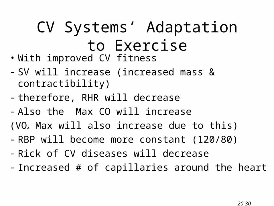
CV Systems’ Adaptation to Exercise
• With improved CV fitness- SV will increase (increased mass & contractibility)- therefore, RHR will decrease- Also the Max CO will increase
(VO2 Max will also increase due to this)- RBP will become more constant (120/80)- Rick of CV diseases will decrease- Increased # of capillaries around the heart
CV Systems’ Adaptation to Exercise
1) Increased Myoglobin (02 binding pigment)- acts as an 02 store aiding in the diffusion of 02
2) Increased oxidation of carbohydrates- training increases the muscles capacity to break
down glycogen in the presence of 02
3) Increased oxidation of fats- training increases the muscles capacity to break
down fatty acids in the presence of 02
20-31
VO2 Max
VO2 Max
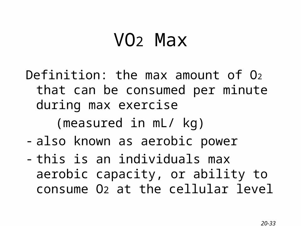
Definition: the max amount of O2 that can be consumed per minute during max exercise
(measured in mL/ kg)
- also known as aerobic power
- this is an individuals max aerobic capacity, or ability to consume O2 at the cellular level
20-33
VO2 Max
• 93% of VO2 Max is under genetic influence, although it can be improved through training, there is a genetic ceiling
• Max VO2 doesn’t differ between boys & girls before puberty, after puberty females are 25 – 230% less than values
20-34
VO2 Max
- Capacity depends on the amount of O2 that can be delivered to the muscles compared to the amount of O2 used by the muscle
- O2 consumption is important to prolonged exercise. (Endurance activities such as marathons, triathletes)
20-35
20-36
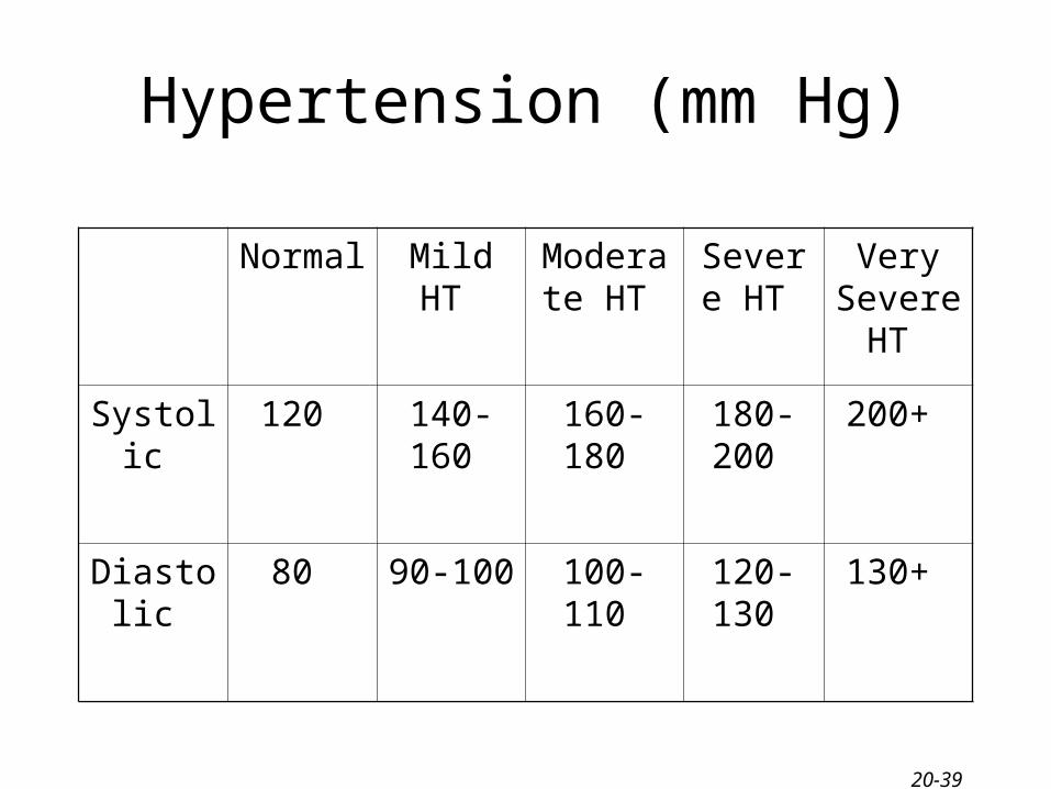
Blood Pressure
Blood pressure refers to the force exerted bycirculating blood on the walls of blood vessels • Systolic - The force your blood exerts
when the heart is contracting
• Diastolic - The force your blood exerts when the heat is relaxing