2016 ANNUAL MEETING OF THE CROATIAN IMMUNOLOGICAL SOCIETY OGULIN, OCTOBER 14 th -15 th 2016 ORGANIZED BY THE CROATIAN IMMUNOLOGICAL SOCIETY University of Rijeka Faculty of Medicine University of Zagreb School of Medicine President: Danka Grčević, Zagreb Vice President: Ines Mrakovčić Šutić, Rijeka Secretary: Alan Šućur, Zagreb ORGANIZING COMMITTEE Sabina Rabatić, Zagreb Tomislav Kelava, Zagreb Alenka Gagro, Zagreb Stipan Jonjić, Rijeka Alemka Markotić, Zagreb Bojan Polić, Rijeka Asja Stipić-Marković, Zagreb Astrid Krmpotić, Rijeka Vanda Juranić Lisnić, Rijeka
Transcript
2016 ANNUAL MEETING OF THE
CROATIAN IMMUNOLOGICAL SOCIETY
OGULIN, OCTOBER 14th-15th 2016
ORGANIZED BY
THE CROATIAN IMMUNOLOGICAL SOCIETY University of Rijeka Faculty of Medicine University of Zagreb School of Medicine
President: Danka Grčević, Zagreb
Vice President: Ines Mrakovčić Šutić, Rijeka
Secretary: Alan Šućur, Zagreb
ORGANIZING COMMITTEE
Sabina Rabatić, Zagreb
Tomislav Kelava, Zagreb
Alenka Gagro, Zagreb
Stipan Jonjić, Rijeka
Alemka Markotić, Zagreb
Bojan Polić, Rijeka
Asja Stipić-Marković, Zagreb
Astrid Krmpotić, Rijeka
Vanda Juranić Lisnić, Rijeka
2
TABLE OF CONTENTS
PROGRAM 5
LECTURES 11
CROSSTALK BETWEEN PENTRAXINS AND COMPLEMENT IN CANCER AND
INFECTION IMMUNOLOGY: NEW INSIGHTS FROM THE LONG PENTRAXIN PTX3 ................. 12
CYTOMEGALOVIRUS EVASION OF DNAM-1 DEPENDENT IMMUNE CONTROL BY
INFLAMMATORY MONOCYTES AND NK CELLS ......................................................................... 13
CYSTEINE CATHEPSINS IN INFLAMMATION: TARGETS FOR NONINVASIVE WHOLE
BODY IMAGING ............................................................................................................................. 14
MESENCHYMAL STEM CELLS AND NOTCH SIGNALING REGULATION OF BONE
THE ROLE OF INNATE IMMUNE CELLS IN DEVELOPMENT OF NAFLD ........................... 51
VIPERA AMMODYTES BITES TREATED WITH ANTIVENOM VIPERATAB®: A CASE
SERIES AND PHARMACOKINETIC EVALUATION ............................................................... 52
CORRELATION OR RECIPROCITY OF NOTCH AND AIOLOS IN LEUKEMIA .................... 53
AUTHOR INDEX 54
5
PROGRAM
FRIDAY October 14th 2016
13:00-14:45 REGISTRATION
14:45-15:00
OPENING
Danka Grčević, president Croatian Immunological Society
15:00-15:30
SESSION I
Chairs: Sabina Rabatić and Felix Wensveen
INVITED LECTURE
Antonio Inforzato Istituto Clinico Humanitas IRCCS, Milan, Italy
Crosstalk between pentraxins and complement in cancer and infection immunology: new insights from the long pentraxin PTX3
15:30-16:00
SELECTED ORAL PRESENTATIONS
Branka Popović: IL-33 drives regulatory T cell suppression of severe liver damage upon mouse cytomegalovirus infection
Tamara Gulić: Purification and characterization of the motogenic properties of Migration Stimulating Factor, a genetically truncated onco-fetal isoform of human fibronectin 1
Ilija Brizić: Perinatal cytomegalovirus infection drives NK cell hyporesponsiveness characterized by downregulation of T-box transcription factor
16:00-16:30 COFFEE BREAK
6
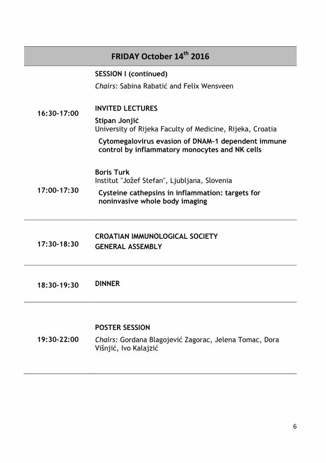
FRIDAY October 14th 2016
16:30-17:00
SESSION I (continued)
Chairs: Sabina Rabatić and Felix Wensveen
INVITED LECTURES
Stipan Jonjić University of Rijeka Faculty of Medicine, Rijeka, Croatia
Cytomegalovirus evasion of DNAM-1 dependent immune control by inflammatory monocytes and NK cells
17:00-17:30
Boris Turk Institut "Jožef Stefan", Ljubljana, Slovenia
Cysteine cathepsins in inflammation: targets for noninvasive whole body imaging
17:30-18:30 CROATIAN IMMUNOLOGICAL SOCIETY
GENERAL ASSEMBLY
18:30-19:30 DINNER
19:30-22:00
POSTER SESSION
Chairs: Gordana Blagojević Zagorac, Jelena Tomac, Dora Višnjić, Ivo Kalajzić
7
SATURDAY October 15th 2016
08:30-09:00
SESSION II
Chairs: Tomislav Kelava and Vanda Juranić Lisnić
INVITED LECTURE
Ivo Kalajzić Center for Regenerative Medicine and Skeletal Development, UConn Health, CT, USA
Mesenchymal stem cells and Notch signaling regulation of bone regeneration
09:00-09:40
SELECTED ORAL PRESENTATIONS
Antonio Markotić: Protective effect of LPS-induced inflammation on Fas-mediated hepatocyte apoptosis
Vilma Dembitz: The role of autophagy in the effects of AMP-kinase modulators on acute myeloid leukemia cells
Lovro Lamot: From symptom to genes: applicability of functional genomic methods in discovering the mechanisms of newly described disease entity
Felix Wensveen: Memory CD8 T cell formation requires induction of Bcl-2 by Eomes in response to low-affinity T cell receptor ligation
09:40-10:05
INVITED LECTURES
Dora Višnjić University of Zagreb School of Medicine, Zagreb, Croatia
AMPK/mTOR, autophagy and differentiation
10:05-10:30
Miroslav Harjaček University of Zagreb School of Medicine, Zagreb, Croatia
From genes to bedside: the current view on pathophysiology of juvenile idiopathic arthritis
10:30-11:00 COFFEE BREAK
11:00-13:00 TOUR TO IVANA BRLIĆ MAŽURANIĆ MUSEUM WITH SIGHTSEEING
8
SATURDAY October 15th 2016
13:00-14:00 LUNCH
14:00-14:30
SESSION III
Chairs: Stipan Jonjić and Astrid Krmpotić
INVITED LECTURE
Ennio Carbone Università degli Studi di Catanzaro "Magna Graecia", Catanzaro, Italy
Solid tumor’s therapy new opportunity: NK cells
14:30-15:10
SELECTED ORAL PRESENTATIONS
Jelena Železnjak: Who wins the fight? A game of cat and mouse between Ly49 receptors and MCMV encoded immunoevasins
Marko Šestan: CMV infection enhances development of glucose intolerance and insulin resistance in obesity
Daria Kveštak: NK cells persisting in the brain following MCMV infection induce polarization of microglia toward proinflammatory phenotype and delay in cerebellar growth via interferon γ
Kristina Vuković: CMV vector expressing RAE-1γ ligand serves as a highly efficient anti-tumor CD8 T cell vaccine
15:10-15:35
INVITED LECTURES
Bojan Polić University or Rijeka Faculty of Medicine, Rijeka, Croatia
The role of NKG2D in development and education of NK cells
15:35-16:00
Jelena Tomac University or Rijeka Faculty of Medicine, Rijeka, Croatia
Multiple overlapping mechanisms of ovarian follicle resistance to CMV infection
16:00-16:30 COFFEE BREAK
9
SATURDAY October 15th 2015
16:30-17:00
SESSION IV
Chairs: Bojan Polić and Danka Grčević
INVITED LECTURE
Matija Rijavec University Clinic of Respiratory and Allergic Diseases, Golnik, Slovenia
The novel role of basophils in anaphylaxis
17:00-17:30
SELECTED ORAL PRESENTATIONS
Ivan-Christian Kurolt: Urinary microRNAs as new early indicators for diseases severity in hemorrhagic fever with renal syndrome
Beata Halassy: Native elution in immunoaffinity chromatography of viruses – a step toward high-purity virus particle purification
Ljerka Karleuša: Disruption of proteasomal function, endosomal acidification and actin integration influence IE1 expression in MCMV infected cells
17:30-17:55
INVITED LECTURES
Gordana Blagojević Zagorac University or Rijeka Faculty of Medicine, Rijeka, Croatia
Chasing recycled molecules by monoclonal antibody-based recycling assays
17:55-18:20
Janoš Terzić University or Split School of Medicine, Split, Croatia
Role of IL-6 in cancer development
18:20-18:30
CLOSING REMARKS AND AWARDS
Danka Grčević, president
Croatian Immunological Society
10
11
LECTURES
12
CROSSTALK BETWEEN PENTRAXINS AND COMPLEMENT IN CANCER AND INFECTION
IMMUNOLOGY: NEW INSIGHTS FROM THE LONG PENTRAXIN PTX3
Antonio Inforzato
Humanitas Research Hospital, Rozzano, Italy
Traditionally regarded as the first line of defense against pathogens, innate immunity plays key
roles in a number of additional processes, including tissue remodelling, inflammation and cancer
development. The innate immune system comprises a cellular and a humoral arm, the latter
encompassing soluble pattern recognition molecules (sPRMs) that cooperate in the recognition of
and response to pathogen and danger associated molecular patterns (PAMPs/”non-self” and
DAMPs/”modified-self”, respectively). The complement system is a major component of the
humoral arm comprised of a cascade of more than 30 proteins, which is activated via three distinct
pathways (alternative, classical and lectin). Recognition and disposal of invading pathogens are the
canonical biological commitments of complement, however it is now appreciated that this system is
actively involved in adaptive immunity, cancerogenesis and cancer-related inflammation. In this
regard, the traditional paradigm describes complement as “good”, in that it recognizes, when not
deceived, the cancer cell, and either directly (via membrane attack complex-mediated lysis) or
indirectly (via complement-dependent cell toxicity) kills or disposes of it. New ideas, however, are
emerging that challenge this dogma and point to the complement system as a component of the
tumor promoting inflammation.
Amongst other sPRMs, pentraxins are a superfamily of highly conserved proteins with distinctive
quaternary structures. C-reactive protein (CRP) and serum amyloid P component (SAP)
collectively form the short pentraxin arm of the superfamily, and share a typical cyclic pentameric
symmetry. Both proteins are mostly produced in the liver in response to IL-6, and are major acute
phase reactants in humans and mice, respectively. Pentraxin 3 (PTX3) is the prototypic member of
the long pentraxin arm; as such, it contains an amino-terminal region linked to a C-terminal
pentraxin domain, and differs from the short counterparts in chromosomal localization, gene
expression, cellular source and ligands. PTX3 is not expressed by hepatocytes, but it is rather
produced by a number of other somatic and immune cells at sites of inflammation and infection.
The locally made protein acts as a non-redundant protective factor in the host defence against
selected microorganisms, most notably the opportunistic fungus Aspergillus fumigatus (AF), the
major etiologic agent of invasive aspergillosis (IA), a lethal infection amongst immunocompromised
individuals. This property relies on a tight molecular crosstalk with the complement system. Indeed,
PTX3 can be regarded as a functional ancestor of antibodies: it has opsonic activity towards AF,
and enhances recognition, phagocytosis and killing of fungal conidia by immune cells, mainly
polymorphonuclear neutrophils, via complement and Fc receptor pathways.
Additional functions have been reported for this long pentraxin in several processes and
mechanisms of innate immunity, inflammation and tissue remodelling, including a novel activity as
extrinsic oncosuppressor gene in cancer-related inflammation, where PTX3 exerts a protective
role, once again through regulation of complement activation. Here I discuss the most recent
findings on the complement/PTX3 crosstalk with major regard to cancer and infection immunology.
Novel vistas will be proposed on this two-sided system, based on a critical revision of current
literature and original data from my own work.
13
CYTOMEGALOVIRUS EVASION OF DNAM-1 DEPENDENT IMMUNE CONTROL BY
INFLAMMATORY MONOCYTES AND NK CELLS
Stipan Jonjić
University of Rijeka Faculty of Medicine, Rijeka, Croatia
The poliovirus receptor (PVR, CD155) is a highly conserved and ubiquitously expressed glycoprotein involved in cellular adhesion and immune recognition. PVR is constitutively expressed on the majority of somatic cells under physiological conditions and its expression is modulated as a consequence of viral infections and oncogenesis. Interestingly, while tumors frequently exhibit abnormally high PVR cell surface levels, some viruses (for example HCMV or HIV) downregulate PVR surface expression presumably to avoid immune cell recognition. PVR serves as a ligand for three receptors: DNAM-1 (CD226), an activating receptor expressed on the majority of immune cells; TIGIT, receptor that inhibits NK and T cell cytotoxicity and CD96 (Tactile), receptor with both activating and inhibitory functions on NK cells. Therefore, the precise mechanism that balances activating and inhibitory signals gathered through these receptors, as well as consequences of the PVR modulation in vivo are important unresolved issues of the PVR biology that might explain differences in tumor and viral modulation of PVR.
To assess the effect of balancing mechanism mediated by PVR receptors in vivo, we took advantage of murine model of cytomegalovirus infection. Our results showed that similar to human cytomegalovirus (HCMV), mouse cytomegalovirus (MCMV), downregulates the surface PVR. We have also characterized the molecular mechanism of this viral regulation that includes PVR retention in endoplasmic reticulum and proteasomal degradation. In addition, using a panel of MCMV deletion mutants it was possible to attribute this function to a novel MCMV protein, within the predicted m20 gene region that we named m20.1. Viral mutant lacking this regulator was severely attenuated in vivo, and this attenuation was reduced or abolished in DNAM KO mice, indicating the dominance of the activating receptor DNAM-1 in deciding the outcome of the modulation of PVR levels. The early attenuation of mutant viruses lacking the PVR inhibitor was only partially dependent on NK cells, which can be explained by the fact that these cells induce both activating and inhibitory PVR receptors upon infection. However, depletion of mononuclear phagocytes abolished the virus control which correlates with dramatic upregulation of DNAM-1 and the absence of inhibitory PVR receptors on these cells, even upon infection. In particular, we identified CCL2 dependent inflammatory monocytes as the major subpopulation controlling virus lacking PVR inhibitor via induction of iNOS. Overall, our data provide the strongest evidence so far for CMV control by mononuclear phagocytes and NK cells in which DNAM-1–PVR pathway plays an essential role and demonstrates novel mechanism of viral regulation of paired receptors. Hence, these results may be instrumental to identifying novel intervention targets and in designing novel vaccines and vaccine vectors.
*This work has recently been published: Lenac Rovis et al, Journal of Experimental Medicine, 213(9):1835-50, 2016.
14
CYSTEINE CATHEPSINS IN INFLAMMATION: TARGETS FOR NONINVASIVE WHOLE BODY
IMAGING
Boris Turk
Jozef Stefan Institute, Department of Biochemistry and Molecular Biology, Jamova 39, 1000 Ljubljana,
Slovenia
Center of Excellence CIPKEBIP, Jamova 39, 1000 Ljubljana, Slovenia
Faculty of Chemistry and Chemical Technology, University of Ljubljana, Slovenia
Inflammation plays an important role in disease onset and progression in a vast number of
diseases, called also inflammation-associated diseases including various cancers, psoriasis,
dermatitis, inflammatory bowel diseases, pancreatitis, various forms of arthritis, osteoarthritis,
erythematosus, and asthma. Proteases play a major role in a number of these diseases. However,
understanding the precise role of an individual protease in a disease remains a major challenge for
successful therapeutic applications. There are several ways how to address this issue, including
the chemical biology approaches including small molecule inhibitors and activity-based probes.
The latter approaches, especially those based on activity-based probes, offer a major potential for
noninvasive optical imaging by monitoring protease activities in situ, i.e. on disease site. Moreover,
the approach enables also validation of proteases as drug targets, in vivo validation of drug
candidates and evaluation of the diagnostic potential of the target proteases. Among the proteases
found to be tightly linked with inflammation-associated diseases are also cysteine cathepsins that
can be found at the sites of inflammation. Furthermore, since they are heavily upregulated in a
number of inflammation-associated diseases, they are therefore perfect targets for such
approaches. There is increasing evidence that monitoring cathepsin activity in vivo may be
applicable to diagnostic imaging, such as demonstrated primarily for cancer, arthritis and
inflammatory bowel diseases. Moreover, cathepsins can be also used as targets for targeted drug
delivery approaches combined with diagnostics, thereby offering a theranostic potential.
15
MESENCHYMAL STEM CELLS AND NOTCH SIGNALING REGULATION OF BONE
REGENERATION
Ivo Kalajzić
Center for Regenerative Medicine and Skeletal Development, UConn Health, CT, USA
Notch signaling has been recently identified as a key player during bone and cartilage
development. Notch inhibits the differentiation of osteoprogenitor cells, but has an osteogenic
effect in mature osteoblasts. We have previously shown that alpha smooth muscle actin (αSMA) is
a marker of mesenchymal progenitor cells that make a significant contribution to fibrous,
osteoblast, and chondrocyte lineages within a fracture callus. Gene expression analysis of isolated
αSMA-labeled progenitor cells revealed that a number of components of the Notch signaling
pathway, including receptors Notch 1, 3 and 4, and target genes Hes1 and Hey1, were significantly
decreased during the early stages of fracture healing. We hypothesize that a decrease in Notch
signaling could regulate the expansion, migration and the differentiation of periosteal cells in the
fracture callus. In this context, inducible Cre-expressing transgenic models enable precise
definition of when Notch signaling is required and in which cell population during the fracture
healing process.
We are using an inducible mouse model overexpressing the Notch1 intracellular domain (NICD1)
in osteoprogenitor cells: αSMACreERT2/Rosa-NICD1. Periosteal progenitor cells (PPC) were
isolated from the periosteum of 8-9 week old mice and cultured. The targeted overexpression of
NICD1 results in increased expression of Notch downstream targets Hes1 and Hey1. PPC
overexpressing NICD1 had increased proliferation and migration compared to tamoxifen treated
cultures from Cre negative littermates. Notch overexpression reduced osteogenic differentiation,
evidenced by reduced von Kossa staining and lower expression of osteocalcin.
The influence of NICD1 overexpression on the fracture healing process was assessed in 8-9
week old αSMACre/NICD1 mice after 3 tamoxifen injections at D0, D2 and D4 post femoral
fracture. Histological analysis was performed 1-3 weeks after fracture. Mice with targeted NICD1
overexpression showed a trend towards a smaller callus displaying first less cartilage and then less
mineralized content than the control mice.
Appropriate regulation of Notch signaling appears to be important for osteogenic differentiation of
PPCs and bone fracture healing. It constitutes a potential target to improve and accelerate fracture
healing by inhibiting its effect in specific cell populations responsible for the bone repair at specific
stages of the process.
16
AMPK/mTOR, AUTOPHAGY AND DIFFERENTIATION
Dora Višnjić
Department of Physiology and Immunology, University of Zagreb School of Medicine, Zagreb, Croatia
Recent studies suggest that drugs targeting metabolism may have some role in differentiation
therapy of leukemia. Adenosine monophosphate (AMP)-activated kinase (AMPK) is an
evolutionary conserved serine/threonine kinase that is activated in response to any decrease in
AMP/ATP ratio. Among many substrates, activated AMPK inhibits the activity of mammalian target
of rapamycin (mTOR), which decreases protein synthesis and cell growth. Physiologically, mTOR
is activated downstream of phosphoinositide 3-kinase (PI3K)/Akt pathway, and our previous
studies demonstrated the activation of PI3K and Akt in nuclei of leukemia cells during
differentiation. PI3K/Akt inhibitors reduce the number of viable cells, but negatively affect their
differentiative capacity. In contrast, use of rapamycin, an mTOR-inhibitor, potentiates differentiation
along granulocytic pathway. To further investigate the role of upstream regulators of mTOR in
leukemia differentiation, we tested the effects of two AMPK-modulators, metformin and AICAR (5-
amino-1-β-D-ribofuranosyl-imidazole-4-carboxamide). Our results demonstrated that AICAR alone
induced the expression of cell surface markers associated with mature monocytes and
macrophages in U937 cells. However, no significant increase in the expression of differentiation
markers was observed in U937 cells treated with metformin alone, although both modulators had
similar effects on proliferation and survival. Although we detected time and dose-dependent
increase in the level of Thr phosphorylated AMPK, a significant decrease in AMPK expression that
was achieved by using commercially available siRNA sequences in U937 cells had no significant
effects on the AICAR-mediated effects on the number of viable cells or the expression of
differentiation markers. Therefore, present studies are aimed to determine the mechanism
responsible for beneficial effects of AICAR in AML cells and to further elucidate signaling
mechanisms and metabolic changes responsible for monocytic and granulocytic differentiation of
AML cell lines in response to other inducers.
Our results show that AICAR and other differentiation agents induce autophagy flux, as measured
by the level of LC3II in the presence and absence of bafilomycin A. No increase in the level of
autophagy was observed in the presence of metformin. PI3KC3 inhibitor, 3-methyladenine,
inhibited the expression of differentiation markers, but increased the level of LC3-II suggesting that
3-MA cannot be used as a specific autophagy inhibitor under nutrient-rich conditions. siRNA
experiments showed that ATG7-dependent autophagy pathway is necessary for AICAR-mediated
effects on the expression of differentiation markers. The role of autophagy in differentiation of
leukemia cells and peripheral blood mononuclear cells will be discussed.
17
FROM GENES TO BEDSIDE: THE CURRENT VIEW ON PATHOPHYSIOLOGY OF JUVENILE
IDIOPATHIC ARTHRITIS
Miroslav Harjaček
Department of Pediatrics, Division of Clinical Immunology and Rheumatology, Clinical Hospital Center
“Sestre milosrdnice, Zagreb, Croatia
Juvenile idiopathic arthritis (JIA) is the most common childhood rheumatic disease. JIA is not a
single disease entity, but rather a group of seven ‘genetically heterogeneous’ and ‘phenotypically
distinct’ disorders (subtypes). The early diagnosis of new-onset JIA has become a major objective
for pediatric rheumatologists in order to identify a management strategy able to change the natural
history of the disease and to prevent joint damage and functional impairment. The term
undifferentiated arthritis (UA) is applied to the most common type of arthritis at the early stage
when, in the absence of current recommended diagnostic criteria, it cannot be classified into the
clinical subtypes of JIA. Patients with UA may progress towards JIA; however in some cases
arthritis may completely resolve. JIA is a multi-factorial disease that is influenced both by
environmental and genetic factors. The fundamental process in JIA is chronic inflammation, in
which the immune system understandably plays a critical role. Both innate and adaptive immune
systems have been implicated in the pathogenesis of various subtypes of JIA. In addition, many
studies have established the magnitude of the genetic basis of JIA. JIA is a complex genetic
condition and the multiple genes that influence susceptibility are actively being sought. Current
dogma supports the concept that the expression of a disease-inducing signature cytokine
phenotype is important to the maintenance stage of chronic synovitis. This cytokine phenotype has
been characterized as a polarization toward type TH1/TH17 cytokines, which are proinflammatory.
Traditionally, JIA has been viewed as “autoimmune” in nature although it has been increasingly
clear that particular subtypes of the disease are predominately “autoinflammatory” in nature (e.g.
systemic onset JIA) or shows overlapping features of both (e.g. enthesitis-related arthritis- ErA).
More recently, the inappropriate responsiveness to various “stressors” like viruses, bacteria,
prolonged antibiotic use, diet, trauma or mechanical stress, as well as psychogenic stress have
been recognized as initial trigger in activation of the orchestrated, complex crosstalk between
various parts of the immune system and CNS leading to “danger” response. Stressors activation of
the various neuroendocrine pathways and oral/gut dysbiosis are triggering simultaneous activation
of the crucial transcription factors like NF-κB, inflammasomes like NLRP-3, and due to failure of
negative immune regulation, ultimately create a pro-inflammatory milieu leading to chronic
synovitis. However, we are still far from having a clear picture of the molecular network that
predisposes a child to develop the disease, to worsen the symptoms, or to successfully respond to
a specific treatment. By using our own data on biomarkers, genetic, gene expression studies, and
epigenetic control of key master genes affecting the pathophysiology of ErA (spondyloarthritis), the
most common subtype of JIA, I would attempt to challenge current dogma and propose the working
platform for the further research of the JIA pathophysiology.
18
SOLID TUMOR’S THERAPY NEW OPPORTUNITY: NK CELLS
Ennio Carbone
Department of Experimenal and Clinical Medicine University Magna Graecia of Catanzaro, Italy
Department of Microbiology and Tumorbiology (MTC), Karolinska Institutet, Stockholm, Sweden
The talk will discuss recent data showing new biological property of NK cells: a) NK cells
capability to efficiently target the Cancer Initiating Cells (CIC) tumor compartment of solid tumor b)
New NK cells subset identified in the melanoma metastatic lymph node exerting a robust
autologous cytotoxicity against tumor b) NK cells potential role in the anti immune check point
therapy prognosis.
In the first part of the talk data showing the in vitro and in vivo NK cells recognition of human
colon adenocarcinoma derived CSC and murine breast adenocarcinoma lesion will be presented
(Tallerico et al under revision Oncoimmunology 2016).
The second part the NK cells recognition and elimination melanoma metastasis will be discussed
in the context of NK cells anatomic localization, NK subsets differentiation, cytokines and
chemokines tumor environment and disease stages.
The last section the anti immune check points era take shape in a study where we investigate the
potential role of NK cells in melanoma patients treated with anti CTL-4 (Ipilimumab).
The lack of knowledge on predictive biomarkers that could assist the anti immune check points
cancer therapy remains a limiting factor. We speculate that, along with additional markers, the
immunoscore is fundamental as prognostic and predictive marker for response to immunotherapies
in metastatic melanoma. Our previous data demonstrate that NK cells control the melanoma
progression in the infiltrated lymph nodes [Ali et al Nature Comm 2014]. Therefore we have
analyzed both T cells and NK cells subsets frequencies and receptors repertoire in the peripheral
blood of 63 Ipilimumab treated patients with Stage IV metastatic melanoma. The study was
performed in one Italian and one Swedish melanoma patient’s cohort (Tallarico et al Submitted
2016). The role of IL-15, TIM-3 and NK cells subset will be discussed in depth.
19
THE ROLE OF NKG2D IN DEVELOPMENT AND EDUCATION OF NK CELLS
Bojan Polić
Department of Histology & Embryology, Faculty of Medicine, University of Rijeka, Rijeka, Croatia
NKG2D is an activating receptor expressed on all NK cells early in NK cell development and have
important role in the cellular stress-surveillance. ‘Stressed’ cells due to viral infection, oncogenic
transformation, metabolic or other reasons up-regulate NKG2D ligands („induced self”) which can
engage the receptor and activate NK cells. MHC class I like-molecules (Rae1 and H60 family,
MULT1) are well characterized ligands for NKG2D (encoded by Klrk1). Previously, our group has
shown that NKG2D plays an important role in NK cell development (Zafirova et al. Immunity, 2009).
NKG2D-deficiency resulted in hyper reactive phenotype of NK cells which caused better control of
MCMV infection. However, Klrk1-/- NK cells still kept impaired ability to kill tumor targets
expressing NKG2D ligands.
In this study, we investigated whether and how NKG2D-deficient mice can control tumors which
do not express NKG2D ligands, but express ligands for other NK receptors. As a tumor model we
used syngeneic melanoma cell line (B16, clone F10). Subcutaneous tumor inoculation resulted in
smaller tumor size in Klrk1-/- mice, while intravenous inoculation resulted in prolonged survival of
Klrk1-/- mice compared to C57BL/6J littermates. The survival differences as well as differences in
tumor size between the groups were lost upon depletion of NK cells, showing NK-cell dependence
of the phenomenon. Using NCR1-/- -/- mice we were also able to show that the tumor
control is NCR1- - dependent. Also, in vitro stimulation of Klrk1-/- NK cells via NCR1 and
CD16, but not other activating NK receptors, resulted in higher production of IFNγ. However,
conditional deletion of Klrk1 at the stage of NCR1 expression (Klrk1flox/flox NCR1Cre mice)
resulted in absence of NK cell hyper reactivity in vivo and in vitro.
In conclusion, our findings for the first time show that NKG2D sets specifically activation
thresholds for NCR1 and CD16, two important activating receptors expressed on all NK cells. This
regulatory role of NKG2D takes place early during the NK cell-development, before the NCR1
expression, which implicates its role in NK cell education.
20
MULTIPLE OVERLAPPING MECHANISMS OF OVARIAN FOLLICLE RESISTANCE TO CMV
INFECTION
Jelena Tomac
Department for Histology and embryology and Center for proteomics, Faculty of Medicine, University of
Rijeka, Croatia
Human cytomegalovirus (HCMV) is a widespread herpesvirus that causes life-long persistent
infections in its host. Although relatively harmless to immunocompetent individuals, it can cause
grave disease in patients with weakened or immature immune system. Infection during pregnancy
can cause pregnancy-loss or numerous long-term developmental disabilities.
HCMV is highly species specific and only infects humans. Murine cytomegalovirus (MCMV) is
biologically similar and related to HCMV; therefore, the infection of mice with MCMV became the
most commonly used model for studying the biology and pathogenesis of CMV, especially in
research that is difficult to conduct in humans. Although CMV’s ability to pass the placenta and
cause devastating congenital disease is well established, very little is known about the CMV
infection of reproductive organs and its consequences on fertility and pregnancy outcome. We
have performed a detailed analysis of CMV pathogenesis in the ovary and observed that CMV very
successfully infects the ovaries. The virus is cleared by day 8 PI, indicating a strong role of innate
immune system in virus control. Moreover, the infection was completely excluded from ovarian
follicles, even in strongly immunosuppressed mouse strains in which nearly whole ovarian stroma
and corpora lutea were infected. Since MCMV is natural pathogen infecting the majority of wild
mice, development of strategies that can act immediately or very early to prevent infection of
follicles in order to preserve reproductive potential is a necessary evolutionary strategy. We have
uncovered several layers of protection that preserve ovarian follicles: physical barriers preventing
the infection and cells of the innate immune system. We show that ovarian follicles are protected
from the infection by a ring of macrophages that rely on NK cells. While the depletion of NK cells
does not result in increased viral titers in the ovary, we could observe infected follicles and
absence of macrophage rings.
21
THE NOVEL ROLE OF BASOPHILS IN ANAPHYLAXIS
Matija Rijavec
University Clinic of Respiratory and Allergic Diseases Golnik, Slovenia
Anaphylaxis is an acute, life-threatening, systemic, allergic reaction with rapid onset. The
activation of mast cells involving IgE and the high-affinity IgE receptor (FcεRI) on these cells is
postulated to have a pivotal role in anaphylaxis and to date, no experimental system has directly
demonstrated that basophils contribute to IgE-mediated anaphylaxis. Therefore, the exact
molecular mechanism as well as the specific role of basophils in human anaphylaxis remains
poorly understood. Using modern laboratory methods, from multi-colour flow cytometry technics to
next generation sequencing platforms, and clinically well characterized patients, who presented
with anaphylaxis at the emergency department or during allergen challenge, our research has been
focus on the determination of the role of basophils in anaphylaxis. Specifically, our aim was to
determine whether blood basophils are activated during anaphylaxis and whether anaphylaxis
induces basophil migration to the site of inflammation and in that case which chemotactic factors
are involved in their migration. Using the global transcriptome profiling of peripheral blood samples
during anaphylaxis we aimed to get a more detailed look at the molecular mechanisms during an
anaphylactic episode.
We have demonstrated that the absolute number of circulating basophils is significantly
decreased during anaphylactic episode, and that decrease was confirmed with the decrease
expression of basophil specific genes, specifically FcεRI, carboxypeptidase A3 and L-histidine
decarboxylase in whole blood samples. In line with that, CCL2, a major basophil chemotactic
factor, significantly increased during anaphylaxis, and there was an inverse correlation between the
absolute number of basophils in blood and CCL2 in sera. Importantly the basophils that remained
in the circulation exhibited a low level of activation. Transcriptome analysis further revealed that
cellular movement, cell-to-cell signalling, interaction and immune cell trafficking are the most
important mechanisms taking place during anaphylaxis. Comparative analysis with expression
signatures of immune cells showed significant under-expression of basophil and over-expression of
activated eosinophil signatures during anaphylactic reaction. Furthermore, measurement of
absolute number of circulating basophils, CCL2 and FcεRI expression have the potential to be
used as biomarkers to confirm the diagnosis of anaphylaxis, showing better AUC, sensitivity and
specificity than serum mast-cell tryptase which is currently the gold standard diagnostic test to
confirm anaphylaxis.
In summary our findings suggest that cellular movement and interactions of distinct immune cells
are taking an important place during anaphylaxis. We have demonstrated a marked migration of
circulating basophils, which correlated with a significant increase in the level of major basophil
chemotactic factor CCL2. These data suggest a novel and specific role for basophils in the
pathobiology of human anaphylaxis.
22
CHASING RECYCLED MOLECULES BY MONOCLONAL ANTIBODY-BASED RECYCLING
ASSAYS
Gordana Blagojević Zagorac
University of Rijeka Faculty of Medicine, Department of Physiology and Immunology
Membrane proteins and their ligands are continuously internalized and directed to recycling or
degradation. Endocytic recycling is a highly regulated, dynamic and complex cellular process and
has irreplaceable role in cellular physiology and pathophysiology processes, includin immune
response. Studies of endosomal trafficking suggest that membrane proteins use various recycling
pathways and much knowledge about recycling route and the regulatory mechanism was
generated for transferrin receptor (TfR), a clathrin-dependent cargo protein. In contrast to clathrin-
dependent cargo proteins, for many proteins that are endocytosed by the clathrin-independent
mechanism, the rate of endocytic recycling and recycling route was not established and remains
largely unknown. The best characterized is the recycling route of Major Histocompatibility Class I
(MHC-I) proteins. For detection and quantification of recycling, several assays were used. Although
antibodies were used in tracing the recycling routes more than a decade, still there were many
inconsistencies in using different experimental approaches, resulting with false positive and false
negative results.
We performed a systematic study of various protocols known in literature in which antibodies are
used as tools to study endosomal recycling. We used TfR recycling, as a paradigm of rapidly
endocytosed clathrin-dependent cargo molecule and fully conformed MHC-I proteins as a
paradigm for constitutively endocytosed clathrin-independent cargo molecule. We also followed
recycling of open MHC-I conformers (peptide-empty MHC-I proteins, eMHC-I) as a control,
because they do not recycle from early endosomal recycling circuit by the fast and slow recycling
route.
Our study demonstrates that direct and indirect detection of recycled mAb:protein complexes at
the cell surface underestimate the recycling pool, especially for clathrin-dependent membrane
proteins that are rapidly reinternalized after recycling. Recycling protocols based on the capture of
recycled mAb:protein complexes require the use of the Alexa Fluor 488 conjugated secondary
antibodies or FITC-conjugated secondary antibodies in combination with inhibitors of endosomal
acidification and degradation. Finally, protocols based on the capture of recycled proteins that are
labeled with Alexa Fluor 488 conjugated primary antibodies and quenching of fluorescence by the
anti-Alexa Fluor 488 displayed the same quantitative assessment of recycling as the antibody-
capture protocols.
This work was supported in part by the Croatian Science Foundation (grant IP-2014-9-9564) and
by the University of Rijeka (grants 13.06.1.1.4, 13.06.2.1.55, and 13.06.2.1.56).
23
ROLE OF IL-6 IN CANCER DEVELOPMENT
Janoš Terzić
University or Split School of Medicine
Inflammation is recently being considered as one of the cancer hallmarks and intereleukin 6 (IL-
6), one of main proinflamatory cytokines, plays major role in inflammation-related cancer
development. We have found that IL-6 has important role in colon cancer development stimulating
development of increased number and size of tumors. These effects were mainly mediated by
Stat3 transcription factor. On the other hand, IL-6 role in inflammation-influenced bladder cancer is
not so well characterized. Using BBN-induced bladder cancer in IL-6 KO and WT mice, we have
determined high activation of immune response genes with tumor phenotype being less distinctive
between tested groups.
24
25
ORAL PRESENTATIONS
26
IL-33 DRIVES REGULATORY T CELL SUPPRESSION OF SEVERE LIVER DAMAGE UPON
MOUSE CYTOMEGALOVIRUS INFECTION
Branka Popović1, Mijo Golemac
1, Lidija Bilić-Zulle
2, Miodrag L Lukić
3, Luka Čičin-Šain
4, Tim
Sparwasser5, Astrid Krmpotić
1, Stipan Jonjić
1
1Department of Histology and Embryology, Faculty of Medicine, University of Rijeka, Croatia
2Clinical Institute of Laboratory Diagnostics, Rijeka Clinical Hospital Center, Rijeka, Croatia
3Department of Microbiology and Immunology, Centre for Molecular Medicine and Stem Cell Research,
Faculty of Medicine, University of Kragujevac, Serbia 4Department of Vaccinology and Applied Microbiology, Helmholtz Centre for Infection Research (HZI),
Braunschweig, Germany 5Institute of Infection Immunology, TWINCORE, Hannover, Germany
Regulatory T cells (Tregs) are crucial for immune homeostasis and for dampening immune
response to several diseased conditions, including viral infections. Mouse cytomegalovirus
(MCMV) is a herpesvirus with pathogenic potential so early immune mechanisms are essential in
controlling virus and protecting from virus-induced pathology. Studies on FoxP3+ Tregs have
revealed their inhibitory role on the early T cell response to MCMV infection and have suggested
Tregs as a target of MCMV’s immunoevasion mechanisms. Here we demonstrate that the number
and activation status of liver Tregs is strongly induced in mice infected with MCMV. The depletion
of Tregs results in an increased virus-specific CD8+ T cell response without alterations in the virus
load. Furthermore, depletion of Tregs leads to severe liver damage and adoptive transfer of Tregs
rescues mice from T cell mediated hepatitis. Interestingly, liver Tregs constitutively express high
amounts of a cellular receptor for IL-33, tissue alarmin strongly upregulated in the liver upon
MCMV infection. The accumulation of Tregs in the liver is dependent on IL-33 signalling and mice
lacking the IL-33 receptor show a more pronounced liver pathology and higher death rate
compared to infected control mice. These results illustrate importance of IL-33 in the accumulation
of liver Tregs and their suppression of MCMV-induced immunopathology.
27
PURIFICATION AND CHARACTERIZATION OF THE MOTOGENIC PROPERTIES OF
MIGRATION STIMULATING FACTOR, A GENETICALLY TRUNCATED ONCO-FETAL
ISOFORM OF HUMAN FIBRONECTIN 1
Gulic T., Laface I., Inforzato A., Oliviera MJ., Sironi M., Lage CC., Bottazzi B., Allavena P.,
Rukavina D., Mantovani A
Humanitas Clinical and Research Institute, Rozzano, Milano
Department of Physiology and Immunology, Medical Faculty University of Rijeka, Croatia
i3S- Institute of Inovation and Research, University of Porto, Porto, Portugal
Migration-stimulating factor (MSF) is a porly studiated oncofetal isoform of human fibronectin 1
(FN1) generated from its primary gene transcript by a intron read-through mechanism. Detailed
molecular characterization indicates that MSF is a 70 kDa soluble protein identical to the N-
terminal portion of full-length FN1 (up to exon III-1a), with the addition of a unique 10 amino acids
long peptide in the C-terminus. MSF is mainly produced by epithelial and stromal cells during foetal
development and by cancer-associated fibroblasts, but not by adult healthy cells. Available data
indicate that it has a potent motogenic activity for fibroblasts, vascular and epithelial cells, and can
induce angiogenesis and matrix remodelling, suggesting a possible role in cancer development.
We focus our attention on the characterization of reagents to study MSF biology. After
immunization with the MSF-specific 10 amino acids long peptide, we selected a monoclonal
antibody recognizing specifically human MSF but not human FN1. The antibody was used to purify
by immunoaffinity chromatography recombinant human MSF (rhMSF) produced by transfected
CHO cells. Initial efforts were aimed at defining the condions ensuring protein stability over the
time. Biological activity of purified rhMSF was tested in migration and invasion assays with
monocytes and cancer cells, using boyden chamber or transwells respectively. Purified
recombinant MSF can promote migration of a different human cancer cell line (PANC-1, MDA-
MB231, 8387), confirming the motogenic effect of this protein. In addition purified rhMSF has a
chemotactic activity for human monocytes and neutrophils, suggesting that it can promote
monocyte/macrophage and neutrophils recruitment into tissues. Further studies are needed to
elucidate the engagement of MSF as protumoral molecule.
28
PERINATAL CYTOMEGALOVIRUS INFECTION DRIVES NK CELL HYPORESPONSIVENESS
CHARACTERIZED BY DOWNREGULATION OF T-box TRANSCRIPTION FACTOR
Ilija Brizic1, Ana Lesac Brizic
1, Berislav Lisnic
1, Vanda Juranic Lisnic
1, Kristina Gotovac
3,
Fran Borovečki3, Astrid Krmpotić
2, Stipan Jonjić
1,2
1 Center for proteomics, Faculty of medicine, University of Rijeka, Croatia
2 Department for histology and embryology, Faculty of medicine, University of Rijeka, Croatia
3 Department for functional genomics, Center for translational and clinical research, School of medicine,
University of Zagreb, Croatia
Human cytomegalovirus (HCMV) is a frequent cause of disease in immunodeficient and
immunologically immature hosts such as newborn infants. For that reason, congenital HCMV
infection presents a significant health concern since it is frequently manifested with
neurodevelopmental sequelae, such as auditory damage and neurodevelopmental disabilities. NK
cells have been shown to play an important role in fighting cytomegalovirus (CMV) infection and
the adaptive features of NK cells in response to CMV infection are being recently increasingly
recognized. However, the extent to which congenital CMV infection affects and shapes NK-cell
mediated immunity is largely unknown. To address this issue, we have used mouse CMV (MCMV)
infection of newborn mice as a model to investigate the impact of congenital CMV infection on the
maturation and functional properties of NK cells. We observed that perinatal MCMV infection leads
to persistent alteration of transcriptional activity and strongly affects the maturation and function of
NK cells. Surprisingly, NK cell expression of T-box transcription factor Eomes, critical for NK cell
development, was dramatically impaired. At the same time the expression of T-bet, another T-box
transcription factor, was unaffected. The downregulation of Eomes correlated with major changes
in NK cell phenotype, indicating most notably NK cell exhaustion, as well as an impaired NK cell
response to different stimuli. To our knowledge this is the first evidence that a viral infection can
lead to the perturbation in NK cell expression of the T-box transcription factors. In addition, we
have observed an NK cell population with a phenotype characteristic of memory-like NK cells. This
population of NK cells persisted for several months in infected mice indicating that congenital CMV
infection is a factor that shapes the NK cell response over a long-term.
29
PROTECTIVE EFFECT OF LIPOPOLYSACCHARIDE-INDUCED INFLAMMATION ON FAS-
MEDIATED HEPATOCYTE APOPTOSIS IN MICE
Antonio Markotić1,2
, Ivan Ćavar1,2
, Petra Turčić3, Alan Šućur
1, Sanja Ivčević
1, Darja Flegar
1,
Helena Markotić4, Danka Grčević
1 Tomislav Kelava
1,2
1 Department of physiology, School of medicine, University of Zagreb, Croatia
2 Department of physiology, School of medicine, University of Mostar, BiH
3 Department of pharmacology, Faculty of Pharmacy and Biochemistry, University of Zagreb, Croatia
4 University Clinical Hospital Mostar
BACKGROUND: Fas/Fas ligand (FasL) apoptotic pathway is involved in the pathogenesis of
various liver diseases. However, the exact effects of acute inflammation on the liver apoptotic
processes are still not well elucidated. We investigated the effect of pro-inflammatory mediators on
Fas/FasL-mediated hepatocyte apoptosis using a model of lipopolysaccharide (LPS)-induced acute
inflammation.
METHODS: Male C57BL/6 mice received intraperitoneal injection of LPS (0.1 µg/g) while the
control group of animals received the vehicle (sterile saline). After 2 hours both groups were
treated with anti-Fas (JO2) activating antibody (0.25 μg/g, intravenously). Mice were sacrificed
after additional 6 hours and plasma (ALT, AST) and liver samples (pathohistology, caspase
activity, qPCR) were harvested. In the second set of experiments mice were treated with saline or
LPS, non-parenchymal liver cells were harvested and leukocytes populations were determined by
flow cytometric analysis. Concentrations of soluble Fas (sFas) in plasma were determined by
ELISA. To induce neutrophil depletion mice were intraperitoneally injected with cyclophosphamide
(250 mg/kg), four days prior to LPS and anti-Fas application.
RESULTS: Mice pre-treated with LPS were protected from Fas/FasL-mediated hepatocyte
apoptosis as evidenced by lower levels of ALT (median (IQR); 82 (32-182) vs. 3709 (1429 – 5922)
U/L, p=0.02) and AST (151.5 (96-256) vs. 3137 (1378-5389) U/L, p=0.02) in plasma compared with
mice which received saline before anti-Fas antibody. Additionally, LPS pre-treated mice had lower
number of apoptotic cells on pathohistological analysis and lower caspase 8 activity than saline
pre-treated mice. LPS alone had no effect on aminotransferase levels and caspase 8 activity, while
it increased expression of inflammatory mediators TNF-alpha, IL-1 and IL-6 in hematopoietic liver
cells, as well as the expression of Fas and antiapoptotic CFLAR and Bcl2l1 in hepatocytes. LPS
did not increase the level of sFas in plasma. Flow cytometric analysis of intrahepatic leukocytes
showed an increase in neutrophil (7.6 fold), NK cell (1.7 fold) and NKT cell (1.6 fold) population.
Accumulation of Fas positive neutrophils in liver following LPS administration was confirmed
immunohistochemically. However, cyclophosphamide-induced neutrophil depletion did not
abrogate protective effect of LPS.
CONCLUSION: Acute inflammation induced by LPS alleviates Fas/FasL-mediated apoptosis by
acting on Fas-apoptotic pathway upstream of caspase 8 activation. We intend to define protective
mechanism more precisely by investigating effects of LPS on expression pattern of broader
spectrum of pro- and antiapoptotic molecules at various time points following LPS treatment.
30
THE ROLE OF AUTOPHAGY IN THE EFFECTS OF AMP-KINASE MODULATORS ON ACUTE
MYELOID LEUKEMIA CELLS
Vilma Dembitz, Hrvoje Lalic, Dora Visnjic
University of Zagreb School of Medicine, Department of Physiology and Croatian Institute for Brain
Research
Autophagy has been shown to contribute to differentiation of leukemia cells in various
experimental settings. Our recent results showed that 5-aminoimidazole-4-carboxamide
ribonucleotide (AICAR), a compound commonly used as a modulator of AMP-kinase (AMPK),
induced differentiation of U937 cells in an AMPK-independent manner. In the present study we
tested for the possible role of autophagy in AICAR-mediated effects. The level of LC3B-II was
increased after 48 h incubation with AICAR and other differentiation inducers, all-trans retinoic acid
(ATRA) and phorbol myristate acetate (PMA). No similar effects were observed in cells treated with
metformin, an AMPK modulator without differentiative properties. The pretreatment of cells with 3-
methyladenine (3-MA) inhibited agonist-mediated increase in the expression of differentiation
markers and decreased the number of viable cells. However, although treatment with 3-MA
reduced the levels of PtdIns(3)P, the levels of LC3B-II increased after addition of 3-MA. Gene
knockdown for Beclin-1 and class III phosphoinositide 3-kinase (PI3KC3) did not abolish the
differentiative effects of AICAR, ATRA and PMA. Still, metformin-mediated decrease in cell viability
was inhibited in cells with down-regulated Beclin-1.
This work has been funded by European Social Fund (ESF) grant to H.L.
31
FROM SYMPTOM TO GENES: APPLICABILITY OF FUNCTIONAL GENOMIC METHODS IN
DISCOVERING THE MECHANISMS OF NEWLY DESCRIBED DISEASE ENTITY
Lovro Lamot1,2
, Fran Borovečki3, Kristina Gotovac
3, Danka Grčević
4, Mandica Vidović
1, Mirta
Lamot1, Edi Paleka Bosak
1, Miroslav Harjaček
1,2
1 Clinical Hospital Center Sestre Milosrdnice, Department of Pediatrics, Division of clinical immunology
and rheumatology
2 University of Zagreb School of Medicine, Department of Pediatrics
3 University of Zagreb School of Medicine, Department for Functional Genomics
4 University of Zagreb School of Medicine, Department of Physiology and Immunology
OBJECTIVE: Clavicular cortical hyperostosis (CCH) is a sterile inflammatory bone disorder of
unknown etiology clinically characterized by pain and/or swelling of the clavicle. It has been
regarded as a variant of chronic nonbacterial/recurrent multifocal osteomyelitis (CNO/CRMO) but
due to lack of other inflammatory sites and recurrence it could also be regarded as a separate
disease in the spectrum. Therefore, it is of high importance to elucidate the exact mechanisms
responsible for the development and progression of the symptoms. METHODS: Total RNA was
isolated from whole blood of 18 new-onset, untreated CCH patients and 8 healthy controls. DNA
microarray gene expression was performed in 5 CCH and 4 control patients along with
bioinformatical analysis of retrieved data. Carefully selected differentially expressed genes