TRANSPLANTATION IMMUNOLOGY 18 • TISSUE COMPATIBILITY • TRANSPLANTATION • HOST-VERSUS-GRAFT DISEASE (HVGD) • GRAFT-VERSUS-HOST REACTION (GVHR) • IMMUNOSUPPRESSION Transplantation is the replacement of an organ or other tissue, such as bone marrow, with organs or tissues (grafts) derived ordinarily from a nonself source such as an allo- geneic donor. Organs include kidney, Hver, heart, lung, pancreas (including pancreatic islets), intestine, or skin. In addition, bone matrix and cardiac valves have been trans- planted. Bone marrow transplants are given for nonmalig- nant conditions such as aplastic anemia, as well as to treat certain leukemias and other malignant diseases. The transplantation of organs has been possible surgi- cally since the early 1900s, when Alexis Carrel perfected the triangulation suture to sew blood vessels together. Yet, significant advances in immunology and immunosuppres- sion required another 75 years. This chapter defines key terms from the field of trans- plantation immunology, which is the study of immunologic reactivity of a recipient to transplanted organs or tissues from a histo-incompatible recipient. Effector mechanisms of transplantation rejection or transplantation immunity consist of cell-mediated immunity and/or humoral antibody immunity, depending upon the category of rejection. For example, hyperacute rejection of an organ such as a renal allograft is mediated by preformed antibodies and takes place soon after the vascular anastomosis is completed in transplantation. By contrast, acute allograft rejection is mediated principally by T cells and occurs during the first week after transplantation. There are instances of humoral vascular rejection mediated by antibodies as a part of the acute rejection response. Chronic rejection is mediated by a cellular response. TISSUE COMPATIBILITY Histocompatibility is tissue compatibility as in the trans- plantation of tissues or organs from one member to another of the same species, an allograft, or from one species to another, a xenograft. The genes that encode antigens that should match if a tissue or organ graft is to survive in the recipient are located in the major histocompatibility com- plex (MHC) region. This is located on the short arm of chromosome 6 in man and of chromosome 17 in the mouse. Class I and class II MHC antigens are important in tissue transplantation. The greater the match between donor and recipient, the more likely the transplant is to survive. For example, a six-antigen match implies sharing of two HLA-A antigens, two HLA-B antigens, and two HLA-DR antigens between donor and recipient. Even though antigenically dissimilar grafts may survive when a powerful immunosuppressive drug such as cyclosporine is used, the longevity of the graft is still improved by having as many antigens match as possible. A histocompatibility locus is a specific site on a chro- mosome where the histocompatibility genes that encode histocompatibility antigens are located. There are major histocompatibility loci such as HLA in man and H-2 in the mouse across which incompatible grafts are rejected within 1-2 weeks. There are also several minor histocom- patibility loci, with more subtle antigenic differences, across which only slow, low-level graft rejection reactions occur. Histocompatibility antigen is one of a group of geneti- cally encoded antigens present on tissue cells of an animal that provoke a rejection response if the tissue containing them is transplanted to a genetically dissimilar recipient. These antigens are detected by typing lymphocytes on which they are expressed. These antigens are encoded in man by genes at the HLA locus on the short arm of chro- mosome 6. In the mouse, they are encoded by genes at the H-2 locus on chromosome 17. Histocompatibility testing is a determination of the MHC class I and class II tissue type of both donor and 475
Transcript
TRANSPLANTATION IMMUNOLOGY 18
• TISSUE COMPATIBILITY
• TRANSPLANTATION
• HOST-VERSUS-GRAFT DISEASE (HVGD)
• GRAFT-VERSUS-HOST REACTION (GVHR)
• IMMUNOSUPPRESSION
Transplantation is the replacement of an organ or other tissue, such as bone marrow, with organs or tissues (grafts) derived ordinarily from a nonself source such as an allogeneic donor. Organs include kidney, Hver, heart, lung, pancreas (including pancreatic islets), intestine, or skin. In addition, bone matrix and cardiac valves have been transplanted. Bone marrow transplants are given for nonmalig-nant conditions such as aplastic anemia, as well as to treat certain leukemias and other malignant diseases.
The transplantation of organs has been possible surgically since the early 1900s, when Alexis Carrel perfected the triangulation suture to sew blood vessels together. Yet, significant advances in immunology and immunosuppression required another 75 years.
This chapter defines key terms from the field of transplantation immunology, which is the study of immunologic reactivity of a recipient to transplanted organs or tissues from a histo-incompatible recipient. Effector mechanisms of transplantation rejection or transplantation immunity consist of cell-mediated immunity and/or humoral antibody immunity, depending upon the category of rejection. For example, hyperacute rejection of an organ such as a renal allograft is mediated by preformed antibodies and takes place soon after the vascular anastomosis is completed in transplantation. By contrast, acute allograft rejection is mediated principally by T cells and occurs during the first week after transplantation. There are instances of humoral vascular rejection mediated by antibodies as a part of the acute rejection response. Chronic rejection is mediated by a cellular response.
TISSUE COMPATIBILITY
Histocompatibility is tissue compatibility as in the transplantation of tissues or organs from one member to another
of the same species, an allograft, or from one species to another, a xenograft. The genes that encode antigens that should match if a tissue or organ graft is to survive in the recipient are located in the major histocompatibility complex (MHC) region. This is located on the short arm of chromosome 6 in man and of chromosome 17 in the mouse. Class I and class II MHC antigens are important in tissue transplantation. The greater the match between donor and recipient, the more likely the transplant is to survive. For example, a six-antigen match implies sharing of two HLA-A antigens, two HLA-B antigens, and two HLA-DR antigens between donor and recipient. Even though antigenically dissimilar grafts may survive when a powerful immunosuppressive drug such as cyclosporine is used, the longevity of the graft is still improved by having as many antigens match as possible.
A histocompatibility locus is a specific site on a chromosome where the histocompatibility genes that encode histocompatibility antigens are located. There are major histocompatibility loci such as HLA in man and H-2 in the mouse across which incompatible grafts are rejected within 1-2 weeks. There are also several minor histocompatibility loci, with more subtle antigenic differences, across which only slow, low-level graft rejection reactions occur.
Histocompatibility antigen is one of a group of genetically encoded antigens present on tissue cells of an animal that provoke a rejection response if the tissue containing them is transplanted to a genetically dissimilar recipient. These antigens are detected by typing lymphocytes on which they are expressed. These antigens are encoded in man by genes at the HLA locus on the short arm of chromosome 6. In the mouse, they are encoded by genes at the H-2 locus on chromosome 17.
Histocompatibility testing is a determination of the MHC class I and class II tissue type of both donor and
475
476 Transplantation Innmunology
Table 18.1 Grafts
Type of graft Dafcition
Autograft A graft of tissue taken from one part of the body and placed in a different site on the body of the same individual,
such as grafts of skin from unaffected areas to burned areas in the same individual
Syngraft A transplant from one individual to another within the same strain; also called isograft
Isograft A tissue transplant from a donor to an isogenic recipient. Grafts exchanged between members of an inbred strain
of laboratory animals, such as mice, are syngeneic rather than isogenic
Homograft Allograft (i.e., an organ or tissue graft) from a donor to a recipient of the same species
Allograft An organ, tissue, or cell transplant from one individual or strain to a genetically different individual or strain within the same species. Also called homograft
Xenograft A tissue or organ graft from a member of one species (i.e., the donor) to a member of a different species (i.e., the recipient); also called a heterograft. Antibodies and cytotoxic T cells reject xenografts several days following transplantation
Heterograft Refer to Xenograft
Orthotopic graft An organ or tissue transplant that is placed in the location that is usually occupied by that particular organ or tissue
Heterotopic graft A tissue or organ transplanted to an anatomic site other than the one where it is usually found under natural conditions - i.e., the anastomosis of the renal vasculature at an anatomical site that would situate the kidney in a place other than the renal fossa, where it is customarily found
recipient prior to organ or tissue transplantation. In man HLA-A, HLA-B, and HLA-DR types are determined, followed by cross-matching donor lymphocytes with recipient serum prior to transplantation. A mixed lymphocyte culture (MLC) was formerly used in bone marrow transplantation, but has now been replaced by molecular DNA typing. The
Table 18.2 Major histocompatibility loci of various species
1 species
1 Human
i Mouse
1 C)og
1 Rhesus monkey
1 Chicken
1 Guinea pig
1 P^g
1 Rat
Major histocompatibility locus 1
HLA j
H-2 1
DLA 1
RhLA 1
B 1 GP-LA 1
SLA 1
RTl 1
MLC may also be requested in living related organ transplants. As in renal allotransplantation, organ recipients have their serum samples tested for percent reactive antibodies, which reveals whether or not they have been presensitized against HLA antigens of an organ for which they may be the recipient.
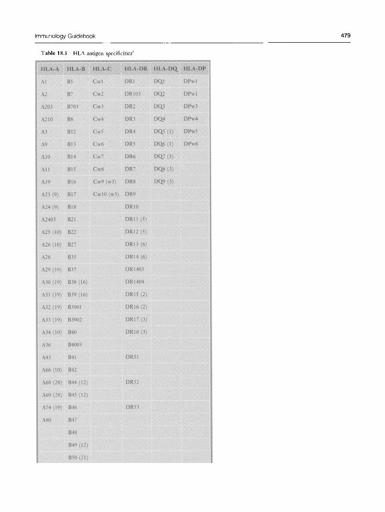
HLA is an abbreviation for human leukocyte antigen. The HLA histocompatibility system in humans represents a complex of MHC class I molecules distributed on essentially all nucleated cells of the body and MHC class II molecules that are distributed on B cells, macrophages, and a few other cell types. These are encoded by genes at the major histocompatibility complex. In humans the HLA locus is found on the short arm of chromosome 6. This has now been well defined, and in addition to encoding surface isoantigens, genes at the HLA locus also encode immune response (/r) genes. The class I region consists of HLA-A, HLA-B, and HLA-C loci and the class II region consists of the D region which is subdivided into HLA-DP, HLA-DQ^ and HLA-DR subregions. Class II molecules play an important role in the induction of an immune response, since antigen-presenting cells must complex an antigen with class II molecules to present it in the presence of interleu-kin-1 to CD4"^ T cells. Class I molecules are important in presentation of intracellular antigen to CD8"^ T cells as well as for effector functions of target cells. Class III molecules
Immunology Guidebook 4 7 7
encoded by genes located between those that encode class I and class II molecules include C2, BF, C4a, and C4b. Class I and class II molecules play an important role in the transplantation of organs and tissues. The microlymphocytotoxi-city assay is used for HLA-A, -B, -C, -DR, and -D(Xtyping. The primed lymphocyte test is used for DP typing. Uppercase letters designate individual HLA loci such as HLA-B and alleles are designated by numbers such as in HLA-B*0701.
HLA-A is a class I histocompatibility antigen in humans. It is expressed on nucleated cells of the body. Tissue typing to identify an individual's HLA-A antigens employs lymphocytes.
HLA-B is a class I histocompatibility antigen in humans which is expressed on nucleated cells of the body. Tissue typing to define an individual's HLA-B antigens employs lymphocytes.
HLA-C is a class I histocompatibility antigen in humans which is expressed on nucleated cells of the body. Lymphocytes are employed for tissue typing to determine HLA-C antigens. HLA-C antigens play little or no role in graft rejection.
The human MHC class II region is the HLA-D region, which is comprised of three subregions designated DR, DQ^ and DP. Multiple genetic loci are present in each of these.
DN (previously DZ) and DO subregions are each comprised of one genetic locus. Each class II HLA molecule is comprised of one a and one P chain that constitute a hetero-dimer. Genes within each subregion encode a particular class II molecule's a and P chains. Class II genes that encode oc chains are designated A, whereas class II genes that encode P chain are designated B. A number is used following A or B if a particular subregion contains two or more A or B genes.
HLA-DR antigenic specificities are epitopes on DR gene products. Selected specificities have been mapped to defined loci. HLA serologic typing requires the identification of a prescribed antigenic determinant on a particular HLA molecular product. One typing specificity can be present on many different molecules. Different alleles at the same locus may encode these various HLA molecules. Monoclonal antibodies are now used to recognize certain antigenic determinants shared by various molecules bearing the same HLA typing specificity. Monoclonal antibodies have been employed to recognize specific class II alleles with disease associations.
An extended haplotype consists of Hnked alleles in positive Hnkage disequiUbrium situated between and including HLA-DR and HLA-B of the major histocompatibility complex of man.
lOCREG
11 30 31
80 29
23
24
2403
210 203
B57- •B58 J? ;
Inter-locus crossreactivjty
26
•69 66
II 25
J o ;
43
32
.19-
74
2 C R E G
Figure 18.1 Serological cross-reactivity HLA-A locus
mmmmmmmm Stfong crossreactivlty
Crossreactivtty
I ' ri) I Broad specificity
CREG (crossreactive group)
478 Transplantation Innnnunology
5CREG
46 57—58^7;,
76—62 — 63
:70 ;
71—72-' 15 — 11
35—53 18
X 78 ' 51 — 52 ,5
5103 5102 -16.
12 CREG
4005 50 50 i 45
I I 49 .21-
64
I 65 '14
3901 39 3902 I
38
44 12,
37
59
67
8 CREG
Figure 18.2 Serological cross-reactivity HLA-B locus
7 CREG
41
13
47
61
;4oj 1
' 6 0 — 4 8
X 81
27-
2708
r 703
73 42
56 — 54
•««• Strong crossreactivity
Crossreactlvlty
" ^ Broad specificity
I CREG (crossreacttve group)
Linkage disequilibrium refers to the appearance of HLA genes on the same chromosome with greater frequency than would be expected by chance.
HLA disease association: certain HLA alleles occur in a higher frequency in individuals with particular diseases than in the general population. This type of data permits estimation of the 'relative risk' of developing a disease with every known HLA allele. For example, there is a strong association between ankylosing spondylitis, which is an autoimmune disorder involving the vertebral joints, and the class I MHC allele, HLA-B27.
HLA tissue typing refers to the identification of major histocompatibility complex class I and class II antigens on lymphocytes by serological and cellular techniques. Class I typing involves reactions between lymphocytes to be typed with HLA antisera of known specificity in the presence of complement. Class II typing detects HLA-DR antigens using purified B cell preparations. It is based on antibody-specific, complement-dependent disruption of the cell membrane of lymphocytes.
Antibody screening: candidates for organ transplants, especially renal allografts, are monitored with relative frequency for changes in their percent reactive antibody (PRA) levels. Obviously, those with relatively high PRA values are considered to be less favorable candidates for renal allotransplants than are those in whom the PRA values are low.
Microlymphocytotoxicity is a widely used technique for HLA tissue typing.
Molecular (DNA) typing: sequence specific priming (SSP) is a method that employs a primer with a single mismatch in the 3 ^-end that cannot be employed efficiently to extend a DNA strand because the enzyme Taq polymerase, during the PCR reaction, and especially in the first PCR cycles which are very critical, does not manifest 3 -S' proofreading endonuclease activity to remove the mismatched nucleotide. If primer pairs are designed to have perfectly matched 3^-ends with only a single allele, or a single group of alleles and the PCR reaction is initiated under stringent conditions, a perfectly matched primer pair results in an amplification product, whereas a mismatch at the 3^-end primer pair will not provide any amplification product. A positive result, i.e., amplification, defines the specificity of the DNA sample. In this method, the PCR amplification step provides the basis for identifying polymorphism. The post-amplification processing of the sample consists only of a simple agarose gel electrophoresis to detect the presence or absence of amplified product. DNA amplified fragments are visualized by ethidium bromide staining and exposure to UV light. A separate technique detects amplified product by color fluorescence. The primer pairs are selected in such a manner that each allele should have a unique reactivity pattern with the panel of primer pairs employed. Appropriate controls must be maintained.
1 ^̂̂^̂ 1 1 ^̂"̂ 1 1 ^̂̂ 1 Notes: ^Antigens as recognized by World Health Organization. Antigens in parentheses are the broad antigens. Antigens followed by broad antigens in parentheses are the antigen splits. Antigens of the Dw series are omitted.
Immunology Guidebook 481
Table 18.4 Comparison
1 Method
1 Number of identifiable
1 typ^s HLA-A
1 HLA-B HLA-C HLA-DR HLA-Da HLA-DP
1 Sample material
1 Reagents
1 Power to identify new 1 alleles
1 Level of resolving power 1 for known alleles
1 Important factors
of HLA typing methods: DNA-based and serologic
Serologic
21 43 10 18 9
—
2™3 million live lymphocytes
Alloantisera (supply exhaustible), some monoclonals
Very limited: depends on availability and specificity of sera
Generic level
Expression of HLA on cell surface Viability of test cells
HLA human leukocyte antigen PCR polymerase chain reaction
CREGs are cross-reactive groups. Public epitope-speci-fic antibodies identify CREGs. Public refers to both similar (cross-reactive) and identical (public) epitopes shared by more than one HLA gene product.
Haplotype designates those phenotypic characteristics encoded by closely linked genes on one chromosome inherited from one parent.
Cross-match testing is an assay used in blood typing and histocompatibility testing to ascertain whether or not donor and recipient have antibodies against each other's cells that might lead to transfusion reaction or transplant rejection. Cross-matching reduces the chances of graft rejection by preformed antibodies against donor cell surface antigens which are usually MHC antigens. Donor lymphocytes are mixed with recipient serum, complement is added and the preparation observed for cell lysis.
Flow cytometry can also be used to perform the crossmatching procedure.
Splits are human leukocyte antigen (HLA) subtypes. A private antigen is an antigen confined to one major
histocompatibility complex (MHC) molecule. A public antigen (supratypic antigen) is an epitope that
several distinct or private antigens have in common.
TRANSPLANTATION
Immunologically privileged sites are certain anatomical sites within the animal body which provide an immunologically privileged environment that favors the prolonged survival of alien grafts. Immunologically privileged areas include: (1) the anterior chamber of the eye, (2) the substantia propria of the cornea, (3) the meninges of the brain.
Sequence-specific FOR primers PCR/gel electrophoresis
Sequence^specific oligonucleotide PCR/hybridization of probes to Probes p c R product
Labeled primers/labeled sequence PCR/nucleotide sequencing of terminators p c R product
Denatured, single-strand DNA/ Reannealing of strands of DNA/ DNA complexes characteristic of Artificial universal heteroduplex electrophoresis of recombined alleles generator (UHG) DNA
Figure 18.3 Serological pubHc epitopes HLA-A and -B molecules
Immunology Guidebook 483
Table 18.6 HLA disease association
1 Disease
1 Ankylosing spondylitis
1 Insulin-dependent diabetes
1 Goodpasture's syndrome
1 Pemphigus vulgaris
1 Acute anterior uveitis
1 Systemic lupus erythematosus
j Multiple sclerosis
1 Graves* disease
1 Rheumatoid arthritis
1 Myasthenia gravis
AatigcB
B27
DR3 + DR4
DR2
DR4
B27
DR3
DR2
DR3
DR4
DR3
Relative 1 risk"* 1
87 1
15 1
16 1
14 j
10 j
6 1
5 I
4 I
^ 1 3
Notes: ^Relative risk (RR) is a measure of the strength of association and is defined as hK/Hk, where h is the frequency of patients with the antigen; k, is the frequency of patients without the antigen; //, is the frequency of healthy controls with the antigen; K is the frequency of controls without the antigen. This form of diabetes is associated independently with
DR3 and DR4, However, the strongest association is with heterozygotes carrying both DR3 and DR4 as shown in the table.
(4) the testis, and (5) the cheek pouch of the Syrian hamster. Foreign grafts implanted in these sites show a diminished ability to induce transplantation immunity in the host.
Allogeneic bone marrow transplantation: hematopoietic cell transplants are performed in patients with hematologic malignancies, certain non-hematologic neoplasms, aplastic anemias and certain immunodeficiency states. In allogeneic bone marrow transplantation the recipient is irradiated with lethal doses either to destroy malignant cells or to create a graft bed. The problems that arise include graft-versus-host (GVH) disease and transplant rejection. GVH disease occurs when immunologically competent cells or their precursors are transplanted into immunologically crippled recipients. Acute GVH disease occurs within days to weeks after allogeneic bone marrow transplantation and primarily affects the immune system and epithelia of the skin, liver, and intestines. Rejection of allogeneic bone marrow transplants appears to be mediated by NK cells and T cells that survive in the irradiated host. NK cells react against allogeneic stem cells that are lacking self MHC Class I molecules and therefore fail to deliver the inhibitory
signal to NK cells. Host T cells react against donor MHC antigens in a manner resembling their reaction against solid tissue grafts.
A xenograft is a tissue or organ graft from a member of one species, i.e., the donor, to a member of a different species, i.e., the recipient. It is also called a heterograft. Antibodies and cytotoxic T cells reject xenografts several days following transplantation.
Xenotransplantation is organ or tissue transplantation between members of different species.
An isograft is a tissue transplant from a donor to an isogenic recipient. Grafts exchanged between members of an inbred strain of laboratory animals such as mice are syngeneic rather than isogenic.
Adoptive transfer is a synonym for adoptive immunization - the passive transfer of lymphocytes from an immunized individual to a non-immune subject with immune system cells such as CD4"^ T cells. Tumor-reactive T cells have been adoptively transferred for experimental cancer therapy.
A skin graft uses skin from the same individual (autologous graft) or donor skin that is applied to areas of the body surface that have undergone third degree burns. A patient's keratinocytes may be cultured into confluent sheets that can be applied to the affected areas, although these may not 'take' because of the absence of type IV collagen 7 S basement membrane sites for binding and fibrils to anchor the graft.
Solid organ allotransplants include kidney, heart, lung, liver, and pancreas. Pancreatic transplantation is a treatment for diabetes. Either a whole pancreas or a large segment of it, obtained from cadavers, may be transplanted together with kidneys into the same diabetic patient. It is important for the patient to be clinically stable and for there to be as close a tissue (HLA antigen) match as possible. Graft survival is 50-80 percent at 1 year.
Islet cell transplantation is an experimental method aimed at treatment of type I diabetes mellitus. The technique has been successful in rats, but less so in man. It requires sufficient functioning islets from a minimum of two cadaveric donors that have been purified, cultured, and shown to produce insulin. The islet cells are administered into the portal vein. The liver serves as the host organ in the recipient who is treated with FK506 or other immunosuppressant drugs.
Autologous bone marrow transplantation (ABMT): leukemia patients in relapse may donate marrow which can be stored and readministered to them following a relapse. Leukemic cells are removed from the bone marrow which is cryopreserved until needed. Prior to reinfiision of the bone marrow, the patient receives supralethal chemo-radiotherapy. This mode of therapy has improved considerably the survival rate of some leukemia patients.
484 Transplantation Innmunology
Hematopoietic stem cell (HSC) transplants are used to reconstitute hematopoietic cell lineages and to treat neoplastic diseases. Twenty-five percent of allogeneic marrow transplants in 1995 were performed using hematopoietic stem cells obtained from unrelated donors. Since only 30 percent of patients requiring an allogeneic marrow transplant have a sibling that is HLA-genotypically identical, it became necessary to identify related or unrelated potential marrow donors. It became apparent that complete HLA compatibility between donor and recipient is not absolutely necessary to reconstitute patients immunologically. Transplantation of unrelated marrow is accompanied by an increased incidence of graft-versus-host disease (GVHD). Removal of mature T cells from marrow grafts decreases the severity of GVHD but often increases the incidence of graft failure and disease relapse. HLA-pheno-typically identical marrow transplants among relatives are often successful. HSC transplantation provides a method to reconstitute hematopoietic cell lineages with normal cells capable of continuous self-renewal. The principal complications of HSC transplantation are graft-versus-host disease, graft rejection, graft failure, prolonged immunodeficiency, toxicity from radio-chemotherapy given pre- and posttransplantation, and GVHD prophylaxis. Methrotrexate and cyclosporine A are given to help prevent acute GVHD. Chronic GVHD may also be a serious complication involving the skin, gut, and liver and an associated sicca syndrome. Allogenic HSC transplantation often involves older individuals and unrelated donors. Thus, blood stem cell transplantation represents an effective method for the treatment of patients with hematologic and non-hematolo-gic malignancies and various types of immunodeficiencies. The in vitro expansion of a small number of CD34~^ cells stimulated by various combinations of cytokines appears to give hematopoietic reconstitution when reinfused after a
high-dose therapy. Recombinant human hematopoietic growth factors (HGF) (cytokines) may be given to counteract chemotherapy treatment-related myelotoxicity. HGF increase the number of circulating progenitor and stem cells, which is important for the support of high-dose therapy in autologous as well as allogeneic HSC transplantation.
Chimerism is the presence of two genetically different cell populations within an animal at the same time.
Corneal transplants are different from most other transplants in that the cornea is a 'privileged site'. These sites do not have a lymphatic drainage. The rejection rate in corneal transplants depends on vascularization; if vascularization occurs, the cornea becomes accessible to the immune system. HLA incompatibility increases the risk of rejection if the cornea becomes vascularized. The patient can be treated with topical steroids to cause local immunosuppression.
HOST-VERSUS-GRAFT DISEASE (HVGD)
Host-versus-graft disease is a consequences of humoral and cell-mediated immune response of a recipient host to donor graft antigens.
Graft rejection is an immunologic destruction of transplanted tissues or organs between two members or strains of a species differing at the major histocompatibility complex for that species (i.e., HLA in man and H-2 in the mouse). The rejection is based upon both cell-mediated and antibody-mediated immunity against cells of the graft by the histoincompatible recipient. First-set rejection usually occurs within 2 weeks after transplantation. The placement of a second graft with the same antigenic specificity as the first in the same host leads to rejection within one week and is termed second-set rejection. This demonstrates the presence of immunological memory learned from the first
Table 18.7
1 "^w^ 1 Hyperacute
1 Acute
1 Chronic
Renal allograft rejection
Time after transplant
Minutes
Days to weeks
>60 days
Mechanism
Preformed antibodies in recipient react with vascular endothelium
Cellular (with humoral antibody episodes)
Cellular
Histopathalogy j
Attraction of polymorphonuclear neutrophils, denuding of | vascular avails; platelets and fibrin plugs blocking blood flow j
Cellular infiltration of interstitium. The cells are mostly j mononuclear cells, plasma cells, lymphocytes, j immunoblasts, some neutrophils. Endothelial cells swollen j and vacuolated, vascular edema, renal tubular necrosis, j sclerosed glomeruli j
Effect of HLA-A, -B and -DR mismatches on primary renal graft
Estimated 10-year graft survival (%)
Study 1 Study 2
S3 65
— 47
42 38
32 32
Half-life
Study 1
12.3
—
9.4
7.5
of graft (years) 1
Study 2 1
20.3 1
10.4 1
B.4 1
7.7 1
Notes: ^Matching for split HLA-A and -B locus antigens. Sources: Study 1 data G Opelz, Collaborative Transplant Study, May 1992. Study 2 data Zhou and Cecka, Clinical Transplants, 1993
experience with the histocompatibility antigens of the graft. When the donor and recipient differ only at minor histocompatibility loci, rejection of the transplanted tissue may be delayed, depending upon the relative strength of the minor loci in which they differ.
Rejection is an immune response to an organ allograft such as a kidney transplant. Hyperacute rejection is due to preformed antibodies and is apparent within minutes following transplantation. Antibodies reacting with endothelial cells cause complement to be fixed, which attracts polymorphonuclear neutrophils, resulting in denuding of the endotheHal lining of the vascular walls. This causes platelets and fibrin plugs to block the blood flow to the transplanted organ, which becomes cyanotic and must be removed. Only a few drops of bloody urine are usually produced. Segmental thrombosis, necrosis, and fibrin thrombi form in the glomerular tufts. There is hemorrhage in the intersti-tium, mesangial cell swelling; IgG, IgM, and C3 may be
deposited in arteriole walls. Acute rejection occurs within days to weeks following transplantation and is characterized by extensive cellular infiltration of the interstitium. These cells are largely mononuclear cells and include plasma cells, lymphocytes, immunoblasts, and macrophages, as well as some neutrophils. Tubules become separated, and the tubular epithelium undergoes necrosis. Endothelial cells are swollen and vacuolated. There is vascular edema, bleeding with inflammation, renal tubular necrosis, and sclerosed glomeruli. Chronic rejection occurs after more than 60 days following transplantation and may be characterized by structural changes such as interstitial fibrosis, sclerosed glomeruH, mesangial proliferative glomerulonephritis, crescent formation, and various other changes.
Orthoclone OKT3 is a commercial antibody against the T cell surface marker CD3. It may be used therapeutically to diminish T cell reactivity in organ allotransplant recipients experiencing a rejection episode.
Table 18.9 Effect of HLA matching on long-term renal allograft survival
Organ donor Number of haplotypes % graft survival Transplant half-life matched' (10 year) (years)
HLA-identical sibling 2
Parent l
Cadaver^ 0
74
54
40
24
12
9
Notes: 'N = 40,765 transplants. Recipient treated with clyclosporine.
Source: Data from PI Terasaki (ed.), Clinical Transplants, 1992; UCLA Tissue Typing Laboratory, 1993, p 501
486 Transplantation Innnnunology
GRAFT-VERSUS-HOST REACTION (GVHR)
The graft-versus-host reaction (GVHR) is the reaction of a graft containing immunocompetent cells against the genetically dissimilar tissues of an immunosuppressed recipient. Criteria requisite for a GVHR include: (1) histoincompat-ibility between the donor and recipient, (2) passively transferred immunologically reactive cells, and (3) a recipient host who has been either naturally immunosuppressed because of immaturity or genetic defect, or deliberately immunosuppresed by irradiation or drugs. The immunocompetent grafted cells are especially reactive against rapidly dividing cells. Target organs include the skin, gastrointestinal tract (including the gastric mucosa), and liver, as well as the lymphoid tissues. Patients often develop skin rashes and hepatosplenomegaly and may have aplasia of the bone marrow. GVHR usually develops within 7-30 days following the transplant or infusion of the lymphocytes. Prevention of the GVHR is an important procedural step in several forms of transplantation and may be accomplished by irradiating the transplant. The chnical course of GVHR
may take a hyperacute, acute, or chronic form, as seen in graft rejection.
IMMUNOSUPPRESSION
Immunosuppression describes either the deliberate administration of drugs such as cyclosporine, azathioprine, corticosteroids, FK506 or rapamycin; the administration of specific antibody; the use of irradiation to depress immune reactivity in recipients of organ or bone marrow allotrans-plants; and the profound depression of the immune response that occurs in patients with certain diseases such as acquired immune deficiency syndrome in which the helper-inducer (004"^) T cells are destroyed by the HIV-1 virus. In addition to these examples of nonspecific immunosuppression, antigen-induced specific immunosuppression is associated with immunologic tolerance.
Clinical immunosuppression has been used to treat immunological diseases, including autoimmune reactions, as well as to condition recipients of soHd organ allografts or of bone marrow transplants.
Table 18.10 Immunosuppressive agents used in organ and tissue transplantation
Agent
Corticosteroids
Azathioprine
Cyclosporine (CSA)
Mechanism of Acttoa
Blocks cytokine gene expression
Inhibits purine synthesis
Suppresses interleukin-2 (IL-2) synthesis
Blocks Ca ^-dependent T cell activation pathway via binding to calcineurin
FK506 (pending FDAapproval) Interferes with synthesis and binding of IL-2. It resembles cyclosporine, with which it may be used synergistically. Immunosuppressive properties 50 times greater than cyclosporine
Rapamycin
0KT3 (Orthoclone)
Mycophenolate mofeteil
Inhibits the response of antigen-activated lymphocytes to growth factors; suppresses B and T cell proliferation, lymphokine synthesis, and T cell responsiveness to IL-2
Monoclonal antibody against T cell surface antigen CD3. Diminishes T cell reactivity. Used to treat post-rejection episodes in organ allotransplant recipients
Induces reversible antiproUferative effects specifically on lymphocytes but does not induce renal, hepatic or neurologic toxicity. Inhibits a lymphocyte-specific guanosine synthesis pathway. Reversibly inhibits final steps in purine synthesis leading to depletion of guanosine and deoxyguanosine nucleotides