124
34 THE GYNAECOLOGICAL IMAGING DAVID SUTTON
| Date post: | 15-Aug-2015 |
| Category: |
Education |
| Upload: | muhammad-bin-zulfiqar |
| View: | 38 times |
| Download: | 1 times |
34THE GYNAECOLOGICAL
IMAGING
DAVID SUTTON

DAVID SUTTON PICTURES
DR. Muhammad Bin Zulfiqar PGR-FCPS III SIMS/SHL
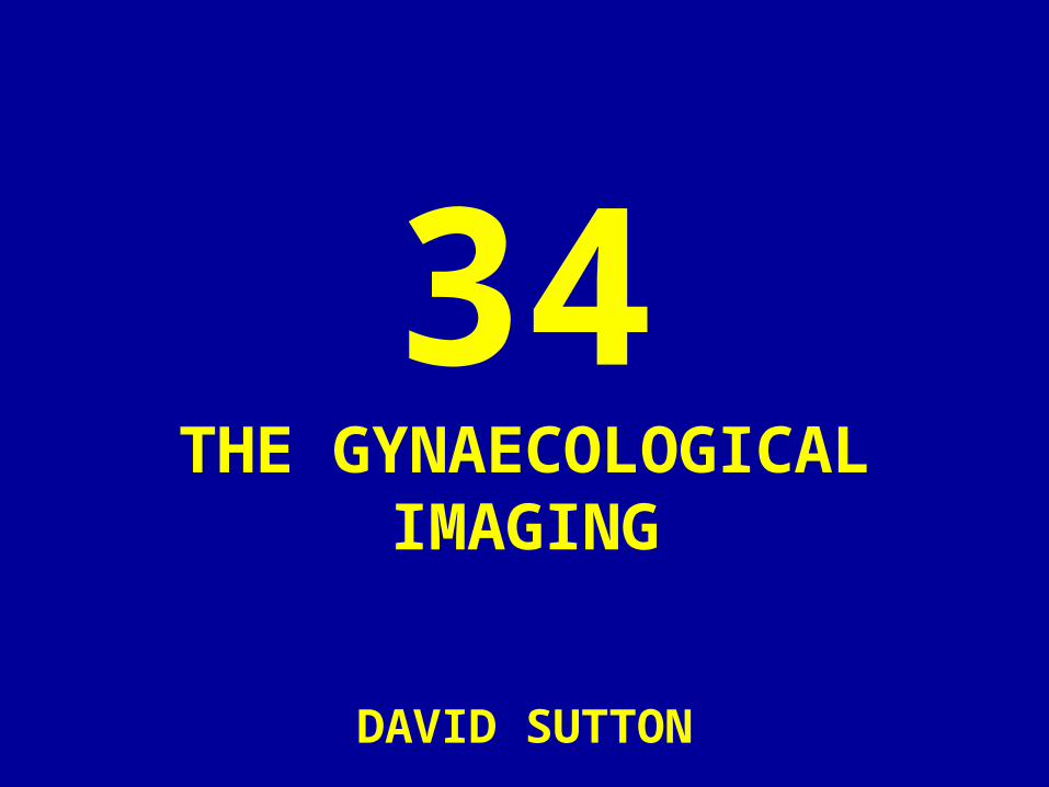
• Fig. 34.1 Sagittal transabdominal scan (IAS) showing measurement of uterine size (white crosses) and a moderately thick luteal phase endometrium (arrowheads). Arrows indicate vaginal walls.
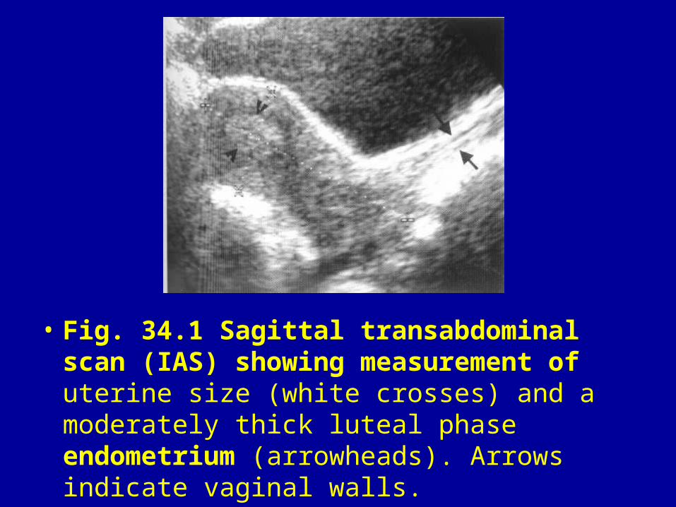
• Fig. 34.2 Sagittal TAS. Deformity of the bladder base and acoustic shadowing due to a vaginal tampon (arrows).

• Fig. 34.3 Endovaginal scan (EVS). Measurement of the endometrium (black arrows). Note there are five layers included in the measurement but the deeper hypoechoic layer (white arrows) is not included. The central echogenic line (black arrowhead) is due to the interface of the two layers of endometrium.
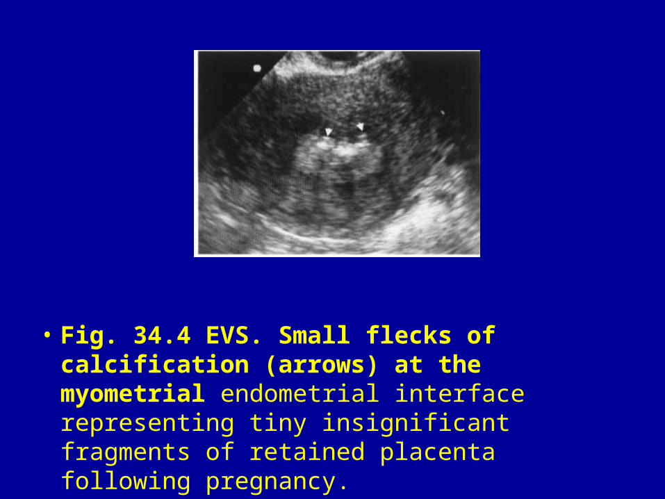
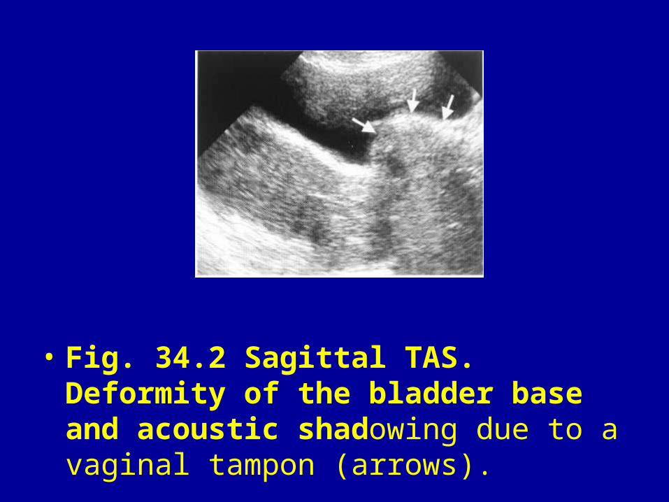
• Fig. 34.4 EVS. Small flecks of calcification (arrows) at the myometrial endometrial interface representing tiny insignificant fragments of retained placenta following pregnancy.
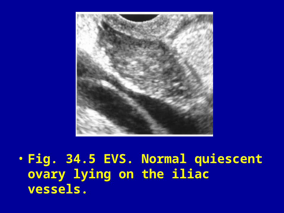
• Fig. 34.5 EVS. Normal quiescent ovary lying on the iliac vessels.

• Fig. 34.6 EVS. Atrophic postmenopausal ovary (arrowheads).

• Fig. 34.7 EVS. Power Doppler showing uterine artery (arrows) running alongside the uterine body.
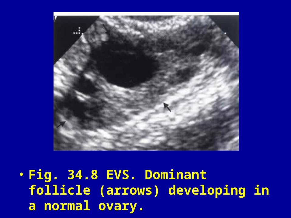
• Fig. 34.8 EVS. Dominant follicle (arrows) developing in a normal ovary.
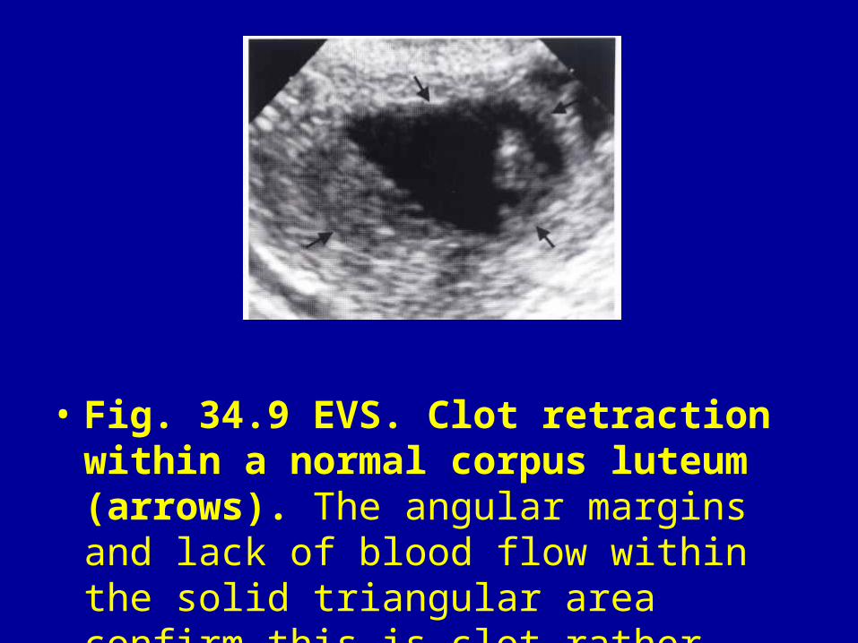
• Fig. 34.9 EVS. Clot retraction within a normal corpus luteum (arrows). The angular margins and lack of blood flow within the solid triangular area confirm this is clot rather than a solid nodule.

• Fig. 34.10 (A) EVS. Irregularly shaped echogenic cyst (arrowheads) due to a collapsing corpus luteum. (B) Colour Doppler. Flow around the corpus luteum (arrows) due to neo vascularisation.
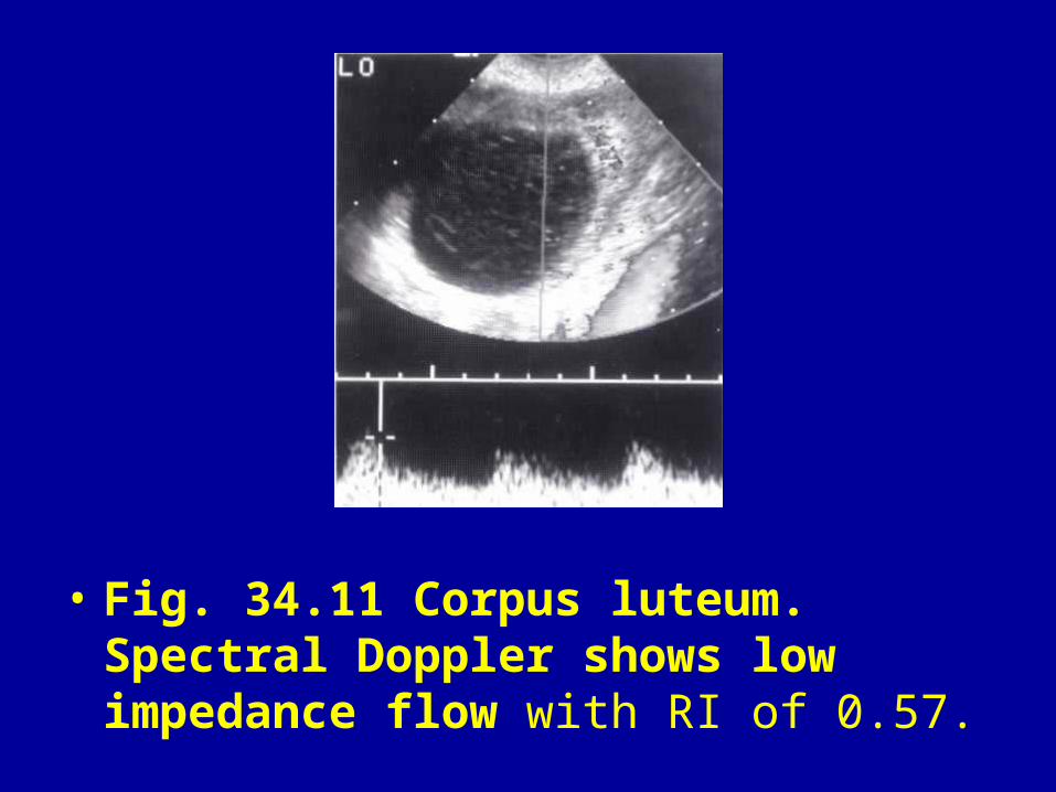
• Fig. 34.11 Corpus luteum. Spectral Doppler shows low impedance flow with RI of 0.57.
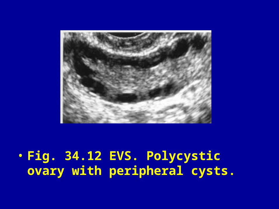
• Fig. 34.12 EVS. Polycystic ovary with peripheral cysts.
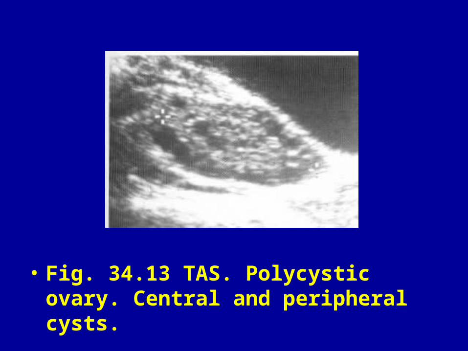
• Fig. 34.13 TAS. Polycystic ovary. Central and peripheral cysts.

• Fig. 34.14 TAS. Multifollicular ovary (arrows) in a patient with amenorrhoea due to anorexia nervosa.
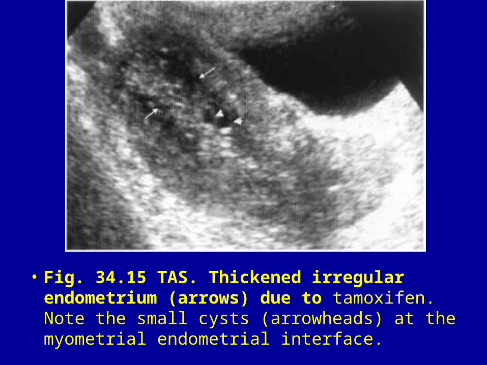
• Fig. 34.15 TAS. Thickened irregular endometrium (arrows) due to tamoxifen. Note the small cysts (arrowheads) at the myometrial endometrial interface.
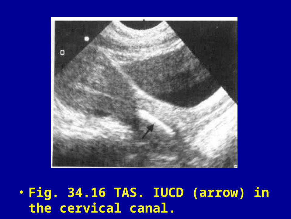
• Fig. 34.16 TAS. IUCD (arrow) in the cervical canal.
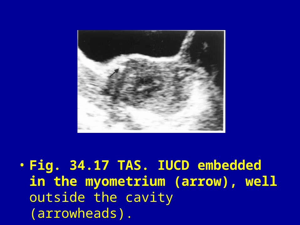
• Fig. 34.17 TAS. IUCD embedded in the myometrium (arrow), well outside the cavity (arrowheads).
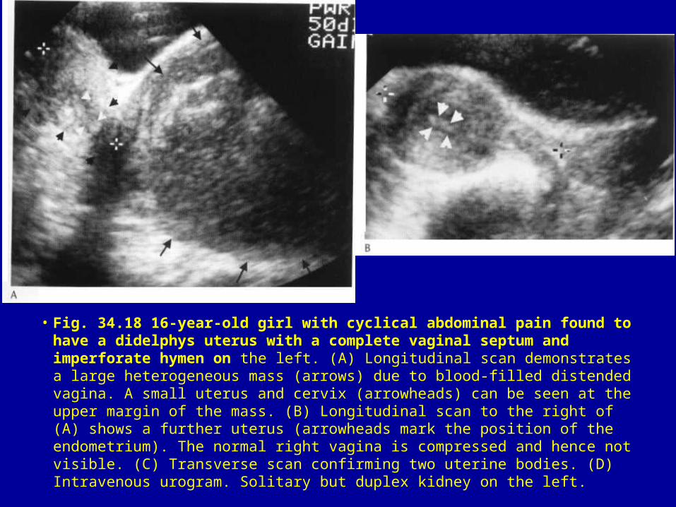
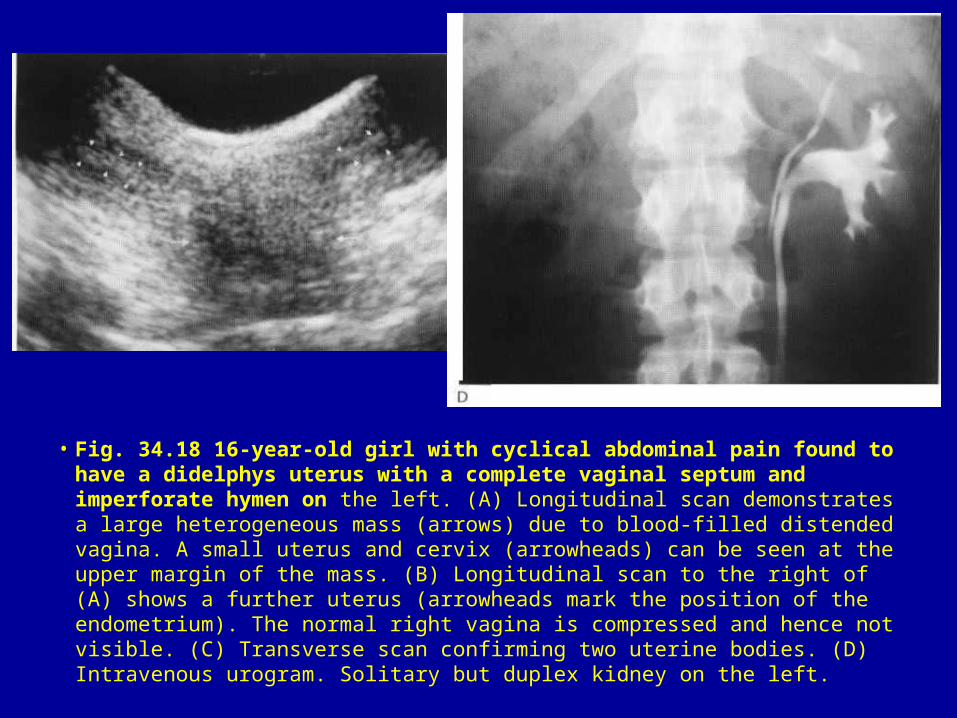
• Fig. 34.18 16-year-old girl with cyclical abdominal pain found to have a didelphys uterus with a complete vaginal septum and imperforate hymen on the left. (A) Longitudinal scan demonstrates a large heterogeneous mass (arrows) due to blood-filled distended vagina. A small uterus and cervix (arrowheads) can be seen at the upper margin of the mass. (B) Longitudinal scan to the right of (A) shows a further uterus (arrowheads mark the position of the endometrium). The normal right vagina is compressed and hence not visible. (C) Transverse scan confirming two uterine bodies. (D) Intravenous urogram. Solitary but duplex kidney on the left.
• Fig. 34.18 16-year-old girl with cyclical abdominal pain found to have a didelphys uterus with a complete vaginal septum and imperforate hymen on the left. (A) Longitudinal scan demonstrates a large heterogeneous mass (arrows) due to blood-filled distended vagina. A small uterus and cervix (arrowheads) can be seen at the upper margin of the mass. (B) Longitudinal scan to the right of (A) shows a further uterus (arrowheads mark the position of the endometrium). The normal right vagina is compressed and hence not visible. (C) Transverse scan confirming two uterine bodies. (D) Intravenous urogram. Solitary but duplex kidney on the left.
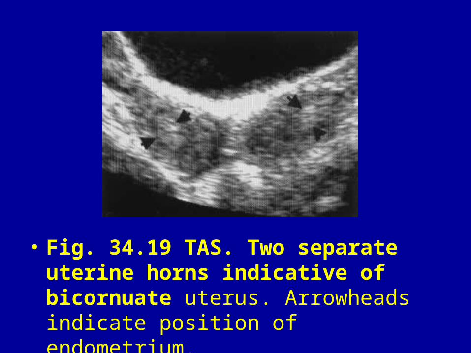
• Fig. 34.19 TAS. Two separate uterine horns indicative of bicornuate uterus. Arrowheads indicate position of endometrium.
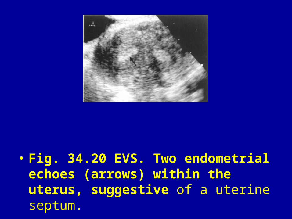
• Fig. 34.20 EVS. Two endometrial echoes (arrows) within the uterus, suggestive of a uterine septum.

• Fig. 34.21 (A) TAS. Fibroid polyp (arrows) within the cervical canal. The stalk of the polyp (arrowheads) can be seen in the uterine cavity. (B) TAS. Same patient 6 months earlier. The fibroid polyp (arrows) is now seen within the uterine cavity.

• Fig. 34.22 TAS. Typical mural fibroids (arrows) abutting but not displacing the cavity (arrowheads). The larger fibroid shows typical recurrent shadowing.
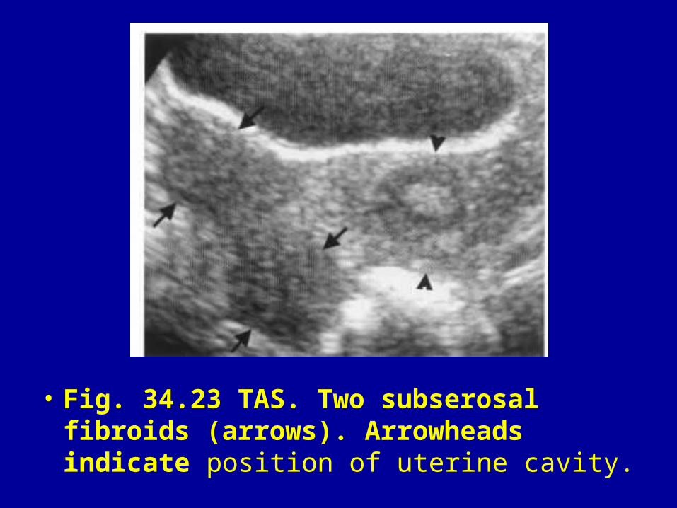
• Fig. 34.23 TAS. Two subserosal fibroids (arrows). Arrowheads indicate position of uterine cavity.
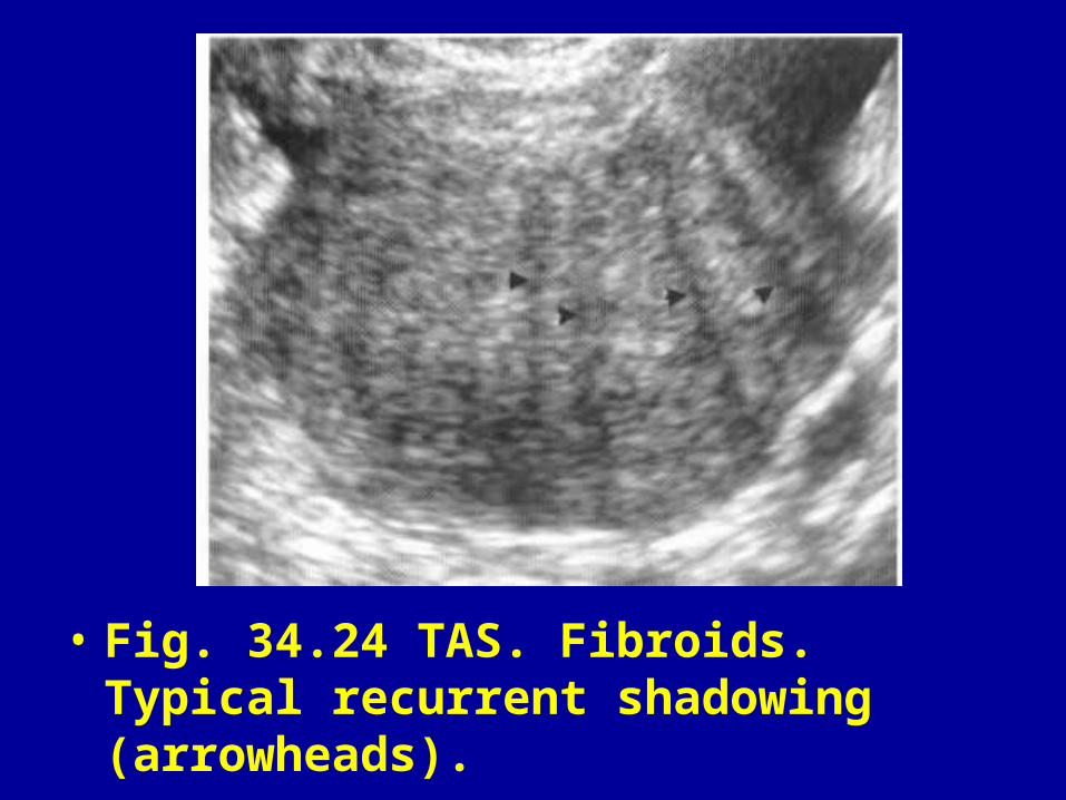
• Fig. 34.24 TAS. Fibroids. Typical recurrent shadowing (arrowheads).

• Fig. 34.25 TAS. Irregular area of increased reflectivity in the centre of a fibroid due to degeneration.
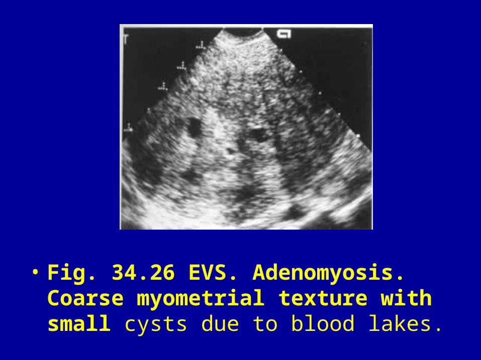
• Fig. 34.26 EVS. Adenomyosis. Coarse myometrial texture with small cysts due to blood lakes.
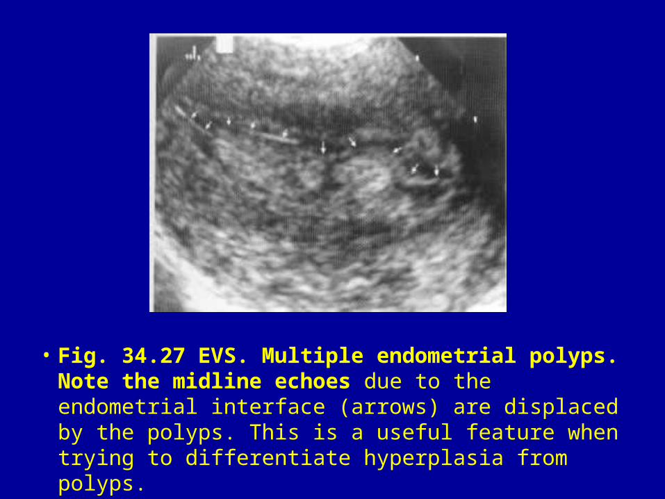
• Fig. 34.27 EVS. Multiple endometrial polyps. Note the midline echoes due to the endometrial interface (arrows) are displaced by the polyps. This is a useful feature when trying to differentiate hyperplasia from polyps.
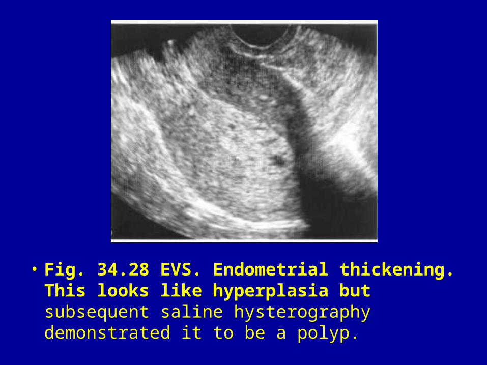
• Fig. 34.28 EVS. Endometrial thickening. This looks like hyperplasia but subsequent saline hysterography demonstrated it to be a polyp.
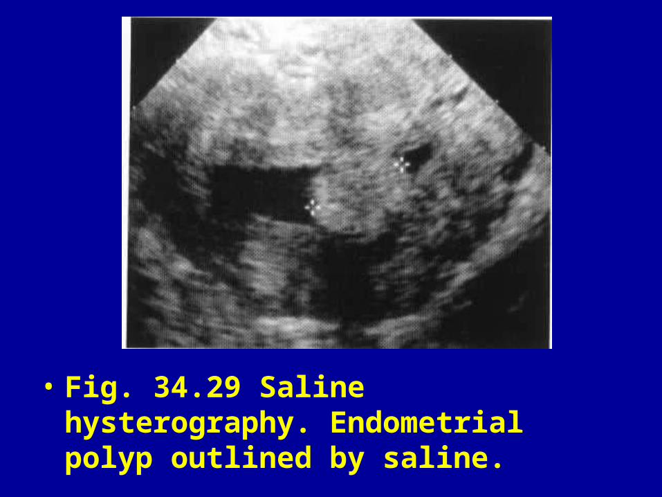
• Fig. 34.29 Saline hysterography. Endometrial polyp outlined by saline.

• Fig. 34.30 EVS. Poorly defined intrauterine mass due to endometrial carcinoma.
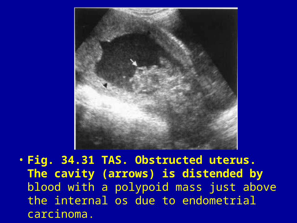
• Fig. 34.31 TAS. Obstructed uterus. The cavity (arrows) is distended by blood with a polypoid mass just above the internal os due to endometrial carcinoma.

• Fig. 34.32 TAS. Carcinoma of cervix. Large irregular cervix (arrowheads) with a small tongue of tumour (arrow) extending towards the bladder.
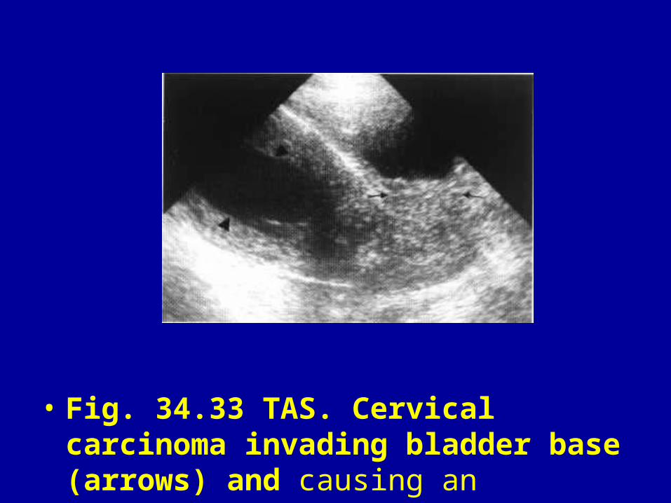
• Fig. 34.33 TAS. Cervical carcinoma invading bladder base (arrows) and causing an obstructed uterus (arrowheads).

• Fig. 34.34 EVS. Fimbrial cyst adjacent to ovary.
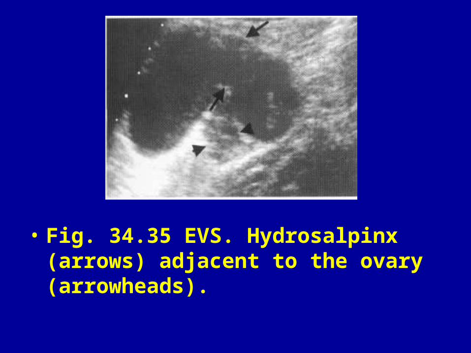
• Fig. 34.35 EVS. Hydrosalpinx (arrows) adjacent to the ovary (arrowheads).

• Fig. 34.36 EVS. Typical endometrioma with diffuse moderately high-level echoes (arrows).

• Fig. 34.37 TAS. Bilateral endometriomas. Note the fluid level on the left and the irregularly thickened wall.

• Fig. 34.38 TAS. Endometriosis. Complex ovarian mass with internal septations and echoes of varying density. Differential diagnosis must include a malignant tumour.

• Fig. 34.39 Huge pleural effusion in a young girl. Aspiration revealed heavily blood-stained fluid with multiple macrophages typical of pleural endometriosis.
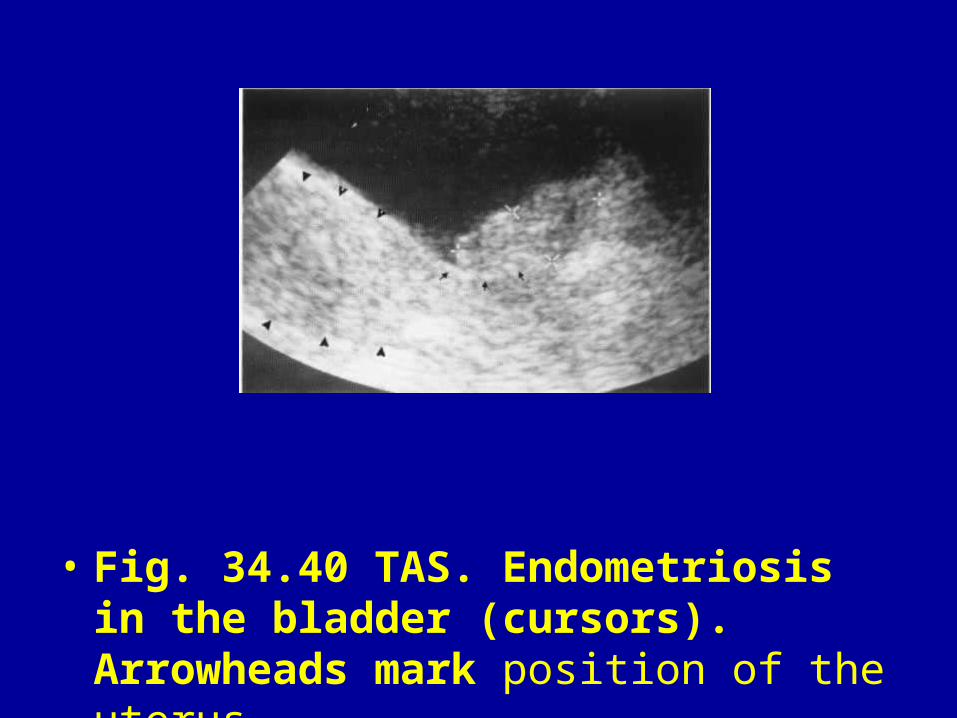
• Fig. 34.40 TAS. Endometriosis in the bladder (cursors). Arrowheads mark position of the uterus.

• Fig. 34.41 TAS. Acute pelvic infection with a thick-walled tuboovarian abscess (arrow) and free pus in the pouch of Douglas (arrowhead).
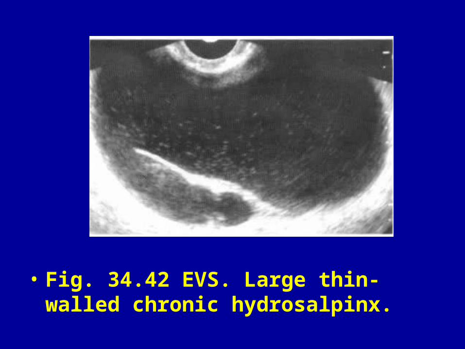
• Fig. 34.42 EVS. Large thin-walled chronic hydrosalpinx.
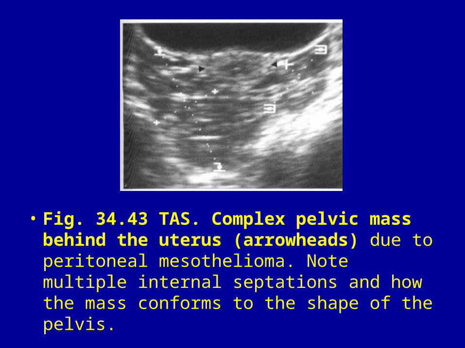
• Fig. 34.43 TAS. Complex pelvic mass behind the uterus (arrowheads) due to peritoneal mesothelioma. Note multiple internal septations and how the mass conforms to the shape of the pelvis.

• Fig. 34.44 TAS. Adnexal cyst with one solid area and some fine internal echoes suggestive of a serous cystadenocarcinoma. Histology confirmed a borderline malignant tumour.
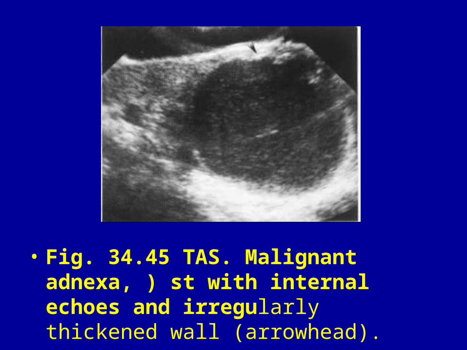
• Fig. 34.45 TAS. Malignant adnexa, ) st with internal echoes and irregularly thickened wall (arrowhead).
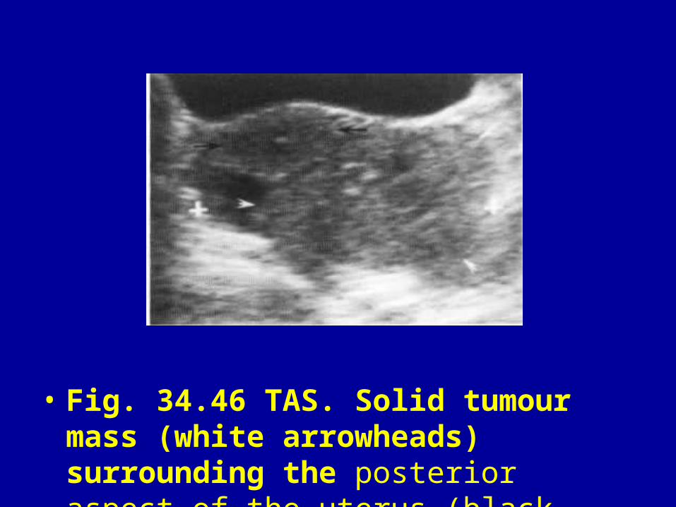
• Fig. 34.46 TAS. Solid tumour mass (white arrowheads) surrounding the posterior aspect of the uterus (black arrows).

• Fig. 34.47 TAS. Benign mucinous cystadenoma showing the typical multiloculated appearance—impossible to differentiate from a malignant tumour.
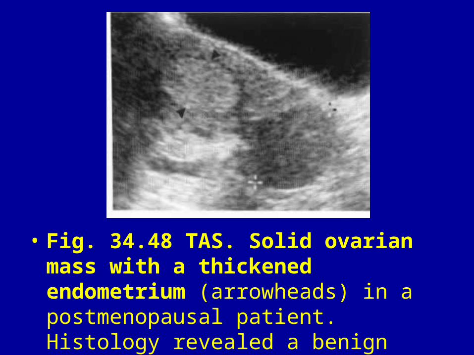
• Fig. 34.48 TAS. Solid ovarian mass with a thickened endometrium (arrowheads) in a postmenopausal patient. Histology revealed a benign functioning thecoma.

• Fig. 34.49 EVS. Ovarian fibroma. Homogeneous solid mass (arrows) arising from the ovary (arrowheads).

• Fig. 34.50 TAS. Typical dermoid with a floating echogenic area with acoustic shadowing due to fat, with or without calcification.

• Fig. 34.51 TAS. Dermoid cyst in a pregnant patient. Note the echogenic nodule (arrows) and dense acoustic shadowing (arrowheads).
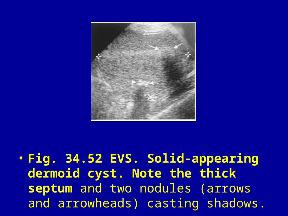
• Fig. 34.52 EVS. Solid-appearing dermoid cyst. Note the thick septum and two nodules (arrows and arrowheads) casting shadows.
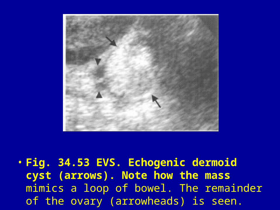
• Fig. 34.53 EVS. Echogenic dermoid cyst (arrows). Note how the mass mimics a loop of bowel. The remainder of the ovary (arrowheads) is seen.
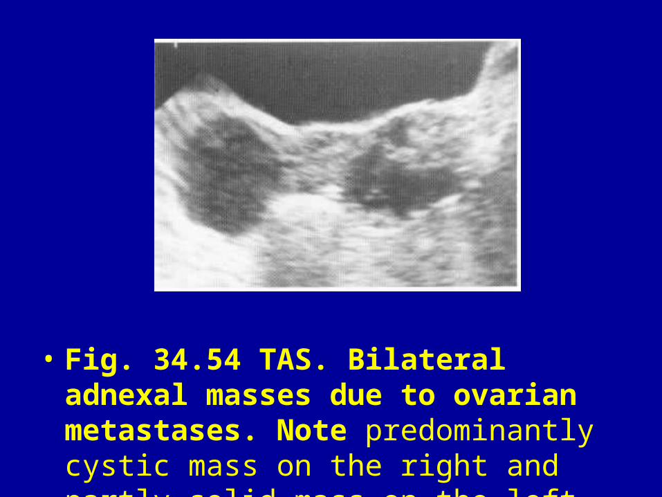
• Fig. 34.54 TAS. Bilateral adnexal masses due to ovarian metastases. Note predominantly cystic mass on the right and partly solid mass on the left.
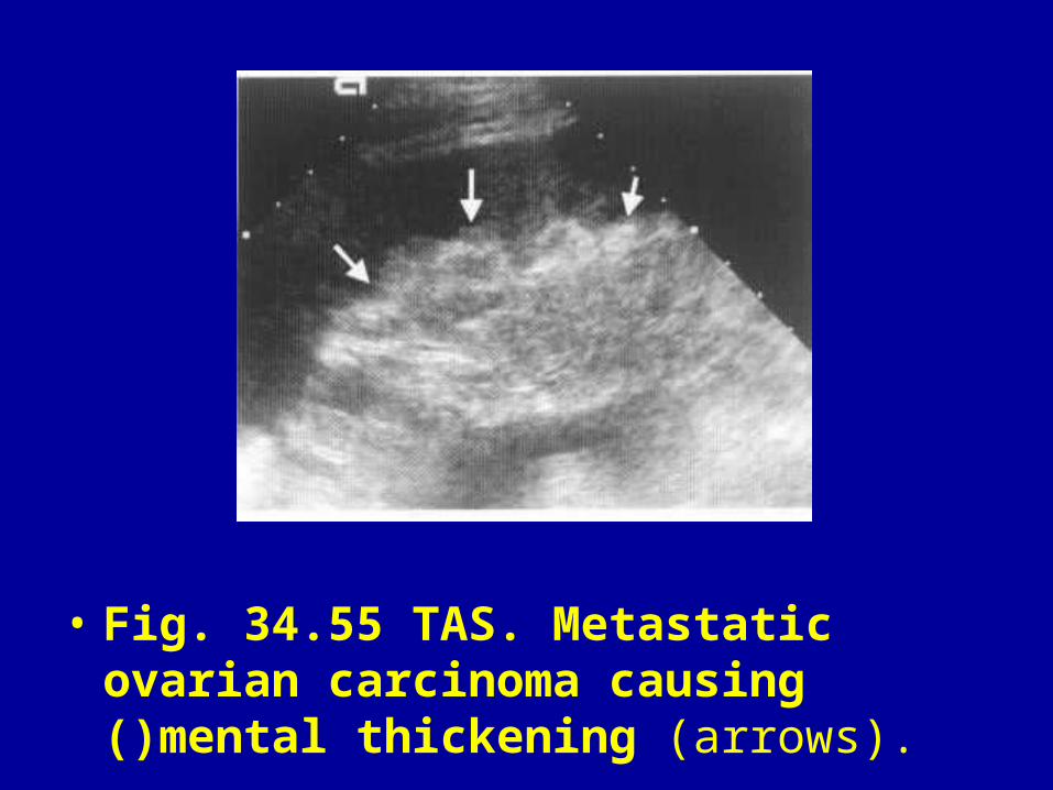
• Fig. 34.55 TAS. Metastatic ovarian carcinoma causing ()mental thickening (arrows).
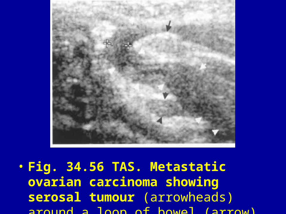
• Fig. 34.56 TAS. Metastatic ovarian carcinoma showing serosal tumour (arrowheads) around a loop of bowel (arrow).
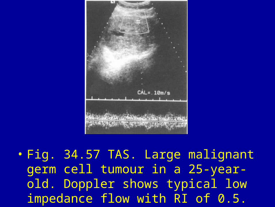
• Fig. 34.57 TAS. Large malignant germ cell tumour in a 25-year-old. Doppler shows typical low impedance flow with RI of 0.5.

• Fig. 34.58 EVS. Ovarian cyst with nodule in a 65-year-old. Doppler shows low impedance flow (RI 0.50) suggestive of a malignant tumour. Histology revealed a benign cystadenofibroma with a Brenner tumour. No evidence of malignancy.

• Fig. 34.59 HyCoSy. Contrast (Echovist) is seen outlining the cavity and entering the fallopian tube (arrows).

• Fig. 34.60 HyCoSy. Echovist outlines a fibroid polyp (arrows) in the uterine cavity.

• Fig. 34.61 Dermoid cyst. Note calcification and teeth with a fat—fluid level (arrow).

• Fig. 34.62 Barium enemas. (A) Serosal metastases from ovarian carcinoma. (B) Short smooth stricture due to endometriosis (arrowheads). Note the puckering of the serosal due to adhesion (arrow).

• Fig. 34.63 HSG. Normal cavity. Both tubes visible with regular mucosal folds and free peritoneal spill. Note how the contrast flows around loops of bowel (arrows).
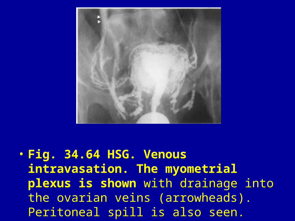
• Fig. 34.64 HSG. Venous intravasation. The myometrial plexus is shown with drainage into the ovarian veins (arrowheads). Peritoneal spill is also seen.

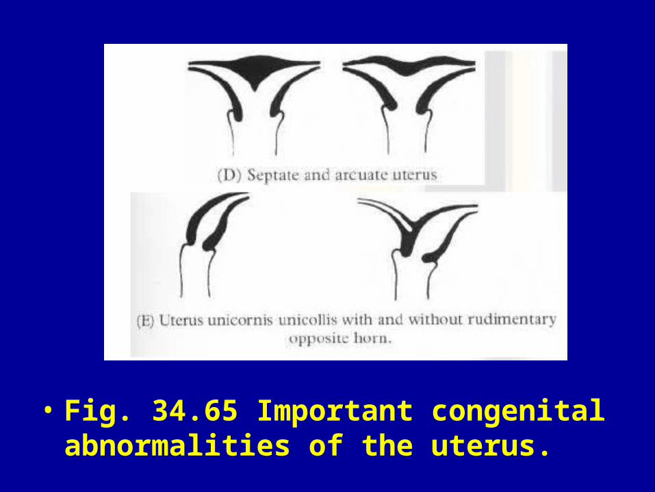
• Fig. 34.65 Important congenital abnormalities of the uterus.
• Fig. 34.65 Important congenital abnormalities of the uterus.
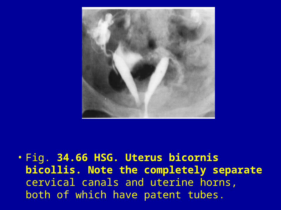
• Fig. 34.66 HSG. Uterus bicornis bicollis. Note the completely separate cervical canals and uterine horns, both of which have patent tubes.
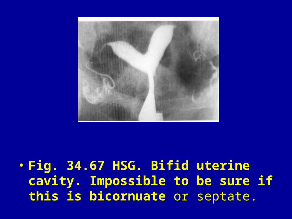
• Fig. 34.67 HSG. Bifid uterine cavity. Impossible to be sure if this is bicornuate or septate.
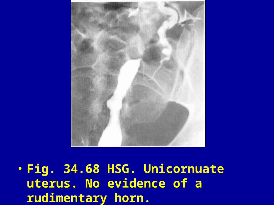
• Fig. 34.68 HSG. Unicornuate uterus. No evidence of a rudimentary horn.
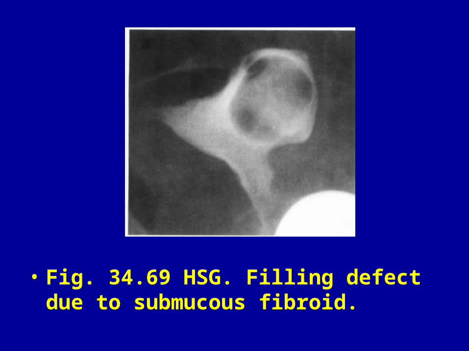
• Fig. 34.69 HSG. Filling defect due to submucous fibroid.

• Fig. 34.70 HSG. Cavity and right fallopian tube being distorted by large mural fibroid. Note small calcified fibroid on the left (arrows).
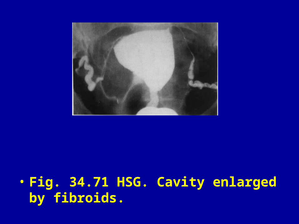
• Fig. 34.71 HSG. Cavity enlarged by fibroids.

• Fig. 34.72 HSG. Polypoid endometrium causing multiple filling defects (arrows) only seen on the early filling film.
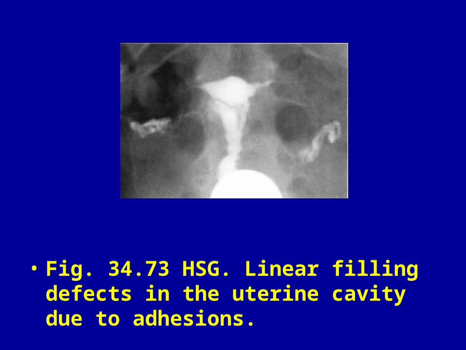
• Fig. 34.73 HSG. Linear filling defects in the uterine cavity due to adhesions.

• Fig. 34.74 HSG. Severe Asherman's syndrome with complete obliteration of the uterine cavity.
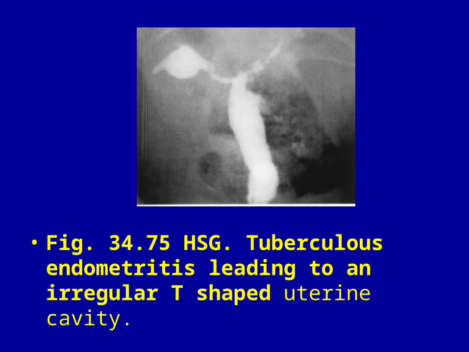
• Fig. 34.75 HSG. Tuberculous endometritis leading to an irregular T shaped uterine cavity.
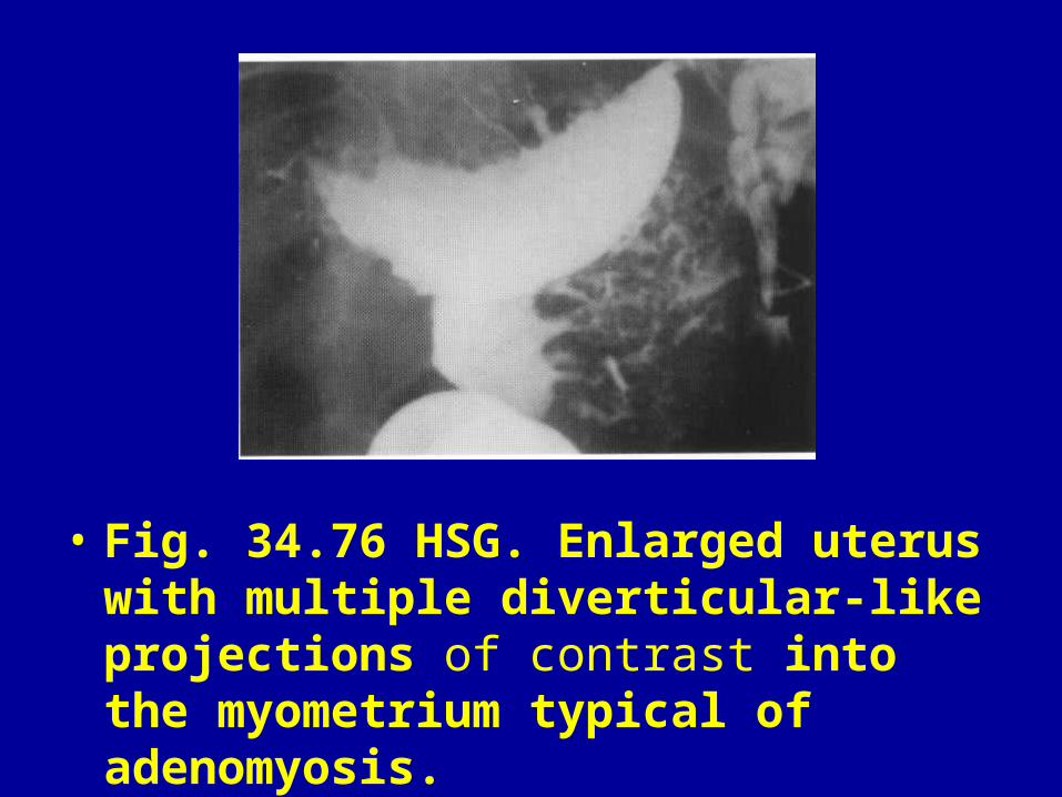
• Fig. 34.76 HSG. Enlarged uterus with multiple diverticular-like projections of contrast into the myometrium typical of adenomyosis.
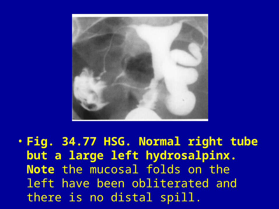
• Fig. 34.77 HSG. Normal right tube but a large left hydrosalpinx. Note the mucosal folds on the left have been obliterated and there is no distal spill.
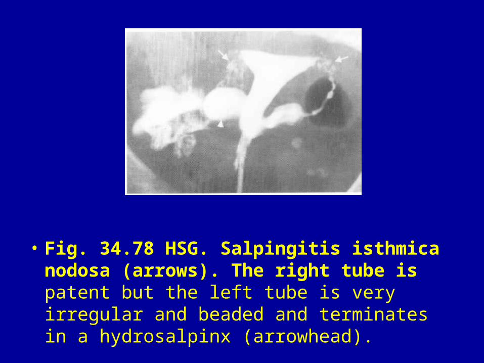
• Fig. 34.78 HSG. Salpingitis isthmica nodosa (arrows). The right tube is patent but the left tube is very irregular and beaded and terminates in a hydrosalpinx (arrowhead).

• Fig. 34.79 HSG. Bilateral cornual occusions.
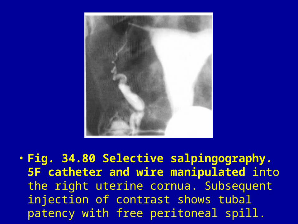
• Fig. 34.80 Selective salpingography. 5F catheter and wire manipulated into the right uterine cornua. Subsequent injection of contrast shows tubal patency with free peritoneal spill.
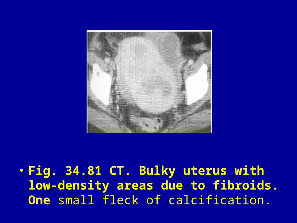
• Fig. 34.81 CT. Bulky uterus with low-density areas due to fibroids. One small fleck of calcification.
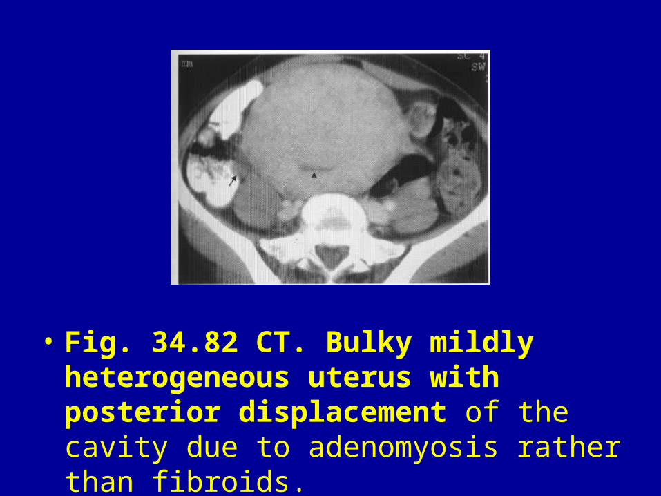
• Fig. 34.82 CT. Bulky mildly heterogeneous uterus with posterior displacement of the cavity due to adenomyosis rather than fibroids.
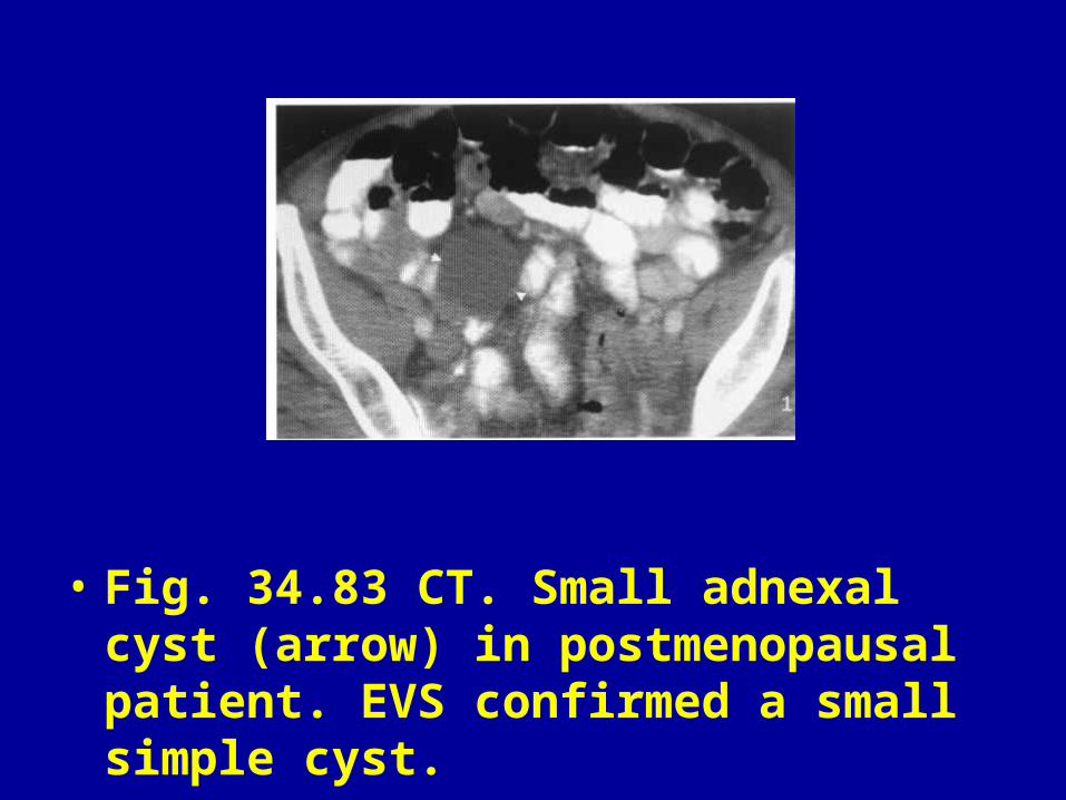
• Fig. 34.83 CT. Small adnexal cyst (arrow) in postmenopausal patient. EVS confirmed a small simple cyst.

• Fig. 34.84 CT. Complex mass in the pelvis typical of a dermoid cyst (arrows). The mass is of mixed attenuation but contains a large amount of fat. It has a calcified rim and a dense area of calcification (arrowheads) inferolaterally due to a tooth.
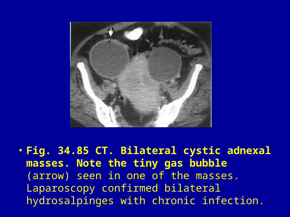
• Fig. 34.85 CT. Bilateral cystic adnexal masses. Note the tiny gas bubble (arrow) seen in one of the masses. Laparoscopy confirmed bilateral hydrosalpinges with chronic infection.

• Fig. 34.86 CT. Patient with ovarian carcinoma, peritoneal deposits (white arrowheads), para-aortic lymphadenopathy (black arrowheads) and 'omental cake' (black arrows).
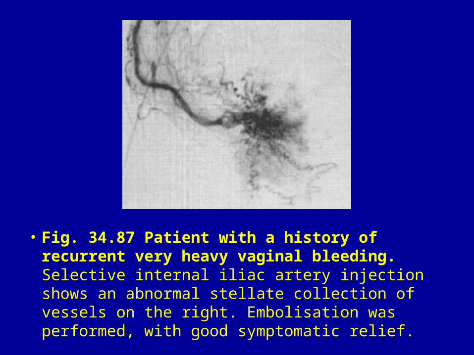
• Fig. 34.87 Patient with a history of recurrent very heavy vaginal bleeding. Selective internal iliac artery injection shows an abnormal stellate collection of vessels on the right. Embolisation was performed, with good symptomatic relief.
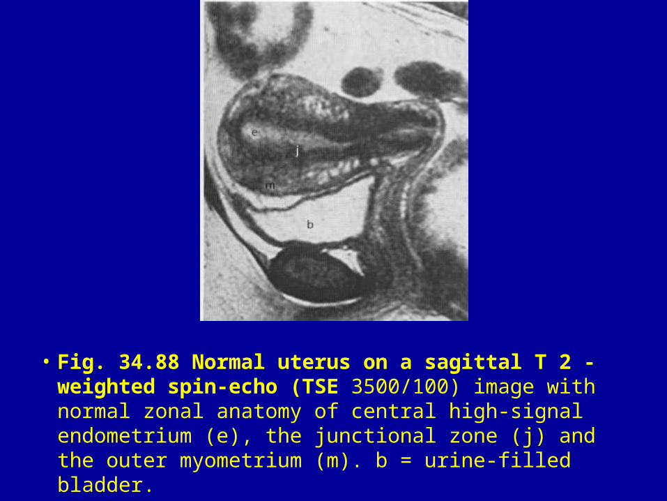
• Fig. 34.88 Normal uterus on a sagittal T 2 -weighted spin-echo (TSE 3500/100) image with normal zonal anatomy of central high-signal endometrium (e), the junctional zone (j) and the outer myometrium (m). b = urine-filled bladder.

• Fig. 34.89 Normal zonal anatomy of the cervix on a transverse T2 -weighted spin-echo (TSE 3500/100) image. b = bladder; s = cervical stroma; straight arrow = cervical mucus and epithelium.
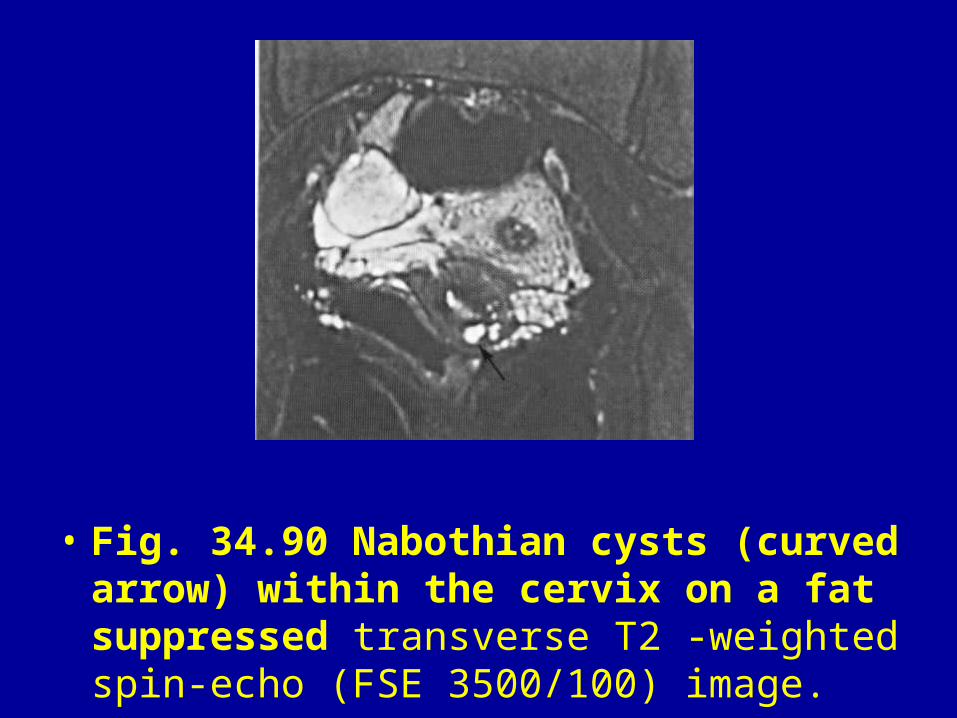
• Fig. 34.90 Nabothian cysts (curved arrow) within the cervix on a fat suppressed transverse T2 -weighted spin-echo (FSE 3500/100) image.
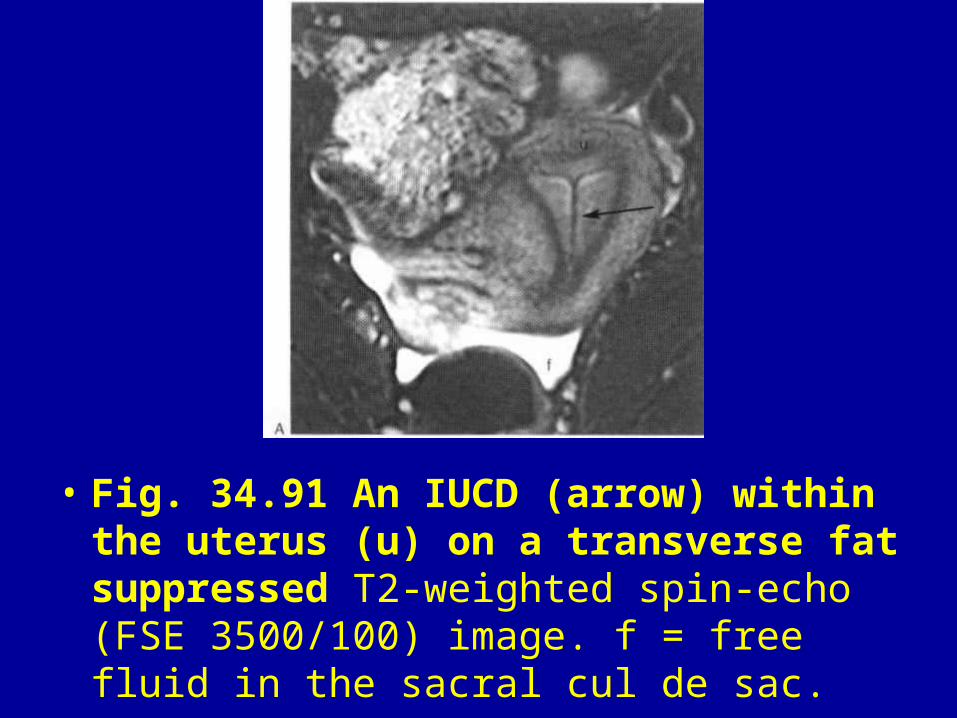
• Fig. 34.91 An IUCD (arrow) within the uterus (u) on a transverse fat suppressed T2-weighted spin-echo (FSE 3500/100) image. f = free fluid in the sacral cul de sac.
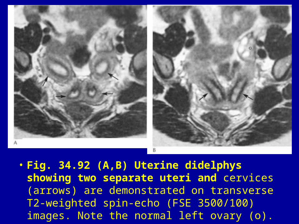
• Fig. 34.92 (A,B) Uterine didelphys showing two separate uteri and cervices (arrows) are demonstrated on transverse T2-weighted spin-echo (FSE 3500/100) images. Note the normal left ovary (o).
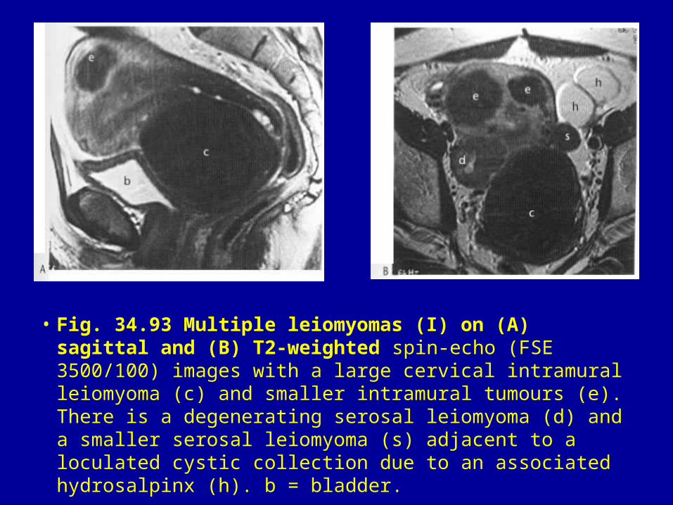
• Fig. 34.93 Multiple leiomyomas (I) on (A) sagittal and (B) T2-weighted spin-echo (FSE 3500/100) images with a large cervical intramural leiomyoma (c) and smaller intramural tumours (e). There is a degenerating serosal leiomyoma (d) and a smaller serosal leiomyoma (s) adjacent to a loculated cystic collection due to an associated hydrosalpinx (h). b = bladder.
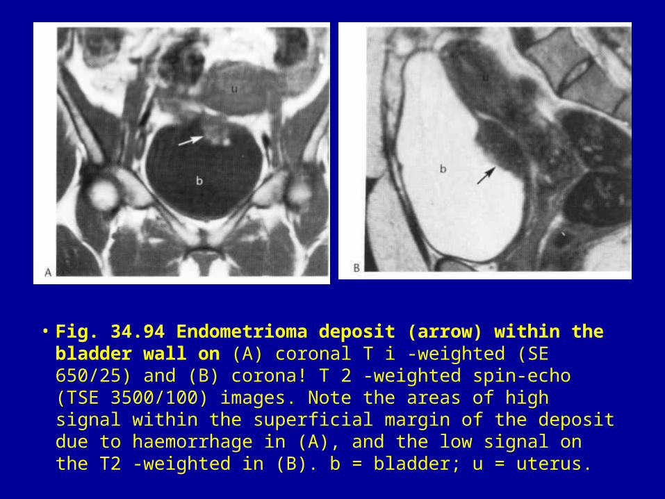
• Fig. 34.94 Endometrioma deposit (arrow) within the bladder wall on (A) coronal T i -weighted (SE 650/25) and (B) corona! T 2 -weighted spin-echo (TSE 3500/100) images. Note the areas of high signal within the superficial margin of the deposit due to haemorrhage in (A), and the low signal on the T2 -weighted in (B). b = bladder; u = uterus.
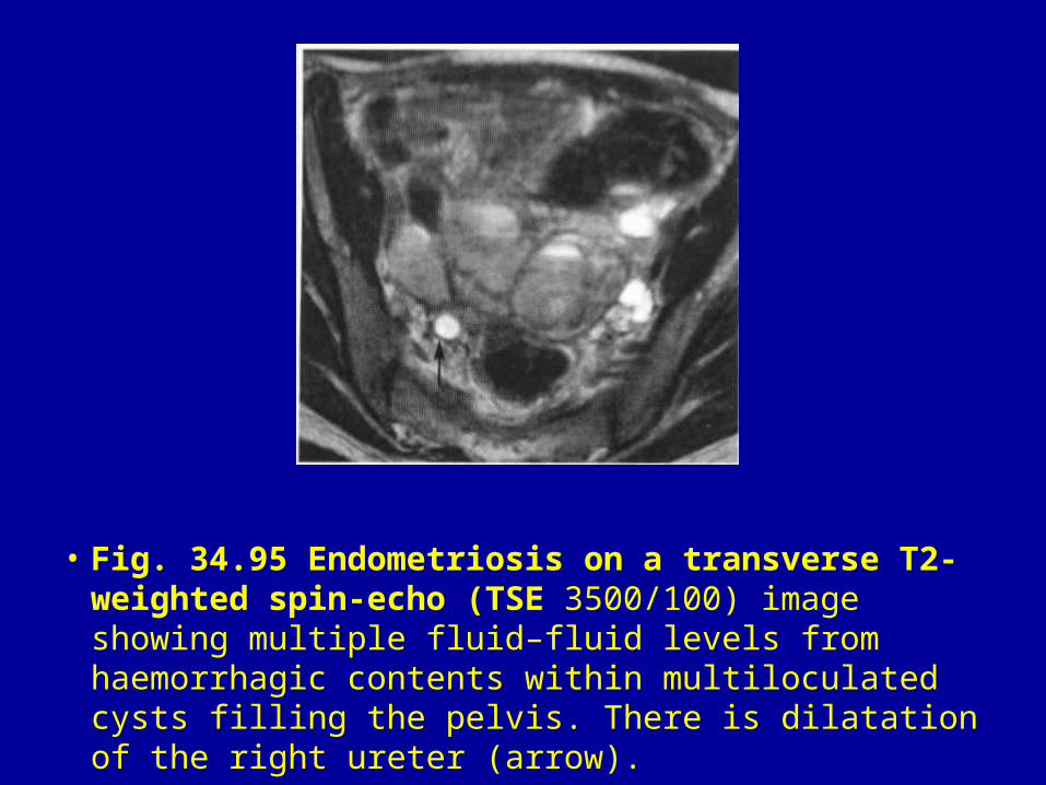
• Fig. 34.95 Endometriosis on a transverse T2-weighted spin-echo (TSE 3500/100) image showing multiple fluid–fluid levels from haemorrhagic contents within multiloculated cysts filling the pelvis. There is dilatation of the right ureter (arrow).
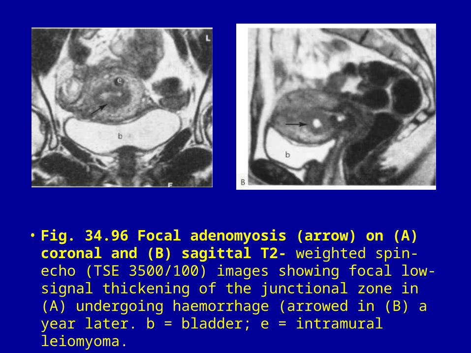
• Fig. 34.96 Focal adenomyosis (arrow) on (A) coronal and (B) sagittal T2- weighted spin-echo (TSE 3500/100) images showing focal low-signal thickening of the junctional zone in (A) undergoing haemorrhage (arrowed in (B) a year later. b = bladder; e = intramural leiomyoma.
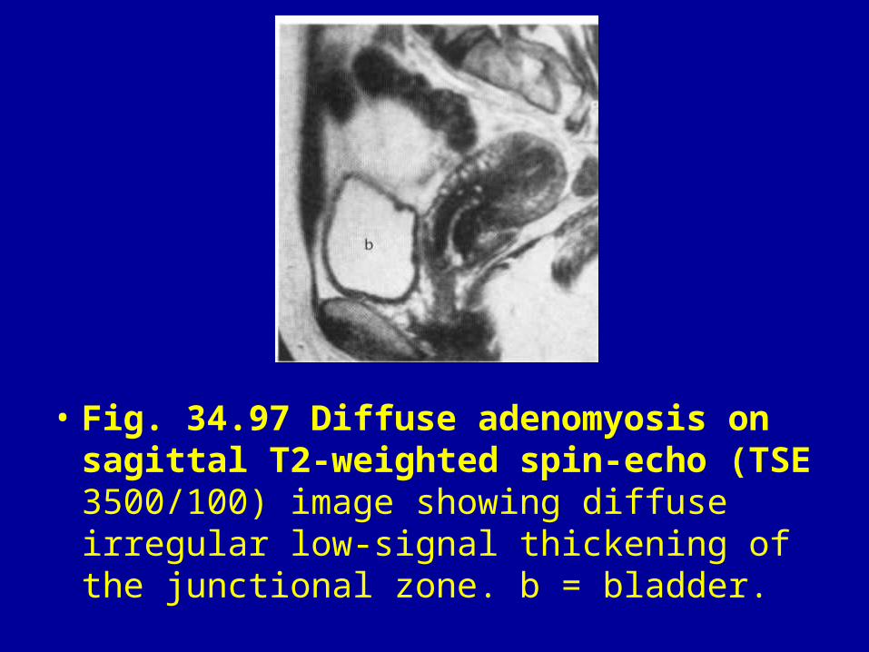
• Fig. 34.97 Diffuse adenomyosis on sagittal T2-weighted spin-echo (TSE 3500/100) image showing diffuse irregular low-signal thickening of the junctional zone. b = bladder.
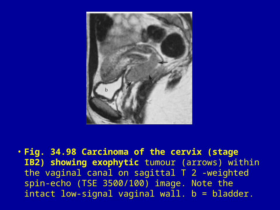
• Fig. 34.98 Carcinoma of the cervix (stage IB2) showing exophytic tumour (arrows) within the vaginal canal on sagittal T 2 -weighted spin-echo (TSE 3500/100) image. Note the intact low-signal vaginal wall. b = bladder.

• Fig. 34.99 Large cervical carcinoma (arrow) infiltrating into the bladder (b) with a separate tumour nodule in the posterior fornix (arrowhead) on a sagittal T2 -weighted spin-echo (FSE 3000/100) image. Note the endometrial obstruction (e). (Courtesy of Dr J. M. Hawnaur, Department of Diagnostic Radiology, University of Manchester.)

• Fig. 34.100 Bulky exophytic cervical carcinoma confined within the cervix on a sagittal T 2 -weighted spin-echo (TSE 5041/132) image. The low signal vaginal wall remains intact apart from an area of tumour infiltration posteriorly (arrow). Within the tumour in the anterior fornix there is an area of necrosis. b = bladder. (Courtesy of Dr R. J. Johnson, Christie Hospital.)

• Fig. 34.101 Carcinoma of the cervix (stage IIB) showing tumour (t) within the parametrium (arrows) on transverse T 2 -weighted spin-echo (FSE 3500/100) image. Note the loss of the normal low signal from the cervical stroma. b = bladder.
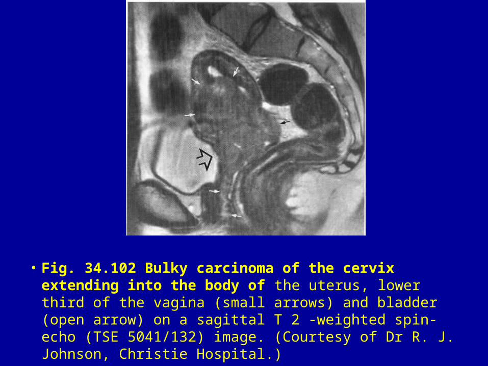
• Fig. 34.102 Bulky carcinoma of the cervix extending into the body of the uterus, lower third of the vagina (small arrows) and bladder (open arrow) on a sagittal T 2 -weighted spin-echo (TSE 5041/132) image. (Courtesy of Dr R. J. Johnson, Christie Hospital.)

• Fig. 34.103 Tumour (t) infiltrating the parametrium and left iliacus muscle (i), with left-sided involved lymph nodes (n) and a right ovarian metastasis (o) on a transverse T 2 -weighted spin-echo (TSE 3500/100) image. b = bladder.

• Fig. 34.104 Cervical carcinoma (t) extending into the parametrium producing left hydronephrosis (straight arrow) and extending posteriorly through the perirectal fascia into the rectal mucosa (curved arrow) on a transverse T2 -weighted spin-echo (TSE 5041 /1 32) image. b = bladder (Courtesy of Dr R.J. Johnson, Christie Hospital.)

• Fig. 34.105 Recurrent carcinoma of the cervix (t) infiltrating into the parametrium and right levator ani on a transverse T 2 -weighted spin-echo (TSE 5041/132) image. Note the low-signal vaginal wall (arrow). b = bladder, r = rectum. (Courtesy of Dr R. J. Johnson, Christie Hospital.)
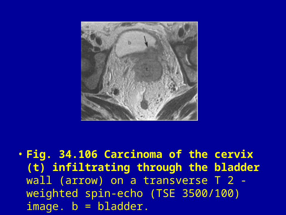
• Fig. 34.106 Carcinoma of the cervix (t) infiltrating through the bladder wall (arrow) on a transverse T 2 -weighted spin-echo (TSE 3500/100) image. b = bladder.
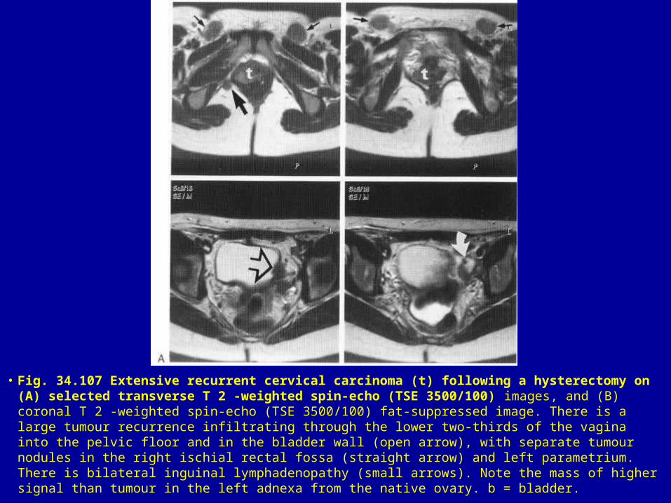
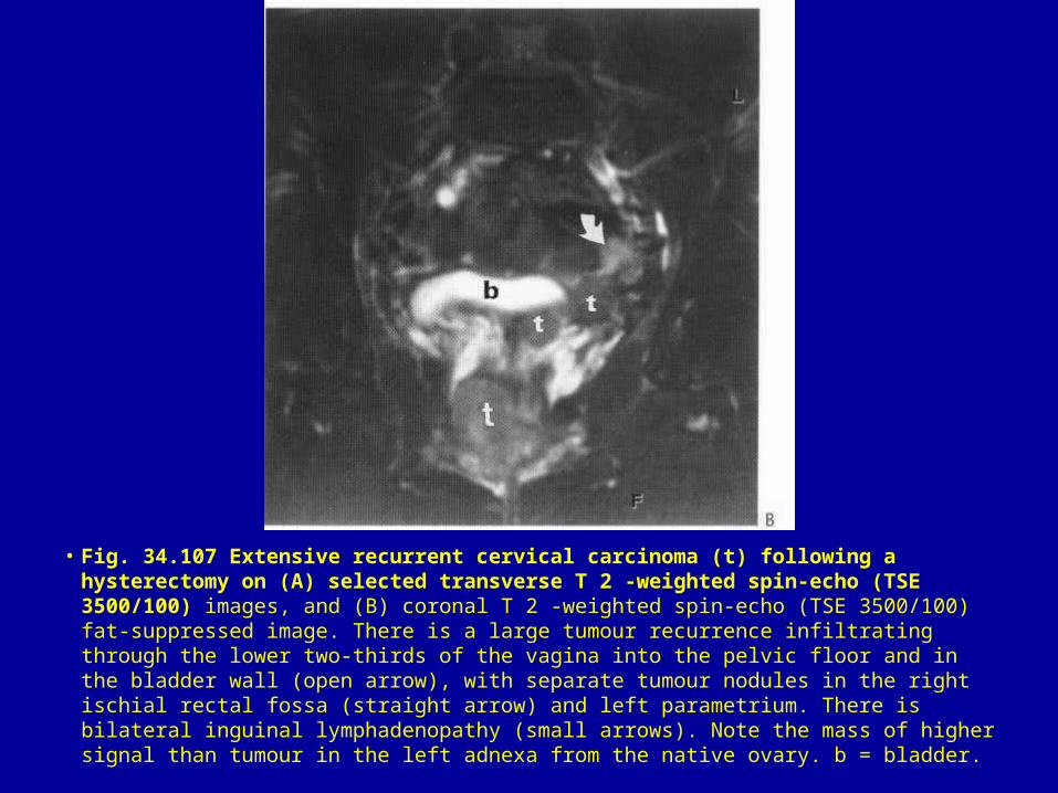
• Fig. 34.107 Extensive recurrent cervical carcinoma (t) following a hysterectomy on (A) selected transverse T 2 -weighted spin-echo (TSE 3500/100) images, and (B) coronal T 2 -weighted spin-echo (TSE 3500/100) fat-suppressed image. There is a large tumour recurrence infiltrating through the lower two-thirds of the vagina into the pelvic floor and in the bladder wall (open arrow), with separate tumour nodules in the right ischial rectal fossa (straight arrow) and left parametrium. There is bilateral inguinal lymphadenopathy (small arrows). Note the mass of higher signal than tumour in the left adnexa from the native ovary. b = bladder.
• Fig. 34.107 Extensive recurrent cervical carcinoma (t) following a hysterectomy on (A) selected transverse T 2 -weighted spin-echo (TSE 3500/100) images, and (B) coronal T 2 -weighted spin-echo (TSE 3500/100) fat-suppressed image. There is a large tumour recurrence infiltrating through the lower two-thirds of the vagina into the pelvic floor and in the bladder wall (open arrow), with separate tumour nodules in the right ischial rectal fossa (straight arrow) and left parametrium. There is bilateral inguinal lymphadenopathy (small arrows). Note the mass of higher signal than tumour in the left adnexa from the native ovary. b = bladder.

• Fig. 34.108 Carcinoma of the cervix (straight arrows) on sagittal T 2 -weighted spin echo (SE 1500/80) (A) before treatment, (B) 6 weeks, and (C) 6 months after radiotherapy. Note the rapid reduction in size of the tumour between (A) and (B). The small area of high signal in the cervix in (C) is due to either residual tumour or post-treatment change. Note the low-signal area in the uterus due to a non-degenerating leiomyoma (curved arrow), and the high mucosal signal in the posterior wall of the bladder (b) from radiotherapy change in (B) and (C).

• Fig. 34.108 Carcinoma of the cervix (straight arrows) on sagittal T 2 -weighted spin echo (SE 1500/80) (A) before treatment, (B) 6 weeks, and (C) 6 months after radiotherapy. Note the rapid reduction in size of the tumour between (A) and (B). The small area of high signal in the cervix in (C) is due to either residual tumour or post-treatment change. Note the low-signal area in the uterus due to a non-degenerating leiomyoma (curved arrow), and the high mucosal signal in the posterior wall of the bladder (b) from radiotherapy change in (B) and (C).

• Fig. 34.109 Extensive radiation change involving the bladder, vagina, rectum and bowel loops on (A) sagittal T2-weighted (SE 1 500/80) and (B) transverse T 1 -weighted (SE 800/40) images. In (A) the bladder (b) has a thickened wall with a high-signal-intensity mucosa around the posterior wall. The uterus (u) is enlarged and the vagina (arrows), rectosigmoid (r), and adjacent small bowel loops (I) show thickened walls with high signal. No evidence of recurrence of cervical carcinoma, which was confirmed on histological review. The high signal from the sacrum and L5 vertebra is due to radiation-induced fatty infiltration of the marrow spaces. The area of signal void within the vagina in (A) is due to a tampon in situ.

• Fig. 34.110 Stage IC endometrial carcinoma (e) on a sagittal T2 3500/100) image. b = bladder. -weighted spin-echo (TSE
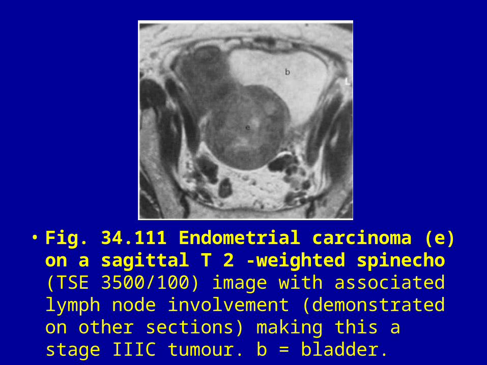
• Fig. 34.111 Endometrial carcinoma (e) on a sagittal T 2 -weighted spinecho (TSE 3500/100) image with associated lymph node involvement (demonstrated on other sections) making this a stage IIIC tumour. b = bladder.
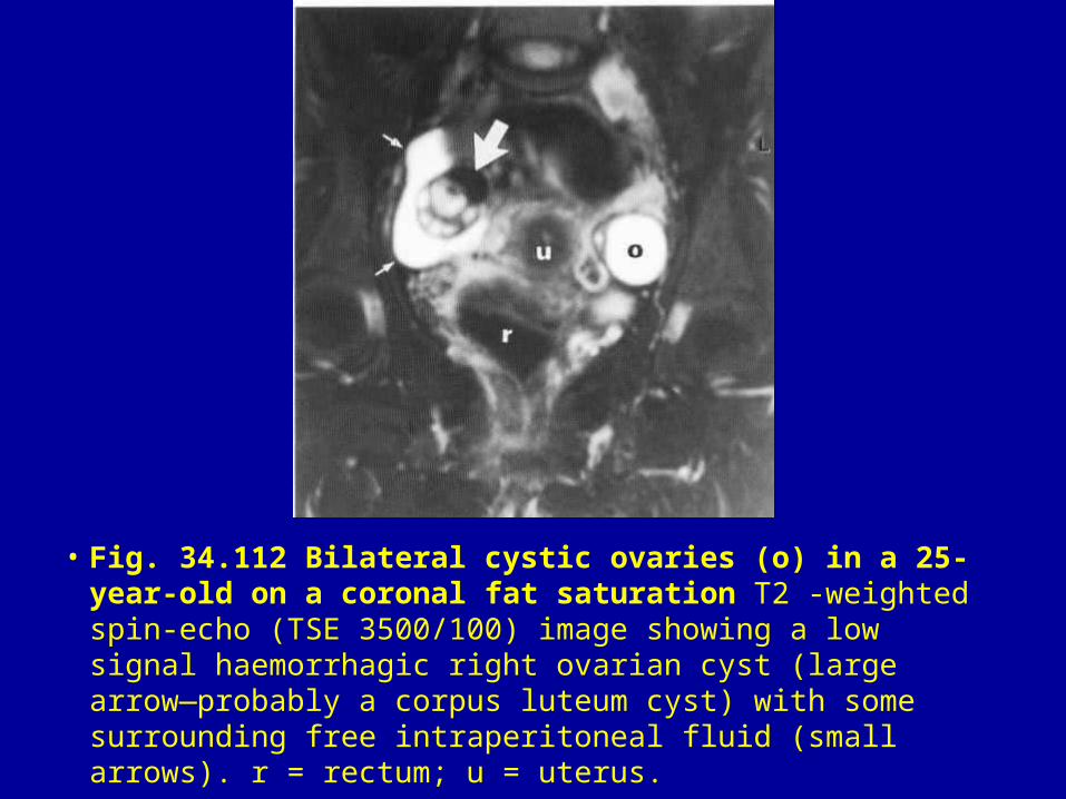
• Fig. 34.112 Bilateral cystic ovaries (o) in a 25-year-old on a coronal fat saturation T2 -weighted spin-echo (TSE 3500/100) image showing a low signal haemorrhagic right ovarian cyst (large arrow—probably a corpus luteum cyst) with some surrounding free intraperitoneal fluid (small arrows). r = rectum; u = uterus.

• Fig. 34.113 Multiloculated thin-walled haemorrhagic benign ovarian cysts (c) showing fluid–fluid levels on a transverse T 2 -weighted spin-echo (SE 2000/120) image. There is a coincidental uterine leiomyoma (I).
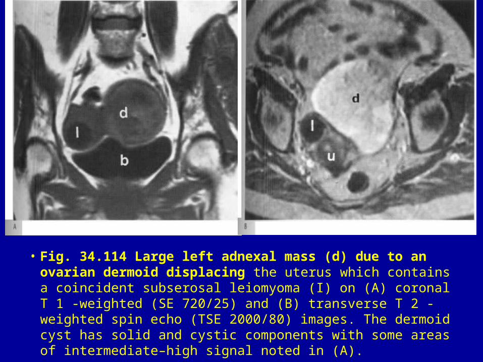
• Fig. 34.114 Large left adnexal mass (d) due to an ovarian dermoid displacing the uterus which contains a coincident subserosal leiomyoma (I) on (A) coronal T 1 -weighted (SE 720/25) and (B) transverse T 2 -weighted spin echo (TSE 2000/80) images. The dermoid cyst has solid and cystic components with some areas of intermediate–high signal noted in (A).

• Fig. 34.115 Dermoid cyst (arrows) showing a unilocular mass with a nodule within high-signal fat on (A) coronal T 1 -weighted spin-echo (TSE 700/12) image and (B) sagittal T2 -weighted spin-echo (TSE 3500/100) image. A fat-suppressed sequence (not shown) was also performed to confirm the fat contents of the cyst. b = bladder.

• Fig. 34.116 Complex large left adnexal mass with solid and cystic components (arrows) compressing and displacing the uterus on transverse T2 - weighted spin-echo (TSE 51 36/1 32) image. Note that there is distension of the endometrial cavity with intermediate to high signal due to a coexisting endometrial tumour (e). Endometrioid carcinoma of the ovary is associated in approximately a third of cases with endometrioid carcinoma of the uterus. (Courtesy of Dr R. J. Johnson, Christie Hospital.)
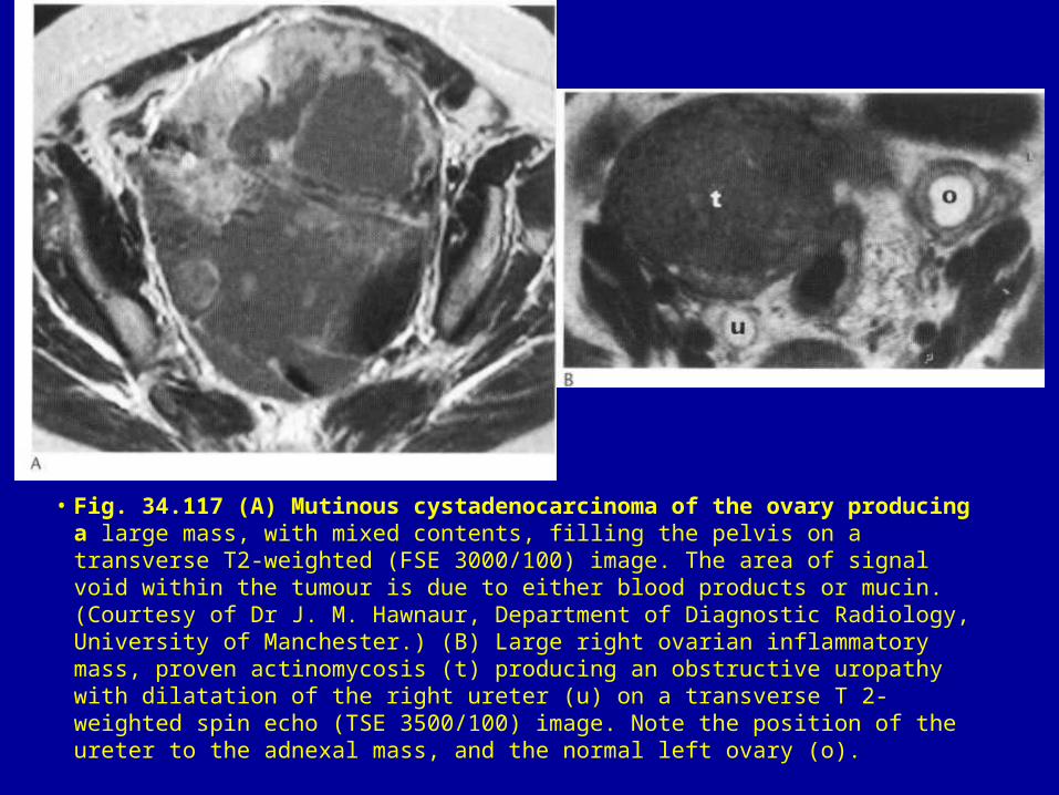
• Fig. 34.117 (A) Mutinous cystadenocarcinoma of the ovary producing a large mass, with mixed contents, filling the pelvis on a transverse T2-weighted (FSE 3000/100) image. The area of signal void within the tumour is due to either blood products or mucin. (Courtesy of Dr J. M. Hawnaur, Department of Diagnostic Radiology, University of Manchester.) (B) Large right ovarian inflammatory mass, proven actinomycosis (t) producing an obstructive uropathy with dilatation of the right ureter (u) on a transverse T 2-weighted spin echo (TSE 3500/100) image. Note the position of the ureter to the adnexal mass, and the normal left ovary (o).


