39
Electrodiagnostics Combination of Ultrasound and Electrical Currents Therapy manual

ElectrodiagnosticsCombination of Ultrasound and
Electrical Currents
Therapy manual

Copyright: Enraf-Nonius B.V. P.O. Box 12080 3004 GB ROTTERDAM The Netherlands Tel: +31 (0)10 - 20 30 600 Fax: +31 (0)10 - 20 30 699 [email protected] www.enraf-nonius.com Part number: 3486.217-42 December 2005

ElectrodiagnosticsCombination of Ultrasound and
Electrical Currents
Therapy manual
By M. van der Esch

GB
Table of contents
1 Preface ..........................................................................................................................................1 2 Introduction ..................................................................................................................................2 3 History...........................................................................................................................................3 4 Aims of electrodiagnostics .........................................................................................................5 5 Equipment and method ...............................................................................................................6 6 Aims of combined electrical current and ultrasound ..............................................................7 7 Working hypotheses....................................................................................................................8 8 Fundamental Neurophysiology ..................................................................................................9
8.1 Introduction ..........................................................................................................................9 8.2 Influence of nociception at the site of the lesion................................................................10 8.3 Influence of nociception on the posterior horn of the spinal cord......................................11 8.4 Influence of nociception on the lateral horn of the spinal cord ..........................................13
9 Types of referred pain ...............................................................................................................15 9.1 Situation I ...........................................................................................................................15 9.2 Situation II ..........................................................................................................................16 9.3 Situation III .........................................................................................................................16
10 Examination method..................................................................................................................18 10.1 Introduction.....................................................................................................................18 10.2 Setting-up the equipment ...............................................................................................19 10.3 Diagnostic/Therapeutic method......................................................................................19
11 Case.............................................................................................................................................22 11.1 Epilogue..........................................................................................................................23
12 References..................................................................................................................................31
GB

Disclaimer This therapy manual and the information it contains are the property of Enraf-Nonius B.V. (Delft, the Netherlands). Insofar as is maximally permitted under the applicable prescriptive law, neither Enraf-Nonius nor its suppliers or dealers are liable under any circumstances for any indirect, exceptional, incidental or consequential damages arising from the use of the therapy manual or from the inability to use it. Enraf-Nonius cannot be held liable for the consequences of any incorrect information furnished by its staff or for any errors in this therapy manual and/or other accompanying documentation (including trade documentation). The other party (the user of the therapy manual or her or his representative) must hold Enraf-Nonius harmless and indemnify it against any third party damages, irrespective of their nature and irrespective of the relationship with the other party. Before you start treating a patient you must be familiar with the operating procedures for each treatment and with the indications, contra-indications, warnings and precautionary measures. For additional information about he use of interferential therapy, consult other sources.

GB

1
GB
1 Preface This book is the result of a lecture I gave on behalf of Enraf-Nonius B.V. Its aim is to provide a review of the present-day working hypotheses with respect to electrodiagnostics and electrotherapy. The subjects dealt with are:
Electrodiagnostics Integration with ultrasound Setting up an examination strategy. In conclusion, electrotherapy is considered in greater detail.
The text of this book has been kept as brief as possible. Readers wishing to go further into the various subjects are referred to the textbooks currently available, and to the articles quoted. The contents are based on extensive consultation with colleagues. Developments in electrotherapy are proceeding extremely rapidly. It is therefore not unlikely that, after study of new publications in the literature and the related discussions, the text will be adapted in the course of the next few years. Finally, a word of thanks Enraf-Nonius B.V. for taking care of the lay-out. M.van der Esch Physiotherapist Manipulative therapist Employed at: Jan van Breemeninstituut, Centrum voor Reumatologie en Revalidatie, Amsterdam. Hogeschool van Amsterdam, afdeling Fysiotherapie, Amsterdam Stichting Opleiding Manuele Therapie, Eindhoven pain following stimuli which, under normal conditions, would not cause pain.

2
GB
2 Introduction A short history of electrotherapy provides a useful background for examining the concept of electro-diagnostics, and considering its consequences for therapy. The use of electrotherapy equipment by physio-therapists is extremely widespread, as can be seen by reference to the recent article by G.J.M.G. van der Heijden et al. in the Nederlandse Tijdschrift voor Fysiotherapie (Dutch Physiotherapy Magazine). Such widespread use can only be justified if it gives rise to detectable clinical effects. The aim of this text is to indicate when the application of electrical stimuli for a therapeutic purpose is justified, and how such stimuli may be applied. A crucial question in this respect is that of why patients with tissue damage and inflammation experience pain following stimuli which, under normal conditions, would not cause pain.

3
GB
3 History The use of electrical stimuli for affecting pathological processes has been known for a long time. However, the effects cannot be attributed solely to the various modalities of electrical currents: there is also a wide variety of electrode positions, although there is no clear registration of which positions are the most effective. The electrode positions according to Trabert are particularly well known, and discussions concerning the most effective positions can also be found in the TENS literature. In many cases, the 'active' electrode is placed at the position where the patient experiences pain, and the 'indifferent' electrode is placed distal to this position. However, this raises the question of whether the patient's subjective experience is really a reliable indication for positioning the electrode. Other questions include those of whether the types of current are important for the positioning of the electrodes, and whether a distal lesion in an extremity or a lesion in the spinal column can still influence the positioning of an electrode. Should the electrode be positioned in the region of the pain, or should there be diagnostic data available to decide on an electrode position for a particular current type? It is interesting to investigate what was written in the 'old' literature concerning the positions of the electrodes. As early as 1906, Dr Fritz Frankenhauser stated in his book 'Die Physiologischen Grundlagen und die Technik der Elektrotherapie' (The Physiological Basis and Technique of Electrotherapy) that the region should be investigated with the 'Reizelectrode' (Stimulating Electrode) in order to find the most sensitive point. In 1926, Dr J.C. Bom said the following in his 'Physische Therapie, Natuurgeneeswijze1 (Physical Therapy, Natural Healing Method), in the chapter 'Electrotherapie of Behandeling met Electriciteit' (Electrotherapy or Treatment with Electricity): 'In the middle of the last century, when electricity first began to be applied in medical practice, it was thought in many circles that a method had been found that would be successful in the majority of cases where other methods of treatment had failed. The enthusiasm appeared to have been too great, and the results failed to meet the high expectations that had been cherished. As so often happens in the world when things that have been highly acclaimed prove disappointing, the ardour rapidly cooled, and the general opinion was that if any benefit could still be attributed to electrical treatment, it was restricted to a sort of suggestion which, while it could lead to success in certain cases, had no real inherent value. However, this opinion was also incorrect, and the investigations of such men as Remak, Jellinek, Kowarschik et al. have shown that, provided that electrical treatment is applied with understanding, its usefulness can no longer be doubted.' Some 64 years later, this quotation is still extremely topical. Particularly the last sentence, which states that electrical treatment can be useful if applied with understanding, is highly relevant to this text. Another old book of great value is that written by Dr L. Bolk in 1910. Even at this early date his description of The Segmental Innervation of the Trunk and Limbs in Man' provided what was, for the time, an outstanding description of the innervation of various tissue structures. The study of pain and the nervous system, which began at that time within the contexts of anatomy and physiology, has now become a science in its own right. Organisations such as the IASP (International Association for the Study of Pain) have, over the last few years, produced an enormous amount of information on the various nociceptive pathways in the nervous system and the origins of pain, with the aim of discovering or optimising forms of therapy for effective application in anaesthesia, neurology and other health care sectors, including physiotherapy. The quantity of information is still increasing, as can be seen from the 30% increase in the number of abstracts at the latest IASP congress in Adelaide, Australia. From the beginning of this century, countless medical and paramedical workers (including physiotherapists) have been engaged in the application of electrical currents, and measurement of the clinical effects.

4
GB
Important examples are the books of Dr Erwin Schliephake in 1938, and Dr Josef Kowarschik in 1940, which described what were, for the time, remarkable results of treatment with high-frequency electromagnetism. However, in the Netherlands, there has never been a major scientific breakthrough in electrotherapy. For a long time, workers have been engaged in optimising the stimulus. Due to the growing knowledge in the area of the neurophysiotogy over the last years, it has become more understanding that the patient's circumstances are determining the result in the end. This is why for each electrotherapeutical application it has to be agreed upon in which way the pathological process can be influenced. To be able to choose which type of electrical current is to be used and where the electrodes have to be placed it is important to apply electrodiagnostics.

5
GB
4 Aims of electrodiagnostics In general it can be said that with the aid of electrical current "trigger points" have to be found in order to identify and influence "organ dysfunctions". Not a tot has changed since FrankhAuser's time (1907). The present-day physiotherapist is likewise looking for "trigger points" to start the therapy. Diagnosis takes place on the afferent and efferent somatic nervous system. Diagnosis on the efferent motor system is disregarded in this paper. Diagnosis in respect of the afferent nervous system relates to: the II, Ilia, Illb and IV afferents of skin, muscle and capsule structures. (Conform Ertanger and Gasser the A beta, A delta and C nerves, see Fig. 1).
Class Group Diameter (µ) Conduction velocity (m/s)
Functions / distribution
A Afferent Ia
Ib (α) II
(βγ) III (δ)
20
5-15
1-7
100
20-90
12-30
Primary endings (la fibres) Secondary endings (lb fibres) Vibration Touch Pain, temperature
A Efferent Α γ
17
50-100
Motor extrafusal muscle fibres Motor intrafusal muscle fibres
B 3 3-15 Preganglionic Autonomic C IV 0,5-1 0,5-2 Pain, temperature (?)
Group III and IV afferents are almost unmyelinated and have a low propogation speed (0.5 - 30 ms, according to Guyton, 1981). When the diameter decreases the resistance increases and thus the stimulation of the nerves is diminished. Thick myelinated nerves (group II) have low stimuli thresholds, whereas, unmyelinated and thin nerves have high stimuli thresholds under physiological circumstances. A purposive search is made for local sensitive points or areas, such as tender points, trigger points and non-sensitive peripheral cutaneous nerves. The reaction to the stimuli, often in the form of a referred sensation, provides information on the neuroretlectory localisation of the disorder and the possibility of therapy. The level of referred sensations is dependent on the strength and duration of the nocisensoric afferents and the modulation of the nocisensoric afferent of the central nerve system. During an inadequate modulation, whereby the stimuli cannot be adequately discriminated, we often find an excessive reffered sensation of pain. The sympathetic nerve system also plays an important part in the origination of reffered sensations just like during the existence of structure tissue changes in the area of referred sensations. Generally one needs to be aware of the fact that a "local problem" can be disguised in the definition of the patient due to referred pain. This is why pain is not an indication in electrotherapy treatments as it is often difficult to demonstrate a change in tissue in the region of referred pain or to demonstrate an increase in the sensitivity of the nerves.
Figure 1 Classification of nerve fibres according to Lee and Warren, 1978 (from: Walsh, D., 1991)

6
GB
5 Equipment and method The following means can be used in electrodiagnostics:
1. Galvanic currents. 2. Direct tow frequency currents such as diadynamic currents and 2-5 currents
according to Trabert. 3. Medium frequency alternating currents such as interferential currents. 4. A combination of the above together with ultrasound.
One of the earliest diagnostic methods was that of Dr Kahane with constant direct current (galvanic current). This was interrupted manually by applying the electrode to and removing it from the skin repeatedly. The result was a highly stimulated current with the purpose of establishing the most sensitive regions. The disadvantage of this method was the extremely aggressive stimulus, which made differentiation between slight and severe hyperalgesia impossible. Because of the strong reaction, it was difficult to observe reactions from the most sensitive regions. Historically, Gierlich's application is also significant. He discovered, more or less by chance, that the simultaneous application of ultrasound and diadynamic currents made hyperalgesic regions easier to detect. It was not uncommon for the hyperalgesic region to be slightly painful when stimulated by electrical current alone, and more painful when ultrasound was added.
7
GB
6 Aims of combined electrical current and ultrasound
- The combination of stimuli gives different effects from those of the stimuli applied separately. - Combined application has the advantage that irritable regions deeper than the skin can be
stimulated. - Latent irritations can be stimulated by ultrasound, enabling the nerve fibres to be
depolarised with a low current intensity.
Ultrasound is able to lightly traumatise the tissue, which can be therapeutically desirable. Following therapy with combined techniques, the therapeutic stimulus can persist for a relatively tang time at the local tissue level. A characteristic phenomenon is that the current intensity has to be continually reduced during the therapy, due to the increasing sensitivity of the nerve fibres or membranes. The combination of techniques advocated by Gierlich is characterised by:
- The use of direct currents. This makes the method somewhat aggressive, causing skin trauma by etching (seen as visible hyperaemia under the electrode).
- Limited depth effect, due to the use of direct current, although the aim is to localise both superficial and deeper-lying hyperalgesic regions.
- An unpleasant sensation when the electrical circuit is opened and closed. All the above-mentioned negative characteristics can be avoided by combining ultrasound with a medium-frequency alternating current. Modern equipment has now reached a level of development that makes this combination quite easy to use. If the hyperalgesia is slight, or tissue trauma is actually desired in order to produce neurogenous inflammation, Gieriich's method is still of value.

8
GB
7 Working hypotheses The following four hypotheses, which will be worked out in further detail, form the basis of present-day electrodiagnostics. The hypotheses have been derived from study of the literature, and from personal experience. There is, as yet, no question of there being a scientifically proven method.
1. Nociceptor stimulation can lead to neurogenous inflammation if the tissue is traumatised. The clinical effect is primary hyperalgesia within the various tissue structures affected (skin/capsule/muscle).
2. Electrodiagnostics has the aim of localising the clinically manifest primary and secondary hyperalgesia, and to elicit reactions at the spinal level (posterior and lateral horn).
3. Electrodiagnosis leads to the decision as to whether electrotherapy is indicated and, if so, which current type should be selected and in which innervation region of the body.
4. Electrodiagnostics with the aid of ultrasound has the advantage of demonstrating hyperalgesia lying deeper than the skin, even in sparsely innervated regions.
The first of these hypotheses is indicated in the literature from various disciplines. The remaining three are derived from physiotherapy.

9
GB
8 Fundamental Neurophysiology 8.1 Introduction Physiotherapists can intervene in the transmission of nociceptive stimuli in various ways. It is desirable that therapists should be able to recognise the various intervention levels. The starting point for both diagnosis and therapy is a knowledge of the neurophysiological relationships in wound-healing processes and pain. Pain is in the foreground, but is difficult to classify or to assess objectively (Vecht et al, 1990). The IASP defines pain as follows: 'Pain is an unpleasant sensory and emotional experience associated with actual or potential tissue damage, or described in terms of such damage'. Hence, pain is always subjective. Every individual learns the significance of the word 'pain1 through experience of trauma during life. It is always unpleasant, and therefore also an emotional experience. Many people complain of pain where little or no tissue damage is present. Stimuli which are, by definition, painful, allow pain to be classified as a physiological reaction (Struppler). There are also situations in which stimuli which are not normally painful can, in fact, cause pain in patients. For this reason the IASP has introduced the new term allodynia 'to refer to the pain that results from tow-intensity (ordinarily innocuous) stimuli'. Thus, allodynia is pain caused by stimuli which, under normal physiological conditions, would not cause pain. Allodynia indicates that a change has occurred in the quality of a sensation. The original modality is not painful, but the reaction to the stimulus is painful. Nociception plays a central role (Besson, 1987), as it can be used as a treatment parameter. Nociceptor activity in response to noxious stimuli need not necessarily lead to pain. However, the physiological effects associated with nociception can, to some extent, be assessed objectively. Definition of a noxious stimulus A noxious stimulus is a mechanical, chemical or thermal stimulus which causes, or is likely to cause, damage to tissue. Definition of noclceptlon Nociception is the transmission of information via the 1Mb and IV afferent nerve fibres to the posterior horn of the spinal cord, informing the nervous system of actual or threatened damage to the body, and initiating the directly associated reactions. One reaction which is of clinical value is the sensrtisation of the free nerve endings resulting, inter alia, in hyperalgesia. Inflammation of the skin and other innervated tissues is characterised by chronic pain and hyperalgesia. Hyperalgesia in the skin is classified into two forms: primary hyperalgesia, originating in the inflamed tissue, and secondary hyperalgesia occurring in the surrounding tissues (Fig. 2).
Figure 2 The triple response of Lewis

10
GB
It is not known whether these two forms of hyperalgesia also occur in other organs (Handwerker et al, 1991). However, Head's 'zones' can probably be interpreted as a special type of secondary hyperalgesia, related to chronic nociception occurring elsewhere. The difference between these two forms of hyperalgesia is of interest for understanding the underlying neurogenous mechanisms, as the primary form can be attributed to changes in the nociception itself, while the secondary form probably originates in the central nervous system. Thus, hyperalgesia is divided into a primary and a secondary form. Primary hyperalgesia is a local increase in the sense of pain at the site of the tissue damage (and is also referred to as local hyperalgia). This increased sensitivity is due, in part, to the sensitising effect of prostaglandin E2 on the non-myelinated free nerve endings, and the sensitising effect of substance P on the peripheral free nerve endings. Secondary hyperalgesia is a time- and location-dependent increase in pain sensitivity arising from the posterior horn of the spinal cord. It does not occur at the site of the tissue damage, but is a consequence of the damage. Ruch described secondary hyperalgesia in 1965, and stated that this form of hyperalgesia was only present for a few hours following mechanical stimulation of a physiologically normal skin. In practice, it appears possible that secondary hyperalgesia can persist for much longer. No conclusive explanation is known, but it seems reasonable to assume that mechanisms in the central nervous system are responsible. In 1989, M.A. Cline et al. defined hyperalgesia as follows: 'towered threshold for pain sensitivity and increased pain magnitude of suprathreshold stimuli1. In diagnostics, including electrodiagnostics, it is of considerable importance to establish the site or sites of allodynia and hyperalgesia. These sites provide information on the affected innervation level. At the same time, the extent of the affected area provides a standard for assessing the acuteness of the clinical problem. Hyperalgesia is also accompanied by the classical signs of redness, swelling, increased temperature and impairment of tissue function. These signs can be assessed objectively and can, at a later stage, serve to indicate the effectiveness of the selected treatment. In 1991, Clifford J. Woolf summarised the pathological aspects of clinical pain as follows: 'There are at least four aspects of clinical pain that are pathological in the sense that they are not present in the absence of tissue damage or inflammation. These are a reduction in the threshold necessary to elicit pain (Allodynia), an increase in the response to noxious stimuli (Hyperalgesia), a prolongation in the response to a transient stimulus (Persistant Pain) and a spatial spread of the pain to uninjured tissue (Referred pain)'. The following three sections deal with the influence of nociception at the site of the lesion, in the posterior horn of the spinal cord, and in the lateral horn. The influence of nociception on the anterior (motor) horn of the spinal cord, and its influence on the central pain modulating systems, are beyond the scope of the present text. 8.2 Influence of nociception at the site of the lesion A tissue lesion leads to activity in the sensory receptors, signalling the damage. The two most common types of receptor are the myelinated mechano- and thermoreceptors, and the non-myelinated polymodal nociceptors. In 1991, R. Dubner demonstrated that both types of receptor are responsible for primary hyperalgesia in slight skin burns. Tachykinins are released in the region of the nerve endings, which lead to sensitisatton of the mechano receptors (Fig. 3).

11
GB
There is simultaneous sensitisation of the neurons of the posterior horn, as well as vasodilation and an increase in the permeability of the blood vessel walls. Inflammation is initiated, which assists in the recovery of the damaged tissue (wound healing). This inflammation has been described in the literature by Levine and others as neurogenous inflammation, as the inflammation is initiated via the nervous system. The clinical signs are increased temperature, swelling, impairment of function and pain. Pain provides information on the lesion, and the appropriate emotional response. However, many lesions are not consciously perceived, and pain as such will not be experienced. In such cases, the resulting nociception will be limited to informing the central nervous system, and initiating the necessary reactions for recovery, without involvement of the consciousness. When the region of the lesion is palpated, there is a fairly rapid nociceptive response in the form of reddening (triple response of Lewis), and the stimulus is often experienced as painful. The stimulus required is gentle and would not, under normal physiological conditions, lead to nociception. 8.3 Influence of nociception on the posterior horn of the spinal cord Nociception leads to sensitisation of the posterior hom of the spinal cord. If the nociception is of long duration, the sensitisation is not restricted to the neurons of the nerves from the damaged area, but also affects the neighbouring neurons. The action potentials of nerve fibres from totally different tissue structures which, however, belong to the same innervation level, depolarise their 'own' neurons more rapidly. The polymodal nociceptors have large receptive fields. When injury occurs, substance P is released in other areas as well as the damaged area. A sufficiently strong stimulus ('supra-threshoid noxious stimulus') can then lead to the 'triple response' of Lewis. This can be regarded as secondary hyperalgesia. This effect is of clinical importance, as it generally occurs at the sites of tissue transitions and/or the neurovascular hilus. Clinically, this is secondary hyperalgesia, as the cause was not primarily in this tissue. This hyperalgesia is only manifest when an applied stimulus is sufficient to initiate nociception. This is a different situation from that at the site of the lesion, where the nociceptors are already sensitised, and even a mild stimulus will lead to depoiarisation (area of primary hyperalgesia).
Fig. 3 Neurogenous inflammation (from M. Zimmermann, 1989)

12
GB
The sensitisation of the posterior horn of the spinal cord results in a reduction in the differentiating capacity of the nervous system. If a stimulus is applied to the site of the lesion, the nervous system can also experience sensation at sites in other tissue structures belonging to the same innervation level. No clinical symptoms can be observed in the tissue structures themselves. This sensation is known as 'referred pain', and indicates that there is an increase in the area of nociceptive activity. The extent of the increase is indicative of the severity of the problem. Initially, the referred pain will occur in the innervation region of the stimulated area. Later, sensations are felt which no longer belong to the innervation area, and the extent increases. The increase occurs in the sympathetic and autonomic nervous system. Definition of referred pain Referred pain is pain projected in or to a richly innervated somatic structure (e.g. skin) at some distance from and generally more superficial than the site of the tissue damage causing the original nociceptor activity (adapted from Ruch). In the case of chronic referred pain, there is a possibility of hyperalgesia in the area where the pain is felt. The phenomenon is known as 'referred pain' in English, and the corresponding term 'ubertragende Schmerz' (Hansen and Schliak, 1962) appears in the German literature. In Dutch, it is known as 'weerpijn' (Voorhoeve), or by the unfortunately chosen term 'uitstralende pijn' - i.e. radiating pain. As long ago as 1909, Mackenzie gave a classic interpretation of referred pain, based on earlier observations by Sturge (1883) and Ross (1887). This interpretation still represents a fundamental concept. According to Mackenzie's interpretation, sensory stimuli from the viscera cause an 'irritable focus' in the afferent segment of the spinal cord. This facilitates afferent impulses from the skin, causing real skin-pain: hyperalgesia and referred pain as a consequence of the irritable focus. This interpretation is known as Mackenzie's convergence-facilitation theory (Ruch, 1965, Fig. 4). In 1960, Ruch proposed a somewhat modified hypothesis. Visceral afferent nerves that cause pain converge with the skin afferents, and end at the same neurons. After processing in the posterior horn, a signal is transmitted to the brain. This is interpreted as originating from the skin, which is the most sensitive structure. The interpretation is based on experience of stimulation of the same skin afferents. This convergence-projection theory can explain the apparent location of the pain, but not the hyperalgesia, according to the dermatomic distribution (Fig. 5).
Many experiments support these two hypotheses by demonstrating that the nociceptive afferents from the viscera and the skin converge at the same level in the spinal cord, the brain stem, the thalamus and the cortex (for a review, see Procacci P. et al., 1986). The convergence of the nociceptive afferents in the nervous system can explain a number of clinical symptoms, including referred pain without hyperalgesia. The presence of hyperalgesia is more difficult to explain and, according to Wolff (in Procacci P. et al, 1986), is characterised by local changes in the tissue. In conclusion, it can be stated that referred pain can be caused by two mechanisms which largely overlap. No structural changes are found in the region of the referred pain. This means that referred pain is purely a perception of pain, with no obvious changes in the tissue structure of the region where the pain is felt.
Fig. 4 The facilitation theory (diagram compiled by R. Hoogland)
Fig. 5 The convergence theory (diagram compiled by R. Hoogland)

13
GB
Clinically, it appears that sensations other than pain are also possible, such as burning or sharp sensations. The two hypotheses mentioned above have led to the formulation of working hypotheses. The general starting point is the assumption that referred pain can only occur via the agency of the central nervous system. Particularly systems descending from the PHG, and the limbic system, can have a strongly modulating effect on the degree of selectivity of the posterior horn neurons. While in the past referred pain was only used to explain the pain in visceral disorders, it is now generally accepted that referred pain can be caused by nociception in the motor apparatus. The site of the referred pain, and the tissues or organs that are most likely to give rise to referred pain, are determined by differences in the density of innervation of the structures and the somatotopic organisation within the posterior horn of the spinal cord. The term innervation density is used to refer to the frequency of occurrence of peripheral receptors in the tissue or organ. In general, it can be stated that the innervation density of acral and superficial structures is greater than that of axial and deep-lying structures. This is valid for the organs themselves, and for the various tissues within a particular organ. In practice it is important that both hypotheses indicate that the site of the referred pain belongs to the same innervation level as the structure from which the chronic nociception originates. If the extent of a lesion is known, as well as the innervation density of the structure (which is directly related to the degree of nociceptor activity), it is possible to estimate the severity of the referred pain. In addition, the extent of the region of referred pain provides information on the modulating role of the nervous system. In the case of non-specific arousal and tow selectivity of the nervous system, it is possible that a small lesion with little nociception will nevertheless give rise to definite referred pain. This is particularly so if the lesion is in a proximal articular capsule with low innervation density and a small nociceptive receptor field in the posterior horn, as in the case of a lumbar facet joint. 8.4 Influence of nociception on the lateral horn of the spinal cord The lateral horn of the spinal cord contains the neurons of the sympathetic nervous system at levels C8 to L2. The regions above and below these levels have no neurons in the lateral horn, and are innervated from C8 to L2. These neurons are highly specialised (Janig), and nine types of neurons with different functions have been identified to date (Fig. 6).
These neurons are under the influence of various integrative control levels, such as that of the hypothalamus (Swett et al, 1984). The influence of nociception on the sympathetic nervous system must therefore be considered in this light. Neuron activity due to nociception is partly determined by controlling systems, such as the hypothalamus. However, every nociceptive stimulus, whether from a visceral or a somatic source, will influence the sympathetic nervous system. The reactions of this system are clearly linked with the reactions of the affective/emotional system (Schmidt).
Figure 6 Subsystems within the sympathetic nervous system (from R.A.B. Oostendorp, Thesis, 1988)

14
GB
Generally speaking, the reaction of the sympathetic nervous system to nociception can be regarded as non-specific, although reference is made to a 'defence reaction1 which is under the control of the limbic system, particularly the nucleus amygdala. However, the sympathetic nervous system can also react very specifically to nociceptive stimuli from somatic and visceral structures. Specific reflexes from lateral horn neurons innervating the striped muscles and the skin have been fairly closely investigated in animals and in man. Nociception activates neurons which cause vasoconstriction in the musculature, while neurons which would cause vasoconstriction in the skin are inhibited (Loven reflex). The consequence is vasodilation in the skin coupled with vasoconstriction in the muscles. This situation can arise in the case of short-duration nociception and a physiologically active nervous system. However, if the nociception is of long duration, i.e. a matter of weeks or months, the trophism of the skin can be disturbed, depending on the degree of nociception and the adaptation of the nervous system. Under pathophysiological conditions, trophic changes will occur in the long term in both muscle and skin. These changes have consequences for the matrix of the collagen in these two structures. In general, the collagen becomes stiff and hard. Eventually, its tensile strength diminishes, and it is easily traumatised under load. In the region of the mechanoceptors, there are relatively many blood vessels. These receptors become sensitised by the trophic disturbance. Due to consequences of nociception that have been described elsewhere, it appears the receptors are also sensitised by changes in the axon plasma. The increased sensitivity to pressure will mainly occur at those sites that are most densely innervated, such as the wrists and hands, and the points where nerves pass through the muscle fasciae. The sympathetic nervous system also innervates the neurons of the spinal cord itself. These, too, become sensitised by the trophic disturbance. Under normal physiological conditions, the neuron circuits in the lateral horn are relatively well separated from each other. For example, the viscera and the thoracic somatic structures are innervated by medially situated neurons, while more peripheral structures such as the lumbar and cervical spinal column, and the head, are innervated by neurons with a more lateral position in the lateral horn. It is beyond the scope of this text to provide a detailed description of the various routes of the sympathetic efferent nerves. It is sufficient to note that the thoracic and, above all, medially situated neurons form the autochthonous innervation level, while those situated more laterally in the lateral hom form the peripheral innervation level for the motor apparatus (extremities, lumbar spine, pelvis, cervical spine and the head). Nociception from the periphery, such as the shoulder innervation at C5, will have to pass down to the lateral group of neurons at the level of T5. The sympathetic efferents from T5 will then have to pass up to the C5 level in order to innervate the organs concerned.

15
GB
9 Types of referred pain Patients may experience other sensations than pain in response to stimuli, particularly during the clinical examination. Such sensations are known as referred sensations, and can vary from slight prickling sensations to tingling, 'electric' sensations, a feeling like ants crawling over the skin, a feeling of pressure or tension in the region of a joint and, finally, actual pain. This pain is then fully comparable with the pain the patient originally presented. Referred pain is therefore regarded as one form of referred sensation. Patients sometimes mention referred sensations when the anamnesis is compiled, but generally such sensations only appear during the examination. Particularly during palpation of hyperalgesic areas, the patient experiences a feeling of pressure, the musculature reacts with a local contraction (a twitch), redness appears and referred sensations occur. Apparently, four types of referred pain (sensation) can occur. The differences are somewhat vague, particularly in practice, as the central nervous system reacts as a totality. The appearance is strongly dependent on the selectivity within the central nervous system and, in fact, the sympathetic nervous system is always involved. This need not necessarily lead to clinical symptoms, provided that the neurogenous inflammation is kept under control. The four types (according to Hoogland, 1989) are: 1. Referred pain (sensation) in the innervation region of the autonomic nervous system. 2. Referred pain (sensation) in the autochthonous innervation region of the sympathetic
nervous system. 3. Referred pain (sensation) in the peripheral innervation region of the sympathetic nervous system. 4. Combinations of the foregoing three forms of referred pain (sensation). Distinguishing between the various types is of essential importance for diagnosis. The examination results can lead to the most accurate possible selection of the therapy parameters. In a lesion of the extremities, the sequence of occurrence is usually for Type I to Type III, and then to Type II. Three situations are presented below, in order to clarify the various aspects. 9.1 Situation I If tissue damage occurs in an extremity, a neurogenous inflammation is initiated at the site of the lesion, which starts the healing process. Primary hyperalgesia appears at the site of the lesion and, under normal physiological conditions, it can be expected that healing will occur within the normal time limits (De Morree). Nociception from the damaged structure will sensitise the corresponding posterior horn neurons of the spinal cord (e.g. level L5-S1). If nociception persists for longer than is physiologically normal, convergence and facilitation will result sensitisation of the whole neuron group at the same innervation level (L5-S1). Referred pain (sensation) of Type I can be expected. If mild electrical stimulation is applied to the nociceptors in the damaged region, the referred sensation may be felt elsewhere in the same innervation level, particularly in the richly innervated regions. However, there will be no objectively detectable clinical symptoms in these regions. It should, once again, be emphasised that stimulation of superficial structures in the peripheral part of an extremity cannot be expected to cause referred sensations. If the sympathetic nervous system is involved in a physiologically normal way in the healing process, no pathological symptoms will be encountered. There is a physiologically normal trophic reaction. No pathological symptoms will be found at the autochthonous (i.e. thoracic) innervation level. If nociception continues, there is a possibility of symptoms occurring elsewhere. The medial column neurons of the sympathetic nervous system will be increasingly activated by the continuing nociception. The innervation region of these neurons will start to show symptoms of the increased activity. Eventually, the trophism will be disturbed. The skin in the peripheral innervation region of the sympathetic nervous system will be pale, the musculature will be hypertonic, and the joints will be stiff. Symptoms of hyperalgesia in these regions can only be expected when the weakened tissue is actually damaged.

16
GB
With increasing and long-duration nociception, and decreased selectivity of the nervous system, Type III referred pain (sensation) can be expected in the region. In the long term, regions of hyperalgesia will be found, due to the sensitisation of the mechanoceptors and the occurrence of microlesions. Stimulation of these regions will result in referred sensation at the site of the primary injury, or in regions with a high innervation density. The strength of the tissue will be reduced under the influence of the trophic disturbance, making it more easily traumatised. If trauma occurs, there will be new neurogenous inflammation at the site of the lesion, and the region will show primary hyperalgesia. In practice, this means that two traumatised regions can be found. 9.2 Situation II The primary problem has always been in the motor apparatus, particularly in the periphery such as the extremities, the cervical spine or the lumbar spine/pelvis. Symptoms can slowly start to appear at the autochthonous level of the sympathetic nervous system due to extension of the neuron activity from the lateral to the medial column. These symptoms will mainly be found in the connective tissue of the musculature, articular capsule, ligaments and skin. The main component of these symptoms is trophic disturbance. The skin is somewhat pale, the musculature rather stiff, and the joints at the thoracic level will show an increased resistance to movement. On palpation, there will be a fast 'Defence Contraction' (twitch) due to the sensitisation of the mechanoceptors. This can also be demonstrated by the application of electrical current. In practice, reference is made to Tender Points or Trigger Points (Simons 1988), although at this time these are only symptoms of (secondary) hyperalgesia. The term Trigger Point indicates that stimulation of these points will result in a reaction elsewhere. Depending on the selectivity of the central nervous system, there will be a referred sensation at the site of the primary lesion or, at least, in the direction of the lesion. Depending on the autochthonous innervation level of the sympathetic nervous system, there is a chance of a Type II referred sensation. At a Tender Point, there is secondary hyperalgesia, but there will be no referred sensation if the applied stimulus is not nociceptive. 9.3 Situation III In this case, we consider the symptoms that can be found if the source of nociception is primarily situated at the autochthonous level of the sympathetic nervous system, e.g. at the C8-L2 level of the spine or in a visceral organ. If the visceral organ has nociceptive afferents to the nervous system, the primary hyperalgesia will be in the region of the organ itself. In other words, the organ will be subject to pressure pain, or will be sensitive to traction due to filling (Gebhart and Ness, 1991). Secondary hyperalgesia can be found in the region of thoracic somatic structures. If the nociception is of long duration, this may be accompanied by referred pain (Type II). If there is simultaneous trauma in part of the motor apparatus, the consequences will be far more serious than they would be under normal physiological conditions, due to the convergence of nociceptive afferents and facilitation of the neurons of the posterior horn of the spinal cord. If nociception from the visceral organ persists for a long time, all the medial column neurons of the sympathetic nervous system will be sensitised. The trophism of the somatic structures will be disturbed. New hyperalgesic regions may be found, due to damage to weakened structures in the autochthonous innervation level of the sympathetic nervous system. Stimulation of the hyperalgesic areas can cause referred sensation (Type II) in richly innervated regions, particularly the skin, but little or none in the visceral organs. To make matters more complex, it is also possible for the hyperactivity of the neurons in the medial column of the lateral horn to spread to those in the lateral column. Symptoms of hyperactivity in the sympathetic nervous system can also appear in the peripheral innervation region of the sympathetic nervous system (combination of Type II and Type III referred sensation and, probably, Type I). In diagnostics, the anamnesis is of fundamental importance. An effort must be made to discover the site of the primary lesion, and much time there has been for it to spread. The degree of selectivity of the central nervous system should be estimated, and taken into account.

17
GB
This type of diagnosis is not simple. In practice, there is always a combination of factors. Owing to the complexity of the symptoms, it is important to be aware of the relationships of the various types of referred sensation. A thorough knowledge of the segmental innervation levels is indispensable. It is also essential to be acquainted with the reaction patterns of the central nervous system.
18
GB
10 Examination method 10.1Introduction In the physiotherapy practice, patients often present complaints of pain which are, in fact, due to referred pain. The anamnesis can give an indication of the degree of the disorder, and the innervation levels which may be involved. In the subsequent examination, it is necessary to localise the region from which the nociception originates, and to observe the reactions to stimulation. In the examination, the dorsal part of thorax is of diagnostic importance. The dorsal part of the trunk, in the region of the spine, is innervated by the dorsal rami of the spinal nerves. The dorsal rami split into medial and lateral branches. These provide the innervation of the skin, the fasciae, the musculature, the bone structures and the joints. This innervation comprises both the afferent/efferent autonomic innervation as well as the afferent sympathetic innervation. The dorsal rarnus of a spinal nerve is a monosegmental nerve. In the course of the dorsal ramus, various collaterals branch off, which are named according to the tissue structures they serve: cutaneous branch, muscular branch and articular branch. The articular branches innervate the articular capsules of the intervertebral joints at the cervical, thoracic and lumbar levels. The intervertebral joints are not monosegmentally innervated. The articular branches of several dorsal rami innervate one intervertebral joint. This means that referred pain originating from nociception in one intervertebral articular capsule can spread over several innervation levels.
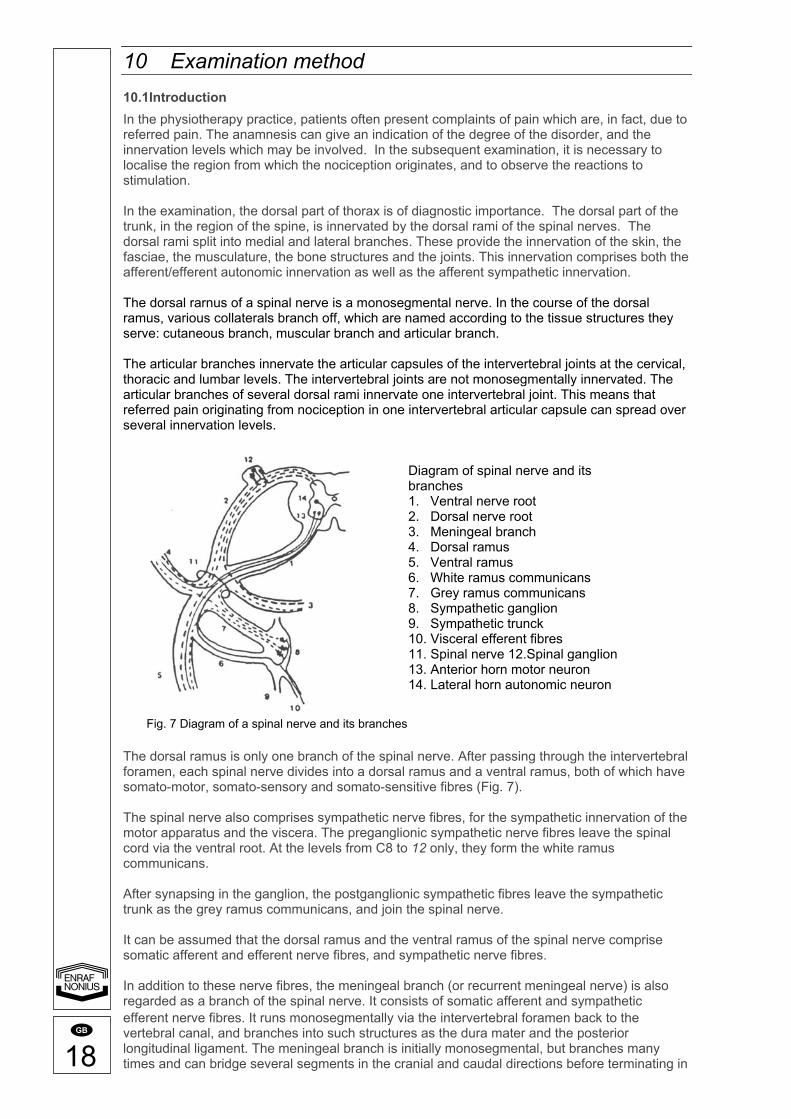
The dorsal ramus is only one branch of the spinal nerve. After passing through the intervertebral foramen, each spinal nerve divides into a dorsal ramus and a ventral ramus, both of which have somato-motor, somato-sensory and somato-sensitive fibres (Fig. 7). The spinal nerve also comprises sympathetic nerve fibres, for the sympathetic innervation of the motor apparatus and the viscera. The preganglionic sympathetic nerve fibres leave the spinal cord via the ventral root. At the levels from C8 to 12 only, they form the white ramus communicans. After synapsing in the ganglion, the postganglionic sympathetic fibres leave the sympathetic trunk as the grey ramus communicans, and join the spinal nerve. It can be assumed that the dorsal ramus and the ventral ramus of the spinal nerve comprise somatic afferent and efferent nerve fibres, and sympathetic nerve fibres. In addition to these nerve fibres, the meningeal branch (or recurrent meningeal nerve) is also regarded as a branch of the spinal nerve. It consists of somatic afferent and sympathetic efferent nerve fibres. It runs monosegmentally via the intervertebral foramen back to the vertebral canal, and branches into such structures as the dura mater and the posterior longitudinal ligament. The meningeal branch is initially monosegmental, but branches many times and can bridge several segments in the cranial and caudal directions before terminating in
Diagram of spinal nerve and its branches 1. Ventral nerve root 2. Dorsal nerve root 3. Meningeal branch 4. Dorsal ramus 5. Ventral ramus 6. White ramus communicans 7. Grey ramus communicans 8. Sympathetic ganglion 9. Sympathetic trunck 10. Visceral efferent fibres 11. Spinal nerve 12.Spinal ganglion 13. Anterior horn motor neuron 14. Lateral horn autonomic neuron
Fig. 7 Diagram of a spinal nerve and its branches

19
GB
free nerve endings. Stimulation of nociceptors in the dura mater thus leads to referred pain over several segments. The ventral rami of the cervical, lumbar and sacral spinal nerves join at various levels to form plexuses. Peripheral nerves arises from these plexuses, comprising nerve fibres from several spinal nerves. Thus, the skin, muscles and joints of the extremities are innervated from various levels of the spinal cord. This means that the peripheral innervation of the skin, skeletal muscles and joints always has a multisegmental character (Fig. 8). From the foregoing it is apparent that practically all structures of the motor apparatus have a sympathetic innervation from the spinal nerves, sometimes directly from the ganglion, and sometimes from the dorsal ramus or the meningeal branch. The intervertebral, costovertebral and costotransverse joints are innervated in this way, as well as the intrinsic muscles and the dorsal skin regions. Due to chronic hyperactivity of the sympathetic nerve system a continuous vasoconstriction occurs in the blood vessels which influences the trophic of the tissues. According to Korr's findings, long term hyperactivity of the sympathetic system ultimately leads to a vasoconstriction in the target tissues. This mostly manifests in the spinal and paraspinal region, in other words in the innervation region of the rami dorsalis medialis and lateralis. Quote: Korr 1978
1. Long term hyperactivity of particular sympathetic pathways is deleterious to the target tissues.
2. Clinical manifestations are determined by the organs or tissues which are innervated by the hyperactive sympathetic neurons, each responding in their own way, even to the sympathetically induced vasoconstriction that may be a common factor.
3. The high impulse traffic in selected sympathetic pathway may be related to musculo/skeletal dysfunction, especially in the spinal and paraspinal area.
In view of the fact that it is never possible to assess absolutely whether there is primary or secondary hyperalgesia at the thoracic innervation level, it is wise to start electrodiagnosis with a mild stimulus. A medium-frequency alternating current as interferential therapy would appear to be highly suitable. In the case of primary hyperalgesia this will certainly lead to discharges of the IV afferents with all the reactions entailed. An area with secondary hyperalgesia will only discharge if nociceptive stimuli such as polarised current are used. Since the examination is not restricted to the skin, but also involves deeper-seated structures, it is useful to add an ultrasound stimulus. In practice, the cause of the discharge and time variation is not always clear. This is why, from the practical point of view, we opt for an examination that starts thoracally and is later extended to the periphery. 10.1 Setting-up the equipment Ultrasound; a fixed intensity of 0.5 W/cm2 continuous. Medium frequency alternating current; AMF 100 Hz bipolar. Polarised current;
- Diadynamic current, DF - 2-5 current according to Trabert.
Time on maximum, depending on the duration of the examination. 10.2 Diagnostic/Therapeutic method The examination starts with a relaxed ventral position. An "indifferent" electrode is placed under the abdomen. The head is turned towards the therapist so that communication is possible. The ultrasound treatment head is the active electrode. Initially, the current is turned up such that the patient has a slight sensation of current. If it is suspected that considerable and severe hyperalgesia will be found, start outside the expected affected area. The ultrasound is then set at 0.5 W/cm2 continuous. At the level of the process! spinosus an examination is carried out into hyperalgesia and referred sensations, working caudally to cranially and back. This is the innervation area of the ramus dorsalis medialis. After this, move somewhat more laterally in the same way until the border with the ramus ventralis area is reached. A sort of "map" is made of the areas of irritation manifested, in order words which innervation regions are affected, which displayed a referred sensation and where the referred sensation was felt. Lengthy stimulation is often necessary to provoke these referred sensations; the phenomenon

20
GB
only occurs after several seconds to minutes. The ultrasound head should therefore not be moved. In the brochure Ultrasound Therapy by R. Hoogland it is explained why it is essential to keep the ultrasound head in continuous motion. In this case, therefore, the ultrasound energy will have to be reduced while the ultrasound head is held still on the area of hyperalgesia. The referred sensation is induced by increasing the current intensity. The ultrasound has served solely to localise the hyperalgesia. Follow-up: On the basis of the data, an analysis is made of the affected innervation level and the structures possibly involved. The examination can be optimised by means of anamnesis and manual examination. It is also possible that the analysis itself is enough to enable a decision to be made as to whether electrotherapy is indicated or whether the electrodiagnostic examination must be extended. Extension is possible:
1. In the same region with a polarised current, since there is probably secondary hyperalgesia and the tissues will have to be mildly traumatised. Combination with ultrasound is again possible to reach deeper-seated tissue.
2. To peripherally located innervation areas, such as the rami dorsales C1-C8 and L2- S2, with a medium frequency alternating current or directly with a polarised current
in combination with ultrasound. The choice is determined by whether there is primary or secondary hyperalgesia.
3. To the innervation area of the rami ventralis, such as in the extremities. As well as localising the hyperalgesic points or areas, it is important that a referred sensation, probably in the sense of pain, is actually induced. This demonstrates the dysfunction of the CNS (Central Nerve System).
Confirmation of the electrotherapy is thereby indicated. Efforts are then made to reduce the dysfunction along the following lines:
1. Medium frequency alternating currents are extremely good at blocking the columna dorsalis neurons. By blocking, and hence the reduction of the quantity of nociception in the spinothalamic system and to the columna lateralis, there is probably a chance of healing from within the body.
2. Ultrasound is capable of mildly traumatising the tissue, in other words nociception will certainly be caused. If this nociception will reduce the healing process, ultrasound should not be used. If mild traumatisation is actually desirable and the CMS interprets the increase in nociception as such, there is an indication.
3. Polarised currents will certainly cause nociception. Recognisable from the hyperaemia under the electrodes after current has been applied for a few minutes. If the CNS is capable of processing this nociception and the CNS is even "woken up", a polarised current is again indicated.
In concrete terms this means:
1. In an area of primary hyperalgesia, an alternating current is often used to impede the great quantity of nociception. Or in a similar area in which no change in the stimulus of the afferents is noticeable.
2. In an area of secondary hyperalgesia, a polarised current is generally needed to traumatise the tissue slightly. This trauma is a stimulus to healing within the same innervation level. The same applies to ultrasound.
Treatment: In principle, the effects of a therapy can be seen immediately after the treatment, for example in the sense of a decrease in muscle tone or increased mobility in a joint. In the first instance, we look for detectable phenomena. Finally, it is possible that there will be immediate subjective changes, such as a reduction in the pain experienced. The extent and the duration of these phenomena determine the frequency and the number of treatments given, and hence also the prognosis. If no referred sensations can be induced, and hence no symptoms either, further treatment is not advisable. At the most, the trigger points can be reconsidered and the diagnostic process started again. It is often found that very clear symptoms occur, however, without an immediate reduction of pain. This phenomenon is not in itself negative and in the long term prognosis is good. Immediate normalisation of the tone of the orthosympathetic nervous system and obvious changes in the motor system, as a result of which improvements in function are immediately visable, point to a good prognosis in the short term.

21
GB
The effects of the treatment thus determine the frequency of the treatment. If an effect only lasts for a few hours, the treatment will have to be given two or three times a day. The duration of a treatment is determined by the degree and the extension of referred sensations. The minimum length of a treatment should be in the order of 5 to 10 minutes. The maximum length depends on the decrease in the referred sensations and is usually reckoned to be half an hour.

22
GB
11 Case A 22 year old man indicates pain complaints in the right buttock and the dorsal side of the thigh. This pain occurs when running, going up stairs and jumping. Especially after sport activities pain occurs when leaning over with straight legs. At the same time stiffness arises in the tower back. After a couple of hours this stiffness and pain make it very difficult to get up. This complaint began during a training session for soccer, about six months ago. After kicking a ball with the right foot the patient suddenly felt a clear, sharp pain in his right buttock approximately at the tuber isciadicum position. Clear pain complaints occurred during a week and, although, the pain reduced it was still felt after a longer period. The General Practitioner diagnosed an irritation of the hamstrings connection at the tuber isciadicum position and sent the patient to a physiotherapist. An X-ray examination of the lumbal spinal column did not give any particularities. Approximately 18 months ago the right anterior cruciate ligament was strained. This lesion was treated conservatively for the period of 6 weeks, by means of a brace. During heavy strain the patient experienced a tired/heavy feeling in the right leg and there appears to be a light instability in the right knee. During examination at the inspection phase a flat lumbal spinal column is diagnosed with a clear thoracal kyphoses. The contours of the right hamstrings are marked with respect to those on the left, as are the monoarticular and the medial muscle belly of the m. gastrocnemius. When moving the right buttock turns slightly backwards when the right hip is extended. Compensatory muscular activities are distinguished when the patient is having difficulties to lean on the right leg only. This exercise is impossible when the eyes are closed. When passively examined the right sacro-iliacal joint is found stiff and painful when pressed. The right "Vorlauf test" is positive. The thoracolumbar transition is limited in flexion and extension. During a length test the muscles of the right hamstrings and the m. iliopsoas appear to be stiff compared to those on the left. When the pressure is sustained the muscles lengthen. When tightening the right hamstrings in prone position with flexion over the knee cramp develops. Power loss does not happen, but the attachment of the hamstrings is painful during the tightening, which also arises during palpation. Palpation also produces the skin to be stiff in the innervation area of L5-S1 and when it is handled more roughly it is slightly painful. The fast occurring redness is remarkable. The same is found at the position of the processi spinosus TH12 and L1. During the fascia gluteal and the hamstrings "string-like" structures can be palpated. Towards the lumbosacral and thoracolumbar transition the m. erectors spinae lumbalis is hypertonia and pressure sensitive. After this examination electrodiagnostic analysis with a medium frequent alternating current in combination with ultrasound was carried out. Near the lumbosacral spinal column transition, in the abdomen part of the hamstrings and near the fascia gluteal painful areas were localised. These areas felt dull and the patient felt the current in an area around the electrode (ultrasound treatment head). After this an examination near the thoracolumbar spinal column was carried out with diadynamic current and ultrasound. Both near the twelfth thoracal vertebra and on the lateral side of the right crista iliaci a burning pain sensation occurred. The ultrasound was turned off and diadynamic current was applied on these areas. After approximately two minutes the patient felt a dull, tickling sensation deep in the right buttock. When this sensation disappeared the patient's entire right leg felt tired/heavy.

23
GB
11.1 Epilogue The purpose of this case is mainly to answer the following questions:
1. Does the healing process comply with the physiological wound healing rules? 2. Give a kinesiologic explanation for the "divergent" course. 3. Where can the changes in the above mentioned muscles be described to? 4. Is it useful to stretch the muscles? 5. Give an explanation for the painful areas near the thoracolumbar spinal column. 6. Is there question of referred sensation during the electrodiagnostic examination? 7. Is it possible that manipulation of the right sacroiiiacai joint causes a centra-indication? 8. In view of the examination data, do you think electrotherapy offers an indication for the
disorder? 9. With which examination technique do you wish to follow up next? 10. How would you treat this patient based on the examination data of the case?
Please follow with a therapy description. This case and questions form an example and are limited. Yet there appears to be sufficient material to have a significant discussion.

24
GB
Nerves of Back
Fig. 8 Cutaneous innervation of the various regions of the body

25
GB
Suboccipital Traingle
Fig. 8 Cutaneous innervation of the various regions of the body

26
GB
Cutaneous nerves and superficial veins of shoulder and arm
Fig. 8 Cutaneous innervation of the various regions of the body

27
GB
Cutaneous nerves and superficial veins of forearm
Fig. 8 Cutaneous innervation of the various regions of the body

28
GB
Wrist and hand: superficial dissection (dorsal view)
Figure 8 Cutaneous innervation of the various regions of the body

29
GB
Superficial veins and cutaneous nerves of lower limb
Fig. 8 Cutaneous innervation of the various regions of the body

30
GB
Figure 8 Cutaneous innervation of the various regions of the body
Fig. 9 An example of hyperalgesic regions, localised by ultrasound and electrical current
31
GB
12 References
1. Besson J.M. and Chaouch. A.; Peripheral and spinal mechanisms of nociception. Physiological reviews Vol. 67, No. 1 January 1987.
2. Bolk L; De segmentale innervatie van romp en ledematen bij den mensch. 1910.
Uitgeverij De Erven F. Bohn Haarlem. 3. Bom J.C.;
Physische therapie, Natuurgeneeswijze. 1926. uitg. La Riviere en Voorhoeve – Zwolle.
4. Cline M.A. et al; Chronic hyperalgia and skin warming. Caused by sensitized C nociceptors. Brain 1989, 112: 621-647.
5. Dubner R.; Neuronal plasticity and pain following peripheral tissue inflammation or nerve injury. Chapter 30. Proceedings of the Vlth World Congress on Pain. Elsevier Science Publishers BV, 1991.
6. Frankenhauser, F.; Die physiologischen Grundlagen und die Technik der Elektrotherapie. Enke Veriag, 1906.
7. Gebhart G.F. and Ness T.J.; Mechanisms of visceral pain. Chapter 42. Proceedings of the Vlth World Congress on Pain. Elsevier Science Publishers BV, 1991.
8. Gierlich K. und Jung A.; Die kombinierte Anwendung von Ultrashall und Reizstrome Physik. Medizin und Reabil. Heft 9. 1968.
9. Handwerker H.O. en Reek P.W.; Pain and Inflammation. Chapter 7. Proceedings of the Vlth World Congress on Pain. Elsevier Science Publishers BV, 1991.
10. Hansen K. und Schliack H.; Segmentale Innervation; Ihre bedeutung fur Klinik und Praxis. Thieme Veriag, Stuttgart 1962.
11. HeadH.; On disturbance of sensation with especial reference to the pain of visceral disease. Brain 1893; 16: 1-132.
12. Heijden van der G.J.M.G., Bouter L.M. en Knottnerus J.A.; De effectiviteit van interferentie, ultrareiz en diadynamische strpmen. Deel I: werkings-mechanisme Deel II: patie"ntgebonden onderzoek. Ned. T. Fysiotherapie vol. 100 no.1 januari 1990 pag 4-22.
13. Hoogland R.; Brochure Ultrageluidtherapie, Enraf-Nonius 1986.
14. Janig W.; Systemic and specific autonomic reactions in pain: efferent, afferent and endocrine components. Eur. J. Anaesth. 1985, 2, p319-346.
15. Janig W. and Mclachlan E.M.; Organisation of lumbar spinal outflow to distal colon and pelvic organs. Physiological Reviews. 1987; vol 67, no 4: 1332-1404.
16. Korrl.M.; Sustained sympathicotonia, In: Neurobiologic Mechanisms in Manipulative Therapy (ed. by Korr, I.M.) Plenum Press, New York, 1978.
17. Kowarschik, J.; Kurzwekkentherapie, Veriag Julius Springer, 1940.
18. Levine J.D. et al; The peripheral nervous system and the inflammatory process. Chapter 4. Proceedings of the Vth World congress on Pain. 1988 Elsevier Science Publishers BV.
19. Morree J.J. de; Dynamiek van het menselijk bindweefsel. Utrecht, Bonn Scheltema en Holkema, 1989.
20. Ness T.J. and Gebhart G.F.; Visceral pain: a review of experimental studies. Pain, 1990; 41:167-234.
21. NetterF.H.; The Ciba Collection of Medical Illustrations, Vol. 8, The Musculoskeletal System Part I, Pharmaceutical Division, Ciba-Geigy Corporation, 1987.

32
GB
22. Oostendorp R.A.B.;Functionele vertebrobasilaire insufficientie. Proefschrift. Drukkerij Leijn, Nijmegen. 1988.
23. Procacci P. et al; Clinical approach to visceral sensation. Progress in Brain Research, Vol 67. Chapter 2, editors F. Cervero en J.B. Morrison. 1986.
24. Ruch T.C.; Pathophysiology of pain. Physiology and biophysics. Chapter 16. Ruch and Patton. Sanders 1965. 25. Schliephake, E.;
Short-wave therapy, 2nd English edition, The Actinic Press London, 1938. 26. Schmidt R.F.;
Pijn veroorzakende stoffen, therapeutische grondbeginselen. Stimulus 1982-2 27. Schmidt R.F. und Thews G.T.;
Physiologic des Menschen. 20* druk, Springer Verlag, 1980. 28. Simons D.G.;
Myofacial pain syndromes of head, neck and low back. Chapter 21. Proceedings of the Vth World Congress on Pain. 1988 Elsevier Science Publishers BV.
29. Struppler A.; Neurofysiologie van pijn. Stimulus 1982
30. Swett J.E and Law J.D.; Analgesie door perifere zenuwstimulatie: geen perifeer mechanisme. Stimulus 1984-3
31. Vecht Ch. J. en Alting van Geusau R.B.; Ned. Tijdschrift Geneeskunde 1990; 134, nr 2: pp 59-63.
32. Voorhoeve, Prof. Dr. P.E.; Leerboek der Neuro-Fysiologie, 2* druk, 1984.
33. Walsh D.; Nociceptive pathways. Relevance to the Physiotherapist. Physiotherapy, May, 1991;
vol 77, no 5: 317-321. 34. Woolf C.J.;
Central mechanisms of acute pain. Chapter 3. Proceedings of the Vlth World Congress on Pain. Elsevier Science Publishers BV, 1991.
35. Zimmermann M.; Pain mechanisms and mediators in osteoarthritis. Seminars in Arthritis and Rheumatism, Vol 18, no 14, Suppl 2 (May), 1989: pp 22-29.
36. International Association for the Study of Pain, Subcommittee on Taxonomy, Merskey H.; Classification of chronic pain: descriptions of chronic pain syndromes and definitions of pain terms. Pain (Suppl.), 1986; 3: S1-S225.


