24
Surgical Management of Middle Cerebral Artery Aneurysms Alexander M. Mason , C. Michael Cawley , Daniel L. Barrow 2/02/59
| Date post: | 22-Jan-2018 |
| Category: |
Health & Medicine |
| Upload: | neurosurgery-vajira |
| View: | 202 times |
| Download: | 0 times |
Surgical Management of Middle Cerebral Artery Aneurysms
Alexander M. Mason , C. Michael Cawley , Daniel L. Barrow2/02/59
• May grow to 20 mm or larger before detection• More likely to present with an
intraparenchymal clot• More likely to cause symptoms of mass
effect than most other intracranial aneurysms• More challenging to treat with endovascular
techniques than other aneurysms within the intracranial circulation
MCA aneurysm
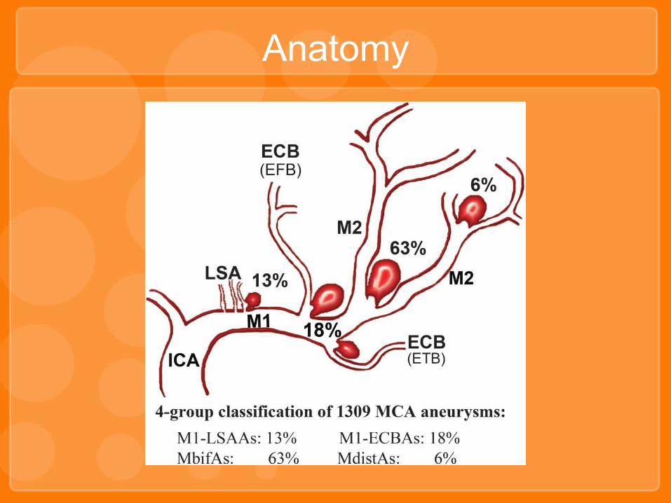
Anatomy
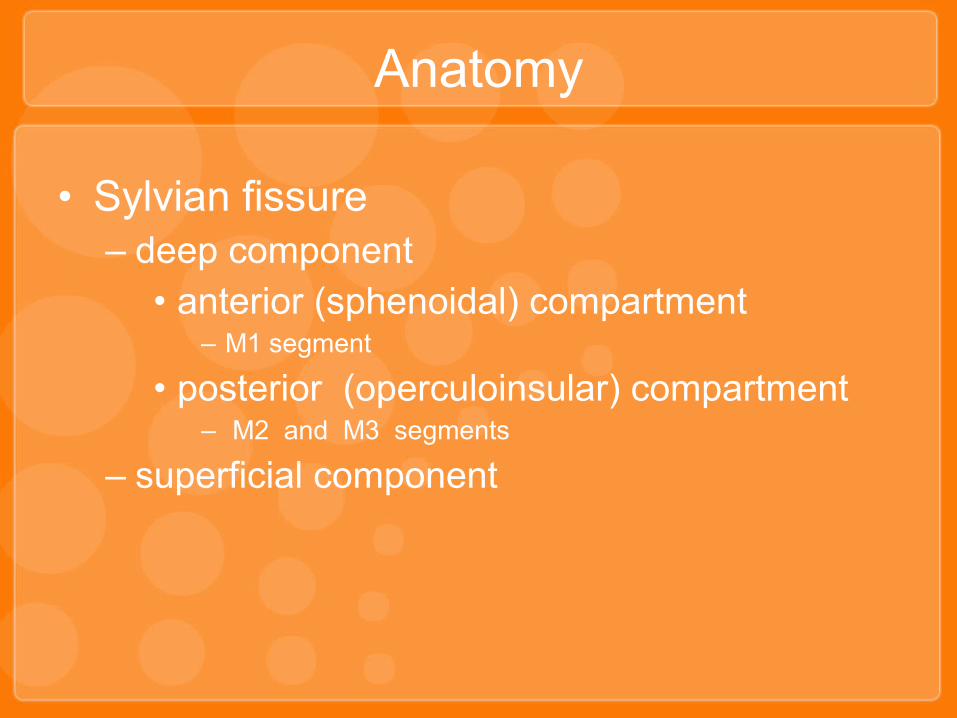
• Sylvian fissure– deep component
• anterior (sphenoidal) compartment– M1 segment
• posterior (operculoinsular) compartment– M2 and M3 segments
– superficial component
Anatomy

• M1 (sphenoidal)– lateral lenticulostriate arteries
• caudate nucleus
– anterior temporal artery• temporal tip
• M2 (insular)• M3 (opercular)• M4 (cortical)• MCA candelabra : duplication of MCA
Anatomy
• Morphology– Saccular aneurysms : most common– Fusiform aneurysms– Blister aneurysms : less common
– Extremely dysmorphic or distal aneurysms are usually infectious : M4 branches
• Size– small (<5 mm)– medium (5 to 10 mm)– large (11 to 25 mm)
– giant (>25 mm)
Classification

Classification
• Location– MCA bifurcation and trifurcation aneurysm 90%
• Surgical consideration
– Proximal MCA aneurysm
• Superior wall type :
– aneurysms arise at the origins of the lenticulostriate arteries
– project into the frontal lobe posterosuperiorly
• Inferior wall type :
– aneurysms arise at the origin of the anterior temporal artery or the temporopolar artery
– project toward the temporal lobe in an anterolateral projection
• Location– Distal MCA aneurysms
• most uncommon of the MCA aneurysms• either infectious or, less frequently, traumatic
• often small and can be challenging to identify
Classification
Classification
• Etiology– Saccular Aneurysms(berry)
• at sites of arterial curves or branching
• hemodynamic forces
– Infectious Aneurysms(mycotic)
• most commonly found along the distal branches of the cerebral arteries
• secondary to infectious emboli
• bacterial endocarditis, idiopathic bacterial or fungal
sources
• neoplastic disease, such as choriocarcinoma and atrial myxoma
• Etiology– Fusiform Aneurysms
• most frequently seen in the posterior circulation
– Dissecting Aneurysms• rare• may be associated with infection, connective tissue diseases
such as Marfan’s syndrome, cystic medial degeneration, and fibromuscular dysplasia
• present with ischemia or SAH
– Traumatic Aneurysms• uncommon• most often associated with the ACA
• most frequently distal on M3 and M4 segments and often present with
• delayed rupture
Classification

• Because of their propensity to become quite large before detection, they may occasionally become symptomatic without SAH
• Seizures in giant aneurysm• Lack of blood necessitates a lumbar puncture (LP) if
the history is suggestive of an aneurysm rupture
• CT
– SAH
– ICH
Presentation and evaluation

• MCA aneurysms are preferentially clipped at most cerebrovascular centers– frequently broad-based configuration– the relatively small caliber of surrounding branches
often precludes the use of stents– their classic bifurcation location makes recurrence
more likely
Surgical treatment

• Cerebrospinal fluid drainage– indicated in patients with acute hydrocephalus after
aneurysmal SAH– relaxed brain makes surgery safer in ruptured
aneurysm surgery– patients with unruptured aneurysms generally do not
have preoperative placement of a lumbar drain
• Position– head is turned about 30 degrees to the opposite
side with the vertex slightly extended
Preparation

Craniotomy
• Pterional incision• Preserve STA• Temporalis muscle splitting
with leaving cuff of muscle• 2 burr holes
– Key hole
– Superior to zygoma

• Dura is stripped from the cranium with a Penfield No. 3
• Pterional craniotomy flap is created with a high-speed drill with footplate
• The lesser wing of the sphenoid is flattened to the level of the superior orbital fissure
• Dural tack-up sutures• Dura is opened in a curvilinear fashion
Craniotomy

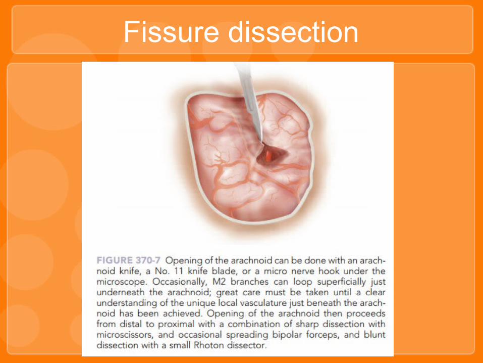
• Microscope• Sylvian fissure is opened sharply with an
arachnoid knife or a No. 11 knife blade• After a 3-4 mm, microscissors are used to
continue to open the superficial arachnoid
over the fissure• Inside out technique or open opticocarotid
cistern
Fissure dissection
Fissure dissection

• Unruptured aneurysm or a small, laterally pointing ruptured aneurysm of the bifurcation– first distally and then ultimately on the medial surface
(undersurface) of the bifurcation to access the M1 segment, gaining proximal control without opening the proximal fissure first
• larger or ruptured aneurysm or uncertain anatomy– fissure dissection should be wide and include
proximal control, initially at the opticocarotid cistern, and then working distally until the proximal M1 is identified
Aneurysm Dissection and Clipping

• Sharp dissection,clear aneurysm neck• Beware small branches adherence to dome• Temporary clip• Clips should be applied parallel to the branch
vessels to avoid possible small remnants and to reestablish normal anatomic flow
Aneurysm Dissection and Clipping

• Multilobed aneurysms– require more than one clip and often imaginative clip
constructs
• Large, multilobulated aneurysms often also have calcified or atheromatous walls– closure of clips tricky
– tandem clipping technique– fenestrated clip through the distal neck and then
completing the proximal neck occlusion with a nonfenestrated clip
Aneurysm Dissection and Clipping

Aneurysm Dissection and Clipping

• Giant MCA aneurysms– visualization of branches or small lenticulostriate
arteries is often poor– usually calcification or atheroma– large sacs contain intraluminal thrombus that must be
removed before clip application
• Temporary trapping and opening of the sac to evacuate the thrombus
• Brain ischemic protection– modest hypothermia– induced hypertension– barbiturates
Aneurysmorrhaphy

Temporary Occlusion
• occlusion times of less than 10 minutes are preferable
• no consensus in the literature regarding specific cerebroprotective agents

Bypass
• Fusiform and giant aneurysms• STA-MCA bypass• M2 side to side


