3D FractalNet: Dense Volumetric Segmentation for Cardiovascular MRI Volumes Lequan Yu 1 , Xin Yang 1 , Jing Qin 2 , and Pheng-Ann Heng 1 1 Dept. of Computer Science and Engineering, The Chinese University of Hong Kong 2 Centre for Smart Health, School of Nursing, The Hong Kong Polytechnic University Abstract. Cardiac image segmentation plays a crucial role and even a prerequisite for diverse medical applications. However, differentiating branchy structures and slicing fuzzy boundaries from cardiovascular MRI volumes remain a very challenging task. In this paper, we propose a novel deeply-supervised 3D fractal network for efficient automated whole heart and great vessel segmentation in MRI volumes. The proposed 3D fractal network takes advantage of fully convolutional architecture to perform efficient, precise, volume-to-volume prediction. Notably, by recursively applying a single expansion rule, we construct our network in a novel self- similar fractal scheme and thus promote it in combining hierarchical clues for accurate segmentation. More importantly, we employ deep supervi- sion mechanism to alleviate the vanishing gradient problem and improve the training efficiency of our network on small medical image dataset. We evaluated our method on the HVSMR 2016 Challenge dataset. Ex- tensive experimental results demonstrated the superior performance of our method, ranking top in both two phases. 1 Introduction Noninvasive cardiac imaging is an invaluable tool for the diagnosis and treat- ment of cardiovascular disease. Cardiac image segmentation plays a crucial role and even a prerequisite for diverse applications, such as quantification of vol- ume, surgical planning for complex congenital heart disease and radio-frequency ablation. However, facing with the explosive growth of volume data, manual delineation is severely inhibited and tends to be tedious, time-consuming, and prone to inter- and intra-observer variability. Subjecting to the low tissue contrast of the myocardium against surround- ings, patient variability and spatial inhomogeneities, it is very challenging to de- velop automatic solutions for efficient whole heart segmentation. Based on hand- crafted features, previous automatic methods typically exploited deformable models [11], non-rigid registration [16] and expert emendation involved inter- active segmentation [10]. Relighted by their powerful feature learning capability, Convolutional Neural Networks (CNNs) have been utilized in a 2D context for biomedical image segmentation [12] and ventricle segmentation [14]. However, leveraging the effectiveness of CNNs in capturing 3D spatial contextual infor- mation to segment the whole heart and great vessel from MRI volume has not
Transcript
3D FractalNet: Dense Volumetric Segmentationfor Cardiovascular MRI Volumes
Lequan Yu1, Xin Yang1, Jing Qin2, and Pheng-Ann Heng1
1 Dept. of Computer Science and Engineering, The Chinese University of Hong Kong2 Centre for Smart Health, School of Nursing, The Hong Kong Polytechnic University
Abstract. Cardiac image segmentation plays a crucial role and evena prerequisite for diverse medical applications. However, differentiatingbranchy structures and slicing fuzzy boundaries from cardiovascular MRIvolumes remain a very challenging task. In this paper, we propose a noveldeeply-supervised 3D fractal network for efficient automated whole heartand great vessel segmentation in MRI volumes. The proposed 3D fractalnetwork takes advantage of fully convolutional architecture to performefficient, precise, volume-to-volume prediction. Notably, by recursivelyapplying a single expansion rule, we construct our network in a novel self-similar fractal scheme and thus promote it in combining hierarchical cluesfor accurate segmentation. More importantly, we employ deep supervi-sion mechanism to alleviate the vanishing gradient problem and improvethe training efficiency of our network on small medical image dataset.We evaluated our method on the HVSMR 2016 Challenge dataset. Ex-tensive experimental results demonstrated the superior performance ofour method, ranking top in both two phases.
1 Introduction
Noninvasive cardiac imaging is an invaluable tool for the diagnosis and treat-ment of cardiovascular disease. Cardiac image segmentation plays a crucial roleand even a prerequisite for diverse applications, such as quantification of vol-ume, surgical planning for complex congenital heart disease and radio-frequencyablation. However, facing with the explosive growth of volume data, manualdelineation is severely inhibited and tends to be tedious, time-consuming, andprone to inter- and intra-observer variability.
Subjecting to the low tissue contrast of the myocardium against surround-ings, patient variability and spatial inhomogeneities, it is very challenging to de-velop automatic solutions for efficient whole heart segmentation. Based on hand-crafted features, previous automatic methods typically exploited deformablemodels [11], non-rigid registration [16] and expert emendation involved inter-active segmentation [10]. Relighted by their powerful feature learning capability,Convolutional Neural Networks (CNNs) have been utilized in a 2D context forbiomedical image segmentation [12] and ventricle segmentation [14]. However,leveraging the effectiveness of CNNs in capturing 3D spatial contextual infor-mation to segment the whole heart and great vessel from MRI volume has not
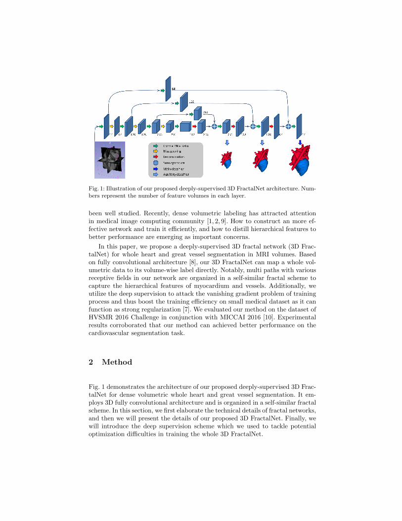
Fig. 1: Illustration of our proposed deeply-supervised 3D FractalNet architecture. Num-bers represent the number of feature volumes in each layer.
been well studied. Recently, dense volumetric labeling has attracted attentionin medical image computing community [1, 2, 9]. How to construct an more ef-fective network and train it efficiently, and how to distill hierarchical features tobetter performance are emerging as important concerns.
In this paper, we propose a deeply-supervised 3D fractal network (3D Frac-talNet) for whole heart and great vessel segmentation in MRI volumes. Basedon fully convolutional architecture [8], our 3D FractalNet can map a whole vol-umetric data to its volume-wise label directly. Notably, multi paths with variousreceptive fields in our network are organized in a self-similar fractal scheme tocapture the hierarchical features of myocardium and vessels. Additionally, weutilize the deep supervision to attack the vanishing gradient problem of trainingprocess and thus boost the training efficiency on small medical dataset as it canfunction as strong regularization [7]. We evaluated our method on the dataset ofHVSMR 2016 Challenge in conjunction with MICCAI 2016 [10]. Experimentalresults corroborated that our method can achieved better performance on thecardiovascular segmentation task.
2 Method
Fig. 1 demonstrates the architecture of our proposed deeply-supervised 3D Frac-talNet for dense volumetric whole heart and great vessel segmentation. It em-ploys 3D fully convolutional architecture and is organized in a self-similar fractalscheme. In this section, we first elaborate the technical details of fractal networks,and then we will present the details of our proposed 3D FractalNet. Finally, wewill introduce the deep supervision scheme which we used to tackle potentialoptimization difficulties in training the whole 3D FractalNet.
Fig. 2: An illustration of the expansion rule in our fractal architecture. We add down-sampling and upsampling operations in the expansion rule to utilize multi-scale feature.
2.1 Fractal Networks
Fractal networks are constructed by repeatedly applying an expansion rule froma base case [6]. Let C denotes the index of a truncated fractal fC(·) (i.e., a fewstacked layers) and the base case of a truncated fractal (Fig. 2(a)) is a singleconvolution:
f1(z) = conv(z). (1)
Then the successive fractals (Fig. 2(b)) can be defined recursively according tothe expansion rule:
z′ = conv(z),
fC+1(z) = conv[conv(z′)⊕ fC(z′)] (2)
where ⊕ is a join operation and conv(·) is a convolution operator. The joinoperation ⊕ merges two blobs, which are the extracted feature volumes result-ing from convolution and fractal operator fC(·) respectively. Because these twoblobs contain features from different visual levels, joining them can enhance thediscrimination capability of networks. Generally, the join operator can be sum-mation, maximization and concatenation. We employ summation in our experi-ments, which can achieve the best performance via cross-validation on trainingdataset.
In order to increase the receptive field and enclose more contextual informa-tion, we add downsampling and upsampling operation in the above expansionrule, as shown in Fig. 2(c). Specifically, we add a max-pooling with stride of 2and a deconvolution also with stride of 2. The receptive field of a fractal thusbecomes broader after the downsample operation. When combining different re-ceptive fields through the join operation, the network can harness multi-scalevisual cues and promote itself in discriminating.
2.2 3D FractalNet
After recursively expanding the base case with the above expansion rule for threetimes, we obtained the 3D FractalNet used in this paper, as shown in Fig. 1. Thejoin operator of fractal expansion in our 3D FractalNet is summation, computingthe element-wise sum of two blobs. The building blocks of our network, such asthe convolutional, max-pooling and deconvolutional layers, are all implementedwith a 3D manner, thus the network can fully preserve and exploit the 3D spatialinformation of the input volumetric data. Note that our network adopts the fullyconvolutional architecture, and hence can take arbitrary-sized volumetric dataas input and output corresponding sized predictions within a single forwardprocess, which is very efficient in handling large MRI dataset.
Previous studies [13] have shown that smaller convolutional kernels are moreefficient in 2D network design. Because the effective receptive field size of stackedsmall kernels is equivalent to that of one large kernel (the effective receptivefield of three 3×3×3 kernels is same as one 7×7×7 kernel), while giving lowercomputation cost. Therefore, we adopt small convolution kernels with size of3×3×3 in convolutional layers. Each convolutional layer is followed by a rectifiedlinear unit (ReLU). Note that we also employ batch normalization layer (BN) [4]before each ReLU layer to accelerate the training process. At the end of thenetwork, we add a 1×1×1 convolutional layer as a main classifier to generatethe segmentation results and further get the segmentation probability volumesafter passing the softmax layer.
2.3 Deeply-supervised 3D FractalNet
Directly training such a deep 3D fractal network is challenging due to the issueof vanishing gradient [3], which makes the back-propagation ineffective in earlylayers. Following previous studies on training deep neural networks with deepsupervision [2, 15], we proposed the deeply-supervised 3D FractalNet by injectingM auxiliary classifiers into the network. Because the dimensions of auxiliaryclassifiers’ output are different from ground truth, we insert deconvolutionallayers to upsample the auxiliary classifiers’ output. This scheme can effectivelyalleviate the problem of vanishing gradient and assist the training process withdirect supervision on the hidden layers.
Sepecially, let W be the weights of main network and w = (w1, w2, .., wM )be the weights of auxiliary classifiers. Then the cross-entropy loss function of themain classifier is
L(X ;W ) =∑xi∈X
−log p(yi = `(xi)|xi;W ), (3)
where X represents the training samples and p(yi = `(xi)|xi;W ) is the proba-bility of target class label `(xi) corresponding to sample xi ∈ X . Similarly, theloss function of the mth auxiliary classifier is
Lm(X ;W,wm) =∑xi∈X
−log p(yi = `(xi)|xi;W,wm). (4)
Therefore, the total loss function of our deeply-supervised 3D FractalNet is:
L(X ;W,w) = L(X ;W ) +
M∑m=1
αmLm(X ;W,wm) + λψ(W ), (5)
where the first two terms are the classifier loss and the last part is the regulariza-tion term (L2 norm in our experiments); αm is the weight of different auxiliaryclassifiers.
3 Experiments and Results
3.1 Dataset and Pre-processing
We evaluated our network on two phases: cropped axial images (phase 2) andcropped short-axial images (phase 3) of HVSMR 2016 Challenge dataset. Thedataset consists of 20 cardiovascular magnetic resonance (CMR) images (10training and 10 testing). Note that the ground truth of testing dataset is heldout by the organizer for independent evaluation. Before training networks, wepre-process the training dataset by normalizing them as zero mean and unitvariance. In order to tackle the insufficiency of training data and avoid overfit-ting, we also utilize the data augmentation to enlarge the training dataset. Theaugmentation operators include rotation (90, 180 and 270 degree) and flip inaxial plane and we totally use 80 examples to train our network.
3.2 Implementation details
The proposed method was implemented with C++ and Matlab under the opensource deep learning library of Caffe [5], using a standard PC with a 2.60GHzIntel(R) Xeon(R) E5-2650 CPU and a NVIDIA TITAN X GPU. The weights ofnetworks were initialized from the Gaussian distribution (µ = 0, σ = 0.01) andupdated using stochastic gradient descend (SGD) method (batch size=4, mo-mentum=0.9, weight decay=0.0005). The learning rate was set as 0.002 initiallyand divided by 10 every 3000 iterations and the network were trained for up to10000 iterations. We added two auxiliary classifiers and the weights αm are 0.33and 0.67, respectively. We randomly cropped a 64×64×64 sub-volume from eachsample in every iterations for the input when training our network, and there-fore we totally extracted 40000 patches in training. We used a overlap-tilingstrategy to generate the whole volume probability map by stitching sub-volumepredictions. We also employed some morphology operators including removingsmall isolated components and filling holes to process the prediction. Generally,it took about 12 seconds to process one volume with size about 200×140×120using above configuration.
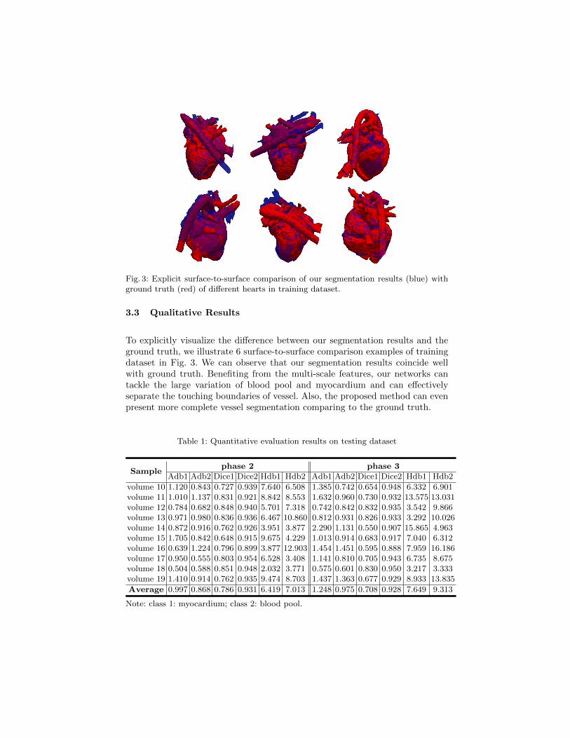
Fig. 3: Explicit surface-to-surface comparison of our segmentation results (blue) withground truth (red) of different hearts in training dataset.
3.3 Qualitative Results
To explicitly visualize the difference between our segmentation results and theground truth, we illustrate 6 surface-to-surface comparison examples of trainingdataset in Fig. 3. We can observe that our segmentation results coincide wellwith ground truth. Benefiting from the multi-scale features, our networks cantackle the large variation of blood pool and myocardium and can effectivelyseparate the touching boundaries of vessel. Also, the proposed method can evenpresent more complete vessel segmentation comparing to the ground truth.
Table 1: Quantitative evaluation results on testing dataset
The main evaluation criteria in the Challenge include Dice coefficient (Dice),Hausdorff Distance of Boundaries (Hdb[mm]) and Average Distance of Bound-aries (Adb[mm]), which emphasize both the region and boundary similarities.Auxiliary metrics, such as Jaccard index, Cohen’s Kappa, Sensitivity and Speci-ficity are also considered. For distance related metrics, lower values indicatesbetter performance.
We report two types of result: testing dataset result and leave-one-out cross-validation result of training dataset on phase 2 and 3. On the Challenge website,these results are reported from our teams CUMED2.3 Table 1 and 2 illustratethe automated segmentation results under the main metrics on testing datasetand cross-validation of training dataset, respectively.
4 Conclusion
In this paper, we propose a novel deeply-supervised 3D FractalNet for automatedwhole heart and great vessel segmentation from cardiovascular magnetic reso-nance (CMR) images. By adopting 3D fully convolutional neural networks, ournetwork can perform accurate, efficient, volume-to-volume prediction. Under arecursive fractal scheme, our network can fuse interacting subpaths of differentconvolution lengths and thus utilize multi-scale features to enhance its discrim-ination capacity. In addition, to facilitate the training process of this networkon small medical dataset, deep supervision is injected to alleviate the vanishinggradients problem. Experimental results on MICCAI 2016 HVSMR Challengedataset demonstrated the superior performance of our proposed method in han-dling large shape variation and delineating branchy structures. Our proposed
network is general and promising to be extended to other medical volumetricsegmentation applications.
Acknowledgments. The work described in this paper was supported by agrant from the Research Grants Council of the Hong Kong Special Adminis-trative Region (Project no. CUHK 412513).
References
1. Cicek, O., Abdulkadir, A., Lienkamp, S.S., Brox, T., Ronneberger, O.: 3d u-net:Learning dense volumetric segmentation from sparse annotation. arXiv preprintarXiv:1606.06650 (2016)
2. Dou, Q., Chen, H., Jin, Y., Yu, L., Qin, J., Heng, P.A.: 3d deeply super-vised network for automatic liver segmentation from ct volumes. arXiv preprintarXiv:1607.00582 (2016)
3. Glorot, X., Bengio, Y.: Understanding the difficulty of training deep feedforwardneural networks. In: Aistats. vol. 9, pp. 249–256 (2010)
4. Ioffe, S., Szegedy, C.: Batch normalization: Accelerating deep network training byreducing internal covariate shift. arXiv preprint arXiv:1502.03167 (2015)
5. Jia, Y., Shelhamer, E., Donahue, J., Karayev, S., Long, J., Girshick, R., Guadar-rama, S., Darrell, T.: Caffe: Convolutional architecture for fast feature embedding.arXiv preprint arXiv:1408.5093 (2014)
7. Lee, C.Y., Xie, S., Gallagher, P., Zhang, Z., Tu, Z.: Deeply-supervised nets. (2015)8. Long, J., Shelhamer, E., Darrell, T.: Fully convolutional networks for semantic
segmentation. In: Proceedings of the IEEE Conference on Computer Vision andPattern Recognition. pp. 3431–3440 (2015)
9. Merkow, J., Kriegman, D., Marsden, A., Tu, Z.: Dense volume-to-volume vascularboundary detection. arXiv preprint arXiv:1605.08401 (2016)
10. Pace, D.F., Dalca, A.V., Geva, T., Powell, A.J., Moghari, M.H., Golland, P.: In-teractive whole-heart segmentation in congenital heart disease. In: InternationalConference on Medical Image Computing and Computer-Assisted Intervention.pp. 80–88. Springer (2015)
11. Peters, J., Ecabert, O., Meyer, C., Schramm, H., Kneser, R., Groth, A., Weese, J.:Automatic whole heart segmentation in static magnetic resonance image volumes.In: International Conference on Medical Image Computing and Computer-AssistedIntervention. pp. 402–410. Springer (2007)
12. Ronneberger, O., Fischer, P., Brox, T.: U-net: Convolutional networks for biomedi-cal image segmentation. In: International Conference on Medical Image Computingand Computer-Assisted Intervention. pp. 234–241. Springer (2015)
13. Simonyan, K., Zisserman, A.: Very deep convolutional networks for large-scaleimage recognition. arXiv preprint arXiv:1409.1556 (2014)
14. Tran, P.V.: A fully convolutional neural network for cardiac segmentation in short-axis mri. arXiv preprint arXiv:1604.00494 (2016)
15. Xie, S., Tu, Z.: Holistically-nested edge detection. In: Proceedings of the IEEEInternational Conference on Computer Vision. pp. 1395–1403 (2015)
16. Zhuang, X., Rhode, K.S., Razavi, R.S., Hawkes, D.J., Ourselin, S.: A registration-based propagation framework for automatic whole heart segmentation of cardiacmri. IEEE transactions on medical imaging 29(9), 1612–1625 (2010)