A modified approach to aortic root reconstruction in children: An extended 2-patch root enlargement technique Ed Peng, FRCS(CTh), and Asif Hasan, FRCS(CTh), Newcastle Upon Tyne, United Kingdom When aortic valve replacement is required in children, our favored approach is the Ross or Ross-Konno procedure. 1,2 The Ross procedure may not be feasible in some settings, however, such as in patients with Marfan syndrome or previous truncus repair. In these settings, prosthetic valve replacement is required, and various annular enlargement techniques have been described to accommodate an adult rigid prosthesis in children. 3-5 We have found these annular enlargement techniques to be inadequate in patients with previous aortic root surgery. We therefore describe here a modified approach to enlarge the aortic annulus, root, and proximal ascending aorta that is different from previously described techniques and demonstrate its use in 2 female pediatric patients. CLINICAL SUMMARIES Patient 1 The first patient had Marfan syndrome. She had under- gone previous valve-sparing aortic root replacement with a 16-mm Vascutek graft (Vascutek Ltd a Terumo Company, Inchinnis, UK) at 6 years and further aortic valve repair at 8 years. At follow-up, progressive aortic stenosis and regurgitation developed. At the age of 12 years (weight 36 kg, body surface area 1.25 m 2 ), she underwent further surgery to implant a mechanical aortic valve. Cardiopulmonary bypass was established with aortic bicaval cannulation, cooling to 32 C, and right superior pulmonary vein venting. A trans- verse aortotomy was performed (Figure 1, A). The previous valve was found to be severely scarred and incompetent. The previous conduit had shrunk, and the root annulus could only accommodate a size 13-mm Hegar dilator. A Manouguian incision was performed vertically between noncoronary and left coronary cusps, extending sparingly into the roof of left atrium and the body of the anterior mitral leaflet (Figure 1, B), which allowed a size 17-mm Hegar dilator to be fitted. Another incision (Figure 1, B) was made to enlarge the root anteriorly, incising between the left and right coronary cusps, which further accommo- dated a size 21-mm Carbomedics valve (Sorin Group, Arvada, Colo). Two separate counterincisions were made on the ascending aorta above the transverse aortotomy. Two separate Vascutek graft patches (Figure 1, C) were used to close the aortotomy. Each patch was fashioned to extend above the transverse aortotomy, so that it would enlarge not only the annulus but also the root and the proximal ascending aorta (Figure 2, A and B). The patient came off bypass in sinus rhythm and without inotropes. Postoperative echocardiography showed good prosthetic valve and ventricular function. Her subsequent recovery was uneventful, and she was well at 2-month follow-up (Figure 2, D). Patient 2 The second patient had previous pulmonary arterial banding at 1 month, truncus repair and truncal valve replacement at 4 months, replacement of the pulmonary arterial conduit at 2.3 years, and replacement of aortic and pulmonary conduits and enlargement of the intramural posterior coronary artery at 5 years. The Manouguian technique was used to implant a 15-mm Shelhigh aortic conduit (Shelhigh Inc, Union, NJ). At the age of 10 years (weight, 24 kg; body surface area, 0.94 m 2 ), the patient underwent further surgery to implant a larger aortic valve prosthesis and to replace the pulmonary conduit. The previous conduit had shrunk, and the root annulus could only accommodate a size 8-mm Hegar dilator. A Nicks incision was performed in the mid non- coronary sinus. A further incision was made anteriorly between the right and left coronary cusps to accommodate a size 16-mm Carbomedics valve. Two separate equine pericardial patches were used to enlarge the root. The patient came off bypass in sinus rhythm on small doses of inotropes. Postoperative echocardiography showed good prosthetic valve and ventricular function. Her subsequent recovery was uneventful, and she was well at 4-month follow-up (Figure 2, E). DISCUSSION In the context of previous aortic surgery, a previously implanted prosthetic conduit will fix the size of the proximal aorta and additionally may show evidence of regression. A modified approach is therefore required to enlarge not only the annulus but also the aortic root and the proximal ascending aorta. In this technique, 2 separate From the Department of Paediatric Cardiothoracic Surgery, The Freeman Hospital, Newcastle Upon Tyne, UK. Disclosures: Authors have nothing to disclose with regard to commercial support. Received for publication June 29, 2013; revisions received Aug 2, 2013; accepted for publication Aug 16, 2013; available ahead of print Sept 30, 2013. Address for reprints: Asif Hasan, FRCS(CTh), Department of Paediatric Cardiotho- racic Surgery, The Freeman Hospital, Newcastle Upon Tyne NE7 7DN, UK (E-mail: [email protected]). J Thorac Cardiovasc Surg 2013;146:1547-9 0022-5223/$36.00 Copyright Ó 2013 by The American Association for Thoracic Surgery http://dx.doi.org/10.1016/j.jtcvs.2013.08.049 The Journal of Thoracic and Cardiovascular Surgery c Volume 146, Number 6 1547 SURGICAL TECHNIQUES
Transcript
SURGICALTECHNIQUES
A modified approach to aortic root reconstruction in children:An extended 2-patch root enlargement technique
Ed Peng, FRCS(CTh), and Asif Hasan, FRCS(CTh), Newcastle Upon Tyne, United Kingdom
When aortic valve replacement is required in children, ourfavored approach is the Ross or Ross-Konno procedure.1,2
The Ross procedure may not be feasible in some settings,however, such as in patients with Marfan syndrome orprevious truncus repair. In these settings, prosthetic valvereplacement is required, and various annular enlargementtechniques have been described to accommodate anadult rigid prosthesis in children.3-5 We have found theseannular enlargement techniques to be inadequate inpatients with previous aortic root surgery. We thereforedescribe here a modified approach to enlarge the aorticannulus, root, and proximal ascending aorta that isdifferent from previously described techniques anddemonstrate its use in 2 female pediatric patients.
CLINICAL SUMMARIESPatient 1
The first patient had Marfan syndrome. She had under-gone previous valve-sparing aortic root replacement witha 16-mm Vascutek graft (Vascutek Ltd a Terumo Company,Inchinnis, UK) at 6 years and further aortic valve repair at8 years. At follow-up, progressive aortic stenosis andregurgitation developed.
At the age of 12 years (weight 36 kg, body surface area1.25 m2), she underwent further surgery to implant amechanical aortic valve. Cardiopulmonary bypass wasestablished with aortic bicaval cannulation, cooling to32�C, and right superior pulmonary vein venting. A trans-verse aortotomy was performed (Figure 1, A). The previousvalve was found to be severely scarred and incompetent.The previous conduit had shrunk, and the root annuluscould only accommodate a size 13-mm Hegar dilator. AManouguian incision was performed vertically betweennoncoronary and left coronary cusps, extending sparinglyinto the roof of left atrium and the body of the anteriormitral leaflet (Figure 1, B), which allowed a size 17-mmHegar dilator to be fitted. Another incision (Figure 1, B)
From the Department of Paediatric Cardiothoracic Surgery, The Freeman Hospital,
Newcastle Upon Tyne, UK.
Disclosures: Authors have nothing to disclose with regard to commercial support.
Received for publication June 29, 2013; revisions received Aug 2, 2013; accepted for
publication Aug 16, 2013; available ahead of print Sept 30, 2013.
Address for reprints: Asif Hasan, FRCS(CTh), Department of Paediatric Cardiotho-
racic Surgery, The Freeman Hospital, Newcastle Upon Tyne NE7 7DN, UK
Copyright � 2013 by The American Association for Thoracic Surgery
http://dx.doi.org/10.1016/j.jtcvs.2013.08.049
The Journal of Thoracic and Car
was made to enlarge the root anteriorly, incising betweenthe left and right coronary cusps, which further accommo-dated a size 21-mm Carbomedics valve (Sorin Group,Arvada, Colo). Two separate counterincisions were madeon the ascending aorta above the transverse aortotomy.Two separate Vascutek graft patches (Figure 1, C) wereused to close the aortotomy. Each patch was fashioned toextend above the transverse aortotomy, so that it wouldenlarge not only the annulus but also the root and theproximal ascending aorta (Figure 2, A and B). The patientcame off bypass in sinus rhythm and without inotropes.Postoperative echocardiography showed good prostheticvalve and ventricular function. Her subsequent recoverywas uneventful, and she was well at 2-month follow-up(Figure 2, D).
Patient 2The second patient had previous pulmonary arterial
banding at 1 month, truncus repair and truncal valvereplacement at 4 months, replacement of the pulmonaryarterial conduit at 2.3 years, and replacement of aorticand pulmonary conduits and enlargement of the intramuralposterior coronary artery at 5 years. The Manouguiantechnique was used to implant a 15-mm Shelhigh aorticconduit (Shelhigh Inc, Union, NJ).At the age of 10 years (weight, 24 kg; body surface area,
0.94 m2), the patient underwent further surgery to implant alarger aortic valve prosthesis and to replace the pulmonaryconduit. The previous conduit had shrunk, and the rootannulus could only accommodate a size 8-mm Hegardilator. A Nicks incision was performed in the mid non-coronary sinus. A further incision was made anteriorlybetween the right and left coronary cusps to accommodatea size 16-mm Carbomedics valve. Two separate equinepericardial patches were used to enlarge the root. Thepatient came off bypass in sinus rhythm on small doses ofinotropes. Postoperative echocardiography showed goodprosthetic valve and ventricular function. Her subsequentrecovery was uneventful, and she was well at 4-monthfollow-up (Figure 2, E).
DISCUSSIONIn the context of previous aortic surgery, a previously
implanted prosthetic conduit will fix the size of theproximal aorta and additionally may show evidence ofregression. A modified approach is therefore required toenlarge not only the annulus but also the aortic root andthe proximal ascending aorta. In this technique, 2 separate
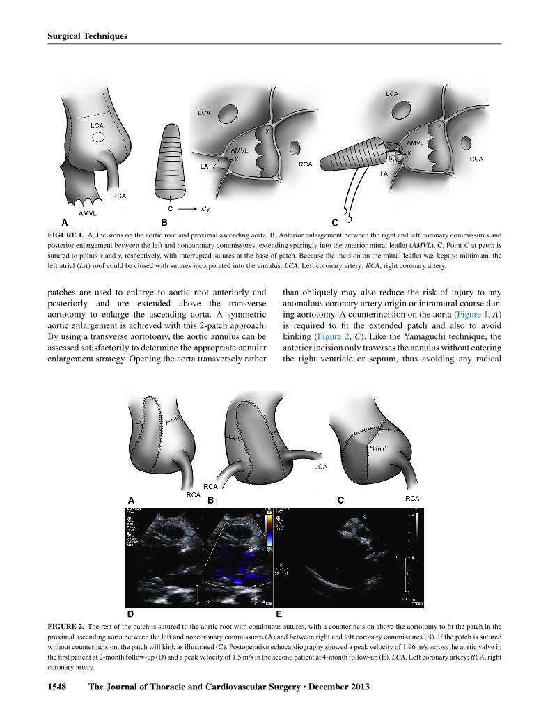
FIGURE 1. A, Incisions on the aortic root and proximal ascending aorta. B, Anterior enlargement between the right and left coronary commissures and
posterior enlargement between the left and noncoronary commissures, extending sparingly into the anterior mitral leaflet (AMVL). C, Point C at patch is
sutured to points x and y, respectively, with interrupted sutures at the base of patch. Because the incision on the mitral leaflet was kept to minimum, the
left atrial (LA) roof could be closed with sutures incorporated into the annulus. LCA, Left coronary artery; RCA, right coronary artery.
Surgical Techniques
patches are used to enlarge to aortic root anteriorly andposteriorly and are extended above the transverseaortotomy to enlarge the ascending aorta. A symmetricaortic enlargement is achieved with this 2-patch approach.By using a transverse aortotomy, the aortic annulus can beassessed satisfactorily to determine the appropriate annularenlargement strategy. Opening the aorta transversely rather
FIGURE 2. The rest of the patch is sutured to the aortic root with continuous
proximal ascending aorta between the left and noncoronary commissures (A) an
without counterincision, the patch will kink as illustrated (C). Postoperative ech
the first patient at 2-month follow-up (D) and a peak velocity of 1.5m/s in the sec
coronary artery.
1548 The Journal of Thoracic and Cardiovascular Sur
than obliquely may also reduce the risk of injury to anyanomalous coronary artery origin or intramural course dur-ing aortotomy. A counterincision on the aorta (Figure 1, A)is required to fit the extended patch and also to avoidkinking (Figure 2, C). Like the Yamaguchi technique, theanterior incision only traverses the annulus without enteringthe right ventricle or septum, thus avoiding any radical
sutures, with a counterincision above the aortotomy to fit the patch in the
d between right and left coronary commissures (B). If the patch is sutured
ocardiography showed a peak velocity of 1.96 m/s across the aortic valve in
ond patient at 4-month follow-up (E). LCA, Left coronary artery; RCA, right
gery c December 2013
Surgical Techniques
Konno procedure.5 An additional annular enlargement ante-riorly also keeps the Manouguian incision to the minimumif needed and minimizes any potential mitral valvedysfunction.
This proposed modified extended 2-patch techniqueenlarges the proximal aorta in 2 directions from the annulusto ascending aorta. In the context of reoperative aorticreconstruction, it can be used as an alternative to total aorticroot replacement and coronary reimplantation.
We thank Ms Angela Butler for the illustrations in this article.
From the Tulane Pediatric Heart Center,a the Department of Pediatrics,b the Depart-
ment of Internal Medicine,c and the Department of Surgery,d Tulane University
School of Medicine, New Orleans, La.
Disclosures: Authors have nothing to disclose with regard to commercial support.
Received for publication July 26, 2013; accepted for publication Aug 1, 2013;
available ahead of print Sept 30, 2013.
Address for reprints: Thomas Yeh, Jr, MD, PhD, 1430 Tulane Ave, SL-22,
New Orleans, LA 70112.
J Thorac Cardiovasc Surg 2013;146:1549-51
0022-5223/$36.00
Copyright � 2013 by The American Association for Thoracic Surgery
http://dx.doi.org/10.1016/j.jtcvs.2013.08.010
The Journal of Thoracic and Car
References1. Vitale N, Hornung T, Ciotti G, Hamilton JR, Pozzi M, Hasan A. The Ross
procedure in children under ten years of age. J Heart Valve Dis. 1999;8:601-4.
2. McBrien A, Chaudhari M, Crossland DS, Aspey H, Heads-Baister A, Griselli M,
et al. Single-centre experience of 101 paediatric and adult Ross procedures:
4. Nicks R, Cartmill T, Bernstein L. Hypoplasia of the aortic root. The problem of
aortic valve replacement. Thorax. 1970;25:339-46.
5. Yamaguchi M, Ohashi H, Imai M, Oshima Y, Hosokawa Y. Bilateral enlargement
of the aortic valve ring for valve replacement in children. New operative technique.
J Thorac Cardiovasc Surg. 1991;102:202-6.
Anomalous aortic origin of the coronary artery: Does pulmonaryartery translocation affect coronary artery course?
Vitor C. Guerra, MD,a,b Michael R. Recto, MD,a,b Corey Goldman, MD, PhD,c andThomas Yeh, Jr, MD, PhD,a,d New Orleans, La
The debate around timing and choice of procedure foranomalous aortic origin of the coronary artery (AAOCA)is now the subject of an ongoing study by the CongenitalHeart Surgeons Society.1 Approaches have includedunroofing, reimplantation, and pulmonary artery (PA)translocation.2 Whether moving the distal PA exerts anyeffect on the proximal PA, and thereby acts to relievecoronary compression, has been questioned.2 Here weanalyze preoperative and postoperative imaging in a singlepatient.
An echocardiogram performed for a murmur in a12-year-old male football player revealed AAOCA. Thepatient had neither chest pain nor shortness of breath.Examination and chest radiography were unrevealing.Electrocardiography revealed normal sinus rhythm withearly repolarization and increased left-sided forces.Transthoracic echocardiography (and later catheterization)revealed a 3-mm left main coronary artery (LM) arisingfrom the right coronary sinus (Figure 1, A1 and D1) thatshared a common ostium with a 2-mm right coronary artery(Figure 1, C1). The LM passed between the aorta and PAand gave rise to the left anterior descending and left
circumflex coronary arteries. There was trivial tricuspidinsufficiency, no outflow tract obstruction, and normalchamber dimensions (left ventricular posterior wall, 9mm; septum, 11 mm). Left ventricular function was hyper-dynamic (shortening fraction 56%, ejection fraction 86%).Computed tomographic (CT) angiography (Figure 1,E1 and F1) confirmed the diagnosis and the absence ofintramural coronaries. Exercise stress echocardiography(Bruce protocol) was terminated at 10 minutes, 22 secondsfor leg fatigue at 12.3 metabolic equivalents. Pulseand blood pressure increased from 83 to196 beats/min(94% of age predicted maximum) and 139/45 to 204/41mmHg, respectively. Therewere no electrocardiographicchanges, arrhythmias, or wall motion abnormalities. Three-dimensional transesophageal echocardiography revealedneither coronary ostial stenosis nor intramural course(Figure 1, B1).At surgery, little distance separated the aorta from the
PA when dissected down to the coronaries to confirmtheir anatomy. The branch PAs were dissected to theirlobar branches to relieve tension. Under bicaval cannula-tion with aortic crossclamping, the right PA wastransected near its origin and its posterior wall reanasto-mosed anterior to the ascending aorta. The right PA wasgenerously patched anteriorly with autologous pericar-dium to avoid narrowing. The patient was weaned easilyfrom cardiopulmonary bypass with good ventricular func-tion. Only when the PA was pressurized did we note ananterior bowing in the PA away from the aorta, undoubt-edly facilitated by adventitial lysis. This accentuatedarching created an obvious clear space away from thecoronaries.