A nanofluidic mixing device for high-throughput fluorescence sensing of single molecules
Klaus Mathwiga,*, Carel Fijenb, Mattia Fontanab, Serge G. Lemayc, Johannes Hohlbeinb,d,*
aPharmaceutical Analysis, Groningen Research Institute of Pharmacy, University of Groningen, P.O. Box 196, 9700 AD, Groningen, The Netherlands
bLaboratory of Biophysics, Wageningen University and Research, Dreijenlaan 3, 6703 HA Wageningen, The Netherlands cMESA+ Institute for Nanotechnology, University of Twente, P.O. Box 217, 7500 AE Enschede, The Netherlands
dMicrospectroscopy Centre, Wageningen University and Research, Dreijenlaan 3, 6703 HA Wageningen, The Netherlands
Keywords: nanochannel; nanofluidic mixing; fluorescence detection; single molecules
Single-molecule detection is important in many areas of the life sciences as it allows studying complex reaction mechanisms, rare events, or subpopulations of complex biomolecules such as DNA or proteins whose individual properties are often hidden in conventional ensemble-based measurements. Fluorescence microscopy is by far the most important and widely used method to study individual molecules. However, conventional state-of-the-art methods (i.e., confocal microscopy and camera based total-internal-reflection fluorescence or widefield microscopy) suffer from severe drawbacks: Often, elaborate surface-immobilization is required, high-throughput detection is rarely achievable and reactions cannot be triggered continuously.
142 Klaus Mathwig et al. / Procedia Technology 27 ( 2017 ) 141 – 142
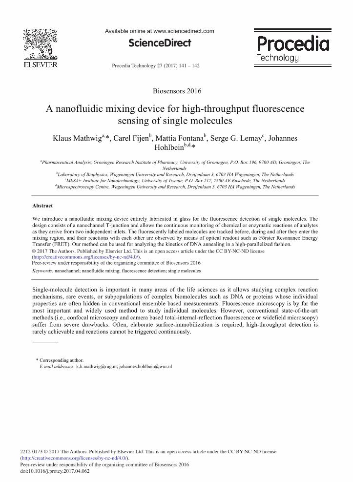
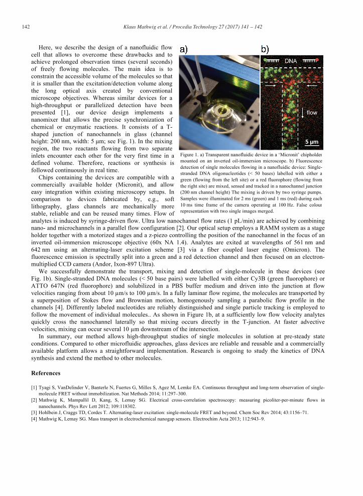
Here, we describe the design of a nanofluidic flow cell that allows to overcome these drawbacks and to achieve prolonged observation times (several seconds) of freely flowing molecules. The main idea is to constrain the accessible volume of the molecules so that it is smaller than the excitation/detection volume along the long optical axis created by conventional microscope objectives. Whereas similar devices for a high-throughput or parallelized detection have been presented [1], our device design implements a nanomixer that allows the precise synchronization of chemical or enzymatic reactions. It consists of a T-shaped junction of nanochannels in glass (channel height: 200 nm, width: 5 μm; see Fig. 1). In the mixing region, the two reactants flowing from two separate inlets encounter each other for the very first time in a defined volume. Therefore, reactions or synthesis is followed continuously in real time.
Chips containing the devices are compatible with a commercially available holder (Micronit), and allow easy integration within existing microscopy setups. In comparison to devices fabricated by, e.g., soft lithography, glass channels are mechanically more stable, reliable and can be reused many times. Flow of analytes is induced by syringe-driven flow. Ultra low nanochannel flow rates (1 pL/min) are achieved by combining nano- and microchannels in a parallel flow configuration [2]. Our optical setup employs a RAMM system as a stage holder together with a motorized stages and a z-piezo controlling the position of the nanochannel in the focus of an inverted oil-immersion microscope objective (60x NA 1.4). Analytes are exited at wavelengths of 561 nm and 642 nm using an alternating-laser excitation scheme [3] via a fiber coupled laser engine (Omicron). The fluorescence emission is spectrally split into a green and a red detection channel and then focused on an electron-multiplied CCD camera (Andor, Ixon-897 Ultra).
We successfully demonstrate the transport, mixing and detection of single-molecule in these devices (see Fig. 1b). Single-stranded DNA molecules (< 50 base pairs) were labelled with either Cy3B (green fluorophore) or ATTO 647N (red fluorophore) and solubilized in a PBS buffer medium and driven into the junction at flow velocities ranging from about 10 µm/s to 100 µm/s. In a fully laminar flow regime, the molecules are transported by a superposition of Stokes flow and Brownian motion, homogenously sampling a parabolic flow profile in the channels [4]. Differently labeled nucleotides are reliably distinguished and single particle tracking is employed to follow the movement of individual molecules.. As shown in Figure 1b, at a sufficiently low flow velocity analytes quickly cross the nanochannel laterally so that mixing occurs directly in the T-junction. At faster advective velocities, mixing can occur several 10 µm downstream of the intersection.
In summary, our method allows high-throughput studies of single molecules in solution at pre-steady state conditions. Compared to other microfluidic approaches, glass devices are reliable and reusable and a commercially available platform allows a straightforward implementation. Research is ongoing to study the kinetics of DNA synthesis and extend the method to other molecules.
References
[1] Tyagi S, VanDelinder V, Banterle N, Fuertes G, Milles S, Agez M, Lemke EA. Continuous throughput and long-term observation of single-molecule FRET without immobilization. Nat Methods 2014; 11:297–300.
[3] Hohlbein J, Craggs TD, Cordes T. Alternating-laser excitation: single-molecule FRET and beyond. Chem Soc Rev 2014; 43:1156–71. [4] Mathwig K, Lemay SG. Mass transport in electrochemical nanogap sensors. Electrochim Acta 2013; 112:943–9.
Figure 1. a) Transparent nanofluidic device in a ‘Micronit’ chipholdermounted on an inverted oil-immersion microscope. b) Fluorescencedetection of single molecules flowing in a nanofluidic device: Single-stranded DNA oligonucleotides (< 50 bases) labelled with either agreen (flowing from the left site) or a red fluorophore (flowing fromthe right site) are mixed, sensed and tracked in a nanochannel junction(200 nm channel height) The mixing is driven by two syringe pumps.Samples were illuminated for 2 ms (green) and 1 ms (red) during each10 ms time frame of the camera operating at 100 Hz. False colourrepresentation with two single images merged.