Full Terms & Conditions of access and use can be found at https://www.tandfonline.com/action/journalInformation?journalCode=iebt20 Expert Opinion on Biological Therapy ISSN: (Print) (Online) Journal homepage: https://www.tandfonline.com/loi/iebt20 Platelet-rich plasma for the treatment of knee osteoarthritis: an expert opinion and proposal for a novel classification and coding system Elizaveta Kon , Berardo Di Matteo , Diego Delgado , Brian J Cole , Andrea Dorotei , Jason L Dragoo , Giuseppe Filardo , Lisa A Fortier , Alberto Giuffrida , Chris H Jo , Jeremy Magalon , Gerard A Malanga , Allan Mishra , Norimasa Nakamura , Scott A Rodeo , Steven Sampson & Mikel Sánchez To cite this article: Elizaveta Kon , Berardo Di Matteo , Diego Delgado , Brian J Cole , Andrea Dorotei , Jason L Dragoo , Giuseppe Filardo , Lisa A Fortier , Alberto Giuffrida , Chris H Jo , Jeremy Magalon , Gerard A Malanga , Allan Mishra , Norimasa Nakamura , Scott A Rodeo , Steven Sampson & Mikel Sánchez (2020): Platelet-rich plasma for the treatment of knee osteoarthritis: an expert opinion and proposal for a novel classification and coding system, Expert Opinion on Biological Therapy, DOI: 10.1080/14712598.2020.1798925 To link to this article: https://doi.org/10.1080/14712598.2020.1798925 Accepted author version posted online: 21 Jul 2020. Published online: 03 Sep 2020. Submit your article to this journal Article views: 80 View related articles View Crossmark data Citing articles: 5 View citing articles
Transcript
Full Terms & Conditions of access and use can be found athttps://www.tandfonline.com/action/journalInformation?journalCode=iebt20
Platelet-rich plasma for the treatment of kneeosteoarthritis: an expert opinion and proposal fora novel classification and coding system
Elizaveta Kon , Berardo Di Matteo , Diego Delgado , Brian J Cole , AndreaDorotei , Jason L Dragoo , Giuseppe Filardo , Lisa A Fortier , AlbertoGiuffrida , Chris H Jo , Jeremy Magalon , Gerard A Malanga , Allan Mishra ,Norimasa Nakamura , Scott A Rodeo , Steven Sampson & Mikel Sánchez
To cite this article: Elizaveta Kon , Berardo Di Matteo , Diego Delgado , Brian J Cole , AndreaDorotei , Jason L Dragoo , Giuseppe Filardo , Lisa A Fortier , Alberto Giuffrida , Chris H Jo ,Jeremy Magalon , Gerard A Malanga , Allan Mishra , Norimasa Nakamura , Scott A Rodeo , StevenSampson & Mikel Sánchez (2020): Platelet-rich plasma for the treatment of knee osteoarthritis:an expert opinion and proposal for a novel classification and coding system, Expert Opinion onBiological Therapy, DOI: 10.1080/14712598.2020.1798925
To link to this article: https://doi.org/10.1080/14712598.2020.1798925
Platelet-rich plasma for the treatment of knee osteoarthritis: an expert opinion and proposal for a novel classification and coding systemElizaveta Kona,b, Berardo Di Matteoa,b,c, Diego Delgado d, Brian J Colee, Andrea Doroteia,b, Jason L Dragoof, Giuseppe Filardog, Lisa A Fortierh, Alberto Giuffrida a,b, Chris H Joi, Jeremy Magalonj,k, Gerard A Malangal, Allan Mishram, Norimasa Nakamuran, Scott A Rodeoo, Steven Sampsonp and Mikel Sánchez d,q
aDepartment of Biomedical Sciences, Humanitas University, Milan, Italy; bHumanitas Clinical and Research Center, IRCCS, Rozzano, Milan, Italy; cFirst Moscow State Medical University - Sechenov University, Moscow, Russia; dAdvanced Biological Therapy Unit, Hospital Vithas San José, Vitoria- Gasteiz, Spain; eDepartment of Orthopaedics, Rush University Medical Center, Chicago, Illinois, USA; fDepartment of Orthopedic Surgery, University of Colorado, Englewood, Colorado, USA; gApplied and Translational Research (ATR) Center, IRCCS Istituto Ortopedico Rizzoli, Bologna, Italy; hDepartment of Clinical Sciences, College of Veterinary Medicine, Cornell University, Ithaca, NY, USA; iDepartment of Orthopedic Surgery, Seoul Metropolitan Government-Seoul National University Boramae Medical Center, Seoul National University College of Medicine, Seoul, Republic of Korea; jAix Marseille Univ, INSERM, INRA, C2VN, Marseille, France; kCell Therapy Laboratory, Hôpital De La Conception, AP-HM, IN, SERM CIC BT, Marseille, France; lNew Jersey Regenerative Institute LLC, Cedar Knolls, NJ; Department of Physical Medicine and Rehabilitation, Rutgers University, New Jersey Medical School, Newark, NJ, USA; mDepartment of Orthopaedic Surgery, Menlo Medical Clinic, Stanford University Medical Center, Menlo Park, CA, USA; nInstitute for Medical Science in Sports, Osaka Health Science University, Osaka, Japan; oOrthopaedic Soft Tissue Research Program, Hospital for Special Surgery, New York, New York, USA; pDavid Geffen School of Medicine at UCLA, Los Angeles, CA, USA; qArthroscopic Surgery Unit, Hospital Vithas San José, Vitoria-Gasteiz, Spain
ABSTRACTIntroduction: Platelet-rich plasma (PRP) is able to modulate the joint environment by reducing the inflammatory distress and promoting tissue anabolism. Therefore, it has gained increasing popularity among clinicians in the treatment of osteoarthritis (OA), and it is currently proposed beside consoli-dated options such as viscosupplementation.Areas covered: A systematic review of all available meta-analyses evaluating intra-articular PRP injec-tions in patients affected by knee OA was performed, to understand how this biologic treatment approach compares to the traditional injective therapies available in clinical practice. Moreover, a novel coding system and ‘minimum reporting requirements’ are proposed to improve future research in this field and promote a better understanding of the mechanisms of action and indications.Expert opinion: The main limitation in the current literature is the extreme variability of PRP products used, with often paucity or even lack of data on the biologic features of PRP, which should not be considered as a simple substance, but rather a ‘procedure’ requiring accurate reporting of the char-acteristics of the product but also all preparation and application modalities. This approach will aid in matching the optimal PRP product to specific patient factors, leading to improved outcomes and the elucidation of the cost-effectiveness of this treatment.
ARTICLE HISTORYReceived 21 April 2020 Accepted 17 July 2020
Osteoarthritis (OA) is a common cause of disability: 9.6% of men and 18.0% of women aged ≥60 years have symptomatic OA characterized by joint pain, swelling, and loss of function with consequently a negative impact on patients’ quality of life [1]. Different treatments have been developed to manage OA and delay joint replacement surgery, especially in younger patients with earlier stages of OA. Available conser-vative treatments include non-pharmacological therapies such as dietary supplements, muscle strengthening exercises, non-steroid and steroid anti-inflammatory drugs, intra-articu-lar corticosteroid (CS) injection, hyaluronic acid (HA) injec-tions and, more recently, newer biological therapies including platelet-rich plasma (PRP) injections. The rationale for using PRP is the restoration of joint homeostasis, which is one of the driving factors of OA disease [2–4]. PRP consists of
a volume of autologous plasma with a concentration of platelets above the baseline [5], containing a high level of several growth factors, such as IGF-1, TGF- β, EGF, PDGF, VEGF, FGF, which have shown anabolic properties [6–8]. PRP also contains cytokines and bioactive molecules with immu-nomodulatory properties able to counteract inflammatory and catabolic molecules characterizing the OA joint environ-ment [9,10]. Various studies, mainly focused on the knee joint, supported PRP efficacy in mild to moderate OA [11,12]. Furthermore, several studies demonstrated that intra-articular PRP injections are safe with a rate of adverse events not higher compared to the other intra-articular injectable products [13,14]. Therefore, PRP has gained increasing popularity among clinicians in the last 10– 15 years and, given its current wide availability even in the outpatient setting [15,16], it has become a common injective option proposed to patients similar to more traditional ‘on
CONTACT Berardo Di Matteo [email protected] Department of Biomedical Sciences, MD - Humanitas University, Rozzano –, Milan, 20089, Italy
EXPERT OPINION ON BIOLOGICAL THERAPY https://doi.org/10.1080/14712598.2020.1798925
the shelf’ approaches such as HA and CS. So nowadays the injective ‘armamentarium’ of the physician can rely on very different therapeutic options, with different mechanisms of action and, inevitably, variable costs. Nevertheless, despite almost 15 years of research in the field of PRP, no consensus or guidelines exist among scientific societies of rheumatolo-gists, orthopedic surgeons, and physiatrists, on the most suitable indications for the use of PRP in the treatment of OA.
Given this current environment, the purpose of this sys-tematic review was to analyze the outcomes of all the avail-able meta-analyses evaluating intra-articular PRP injections in patients affected by knee OA, to understand how this biologic treatment approach compares to the traditional injective therapies available in the clinical practice. Moreover, a novel classification and coding system is proposed to improve future research in this field, to promote a better understanding of the mechanisms of action and indications for this biological approach and, in the end, foster a better use of PRP products for the treatment of OA.
2. Materials and methods
A literature search was carried out on the PubMed, EMBASE, Scopus, and PEDro databases on 15 March 2020, using the following keywords that were combined together to achieve maximum search strategy sensitivity: ‘PRP,’ ‘platelet rich plasma,’ ‘platelet gel,’ ‘platelet derived growth factors,’ ‘plate-let concentrate,’ ‘PRGF,’ ‘ACP,’ ‘autologous conditioned plasma,’ ‘platelet lysate,’ ‘platelet rich fibrin,’ ‘platelet rich membrane,’ ‘platelet derived,’ ‘autologous protein solution’ in association with: ‘meta-analysis’ and in association with: ‘osteoarthritis,’ ‘OA,’ ‘chondropathy,’ ‘articular degeneration,’ ‘cartilage.’
First, all the retrieved articles were screened by title and abstract, using the following inclusion criteria for article selec-tion: 1) meta-analysis, 2) dealing with knee OA, 3) comparing the use of intra-articular PRP injections to other injectables such as HA, CS, or placebo, 4) written in the English language, and 5) published from 2005 to 2020. Exclusion criteria were: 1) studies not containing any meta-analysis, 2) dealing with other
applications of PRP than knee OA, 3) written in other lan-guages than English, 4) published before 2005. We further excluded all duplicate articles and articles from non-peer- reviewed journals. Conference presentations, narrative reviews, editorials, and expert opinions were also excluded. A PRISMA flowchart of the selection and screening method is provided in Figure 1. Two investigators extracted relevant data independently. The following data were extracted from each included meta-analysis: first author, year of publication, num-ber of studies included in each meta-analysis, number of patients evaluated, age, OA grade, outcome measures, meth-ods, overall clinical findings, which are summarized in Table I. Discrepancies were resolved by discussion and consensus, and the final results were reviewed by the senior investigators.
3. Results
3.1. Identification of studies
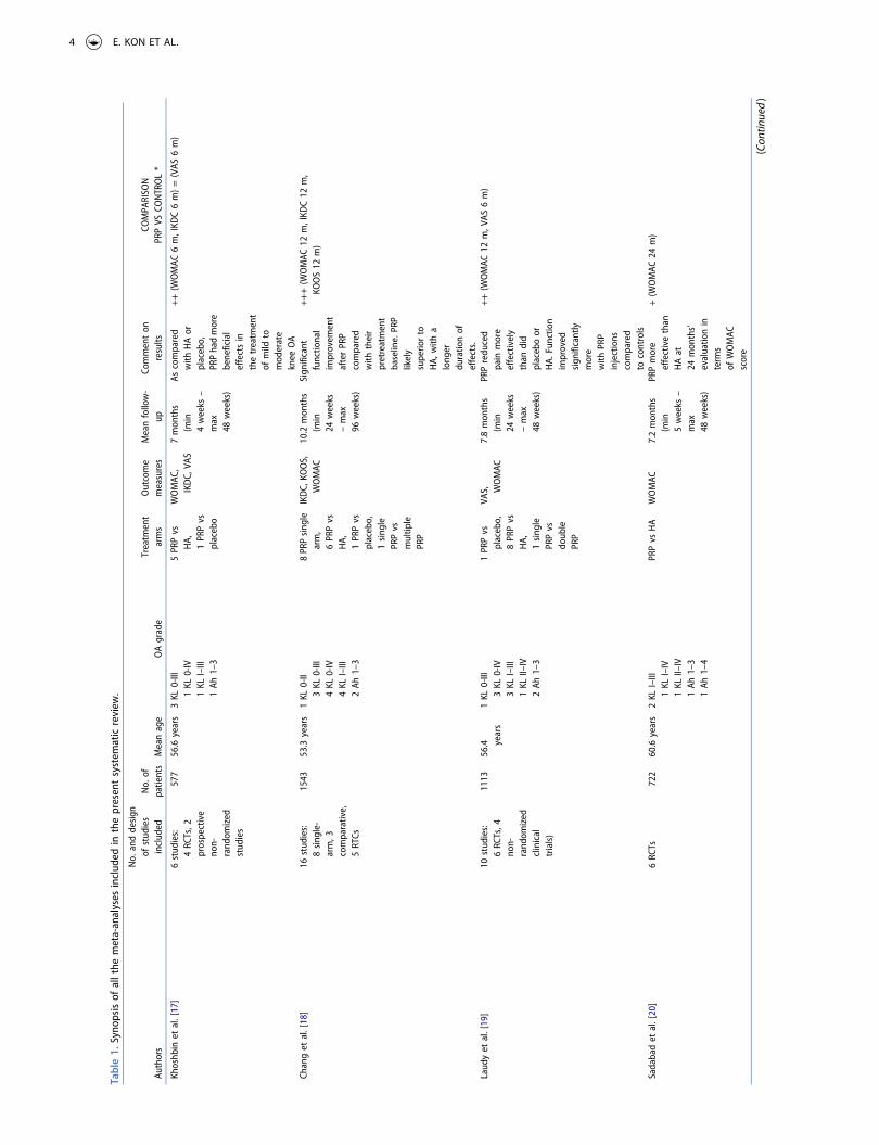
A total of 48 articles were identified through databases search-ing. After title and abstract screening, 47 studies were included. As shown in Figure 1, 34 articles were excluded and, ultimately, a total of 12 meta-analyses published from 2013 to 2020 were included, dealing with the comparison of intra-articular PRP injection to intra-articular HA, CS, or pla-cebo for the treatment of knee OA [17–28]. A synopsis of all papers included in the present systematic review is shown in Table 1.
3.2. Patients and evaluation methods
Twelve meta-analyses on knee OA were included, with a number of patients ranging from 577 to 1,543. Patients with all grades of OA were included, with a majority of patients being affected by mild to moderate OA (Kellgren-Lawrence I– III and Ahlbäck I–III). Of the 12 meta-analyses, 6 were con-ducted in China [22–26,28], 1 in Canada [17], 1 in Taiwan [18], 1 in the Netherlands [19], 1 in Iran [20], 1 in Thailand [21] and 1 in South Africa [27]. Knee pain and function were evaluated using the scores Western Ontario and McMaster Universities Arthritis Index (WOMAC), International Knee Documentation Committee (IKDC), Lequesne, Visual Analog Scale (VAS), EuroQol-VAS (EQ-VAS), Knee injury and Osteoarthritis Outcome Score (KOOS).
3.3. Reported clinical outcomes
Overall, considering the findings of the meta-analyses included, intra-articular PRP injection led to an improvement in all clinical scores (WOMAC, IKDC, VAS, EQ-VAS, Lequesne, KOOS) with greater and lasting efficacy compared to HA or placebo.
Western Ontario and McMaster Universities Arthritis Index (WOMAC)
WOMAC score was considered in 12 meta-analyses, and 11 found superiority of PRP compared to HA or placebo in terms of WOMAC score. In particular: 3 meta-analyses
Article highlights
● The use of PRP as an injective treatment for osteoarthritis is safe and provides symptomatic relief and functional recovery.
● PRP injections do not provide direct cartilage regeneration but rather a modulation of the articular environment, with a reduction of the inflammatory distress.
● The evaluation of literature revealed that the majority of meta-ana-lyses found better results of PRP compared to viscosupplementation at short to middle term evaluation.
● PRP should be considered a ‘procedure’ rather than a simple injective substance; therefore, preparation methods, storage, activation mod-alities, therapeutic protocols need to be clearly reported in trials.
● A great inter-product variability exist among different PRP products, so there is a stringent need for a classification system and ‘minimum reporting requirements’ to be universally adopted by basic research-ers and clinicians.
2 E. KON ET AL.
[17,22,28] documented better results for PRP at the 6 months’ evaluation; 7 meta-analyses [18,19,21,23–26] revealed better results up to the 12 months’ evaluation; 1 meta-analysis [20] showed superior results of PRP at 24 months.
In one meta-analysis, no statistically significant differences was observed between PRP and HA group up to 12 months [28].
3.4. International knee documentation committee (IKDC)
The IKDC subjective score was considered in eight meta-ana-lyses, and seven of them found superiority of PRP: four of them reported better results for PRP at 6 months [17,22,26,28] and three meta-analyses [18,21,23] showed the superior outcome of PRP compared to HA or placebo up to the 12 months’ follow-up.
In one meta-analysis no statistically significant differences were observed between PRP and HA groups up to 12 months [27].
3.4.1. VAS for painSix meta-analyses considered VAS for pain in knee-treated patients, and three of them documented some superiority of PRP: in two meta-analyses PRP injections were superior to HA or placebo at 6 months [19,28]; in one meta-analysis PRP (and ACP – autologous-conditioned plasma, which was separately analyzed) was demonstrated to be clearly super-ior over HA up to 12 months [27].
In three meta-analyses there was no statistical difference in VAS scores between the PRP group and HA or placebo at 6 months [17,22,26].
3.4.2. EQ-VASTwo meta-analyses evaluated the EQ-VAS: in one study [25] no difference emerged between PRP and HA for up to 12 months, whereas in another study [21], the PRP group had statistically significantly better quality of life than the HA group at 12 months.
Records identified through database searching
(n = 48)
Scre
enin
gIn
clud
ed
Elig
ibili
ty
Iden
tific
atio
n
Records after duplicates removed (n = 47)
Records screened (n = 47)
Records excluded (n = 31)
Full-text articles assessed for eligibility
(n = 16)
Full-text articles excluded, with reasons
(n = 4)
Studies included in qualitative synthesis
(n = 12)
Figure 1. PRISMA Flowchart resuming the papers’ selection process.
EXPERT OPINION ON BIOLOGICAL THERAPY 3
Tabl
e 1.
Syn
opsi
s of
all
the
met
a-an
alys
es in
clud
ed in
the
pre
sent
sys
tem
atic
rev
iew
.
Auth
ors
No.
and
des
ign
of s
tudi
es
incl
uded
No.
of
patie
nts
Mea
n ag
eO
A gr
ade
Trea
tmen
t ar
ms
Out
com
e m
easu
res
Mea
n fo
llow
- up
Com
men
t on
re
sults
COM
PARI
SON
PR
P VS
CO
NTR
OL
*
Khos
hbin
et
al. [
17]
6 st
udie
s:
4 RC
Ts, 2
pr
ospe
ctiv
e no
n-
rand
omiz
ed
stud
ies
577
56.6
yea
rs3
KL 0
-III
1 KL
0-IV
1
KL I–
III
1 Ah
1–3
5 PR
P vs
H
A,
1 PR
P vs
pl
aceb
o
WO
MAC
, IK
DC,
VAS
7 m
onth
s (m
in
4 w
eeks
–
max
48
wee
ks)
As c
ompa
red
with
HA
or
plac
ebo,
PR
P ha
d m
ore
bene
ficia
l ef
fect
s in
th
e tr
eatm
ent
of m
ild t
o m
oder
ate
kn
ee O
A
++
(W
OM
AC 6
m, I
KDC
6 m
) =
(VA
S 6
m)
Chan
g et
al.
[18]
16 s
tudi
es:
8 si
ngle
- ar
m, 3
co
mpa
rativ
e,
5 RT
Cs
1543
53.3
yea
rs1
KL 0
-II
3 KL
0-II
I 4
KL 0
-IV
4 KL
I–III
2
Ah 1
–3
8 PR
P si
ngle
ar
m,
6 PR
P vs
H
A,
1 PR
P vs
pl
aceb
o,
1 si
ngle
PR
P vs
m
ultip
le
PRP
IKD
C, K
OO
S,
WO
MAC
10.2
mon
ths
(min
24
wee
ks
– m
ax
96 w
eeks
)
Sign
ifica
nt
func
tiona
l im
prov
emen
t af
ter
PRP
com
pare
d w
ith t
heir
pr
etre
atm
ent
base
line.
PRP
lik
ely
su
perio
r to
H
A, w
ith a
lo
nger
du
ratio
n of
ef
fect
s.
++
+ (
WO
MAC
12
m, I
KDC
12 m
, KO
OS
12 m
)
Laud
y et
al.
[19]
10 s
tudi
es:
6 RC
Ts, 4
no
n-
rand
omiz
ed
clin
ical
tr
ials
)
1113
56.4
year
s1
KL 0
-III
3 KL
0-IV
3
KL I–
III
1 KL
II–I
V 2
Ah 1
–3
1 PR
P vs
pl
aceb
o,
8 PR
P vs
H
A,
1 si
ngle
PR
P vs
do
uble
PR
P
VAS,
W
OM
AC7.
8 m
onth
s (m
in
24 w
eeks
–
max
48
wee
ks)
PRP
redu
ced
pain
mor
e ef
fect
ivel
y
than
did
pl
aceb
o or
H
A. F
unct
ion
im
prov
ed
sign
ifica
ntly
m
ore
w
ith P
RP
inje
ctio
ns
com
pare
d
to c
ontr
ols
++
(W
OM
AC 1
2 m
, VAS
6 m
)
Sada
bad
et a
l. [2
0]6
RCTs
722
60.6
yea
rs2
KL I–
III
1 KL
I–IV
1
KL II
–IV
1 Ah
1–3
1
Ah 1
–4
PRP
vs H
AW
OM
AC7.
2 m
onth
s (m
in
5 w
eeks
–
max
48
wee
ks)
PRP
mor
e ef
fect
ive
than
H
A at
24
mon
ths’
eval
uatio
n in
te
rms
of W
OM
AC
scor
e
+ (
WO
MAC
24
m)
(Con
tinue
d)
4 E. KON ET AL.
Tabl
e 1.
(Co
ntin
ued)
.
Auth
ors
No.
and
des
ign
of s
tudi
es
incl
uded
No.
of
patie
nts
Mea
n ag
eO
A gr
ade
Trea
tmen
t ar
ms
Out
com
e m
easu
res
Mea
n fo
llow
- up
Com
men
t on
re
sults
COM
PARI
SON
PR
P VS
CO
NTR
OL
*
Kanc
hana
taw
an e
t al
. [21
]9
RCTs
1146
58.2
yea
rs2
KL 0
-III
1 KL
0-IV
1
KL I–
III
2 KL
I–IV
1
KL II
–IV
2 Ah
1–3
7 PR
P vs
HA
2 PR
P vs
pl
aceb
o
WO
MAC
, Le
ques
ne
scor
e,
IKCD
, EQ
- VA
S
9 m
onth
s (m
in
24 w
eeks
–
max
48
wee
ks)
PRP
mor
e ef
fect
ive
than
H
A
and
plac
ebo
in r
educ
ing
sym
ptom
s,
impr
ovin
g fu
nctio
n an
d im
prov
ing
qu
ality
of
life
in p
atie
nts
with
mild
-to-
m
oder
ate
OA
of t
he k
nee
++
+ (
WO
MAC
12
m, I
KDC
12 m
, EQ
-VAS
12
m)
= (
Lequ
esne
sco
re 1
2 m
)
Xu e
t al
. [22
]10
RCT
s11
8457
.1 y
ears
1 KL
0-II
I 2
KL 0
-IV
1 KL
I–III
1
KL I–
IV
2 KL
II–I
II 1
KL II
–IV
2 Ah
1–3
8 PR
P vs
H
A,
2 PR
P vs
pl
aceb
o
WO
MAC
, IK
CD, V
AS,
Lequ
esne
sc
ore
9 m
onth
s (m
in
1 w
eek
– m
ax
48 w
eeks
)
PRP
was
fou
nd
effe
ctiv
e to
re
lieve
pa
in a
nd
impr
ove
func
tion,
with
a
satis
fact
ory
leve
l ob
serv
ed f
or
at le
ast
6 m
++
(W
OM
AC 6
m, I
KDC
6 m
) =
(VA
S 6
m,
Lequ
esne
sco
re 6
m)
Dai
et
al. [
23]
10 R
CTs
1069
57.9
yea
rs1
KL 0
-III
1 KL
0-IV
1
KL I–
III
1 KL
I–IV
3
KL II
–III
1 KL
II–I
V 2
Ah 1
–3
8 PR
P vs
H
A,
2 PR
P vs
pl
aceb
o
WO
MAC
, IK
CD,
Lequ
esne
sc
ore
8.7
mon
ths
(min
12
wee
ks,
max
48
wee
ks)
Com
pare
d w
ith
HA
and
salin
e,
intr
a-ar
ticul
ar
PRP
inje
ctio
n m
ay
have
mor
e be
nefit
in
pain
rel
ief
and
func
tiona
l im
prov
emen
t
++
+ (
WO
MAC
12
m, I
KDC
12 m
, Leq
uesn
e sc
ore
12 m
) =
(W
OM
AC 6
m, I
KDC
6 m
, Leq
uesn
e sc
ore
6 m
)
Shen
et
al. [
24]
14 R
CTs
1423
59.1
yea
rs3
KL 0
-IV
3 KL
I–III
1
KL I–
IV
4 KL
II–I
II 1
KL II
–IV
2 Ah
1–3
PRP
vs H
A/
plac
ebo/
oz
one/
CS
WO
MAC
9.9
mon
ths
(min
8
wee
ks,
max
52
wee
ks)
PRP
prob
ably
is
mor
e ef
fect
ive
in
term
s of
pai
n re
lief
and
self-
repo
rted
fu
nctio
n im
prov
emen
t, co
mpa
red
with
pla
cebo
, H
A, a
nd C
S
+ (
WO
MAC
12
m)
(Con
tinue
d)
EXPERT OPINION ON BIOLOGICAL THERAPY 5
Tabl
e 1.
(Co
ntin
ued)
.
Auth
ors
No.
and
des
ign
of s
tudi
es
incl
uded
No.
of
patie
nts
Mea
n ag
eO
A gr
ade
Trea
tmen
t ar
ms
Out
com
e m
easu
res
Mea
n fo
llow
- up
Com
men
t on
re
sults
COM
PARI
SON
PR
P VS
CO
NTR
OL
*
Zhan
g et
al.
[25]
13 s
tudi
es:
10 R
CTs,
3 pr
ospe
ctiv
e st
udie
s
1390
58.2
yea
rs1
KL 0
-III
2 KL
0-IV
5
KL I–
III
1 KL
I–IV
2
KL II
–III
1 KL
II–I
V 1
Ah 1
–3
PRP
vs H
A,W
OM
AC, E
Q-
VAS
8.7
mon
ths
(min
4
wee
ks –
m
ax
48 w
eeks
)
PRP
redu
ced
pain
mor
e ef
fect
ivel
y th
an H
A.
WO
MAC
sc
ore
at
6 m
onth
s’ f-
up
was
su
perio
r in
PR
P gr
oup
+ (
WO
MAC
6 m
) =
(W
OM
AC 1
2 m
, EQ
-VAS
12
m)
Han
et
al. [
26]
15 R
CTs
1314
56.5
yea
rs5
KL I–
III
3 KL
I–IV
4
KL II
–III
1 KL
II–I
V 1
KL II
I–IV
1
Ah 1
–3
PRP
vs H
AW
OM
AC,
VAS,
IKD
C,
Lequ
esne
sc
ore
9 m
onth
s (m
in
12 w
eeks
–
max
72
wee
ks)
PRP
has
a po
sitiv
e ef
fect
on
pai
n le
vels
an
d fu
nctio
nal
outc
omes
co
mpa
red
with
HA
inje
ctio
ns.
++
(W
OM
AC 1
2 m
, IKD
C 6
m)
= (
IKD
C 12
m, V
AS
6 m
, Leq
uesn
e sc
ore
6 m
)
Hoh
man
n et
al.
[27]
12 s
tudi
es12
4858
.8 y
ears
2 KL
0-II
I 4
KL I–
III
1 KL
I–IV
2
KL II
–III
1 KL
II–I
V 2
Ah 1
–3
9 PR
P vs
H
A,
3 AC
P vs
H
A
WO
MAC
, IK
CD,
VAS
10 m
onth
s (m
in
24 w
eeks
–
max
48
wee
ks)
PRP
is s
uper
ior
to H
A in
kne
e pa
in
redu
ctio
n; in
pa
rtic
ular
, AC
P ap
pear
s al
so t
o be
cl
early
su
perio
r ov
er
HA;
the
re
wer
e no
ad
vant
ages
of
PRP
over
HA
in o
ther
cl
inic
al
para
met
ers
+ (
VAS
12 m
) =
(W
OM
AC 1
2 m
, IKD
C 12
m)
Wu
et a
l. [2
8]9
RCTs
1063
56.5
yea
rs2
KL 0
-III
1 KL
0-IV
2
KL I–
II 4
KL I–
III
PRP
vs H
AIK
CD,
WO
MAC
, VA
S,
KOO
S
8.1
mon
ths
(min
4
wee
ks –
m
ax
52 w
eeks
)
PRP
appe
ars
to
be b
ette
r fo
r pa
in r
elie
f an
d se
lf-
repo
rted
fu
nctio
nal
impr
ovem
ent
than
HA.
++
+ (
WO
MAC
6 m
, IKD
C 6
m, V
AS 6
m)
= (
KOO
S 6
m)
KL:
Kellg
ren-
Law
renc
e O
A c
lass
ific
atio
n A
h: A
hlbä
ck O
A c
lass
ific
atio
n *
In t
his
colu
mn
we
repo
rt e
vent
ual s
uper
iorit
y of
PRP
com
pare
d to
con
trol
. ‘+
’ m
eans
tha
t PR
P is
bet
ter
in t
he s
peci
fic p
aram
eter
des
crib
ed in
par
enth
eses
. Whe
n m
ultip
le +
wer
e us
ed (
max
imum
thr
ee +
) th
at m
eans
tha
t PR
P pr
oved
to
be m
ore
effe
ctiv
e in
mul
tiple
clin
ical
par
amet
ers.
‘ =
’ m
eans
no
diffe
renc
e be
twee
n PR
P an
d co
ntro
ls.
6 E. KON ET AL.
3.4.3. Lequesne scoreFour meta-analyses considered the Lequesne score, and three of them [21,22,26] found no difference in favor of PRP, neither at 6 [22,26] nor at 12 months’ follow-up [21].
In one meta-analysis instead, PRP provided better results than HA at the 12 months’ evaluation [23].
3.5. Knee injury and osteoarthritis outcome score (KOOS)
Two meta-analyses analyzed the KOOS score: one did not find any difference between PRP and HA at 6 months [28], whereas the other found that PRP injections led to a better KOOS score at 12 months compared to HA or placebo [18].
3.6. A novel coding system of PRP products and minimum reporting requirements for future trials
One of the major limitations in the field of PRP is that most studies include PRP formulations obtained by different meth-ods, with different compositions and characteristics, and therefore the outcomes could be different depending on the product used, even though they are all called PRP. This makes the comparison among results of different studies often con-fusing and contradictory. Beyond the wide variability among products, literature is often characterized by the paucity of data provided by authors on the composition and biologic activity of the particular PRP adopted [29]. This limitation occurs both in clinical and pre-clinical research [31–33]: a recent paper by Chahla et al. [34] found that only 11/105 studies (10%) provided comprehensive reporting of the PRP preparation protocol, and only 17/105 studies (16%) provided quantitative metrics on the composition of the final PRP product.
In response to this, several classification systems have been proposed to report the most relevant parameters of PRP [35– 38] and, in recent years, these classifications have become more sophisticated by including features such as erythrocytes, recovery efficiency, or centrifugation type [39–41]. However, none of them have been able to reach the agreement of experts. Our aim is to present a novel classification and mini-mal reporting requirements reached by consensus among main opinion leaders in this field. This consensus statement consists of: (1) a code that quickly identifies and gives an idea of the type of PRP used in each study, based on parameters that are easy to measure and affordable for any research team; (2) three tables to be used depending on the study (in vitro, in vivo, or clinical) in which the preparation, characterization, and application of PRP are described in a concise and struc-tured way.
3.6.1. Code systemThe code is a sequence of six digits grouped in pairs indicating parameters of platelet composition, purity, and activation: N1N2-N3N4-N5N6. We tried to simplify the code as much as possible because the complex never prevails. Besides, it is only composed of numbers, since these are ‘the most universal
language.’ Each digit refers to the following parameters sum-marized in Table 2.
The digits N1 and N2 indicate the platelet composition of PRP. It takes into account the concentration of PRP with respect to the basal levels in blood. By associating the digits N1 and N2 with platelet concentrations (2 = 200,000–300,000 or 4 = 400,000–500,000), it is very simple to deduce both the platelet concentration and the concentration ratio. If a PRP is ‘24-N3N4-N5N6,’ it can be already deduced that this PRP has a platelet concentration around double above the basal values (which are around 200,000 platelets/µL in blood), and that PRP platelet concentration is between 400,000 and 500,000 plate-lets/µL. In addition to providing information about platelet concentration and concentration ratio, the total number of platelets administered can also be calculated by a simple multiplication taking into account the volume of PRP injected.
Table 2. Explanation of the digits of the novel PRP coding system.
White Blood Cells in PRP 0 = Less than baseline 1 = 1.01 to 2 x baseline 2 = 2.01 to 3 x baseline 3 = 3.01 to 4 x baseline 4 = 4.01 to 5x baseline 5 = > 5x baseline
-Number 5
(N5)External Activation 0 = No (endogenous)
1 = YesNumber 6
(N6)Calcium Addition 0 = No
1 = Yes
EXPERT OPINION ON BIOLOGICAL THERAPY 7
These data are indicated in the summary tables below (Table 1, 2, and 3).
The digits N3 and N4 indicate the purity of the PRP, refer-ring to the absence (0) or presence (1) of erythrocytes and the concentration of leukocytes (0, 1, 2, 3 …). The effect of the PRP is not only conditioned by the presence of leukocytes, which is a widely studied issue, but also by the presence of erythro-cytes (red blood cells – RBCs), which can affect PRP due to possible toxicity produced by hemolysis and eryptosis [42]. The limit of 1x10⁶/µL was defined based on the data coming from studies were RBC count in PRP products was specifically assessed: depletion of RBCs always led to products with RBC<1x10⁶/µL [43,44].
Finally, the digits N5 and N6 refer to the activation. N5
indicates if activation is endogenous (0) or if PRP is activated before its injection (1). N6 mentions the addition of calcium for activation (0 = no, 1 = yes), as it is important to know if the calcium sequestered to prevent blood clotting during PRP preparation has been added again: previous studies reported that calcium concentration has an influence on the cellular and tissue response produced by the PRP [45].
3.6.2. Examples(1) PRP obtained from blood with a mean concentration of 150,000 platelets/µL and reaching a mean concentration of 430,000 platelets/µL; presence of erythrocytes and no leuko-cytes; activation with thrombin: PRP code is 14–10-10.
(2) PRP obtained from blood with a mean concentration of 230,000 platelets/µL and reaching a mean concentration of 310,000 platelets/µL; no erythrocytes; double leukocyte con-centration compared to blood levels and without exogenous activation: PRP code is 23–02-00.
(3) PRP obtained from blood with a mean concentration of 212,900 platelets/µL and reaching a mean concentration of 420,000 platelets/µL; with no leukocytes and no erythrocytes; and activation with calcium: PRP code is 24–00-11.
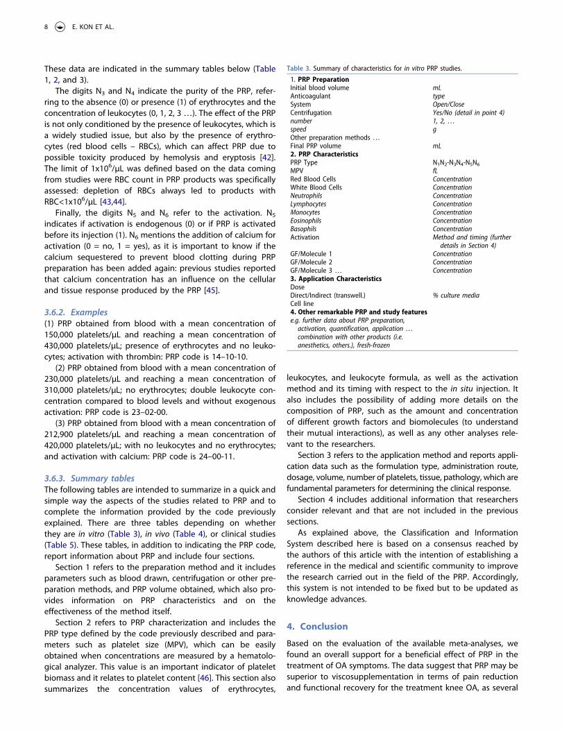
3.6.3. Summary tablesThe following tables are intended to summarize in a quick and simple way the aspects of the studies related to PRP and to complete the information provided by the code previously explained. There are three tables depending on whether they are in vitro (Table 3), in vivo (Table 4), or clinical studies (Table 5). These tables, in addition to indicating the PRP code, report information about PRP and include four sections.
Section 1 refers to the preparation method and it includes parameters such as blood drawn, centrifugation or other pre-paration methods, and PRP volume obtained, which also pro-vides information on PRP characteristics and on the effectiveness of the method itself.
Section 2 refers to PRP characterization and includes the PRP type defined by the code previously described and para-meters such as platelet size (MPV), which can be easily obtained when concentrations are measured by a hematolo-gical analyzer. This value is an important indicator of platelet biomass and it relates to platelet content [46]. This section also summarizes the concentration values of erythrocytes,
leukocytes, and leukocyte formula, as well as the activation method and its timing with respect to the in situ injection. It also includes the possibility of adding more details on the composition of PRP, such as the amount and concentration of different growth factors and biomolecules (to understand their mutual interactions), as well as any other analyses rele-vant to the researchers.
Section 3 refers to the application method and reports appli-cation data such as the formulation type, administration route, dosage, volume, number of platelets, tissue, pathology, which are fundamental parameters for determining the clinical response.
Section 4 includes additional information that researchers consider relevant and that are not included in the previous sections.
As explained above, the Classification and Information System described here is based on a consensus reached by the authors of this article with the intention of establishing a reference in the medical and scientific community to improve the research carried out in the field of the PRP. Accordingly, this system is not intended to be fixed but to be updated as knowledge advances.
4. Conclusion
Based on the evaluation of the available meta-analyses, we found an overall support for a beneficial effect of PRP in the treatment of OA symptoms. The data suggest that PRP may be superior to viscosupplementation in terms of pain reduction and functional recovery for the treatment knee OA, as several
Table 3. Summary of characteristics for in vitro PRP studies.
1. PRP PreparationInitial blood volume mLAnticoagulant typeSystem Open/CloseCentrifugation Yes/No (detail in point 4)number 1, 2, …speed gOther preparation methods …Final PRP volume mL2. PRP CharacteristicsPRP Type N1N2-N3N4-N5N6
details in Section 4)GF/Molecule 1 ConcentrationGF/Molecule 2 ConcentrationGF/Molecule 3 … Concentration3. Application CharacteristicsDoseDirect/Indirect (transwell.) % culture mediaCell line4. Other remarkable PRP and study featurese.g. further data about PRP preparation,
activation, quantification, application … combination with other products (i.e. anesthetics, others.), fresh-frozen
8 E. KON ET AL.
meta-analyses were identified and the majority reported superiority of PRP in one or more clinical parameters: in parti-cular, looking at the functional scores most commonly used by clinicians, 11 out of 12 meta-analyses found better WOMAC score, and 7 out of 8 meta-analyses found superior IKDC- subjective in PRP group compared to HA or placebo.
The main limitation in the analysis of the literature comes from the extreme variability of PRP products used, with often paucity or even lack of data provided by authors to under-stand the biologic features of the PRP used in the specific trial considered. Comparing the results of similar substances, as well as distinguishing those of different products, would be fundamental to understand what works better for a selected clinical indication. Only recently there has been increasing awareness of the necessity of measuring and reporting the complete features of PRP, and the classification and coding system presented in this review should be considered as a proposal for a ‘roadmap’ to guide future pre-clinical and clin-ical research. PRP should not be simply considered a product to inject but rather we should think it as a ‘procedure,’ since the number of injections, the time interval among administra-tions, the storage, and activation methods could also play a significant role in determining the clinical outcomes.
Therefore, more useful data will be obtained when researchers compare similar ‘PRP procedures,’ dealing not only with com-parable substances but also with comparable application methods.
5. Expert opinion
At the beginning of the application of PRP in musculoske-letal diseases, there were high expectations supported by encouraging data from in vitro and animal trials, both in terms of safety and efficacy [47–49]. Therefore, PRP was soon applied in a wide range of pathologies, from OA to tendinopathies and muscle injuries [50–53]. Despite the initial enthusiasm leading often to an indiscriminate use of this product [54], based on the current knowledge and technologies available, tissue regeneration by simple PRP injection is still a difficult challenge to achieve. As largely expected, the promising laboratory or animal results could not be reproduced in the clinical practice, as the complex networks and the many etiopathogenetic factors that come into play in-vivo cannot be easily mimicked in con-trolled experimental conditions, a general rule that should always be remembered when dealing with biologic pro-ducts [55].
Table 4. Summary of characteristics for in vivo PRP studies.
1. PRP PreparationInitial blood volume mLAnticoagulant typeSystem Open/CloseCentrifugation Yes/No (detail in point 4)number 1, 2, …speed gOther preparation methods …Final PRP volume mL2. PRP CharacteristicsPRP Type N1N2-N3N4-N5N6
details in Section 4)GF/Molecule 1 ConcentrationGF/Molecule 2 ConcentrationGF/Molecule 3 … Concentration3. Application CharacteristicsFormulation type Liquid, gel, scaffold …Administration route including image guidanceDosage number of applications and
intervalVolume ml PRPDose number of injected platelets
(range)TissuePathologyAnimal4. Other remarkable PRP and study featurese.g. further data about PRP preparation,
activation, quantification, application … combination with other products (i.e. anesthetics, others.), fresh-frozen
Table 5. Summary of characteristics for clinical PRP studies.
1. PRP PreparationInitial blood volume mLAnticoagulant typeSystem Open/CloseCentrifugation Yes/No (detail in point 4)number 1, 2, …speed gOther preparation methods …Final PRP volume mL2. PRP CharacteristicsPRP Type N1N2-N3N4-N5N6
details in Section 4)GF/Molecule 1 ConcentrationGF/Molecule 2 ConcentrationGF/Molecule 3 … Concentration3. Application CharacteristicsFormulation type Liquid, gel, scaffold …Administration route including image guidanceDosage number of applications and
intervalVolume ml PRPDose number of injected platelets
(range)TissuePathology4. Other remarkable PRP and study featurese.g. further data about PRP preparation,
activation, quantification, application … combination with other products (i.e. anesthetics, others.), fresh-frozen
EXPERT OPINION ON BIOLOGICAL THERAPY 9
So, what have we learned in the last 10–15 years of research about PRP and its application? First, it has become clear that the aggressive marketing of novel biologic products should be limited until we gain enough data [54]. The clinical application of PRP increased markedly in a relatively short timespan, much before reliable data and sound randomized trials were available to support its use. Recently, some clini-cians even proposed PRP as a first-line treatment for the management of OA and tendinopathies without the backup of solid evidence [56]. This was made possible by a ‘loop whole’ in regulations concerning blood-derived products. In the USA, for example, biotech companies took advantage of the 510(k) exemption [57], based on which new medical devices ‘substantially equivalent’ to others already marketed can skip the ‘standard’ FDA approval process. So, after the first devices for the preparation of PRP were released on the market, quickly similar devices directly came to the market supported by a great deal of media exposure for this ‘innova-tive’ biologic product. With the market full of devices, various PRP products were provided to patients with great inter-pro-duct variability. Different devices and different preparation methods produce different PRPs in terms of number of plate-lets, presence of red blood cells, leukocytes, platelet activation status, and so on [39].
Platelet content has been the first field of debate among scientists. Despite the recognition that both platelet concen-tration and their total amount are key factors, since the major-ity of growth factors are stored in their alpha-granules, no clinical studies have yet reported a correlation between plate-let count and clinical outcome. Even platelets’ ratio (compared to whole blood) has not been correlated with the results. This could be due to several causes, such as the different respon-sivity of platelets in releasing their growth factors, the pre-sence of many other relevant bioactive molecules in the plasma (separate from platelets), and the influence of the individual patients’ features, comorbidities, and concurrent medications [58,59]. Some recent insights have suggested that the mean platelet volume (MPV) might be a parameter worth of further investigation, since it could reflect the ‘sto-rage capability’ of platelets, which are the most heteroge-neous blood components in terms of sizes: larger MPV could, therefore, mean higher content of bioactive molecules. Furthermore, the variability in sizes influences platelet density, so that the same centrifugation process could produce PRP with similar platelets’ concentration but different MPV and, therefore, different biologic properties [60]. Another relevant issue is the micro-environment where PRP is applied, since this could also influence PRP biologic actions, given the difference existing between joints and soft tissues such as muscles and tendons [61,62].
Beyond platelet count, another debated issue has been the role of leukocytes which, based on in vitro experiments, have been considered to potentially be detrimental due to the release of pro-inflammatory and catabolic mediators such as metalloproteinases [10,63]. Despite these premises, there are still limited clinical data on the comparison between leuko-cyte-rich and leukocyte-poor PRP products [64–66], and
actually, a trend reversal has been observed in the last years, with attempts to ‘take advantage’ of the properties of leuko-cytes in modulating the joint environment [67]. This may be due to the fact that leukocytes include a variety of cell types including neutrophils, lymphocytes, and monocytes with var-ious biologic activities. Some studies have shown that it is possible to stimulate monocytes to become M2 pro-healing macrophages in the joint [68,69] and that white blood cells may down-regulate NFκβ expression through both an inhibi-tion of cyclooxygenase 2 (COX2) expression and a higher production of NFκβ inhibitor α (Iκβα) by chondrocytes [70]. In addition, aspects such as pathology and tissue where PRP is applied may also be a determining factor in whether the presence of white blood cells is beneficial or harmful. Similarly, the role of red blood cells within PRP products should be carefully considered: although in vitro studies demonstrate a dose-responsive detrimental effect of RBC on the intra-articular environment with decreased proteoglycan synthesis and chondrocyte apoptosis, the effects on RBC in vivo are still not elucidated [71,72].
Additional variables to consider include: 1) the volume of blood to harvest, which is related to the volume of PRP to inject; 2) the preparation method (number of centrifugations, revolutions per minute, timing, etc., …); 3) the use of fresh or freeze-thawed product; 4) activation by different substances (calcium, thrombin, polyacrylamide beads, etc., …); 5) the timing of activation with respect to intra-articular PRP injec-tion, which could influence the physical state of PRP and also the kinetic of growth factors’ release; 6) the number and the time interval between injections [73,74]. Unresolved issues related to each of these parameters must be addressed to optimize the therapeutic strategy in the use of PRP.
All of these factors highlight that PRP should not be con-sidered as a simple substance, but rather a ‘procedure’ requir-ing the above-noted parameters to be clearly reported. In the last 10 years, at least 6 different classification systems of PRP products have been proposed, but none of them has reached universal acceptance or widespread use [38]. This could be due to various reasons, such as the lack of a comprehensive method that includes all the biologic and ‘procedural’ aspects related to PRP preparation and administration, and also to the impossibility for many researchers to obtain ‘first-hand’ data on PRP products, especially in case they were using commer-cial kits in an outpatient setting.
The coding system and the ‘minimum reporting require-ments’ presented here are suggested as a tool to help basic researchers as well as clinicians in the difficult process of delineating more precise information. This requires that researchers and clinicians use a common reference system: starting from the classification systems already available, we tried to provide a comprehensive instrument that conveys all the essential data that researchers need to compare results of different trials and orient future investigations. The proposed system suffers some limitations: for example, some cutoff values have been arbitrarily established and likely they will need adjustment over time. Furthermore, we still do not know all the molecules with a relevant biologic action within PRP, so
10 E. KON ET AL.
the list shall be updated in the future. Lastly, the data con-cerning PRP should always be matched to those concerning the population of patients treated, since responsiveness might be influenced by a wide range of receiver’s features: therefore a ‘definitive classification system’ will need to include simulta-neously data on PRP procedure, type of disease, and patients’ features. Applying this strategy at the onset of research and clinical use would have likely contributed to a better current understanding of the true scientific evidence for PRP in the treatment of OA and other conditions.
Today we have an abundance of clinical trials but just a few of them are high quality, double-blinded RCTs. This is a major limitation considering the huge impact of placebo in the setting of intra-articular injections and the need for a strong study design to properly investigate PRP results [75]. There is also a plethora of systematic reviews (perhaps more than high-quality trials) and an interesting number of meta- analyses, mainly comparing PRPs to viscosupplementation in the knee, the most commonly injected joint. The majority of these studies and meta-analyses are flawed by the lack of consistent reporting of the PRP products used [34]. Still, the analysis of the outcomes from all the available meta-analyses, performed by different authors with different methods, pro-vided us some insights into the therapeutic potential of PRP. The fact that PRP provides better outcomes compared to saline has been confirmed by the available RCTs regardless of the different PRP procedures. Even with the aforemen-tioned limitations, the current literature supports the conclu-sion that PRP is superior to HA. Our research included meta- analyses published in the timespan of 7 years (2013 to 2020): none of the included meta-analyses revealed inferiority of PRP compared to HA and, instead, the majority of them revealed the superiority of PRP in at least one clinical para-meter, as summarized in Table 1. The evaluations were mainly performed in the range of 6–12 months’ follow-up, which is a common time-point for injective treatments, since the duration of their beneficial effects is limited and just a few trials evaluated the long-term survival curve of intra- articular injections [76–78]. Interestingly, we found substan-tial agreement among meta-analysis independently of the publication time: ‘early’ meta-analyses reported similar find-ings compared to more recent publications. This is partly due to the fact that the same trials were included in most of the meta-analyses but, in the last years, it should be noted an increasing number of RCTs published: we believe that the higher quality of the ‘recent literature’ could further endorse the role of PRP. In any case, it should be acknowledged that meta-analyses are not always characterized by flawless meth-odology: for example, some of them included also compara-tive nonrandomized trials, thus reducing the overall quality of the evidence found. However, the apparent superiority of PRP should be interpreted carefully in light of the drawbacks of the available evidence, as it is acknowledged that pooling different PRP products is not ideal from a methodological point of view. The literature on HA is also confounded by the many HA preparations available on the market, differing in terms of molecular weight and chemical structure [79,80].
Furthermore, a flaw of the present review should be acknowl-edged: the lack of an analysis of studies reporting objective assessment (i.e. radiologic and histologic data following PRP application). This could be justified by the intention of the authors to focus more on the clinical efficacy of PRP, which prompted to prioritize subjective patients’ reported outcomes.
Based on the available data, PRP appears to be safe and effective in the treatment of knee OA but patients should be fully informed of its potential, avoiding unrealistic expecta-tions. In particular, despite in some trials PRP has been used to treat end-stage knee OA (Kellgren Lawrence grade 4 or Alback grade more than 3), it should be pointed out that PRP injections are not routinely indicated in severe OA with concurrent bone deformity. This is an important consideration, since PRP is not covered by National Health Systems in most countries and requires the patient to pay for this treat-ment [81].
Over time, the scientific understanding of PRP effects led to a change in perspective, progressing from the expecta-tion of tissue regeneration to rather a modulation of the articular environment, especially at the synovial level, down- regulating the degenerative process promoted by chronic inflammation while improving anabolic pathways [2,82]. In this regard, recent studies have been oriented toward the development of ‘autologous anti-inflammatory’ blood- derived products, which can modulate inflammation with-out the well-known side effects associated with traditional synthetic drugs (i.e. CS and NSAIDs) [83,84]. The real game- changers in the field will be, on one hand, the possibility of studying the interactions of single bioactive molecules and their behavior within the joint [85], and, on the other hand, the patient profiling strategy, i.e. identifying specific fea-tures of the subject that play a role in determining the success or failure of PRP administration. To this purpose, international electronic-based registries could help in pool-ing together a significant amount of data to find prognostic factors and elucidating the cost-effectiveness of this treat-ment. Moreover, an important aspect is a better under-standing of the pathology itself, as OA is a multifaceted disease with a variety of features triggered by different etiopathogenetic pathways [86,87].
The advent of biologic strategies has initiated a path to ‘personalized medicine’ which has tremendous potential for improved treatments, but will also require further significant research effort since tailoring a therapy to target a specific category or even a single patient will require close collabora-tions between basic scientists and clinicians. In this light, having a common language is paramount, and this new PRP classification and coding system could provide a step forward in this direction, fostering further development of this promis-ing biological approach for the treatment of OA and other musculoskeletal conditions.
Funding
This paper is not funded.
EXPERT OPINION ON BIOLOGICAL THERAPY 11
Declaration of interestElizaveta Kon is speaker for Fidia (Italy) and Zimmer Biomet; paid consultant for Cartiheal (Israel) and Green Bone. Owns stock in Cartiheal (Israel), Geistlich and Zimmer Biomet. Brian J. Cole has royal-ties from Arthrex, Inc. and Elsevier Publishing. Is paid consultant for Arthrex, Inc and Regentis. Owns stock in Bandgrip Inc, Ossio and Regentis. Has received research support from Aesculap/B.Braun, Arthrex, Inc, National Institutes of Health (NIAMS & NICHD) and Regentis. Has received financial or material support from Athletico, JRF Ortho and Smith & Nephew. Jason L. Dragoo is speaker for Ossur. Is paid consultant for Beckman Dickenson; Biomet; Breg; CONMED Linvatec; DePuy, a Johnson & Johnson Company; DJ Orthopaedics; Flexion Therapeutics; Genzyme; Harvest Technologies; Joint Restoration Foundation (JRF); KCRN Research; Moximed; Ossur; Regeneration Technologies, Inc.; RNL Bio; Sideline Sports Docs, LLC; Zimmer. Has received research support from CONMED Linvatec; Linvatec; Ossur; RTI; Zimmer. Has received financial or material support from Emcyte; Harvest Technologies and RTI. Lisa a. Fortier has royalties from Arthrex, Inc. Is paid consultant for Arthrex, Inc. Has received research support from Arthrex, Inc. Jeremy Malagon has received hon-orarium from Macopharma, Fidia and Horiba. Allan Mishra has royalties from DePuy, a Johnson & Johnson Company and Zimmer. Scott Rodeo has received consulting fees from Flexion, has received speaking fees from Smith & Nephew, has received honoraria from Fidia Pharma, has received royalties from Zimmer Biomet, and has stock/stock options in Ortho Regenerative Technologies. Steven Sampson has been a paid speaker for Sonosite. Berardo Di Matteo, Andrea Dorotei, Giuseppe Filardo, Alberto Giuffrida, Chris H Jo, Gerard a Malanga, Norimasa Nakamura, Diego Delgado, Mikel Sánchez have nothing to disclose. The authors have no other relevant affiliations or financial involvement with any organization or entity with a financial interest in or financial conflict with the subject matter or materials discussed in the manu-script apart from those disclosed.
Reviewer disclosuresPeer reviewers on this manuscript have no relevant financial relationships or otherwise to disclose.
Papers of special note have been highlighted as either of interest (•) or of considerable interest (••) to readers.
1. Woolf AD, Pfleger B. Burden of major musculoskeletal conditions. Bull World Health Organ. 2003;81(9):646–656.
2. Goldring MB, Otero M. Inflammation in osteoarthritis. Curr Opin Rheumatol. 2011;23(5):471–478.
3. Maldonado M, Nam J. The role of changes in extracellular matrix of cartilage in the presence of inflammation on the pathology of osteoarthritis. Biomed Res Int. 2013;2013:284873.
• This paper demonstrated how inflammation-induced ecm remodeling disturbs cellular activities to prevent self-regen-eration of cartilage in the pathology of OA.
4. Kapoor M, Martel-Pelletier J, Lajeunesse D, et al. Role of proinflam-matory cytokines in the pathophysiology of osteoarthritis. Nat Rev Rheumatol. 2011;7(1):33–42.
5. Marx RE. Platelet-rich plasma (PRP): what is PRP and what is not PRP? Implant Dent. 2001;10(4):225–228.
• First definition of PRP available in literature.
6. Sun Y, Feng Y, Zhang CQ, et al. The regenerative effect of platelet- rich plasma on healing in large osteochondral defects. Int Orthop. 2010;34(4):589–597.
7. Sundman EA, Cole BJ, Karas V, et al. The anti-inflammatory and matrix restorative mechanisms of platelet-rich plasma in osteoar-thritis. Am J Sports Med. 42(1): 35–41. 2014..
•• This article demonstrated the antinociceptive and anti-inflam-matory activities of prp supporting its use in OA Joints to reduce pain and modulate the disease process
8. Zhu Y, Yuan M, Meng HY, et al. Basic science and clinical applica-tion of platelet-rich plasma for cartilage defects and osteoarthritis: a review. Osteoarthritis Cartilage. 21(11): 1627–1637. 2013..
•• Interesting review on PRP and analogue products starting from basic science and arriving to clinical applications in car-tilage defects (CD) and OA.
9. Rutjes AW, Jüni P, da Costa BR, et al. Viscosupplementation for osteoarthritis of the knee: a systematic review and meta-analysis. Ann Intern Med. 2012;157(3):180–191.
10. Sundman EA, Cole BJ, Fortier LA. Growth factor and catabolic cytokine concentrations are influenced by the cellular composi-tion of platelet-rich plasma. Am J Sports Med. 2011;39(10):2135– 2140.
• This study demonstrated how the concentration of growth factors and catabolic cytokines is dependent on the cellular composition of PRP.
11. Cerza F, Carnì S, Carcangiu a, et al. Comparison Between Hyaluronic Acid and Platelet-Rich Plasma, Intra-articular Infiltration in the Treatment of Gonarthrosis. Am J Sports Med. 40(12): 2822–2827. 2012..
• RCT involving 120 patients demonstrating better effects of PRP over HA in terms of clinical outcome at 6M.
12. Sánchez M, Fiz N, Azofra J, et al. a randomized clinical trial evaluat-ing plasma rich in growth factors (PRGF-Endoret) versus hyaluronic acid in the short-term treatment of symptomatic knee osteoarthri-tis. Arthroscopy. 28(8): 1070–1078. 2012..
• Multicenter RCT involving 176 patients demonstrating plasma rich in growth factors (PRGF) to have superior short-term results when compared with HA.
13. Campbell KA, Saltzman BM, Mascarenhas R, et al. Does intra-articu-lar platelet-rich plasma injection provide clinically superior out-comes compared with other therapies in the treatment of knee osteoarthritis? A systematic review of overlapping meta-analyses. Arthroscopy. 31(11): 2213–2221. 2015..
•• Network meta-analysis including 3 high quality studies selected by applying the jadad algorithm that demonstrated ia-prp to be a valid treatment for knee OA ensuring sympto-matic relief for up to 12 months.
14. Dold AP, Zywiel MG, Taylor DW, et al. Platelet-rich plasma in the management of articular cartilage pathology: a systematic review. Clin J Sport Med. 2014;24(1):31–43.
15. Hepper CT, Halvorson JJ, Duncan ST, et al. The efficacy and dura-tion of intra-articular corticosteroid injection for knee osteoarthritis: a systematic review of level I studies. J Am Acad Orthop Surg. 2009;17(10):638–646.
16. Trigkilidas D, Anand a. The effectiveness of hyaluronic acid intra- articular injections in managing osteoarthritic knee pain. Ann R Coll Surg Engl. 2013;95(8):545–551.
17. Khoshbin a, Leroux T, Wasserstein D, et al. The efficacy of platelet- rich plasma in the treatment of symptomatic knee osteoarthritis: a systematic review with quantitative synthesis. Arthroscopy. 2013;29 (12):2037–2048.
18. Chang KV, Hung CY, Aliwarga F, et al. Comparative effectiveness of platelet-rich plasma injections for treating knee joint cartilage degenerative pathology: a systematic review and meta-analysis. Arch Phys Med Rehabil. 2014;95(3):562–575.
19. Laudy AB, Bakker EW, Rekers M, et al. Efficacy of platelet-rich plasma injections in osteoarthritis of the knee: a systematic review and meta-analysis. Br J Sports Med. 2015;49(10):657–672.
12 E. KON ET AL.
20. Sadabad HN, Behzadifar M, Arasteh F, et al. Efficacy of platelet-rich plasma versus hyaluronic acid for treatment of knee osteoarthritis: a systematic review and meta-analysis. Electron Physician. 2016;8 (3):2115–2122.
21. Kanchanatawan W, Arirachakaran a, Chaijenkij K, et al. Short-term outcomes of platelet-rich plasma injection for treatment of osteoarthritis of the knee. Knee Surg Sports Traumatol Arthrosc. 2016;24(5):1665–1677.
22. Xu Z, Luo J, Huang X, et al. Efficacy of platelet-rich plasma in pain and self-report function in knee osteoarthritis: a best-evidence synthesis. Am J Phys Med Rehabil. 2017;96(11):793–800.
23. Dai W-L, Zhou a-G, Zhang H, et al. Efficacy of platelet-rich plasma in the treatment of knee osteoarthritis: a meta-analysis of randomized controlled trials. Arthroscopy. 2017;33(3):659–670.e1.
24. Shen L, Yuan T, Chen S, et al. The temporal effect of platelet-rich plasma on pain and physical function in the treatment of knee osteoarthritis: systematic review and meta-analysis of randomized controlled trials. J Orthop Surg Res. 2017;12(1):16.
25. Zhang HF, Wang CG, Li H, et al. Intra-articular platelet-rich plasma versus hyaluronic acid in the treatment of knee osteoarthritis: a meta-analysis. Drug Des Devel Ther. 2018;12:445–453.
26. Han Y, Huang H, Pan J, et al. Meta-analysis Comparing platelet-rich plasma vs hyaluronic acid injection in patients with knee osteoar-thritis. Pain Med. 2019;20(7):1418–1429.
27. Hohmann E, Tetsworth K, Glatt V. Is platelet-rich plasma effective for the treatment of knee osteoarthritis? a systematic review and meta-analysis of level 1 and 2 randomized controlled trials. Eur J Orthop Surg Traumatol. 13 14 Feb 2020;doi:10.1007/s00590-020- 02623-4
28. Wu Q, Luo X, Xiong Y, et al. Platelet-rich plasma versus hyaluronic acid in knee osteoarthritis: a meta-analysis with the consistent ratio of injection. J Orthop Surg (Hong Kong). 2020;28 (1):2309499019887660.
29. Rodeo S. The need for minimum reporting standards for studies of “biologics” in sports medicine. Am J Sports Med. 2019;47(11):2531– 2532.
30. Vannini F, Di Matteo B, Filardo G. Platelet-rich plasma to treat ankle cartilage pathology - from translational potential to clinical evi-dence: a systematic review. J Exp Orthop. 2015;2(1):2.
31. Giusti I, D’Ascenzo S, Mancò a, et al. Platelet concentration in platelet-rich plasma affects Tenocyte behavior in vitro. Biomed Res Int. 2014;2014:630870.
32. Xu Z, Yin W, Zhang Y, et al. Comparative evaluation of leukocyte- and platelet-rich plasma and pure platelet-rich plasma for cartilage regeneration. Sci Rep. 2017;7:43301.
33. Kreuz PC, Krüger JP, Metzlaff S, et al. Platelet-Rich Plasma Preparation Types Show Impact on Chondrogenic Differentiation, Migration, and Proliferation of Human Subchondral Mesenchymal Progenitor Cells. Arthroscopy. 2015;31(10):1951–1961.
34. Chahla J, Cinque ME, Piuzzi NS, et al. A call for standardization in platelet-rich plasma preparation protocols and composition report-ing: a systematic review of the clinical orthopaedic literature. J Bone Joint Surg Am. 2017;99(20):1769–1779.
35. Rossi LA, Murray IR, Chu CR, et al. Classification systems for platelet- rich plasma. Bone Joint J. 2019;101(–B(8)):891–896.
36. Dohan Ehrenfest DM, Rasmusson L, Albrektsson T. Classification of platelet concentrates: from pure platelet-rich plasma (P-PRP) to leucocyte- and platelet-rich fibrin (L-PRF). Trends Biotechnol. 2009;27(3):158–167.
37. Mishra a, Harmon K, Woodall J, et al. Sports medicine applications of platelet rich plasma. Curr Pharm Biotechnol. 2012;13(7):1185–1195.
38. DeLong JM, Russell RP, Mazzocca AD. Platelet-rich plasma: the PAW classification system. Arthroscopy. 2012;28(7):998–1009.
39. Mautner K, Malanga GA, Smith J, et al. a call for a standard classification system for future biologic research: the rationale for new PRP nomenclature. PMR. 2015;7(4 Suppl):S53–S59.
40. Magalon J, Chateau AL, Bertrand B, et al. DEPA classification: a proposal for standardising PRP use and a retrospective
application of available devices. BMJ Open Sport Exerc Med. 2016;2(1):e000060.
41. Lana JFSD, Purita J, Paulus C, et al. Contributions for classification of platelet rich plasma - proposal of a new classification: MARSPILL. Regen Med. 2017;12(5):565–574.
42. Everts PA, Malanga GA, Paul RV, et al. Assessing clinical implications and perspectives of the pathophysiological effects of erythrocytes and plasma free hemoglobin in autologous biologics for use in musculoskeletal regenerative medicine therapies a review. Regen Ther. 2019;11:56–64.
43. Castillo TN, Pouliot MA, Kim HJ, et al. Comparison of growth factor and platelet concentration from commercial platelet-rich plasma separation systems. Am J Sports Med. 2011;39(2):266– 271.
44. Yin W, Xu Z, Sheng J, et al. Erythrocyte sedimentation rate and fibrinogen concentration of whole blood influences the cellular composition of platelet-rich plasma obtained from centrifugation methods. Exp Ther Med. 2017;14(3):1909–1918.
45. Oneto P, Zubiry PR, Schattner M, et al. Anticoagulants interfere with the angiogenic and regenerative responses mediated by pla-telets. Front Bioeng Biotechnol. 2020;8:223.
47. Mariani E, Filardo G, Canella V, et al. Platelet-rich plasma affects bacterial growth in vitro. Cytotherapy. 2014;16(9):1294–1304.
48. Mariani E, Canella V, Berlingeri a, et al. Leukocyte presence does not increase microbicidal activity of Platelet-rich Plasma in vitro. BMC Microbiol. 2015;15:149.
49. Filardo G, Kon E, Roffi a, et al. Platelet-rich plasma: why intra- articular? a systematic review of preclinical studies and clinical evidence on PRP for joint degeneration. Knee Surg Sports Traumatol Arthrosc. 2015;23(9):2459–2474.
50. Di Matteo B, Loibl M, Andriolo L, et al. Biologic agents for anterior cruciate ligament healing: a systematic review. World J Orthop. 2016;7(9):592–603.
51. Andriolo L, Di Matteo B, Kon E, et al. PRP Augmentation for ACL Reconstruction. Biomed Res Int. 2015;2015:371746.
52. Perdisa F, Filardo G, Di Matteo B, et al. Platelet rich plasma: a valid augmentation for cartilage scaffolds? a systematic review. Histol Histopathol. 2014;29(7):805–814.
53. Guenoun D, Magalon J, de Torquemada I, et al. Treatment of degenerative meniscal tear with intrameniscal injection of platelets rich plasma. Diagn Interv Imaging. 2020;101(3):169–176.
54. Di Matteo B, Kon E. Editorial Commentary: biologic Products for Cartilage Regeneration-Time to Redefine the Rules of the Game? Arthroscopy. 2019;35(1):260–261.
55. Pagani S, Borsari V, Veronesi F, et al. Increased chondrogenic potential of mesenchymal cells from adipose tissue versus bone marrow-derived cells in osteoarthritic in vitro models. J Cell Physiol. 2017;232(6):1478–1488.
56. Chen PC, Wu KT, Chou WY, et al. Comparative effectiveness of different nonsurgical treatments for patellar tendinopathy: a sys-tematic review and network meta-analysis. Arthroscopy. 2019;35 (11):3117–3131.e2.
57. Hadley CJ, Shi WJ, Murphy H, et al. The clinical evidence behind biologic therapies promoted at annual orthopaedic meetings: a systematic review. Arthroscopy. 2019;35(1):251–259.
58. Di Matteo B, Filardo G, Lo Presti M, et al. Chronic anti-platelet therapy: a contraindication for platelet-rich plasma intra-articu-lar injections? Eur Rev Med Pharmacol Sci. 2014;18(1 Suppl): 55–59.
59. Velier M, Magalon J, Daumas a, et al. Production of platelet-rich plasma gel from elderly patients under antithrombotic drugs: perspectives in chronic wounds care. Platelets. 2018;29(5):496– 503.
60. Ozer K, Kankaya Y, Çolak Ö. An important and overlooked para-meter in platelet rich plasma preparation: the mean platelet volume. J Cosmet Dermatol. 2019;18(2):474–482.
61. Chellini F, Tani a, Zecchi-Orlandini S, et al. Influence of platelet-rich and platelet-poor plasma on endogenous mechanisms of skeletal muscle repair/regeneration. Int J Mol Sci. 2019;20(3):3.
62. Sánchez M, Delgado D, Pompei O, et al. Treating severe knee osteoarthritis with combination of intra-osseous and intra-articular infiltrations of platelet-rich plasma: an observational study. Cartilage. 2019;10(2):245–253.
63. Braun HJ, Kim HJ, Chu CR, et al. The effect of platelet-rich plasma formulations and blood products on human synoviocytes: implica-tions for intra-articular injury and therapy. Am J Sports Med. 2014;42(5):1204–1210.
64. Filardo G, Kon E, Pereira Ruiz MT, et al. Platelet-rich plasma intra- articular injections for cartilage degeneration and osteoarthritis: single- versus double-spinning approach. Knee Surg Sports Traumatol Arthrosc. 2012;20(10):2082–2091.
65. Mariani E, Canella V, Cattini L, et al. Leukocyte-rich platelet-rich plasma injections do not up-modulate intra-articular pro-inflammatory cyto-kines in the osteoarthritic knee. PLoS One. 2016;11(6):e0156137.
66. Filardo G, Kon E, Di Matteo B, et al. Leukocyte-poor PRP application for the treatment of knee osteoarthritis. Joints. 2014;1(3):112–120.
67. Kon E, Engebretsen L, Verdonk P, et al. Clinical outcomes of knee osteoarthritis treated with an autologous protein solution injection: a 1-year pilot double-blinded randomized controlled trial. Am J Sports Med. 2018;46(1):171–180.
68. Manferdini C, Paolella F, Gabusi E, et al. Adipose stromal cells mediated switching of the pro-inflammatory profile of M1-like macrophages is facilitated by PGE2: in vitro evaluation. Osteoarthritis Cartilage. 2017;25(7):1161–1171.
69. Vasina EM, Cauwenberghs S, Feijge MA, et al. Microparticles from apoptotic platelets promote resident macrophage differentiation. Cell Death Dis. 2011;2:e211.
70. Bendinelli P, Matteucci E, Dogliotti G, et al. Molecular basis of anti- inflammatory action of platelet-rich plasma on human chondro-cytes: mechanisms of NF-kB inhibition via HGF. J Cell Physiol. 2010;225(3):757e766.
71. Jansen NW, Roosendaal G, Bijlsma JW, et al. Exposure of human cartilage tissue to low concentrations of blood for a short period of time leads to prolonged cartilage damage: an in vitro study. Arthritis Rheum. 2007;56(1):199–207.
72. Hooiveld M, Roosendaal G, Wenting M, et al. Short-term exposure of cartilage to blood results in chondrocyte apoptosis. Am J Pathol. 2003;162(3):943–951.
73. Roffi a, Filardo G, Assirelli E, et al. Does platelet-rich plasma freeze- thawing influence growth factor release and their effects on chon-drocytes and synoviocytes? Biomed Res Int. 2014;2014:692913.
74. Cavallo C, Roffi a, Grigolo B, et al. Platelet-rich plasma: the choice of activation method affects the release of bioactive molecules. Biomed Res Int. 2016;2016:6591717.
75. Previtali D, Merli G, Di Laura Frattura G, et al. The long-lasting effects of “placebo injections” in knee osteoarthritis: a meta- analysis. Cartilage. 2020 Mar 18;12. doi:10.1177/1947603 520906597
76. Filardo G, Kon E, Buda R, et al. Platelet-rich plasma intra-articular knee injections for the treatment of degenerative cartilage lesions and osteoarthritis. Knee Surg Sports Traumatol Arthrosc. 2011;19 (4):528–535.
77. Altamura SA, Di Martino a, Andriolo L, et al. Platelet-rich plasma for sport-active patients with knee osteoarthritis: limited return to sport. Biomed Res Int. 2020;2020:8243865.
78. Di Martino a, Di Matteo B, Papio T, et al. Platelet-rich plasma versus hyaluronic acid injections for the treatment of knee osteoarthritis: results at 5 years of a double-blind, randomized controlled trial. Am J Sports Med. 2019;47(2):347–354.
79. Acuña AJ, Samuel LT, Jeong SH, et al. Viscosupplementation for hip osteoarthritis: does systematic review of patient-reported outcome measures support use? J Orthop. 2020;21:137–149.
80. Vincent P. Intra-articular hyaluronic acid in the symptomatic treat-ment of knee osteoarthritis: a meta-analysis of single-injection products. Curr Ther Res Clin Exp. 2019;90:39–51.
81. Bendich I, Rubenstein WJ, Cole BJ, et al. What is the appropriate price for prp injections for knee osteoarthritis? A cost-effectiveness analysis based on evidence from level 1 randomized controlled trials. Arthroscopy. 2020 Feb 13;36(21):1983–1991.e1.
82. Khatab S, van Buul GM, Kops N, et al. Intra-articular injections of platelet-rich plasma releasate reduce pain and synovial inflamma-tion in a mouse model of osteoarthritis. Am J Sports Med. 2018;46 (4):977–986.
83. Vitale ND, Vandenbulcke F, Chisari E, et al. Innovative regenerative medicine in the management of knee OA: the role of Autologous Protein Solution. J Clin Orthop Trauma. 2019;10(1):49–52.
84. O’Shaughnessey K, Matuska a, Hoeppner J, et al. Autologous protein solution prepared from the blood of osteoarthritic patients contains an enhanced profile of anti-inflammatory cyto-kines and anabolic growth factors. J Orthop Res. 2014;32 (10):1349–1355.
85. Louis ML, Magalon J, Jouve E, et al. Growth factors levels determine efficacy of platelets rich plasma injection in knee osteoarthritis: a randomized double blind noninferiority trial compared with viscosupplementation. Arthroscopy. 2018;34 (5):1530–1540.e2.
86. De Santis M, Di Matteo B, Chisari E, et al. The role of wnt pathway in the pathogenesis of oa and its potential therapeutic implications in the field of regenerative medicine. Biomed Res Int. 2018;2018:7402947.
87. Geyer M, Schönfeld C. Novel insights into the pathogenesis of osteoarthritis. Curr Rheumatol Rev. 2018;14(2):98–107.