A Pearl Protein Self-Assembles To Form Protein Complexes ThatAmplify MineralizationIva Perovic,† Trinanjana Mandal,‡ and John Spencer Evans*,†
†Laboratory for Chemical Physics, Division of Basic Sciences and Craniofacial Biology, New York University, 345 E. 24th Street, NY,New York 10010, United States‡Department of Chemistry and the Molecular Design Institute, New York University, 100 Washington Square East, New York, NewYork 10003-6688, United States
*S Supporting Information
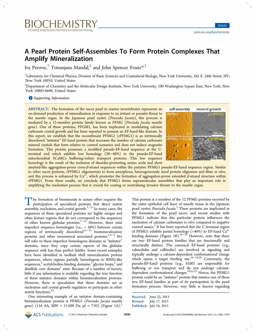
ABSTRACT: The formation of the nacre pearl in marine invertebrates represents anon-demand production of mineralization in response to an irritant or parasite threat tothe mantle organ. In the Japanese pearl oyster (Pinctada fucata), this process ismediated by a 12-member protein family known as PFMG (Pinctada fucata mantlegene). One of these proteins, PFGM1, has been implicated in modulating calciumcarbonate crystal growth and has been reported to possess an EF-hand-like domain. Inthis report, we establish that the recombinant PFMG1 (rPFMG1) is an intrinsicallydisordered “imitator” EF-hand protein that increases the number of calcium carbonatemineral crystals that form relative to control scenarios and does not induce aragoniteformation. This protein possesses a modified pseudo-EF-hand sequence at the C-terminal end which exhibits low homology (30−40%) to the pseudo-EF-handmitochondrial SCaMCs buffering/solute transport proteins. This low sequencehomology is the result of the inclusion of disorder-promoting amino acids and shortamyloid-like aggregation-prone cross-β-strand sequences within the putative PFMG1 pseudo-EF-hand sequence region. Similarto other nacre proteins, rPFMG1 oligomerizes to form amorphous, heterogeneously sized protein oligomers and films in vitro,and this process is enhanced by Ca2+, which promotes the formation of aggregation-prone extended β-strand structure withinrPFMG1. From these results, we conclude that PFMG1 forms supramolecular assemblies that play an important role inamplifying the nucleation process that is crucial for coating or neutralizing invasive threats to the mantle organ.
The formation of biominerals in nature often requires theparticipation of specialized proteins that direct matrix
assembly, nucleation, and crystal growth.1−12 In many cases, thesequences of these specialized proteins are highly unique andoften feature regions that do not correspond to the sequencesof other known globular proteins.1−11 However, there existimperfect sequence homologies (i.e., < 50%) between certainregions of intrinsically disordered13−15 biomineralizationproteins and other nonmineral associated proteins.1,3−5 Wewill refer to these imperfect homologous domains as “imitator”domains, since they copy certain aspects of the globularsequence with less than perfect fidelity. Some recent exampleshave been identified in mollusk shell mineralization proteinsequences, where regions partially homologous to RING-likesequences,5 acetylcholine-binding domains,3 glycine loops,1 anddisulfide core domains1 exist. Because of a number of factors,little if any information is available regarding the true functionof these imitator domains within biomineralization proteins.However, there is speculation that these domains act asnucleation and crystal growth regulators or participate in othermatrix functions.3,5
One interesting example of an imitator domain-containingbiomineralization protein is PFMG1 (Pinctada fucata mantlegene) (116 AA, MW = 13 609 Da, pI = 7.93) (Figure 1A).3
This protein is a member of the 12 PFMG proteins secreted bythe outer epithelial cell layer of mantle tissue in the Japanesepearl oyster, Pinctada fucata.3 These proteins are implicated inthe formation of the pearl nacre, and recent studies withPFMG1 indicate that this particular protein influences thenucleation of calcium carbonates in vitro compared to negativecontrol assays.3 It has been reported that the C-terminal regionof PFMG1 exhibits partial homology (<40%) to EF-hand Ca2+
binding domains (Figure 1B).16−22 However, note that thereare two EF-hand protein families that are functionally andstructurally distinct. The canonical EF-hand proteins (e.g.,calmodulin and calbindin) are involved in signaling andtypically undergo a calcium-dependent conformational changewhich opens a target binding site.16−18,20 Conversely, thepseudo-EF-hand proteins (e.g., S100) are responsible forbuffering or ion transport and do not undergo calcium-dependent conformational changes.19,21,22 Hence, the PFMG1protein could be an “imitator” protein that mimics one of thesetwo EF-hand families as part of its participation in the pearlformation process. However, very little is known regarding
Received: June 22, 2013Revised: July 17, 2013Published: July 18, 2013
PFMG1 function and structure, and hence it is not clear howthis biomineralization protein, or its putative EF-hand region,should be classified.In this paper we report new experiments which establish the
functional and structural relevance of PFMG1 and its putativeEF-hand region with regard to the pearl biomineralizationprocess. Specifically, we document the ability of recombinantPFMG1 (rPFMG1) to boost the nucleation process asexhibited by increased in vitro crystal production with noevidence of polymorph (aragonite) formation. We alsodiscovered that rPFMG1, like other nacre-associated proteins,is intrinsically disordered, exhibits unique pH- and Ca2+-dependent aggregation-prone behavior in solution, and self-assembles to form heterogeneously sized protein oligomersunder mineralization conditions. Using bioinformatics, weconfirm a 30−40% sequence homology between the C-terminalregion of PFMG1 (positions 61−110) and the pseudo-EF-handdomains of small calcium-binding mitochondrial carrier(SCaMCs) subfamilies that possess solution buffering capa-bilities.19,21,22 Hence, PFMG1 is an aggregation-prone pseudo-EF-hand “imitator” protein that amplifies the nucleationprocess during pearl formation.
■ EXPERIMENTAL PROCEDURES
Synthesis and Purification of rPFMG1. The genesynthesis, cloning, bacterial expression, and purification ofrPFMG1 were performed by GenScript USA (Piscataway, NJ)using their proprietary OptimumGene system and recombinantexpression systems. The synthetic DNA was created from thecomplete PFMG1 sequence (UniProt accession numberQ3YL59) minus the membrane leader sequence (residues 1−20) yielding a target sequence of 116 AA that now starts atresidue 21 (new N-terminus, R1) and ends at residue 136 (nowD116) (Figure 1A). To this DNA sequence a hybrid poly(His)6tag−enterokinase (EK) protease cleavage site was incorporatedat the N-terminus to facilitate the purification of rPFMG1 in aone-step fashion using high-affinity Ni chelation chromatog-raphy followed by enzyme digestion/affinity removal. Therecombinant plasmid was transiently transfected into a 100 mLsuspension of HEK 293 cell culture, which was then grown inserum-free media, collected, and lysed at day 5 post-transfection. Cell pellets were resuspended in phosphatebuffered saline (PBS) and sonicated for 2 min to reduce
solution viscosity. The solution was then centrifuged, and thetarget protein was captured from the cell lysate using HiTrapchelating Ni HP column and eluted with 200 mM imidazolebuffer.Following Ni elution, poly(His)6 tag removal was accom-
plished via EK digestion and subsequent affinity capture of theEK enzyme. The final protein purity was determined to be 93%(4%−20% gradient SDS-PAGE/Coomassie Blue staining,reducing conditions). The minor impurity was determined tobe an aggregate of the rPFMG1 protein (>100 kDa), which wassubsequently removed from the protein sample via ultra-filtration (Amicon Ultra 0.5, 30 kDa MWCO, MilliporeCorporation) at 14 000 rpm, 15 min, and 25 °C, withrPFMG1 recovered in the flow-through supernatant. Using aBruker Daltonics MALDI-TOF, we determined the apparentMW of this purified material to be 13 625 kDa (Figure S1,Supporting Information), which is close to the hypotheticalMW of 13 609 Da. Purified protein stock aliquots were storedin 50 mM Tris-HCl/10% v/v glycerol (pH 8.0) at −80 °C untilneeded. For subsequent experimentation, rPFMG1 sampleswere created by exchanging and concentrating appropriatevolumes of stock solution into unbuffered deionized distilledwater (UDDW) or other appropriate buffers using AmiconUltra 0.5, 3 kDa MWCO (Millipore Corporation).
Calcium Carbonate Nucleation. Using the same assayconditions employed in nacre protein−aragonite formationstudies (16 h, 16 °C, 12.5 mM CaCl2, with the final pH = 8.0−8.3 at the conclusion of the assay), we monitored the effect ofrPFMG1 on calcium carbonate crystal growth using standardsolid ammonium carbonate vapor decomposition meth-ods.8,11,12,23,24 These assay solutions contained either noprotein (negative control) or final assay concentrations of 0.7,1.8, 3.7, and 7.3 μM rPFMG1 protein. The collectionprocedure for SEM analysis of assay precipitates involved theuse of Si wafer fragments (1 cm2 or less in size, “P” type [1 0 0],20 ohm·cm, 250−350 μm, Silicon Quest Intl., Santa Clara, CA)or freshly cleaved geologic calcite fragments (Iceland spar, nolarger than 3 mm × 3 mm) that were placed at the bottom ofthe wells prior to the start of the assay.5,8 Si wafers and calcitefragments containing assay deposits were washed with calciumcarbonate saturated methanol and then dried at 37 °Covernight. For SEM imaging dried Si wafer or calcite fragmentsamples were coated with a thin layer of gold and then imagedusing a Hitachi S-3500N scanning electron microscope (5 kV)
Figure 1. (A) Bioinformatics analysis of the PFMG1 primary sequence (UniProt accession number Q3YL59). Disordered sequence regions (solidlines) were identified using IUP_PRED and DISOPRED2 prediction algorithms, and cross-β-strand sequence regions (dashed lines) which exhibitassociation propensities were identified using TANGO and AGGRESCAN. The gray highlighted region represents the putative EF-hand domain.(B) BLAST sequence alignment of PFMG1 with other EF-hand protein sequences. Gray residues denote homologies with PFMG1. The SCaMC1proteins are members of the pseudo-EF-hand family.
utilizing secondary emission detection. Energy dispersivespectra (EDS) were collected on carbon-coated samples usingthe PGT-Bruker X-ray microanalysis system at 25 kV.Dynamic Light Scattering. The hydrodynamic radii of
rPFMG1 complexes were measured over a pH range of 7−10.5using a DynaPro MS/X dynamic light scattering instrument(Protein Solutions, Inc.) and experimental protocols developedfor nacre polypeptides.11,24,25 Using 10 mM buffers (pH 7.0−8.5, Tris-HCl buffer; pH 9.0−10.5, sodium carbonate−bicarbonate buffer), we created final rPFMG1 concentrations= 3.7, 7.3, 20, 50, 75, and 100 μM, which correspond to theconcentration range utilized in earlier nacre protein self-association DLS studies.11,24,25 All samples were filtered using0.22 μm poly(vinylidene fluoride) syringe filter (FisherScientific) and then incubated at 16 °C (same temperature asfor mineral assays) for 5 min in the cuvette prior tomeasurement.11,22,24,25 Studies conducted below pH 7 led toexcessive spontaneous protein precipitation, and for this reasonwere excluded from our analysis. Ten acquisitions were takenper pH point per sample. Data regularization analysis wasperformed using the Dynamics v6.0 software. By measuring thefluctuations in the laser light intensity scattered by the sample,the instrument is able to detect the speed (diffusion coefficient)at which the particles are moving through the medium.26
X-ray Diffraction. Washed and dried control and rPFMG1precipitates from mineralization assays were analyzed using aBruker D8 DISCOVER GADDS microdiffractometer equippedwith a VANTEC-2000 area detector in a φ rotation method.The X-ray generated from a sealed copper tube ismonochromated by a graphite crystal and collimated by a 0.5mm MONOCAP (λ Cu Kα = 1.541 78 Å). The sample−detector distance is 150 mm. Two runs with θ1 = θ2 = 15° and30° are collected for each specimen, and the exposure time is600 s per run. Data were merged and integrated by theXRD2EVAL program in the Bruker PILOT software. Thecomposition of calcium carbonate polymorphs was calculated ineach sample with reference to calcite, aragonite, and vaterite-specific X-ray diffraction data that were obtained from thePower Diffraction File (PDF), a database of X-ray powderdiffraction patterns maintained by the International Center forDiffraction Data (ICDD).AFM Imaging of rPFMG1 Assemblies. To complement
our DLS studies, we investigated the dimensional andmorphological characteristics of rPFMG1 assemblies capturedfrom solution onto mica substrates. AFM experiments wereexecuted at 25 °C using an Asylum MFP-3D standalone AFMoperating in tapping mode in buffer solution. V-shaped Si3N4cantilevers (reported spring constant 0.09 N/m) were used forimaging. A precise drive frequency in fluid (8 kHz) wascalculated for each cantilever prior to imaging by overlaying thethermal spectrum over the frequency sweep. Two rPFMG1samples were imaged: an apo sample (7.3 μM in 10 mM Tris-HCl, pH 8.0) and a Ca(II)-loaded sample (7.3 μM in 10 mMTris-HCl, 12.5 mM CaCl2, pH 8.0). All samples were deliveredonto a freshly stripped surface of mica (Ted Pella, Inc., 0.9 mmthick) and incubated for a period of 15 min at ambienttemperature prior to measurement. Igor Pro 6.01 software wasused for image acquisition at a scan rate of 2 Hz. Gwyddionsoftware27 was implemented for image processing, noisefiltering, and analysis.Circular Dichroism Spectrometry. CD spectra (190−260
nm) of r-PFMG1 in 10 mM Tris pH 8.0 and in the presence ofCaCl2 were collected at 25 °C on the AVIV stopped flow
202SF CD spectropolarimeter. A total of eight scans per samplewere collected in a cuvette with 0.1 cm path length, using 1 nmbandwidth, 1 nm wavelength step, and 0.5 s averaging time.The instrument was previously calibrated with D-10-camphor-sulfonic acid. Initial CD studies examined a range ofconcentrations from 0.7 to 7.3 μM. Calcium titrations wereperformed in 10 mM Tris pH 8.0, using 7.3 μM proteinsamples that contained Ca2+:protein mole ratios of 1:2, 1:1, 2:1,10:1, and 1700:1 (i.e., 12.5 mM CaCl2). The recorded spectrawere averaged and the appropriate background spectra (Trisbuffer with and without CaCl2) subtracted. Spectra weresmoothened using the binomial algorithm included in the AVIVCD software. Ellipticity is reported as mean residue ellipticity(deg cm2 dmol−1). Secondary structure estimates of rPFMG1samples were obtained using three different algorithms includedin the CDPro software package (SELCON3, CDSSTR,CONTILL) and the SMP56 Reference Protein Set.28−30
Bioinformatics. The Basic Local Alignment Search Tool(BLAST, National Center for Biotechnology Information) andInterProScan/SWISS MODEL programs (SwissProt) wereused to determine the sequence homology between PFMG1and protein database EF-hand protein sequences (Figure1B).31−33 To determine the location of disordered sequenceregions within the PFMG1 sequence, we employed theIUP_PRED34 and DISOPRED235 prediction algorithms usingdefault parameters. Subsequently, we utilized TANGO36 andAGGRESCAN37 to globally identify putative cross-β-strandsequence regions which exhibit association propensities.
■ RESULTSCrystal Growth Experiments Reveal That rPFMG1 Is a
Nucleation Enhancer. In previous light microscopy studies, arecombinant version of PFMG1 was demonstrated to affectcalcium carbonate crystal growth.3 However, smaller lengthscale measurements and quantitative analysis were notperformed on these deposits, and so the in vitro function ofthis protein was not firmly established. Using standardammonium carbonate vapor diffusion mineralization assays,we extend this knowledge and find that rPFMG1 enhances thenucleation of calcite crystals in vitro (Figure 2 and Figure S2).Compared to control assays (no protein added), we observethat the number of rhombohedral single and polycrystallinecalcite crystals increases as a function of rPFMG1 concen-tration. At the same time, we note that calcite crystaldimensions diminish (Figure 2). These rPFMG1-generatedmineral deposits were confirmed to be calcite (powder X-raydiffraction, Figure 3), with no detectable evidence of vaterite oraragonite formation. Thus, rPFMG1 increases the number ofnucleating mineral clusters at the expense of mineral clustersize, with no evidence of polymorph selection; i.e., the proteinacts as a nucleation enhancer.A high-magnification comparison of the mineral crystal
surfaces obtained in both the negative control and rPFMG1assays (Figure S3) reveals that control and rPFMG1 assaymineral samples have similar surface morphologies. Thus,under these assay conditions,2,4,5,11,12 rPFMG1 does not appearto alter calcite crystal morphologies or surfaces to a significantdegree. These findings correlate with our X-ray microanalyses,where we were unable to detect rPFMG1 protein on the surfaceof these crystals (via Cys-specific thiol S detection, Figure S4).Understandably, the fact that our mineral surfaces are rough intexture and that PFMG1 contains only two Cys residues(Figure 1A) prevents us from conducting an accurate
quantitative analysis. Thus, our SEM and X-ray microanalysisdate point to several possibilities: (a) the adsorption ofrPFMG1 onto calcite crystal surfaces is either very low ornonexistent (Figures S3 and S4); (b) the protein may beadsorbed onto crystal surfaces, but due to the low S content inthe protein, it is not possible to detect adsorbed rPFMG1; (c)the protein may be entrapped within the crystals and thusunavailable for surface detection by X-ray microanalysis.rPFMG1 Oligomerizes in Solution To Form Hetero-
disperse Supramolecular Assemblies. It is known thatsome nacre-associated proteins spontaneously form mineral-stabilizing protein oligomers in solution.11,12,23−25,38,39 Usingdynamic light scattering (DLS), we find that the same holdstrue for the pearl protein, rPFMG1 (Figure 4). At rPFMG1concentrations corresponding to those utilized in our assays
(7.3 μM, Figure 2), we observe that rPFMG1 oligomerizes overthe pH range 7−10.5 (Figure 4 and Figure S5). At all pH pointsthere is significant size heterogeneity as evidenced bypolydispersity values >15% (represented by error bars oneach histogram bar). On the basis of mass percent, twooligomer populations are noted at pH = 7, 9, 10, and 10.5 withhydrodynamic radii (RH) values ranging from 4.7−7.6 nm and27.7−48.2 nm. At pH 8.0, which coincides with themineralization assay pH and the pI of PFMG1, a singleoligomer population is noted (RH = 75 nm), and this representsthe largest apo-rPFMG1 oligomer that we can detect with DLS.In contrast, three populations exist at pH 9.5 (RH = 2.6, 7.4, and47.3 nm; Figure 4). The variations in mass percent and particlesize suggest that rPFMG1 oligomerization is pH-dependent,and thus protein electrostatics plays a role in the self-assemblyprocess.We next explored rPFMG1 self-assembly in the presence of
Ca2+ at pH 8.0 (Figures S6 and S7). At all stoichiometries thereis significant size heterogeneity as evidenced by polydispersityvalues >15%. At Ca2+:protein stoichiometries of 1:1 and 2:1, weobserve three rPFMG1 oligomer populations, and at all otherstoichiometry points we observe two oligomer populations. Ingeneral, Ca(II)-induced oligomer dimensions (RH = 4−20 nm,40−64 nm) exceed those obtained for the apo state (Figure 4).Hence, rPFMG1 self-assembles to form heterodisperseoligomers over a wide pH range, with a higher degree ofoligomerization noted in the presence and absence of Ca2+ atpH 8.0 (Figure 2).
rPFMG1 Oligomerization Is Enhanced by Ca2 andPromotes Protein Film Formation. Our DLS experimentscan only detect submicrometer-sized protein assemblies, andpotentially any oligomers >0.2 μm would be purged viafiltration. To probe beyond this limit, we performed tappingmode AFM imaging of 7.3 μM rPFMG1 protein oligomers thatsettle onto untreated mica substrates (Figure 5) underconditions reflecting assay scenarios (Figure 2) and highestparticle sizes (Figure 4). In the apo state, we confirm theformation of heterogeneously sized amorphous-appearingoligomers that are spherical to ellipsoidal in morphology(Figure 5). Using line profile extraction of amplitude and heightimages of 10 typical oligomers, we estimate that apo-rPFMG1protein oligomers possess average diameters = 31 ± 9 nm andheights = 1 ± 0.7 nm. With the introduction of Ca2+, we nowobserve larger complexes and evidence of particle fusion withaverage diameters = 86 ± 34 nm (2−2.25-fold increase) andheights = 6 ± 3 nm (3−4-fold increase) for individualcomplexes (Figure 5). Thus, we confirm our DLS findings,namely, that rPFMG1 possesses self-associative capabilities thatare enhanced by Ca2+.The apo-rPFMG1 oligomers appear contiguous and suggest
that a film-like protein coating is forming on the mica surface,with larger particles projecting above the film layer (Figure 5).To confirm this, we measured the Rq, or root-mean-squaresurface roughness, of a freshly cleaved mica surface in buffer (Rq= 0.078 nm) versus untreated mica surfaces exposed to apo-rPFMG1 (Rq = 0.17 nm, ∼2-fold increase) and rPFMG1 in12.5 mM CaCl2 (Rq = 0.26, ∼3-fold increase) and confirmedthat under apo and Ca2+ conditions the mica surfaces arecoated with rPFMG1 oligomers. Other calculated surfaceparameters [i.e., roughness average (Ra), maximum roughness(Rp)] were found to exhibit similar trends between plain micaand rPFMG1-treated mica (data not shown), and this confirms
Figure 2. Scanning electron micrograph of mineral deposits capturedby geologic calcite fragments in ammonium carbonate vapor diffusionmineralization assays. (A) Protein-free assay (negative control). Notepredominance of single crystal and polycrystalline calcite crystals. (B,C, D) 1.8, 3.7, and 7.3 μM rPFMG1 assays, respectively. Note that asrPFMG1 concentrations increase, the number of calcite crystalincreases. Scale bars = 100 μm.
Figure 3. Representative powder micro-X-ray diffraction spectra of Siwafers taken from protein-free (control) and rPFMG1-containingmineralization assays. Calcite (c) reference peaks are denoted andwere obtained from known data sets (International Center forDiffraction Data (ICDD)). Note that diffraction data for 7.3 μMrPFMG1 is identical to that obtained for 3.7 μM.
the formation of a protein coating or film comprised ofoligomers on these mica surfaces.Structural Features of Oligomeric rPFMG1. Earlier
studies with oligomeric nacre-associated proteins demonstratedthe presence of intrinsic disorder that persists in the assembledstate.4,9,11,12,23−25,38,39 Since rPFMG1 spontaneously formsprotein assemblies (Figures 4 and 5), it is likely that disorderedregions are present in this protein, but this has yet to beestablished. Moreover, canonical and pseudo-EF-hand proteinspossess specific structural features in the presence and absenceof Ca2+,16−20 and to date no specific studies have establishedwhich structural features are found in PFMG1.To remedy this, we first performed circular dichroism (CD)
spectrometry on apo-rPFMG1 protein oligomer samples (i.e.,7.3 μM rPFMG1, 10 mM Tris-HCl, pH 8.0). Here, we find thatthe CD spectra for rPFMG1 oligomers feature a (+) ellipticityband with a maxima at 187 nm and three broad (−) ellipticitybands with minima at 208, 215, and 220 nm (Figure 6). With208 nm/222 and 215 nm being characteristic for α-helix and β-
strand, respectively,8,9,11,12,28−30 it is clear that rPFMG1assemblies are comprised of protein molecules that possessresidual structure.8,9,11,12 However, since these bands are broadand weak in intensity, it is likely that the protein contains somedegree of unfolding or disorder.8,9,11,12,28−30 This is confirmedby comparison to CD database reference spectra (CDSSTR,SMP56, inset, Figure 6) where rPFMG1 can be characterized asapproximately 38% random coil, 21% turn, 35% total β-strand,and 6% helix.28−30 We stress caution when interpreting theseresults as absolute values and prefer to rely on them in relativeterms. Nonetheless, we note that the low content of α-helicalstructure is atypical for folded EF-hand proteins that contain astable 4-helix bundle.16−20 Hence, on a structural basis, weconclude that rPFMG1 is intrinsically disordered and does notfit the description of a true canonical 4-helix bundle EF-handprotein.Next, to qualitatively classify the type of putative EF-hand
domain sequence in PFMG1, we utilized stoichiometric Ca2+
titration in tandem with CD to determine the Ca2+-inducedconformational response of rPFGM1 within assemblies (Figure
Figure 4. Dynamic light scattering analysis of r-PFMG1 oligomerization (7.3 μM) in various pH buffers at 16 °C. (A) Hydrodynamic radiidetermined for distinct oligomer populations as defined in Figure S5. Error bars represent polydispersity of particle sizes obtained for each pH. (B)Pie chart representation of distinct oligomer populations and corresponding mass % as a function of pH. Mass % and corresponding RH radii arematched by comparable grayscale shading of pie charts and histogram bars.
Figure 5. Tapping mode AFM images of 7.3 μM rPFMG1 proteinoligomers (10 mM Tris-HCl, pH 8.0) in the presence and absence of12.5 mM CaCl2 on untreated mica surfaces. Note in the height plotsthat the rPFMG1 oligomers form a contiguous film-like layer orcoating on the mica surface.
Figure 6. Circular dichroism spectra of 7.3 μM oligomeric rPFMG1(10 mM Tris-HCl buffer, pH 8.0) in the presence and absence ofstoichiometric levels of CaCl2. “Assay” refers to 12.5 mM CaCl2 (i.e.,Ca2+:rPFMG1 1700:1). Inset histogram plot details % secondarystructure content determined by CDPro (CDSSTR) and the SMP56Reference Protein Set.28−30 The histogram data are reported asfractional weight distributed between regular helix (αr), distorted helix(αd), regular strand (βr), distorted strand (βd), turn (t), and randomcoil or unordered (rc) structures.
6). Compared to the apo-state, we note that CD bandintensities initially increase at Ca2+:protein = 1:1 but thendiminish as Ca2+ levels increase. We attribute this reduction inmolar ellipticity intensities to increases in either proteinassembly dimensions or populations (Figure 5; Figures S6and S7) that would lead to increased beam scattering andsubsequent loss in signal intensities. In terms of secondarystructure content, we note that at Ca2+:protein = 1:1 therandom coil and helix contents decrease relative to the apo-state. Simultaneously we note concomitant increases in β-strand and turn structures relative to the apo-state. AtCa2+:protein ratios >1:1 we note that these trends persist andthat there are no further changes in secondary structure content(Figure 6), even when we reach mineral assay levels (12.5 mMor 1700:1). We conclude the following: (a) The structuralresponse of rPFMG1 to Ca2+ is similar to that described forpseudo-EF-hand proteins, i.e., limited conformational sensitiv-ity to Ca2+;18−21 (b) Ca2+ induces increases in β-structureswithin rPFMG1 at the expense of helical and random coilcontents, and since β-structures are associated with aggrega-tion,36−38 this explains why rPFMG1 forms larger aggregatesand films in the presence of Ca2+ compared to the apo-state(Figure 5; Figures S6 and S7).Bioinformatics Confirm That PFMG1 Is a Pseudo-EF-
Hand Protein That Possesses Intrinsically Disorderedand Aggregation-Prone Regions. It is known that nacreextracellular matrix proteins possess two types of sequencecharacteristics that promote self-association: intrinsic disorder/unfolding and amyloid-like aggregation prone cross-β-strandsupersecondary structures.38 Using predictive algorithms IUPand DISOPRED, we identified an intrinsically disorderedregion in both the N-terminal and C-terminal domains ofPFMG1 (Figure 1A). Interestingly, the predicted region ofdisorder in the C-terminal domain overlaps with a 13 AAsequence region that comprises the putative EF-hand domain(3). The presence of disorder in this region may explain whyrPFMG1 possesses a lower α-helical content compared to otherEF-hand proteins (Figure 6).16−22 Similarly, using predictivealgorithms TANGO and AGGRESCAN for identifyingaggregation-prone cross-β-strand sequences, we identifiedthree putative regions (representing 22% of the total sequencelength) at positions 26−34, 46−52, and 81−89 (coincidingwith the EF-hand region) (Figure 1A). Once again, there existsoverlap between a predicted assembly domain and the EF-handdomain.3 The prediction of 22% extended β-strand contentroughly correlates with the detection of regular (20%) anddistorted (14%) β-strand content detected by CD spectrometry(Figure 6). Thus, the PFMG1 protein contains intrinsicallydisordered and aggregation-prone sequences, with examples ofeach sequence type found within the putative EF-hand domain.The occurrence of aggregation-prone sequence features
within the PFMG1 EF-hand region provides the context for are-examination of sequence homology of this protein (Figure1B). Initial homology studies reported that PFMG1 exhibited40% homology with the EF-hand calcium binding protein,CBP-1,3 but no other homologous sequences were reported atthat time. Using BLAST and InterProScan/SWISS MODEL,we identified seven additional pseudo-EF-hand proteinsequences that are homologous (27−43%) to PFMG1 (Figure1B). This list of proteins includes fucosytransferases(FUTRAse), phosphodiesterases (PIBP), calcium bindingprotein (CaBP), and solute carrier calcium-binding mitochon-drial carriers (SCaMCs) subfamilies that act as buffering
agents.19,21,22 Note that the SCaMCs exhibit the best homologymatch to the putative PFMG1 EF-hand sequence region, andthus PFMG1 appears to be a pseudo-EF-hand “imitator”protein whose sequence region are more closely related to thesolution buffering protein families.19,21,22
Interestingly, we note that the PFMG1 regions that exhibitthe lowest homology to SCaMCs possess aggregation-proneand intrinsically disordered features. Specifically, the -KLAE-VFFSEA- region (residues 81−90, 27% homology) is predictedto be an amyloid-like aggregation-prone sequence (Figure 1B).Conversely, the -ADENDDEQISTSE− region (residues 90−102, 46% homology) is predicted to be an intrinsicallydisordered sequence (Figure 1) due to the introduction ofdisorder-promoting residues13−15,39 E, D at sequence positions92, 94, 96, and S, Q at positions 99 and 97, respectively (Figure1B). We believe that the disordered and aggregation proneregions within the PFMG1 pseudo-EF-hand sequence havealtered the functional properties of this SCaMCs-relateddomain, such that this sequence region can participate inprotein−protein interactions (Figure 5) and protein−mineralmodulation (Figure 2).
■ DISCUSSIONThe formation of a natural pearl initiates when a foreignsubstance or parasite becomes trapped between the shell nacrelayer and the mantle organ of the mollusk.40 This triggerscellular migration and encapsulation around the irritant,followed by the release of the “maintenance crew” (i.e., mantleproteins that initiate a new cycle of nacre formation) into theencapsulated space around the irritant. Ultimately, a new layerof nacre (pearl) is created around the irritant, rendering itharmless to the mantle organ. The PFMG protein family is apart of this “crew”, and as we have demonstrated in this report,PFMG1 protein potentially plays an important part in the nacredeposition process. Our in vitro mineralization experimentsconfirm that oligomeric PFMG1 is a nucleation enhancer(Figure 2 and Figure S2) that increases the number of calciumcarbonate crystals formed relative to control scenarios yet doesnot participate in aragonite selection/stabilization (Figure 3;Figures S3 and S4). Within the context of pearl formation40 andthe current prenucleation cluster hypothesis,41−43 we believethat oligomeric PFMG1 rapidly induces mineral formation inorder to ensure that the irritant becomes rapidly coated andneutralized. Hypothetically, this could be accomplished byincreasing the number of prenucleation clusters that are formedin solution.42,43 This would give rise to a larger number ofsmaller-sized ACC clusters, which, in turn, would result in thegeneration of more crystals with smaller dimensions comparedto control scenarios (Figure 2). Clearly, additional exper-imentation will be required to delineate the pearl nacreformation process in more detail.In earlier studies we investigated the mineralization activities
of the N- and C-terminal 30 AA regions of PFMG1 usingmodel peptides.45 We found that both sequences form peptideassemblies under mineralization assay conditions similar tothose employed in the present study. Furthermore, bothpeptide sequences possessed secondary structure characteristicsthat are also similar to those found in rPFMG1 (Figure 6).However, the notable difference between rPFMG1 and thesetwo model peptides lies in their mineralization activities: WhilerPFMG1 amplifies crystal deposition and does not affect crystalmorphologies, both of the PFMG1-derived terminal sequencesnucleate aragonite within peptide assemblies, albeit at low
frequency.45 It is likely that these isolated, shorter sequencesnow exhibit functional aberrations from their true roles withinthe parent protein. With that in mind, and in light of thepresent rPFMG1 study, we advise the reader to interpret ourprevious PFMG1 model peptide study45 with caution andwithin the context of the current study.We believe that protein oligomerization is critical to the
PFMG1-mediated mineral amplification process (Figures 4 and5). We discovered that the optimal formation of rPFMG1assemblies occurs near the pI of the protein (pH 8.0, Figure 4)and in the presence of Ca2+ (Figure 5; Figures S6 and S7). Thisfinding strongly suggests that protein electrostatics (i.e., sidechain charge) is one of the driving forces for oligomerization. Inaddition, the detection of intrinsically disordered andaggregation-prone “interactive” sequences (Figures 1 and 6)makes a strong case for the participation of these sequences inoligomerization as well. Given that PFMG1 contains intrinsi-cally disordered regions, it is not surprising that rPFMG1oligomers adopt an amorphous morphology and are heteroge-neous in dimension (Figures 4 and 5). We believe this is animportant finding, for similar traits have been noted in otherdisordered biomineralization protein assemblies4,9,11,12,23−25,38
and in disordered polymers that form mineral-stabilizingpolymer-induced liquid phases (PILP).46,47 Comparativemineralization studies of PILP polymers and biomineralizationproteins will undoubtedly reveal if these disordered systems aremechanistically similar.In addition to oligomerization, it is likely that the C-terminal
EF-hand-like sequence (residues 61−110 Figure 1B) plays animportant role in rPFMG1 nucleation enhancement. Our CDand bioinformatics studies indicate that this sequence does notshare the same features and properties of the canonical EF-hand proteins. Rather, this sequence is qualitatively similar tothe pseudo-EF-hand sequences found in SCaMCs (Figure1).19,21,22 However, this affiliation with SCaMCs is tempered bythe occurrence of disordered and aggregation-prone sequenceswithin the pseudo-EF-hand sequence of PFMG1 (Figure 1A).As a result of these alterations, we believe that the PFMG1pseudo-EF-hand sequence region has been “tuned” tospecifically function within the pearl mineralization scheme.For example, we speculate that the PFMG1 pseudo-EF-hand“imitator” sequence may be responsible for the Ca2+-enhancedassembly of PFMG1 protein molecules (Figures 4 and 5;Figures S5 and S6). Alternatively, since PFMG1 proteinoligomers would contain numerous pseudo-EF-hand domainsconcentrated within a limited molecular volume (Figures 4 and5), these domains could play a role in the regulation ofmineralization conditions (e.g., pH buffering, Ca2+ availability)in the vicinity of these protein oligomers. This, in turn, couldsignificantly impact prenucleation cluster formation ki-netics41−43 and crystal quantities in solution (Figure 2).Obviously, other functions may be assignable to this domain,and additional studies will be required to establish theparticipation of the pseudo-EF-hand sequence within theoyster pearl mineralization process.
■ ASSOCIATED CONTENT*S Supporting InformationMALDI-TOF spectra of purified rPFMG1 (Figure S1), SEMimages of mineral deposits captured by Si wafers inmineralization assays (Figure S2), SEM and X-ray surfaceanalyses of control and rPFMG1-treated crystals (Figures S3and S4), DLS mass % plots for rPFMG1 as a function of pH
and Ca2+:protein mole ratios (Figures S5 and S6), and DLSanalysis of rPFMG1 oligomerization (Figure S7). This materialis available free of charge via the Internet at http://pubs.acs.org.
FundingThis research was supported by the U.S. Department of Energy,Office of Basic Energy Sciences, Division of Materials Sciencesand Engineering under Award DE-FG02-03ER46099.
NotesThe authors declare no competing financial interest.
■ ACKNOWLEDGMENTSWe thank Dr. Moise Ndao for performing the MALDI-TOFmass spectrometry analysis of rPFMG1. This report representscontribution number 67 from the Laboratory for ChemicalPhysics, New York University.
■ ABBREVIATIONSPFMG1, PNC, DLS, SCaMC1, solute carrier mitochondrialcarrier protein-1; S100, calcium binding protein that is 100%soluble in ammonium sulfate at neutral pH; CABP, calciumbinding protein; FUTRAse, α-1,3-fucosyltransferase XI; PIBP,1-phosphatidylinositol 4,5-bisphosphate phosphodiesteraseΔ3A.
■ REFERENCES(1) Shen, X., Belcher, A. M., Hansma, P. K., Stucky, G. D., andMorse, D. E. (1997) Molecular cloning and characterization of LustrinA, a matrix protein from shell and pearl nacre of Haliotis rufescens. J.Biol. Chem. 272, 32472−32481.(2) Samata, T., Hayashi, N., Kono, M., Hasegawa, K., Horita, C., andAkera, S. (1999) A new matrix protein family related to the nacreouslayer formation of Pinctada fucata. FEBS Lett. 462, 225−232.(3) Ma, Z., Huang, J., Sun, J., Wang, G., Li, C., Xi, L., and Zhang, R.(2007) A novel extrapallial fluid protein controls the morphology ofnacre lamellae in the pearl oyster, Pinctada fucata. J. Biol. Chem. 282,23253−23260.(4) Suzuki, M., Saruwatari, K., Kogure, T., Yamamoto, Y., Nishimura,T., Kato, T., and Nagasawa, H. (2009) An acidic matrix protein, Pif, isa key macromolecule for nacre formation. Science 325, 1388−1390.(5) Michenfelder, M., Fu, G., Lawrence, C., Weaver, J. C., Wustman,B. A., Taranto, L., and Evans, J. S. (2003) Characterization of twomolluscan crystal-modulating biomineralization proteins and identi-fication of putative mineral binding domains. Biopolymers 70, 522−533errata 73, 522..(6) Fu, G., Qiu, S. R., Orme, C. A., Morse, D. E., and DeYoreo, J. J.(2007) Acceleration of calcite kinetics by nacre proteins. Adv. Mater.17, 2678−2683.(7) Falini, G., Albeck, S., Weiner, S., and Addadi, L. (1996) Controlof aragonite or calcite polymorphism by mollusk shell macromolecules.Science 271, 67−69.(8) Ndao, M., Keene, E., Amos, F. A., Rewari, G., Ponce, C. B.,Estroff, L., and Evans, J. S. (2010) Intrinsically disordered molluskshell prismatic protein that modulates calcium carbonate crystalgrowth. Biomacromolecules 11, 2539−2544.(9) Delak, K., Harcup, C., Lakshminarayanan, R., Zhi, S., Fan, Y.,Moradian-Oldak, J., and Evans, J. S. (2009) The tooth enamel protein,porcine amelogenin, is an intrinsically disordered protein with anextended molecular configuration in the monomeric form. Biochemistry48, 2272−2281.
(10) Mann, K., Siedler, F., Treccani, L., Heinemann, F., and Fritz, M.(2007) Perlinhibin, a cysteine, histidine, and arginine-rich miniproteinfrom abalone (Haliotis laevigata) nacre, inhibits in vitro calciumcarbonate crystallization. Biophys. J. 93, 1246−1252.(11) Amos, F. F., and Evans, J. S. (2009) AP7, a partially disorderedpseudo C-RING protein, is capable of forming stabilized aragonite invitro. Biochemistry 48, 1332−1339.(12) Amos, F. F., Ndao, M., Ponce, C. B., and Evans, J. S. (2011) AC-RING-like domain participates in protein self-assembly and mineralnucleation. Biochemistry 50, 8880−8887.(13) Uversky, V. N. (2002) Natively unfolded proteins: A pointwhere biology waits for physics. Protein Sci. 11, 739−756.(14) Tompa, P. (2002) Intrinsically unstructured proteins. TrendsBiochem. Sci. 27, 527−533.(15) Meng, J., Romero, P., Yang, J. Y., Chen, J. W., Vacic, V.,Obradovic, Z., and Uversky, V. N. (2008) The unfoldomics decade:An update on intrinsically disordered proteins. BMC Genomics 9, 1−26.(16) Chou, J. J., Li, S., Klee, C. B., and Bax, A. (2001) Solutionstructure of Ca(2+) calmodulin reveals flexible hand-like properties ofits domains. Nat. Struct. Biol. 8, 990−997.(17) Takeda, S., Yamashita, A., Maeda, K., and Maeda, Y. (2003)Structure of the core domain of human cardiac troponin in theCa(2+)-saturated form. Nature 424, 35−41.(18) Kojetin, D. J., Venters, R. A., Kordys, D. R., Thompson, R. J.,Kumar, R., and Cavanagh, J. (2006) Structure, binding interface andhydrophobic transitions of Ca2+-loaded calbindin-D(28k). Nat. Struct.Mol. Biol. 13, 641−647.(19) Del Arco, A., and Satrustegui, J. (1998) Molecular cloning ofAralar, a new member of the mitochondrial carrier superfamily thatbinds calcium and is present in human muscle and brain. J. Biol. Chem.273, 23327−23334.(20) Nelson, M. R., Thulin, E., Fagan, P. A., Forsen, S., and Chazin,W. J. (2002) The EF-hand domina: a globally cooperative structuralunit. Protein Sci. 11, 198−205.(21) Traba, J., Del Arco, A., Duchen, M. R., Szabadkai, G., andSatrustegui, J. (2012) SCaMC-1 promotes cancer cell survival bydesensitizing mitochondrial permeability transition via ATP/ADP-mediated matrix Ca(2+) buffering. Cell Death Differ. 19, 650−660.(22) Del Arco, A., and Satrustegui, J. (2004) Identification of a novelhuman subfamily of mitochondrial carriers with calcium bindingdomains. J. Biol. Chem. 279, 24701−24713.(23) Keene, E. C., Evans, J. S., and Estroff, L. A. (2010) Silk fibroinhydrogels coupled with the n16N-beta-chitin complex: An in vitroorganic matrix for controlling calcium carbonate mineralization. Cryst.Growth Des. 10, 5169−5175.(24) Amos, F. F., Ponce, C. B., and Evans, J. S. (2011) Formation offramework nacre polypeptide supramolecular assemblies that nucleatepolymorphs. Biomacromolecules 12, 1883−1889.(25) Ponce, C. B., and Evans, J. S. (2011) Polymorph crystalselection by n16, an intrinsically disordered nacre framework protein.Cryst. Growth Des. 11, 4690−4696.(26) Schartl, W. (2007) Light Scattering from Polymer Solutions andNanoparticle Dispersions 1st ed., Springer-Verlag, Heidelberg, Germany.(27) Necas, D., and Klapetek, P. (2012) Gwyddion: an open-sourcesoftware for SPM data analysis. Cent. Eur. J. Phys. 10, 181−188.(28) Sreerama, N., and Woody, R. W. (2000) Estimation of proteinsecondary structure from circular dichroism spectra: Comparison ofCONTIN, SELCON, and CDSSTR methods with an expandedreference set. Anal. Biochem. 287, 252−260.(29) Lobley, A., Whitmore, L., and Wallace, B. A. (2002)DICHROWEB: An interactive website for the analysis of proteinsecondary structure from circular dichroism spectra. Bioinformatics 18,211−212.(30) Sreerama, N., and Woody, R. W. (2004) On the analysis ofmembrane protein circular dichroism spectra. Protein Sci. 13, 100−112.(31) Zdobnov, E. M., and Apweiler, R. (2001) InterProScan anintegration platform for the signature recognition methods in InterPro.Bioinformatics 17, 847−848.
(32) Arnold, K., Bordoli, L., Kopp, J., and Schwede, T. (2006) TheSWISS-MODEL Workspace: A web-based environment for proteinstructure homology modeling. Bioinformatics 22, 195−201.(33) Kiefer, F., Arnold, K., Kunzli, M., Bordoli, L., and Schwede, T.(2009) The SWISS-MODEL Repository and associated resources.Nucleic Acids Res. 37, D387−D392.(34) Dosztanyi, Z., Csizmok, V., Tompa, P., and Simon, I. (2005)IUPred: web server for the prediction of intrinsically unstructuredregions of proteins based on estimated energy content. Bioinformatics21, 3433−3434.(35) Ward, J. J., Sodhi, J. S., McGuffin, L. J., Buxton, B. F., and Jones,D. T. (2004) Prediction and functional analysis of native disorder inproteins from the three kingdoms of life. J. Mol. Biol. 337, 635−645.(36) Linding, R., Russell, R. B., Neduva, V., and Gibson, T. J. (2004)A comparative study of the relationship between protein structure andbeta-aggregation in globular and intrinsically disordered proteins(TANGO). J. Mol. Biol. 342, 345−353.(37) Conchillo-Sole, O., de Groot, N. S., Aviles, F. X., Vendrell, J.,Daura, X., and Ventura, S. (2007) AGGRESCAN: a server for theprediction and evaluation of “hot spots” of aggregation inpolypeptides. BMC Bioinformatics 8, 65−82.(38) Evans, J. S. (2012) Identification of intrinsically disordered andaggregation - promoting sequences within the aragonite-associatednacre proteome. Bioinformatics 28, 3182−3185.(39) Ndao, M., Dutta, K., Bromley, K., Sun, Z., Lakshminarayanan,R., Rewari, G., Moradian-Oldak, J., and Evans, J. S. (2011) Probing theself-association, intermolecular contacts, and folding propensity ofamelogenin. Protein Sci. 20, 724−734.(40) Southgate, P., and Lucas, J. (2008) in The Pearl Oyster: ABeginner’s Guide to Programming Images, Animation, and Interaction pp77−102, Elsevier BV, Oxford, UK.(41) Gebauer, D., Volkel, A., and Coelfen, H. (2008) Stableprenucleation of calcium carbonate clusters. Science 322, 1819−1822.(42) Gebauer, D., and Coelfen, H. (2011) Prenucleation clusters andnon-classical nucleation. Nano Today 6, 564−584.(43) Demichelis, R., Raiteri, P., Gale, J. D., Quigley, D., and Gebauer,D. (2011) Stable prenucleation mineral clusters are liquid-likepolymers. Nat. Commun. 2, 1−8.(44) Gower, L. B. (2008) Biomimetic model systems for investigatingthe amorphous precursor pathway and its role in biomineralization.Chem. Rev. 108, 4551−4627.(45) Amos, F. F., Destine, E., Ponce, C. B., and Evans, J. S. (2010)The N- and C-terminal regions of the pearl-associated EF Handprotein, PFMG1, promote the formation of the aragonite polymorphin vitro. Cryst. Growth Des. 10, 4211−4216.(46) Wolf, E., Leiterer, J., Pipich, V., Barrea, R., Emmerling, F., andTremel, W. (2011) Strong stabilization of amorphous calciumcarbonate emulsion by ovalbumin Gaining insight into the mechanismof ’polymer-induced liquid precursor’ processes. J. Am. Chem. Soc. 133,12642−12649.(47) Freeman, C. L., Harding, J. H., Quigley, D., and Rodger, P. M.(2010) Structural control of crystal nuclei by an eggshell protein.Angew. Chem., Int. Ed. 49, 5135−5137.