1 Università degli Studi ROMA TRE SCUOLA DOTTORALE IN BIOLOGIA (XXII CICLO) Sezione: SCIENZE BIOMOLECOLARI E CELLULARI A possible molecular mechanism for parasitic inhibition by Lactoferrin Dottorando: Docente Guida: Dr. Loris Leboffe Prof. Giovanni Antonini ANNO ACCADEMICO 2008-2009
Transcript
1
Università degli Studi ROMA TRE
SCUOLA DOTTORALE IN BIOLOGIA (XXII CICLO)
Sezione: SCIENZE BIOMOLECOLARI E CELLULARI
A possible molecular mechanism
for parasitic inhibition by Lactoferrin
Dottorando: Docente Guida:
Dr. Loris Leboffe Prof. Giovanni Antonini
ANNO ACCADEMICO 2008-2009
2
A POSSIBILE MOLECULAR MECHANISM FOR PARASITIC INHIBITION BY LACTOFERRIN
Summary
Transferrins (Tfs) belong to a family of iron-binding glycoproteins possessing similar aminoacid sequence though they have different biological functions and locations. Lactoferrin (Lf) is expressed and secreted from glandular epithelial cells and from mature neutrophiles of mammalian and it is an important component of the aspecific host defence or natural immunity, including resistance to parasitic infections. Serum transferrin (sTf) is synthesized by the liver of mammals and secreted into the blood stream; its primary function is iron transport. Ovotransferrin (Otrf), synthesized by avians, displays both iron transport and protective functions.
Parasites synthesize papain-like cysteine proteases that are relevant for the virulence and pathogenicity of parasites, being involved in several aspects of the parasite life cycle, it is therefore possible that the antiparasitic activity of Lf could be due to the inhibition of parasitic papain-like cysteine protease that we have recently observed.
In this study we have investigated the thermodynamic parameters of hLf, bLf and Otrf inhibition of the parasitic papain-like type I cysteine proteases from Leishmania infantum, Trypanosoma cruzi and Trypanosoma brucei. bLf, hLf and Otrf, both in the apo- and olo-forms, showed time- and concentration-dependent inhibition of the catalytic activity of papain and of type I proteases from L.infantum, T. cruzi and T. brucei. The KI values observed for bLf and hLf inhibition of L. infantum, T. cruzi and T. brucei proteases were in the nanomolar range (KI = 3.1 nM), lower than KI values observed for papain inhibition (KI = 24 nM). Otrf showed lower inhibition of cysteine proteases (KI = 0.6 µM). On the contrary, sTf did not display any inhibition towards parasitic proteases, according to its different role in mammals. The inhibition of parasitic cysteine proteases by hLf, bLf and Otrf appeared to conform to a competitive mechanism. The observed pH
3
optimum for bLf inhibition of parasitic proteases was around neutrality, while it was acidic for hLf and alkaline for Otrf. The further quantitative analysis of pH dependence of the intrinsic ligand-independent inhibition constant KI allowed the evaluation of pKa values that define the acid-base equilibrium of amino acidic residue(s) modulating the enzyme(s)-inhibitor recognition events. SDS-PAGE showed that hLf, bLf and Otrf were easily degraded by either papain or parasitic type I protease during the assay incubation time (few minutes) and it is likely that one or more protease inhibitory peptides were generated from protein hydrolysis.
As a matter of fact, a sequence present near the C-terminus of human (hLf) and bovine (bLf) lactoferrin shows homology with the sequence of the active site of cystatins, which are competitive inhibitors of papain-like cysteine proteases. The same sequence is present, though with lower homology, in Otrf and, with even lower homology, in sTf.
Therefore, we have charactherized by MALDI-TOF the profile of Lf cleavage by papain and preliminary data suggest the presence of a cystatin-like peptide in two proteolytic fragments of hLf and in one proteolytic fragment of bLf.
Le transferrine (Tfs) sono una famiglia di glicoproteine in grado di legare reversibilmente il ferro, e presentano un’elevata omologia di sequenza tra tutti i membri di questa famiglia. In particolare la lattoferrina (Lf) è prodotta e secreta nelle cellule ghiandolari epiteliali ed è presente nei granuli dei granulociti neutrofili dei mammiferi. Essa rappresenta un’importante componente della difesa immunitaria aspecifica dell’ospite. La siero transferrina (sTf) invece è sintetizzata nel fegato, ed è coinvolta unicamente nel trasporto del ferro. L’ovotransferrina (Otrf), l’omologa aviaria della Lf , svolge sia una funzione di difesa che di trasporto del ferro.
Le proteasi a cisteina papaina-simili da protozoi e metazoi parassiti, secretorie e di membrana, partecipano ai processi di morfogenesi degli organismi patogeni, sono implicate nell’invasione delle cellule e dei tessuti da parte dei parassiti, riducono la risposta immunitaria dell’ospite e costituiscono uno dei fattori più rilevanti nelle patologie associate alle infestazioni da parassiti. Pertanto, a fronte della capacità della Lf di svolgere un’azione antiparassitaria, noi abbiamo ipotizzato che la Lf possa svolgere tale azione grazie all’inattivazione delle suddette proteasi a cisteina.
4
Nel presente lavoro abbiamo studiato le proprietà termodinamiche dell’inibizione delle proteasi a cisteina di alcuni parassiti, quali Leishmania infantum, Tripanosoma cruzi e Trypanosoma brucei. Tale studio è stato esteso anche verso la papaina, capostipite di questa famiglia. Innanzitutto è emerso una maggiore affinità delle transferrine studiate (bLf, hLf e Otrf, in forma apo e olo) verso le proteasi parassitarie (K I = 3.1 nM) rispetto a quella verso la papaina (KI = 24 nM). Otrf risulta essere l’inibitore con minore affinità (KI = 0.6 µM). Inoltre è emerso che la sTf non è in grado di svolgere una’zione inibitoria, coerentemente con la sua funzione nei mammiferi. L’inibizione di tutte le proteasi risulta essere conforme ad un meccanismo di inibizione competitivo.
Da un’analisi della dipendenza del pH dei suddetti fenomeni di inibizione si è osservato una maggiore attività della bLf a pH fisiologico mentre la hLf inibisce più efficacemente a valori di pH acidi (pH = 5.0) e la Otrf a valori di pH alcalini (pH = 9.0). La dipendenza dal pH del parametro KI relativo all’interazione degli inibitori studiati con gli enzimi, ha permeso di evidenziare la variazione di diversi valori di pKa, ascrivibili ad eventi di protonazione e deprotonazione di differenti gruppi ionizzabili.
Inoltre si è osservato, a seguito di SDS-page, una parziale idrolisi della Lf e della Otrf durante diverse incubazioni con la papaina e con le proteasi parassitarie, e l’insieme di tali dati suggerisce la liberazione di un peptide, che può quindi inibire competitivamente le suddette proteasi a cisteina.
A supporto di tale ipotesi, si è osservato in presenza della porzione C-terminale della Lf, una sequenza aminoacidica che presenta un’elevata omologia con il sito attivo delle cistatine, una (super)famiglia di proteina, riconosciute essere gli inibitori reversibili per eccelenza delle proteasi a cisteina papaina-simili. Abbiamo quindi caratterizzato, mediante MALDI TOF, il profilo di idrolisi della Lf ad opera della papaina, ed è stata riscontrata la presenza di frammenti contenenti tale sequenza cistatino-simile, sia nella bLf che nell’hLf.
Among parasitic Protozoa, trypanosomatids belonging to the Leishmania and Trypanosoma genres comprise etiological agents of endemic diseases mainly localised in developing countries. All the human pathogenic trypanosomatids are dixenic parasites: namely with a complex life-cycle characterized by a vector host (an ematophagous insect) which became infected after a blood meal on a infected mammalian and which is responsible to bear and to transmit the flagellate parasite to the next mammalian host. The last one becoming in turn a source of infection for further insect vectors (Cox, 1993; Gilles, 1999; WHO, 2002).
In particular, Trypanosoma cruzi is the agent of the American trypanosomiasis (Chagas’ disease), affecting at least 20 million people, being responsible of the chagasic cardiopathy, the most relevant clinical manifestation of Chagas’ chronic disease, affecting about a third of the infected people (Cox, 1993; Gilles, 1999; WHO, 2002).
Tripanosoma brucei is the causative agent of African sleeping sickness (human African trypanosomosis), a vector-borne disease that is fatal if untreated (Barrett et al., 2003) and ranks second among parasitic diseases in sub-Saharan Africa only to malaria in terms of mortality (Cox, 1993; Gilles, 1999).
Furthermore, several species of the genre Leishmania (i.e. those belonging to the L. mexicana, L. brasiliensis, L. dononvani and L. tropica complexes) cause a broad spectrum of diseases, affecting 12 million of people in both the Old and New Worlds. Leishmaniasis can be fatal (visceral leishmaniasis), grossly disfiguring (mucocutaneous leishmaniasis), or relatively mild, localized, and in some cases self-healing (some forms of cutaneous leishmaniasis) (Cox, 1993; Gilles, 1999; WHO, 2002).
These obligate parasites have two hosts - an insect vector and mammalian host. Because of the large difference between these hosts the trypanosome undergoes complex changes during its life cycle to facilitate its survival in the insect gut and the mammalian bloodstream (Barret et al., 2003; Matthews, 2005).
In the last years growing evidences arose pointing to the importance between the parasitic cisteine proteases and the virulence and pathogenicity of parasites. In fact, cysteine proteases belonging to the
7
papain family are relevant to several aspects of the parasite life cycle and of the parasite-host relationship and are thus seen as promising therapeutic target of parasitic diseases. As an example, the major papain-like cysteine proteases from T. Cruzi, T. brucei and L. infantum, are mainly expressed in all stages of the parasites life cycle. Among others, cruzipain participates in the penetration of T. cruzi trypomastigotes into host cells, in the nutrition of the parasite at the expense of the host, and in the escape mechanisms of the parasite from the immune system of the host. Similar biological roles, even though with distinct molecular mechanisms, are been proposed for cysteine proteases from Leishmania (Coombs & Mottram, 1997; Del Nery et al., 1997; McKerrow, 1999; McKerrow et al., 1993, 1995, 1999; Frame et al., 2000; Caffrey et al., 2000; Lecaille et al., 2002; Sajid & McKerrow, 2002; Mottram et al., 2004). In fact the cysteine proteases of L. mexicana are critical in suppressing protective immune responses, inhibiting host TH1 responses (Buxbaum et al., 2003).
1.1. Cysteine proteases: the papain family
Cysteine proteases are divided into four main groups referred to as clans, CA, CB, CC and CD. Conventionally, proteases are assigned to clans and families depending on a number of characteristics including sequence similarity, common peptide loops, and substrate specificity to small peptide substrates. The clans CB and CC include viral proteases, while in the clan CD (Legumain-like family) there are asparagynil peptidase and transamidase. Asparaginyl endopeptidases exclusively hydrolyse peptides and proteins on the carboxyl side of asparagines residues. These enzymes are often referred to as ‘legumain-like’ as the template protease was first identified and characterised from the plant legume, Canavalia ensiformis, the jack bean. Legumain-like proteases have been identified in many plants, mammals including, human, mouse, rat and pig, and in many parasite organisms like Fasciola hepatica, Schistosoma mansoni and Caenorhabditis elegans (Lecaille et al., 2002; Sajid & McKerrow, 2002; Mottram et al., 2004).
1.1.1 Papain-like proteases
8
In 1879 the first cysteine protease was purified and characterised from Carica papaya, the papaya fruit, and was thus named papain. Papain was also the first cysteine protease structure to be solved. Since its discovery, numerous proteases that have sequences in common with papain have been loosely called ‘papain-like’(Lecaille et al., 2002).
Recently, on the base of structural and catalytic properties, the papain-like proteases have been divided in three different subfamilies (Lecaille et al., 2002; Sajid & McKerrow, 2002; Barrett et al., 2004).
Members of the Cathepsin L-like subfamily have a conserved inter-spaced motif in the pro-region, Glu-X3-Arg-X2-Phe-X2-Asn-X3-Ile-X3-Asn (‘ERFNIN’; named after the single letter code for amino acids; X is any amino acid) and dispaly an endopeptidase activity (Lecaille et al., 2002; Sajid & McKerrow, 2002; Barrett et al., 2004).
Members of the subfamily Cathepsin B-like lack the ERFNIN motif but do have an inserted peptide loop in the catalytic domain, referred to as the ‘occluding loop’, between residues Tyr103 and Cys128 (human cathepsin B-like numbering). In addition to endopeptidase activity (like cathepsin L), cathepsin Bs have a dipeptidyl carboxypeptidase activity. This has been attributed to the occluding loop (Lecaille et al., 2002; Sajid & McKerrow, 2002; Barrett et al., 2004).
Members of the Cathepsin F-like family, the latest enzymes included in papain-like family, have a consensus motif in the pro-region Glu-X3-Arg-X2-Phe-X2-Asn-X3-Ala-X3-Gln/Ala (‘ERFNAQ’; named after the single letter code for amino acids) (Wex et al., 1999, 2000; Barrett et al., 2004).
1.1.2. Structural aspects and catalytic mechanism
The enzyme is assembled from two domains, each comprising residues from both the N- and C-terminal sections of the polypeptide. One domain consists of a six-stranded antiparallel β-sheet (R-domain) and contains the catalytic residues histidine (His159: papain numbering) and asparagine (Asn175: papain numbering) (Fig. 1) (McGrath, 1999; Sajid & McKerrow, 2002; Barrett et al., 2004).
9
The L-domain, instead, consists mainly of three α-helices. The cysteine residue (Cys25 based on papain numbering) is embedded in a highly conserved peptide sequence, Cys-Gly-Ser-Cys-Trp-Ala-Phe-Ser (active site cysteine residue in bold) (Fig. 1) (Rawlings & Barrett, 1994; Turk et al., 1997, 1998; Barrett et al., 2004).
Figure 1: Structure of papain (code PDB: 9PAP) (Kamplius et al., 1994). Molecular graphics images were produced using the UCSF Chimera package.
The catalytic site of papain-like cysteine proteases is highly conserved and formed by three residues: Cys25, His159, and Asn175 (papain numbering). Cys25 and His159 form an ion pair which is stabilized by Asn175 via a hydrogen bond. This triad has some similarities to the active site present in serine proteases (Ser, His, Asp). However, in contrast
10
to serine proteases the nucleophilic cysteine residue is already ionized prior to substrate binding and thus cysteine proteases can be regarded as a priori activated enzymes (Polgard & Halasz, 1982).
Figure 2: Mechanism of substrate hydrolysis by papain-like cysteine proteases (Lecaille et al., 2002).
During peptide hydrolysis, the nucleophilic thiolate cysteine attacks the carbonyl carbon of the scissile bond of the bound substrate and forms a tetrahedral intermediate which is stabilized by the so-called oxyanion hole (Ménard et al., 1991). The tetrahedral intermediate transforms into an acyl enzyme (enzyme-substrate thiol ester) with the simultaneous release of the C-terminal portion of the substrate (acylation). This step is followed by the hydrolysis of the acyl enzyme with water, forming a second tetrahedral intermediate which finally splits into the free enzyme and the N-terminal portion of the substrate (deacylation) (Storer & Ménard, 1994, Vernet et al., 1995) (see Fig. 2).
11
1.1.3. Cruzipain and Brucipain
Trypanosoma cruzi is a protozoan parasite which is transmitted to humans, as an infectious trypomastigote form, from the bite of a blood-sucking insect. The trypomastigote enters the host bloodstream and ultimately invades a cardiac muscle cell, where it transforms into the intracellular amastigote. Amastigotes replicate within cells, transform back to trypomastigotes, and rupture the cell, releasing the infectious form back into the bloodstream and other cells, amplifying the infection (Duschak & Couto, 2009).
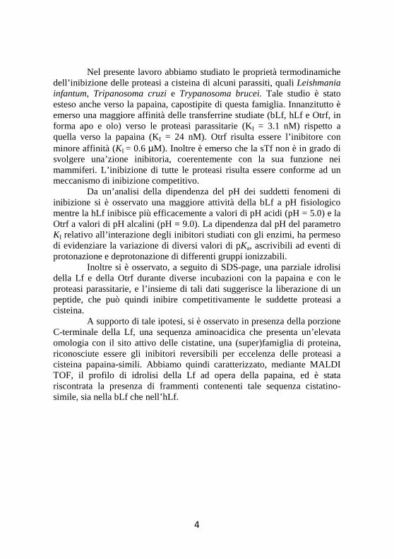
Cysteine proteases appear to be relevant to several aspects of the T. cruzi life cycle and the parasite-host relationship. In particular, cruzipain (Fig. 3), the major cysteine protease from T. cruzi epimastigotes, is expressed in all stages of the parasite life cycle, being more abundant in replicating forms and particularly in the insect epimastigote stage. Notably, cruzipain participates in the penetration of T. cruzi trypomastigotes into host cells, in the nutrition of the parasite at the expense of the host, and in the escape mechanisms of the parasite from the immune system of the host (Duschak & Couto, 2009).
Cruzipain has a structure very similar to that of papain. In fact is composed of polypeptide chain of 215 amino acid residues, folded into two domains (Figure 3). One domain is mainly α-helical (L-domain, residues 12-112 and 208-212), and the other consists of extensive antiparallel β-sheet interactions (R domain, residues 1-11 and 113-207) (Kamphuis et al., 1984, 1985; McGrath et al., 1995). The catalytic residues Cys25, His159 and Asn175, and the extended substrate-binding site, are found in a cleft between the two domains (McGrath et al., 1995).
12
Figure 3: Structure of Cruzipain (code PDB: 1AIM) (Gillmor et al., 1997). Molecular graphics images were produced using the UCSF Chimera package.
Trypanosoma brucei is a parasitic protist species that causes African trypanosomiasis (or sleeping sickness) in humans and in animals in Africa. There are 3 sub-species of T. brucei: T. b. brucei, T. b. gambiense and T. b. Rhodesiense (Barret et al., 2003).
In T. brucei species, the major cysteine protease has primary sequence and biochemical characteristics that are broadly similar to those of mammalian cathepsin L-like (Lonsdale-Eccles & Grab, 1987; Troeberg et al., 1999; Caffrey et al., 2001), and is encoded by a tandem array of 11 nearly identical gene copies (Berriman et al., 2005). The enzymes in T. b. rhodesiense and T. b. brucei are termed rhodesain and brucipain (or trypanopain) respectively (Lonsdale-Eccles & Grab, 1987; Caffrey et al., 2001 Mackey et al., 2004).
13
Brucipain shows 80% homology and 60% identity with the sequences of cruzipain (Duschak & Couto, 2009).
1.1.4. Protease of type I from L. infantum
The proteases of type I from L. infantum is a glycoprotein, synthesized like pre-pro-enzyme and it has a N-domain of 27 amino acidic residues (aa), a pre-peptidic element of 98 aa, a catalytic domain of 214 aa and a C-domain of 104 aa (Souza et al., 1992; Mottram et al., 1997; Campos-Ponce et al., 2005).
Like cruzipain, the proteases from L.infantum are cathepsins L-like and these proteases are required for parasite replication and virulence (Sakanari et al., 1997; Campos-Ponce et al., 2005; Mundodi et al., 2005).
Sequence analysis showed that they are 90% identical to L. major Cathepsin B, 66% identical to L. mexicana cathepsin B, and 45% identical and 57% homology to cruzipain, indicating high sequence conservation among the trypanosomatid species (Mottram et al., 1997).
1.2. Cystatins family
The inactivation of parasite cysteine proteinase mediated by synthetic inhibitors block replication and differentiation of T. cruzi, T. Brucei and L. infantum both in vitro and in vivo, providing an alternative to traditional therapy in drug-resistant parasites (Coombs & Mottram, 1997; Maekawa et al., 1998; McKerrow, 1999; McKerrow et al., 1993, 1995, 1999; McKerrow, 1999; Caffrey et al., 2000; Frame et al., 2000; Lecaille et al., 2002; Lima et al., 2002; Sajid & McKerrow, 2002; Somanna et al., 2002; Ascenzi et al., 2004).
Cysteine protease inhibitors (CPI) belonging to the cystatin (super)family inactivate proteases by trapping them in a(n) (ir)reversible, tight equimolar complex (Barrett et al., 1998). The representatives of this group of CPI is characterized by a wide distribution, being present in
14
mammals, birds, fish, insects, plants and some protozoa (Abrahamson et al., 2003).
The human superfamily of cystatins is divided into three families. Family I, called stefins, comprises intracellular cystatins A and B. Family II includes extracellular and/or transcellular cystatins (cystatins: C, D, E, F, S, SA, and SN). Kininogens, the intravascular cystatins, form family III of cystatins (Barrett, 1987; Rawlings & Barrett, 1990; Turk & Bode, 1991).
All cystatins have the same structure consisting of a five stranded β-sheet wrapped around a five turn α-helix. Cystatins contain three segments which are recognized as responsible for the interaction with cysteine proteases. These are the N-terminal fragment and the so-called first and second loops (L1 ans L2, respectively), which are arranged at one edge of the molecule and are believed to directly interact with the catalytic cleft of cysteine proteases (Grzonka et al., 2001; Turk et al., 2008).
These three cystatin regions, containing evolutionarily conserved amino-acid residues, form a wedge-like structure, which interacts with the catalytic cleft of cysteine proteases. These interactions are hydrophobic binding between the regions of cystatins and the corresponding residues forming the binding pockets of the enzyme. In particular, I-loop contains the conserved residues QVVAG, that interact with the active site of cysteine proteases, whereas the N-terminal fragment contains the conserved residues Leu9 and Gly11 (human cystatin numbering) (Grzonka et al., 2001; Turk et al., 2008). These residues form the reactive syte of cystatins.
15
Figure 4: Structure of Stefin A (code PDB: 1DVD) (Gillmor et al., 1997). Molecular graphics images were produced using the UCSF Chimera package.
Recently a sequence present at the C-terminus of human (hLf) and bovine (bLf) lactoferrin, a mammalian glycoprotein, shows a homology with the sequence of the reactive site of the cystatins (Katunuma et al., 2003). Moreover, this sequence is present in Ovotrasnferrin (Otrf), avian homologous of Lf, and in serum Transferrin (sTf), synthesized in the liver of mammals and secreted into the blood (Anderson et al., 2009).
16
Table 1: Alignment of Lf sequence with cystatine active site
Cystatin active site LG QVVAG
bLf (680) LGTEYVTA (687)
hLf (683) LGPQYVAG (690)
Otrf (657) LGDKFYTV (664)
sTf (661) LGEEYVKA (688)
1.3. Lactoferrin
Lactoferrin (Lf) is a non-haem iron-binding protein that is part of the transferrin protein family, along with serum transferrin (sTf), ovotransferrin (Otrf), melanotransferrin and the inhibitor of carbonic anhydrase, whose function is to transport iron in the blood serum (González-Chávez et al., 2009).
LF is produced by mucosal epithelial cells in various mammalian species, including humans, cows, goats, horses, dogs and several rodents. Recent studies have shown that Lf is also produced by fish, as it has been identified in rainbowtrout eggs using molecular biology techniques (González-Chávez et al., 2009).
This glycoprotein is found in mucosal secretions, including tears, saliva, vaginal fluids, semen (van der Strate et al., 2001), nasal and bronchial secretions, bile, gastrointestinal fluids, urine (Öztas & Özgünes, 2005) and most highly in milk and colostrum (7 g/L) (Rodriguez et al., 2005) making it the second most abundant protein in milk (Connely, 2001), after caseins. It can also be found in bodily fluids such as blood plasma and amniotic fluid. Lf is also found in considerable amounts in secondary neutrophil granules (15 µg/106 neutrophils) (Bennett & Kokocinski, 1987; González-Chávez et al., 2009), where it plays a significant physiological role.
Lf possesses a great iron-binding affinity and is the only transferring with the ability to retain the metal over a wide pH range (Aisen & Leibman, 1972) including extremely acidic pH. It also exhibits a great
17
resistance to proteolysis. In addition to these differences, Lf net positive charge and its distribution in various tissues make it a multifunctional protein (Baker & Baker, 2009).
1.3.1 Structure
Lf (Fig. 5) is an80 kDa glycosylated protein of ca. 700 aminoacids (711 aa for hLf and 689 aa bLf) with high homology among species. It is a simple polypeptide chain folded into two symmetrical lobes (N and C lobes) which are highly homologous with one another (33–41% homology) (Anderson et al., 1987, 1989; Baker, 1994; Moore et al., 1997; Sharma et al., 1998; Baker & Baker, 2009).
Figure 5: Structure of hLf (code PDB: 1FCK) (Baker et al., 2000). Molecular graphics images were produced using the UCSF Chimera package.
18
These two lobes are connected by a hinge region containing parts of an α-helix between residues 333 and 343 in human Lf (hLF), which provides additional flexibility to the molecule. The polypeptide chain includes amino acids 1–332 for the N lobe and 344–703 for the C lobe and is made up of α-helix and β-pleated sheet structures that create two domains for each lobe (domains I and II) (Moore et al., 1997). Each lobe can be further divided into two subdomains (N1 and N2 in the N-lobe and C1 and C2 in the C-lobe) that form a cleft inside of which the iron is bound. The subdomain N1 contains residues 1-90 and 251-333, while N2 contains the residues 91-250) (Baker et al., 1987; Moore et al., 1997; Baker & Baker, 2009).
Each lobe can bind a metal atom in synergy with the carbonate ion (CO3
2−). Lf binds notably Fe2+ and Fe3+ ions, but also Cu2+, Zn2+ and Mn2+ ions (Aisen & Harris, 1989; Baker et al., 1994, 2005; Baker & Baker, 2009).
1.3.2 Functions of Lf
Lf is involved in several physiological functions, including: regulation of iron absorption in the bowel; immune response; antioxidant, anticarcinogenic and anti-inflammatory properties. Protection against microbial infection, whichis the most widely studied function to date (Sanchez et al., 1992; Brock, 1995; Lonnerdal & Iver, 1995; Vorland, 1999; Brock, 2002; Antonini et al., 2005; Baker & Baker, 2009; Leboffe et al., 2009). The antimicrobial activity of LF is mostly due to two mechanisms. The first is iron sequestration in sites of infection, which deprives the microorganism of this nutrient, thus creating a bacteriostatic effect. The other mechanism is the direct interaction of LF with the infectious agent. Positively charged amino acids of LF can interact with anionic molecules on some bacterial, viral, fungal and parasite surfaces, causing cell lysis (Bullen, 1981; Braun & Braun, 2002; Valenti & Antonini, 2005).
Considering the physiological capabilities of Lf in host defence, in addition to current pharmaceutical and nutritional needs, Lf is considered to be a nutraceutical and for several decades investigators have searched for the most convenient way to produce it (González-Chávez et al., 2009).
19
The antiviral activity of hLf was first demonstrated in mice infected with the polycythemia-inducing strain of the Friend virus complex (FVC-P) (Lu et al., 1987). Since 1994, potent antiviral activity of hLf and bLf has been demonstrated against both enveloped and naked viruses, like Cytomegalovirus (CMV) (Harmsen et al., 1995; Andersen et al., 2001), Herpes simplex virus (HSV) (Marchetti et al., 1996, 1998; Siciliano et al., 1999; Valenti & Antonini, 2005), Human immunodeficiency virus (HIV) (Swart et al., 1996 ; Puddu et al., 1998; Berkhout et al., 2004), as well as Human hepatitis C (HCV) and human hepatitis B (HBV) viruses (Ikeda et al., 1998; Hara et al., 2002).
Molecular mechanims of Lf antiparasitic activity are even more complex. Antiparasitic activities of Lf appear often to involve interference with iron acquisition by some parasites, e.g. Pneumocystis carinii, while Lf appears to act as a specific iron donor in other parasites such as Trichomonas foetus; in the latter case, Lf could be expected to enhance infection. It was recently reported that two T. brucei proteins bind human serum transferrin as well as human and bovine Lf. Preincubation of Toxoplasma gondii and Eimeria stiedai sporozoites with a Lf-derived peptide, lactoferricin, reduces their infectivity in animal models. Lf antiparasitic activity is also, sometimes, mediated by interaction with host cells. Thus, iron-saturated Lf enhances intramacrophage killing of T. cruzi amastigotes and decreases intra-erythrocytic growth of Plasmodium falciparum. Lf is able to inhibit the invasion of cultured cells by Plasmodium spp. sporozoites through specific binding to HS. In the case of Plasmodium berghei, Lf reduces invasion by inhibiting the binding of the plasmodial CS protein, with or without HS, suggesting the possibility that Lf can also bind to the same site on LDL receptor-related protein (LRP) as the CS protein (see Leboffe et al., 2009).
20
AIM
The protozoan order Kinetoplastida includes the genus Trypanosoma, members cause some of the most neglected human diseases, the trypanosomiases. There are many species of trypanosome, and the group infects most vertebrate genera. Several trypanosoma species cause important veterinary diseases, but only two cause significant human diseases (Gilles, 1999). In sub-Saharian Africa, Trypanosoma brucei causes sleeping sickness or human African trypanosomiasis, and in America, Trypanosoma cruzi causes Chagas’ disease (Gilles, 1999). The kinetoplastida also contains species of the genus Leishmania that cause a range of diseases in the tropics and subtropics (Gilles, 1999). All microrganisms are transmitted by insect vectors. The transmission in the human host follows blood transfusion, contamined needles, biting insects, vector faeces, and contamined food (Gilles, 1999).
Like all microorganisms, the genus Trypanosoma has an absolute necessity of iron to sustain their growth (Loo & Lalond, 1984; Leboffe et al,, 2009). In particular, some parasites (e.g. Trypanosoma cruzi) survive in mammalian blood and could utilize serum mammalian chelates directly. Other parasites, such as leishmania spp., survie intracellularly in mammals (Gilles, 1999). Therefore they have envolved a mechanism to utilize mammalian holo-transferrin (Tf) and/or holo-Lactoferrin (Lf) via several means.
The Tf uptake by T. brucei has been well characterized. In this protozoan, esag6 and esag7 genes encoded for a protein associated to a heterodimeric transferrin-binding protein complex (TFBP) present on the cell membrane (Leboffe et al., 2009). These receptors have the ability to bind Tf from different mammalian host species (Leboffe et al., 2009). Fluorescence and immunoelectron microscopy showed that the T. brucei TFBP–Tf complex is internalized and transported to lysosomes, where Tf is proteolytically degraded to release iron (Leboffe et al., 2009).
On the contrary, the promastigote uptake of iron for Leishmania is different and involves reduction of extracellular iron. Infact holo-Tf binds to a binding protein on the promastigote surface, as well as for Trypanosoma brucei. One electron is donated to Fe3+ through the action of a membrane-associated or secreted iron reductase complex. The complex could include a cytoplasmic NADPH-requiring reductase which has specificity for NADPH
21
and which donates an electron to a membrane electron carrier (Leboffe et al., 2009). Fe2+ has a very low affinity for Tf and therefore dissociates easily (Baker, 2009).
In the last years growing evidences arose pointing to the importance between parasitic cisteine proteases and the virulence and pathogenicity of trypanosomatid parasites. In fact, cysteine proteases belonging to the papain family are relevant to several aspects of the parasite life cycle and of the parasite-host relationship and are thus seen as promising therapeutic target of parasitic diseases (Coombs & Mottram, 1997; Del Nery et al., 1997; McKerrow, 1999; McKerrow et al., 1993, 1995, 1999; Frame et al., 2000; Caffrey et al., 2000; Lecaille et al., 2002; Sajid & McKerrow, 2002; Mottram et al., 2004).
As an example, the major papain-like cysteine proteases from T. cruzi and L. infantum are mainly expressed in all stages of the parasites life cycle. Among others, cruzipain participates in the penetration of T. cruzi trypomastigotes into host cells, in the nutrition of the parasite at the expense of the host, and in the escape mechanisms of the parasite from the immune system of the host. Similar biological roles, even though with distinct molecular mechanisms, are been proposed for cysteine proteases from Leishmania (Coombs & Mottram, 1997; Del Nery et al., 1997; McKerrow, 1999; McKerrow et al., 1993, 1995, 1999; Frame et al., 2000; Caffrey et al., 2000; Lecaille et al., 2002; Sajid & McKerrow, 2002).
Notably, the inactivation of parasite cysteine proteinases by synthetic inhibitors block replication and differentiation of T. cruzi and L. infantum both in vitro and in vivo, providing an alternative to traditional therapy in drug-resistant parasites (Coombs & Mottram, 1997; Maekawa et al., 1998; McKerrow, 1999; McKerrow et al., 1993, 1995, 1999; McKerrow, 1999; Caffrey et al., 2000; Frame et al., 2000; Lecaille et al., 2002; Lima et al., 2002; Sajid & McKerrow, 2002; Somanna et al., 2002).
Cysteine protease inhibitors (CPI) belonging to the cystatin (super)family inactivate proteases by trapping them in a(n) (ir)reversible, tight equimolar complex (Barrett et al., 1998). The representatives of this group of CPI is characterized by a wide distribution, being present in mammals, birds, fish, insects, plants and some protozoa (Abrahamson et al., 2003; Turk et al., 2008).
22
Recently it was demonstrated that human and bovine milk has a new function against cysteine proteases (Katunuma et al., 2003). These data strongly support the hypothesis that Lf antiparasitic activity could be due, at least partially, to the inhibition of parasitic proteases.
Therefore, in the present work we report a quantitative investigation on the thermodynamic parameters for Tf inhibition of the cysteine protease from L. infantum, cruzipain and brucipain by hLf and bLf. Moreover, Lf binding to papain has been investigated. Moreover, the influence of the pH on the inhibition constants of Lf with papain and with the protease from L. Infantum have been determinated. The analysis of data allowed the identification of putative aminoacidic residues that may presumably modulate the interaction of the cysteine proteases studied with the Lf. Since this sequence is present also in Otrf (ovotransferrin), avian homologous of Lf, we have investigated the Otrf binding to protozoan proteases.
23
Results and discussion
3.1. Thermodynamic characterization of papain and cysteine proteases from L. Infatum, T. cruzi and T. brucei
3.1.1 The catalytic mechanism
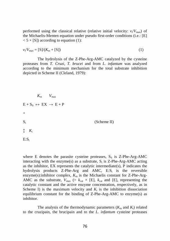
As shown in Fig. 6, the hydrolysis of the fluorogenic substrate Z-Phe-Arg-AMC catalyzed by the papain follows the simple Michaelis-Menten mechanism depicted in Scheme I and defined by equation (1).
Altought similar to the papain, the cysteine proteases from T. cruzi, T. brucei and L. infantum display the phenomenon of the substrate inhibition. In fact, as can be seen from the dependence of the relative initial velocity (i.e.: vi/Vmax) of Z-Phe-Arg-AMC hydrolysis on the substrate concentration, at 25 °C and pH 6.0 (Fig. 6) under steady state conditions (i.e.: [E] < 5 × [S]), the parasite enzymes follow the substrate inhibition mechanism described by Scheme II and defined by equation (2) (Fig.6: theoretical curves as solid lines and experimental data as squares and rumbles for the cysteine proteases from T. Cruzi, T. Brucei and L. infantum, respectively).
24
Figure 6: Effect of the substrate concentration on the hydrolysis of Z-Phe-Arg-AMC catalyzed by papain (black squares), cruzipain (black rumbles), the protease from L. infantum (rumbles) and brucipain (squares). Data were obtained at pH 6.0 and 25 °C. Solid lines, representing the theoretical curves, were calculated through an iterative non-linear least squares procedure with sets of parameters given in Table 2 and Table 3, according to Equation (1) for papain and to Equation (2) for cysteine proteases from T. Cruzi, from T. Brucei and from L. infantum. The standard deviation for each experimental point was equal to ± 8%. For further details, see text.
0 25 50 75 1000.00
0.25
0.50
0.75
1.00
[Z-Phe-Arg-AMC] (µM)
v i/V
max
25
Table 2: Values of Km (µM) for the hydrolysis of Z-Phe-Arg-AMC by proteases from L. infantum, T. cruzi, T . brucei and by papain (25,0°C).
pH Papain Cruzipain Brucipain Prot. from L. infantum
4.0 225±21 4.4±0.3 42.9±0.8 10.9±0.8
5.0 94±8 3.0±0.2 31.4±0.5 5.7±0.8
6.0 70±7 1.6±0.1 16.0±0.3 2.7±0.8
7.0 80±9 1.3±0.1 8.8±0.2 1.4±0.8
8.0 116±10 1.1±0.1 8.4±0.2 1.1±0.8
9.0 224±15 1.1±0.1 8.2±0.1 1.1±0.8
26
Table 3: Values of Ki (µM) for the hydrolysis of Z-Phe-Arg-AMC by the proteases from T. cruzi, T. brucei and L. infantum and (25.0°C).
pH Cruzipain Brucipain Protease from L. infantum
4.0 -----* ------* 33±0.8
5.0 48±8 55±0.2 36±0.5
6.0 59±7 68±0.1 40±0.3
7.0 67±9 77±0.1 55±0.2
8.0 73±10 84±0.1 62±0.2
9.0 74±15 85±0.1 57±0.1
* At such pH value cruzipain and brucipain do not undergo to substrate inhibition phenomenon.
Although the molecular details of substrate inhibition of cysteine proteases are not known, it often occur with synthetic and small substrates (Cazzulo et al., 1990; Lima et al., 1992; Stoka et al., 1998; Venturini et al., 2000; Salvati et al., 2001a). These chemicals are supposed to be able to bind the cysteine proteases interacting with only few residues of the protein and thus leading to the possibility for the synthetic molecule to assume different
27
configurations in respect with the catalytic Cys25 residue (mature cruzipain numbering). On the geometry of the interaction will then depend the result of the substrate-enzyme reaction (Cleland, 1979). Therefore, Z-Phe-Arg-AMC presumably is capable to bind the active site of parasite enzymes with, at least, two alterative configurations: either productively (Ss, Scheme II), leading to the hydrolysis of the substrate itself, or following a binding mode that lead to the enzyme inhibition (Si, Scheme II) (Salvati et al., 2001a; Salvati et al., 2002). Vice versa, papain, although with an active site very similar in the structure to the ones held by the parasite proteases, can interact with Z-Phe-Arg-AMC only through a productive configuration (Ss, Scheme I) (Szabelski et al., 2001) These considerations agree with the different specificity that characterizes papain, on one side, and parasite cysteine proteases from T. cruzi, T. brucei and from L. infantum, on the other. It has been proposed that the occurrence of the substrate inhibition phenomenon can reflect the presence of Glu residue (Glu205, mature cruzipain numbering) lying at the bottom of the S2 sub-site of the active site of papain-like cysteine proteases (as in the case of cruzipain, brucipain and of the protease from L .infantum) while the corresponding papain residue (Ser) should not be able to permit to the synthetic molecule to assume alternative (and inhibitory) geometries (see Salvati et al., 2001a). Remarkably, Glu205 was found to be capable of adopting different conformations according to the nature of the pairing P2 residue, thus enabling the parasite cysteine proteinase to bind substrates and inhibitors with charged (i.e. Arg) or hydrophobic (i.e. Tyr and Phe) residues at their P2 position (Gillmor et al., 1997; McGrath et al., 1995; Polticelli et al., 2005). Accordingly, if Z-Phe-Arg-AMC binds productively to the cruzipain active centre (i.e. Phe(P2) and Arg(P1) residues fit the S2 and S1 clefts, respectively), then it acts as substrates. On the other hand, if the Arg(P1) residue or the Z and AMC substituents of Z-Phe-Arg-AMC fit the S2 recognition sub-site of cruzipain, the substrate inhibition occurs (i.e. Z-Phe-Arg-AMC acts as an inhibitors) (Salvati et al., 2001a).
3.1.2 Effect of pH on values of Km and Ki
The analysis of the experimental data according to equation (1) and (2) for papain and parasite cysteine proteases action, respectively, allowed the evaluation, independently from the Vmax estimation, of the Michaelis constant (Km, for all the four proteases, Fig. 7 and Table 2) and of the substrate inhibition constant Ki (related only to the parasite enzymes, Fig. 8
28
and Table 3) for the hydrolysis of the Z-Phe-Arg-AMC between pH 4.0 to 9.0.
Figure 7: Effect of pH on values of Km for the hydrolysis of Z-Phe-Arg-AMC catalyzed by papain (black squares), cruzipain (black rumbles), the protease from L. infantum (rumbles) and brucipain (squares). Solid lines represent the best fit of experimental data was calculated, according to Equation (3) for parasite cysteine proteases and according to Equation (4) for papain, through an iterative non-linear least squares procedure with sets of parameters given in Table 1. The standard deviation for each experimental point is given in Table 1. The analysis allowed the determination of the pK’ and pK’’ parameters given in Table 3. For further details, see text.
4 5 6 7 8 9
1.50
1.75
2.00
pH
Log
Ki
29
Figure 8: pH dependence of the substrate inhibition constant Ki (µM) for the hydrolysis of the Z-Phe-Arg-AMC catalyzed by cruzipain (black rumbles), L. infantum protease (rumbles) and brucipain (squares) at 25,0 °C. Solid lines, representing the best fit of experimental data calculated according to Equation (5) through an iterative nonlinear least squares procedure with sets of parameters given in Table 2. Standard deviation for each of the experimental point is reported in Table 2. For pH values below pH 5 cruzipain does not show the substrate inhibition. The analysis allowed the determination of the pK° parameters given in Table 3. Standard deviation regarding experimental points is given in Table 2. For further details, see text.
4 5 6 7 8 90
1
2
3
pH
Log
Km
30
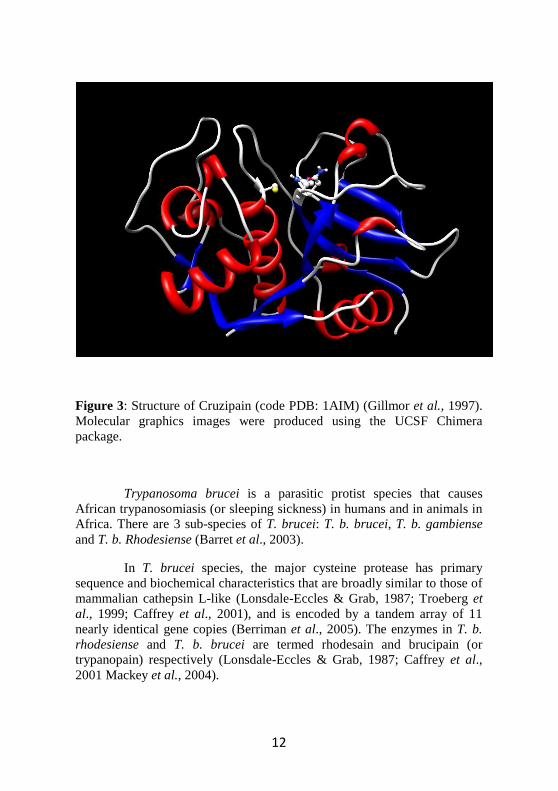
Table 4: Values of pKa parameters which describe the pH dependence of the Michaelis constant Km (in black) and the pH dependence of the substrate inhibition constant Ki (in blue) for the hydrolysis of the Z-Phe-Arg-AMC by papain and by the cysteine proteases from L. Infantum, from T. cruzi and from T. brucei (25,0°C).
pKa Values
Papain pK’unl = 4.6±0,1 pK’ lig = 3.9±0,2
pK” unl = 7.9±0,1 pK” lig = 8.5±0,1
Cruzipain pK’unl = 5.7±0,1 pK’ lig = 5.1±0,1
pK°unl = 5.9±0,2 pK°lig = 6.1±0,2
Brucipain pK’unl = 6.1±0,1 pK’ lig = 5.4±0,1
pK°unl = 5.9±0,2 pK°lig = 6.1±0,2
Protease pK’unl = 6.1±0,2 pK’ lig = 5.2±0,2
from L. infantum pK°unl = 6.2±0,2 pK°lig = 6.4±0,2
Values of Km for the papain (Fig. 2) depend on the acid-base equilibrium of two ionising residues. The analysis of data allowed to determine the values of pK’ unl (= 4.6±0.1), pK’ lig (= 3.9±0.2), pK” unl (= 7.9±0.1) e pK” lig (= 8.5±0.1) (Table 5) and values of pKa reflecte the equilibrium of two amino acidic residue (Fig. 7 and Table 4), classically recognised as the catalytic residues Cys25 (i.e. pK’unl = 4.6±0.1 and pK’ lig =
31
3.9±0.2) and His159 (i.e. pK’’ unl = 7.9±0.1 and pK’’ lig = 8.5±0.1) (Hollaway et al., 1969; Shipton et al., 1976; Ascenzi et al., 1987; Ménard et al., 1990). Nonetheless, the influence of further residues lying in the active site of the enzyme, like Lys156 and Asp158 can not be excluded and they may account for minor contributions in the enzyme-substrate binding energetic, at least modifying the electric field acting on catalytic residues Cys25 and His159 and thus on their observed pKa values.
On the other hand, the analysis of data concerning cruzipain, brucipain and L. infantum protease action (Fig. 7) allowed the estimation of the pKa values that modulates the protease-substrate binding event, for cruzipain (pK’unl = 5.7±0.1 e pK’ lig = 5.1±0.1), brucipain (pK’unl = 6.1±0.1 e pK’ lig = 5.4±0.1) and for the L. infantum protease (pK’unl = 6.1±0.1 e pK’ lig = 5.2±0.2) (Table 4).
Therefore, while Z-Phe-Arg-AMC binding to papain imply a shift of the apparent pKa of 0.7 unit toward acidic pH values (pK’ unl > pK’ lig) and of 0.6 unit toward the alkaline (pK” unl < pK” lig) the interaction of Z-Phe-Arg-AMC with cruzipain, brucipain and L. infantum protease determines a shift of the apparent pKa to acidic values (pK’unl > pK’ lig) of about 0.7 pH units.
On the other hand, only one apparent amino acidic residue is able to modulate the interaction of Z-Phe-Arg-AMC with cruzipain and with L. Infantum protease regarding to the productive as well as to the unproductive binding (SS and Si respectively, see Scheme II). Notably, for all the parasite enzymes the ligand-independent pKa values which define the pH dependence of both the productive (pK’unl) and the non-productive binding process (pK°unl) converge in the limits of the experimental indeterminateness (i.e. for cruzipain and for L. Infantum protease it results pK’unl ~ pK°unl, see Table 3). These values for cruzipain (pK’unl = 6.2±0.2, pK°unl = 6.2±0.2) are very close to those that characterize L. Infantum protease (pK’unl = 5.7±0.1, pK°unl = 5.9±0.2). The main structural determinant that may account for the observed pH dependence of Z-Phe-Arg-AMC binding to parasite cysteine proteases is probably the Glu205 residue (mature cruzipain numbering). As reported above, this residue, Glu in parasite proteases and Ser in papain, is responsible for the different specificity showed by papain and by parasite cysteine proteases. Notably, previous investigations on cysteine proteases from T. cruzi, L. infantum and Plasmodium falciparum (Ascenzi et al., 2004). Human cathepsin B (Khouri et al., 1991) and viral cathepsin from the Autographa californica nuclear
32
polyhedrosis virus (AcNPV) (Bromme & Okamoto, 1995) have shown similar results with reference to the pH dependence of the binding of synthetic ligands to papain-like proteases with a Glu residue equivalent to the Glu205 of cruzipain. It seems to suggest a general model of recognition and of pH-modulation for cysteine proteases with a cathepsin B-like S2 specificity sub-site (Sajid & McKerrow, 2002).
3.2. Inhibition of papain and related proteases by members of the Transferrin family
3.2.1. Tfs are a competitive inhibitors
Lf and Otrf (Tfs) inhibit papain, cruzipain, brucipain and L. infantum protease (Fig. 9 and Table 5). The inhibition of cysteine proteases by Lf and Ortf conforms to competitive inhibition according to the classic competitive inhibition mechanism (Scheme III) described by Equations (6), (7) and (8). On the contrary, sTf does not inactive cysteine proteases, according to its different role in mammals (Anderson et al., 2009).
33
0.000 0.025 0.050 0.0750.00
0.25
0.50
0.75
1.00
[bLf] (µM)
v i/v
0
Figure 9: Dependence of the initial relative velocity vi/v0 for the hydrolysis of Z-Phe-Arg-AMC by papain (black squares), cruzipain (black rumbles), L. infantum protease (rumbles) and brucipain (squares) on the bLf concentration. Experimental data are been obtained at pH 6.0 and 25 °C. The Z-Phe-Arg-AMC concentration was 50 µM for papain and 5.0 for parasite cysteine proteases. Solid lines represent the theoretical curves, described by Equation (6), obtained through an iterative non-linear least squares procedure with sets of parameters given in Table 5. Standard deviation for each experimental point was equal to ± 8%.
34
0 25 50 75 1000.00
0.02
0.04
0.06
[Z-Phe-Arg-AMC] (µM)
KIap
p
0.0 2.5 5.0 7.50.00
0.01
0.02
0.03
[Z-Phe-Arg-AMC] (µM)
KIap
p
A
B
Figure 10: Dependence of the relative inhibition constant KIapp/KI by bLf binding to papain (Panel A, black squares), with cruzipain (Panel B, black rumbles), L. infantum protease (Panel b, rumbles) and brucipain (Panel B, squares) on the substrate concentration (Z-Phe-Arg-AMC). Experimental data were carried out at 25°C and pH 6.0. Straight lines, representing the best fit of experimental data, were calculated according to Equation (8) through an iterative linear least squares procedure with sets of parameters given in Tables 5-8. Standard deviation for each experimental point was to ± 8%.
35
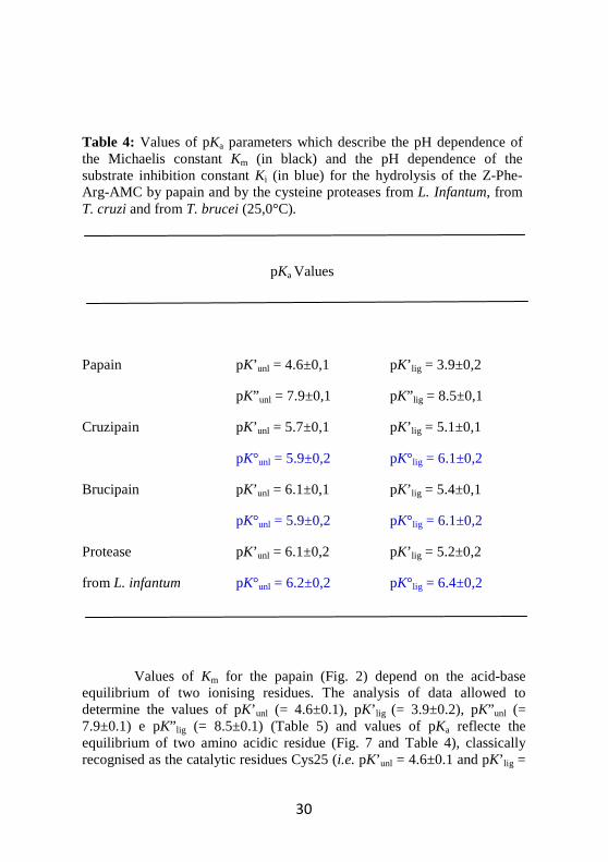
Table 5: Values of KI (µM) for bLf, hLf (holo and apo) and Otrf binding to papain (25.0 °C).
For all the proteases considered in this study, the dependence of the apparent inhibition constant KIapp on the Z-Phe-Arg-AMC concentration, was linear over the substrate concentration range explored (Fig. 10). These results are compatible only with a complete competitive inhibition mechanism (Scheme III), in fact, the observed relative inhibition constant KIapp/KI increases linearly with the substrate concentrations (Fig. 10). Notably, the Y intercept of straight lines is 1. This means that in the absence of the substrate ([Z-Phe-Arg-AMC] = 0, corresponding to the X interpolation value of the Y intercept) the apparent inhibition constant KIapp
39
corrisponds to the intrinsic ligand-independent equilibrium constant KI (KIapp= KI).
Moreover, if we compare values the intrinsic substrate-independent constant KI with those cystatins, it appears that Lf a very good inhibitor of cisteine papain-like proteases (Table 9).
Table 9. Values of the intrinsic ligand-independent constant KI (µM) of stefin A, of stefin B (cystatins of type I), of chicken egg-white cystatin (CEW Cys, cystatin of type II).
Stefin A 1.8 × 10⁻⁷ a 9.1 × 10⁻⁴ a 1.9 × 10⁻⁵ a 2.1 × 10⁻⁵ c
Stefin B 4.9 × 10⁻⁸ b 1.8 × 10⁻² b 3.0 × 10⁻⁶ b 6.0 × 10⁻⁵ c
CEW Cys 1.0 × 10⁻⁵ d 8.0 × 10⁻⁵ d 3.0 × 10⁻⁶ d 1.4 × 10⁻⁵ c
a Estrada et al. (1998).
b Pol & Björk (2001).
c Stoka et al. (1995).
d Anastasi et al. (1983).
40
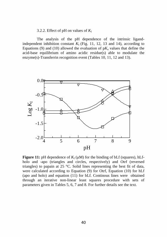
3.2.2. Effect of pH on values of KI
The analysis of the pH dependence of the intrinsic ligand-independent inhibition constant KI (Fig. 11, 12, 13 and 14), according to Equations (9) and (10) allowed the evaluation of pKa values that define the acid-base equilibrium of amino acidic residue(s) able to modulate the enzyme(s)-Transferrin recognition event (Tables 10, 11, 12 and 13).
Figure 11: pH dependence of KI (µM) for the binding of bLf (squares), hLf-holo and -apo (triangles and circles, respectively) and Otrf (reversed triangles) to papain at 25 °C. Solid lines representing the best fit of data, were calculated according to Equation (9) for Otrf, Equation (10) for hLf (apo and holo) and equation (11) for bLf. Continous lines were obtained through an iterative non-linear least squares procedure with sets of parameters given in Tables 5, 6, 7 and 8. For further details see the text.
4 5 6 7 8 9-2.0
-1.5
-1.0
-0.5
0.0
pH
Log
KI
41
Table 10: Values of pKa modulating the inhibition constant KI (µM) for bLf (squares), hLf-apo and –holo (circles and triangles) and Otrf (overturned triangles) to papain (Fig. 6) at 25 °C.
pKa Values
bLf pK’unl = 6.9±0.3 pK’ lig = 5.0±0.2
pK” unl = 7.9±0,1 pK” lig = 8.5±0,1
hLf-apo pK’unl = 7.1±0.3 pK’ lig = 8.0±0.2
hLf-holo pK’unl = 7.1±0.3 pK’ lig = 8.0±0.2
Otrf pK’unl = 8.2±0.2 pK’ lig = 7.3±0.2
42
Figure 12: pH dependence of KI (µM) for the binding of bLf (squares), hLf-holo and -apo (triangles and circles, respectively) and Otrf (reversed triangles) to L. infantum protease at 25 °C. Solid lines representing the best fit of data, were calculated according to Equation (9) for Otrf and according to equation (11) for bLf and for hLf (apo and holo). Continous lines were obtained through an iterative non-linear least squares procedure with sets of parameters given in Tables 5, 6, 7 and 8. For further details see the text.
4 5 6 7 8 9
-2.0
-1.5
-1.0
-0.5
0.0
pH
Log
KI
43
Table 11: Values of pKa modulating the inhibition constant KI (µM) regarding for bLf (squares), hLf-apo and –holo (circles and triangles) and Otrf (overturned triangles) to L. infantum protease (Fig. 7) at 25 °C.
pKa Values
bLf pK’unl = 6.4±0.3 pK’ lig = 5.6±0.1
pK” unl = 7.7±0.2 pK” lig = 8.3±0.4
hLf-apo pK’unl = 6.2±0.5 pK’ lig = 5.8±0.2
pK” unl = 7.9±0.2 pK” lig = 8.5±0.3
hLf-holo pK’unl = 6.2±0.5 pK’ lig = 5.8±0.2
pK” unl = 7.9±0.2 pK” lig = 8.5±0.3
Otrf pK’unl = 8.5±0.2 pK’ lig = 7.5±0.3
44
4 5 6 7 8 9
-2.0
-1.5
-1.0
-0.5
0.0
pH
Log
KI
Figure 13: pH dependence of KI (µM) for the binding of bLf (squares), hLf-holo and -apo (triangles and circles, respectively) and Otrf (reversed triangles) to cruzipain at 25 °C. Solid lines representing the best fit of data, were calculated according to Equation (9) for Otrf and according to equation (11) for bLf and for hLf (apo and holo). Continous lines were obtained through an iterative non-linear least squares procedure with sets of parameters given in Tables 5, 6, 7 and 8. For further details see the text.
45
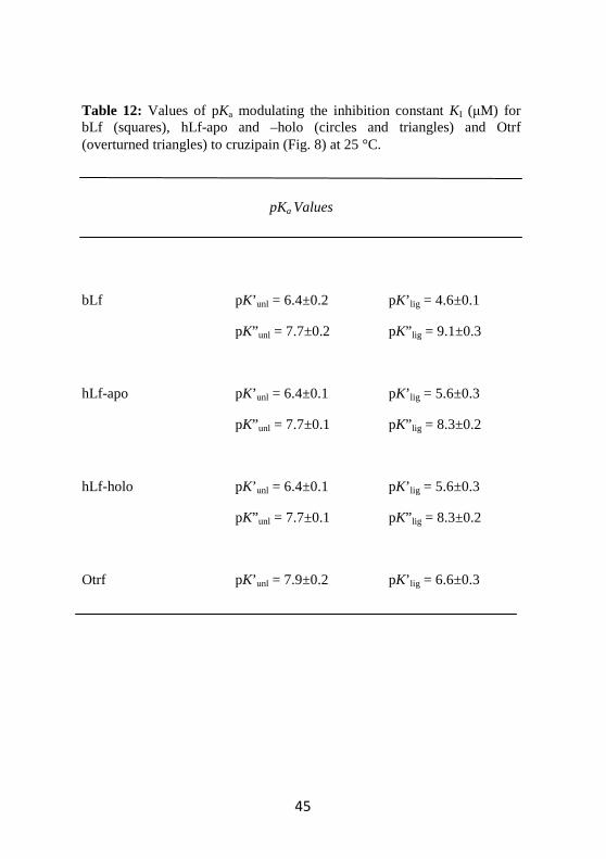
Table 12: Values of pKa modulating the inhibition constant KI (µM) for bLf (squares), hLf-apo and –holo (circles and triangles) and Otrf (overturned triangles) to cruzipain (Fig. 8) at 25 °C.
pKa Values
bLf pK’unl = 6.4±0.2 pK’ lig = 4.6±0.1
pK” unl = 7.7±0.2 pK” lig = 9.1±0.3
hLf-apo pK’unl = 6.4±0.1 pK’ lig = 5.6±0.3
pK” unl = 7.7±0.1 pK” lig = 8.3±0.2
hLf-holo pK’unl = 6.4±0.1 pK’ lig = 5.6±0.3
pK” unl = 7.7±0.1 pK” lig = 8.3±0.2
Otrf pK’unl = 7.9±0.2 pK’ lig = 6.6±0.3
46
4 5 6 7 8 9
-2.0
-1.5
-1.0
-0.5
0.0
pH
Log
KI
Figure 14: pH dependence of KI (µM) for the binding of bLf (squares), hLf-holo and -apo (triangles and circles, respectively) and Otrf (reversed triangles) to brucipain at 25 °C. Solid lines representing the best fit of data, were calculated according to Equation (9) for Otrf and according to equation (11) for bLf and for hLf (apo and holo). Continous lines were obtained through an iterative non-linear least squares procedure with sets of parameters given in Tables 5, 6, 7 and 8. For further details see the text.
47
Table 13: Values of pKa modulating the inhibition constant KI (µM) for bLf (squares), hLf-apo and –holo (circles and triangles) and Otrf (overturned triangles) to cruzipain (Fig. 9) at 25 °C.
pKa Values
bLf pK’unl = 6.2±0.2 pK’ lig = 4.3±0.1
pK” unl = 7.5±0.2 pK” lig = 8.9±0.3
hLf-apo pK’unl = 6.1±0.2 pK’ lig = 5.0±0.1
pK” unl = 7.4±0.2 pK” lig = 8.0±0.3
hLf-holo pK’unl = 6.1±0.2 pK’ lig = 5.0±0.1
pK” unl = 7.4±0.2 pK” lig = 8.0±0.3
Otrf pK’unl = 7.1±0.1 pK’ lig = 5.9±0.2
48
In particular, bLf binding to papain (pK’unl = 6.9±0.3, pK’ lig = 5.0±0.2, pK” unl = 7.9±0,1 and pK” lig = 8.5±0,1 ), L.infantum protease (pK’unl = 6.4±0.3, pK’ lig = 5.6±0.1, pK” unl = 7.7±0.2 and pK” lig = 8.3±0.4), cruzipain (pK’unl = 6.4±0.2, pK’ lig = 4.6±0.1, pK” unl = 7.7±0.2 and pK” lig = 9.1±0.3) and brucipain (pK’unl = 6.2±0.2, pK’ lig = 4.3±0.1, pK” unl = 7.5±0.2 and pK” lig = 8.9±0.3) is modulated by the acid-base equilibrium of two amino acidic residues (Fig. 6-9 and Tables 9-12). Inhibitor binding induces a pKa shift of about 1.9 pH units toward acidic values and of about 0.6 pH units toward the alkaline region for papain (pK*unl > pK* lig and pK** unl < pK** lig), a pKa shift of about 0.8 pH units toward acidic values and of about 0.6 pH units toward the alkaline region for protease from L. Infantum, a pKa shift of about 1.8 pH units toward acidic values and of about 1.4 pH units toward the alkaline region for cruzipain and a pKa shift of about 1.9 pH units toward acidic values and of about 1.4 pH units toward the alkaline region for brucipain.
The interaction of hLf (apo and holo) with papain is modulated by one apparent acidbase ionization (Fig. 6-9 and Tables 9-12) causing a pKa shift of about 0.9 pH units toward alkaline values (pK’unl = 7.1±0.3 and pK’ lig = 8.0±0.2, both apo-form and holo-form). On the other hand, hLf (apo and holo) binding to parasitic proteases is modulated by the acid-base equilibrium of two amino acid residues (Fig. 6-9 and Tables 9-12) causing a pKa shift of about 0.4 pH units toward acidic values and of about 0.6 pH units toward the alkaline region for L. Infantum protease (pK’unl = 6.2±0.5, pK’ lig = 5.8±0.2, pK” unl = 7.9±0.2 and pK” lig = 8.5±0.3), a pKa shift of about 0.8 pH units toward acidic values and of about 0.6 pH units toward the alkaline region for cruzipain (pK’unl = 6.4±0.1, pK’ lig = 5.6±0.3, pK” unl = 7.7±0.1 and pK” lig = 8.3±0.2) and a pKa shift of about 1.1 pH units toward acidic values and of about 0.6 pH units toward the alkaline region for brucipain (pK’unl = 6.1±0.2, pK’ lig = 5.0±0.1, pK” unl = 7.4±0.2 and pK” lig = 8.0±0.3).
Instead, the binding of Otrf bLf to papain (pK’unl = 8.2±0.2 and pK’ lig = 7.3±0.2), to protease of type I from L. infantum (pK’unl = 8.5±0.2 and pK’ lig = 7.5±0.3), to cruzipain (pK’unl = 7.9±0.2 and pK’ lig = 6.6±0.3) and to brucipain (pK’unl = 7.1±0.1 and pK’ lig = 5.9±0.2) is modulated by one apparent amino acidic residue (Fig. 6-9 and Tables 9-12). It causes a pKa shift of about 0.9 pH units for papain, 1.0 pH units for protease of type I from L. infantum, 1.3 pH units for cruzipain and 1.2 pH units for brucipain toward acidic values.
49
Tfs inhibit preferentially parasitic proteases; the pH optimum is around neutrality for bLf, while it is acidic for hLf and alkaline for Otrf. On the contrary, sTf does not inhibit any enzyme, according to its different role in mammals. In fact sTf function is to transport iron in blood serum. Note that values of KI are independent of iron saturation (Tables 8-12).
According to ‘linked functions’ (Wyman, 1964) the (de)protonation of amino acidic residue involved in the ligand binding lead to the modulation of the binding itself in such a way that the effect of pH on the thermodynamic constant(s) (Km, Ki and KI) is quantitatively the same of the effect on the pKa shift (regarding to the acid-base equilibrium of the modulating residues of the protein) induced by the binding of the ligand (∆pKa = ∆pKm, ∆pKa= ∆pKi or ∆pKa= ∆pKI, depending on the ligand) (Wyman, 1968). Therefore, the higher pKa values observed in the binding of inhibitors respect with the synthetic substrate Z-Phe-Arg-AMC probably indicates a much larger free energy variation upon ligand binding, reflecting a wider surface contacting protease-inhibitor complex formation (Wyman, 1964).
Macromolecular recognition occuring between the active site of the cysteine proteases studied and the (macro)molecular species (regardless if they act as inhibitors or substrates) is defined by a thermodynamic constant K (Km, Ki and KI, as previously seen) which represents the all-in affinity that exists between two macromolecular species (i.e. the enzyme and its ligand). Therefore, a K constant must be seen as the result of the sum of all the thermodynamic contribution that came from each enzyme-ligand interacting residues.
Accordingly, the calculated pKa must to be intended as apparent: namely not necessary attributable to the acid-base equilibrium of one amino acidic residue, but rather as a mean value related to the acid-base equilibrium constant of all the ionizing residues which contribute energetically to the enzyme/ligand recognition event.
50
3.3. Inhibition mechanism of Lactoferrin As shown in Fig. 15 Lf and Otrf are easily degraded by papain and
by parasite cysteine proteases during the assay incubation time.
Figure 15: SDS-PAGE showing that Lf is easily degraded by cysteine proteases during the assay incubation time. Line 1: Lf or Otrf. Lines 2,3,4, 5 and 6: Lf/Otrf and cysteine proteases at times 0, 1, 10, 30 and 60 min. SDS-PAGE was obtained at 12% polyacrylamide
Analysis by SDS–PAGE revealed different peptide profiles from Lf hydrolysis (Fig. 15). The incubation time also influenced the degradation extent of Lf. In fact papain and the parasite cysteine proteases generate peptides of range between 35 kDa and 10 kDa. The maximum hydrolysis values (100%) were reached after 30 minutes of incubation for all studied enzymes.When the incubation period was prolonged up to 24 h, smaller size peptides are detected for all strains.
51
In presence of antipain, an inhibitor of papain-like proteases, Lf and Otrf are not degraded (Fig 16).
Figure 16: Effect of antipain on Lf hydrolysis by papain. Line 1 absence of antipain. Line 2, 3, 4 and 5 increased concentration of antipain (0.25:1; 0.50:1; 0.75:1 and 1:1 antipain:papain)
Since the Lf sequence 679-695 is similar to the cystatin active site, it is possible that a peptidic cystatin-like inhibitor is generated by enzymes-induced Lf hydrolysis.
52
3.4. MALDI TOF/TOF analysis
In order to characterize the peptides produced by bLf and hLf papain digestions, a mass spectrometry (MS) analysis was performed. To this end, each inhibitor digestion was analyzed by peptide mass fingerprinting and identified peptides are listed in Tables 14 and 15.
In bLf MS analysis we covered the 60% of protein sequence by detection of 57 peptides.
Figure 17. MS spectrum of bLf digestion by papain
53
Table 14:MS analysis of peptides produced by bLf papain digestion
54
Particularly, we were able to detect a signal at 1733.63 m/z which corresponds to residues 671–685 (GGRPTYEEYLGTEYV) of the bLf protein (Fig 18). Interestingly, this peptide was found eluited in HPLC fraction able to inhibit the cysteine papain-like proteases in dose dependent way and shows in the C-terminal region a cystatine active site sequence (Table 1).
Figure18. MS spectrum of bLf digestion by papain. The image shows the zoom range of 1733.64 peak MS/MS spectrum
In hLf MS analysis we covered more of 60% of sequence (72 peptides matched) (Table 15) detecting two signals (i.e.: 1266.69 and 1646.84 m/z) corresponding to residues YVAGITNLKKC (687-697) and GKTTYEKYLGPQYV (675-688) respectively (Fig. 19 and 20). Both of them were found in two different HPLC fractions with inhibitor activity and show a portion of cystatin-like sequence.
Table 15: MS analysis of peptides produced by hLf papain digestion
55
56
Figure18. MS spectrum of hLf digestion by papain
57
Figure 19: MS spectrum of hLf digestion by papain. The image shows the zoom range of 1266.69 and 1646.84 peaks MS/MS spectrum
These data suggest that these peptides generated from papain hydrolysis could inhibit the protease itself.
58
3.5. Molecular modelling of putative complex Y687-G690-Papain
The sequences of hypotetical inhibitor fragments are aligned (Table 16) using ClustalW alignament software.
Table 16: Alignment of inhibitor peptides with cystatine active site
cystatine active site LG QVVAG
hLf YVAGITNLKKC
hLf GKTTYEKYLGPQYV
bLf GGRPTYEEYLGTEYV
In fig. 20 the result of this procedue is showed. The “YV” motiv seems to be conserved in all fragment tested. On the basis of the proposed alignments a structural preliminary model is done to test the compatibility of these residues with the mechanism of catalysis. To this aim, a site-specific mutation was performed in silico through the utilization of “Swiss pdb viewer” (Guex, N. & Peitsch, M.C. 1997) molecular modeling software. The residues VVAG of Stefin B are mutated in YVAG. For each mutation, the software offers, through an assessment of possible interactions with the substrate, the best rotamers (different disposition of the amino acid residues in three-dimentionl space). The crystal structure of the papain-stefin B complex (code PDP: 1STF) is used to model the interaction (Stubbs et al, 1990). The very preliminary results show a likely favorable interaction between Tyr present on the putative inhibitory fragments and the residue Gly23 of papain. Further observation are required to identify the exact bond-condition. In other hands, this evidence, if properly confirmed by further experimental analysis, seems to show that the patterns identified can be regarded as foundamental in the competitive inhibition process.
59
Figure 20: Molecular modelling of putative complex Y687-G690-Papain. Site-specific mutation was performed in silico through the utilization of “Swiss pdb viewer” (Guex, N. & Peitsch, M.C. 1997) molecular modeling software. The crystal structure of the papain-stefin B complex (code PDP: 1STF) is used to model the interaction (Stubbs et al, 1990).
60
Conclusions
In this study we have investigated the thermodynamic parameters of human lactoferrin (hLf), bovine lactoferrin (bLf) and hen’s ovotransferrin (Otrf) inhibition of the parasitic papain-like type I cysteine proteases from Leishmania infantum, Trypanosoma cruzii and Trypanosoma brucei.
Parasites synthesize papain-like cysteine proteases that are relevant for the virulence and pathogenicity of parasites, being involved in several aspects of the parasite life cycle, it is therefore possible that the antiparasitic activity of lactoferrin could be due to the inhibition of parasitic papain-like cysteine protease that we have recently observed.
Transferrins belong to a family of iron-binding glycoproteins possessing similar aminoacid sequence though they have different biological functions and locations. Lactoferrin is expressed and secreted from glandular epithelial cells and from mature neutrophiles of mammalian and it is an important component of the aspecific host defence or natural immunity, including resistance to parasitic infections. Serum transferrin is synthesized by the liver of mammals and secreted into the blood stream; its primary function is iron transport. Ovotransferrin, synthesized by avians, displays both iron transport and protective functions.
bLf, hLf and Otrf, both in the apo- and olo-forms, showed time- and concentration-dependent inhibition of the catalytic activity of papain and of type I proteases from L.infantum, T. cruzii and T. brucei. The KI values observed for bLf and hLf inhibition of L. infantum, T. cruzi and T. brucei proteases were in the nanomolar range (KI = 3.1 nM), lower than KI values observed for papain inhibition (KI = 24 nM). Otrf showed lower inhibition of cysteine proteases (KI = 0.6 µM). On the contrary, serum transferrin did not display any inhibition towards type I parasitic proteases, according to its different role in mammals. The inhibition of type I parasitic cysteine proteases by hLf, bLf and Otrf appeared to conform to a competitive mechanism. The observed pH optimum for bLf inhibition of parasitic proteases was around neutrality, while it was acidic for hLf and alkaline for Otrf. The further quantitative analysis of the pH dependence of the intrinsic ligand-independent inhibition constant KI allowed the evaluation of pKa values that define the acid-base equilibrium of amino acidic residue(s) modulating the enzyme(s)-inhibitor recognition events. SDS-PAGE showed that hLf, bLf and Otrf were easily degraded by either
61
papain or parasitic type I protease during the assay incubation time (few minutes) and it is likely that one or more protease inhibitory peptides were generated from protein hydrolysis.
As a matter of fact, a sequence present near the C-terminus of human (hLf) and bovine (bLf) lactoferrin shows homology with the sequence of the active site of cystatins, which are competitive inhibitors of papain-like cysteine proteases. The same sequence is present, though with lower homology, in Otrf and, with even lower homology, in sTf. Therefore, we have studied by MALDI-TOF the profile of Lf cleavage by papain and preliminary data suggest the presence of a cystatin-like peptide in two proteolytic fragments of hLf and in one proteolytic fragment of bLf. The work will continue with the characterization of the active peptides looking at the possible design of a peptidomimetic drug for the therapy of parasitic infections.
62
Bibliography
Abrahamson, M., Alvarez-Fernandez, M. & Nathanson, C. M. (2003) Cystatins. Biochem. Soc. Symp. 70, 179-199.
Aisen P, Leibman A. (1972) Lactoferrin and transferrin: a comparative study. Biochim Biophys Acta; 257:314–23. Anastasi, A., Brown, M. A., Kembhavi, A. A., Nicklin, M. J., Sayers, C. A., Sunter, D. C. & Barrett, A. J. (1983) Cystatin, a protein inhibitor of cysteine proteinases. Improved purification from egg white, characterization, and detection in chicken serum. Biochem. J. 211, 129-138.
Andersen, J.H., Osbakk, S.A., Vorland, L.H., Traavik, T., and Gutteberg, T.J. (2001). Lactoferrin and cyclic lactoferricin inhibit the entry of human cytomegalovirus into human fibroblasts. Antiviral Res. 51: 141–149.
Anderson, B.F., Baker, H.M., Dodson, E.J., Norris, G.E., Rumball, S.V., Waters, J.M., and Baker, E.N. (1987). Structure of human lactoferrin at 3.2-Å resolution. Proc. Natl. Acad. Sci. USA 84: 1769–1773.
Anderson, B.F., Baker, H.M., Norris, G.E., Rice, D.W., and Baker, E.N. (1989). Structure of human lactoferrin: crystallographic structure analysis and refinement at 2.8 Å resolution. J. Mol. Biol. 209: 711–734. Anderson GJ, Frazer DM, McLaren GD. (2009) Iron absorption and metabolism. Curr Opin Gastroenterol. 25(2):129-35. Antonini, E. & Ascenzi, P. (1981) The mechamism of trypsin catalysis at low pH: proposal for a structural model. J. Biol. Chem. 256, 12449–12455. Ascenzi, P., Aducci, P., Torroni, A., Amiconi, G., Ballio, A., Menegatti, E. & Guarnieri, M. (1987) The pH dependence of pre-steady-state and steady-state kinetics for the papain catalyzed hydrolysis of N-α-carbobenzoxyglycine p-nitrophenyl ester. Biochim. Biophys. Acta 912, 203-210.
63
Ascenzi, P., Menegatti, E. & Amiconi, G. (1988) pH effects in biochemical reactions: what’s the meaning of pK and midpoint? Biochem. Educ. 16, 93-94.
Ascenzi, P., Bocedi, A., Gentile, M., Visca, P. & Gradoni, L. (2004) Inactivation of parasite cysteine proteinases by the NO-donor 4-(phenylsulfonyl)-3-((2-(dimethylamino)ethyl)thio)-furoxan oxalate. Biochim. Biophys. Acta 1703, 69-77.
Baker, E.N. (1994). Structure and reactivity of transferrins. Adv. Inorg. Chem. 41: 389–463.
Baker EN, Baker HM (2009) A structural framework for understanding the multifunctional character of lactoferrin. Biochimie. 91(1):3-10. Barrett, A.J. (1980) Fluorimetric assays for cathepsin B and cathepsin H with methylcoumarylamide substrates. Biochem. J. 187, 909-912.
Barrett, A. J. & Salvesen, G. (1986) Proteinase Inhibitors. Volume 12. Elsevier, Amsterdam. Barrett, A.J., Rawlings, N.D. & Woessner, J.F. (1998) Handbook of Proteolytic Enzymes. Academic Press, London and San Diego Barrett, A. J., Rawlings, N. D. & Woessner, J. F. (2004) Handbook of Proteolytic Enzymes. 2nd ed. Academic Press, London. Barrett MP, Burchmore RJ, Stich A, Lazzari JO, Frasch AC, Cazzulo JJ, Krishna S. (2003) The trypanosomiases. Lancet. 362(9394):1469-80. Belenghi, B., Acconcia, F., Trovato, M., Perazzolli, M., Bocedi, A., Polticelli, F., Ascenzi, P. & Delledonne, M. (2003) AtCYS1, a cystatin from Arabidopsis thaliana, suppresses hypersensitive cell death. Eur. J. Biochem. 270, 2593-2604.
Bennett RM, Kokocinski T. (1987) Lactoferrin content of peripheral blood cells. Br J Haematol; 39:509–21 Berkhout B., Floris R., Recio I. and Visser S. (2004). The antiviral activity of the milk protein lactoferrin against the human immunodeficiency virus type 1. Biometals 17: 291–294
64
Bocedi, A., Gradoni, L., Menegatti, E. & Ascenzi, P. (2004) Kinetics of parasite cysteine proteinase inactivation by NO-donors. Biochem. Biophys. Res. Commun. 315, 710-718. Braun V. and Braun M. 2002. Active transport of iron and siderophore antibiotics. Curr. Opin. Microbiol., 5: 194–201.
Bullen J. J. 1981. The significance of iron in infection. Rev. Infect .Dis. 3: 1127–1138.
Buxbaum LU, Denise H, Coombs GH, Alexander J, Mottram JC, Scott P. (2003) Cysteine protease B of Leishmania mexicana inhibits host Th1 responses and protective immunity. J Immunol. 171(7):3711-7. Caffrey, C. R., Scory, S. & Steverding, D. (2000) Cysteine proteinases of trypanosome parasites: novel targets for chemotherapy. Curr. Drug. Targets 1, 155-162. Caffrey, C. R., Hansell, E., Lucas, K. D., Brinen, L. S., Alvarez, Hernandez, A., Cheng, J., Gwaltney, S. L. 2nd., Roush, W. R., Stierhof, Y. D., Bogyo, M., Steverding, D. & McKerrow, J. H. (2001) Active site mapping, biochemical properties and subcellular localization of rhodesain, the major cysteine protease of Trypanosoma brucei rhodesiense. Mol. Biochem. Parasitol. 118, 61-73.
Campos-Ponce, M., Ponce, C., Ponce, E. & Maingon, R. D. (2005) Leishmania chagasi/infantum: further investigations on Leishmania tropisms in atypical cutaneous and visceral leishmaniasis foci in Central America. Exp. Parasitol. 109, 209-219.
Cazzulo, J. J., Cazzulo Franke, M. C., Martínez, J. & Franke de Cazzulo, B. M. (1990) Some kinetic properties of a cysteine proteinase (cruzipain) from Trypanosoma cruzi. Biochim. Biophys. Acta 1037, 186-191.
Chevallet M, Luche S and Rabilloud T. (2006) Silver staining of proteins in polyacrylamide gels. Nat Protoc. 4, 1852–1858.
Cleland, W. W. (1979) Substrate inhibition. Methods Enrymol. 63, 501-511.
Connely OM. (2001) Antiinflammatory activities of lactoferrin. J Am Coll Nutr; 20(5 Suppl.):389S–95S.
65
Coombs, G. H. & Mottram, J. C. (1997) Proteinases of Trypanosomes and Leishmania, pp. 177-197. In: Hide, G., Mottram, J. C., Coombs, G. H. & Holmes, P. H. (eds.) Trypanosomiasis and Leishmaniasis: Biology and Control. CAB Intemational, Oxford. Cox F.E.G. (Ed.), Modern Parasitology: A textbook of Parasitology, second ed., Blackwell Science, Oxford, 1993. Del Nery, E., Juliano, M. A., Lima A. A., Scharfstein, J. & Juliano, L. (1997) Kininogenase activity by the major cysteinyl proteinase (cruzipain) from Trypanosoma cruzi. J. Biol. Chem. 272, 25713-25718. Duschak VG, Couto AS (2009) Cruzipain, the major cysteine protease of Trypanosoma cruzi: a sulfated glycoprotein antigen as relevant candidate for vaccine development and drug target. A review. Curr Med Chem. 16(24):3174-202. Estrada, S., Nycander, M., Hill, N. J., Craven, C. J., Waltho, J. P. & Björk, I. (1998) The role of Gly-4 of human cystatin A (stefin A) in the binding of target proteinases. Characterization by kinetic and equilibrium methods of the interactions of cystatin A Gly-4 mutants with papain, cathepsin B, and cathepsin L. Biochemistry 37, 7551-7560.
Frame, M. J., Mottram, J. C. & Coombs, G. H. (2000) Analysis of the roles of cysteine proteinases of Leishmania mexicana in the host-parasite interaction. Parasitology 121, 367-377.
Giansanti F, Rossi P, Massucci MT, Botti D, Antonini G, Valenti P and Seganti L. 2002 Antiviral activity of ovotransferrin discloses an evolutionary strategy for the defensive activities of lactoferrin. Biochem Cell Biol 80, 125–130. Gilles, K. M. (ed.) (1999) Protozoal Diseases. Arnold, London. Gillmor, S. A., Craik., C. S. & Fletterick, R. J. (1997) Structural determinants of specificity in the cysteine protease cruzain. Protein Sci. 6, 1603-1611.
66
González-Chávez SA, Arévalo-Gallegos S, Rascón-Cruz Q. (2009) Lactoferrin: structure, function and applications. Int J Antimicrob Agents. 33(4):301. Grzonka, Z., Jankowska, E., Kasprzykowski, F., Kasprzykowska, R., Łankiewicz, L., Wiczk, W., Wieczerzak, E., Ciarkowski, J., Drabik, P., Janowski, R., Kozak, M., Jaskólski, M. & Grubb, A. (2001) Structural studies of cysteine proteases and their inhibitors. Acta Biochim. Pol. 48, 1-20.
Guex, N. and Peitsch, M.C. (1997) SWISS-MODEL and the Swiss-PdbViewer: An environment for comparative protein modeling. Electrophoresis 18, 2714-2723.
Hara, K., Ikeda, M., Saito, S., Matsumoto, S., Numata, K., Kato, N. et al., 2002. Lactoferrin inhibits hepatitis B virus infection in cultured human hepatocytes. Res. Hepatol. 24: 228–236
Harmsen, M.C., Swart, P.J., de Béthune M.P., Pawels, R., De Clercq, E., The, T.H., and Meijer, D.K.F. 1995. Antiviral effects of plasma and milk proteins: lactoferrin shows a potent activity against both human immunodeficiency virus and human cytomegalovirus replication in vitro. J. Infect. Dis. 172: 280–388.
Hollaway, M.R., Antonimi, E. & Brunori, M. (1971) The pH-dependence of rates of individual steps in ficin catalysis. Eur. J. Biochem. 24, 332-341
Kamphuis, I. G., Kalk, K. H., Swarte, M. B. & Drenth, J. (1984) Structure of papain refined at 1.65 Å resolution. J. Mol. Biol. 179, 233-256.
Kamphuis, I. G., Drenth, J. & Baker, E. N. (1985) Thiol proteases. Comparative studies based on the high-resolution structures of papain and actinidin, and on amino acid sequence information for cathepsins B and H, and stem bromelain. J. Mol. Biol. 182, 317-329.
Katunuma, N., & Kominami, E. (1985) Lysosomal thiol cathepsins and their endogenous inhibitors. Distribution and localization. Prog. Clin. Biol. Res. 180, 71-79.
67
Katunuma N, Shiota H, Le QT. (2003) Medical significance of cysteine protease inhibitors in mammalian secretory fluids. J Med Invest;50(3-4):154-61. Ikeda, M., Sugiyama, K., Tanaka, T., Tanaka, K., Sekihara, H., Shimotohno, K., and Kato, N. 1998. Lactoferrin markedly inhibits hepatitis C virus infection in cultured human hepatocytes. Biochem. Biophys. Res. Commun. 245: 549−553.
Labriola, C., Sousa, M. & Cazzulo, J. J. (1993) Purification of the major cysteine proteinase (cruzipain) from Trypanosoma cruzi by affinity cromatography. Biol. Res. 26, 101-107.
Leboffe L, Giansanti F, Antonini G. (2009) Antifungal and antiparasitic activities of lactoferrin Anti-Infective Agents in Medicinal Chemistry (Formerly Current Medicinal Chemistry - Anti-Infective Agents), 8(2);114-127
Lecaille, F., Kaleta, J. & Brömme D. (2002) Human and parasitic papain-like cysteine proteases: their role in physiology and pathology and recent developments in inhibitor design. Chem. Rev. 102, 4459-4488.
Lima, A. P. C. A., Scharfstein, J., Storer, A. C. & Ménard, R. (1992) Temperature-dependent substrate inhibition of the cysteine proteinase (GP57/51) from Trypanosoma cruzi. Mol. Biochem. Parasitol. 56, 335-338. Lima, A. P., Almeida, P. C., Tersariol, I. L., Schmitz, V., Schmaier, A. H., Juliano, L., Hirata, I. Y., Müller-Esterl, W., Chagas, J. R. & Scharfstein, J. (2002) Heparan sulfate modulates kinin release by Trypanosoma cruzi through the activity of cruzipain. J. Biol. Chem. 277, 5875-5881. Lonsdale-Eccles JD, Grab DJ. (1987) Lysosomal and non-lysosomal peptidyl hydrolases of the bloodstream forms of Trypanosoma brucei brucei. Eur J Biochem. 169(3):467-75. Mackey, Z. B., O'Brien, T. C., Greenbaum, D. C., Blank, R. B. & McKerrow, J. H. (2004) A cathepsin B-like protease is required for host protein degradation in Trypanosoma brucei. J. Biol. Chem. 279, 48426-48433.
68
Maekawa, Y., Himeno, K., Ishikawa, H., Hisaeda, H., Sakai, T., Dainichi, T., Asao, T., Good, R. A. & Katunuma, N. (1998) Switch of CD4+ T cell differentiation from Th2 to Th1 by treatment with cathepsin B inhibitor in experimental leishmaniasis. J. Immunol. 161, 2120-2127. Marchetti, M., Longhi, C., Conte, M.P., Pisani, S., Valenti, P., and Seganti, L. 1996. Lactoferrin inhibits herpes simplex virus type 1 adsorption to Vero cells. Ativiral Res. 29: 221–231.
Marchetti, M., Pisani, S., Antonini, G., Valenti, P., Seganti, L., and Orsi, N. 1998. Metal complexes of bovine lactoferrin inhibit in vitro replication of herpes simplex virus type 1 and 2. Biometals, 11: 89–94.
Matthews KR. (2005) The developmental cell biology of Trypanosoma brucei. J Cell Sci. 118(Pt 2):283-90. McGrath, M. E., Eakin, A. E., Engel, J. C., McKerrow, J. H., Craik, C. S. & Fletterick, R. J. (1995) The crystal structure of cruzain: a therapeutic target for Chagas’ disease. J. Mol. Biol. 247, 251-259. McGrath, M. E. (1999) The lysosomal cysteine proteases. Annu. Rev. Biophys. Biomol. Struct. 28, 181-204.
McKerrow, J. H., Sun, E., Rosenthal, P. J. & Bouvier, J. (1993) The proteases and pathogenicity of parasitic protozoa. Annu. Rev. Microbiol. 47, 821-853.)
McKerrow, J. H., McGrath, M. E. & Engel, J. C. (1995) The cysteine protease of Trypanosoma cruzi as a model for antiparasite drug design. Parasitol. Today 11, 279-282.
McKerrow, J. H. (1999) Development of cysteine protease inhibitors as chemotherapy for parasitic diseases: insights on safety, target validation, and mechanism of action. Int. J. Parasitol. 29, 833-837.
McKerrow, J. H., Engel, J. C. & Caffrey, C. R. (1999) Cysteine protease inhibitors as chemotherapy for parasitic infections. Bioorg. Med. Chem. 7, 639-644.
69
Ménard, R., Khouri, H. E., Plouffe, C., Dupras, R., Ripoll, D., Vernet, T., Tessier D. C., Lalberte, F., Thomas, D. Y. & Storer, A. C. (1990) A protein engineering study of the role of aspartate 158 in the catalytic mechanism of papain. Biochemistry 29, 6706-6713.
Ménard, R., Khouri, H. E., Plouffe, C., Laflamme, P., Dupras, R., Vernet, T., Tessier, D. C., Thomas, D. Y. & Storer, A. C. (1991) Importance of hydrogen-bonding interactions involving the side chain of Asp158 in the catalytic mechanism of papain. Biochemistry 30, 5531-5538.
Moore, S.A., Anderson, B.F., Groom, G.R., Haridas, M., and Baker, E.N. (1997). Three dimensional structure of diferric bovine lactoferrin at 2.8 Å resolution. J. Mol. Biol. 274: 222–236.
Mottram, J. C., Frame, M. J., Brooks, D. R., Tetley, L., Hutchinson, J. E., Souza, A. E. & Coombs, G. H. (1997) The multiple cpb cysteine proteinase genes of Leishmania mexicana encode isoenzymes which differ in their stage-regulation and substrate preferences. J. Biol. Chem. 272, 14285-14293.
Mottram, J. C., Coombs, G. H. & Alexander, J. (2004) Cysteine peptidases as virulence factors of Leishmania. Curr. Opin. Microbiol. 7, 375-381.
Mundodi, V., Kucknoor, A. S. & Gedamu, L. (2005) Role of Leishmania (Leishmania) chagasi amastigote cysteine protease in intracellular parasite survival: studies by gene disruption and antisense mRNA inhibition. BMC Mol. Biol. 6, 3. Öztas¸ Yes¸ im ER, Özgünes¸ N. (2005) Lactoferrin: a multifunctional protein. Adv Mol Med;1:149–54. Pol, E. & Björk, I. (2001) Role of the single cysteine residue, Cys 3, of human and bovine cystatin B (stefin B) in the inhibition of cysteine proteinases. Protein Sci. 10, 1729-1738. Polgár L, Halász P. (1982) Current problems in mechanistic studies of serine and cysteine proteinases. Biochem J. 207(1):1-10. Polticelli, F., Zaini, G., Bolli, A., Antonini, G., Gradoni, L. & Ascenzi, P. (2005) Probing the cruzain S2 recognition subsite: a kinetic and binding energy calculation study. Biochemistry 44, 2781-2789.
70
Puddu, P., Borghi, P., Gessani, S., Valenti, P., Belardelli, F., and Seganti, L. 1998. Antiviral effects of bovine lactoferrin saturated with metal ions on early steps of human immunodeficiency virus type 1 infection. Int. J. Biochem. Cell Biol. 30: 1055–1062.
Rawlings, N. D. & Barrett, A. J. (1990) Evolution of proteins of the cystatin superfamily. J. Mol. Evol. 30, 60-71.
Rawlings, N. D. & Barrett, A. J. (1994). Families of cysteine peptidases. Methods Enzymol. 244, 461-486..
Sajid, M. & McKerrow, J. H. (2002) Cysteine proteases of parasitic organisms. Mol. Biochem. Parasitol. 120, 1-21.
Sakanari, J., Nadler, S. A., Chan, V. J., Engel, J. C., Leptak, C. & Bouvier, J. (1997) Leishmania major: comparison of the cathepsin L- and B-like cysteine protease genes with those of other trypanosomatids. Exp. Parasitol. 85, 63-76.
Salvati, L., Mattu, M., Polticelli, F., Tiberi, F., Gradoni, L., Venturini, G., Bolognesi, M. & Ascenzi, P. (2001a) Modulation of the catalytic activity of cruzipain, the major cysteine proteinase from Trypanosoma cruzi, by temperature and pH. Eur. J. Biochem. 268, 3253-3258.
Salvati, L., Mattu, M., Colasanti, M., Scalone, A., Venturini, G., Gradoni, L. & Ascenzi, P.(2001b) NO donors inhibit Leishmania infantum cysteine proteinase activity. Biochim. Biophys. Acta 1545, 357-366.
Selzer, P. M., Chen, X., Chan, V. J., Cheng, M., Kenyon, G. L., Kuntz, I. D., Sakanari, J. A., Cohen, F. E. & McKerrow, J. H. (1997) Leishmania major: molecular modeling of cysteine proteases and prediction of new nonpeptide inhibitors. Exp. Parasitol. 87, 212-221.
Serveau, C., Lalmanach, G., Juliano, M. A., Scharfstein, J., Juliano, L. & Gauthier, F. (1995) Investigation of the substrate specificity of cruzipain, the major cysteine proteinase of Trypanosoma cruzi, through the use of cystatin-derived substrates and inhibitors. Biochem. J. 313, 951-956.
71
Sharma, A.K., Paramasivam, M., Srinivasan, A., Yadav, M.P., and Singh, T.P. 1998. Three-dimensional structure of mare diferric lactoferrin at 2.6 Å resolution. J. Mol. Biol. 289: 303–317.
Shipton, M., Stuchbury, T. & Brocklehurst, K. (1976) 4-Chloro-7-nitrobenzo-2-oxa-1,3-diazole as a reactivity probe for the investigation of the thiol proteinases. evidence that ficin and bromelain may lack carboxyl groups conformationally equivalent to that of aspartic acid-158 of papain. Biochem. J. 159, 235-44.
Siciliano, R., Rega, B., Marchetti, M., Seganti, L., Antonini, G., and Valenti, P. 1999. Bovine lactoferrin peptidic fragments involved in inhibition of herpes simplex virus type 1 infection. Biochem. Biophys. Res. Commun. 264: 19–23.
Somanna, A., Mundodi, V. & Gedamu, L. (2002) Functional analysis of cathepsin B-like cysteine proteases from Leishmania donovani complex. Evidence for the activation of latent transforming growth factor β. J. Biol. Chem. 277, 25305-25312.
Souza, A. E., Waugh, S., Coombs, G. H. & Mottram, J. C. (1992) Characterization of a multicopy gene for a major stage-specific cysteine proteinase of Leishmania mexicana. FEBS Lett. 311, 124-127.
Stoka, V., Nycander, M., Lenarčič, B., Labriola, C., Cazzulo, J. J., Björk, I. & Turk, V. (1995) Inhibition of cruzipain, the major cysteine proteinase of the protozoan parasite, Trypanosoma cruzi, by proteinase inhibitors of the cystatin superfamily. FEBS Lett. 370, 101-104.
Storer, A. C. & Ménard, R. (1994). Catalytic mechanism in papain family of cysteine peptidases. Methods Enzymol. 244, 486-500.
Stubbs, M. T., Laber, B., Bode, W., Huber, R., Jerala, R., Lenarčič, B. & Turk, V. (1990) The refined 2.4 Å X-ray crystal structure of recombinant human stefin B in complex with the cysteine proteinase papain: a novel type of proteinase inhibitor interaction. EMBO J. 9, 1939-1947.
Swart, P.J., Kuipers, M.E., Smith, C., Pawels, R., de Béthune, M.P., De Clerck, E., Meijer, D.K.F., and Huisman, J.G. 1996. Antiviral effects of milk proteins: acylation results in polyanionic compounds with potent
72
activity against human immunodeficiency virus types 1 and 2 in vitro. AIDS Res. Human Retrov. 12: 769−775.
Szabelski, M., Stachowiak, K. & Wiczk, W. (2001) Influence of Me2SO and incubation time on papain activity studied using fluorogenic substrates. Acta Biochim. Pol. 48, 995-1002.
Tipton, K. F. & Dixon, H. B. (1979) Effects of pH on enzymes. Methods Enzymol. 63, 183-234.
Troeberg, L., Morty, R. E., Pike, R. N., Lonsdale-Eccles, J. D., Palmer, J. T., McKerrow, J. H. & Coetzer, T. H. T. (1999) Cysteine proteinase inhibitors kill cultured bloodstream forms of Trypanosoma brucei brucei. Exp. Parasitol. 91, 349-355.
Turk, V. & Bode, W. (1991) The cystatins: protein inhibitors of cysteine proteinases. FEBS Lett. 285, 213-219.
Turk, B., Turk, V. & Turk, D. (1997) Structural and functional aspects of papain-like cysteine proteinases and their protein inhibitors. Biol. Chem. 378, 141-50.
Turk, D., Gunčar, G., Podobnik, M. & Turk, B. (1998) Revised definition of substrate binding sites of papain-like cysteine proteases. Biol. Chem. 379, 137-147. Turk V, Stoka V, Turk D. (2008) Cystatins: biochemical and structural properties, and medical relevance. Front Biosci. 13:5406-20 Valenti, P. and Antonini G., 2005. Lactoferrin: an important host defence against microbial and viral attack. Cell. Mol. Life Sci. 62, 2576–2587 van der Strate BWA, Belijaars L, Molema G, Harmsen MC, Meijer DK. (2001) Antiviral activities of lactoferrin. Antiviral Res; 52:225–39. Venturini, G., Salvati, L., Muolo, M., Colasanti, M., Gradoni L. & Ascenzi P. (2000) Nitric oxide inhibits cruzipain, the major papain-like cysteine proteinase from Trypanosoma cruzi. Biochem. Biophys. Res. Commun. 270, 437–441.
Vernet, T., Tessier, D. C., Chatellier, J., Plouffe, C., Lee, T. S., Thomas, D. Y., Storer, A. C. & Ménard, R. (1995) Structural and functional roles of
73
asparagine 175 in the cysteine protease papain. J. Biol. Chem. 270, 16645-16652.
Wyman, J. Jr. (1964) Linked functions and reciprocal effects in hemoglobin: a second look. Adv. Protein Chem. 19, 223-286.
Wex, T., Levy, B., Wex, H. & Brömme, D. (1999) Human cathepsins F and W: A new subgroup of cathepsins. Biochem. Biophys. Res. Commun. 259, 401-407.
Wex, T., Levy, B., Wex, H. & Brömme, D. (2000) Human cathepsins W and F form a new subgroup of cathepsins that is evolutionary separated from the cathepsin B- and L-like cysteine proteases. Adv. Exp. Med. Biol. 477, 271-280.
74
Materials and Methods
Materials Enzymes: papain was purchased from Sigma-Aldrich (St. Louis,
MO, USA). Cruzipain was prepared from T. cruzi (CAN-III strain) epimastigotes (Lima et al., 1992; Labriola et al., 1993; Stoka et al., 1995; Salvati et al., 2001a). Brucipain was prepared from T. Brucei (MBOS/NG/94/NITR strain) (Lima et al., 1992; Labriola et al., 1993; Stoka et al., 1995; Salvati et al., 2001a). The L. infantum cysteine protease was prepared from parasite (strain MHOM/TN/80/IPT1) promastigotes (Labriola et al., 1993; Selzer et al., 1997; Salvati et al., 2001b). The reducing agent dithiothreitol does not significantly activate (<10%) the freshly prepared cruzipain and the L. infantum cysteine protease (Salvati et al., 2001a, 2001b; Venturini et al., 2000). The concentration of active cruzipain, brucipain, L. infantum cysteine proteases and papain was determined by active site titration with antipain (Barrett & Salvesen, 1986; Rosenthal et al., 1989).
Inhibitors: Bovine Lactoferrin (bLf) was extracted and purified from raw cow milk according to Sharma et al. (1999). Human Lactoferrin (hLf) was purchased from Sigma-Aldrich (St. Louis, MO, USA). Holo- and apo-forms were obtained according to Giansanti et al. (2002). Ovotransferrin (Otrf) was purified from chicken egg white, as described previously (Phelps & Antonini, 1975). Serum Transferrin (sTf) was purchased from Sigma-Aldrich (St. Louis, MO, USA).
Chemicals: N-α-benzyloxycarbonyl-L-phenylalanyl-L-arginine-(7-amino-4-methyl coumarin) (Z-Phe-Arg-AMC), dithiothreitol, dimethylsulfoxide DMSO and antipain were purchased from Sigma-Aldrich (St. Louis, MO, USA).
The degree of purity of proteins used in this study was ≥ 99%. All other chemicals were of analytical grade.
Sodium Dodecyl Sulfate-Polyacrylamide Gel Electrophoresis (SDS-PAGE). was performed on a 12% polyacrylamide gel and was stained according to Chevallet et al. (2006) Methods
Cysteine protease assay. The catalytic activity of papain, of cruzipain and of the protease from L. infantum was determined following the continuous assays of Barrett (Barrett, 1980), based on the determination of the fluorescence emission due to the proteases catalyzed-hydrolysis of the fluorogenic substrate Z-Phe-Arg-AMC. Briefly, the enzymes were
75
activated incubating the protein solutions with DTT (final concentration, 1mM). Z-Phe-Arg-AMC (final concentration, 1.0 × 10⁻⁷ M to 5.0 × 10⁻⁵ M for parasite cysteine proteases and 1×10⁻⁵M to 1.5×10⁻⁴M for papain) was added to the activated cysteine proteases solution after 40 minutes (final concentration, 2.0 × 10⁻⁹ M, 1.0 × 10⁻⁷ M and 1 × 10⁻⁸ M for papain, cruzipain, brucipain and for protease from L. infantum, respectively). The inhibitor (i.e. antipain or Lactoferrin), if present, was added after the first 10 minutes of incubation (Lima et al., 1992; Stoka et al., 1998; Venturini et al., 2000). Fluorescence (380 nm excitation wavelength, and 460 nm emission wavelength) was measured continuously over 10 min using a Jasco FP-6500 fluorimeter (Jasco Corporation, Tokyo, Japan). Under all the experimental conditions, the fluorescence change (i.e. the product formation) was linear over the assay time (10 min). All the experiments was carried out at 25 °C.
The thermodynamic parameters related to the hydrolysis of the substrate (i.e. the Michaelis constant, Km and the substrate inhibition constant, Ki) and to the inhibition of the Lf (i.e. the inhibition constant, KI) were determined for papain, for cruzipain, for brucipain and for the protease from L. infantum in a pH range spanning from pH 4.0 to pH 9.0 (acetate buffer, from pH 4.0 to 6.0; phosphate buffer, from pH 6.0 to 7.8; borate buffer, from pH 7.8 to 9.0; 0.1 M) at 25 °C. No specific effects were observed using different buffers with overlapping pH values. The amount of Z-Phe-Arg-AMC hydrolysed by cysteine proteases was calibrated at every pH and temperature, letting the enzymatic reaction go to completion and measuring the amplitude of the signal.
The hydrolysis of the Z-Phe-Arg-AMC catalyzed by papain was analyzed (Hollaway et al., 1971; Antonini & Ascenzi, 1981) in the framework of the minimum mechanism described by Scheme I:
Km Vmax
E + S ↔ EX → E + P (Scheme I) where E is the papain, S is the fluorogenic substrate Z-Phe-Arg-AMC, EX indicate the catalytic intermediate(s) and P are the hydrolysis products, namely N-α-benzyloxycarbonyl-L-phenylalanyl-L-arginine (Z-Phe-Arg) and 7-amino-4-methyl coumarin (AMC). Moreover, Km is the Michaelis constant and Vmax (= kcat × [E]) denotes the maximum hydrolysis velocity for each enzyme (that depend on the product of the enzyme concentration [E] with the catalytic constant kcat). The analysis of the thermodynamic parameter Km related to the papain catalyzed-hydrolysis of the substrate was
76
performed using the classical relative (relative initial velocity: vi/Vmax) of the Michaelis-Menten equation under pseudo first-order conditions (i.e.: [E] < 5 × [S]) according to equation (1): vi/Vmax = [S]/(Km + [S]) (1)
The hydrolysis of the Z-Phe-Arg-AMC catalyzed by the cysteine proteases from T. Cruzi, T. brucei and from L. infantum was analyzed according to the minimum mechanism for the total substrate inhibition depicted in Scheme II (Cleland, 1979):
Km Vmax
E + SS ↔ EX → E + P
+
Si (Scheme II)
↕ Ki
E:Si
where E denotes the parasite cysteine proteases, SS is Z-Phe-Arg-AMC interacting with the enzyme(s) as a substrate, Si is Z-Phe-Arg-AMC acting as the inhibitor, EX represents the catalytic intermediate(s), P indicates the hydrolysis products Z-Phe-Arg and AMC, E:Si is the reversible enzyme(s):inhibitor complex, Km is the Michaelis constant for Z-Phe-Arg-AMC as the substrate, Vmax (= kcat × [E], kcat and [E], representing the catalytic constant and the active enzyme concentration, respectively, as in Scheme I) is the maximum velocity and Ki is the inhibition dissociation equilibrium constant for the binding of Z-Phe-Arg-AMC to enzyme(s) as inhibitor.
The analysis of the thermodynamic parameters (Km and Ki) related to the cruzipain, the brucipain and to the L. infantum cysteine proteases
77
catalyzed-hydrolysis of the substrate were determined from the dependence of the apparent relative initial velocity (vi/Vmax) on the substrate concentration under conditions where [E] < 5 × [S] according to equation (2) (Venturini et al., 2000; Stoka et al., 2000):
vi/Vmax = [S]/(Km + [S] + [S]2/Ki) (2)
The pH dependence of the Michaelis constant (Km) for the parasite enzymes-catalyzed hydrolysis of the Z-Phe-Arg-AMC was analyzed according to equation (3) (Wyman, 1964; Tipton & Dixon, 1979; Ascenzi et al., 1988; Salvati et al., 2001a):
LogKm = LogKm++ - Log{([H+] + K’ lig)/( [H
+] + K’unl)} (3)
where Km++ is the pH-independent acidic value of Km, K’unl is the apparent
acidic dissociation constant referring to the acid-base equilibrium of one amino-acid residue for the Z-Phe-Arg-AMC-free active parasite enzyme(s), and K’ lig is the apparent acidic dissociation constant referring to the acid-base equilibrium of the same amino-acid residue for the Z-Phe-Arg-AMC-bound active parasite enzyme(s). Equation (3) accounts for the pKa shift of one ionizing group of active parasite cysteine proteases upon Z-Phe-Arg-AMC binding (Wyman, 1964; Ascenzi et al., 1988; Salvati et al., 2001a).
The pH dependence of the thermodynamic parameter Km for the papain catalyzed-hydrolysis of the Z-Phe-Arg-AMC was analyzed according to equation (4) (Wyman, 1964; Tipton & Dixon, 1979; Ascenzi et al., 1988; Salvati et al., 2001a):
LogKm = LogKm++ - Log{([H+] + K’ lig)/( [H
+] + K’unl)} +
+ Log{([H+] + K” unl)/( [H+] + K” lig)} (4)
78
where Km++ is the pH-independent acidic value of Km and K’ lig, K’unl, K” lig e
K” unl are the apparent acidic dissociation constants referring to the acid-base equilibrium of two amino-acid residues (denoted as K’ for the first and as K’’ for the second residue) for the Z-Phe-Arg-AMC-free (Kunl) and for the Z-Phe-Arg-AMC-bound (Klig) active papain, respectively. Therefore, equation (4) accounts for the pKa shift of two ionizing group of active papain upon Z-Phe-Arg-AMC binding (Wyman, 1964; Ascenzi et al., 1988; Salvati et al., 2001a).
The pH dependence of the thermodynamic parameter Ki which describe the binding equilibrium of the Z-Phe-Arg-AMC, acting as inhibitor, to the parasite cysteine proteases has been analized according to the equation (5) (Wyman, 1964; Tipton & Dixon, 1979; Ascenzi et al., 1987; Salvati et al., 2001a): LogKi = LogKi
++ + Log{([H+] + K°unl)/( [H+] + K°lig)} (5)
where Ki++ define the pH-independent acidic value of Ki, K°unl and K°lig are
the apparent acidic dissociation constants referring to the acid-base equilibrium of one amino-acid residue for the Z-Phe-Arg-AMC-free (K°unl) and Z-Phe-Arg-AMC-bound to the parasite cysteine proteases (K°lig).
The inhibition of the papain and of the cysteine proteases from T. cruzi, T. Brucei and from L. infantum by the Lf was analyzed in the framework of the minimum mechanism, describing the complete competitive inhibition, depicted in Scheme III:
Km Vmax
E + S ↔ EX → E + P
+
I (Schema III)
↕ KI
E:I
79
Although Scheme III is formally identical to Scheme II, in the former I indicate the proteinaceous inhibitor and KI refers to the inhibition constant that define the affinity of Lf itself for papain, for cruzipain, for brucipain and for the protease from L. infantum. For each protease, the apparent inhibition constant KIapp related to the enzyme:inhibitor complex was determined according to equation (6), which correlate the dependence of the apparent relative initial velocity vi/(v0 × Vmax) to the inhibitor concentration [I], under pseudo first-order conditions (i.e.: [E] < 5 × [S]) (Belenghi et al., 2003):
vi/v0 = [S]/(Km × (1+[I]/KIapp) + [S]) (6)
where v0 is the initial apparent velocity for the hydrolysis of the substrate (Z-Phe-Arg-AMC) in the absence of inhibitor, vi denote the initial apparent velocity in the presence of increasing concentrations of inhibitor. The values of KI obtained with equation (6) must to be intended as apparent values (i.e.: KIapp) thanks to the competition for free enzyme(s) that occur between the substrate (Z-Phe-Arg-AMC) and the inhibitor (Lf). Therefore, the substrate-independent values of the inhibition constant (KI) were calculated according to equation (7) which relates KI to the affinity of each enzyme for the substrate (i.e.: the Michaelis constant Km) and to the substrate concentration used ([S]) (Serveau et al., 1995):
KI = KIapp/(1+[S]/Km) (7)
The linear equation relative to Equation (7) is defined by Equation (8):
KIapp/KI = Km-1 × [S] + 1 (8)
Moreover, the pH-dependent interaction of the Transferrin with the papain, with the cruzipain and with the protease from L. infantum, described by the inhibition thermodynamic parameter KI, was analyzed according to
80
equations (9), (10) and (11), depending on the number of ionizing group(s) which modulate the macromolecular interaction. In particular, the data related to enzyme(s):Otrf complexe, depending on the acid-base equilibrium of only one group (descending curve), have been analyzed according to equation (9) (Ascenzi et al., 2004; Bocedi et al., 2004):
LogKI = LogKI++ - Log{([H+] + K* lig)/( [H
+] + K*unl)} (9)
where KI++ is the pH-independent acidic value of KI, K* lig and K*unl are the
apparent acidic dissociation constants referring to the acid-base equilibrium of one amino-acid residue for the enzyme(s)-free (K*unl) and for the enzyme(s):Otrf-bound (K* lig).
The data related to papain:hLf (apo and holo) complexe, depending on the acid-base equilibrium of only one group (ascending curve), have been analyzed according to equation (10) (Ascenzi et al., 2004; Bocedi et al., 2004):
LogKI = LogKI++ + Log{([H+] + K** unl)/([H
+] +K** lig)} (10)
where KI++ is the pH-independent acidic value of KI, K* lig and K*unl are the
apparent acidic dissociation constants referring to the acid-base equilibrium of one amino-acid residue for the enzyme(s)-free (K*unl) and for the papaina:hLf (apo and holo)-bound (K* lig).
The data related to the interaction of the papain with bLf and of the parasitic proteases with bLf, hLf (apo and holo) are analyzed on the basis of the acid-base equilibrium of two ionising groups, according to the equation (11) (Ascenzi et al., 2004; Bocedi et al., 2004):
81
LogKI = LogKI++ - Log{([H+] + K* lig)/( [H
+] + K*unl)} +
+ Log{([H+] + K** unl)/([H+] +K** lig)} (11)
where K* lig, K*unl, K** lig e K** unl are the apparent acidic dissociation constant referring to the acid-base equilibrium of two amino-acid residues (denoted as K* for the first and as K** for the second residue) for the enzyme-free (Kunl) and for the enzyme:inhibitor-bound (Klig) from L. infantum, respectively.
MALDI-TOF/TOF analysis. Papain digestion was independently performer on each inhibitor sample by adding approximately 1µM of enzyme and incubating at RT for 1 h. Peptide mixture was micropurified by ZipTip µC18 pipette tips (Millipore). Coelution was performed directly onto a MALDI target with 0.6 µL of CHCA matrix (5 mg/mL in 50% ACN, 0.1% TFA) and allowed to air-dry at room temperature. MS and MS/MS analyses were performed on an Applied Biosystems 4800 MALDI-TOF/TOF.
Data were acquired in positive reflector mode with five spots of standard mixtures (ABI 4700 Calibration Mixture) for calibration. Mass spectra were obtained from each spot by 30 subspectra accumulation (each consisting of 50 laser shots) in the m/z interval ranging between 800 and 4000. For MS/MS spectra, the collision energy was 1 keV and the collision gas was air. The interpretation of both the MS and MS/MS spectra was carried out by using the GPS Explorer software (Version 1.1, Applied Biosystems), which acts as an interface between the Oracle database containing raw spectra and a local copy of the MASCOT search engine (Version 1.8). A monoisotopic mass list from each protein spot was obtained from the MALDI-TOF data according to default parameters. Peptide mass fingerprints were used to search for protein candidates in Swiss-Prot database (Version 54.2) and using the MASCOT software (www.matrixscience.com Version 2.2) according to the following parameters: one missed cleavage permission, cysteine carboamidomethylated and 50ppm measurement tolerance. Oxidation at methionine (variable modification) was also considered. No post-translational modifications were allowed.