Mitchell, S.F. 2013. A revision of selected Lower Cretaceous American caprinoid rudists: implications for phylogeny
and biostratigraphy. Caribbean Journal of Earth Science, 45, 47-75. © Geological Society of Jamaica. Available online
18th March 2013.
47
A revision of selected Lower Cretaceous American caprinoid
rudists: implications for phylogeny and biostratigraphy
SIMON F. MITCHELL
Department of Geography and Geology, The University of the West Indies, Mona, Kingston 7, Jamaica
(email: [email protected])
ABSTRACT. A revision of the Caprinuloideidae Damestoy is undertaken investigating the myocardinal
arrangements, the presence or absence of an external ligamental groove, and the distribution of pallial canals in
order to arrive at a phylogenetically sound method of classification of the family. The Caprinuloideidae are
placed in the Caprinoidea d’Orbigny under the suborder Radiolitidina Skelton, together with three other
families: the Caprinidae d’Orbigny, the Ichthyosarcolitidae Douvillé and the Antillocaprinidae Mac Gillavry.
The Caprinuloideidae is here subdivided into three subfamilies: the Amphitriscolinae subfam. nov., the
Caprinuloideinae Damestoy, and the Youngicaprininae subfam. nov. The Amphitriscolinae subfam. nov. are
characterized by a thin ventral wall adjacent to the body cavity that lacks pallial canals, the Caprinuloideinae,
by a thickened ventral wall with pallial canals, and the Youngicaprininae subfam. nov. by a modified
myocardinal arrangement with a tooth-like anterior myophore and a ribbed posterior myophore. Two new
genera are described; Neokimbleia gen. nov. and Youngicaprina gen. nov.; and three new species: Neokimbleia
acutus gen. et sp. nov., Youngicaprina sangabrieli gen. et sp. nov. and Y. gloria gen. et sp. nov.
Key words: Rudist bivalves, Cretaceous, Albian, Caprinuloideidae, Youngicaprina, Neokimbleia.
1. INTRODUCTION
Lower Cretaceous caprinoid (formerly caprinid)
rudists are important biostratigraphic indicators
for the carbonate platforms of the New World
(e.g., Palmer, 1928; Mac Gillavry, 1937; Coogan,
1973, 1977; Alencáster, 1999; Alencáster and
Aguilar-Pérez 1996, Alencáster and Pantoja Alor,
1996a, b, c, 1998; Pantoja-Alor et al., 2004;
Chartrousse and Masse, 2004; Scott, 2002; Scott
and Filkorn, 2007). Their taxonomy has been
developed during the last 100 years, or so, largely
in a piecemeal fashion. In this study a revision of
the group is undertaken with the intention of
determining which characters are important for
establishing genera and in refining their
phylogeny. An additional objective of this paper is
to provide a framework into which specimens can
be placed to refine their biostratigraphic value.
The systematics of the caprinuloideid rudists
are currently based largely on the distribution and
form of the pallial canals, particularly which
elements (teeth and myophores), if any, of the
myocardinal system are invaded by pallial canals
(Palmer, 1928; Coogan, 1973, 1977). Less
attention has been paid to the form of the
myocardinal arrangement, which, rather than
pallial canals, would seem to represent a better
way of classifying the group.
A particular problem also exists with the genus
Texicaprina. Coogan (1973) erected this genus and
nominated Sabinia vivari Palmer, 1928, as its type
species. The type material of Sabinia vivari is
strongly recrystallized and the form of the teeth
and myophores cannot be distinguished. Much of
Coogan’s (1973) description is based on Sabinia
kugleri (Bouwman, 1937) from Trinidad, and most
of these features cannot be seen in S. vivari.
Because the type material of Sabinia vivari is too
poorly preserved to fix the characteristics of a
genus, we need to look to the Code for Zoological
Nomenclature to address this problem.
In this study, nearly all the type and figured
specimens of Albian caprinuloideids available in
museum collections around the world have been
re-examined and photographed, together with
studies of abundant additional material in the
Smithsonian Institute and the Texas Memorial
Museum. The following is a list of institutions
from which material has been studied together
with their designation: The American Museum of
Natural History (AMNH); California Academy of
Sciences, San Francisco, California (CAS);
Museum of Paleontology, Institute of Geology,
Universidad Nacional Autónoma de México,
Ciudad Universitaria, México (IGM);
Naturhistorisches Museum Basel (NHB); The
Natural History Museum, London (BMNH); The
National Natural History Museum, Havana, Cuba
(NNHMC); Naturalis, Leiden, The Netherlands
Simon Mitchell - Revision of American caprinoid rudists
48
(NL); Smithsonian National Museum of Natural
History, Washington D.C. (SNMNH); Texas
Memorial Museum, University of Texas at Austin,
Texas (TMM); Geological Museum, University of
Puerto Rico, Mayaguez (UPRGM); and The
University of the West Indies Geological
Museum, Kingston, Jamaica (UWIGM).
2. SYSTEMATIC PALAEONTOLOGY
In this paper new genera and species are
described, and where necessary new descriptions
of previously published species are given. In order
to understand the distribution of taxa both
geographically and stratigraphically, synonym
lists together with discussions are given for some
other taxa. The classification adopted here broadly
follows Skelton (2011, 2013 [this volume]) and
Carter et al. (2011).
Order Hippuritida Newell, 1965
Suborder Radiolitidina Skelton, 2013
Discussion. Two suborders are recognized by
Skelton (2013 [this volume]) in the Hippuritida,
the Radiolitidina Skelton, 2013, for those forms
attached by the right valve, and the Requieniidina
Skelton, 2013, for those forms attached by the left
valve. The Radiolitidina is divided into two
superfamilies, the Caprinoidea d’Orbigny, 1847,
which includes four families (see below), and the
Radiolitoidea d’Orbigny, 1847, which includes all
other families in the Radiolitidina.
Superfamily Caprinoidea d’Orbigny, 1847
Diagnosis. Trace of ligamentary invagination
marked by an external groove in primitive forms,
but lost in several later derived forms. The
posterior myophore of the left valve is rooted on
the posterior valve wall and separated from the
body cavity by a large endomyophoral cavity
formed by a lamina extending from the anterior
tooth to the postero-ventral margin. Primitive
forms have the anterior tooth much larger than the
posterior tooth, but this is lost in some more
derived forms.
Discussion. Four families are recognized in the
Caprinoidea d’Orbigny: the Caprinidae
d’Orbigny, 1847 (Hauterivian to Albian), the
Caprinuloideidae Damestoy, 1971 (Hauterivian to
Cenomanian), the Ichthyosarcolitidae Douvillé,
1887 (Albian to Cenomanian), and the
Antillocaprinidae Mac Gillavry, 1937 (Santonian
to Maastrichtian). Only the Caprinuloideidae are
discussed here, for further details see Mitchell
(2013 [this volume]) for the Ichthyosarcolitidae
and Mitchell (in press) for the Antillocaprinidae.
Family Caprinuloideidae Damestoy, 1971
Diagnosis. An external groove marking the trace
of ligamentary invagination is present as a
primitive character but is secondarily lost in some
more advanced genera. The posterior myophore of
the left valve is a plate which projects into the
endomyophoral cavity in the right valve and faces
outwards towards the posterior wall of the right
valve; the endomyophoral cavity of the right valve
is separated from the body cavity by a septum.
Pallial canals are developed in both valves of all
but the earliest forms.
Discussion. The family is distinguished from the
Caprinidae by the orientation of the posterior
myophore (Chartrousse, 1998a). In the Caprinidae,
the posterior myophore of the right valve is a
projecting vertical plate separated from the
posterior valve wall by a narrow ectomyophoral
cavity and rotated into the left valve where it faces
outward onto the inner face of the posterior
myophore of the left valve. In contrast, in the
Caprinuloideidae, it is the posterior myophore of
the left valve that is rotated into the right valve to
face outwards onto the posterior shell wall. Three
subfamilies are recognized in the Caprinuloideidae
here, the Amphitriscoelinae subfam. nov., the
Caprinuloideinae Damestoy 1971, and the
Youngicaprininae subfam. nov.
Members of the Amphitriscoelinae subfam.
nov. are characterized by a thin ventral shell wall
in which pallial canals are not developed, pallial
canals are only devepoed in ‘gutters’ between the
myophores and the shell margin. The
Caprinuloideinae Damestoy 1971 are characterized
by a thickened ventral shell wall in which pallial
canals are developed around most or all of the
circumference including the ventral shell wall.
Both these families have plate-like myophores,
whereas in the Youngicaprininae subfam. nov. the
anterior myophore is modified, enwrapping the
anterior tooth, whereas the posterior myophore
becomes tooth-like.
Subfamily Amphitriscoelinae subfam. nov.
Diagnosis. A subfamily in which the ventral wall
of the shell adjacent to the body cavity is not
thickened and this part of the shell does not
contain pallial canals. Pallial canals are therefore
either absent (in primitive forms) or occur on the
anterior or posterior sides of the shell where the
Simon Mitchell - Revision of American caprinoid rudists
49
gutters between the myophores and the shell
margin are subdivided to produce pallial canals.
The posterior and anterior myophores of the left
valve are wall-like and connected to the posterior
and anterior teeth respectively.
Discussion. The subfamily Amphitriscoelinae
subfam. nov. include five genera: Retha Felix,
1891, Hauterivian to Early Aptian (Skelton and
Masse, 1998; Brown and Mitchell, 2010; Mitchell
and Green, 2011); Amphitriscoelus Harris and
Hodson, 1922, Late Barremian to Earrly Aptian
(Pantoja-Alor et al., 2004; Skelton and Masse,
1998; Brown and Mitchell, 2010; Mitchell and
Green, 2011); Pantojaloria Alencáster, 1996 (in
Alencáster and Pantoja-Alor 1996b), Late
Barremian to Early Aptian (Alencáster and
Pantoja-Alor 1996b; Pantoja-Alor et al., 2004);
Conchemipora Chartrousse and Masse, 1998,
Early Aptian (Chartrousse and Masse, 1998); and
Oedomyophorus Skelton, 2004, Early Aptian. The
first three are recorded from the Caribbean-
Central American region (Felix, 1891; Harris and
Hodson, 1922; Skelton and Masse, 1998; Pantoja-
Alor et al., 2004; Mitchell and Brown, 2010;
Mitchell and Green, 2011); the fourth is recorded
from the Pacific (Chartrousse and Masse, 1998);
and the fifth is from Saudi Arabia (Skelton, 2004).
Subfamily Caprinuloideinae Damestoy, 1971
Diagnosis. A subfamily in which the ventral wall
of the shell between the body cavity and the shell
margin is thickened and contains one or more
rows of pallial canals. The posterior and anterior
myophores of the left valve are wall-like and
connected to the posterior and anterior teeth
respectively.
Discussion. The Caprinuloideinae Damestoy,
1971, includes a large number of genera:
Huetamia Alencáster and Pantoja-Alor, 1998
(Early Aptian); Coalcomana Harris and Hodson,
1922 (Early Albian); Planocaprina Palmer, 1928
(Early Albian); Caprinuloidea Palmer, 1928
(Early to Late Albian); Mexicaprina Coogan,
1973 (Late Albian); Kimbleia Coogan (Late
Albian); and Neokimbleia gen. nov. (Late Albian).
The myocardinal arrangement in Muellerreidia
Alencáster, 1998, is essentially identical to that of
Mexicaprina Coogan, 1973, and therefore
Muellerreidia boesei Alencáster, 1998, which has
a short right valve, is placed in Mexicaprina here.
Huetamia is seen here as the route of all the
Caprinuloideinae. Chatrousse (1998b, p. 118)
noted that in some of the pyriform canals in this
genus, a transverse septum divided off an inner
rounded canal from the outer canal. The same
feature is also seen in some specimens of
Coalcomana, and numerous canals are seen in
other taxa (Caprinuloidea, Mexicaprina, Kimbleia,
etc.).
Genus Caprinuloidea Palmer, 1928
Type species. Caprinuloidea perfecta Palmer,
1928, from Soyatlan de Adentro (?Early Albian),
state of Jalisco, Mexico.
Diagnosis. A genus of Caprinuloideinae
characterized by an external ligamental groove that
is connected by a fissure to an inner crescentric or
hook-shaped ligament, two or more rows of pallial
canals within the shell wall, and wall-like anterior
and posterior myophore plates in the left valve, the
former fitting onto a ledge and the latter into a
cavity in the right valve.
Discussion. Huetamia, Coalcomana and
Caprinuloidea form an evolutionary plexus
beginning in the Early Aptian and ending in the
Late Albian. It would appear that there is a
gradation between these forms in relation to the
distribution of pallial canals, beginning with an
incomplete circuit of simple canals (with
occasional rounded canals) in Huetamia, canals
which are subdivided in Coalcomana leading to
first a few and later many rows of polygonal canals
(earlier Caprinuloidea) and finally the invasion of
canals into other elements of the myocardinal
structure (later Caprinuloidea). Although the
names are somewhat artificial (although
transitional forms between Coalcomana and
Caprinuloidea have not been seen), they are
valuable in biostratigraphic studies because forms
having a single row (albeit having occasional
rounded canals) of complex pallial canals
(Coalcomana) are limited to the Early Albian; and
for this reason the generic names are retained here.
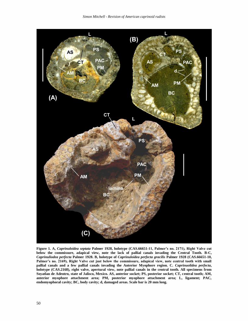
The holotype of Caprinuloidea perfecta Palmer
is a large bivalved specimen (but see Chartrousse,
1998b, and below under type specimen) that was
apparently disarticulated to show the apertural
surfaces of the left and right valves, although the
myophores of the left valve are partly damaged.
The diagnoses of the genus as given by Palmer
(1928) and Coogan (1977), refer to an external row
of pyriform canals and an internal row or rows of
polygonal canals; they do not mention the presence
of canals in the central tooth or the myophoral
Simon Mitchell - Revision of American caprinoid rudists
50
Figure 1. A, Caprinuloidea septata Palmer 1928, holotype (CAS.66651-11, Palmer’s no. 2171), Right Valve cut
below the commissure, adapical view, note the lack of pallial canals invading the Central Tooth. B-C,
Caprinuliodea perfecta Palmer 1928. B, holotype of Caprinuloidea perfecta gracilis Palmer 1928 (CAS.66651-10,
Palmer’s no. 2169), Right Valve cut just below the commissure, adapical view, note central tooth with small
pallial canals and a few pallial canals invading the Anterior Myophore region. C. Caprinuolidea perfecta,
holotype (CAS.2168), right valve, apertural view, note pallial canals in the central tooth. All specimens from
Soyatlan de Adentro, state of Jalisco, Mexico. AS, anterior socket; PS, posterior socket; CT, central tooth; AM,
anterior myophore attachment area; PM, posterior myophore attachment area; L, ligament; PAC,
endomyophoral cavity; BC, body cavity; d, damaged areas. Scale bar is 20 mm long.
Simon Mitchell - Revision of American caprinoid rudists
51
areas which are clearly visible in the type
specimens of Caprinuloidea perfecta (Figure 1C)
and less distinctly in Caprinuloidea perfecta
gracilis Palmer (Figure 1B). Coogan’s (1973,
1977) diagnosis of Texicaprina relies on the
invasion of pallial canals into the teeth and
myophores (since other characters he mentions are
not visible in the type specimens of the type
species of his genus), something that is already
apparent in the right valves of the type specimens
of C. perfecta and C. perfecta gracilis (see also
Chartrousse, 1998b, p. 137, figs. 38, 39).
From an examination of type specimens of
the various species assigned to Caprinuloidea as
well as an analysis of a large numbers of
specimens preserved in the Smithsonian
Institution and the Texas Memorial Museum,
specimens of Caprinuloidea can be divided into
three main groups: (1) those in which the central
tooth is completely solid, as in the holotypes of
Caprinuloidea septata Palmer and C.
multitubifera Palmer; (2) those specimens in
which the central tooth is invaded by small to
normal sized pallial canals as in the holotypes of
Caprinuloidea perfecta and C. perfecta gracilis;
and (3) those forms in which large canals invade
the myocardinal areas as in Caprinuloidea
romeri sp. nov. The type specimens of
Caprinuloidea septata, C. multitubifera and C.
perfecta come from Soyatlan de Adentro
(Palmer, 1928) where they occur with
Coalcomana ramosa, yet outcrops here (written
commun., Peter Skelton 2013) consist of
roadside exposures and the stratigraphic
relationship of these forms cannot be determined.
Elsewhere (e.g., the Seafield Limestone of
Jamaica), Caprinuloidea multitubifera and C.
perfecta and Coalcomana ramosa occur together
(Brown and Mitchell, 2011; Mitchell and Green,
2011) although this does not appear to be the
case in Texas (Scott, 2002). Whether forms of
Caprinuloidea that lack pallial canals in the
central tooth (C. septata) occur at a lower level
than those that have them (C. perfecta) is not
known. Yet, the progressive invasion of pallial
canals into the central tooth of the right valve,
the myocardinal areas of the right valve, and the
myophores of the left valve, as described here,
could represent a progressive sequence that might
allow the erection of chronospecies for high-
resolution biostratigraphy. This is further
strengthened by the case that the holotype of C.
perfecta already has canals invading the central
tooth. In this paper, four species of Caprinuloidea
are recognized and discussed below.
Caprinuloidea septata Palmer, 1928.
Figure 1A
? 1898 Sphaerucaprina Felixi n. sp.: G. Boehm, p. 329, fig. 6 (LV).
? 1898 Sphaerucaprina Lenki n. sp.: G. Boehm, p. 330,
fig. 7. v. 1928 Caprinuloidea septata: Palmer, p. 62, pl. 9, fig. 4,
pl. 10, fig. 3, pl. 11, fig. 1.
? 1977 Caprinuloidea felixi (Boehm); Coogan, pl. 13, fig. 7 (LV from Boehm 1898, fig. 6)
? 1977 Caprinulodea lenki (Boehm); Coogan, pl. 13, fig.
2. (LV from Boehm 1898, fig. 7)
Type specimen. Holotype (CAS.66651-11, Palmer
No. 2171) from Soyatlan de Adentro, State of
Jalisco, Mexico.
Discussion. The name Caprinuloidea septata
Palmer is used here for forms in which the central
tooth and myophore areas of the right valve are
composed of compact shell material and lack
pallial canals and in which both the anterior and
posterior myophores are represented by narrow
wall-like plates. Whereas right valves can be easily
assigned to this species, left valves do not show
sufficiently diagnostic characters that allow
distinction between C. perfecta and C. septata.
Boehm (1898) figured several left valves under the
names Sphaerucaprina Lenki Boehm and S. Felixi
Boehm that might belong to this species. Since
these species are based on left valves, they cannot
be unambiguously placed in current species
concepts.
Distribution. Caprinuolidea septata has only been
described from the ?Early Albian of Soyatlan de
Adentro, State of Jalisco, Mexico (Palmer, 1928),
but might be found where the details of the Right
Valve can be seen.
Caprinuloidea perfecta Palmer, 1928.
Figure 1B-C
v.1928 Caprinuloidea perfecta: Palmer, p. 59-60,
fig. 6., pl. 8, fig. 8, pl. 9, figs. 1-2.
v. 1928 Caprinuloidea perfecta gracilis: Palmer, p.
60-61, pl. 9, fig. 3, pl. 10, fig. 1.
?. 1936 Caprinuloidea perfecta Palmer; Thiadens, p.
1134-1138, fig. 4.6.
? 1977 Caprinuloidea septata Palmer; Coogan, pl.
13, fig. 3.
1977 Caprinuloidea perfecta Palmer; Coogan, pl.
12, fig. 2a-c (holotype).
? 1991 Caprinuloidea perfecta Palmer; Scott and
Gonzalez-Leon, p. 62, figs. 7C-F.
2002 Caprinuloidea perfecta gracilis Palmer;
Scott, fig. 3.3 (from Palmer, 1928).
?. 2011 Caprinuloidea perfecta Palmer; Mitchell and
Green, fig. 5D.
Simon Mitchell - Revision of American caprinoid rudists
52
Type specimen. Palmer nominated as Holotype
(CAS.66651-10, Palmer No. 2168) a bivalve
specimen from Soyatlan de Adentro, State of
Jalisco, Mexico. Chartrousse (1998b) noted that
there were differences in the sizes of the CT, CTS
and that the anterior socket of the RV contains a
fragment of the anterior tooth, whereas in the left
valve the anterior tooth is complete. He concluded
that the two valves came from different
individuals and nominated the RV as Lectotype
and the LV as Paralectotype. This is formally
accepted here.
Discussion. The name Caprinuloidea perfecta is
used here for specimens of Caprinuloidea with
small pallial canals in the central tooth, and also
locally in the myophoral areas of some right
valves. The holotype (which is a very large
specimen) has somewhat larger pallial canals, but
since it occurs with the other specimens, all
material is placed in the same species. Specimens
assigned to C. perfecta gracilis by Palmer (1928)
only differ by being smaller and narrower than C.
perfecta, and are also included in C. perfecta here.
Geographical and Stratigraphical Range. Caprinuloidea perfecta has been widely reported
from New World localities including: Soyatlan de
Adentro, State of Jalisco, Mexico (Palmer, 1928);
the Río Hatillo Limestone of the Dominican
Republic (Myczyński and Iturralde-Vinent, 2005);
the Seafield Limestone of Jamaica (Chubb, 1971;
Mitchell and Green, 2011); the Barranquitas
Limestone of Puerto Rico (Skelton, 1996); and
from limestones in Southern Santa Clara Province
in Cuba (Thiadens, 1936). All of these forms are
tentatively placed in the species C. perfecta,
although the details of the right valve have not
been reported in many instances. Specimens
reported as C. perfecta from the Edwards
Limestone in Texas are here placed in the species
C. romeri sp. nov. because their pallial canals are
regularly developed and some pallial canals
invade the myophores.
Caprinuloidea multitubifera Palmer, 1928.
Figure 2A-C
v. 1928 Caprinuloidea multitubifera: Palmer, p. 61-
62, pl. 10, fig. 2.
v. 1977 Caprinuloidea multitubifera Palmer; Coogan,
pl. 13, fig. 8 (holotype after Palmer, 1928,
pl. 10, fig. 2).
. 1996 Caprinuloidea multitubifera Palmer; Rojas
et al., pl. 2, figs. 2-3.
Type specimen. Holotype (CAS.66651-06, Palmer
No. 2170) from Soyatlan de Adentro, State of
Jalisco, Mexico.
Discussion. Specimens assigned to Caprinuloidea
multitubifera have broad posterior myophores, as
well as having a greater number of smaller pallial
canals than other species of Caprinuloidea. The
pallial canals also have very thick walls which
contrasts with the other species of Caprinuloidea.
Geographical and Stratigraphical Range. Caprinuloidea multitubifera occurs in the ?Lower
Albian of Soyatlan de Adentro, State of Jalisco,
Mexico (Palmer, 1928). Specimens have also been
seen in the Lower Albian Seafield Limestone of
Jamaica which are attributed to this species here.
Caprinuloidea romeri sp. nov.
Figures 3A-E, 4A-D
? 1888 Ichthyosarcolites anguis n. sp.: Roemer, p.
9-10, pl. II (XXXII), fig. 2a-d.
1900 Caprinula anguis Roemer; H. Douvillé, p.
220, fig 16-17.
1969 Caprinuloidea sp.: Perkins, p. N762, fig.
E232.
1973 Caprinuloidea gracilis Palmer; Coogan, p.
61, pl. 5, fig. 2 (poorly preserved).
? 1973 Caprinuloidea multitubifera Palmer; Coogan,
p. 61, pl. 5, fig. 3 (this RV has many more
Canals in the Shell layer than usual, and the
CT is poorly preserved).
? 1977 Caprinuloidea perfecta Palmer; Coogan, pl.
12, fig. 4 (RV Canals in AM area, possibly
not in CT)
1977 Caprinuloidea anguis (Roemer); Coogan, pl.
12, figs 3a (questionably referred to this
species), fig 3b (reproduction of Roemer
1888, fig. 2a), fig. 3c (reproduction of
Douvillé 1900, fig. 16); pl. 13, fig. 1.
1977 Caprinuloidea gracilis Palmer; Coogan, pl.
13, fig. 5-6.
1977 Caprinuloidea sp.: Coogan, pl. 13, fig. 9
.? 1977 Texicaprina vivari Palmer; Coogan, pl. 14,
figs. 3-4.
.? 1977 Texicaprina sp.: Coogan, pl. 15, figs. 4-5.
2002 “Ichthyosarcolites” anguis Roemer, 1888;
Scott, p. 420-421.
. 2007 Caprinuloidea perfecta Palmer; Molineux,
Scott, Ketcham, and Maisano, p. 3-5, figs. 1-
4.
v. 2010 Caprinuloidea perfecta; Scott and Weaver,
fig. 8D.
Diagnosis. A species of Caprinuloidea with a long
straight or twisted right valve and a shorter
partially coiled left valve with an external
ligamental groove, in which pallial canals invade
the central tooth and myocardinal areas of the right
valve; pallial canals partially invade the posterior
Simon Mitchell - Revision of American caprinoid rudists
53
Figure 2. A-C, Caprinuloidea multitubifera Palmer 1928. A, holotype (CAS.66651-06, Palmer’s no. 2170), Left Valve,
apertural view; note the long narrow cavity for the central tooth; scale bar is 20 mm. B-C, Right Valve (TMM.UT-
50694) cut below commissure with dentition elements from the left valve preserved in place (posterior and anterior
myophores still partially encased in matrix); B (scale bar = 20 mm) shows the narrow central tooth, and the broad
posterior myophore filling most of the posterior accessory cavity (although the lamina separating the posterior
accessory cavity and body cavity is not preserved); C (scale bar = 10 mm) shows the construction of the pallial canals,
with a central narrow wall and a thick layer of shell material around each canal; differential thickening gives rise to
pyriform marginal canals. Both specimens from Soyatlan de Adentro, State of Jalisco, Mexico. Scale bar 20 mm. AT,
anterior tooth; AS, anterior socket; PT, posterior tooth; PS, posterior socket; CT, central tooth; CS, central socket;
AM, anterior myophore; PM, posterior myophore; L, ligament; BC, body cavity.
myophore and anterior myophore in the left valve;
there is a marginal row of more-or-less uniform
pallial canals with their long axes orientated
radially; and the shell wall contains three to six
rows of pallial canals inclusive of the marginal
row.
Type material. Holotype (TMM.UT.10932) and
Paratypes (TMM.UT-10930.1, 11268, 33867,
10922), Edwards Limestone, Austin, Texas.
Material. This species is common in the Edwards
Limestone of Texas, and ranges from small
specimens to gigantic specimens.
Description. Right valve generally elongate and
may be straight to somewhat irregularly twisted,
cylindro-conical to cylindrical; grows communally
as elevators and possibly isolated as a recumbent.
The right valve has a well defined external
ligamental groove, extending along the length of
the valve; the ligamental groove is connected by a
fissure, lined with a thin layer of outer shell layer
to the hook-shaped infolded ligament. Transverse
sections are circular to subquadrate to pear-shaped
with a narrow rounded elongate ventral margin,
Simon Mitchell - Revision of American caprinoid rudists
54
Figure 3. Caprinuloidea romeri sp. nov. All specimens from the Edwards Limestone of Texas. A-B, Right Valve
(SI.USNM.221518), Whitney Dam: A, commissure view, with myocardinal elements of the LV in place: note the
narrow wall like form of the PM and AM and the CT filled with pallial canals; B, view of dorsal flank showing
ligamental groove. C, Right valve (TMM.UT-10930.1), San Gabriel River, Roy Gunn’s Ranch, Texas, commissural
Simon Mitchell - Revision of American caprinoid rudists
55
and the anterior margin generally flattened. The
central tooth is broadly rectangular, orientated at a
low angle (about 45 degrees) to a line joining the
posterior and anterior tooth sockets. The sockets
for the teeth are deep, the posterior tooth socket
connected, via a constriction, to the posterior
myophore cavity. There is an embayment for the
projecting (rotated) anterior myophore of the left
valve. The anterior myophore of the left valve is a
vertical wall that starts on the anterior margin of
the anterior tooth socket and is orientated towards
the wall of the body chamber. The anterior tooth
socket is separated from the body cavity by a
septum that stops at the same level as the ledge
for the anterior myophore. The cavity for the
posterior myophore is separated from the body
cavity by a septum, which in small forms in
composed of compact shell material but in larger
forms has a single row of pallial canals. The
ligament is invaginated with the invagination
marked by an external groove connected to the
interior hook-shaped ligament. The body cavity is
broadly rounded, and the lower part is filled with
concave tabulae. The margin of the shell is
composed of rather uniform, radially elongated,
elliptical rather than pyriform canals; some
pyriform canals may be present but either the
inner or outer ends may be narrower. The
remainder of the shell is composed of uniformly
sized canals up to 1.5 mm in diameter. The
marginal pallial canals lack tabulae, whereas the
interior pallial canals have ‘concave towards the
aperture’ tabulae spaced at irregular (1 to 8 mm)
intervals. The central tooth is filled with normal
pallial canals identical to the remainder of the
shell wall. The anterior tooth socket contains
concave tabulae; the details of the fill of the
posterior tooth socket have not been seen.
The left valve is coiled, forming up to three-
quarters of a revolution. In cross section it ranges
from circular to pear-shaped with the ventral
margin narrower and elongated. The anterior
surface is generally gently flattened. A prominent
ligamental groove extends around the anterior-
dorsal margin and is folded in to form a small
hook. The anterior tooth is generally triangular in
cross-sectional shape; the posterior tooth is
elongated anterio-posteriorly, and narrows both
towards the posterior and the anterior. The anterior
myophore is wall-like and extends along the wall
of the body cavity from the anterior tooth. The
posterior myophore skirts around the posterior
margin of the LV endomyomorphal cavity and
only extends a little way beyond the ventral end of
the septum separating the endomyophoral cavity
from the body cavity. The marginal row of pallial
canals is weakly to moderately radially elongate,
they are not pyriform. The remainder of the shell
structure is filled with pallial canals; in many
individuals the two accessory cavities on the
outside of the myophores are filled with a row of
larger pallial canals (up to two times the size of the
‘normal’ pallial canals), but is some forms the
pallial canals are of more uniform size throughout
the shell. The body cavity is regularly elliptical;
the presence or absence of tabulae in the body
cavity or the central tooth socket has not been
confirmed.
Stratigraphic distribution. The material preserved
in the Texas Memorial Museum comes from a
variety of locations in the Edwards Limestone of
Texas. These include: South San Gabriel River,
Roy Gunn’s Ranch, Texas (this locality now lies
under Lake Georgetown); and Witney Dam, Texas.
Discussion. The material described and figured as
Ichthyosarcolites anguis by Roemer (1888), which
he collected from two miles above the mouth of
Barton Creek, Austin, Travis Co., Texas, from
somewhere in the Edwards Limestone (Scott,
2002, p. 421), appears to be heterogeneous.
Roemer’s specimens in his plate II (XXXII), figs.
2a-d, have external ligamental grooves and could
be referred to Caprinuloidea romeri sp. nov.; in
contrast, the specimens in his plate I (XXXI), figs.
7a-b, lack external ligamental grooves and would
appear to represent Youngicaprina sangabrieli
gen. et sp. nov. Both species are common in the
Edwards Limestone of Texas, but the figures given
by Roemer (1888) are too stylistic to determine the
true affinity of these specimens. Furthermore, the
[Figure 3 continued] view, containing teeth from left valve, note the CT filled with pallial canals, pallial canals in the
ledge below for the AM (b) and a row of pallial canals in the lamina dividing the damaged posterior accessory cavity
from the BC; the sockets are filled by the broken teeth of the LV which are lined by chert suggesting that they were
originally solid. D, Right Valve (TMM.UT-11268), abumbonal view, note sockets for teeth without apparent tabulae,
area of AM filled with pallial canals (d), PAC divided into three by septa (as in Mexicaprina), and the elongate pallial
canal filled CT (unlike in Mexicaprina). E, Left valve (TMM.UT-10922), commissural view, showing myocardinal
elements; the PT and AT are broken off and lined by crystals suggesting they were solid, the PM is wall like, the AM,
is triangular with a single pallial canal, and the ectomyophoral cavity is filled with large pallial canals (e). Scale bar 20
mm. AT, anterior tooth; AS, anterior socket; PT, posterior tooth; PS, posterior socket; CT, central tooth; CS, central
socket; AM, anterior myophore; PM, posterior myophore; L, ligament; BC, body cavity.
Simon Mitchell - Revision of American caprinoid rudists
56
Figure 4. Caprinuloidea romeri sp. nov. All specimens from the Edwards Limestone of Texas. A-B, Bivalve
(SI.USNM.PAL.534223), Whitney Dam: A, transverse section of Right Valve, abumbonal view, showing teeth and
myophores; B, transverse section of Left Valve, abumbonal view showing teeth and myophores. C, Left Valve
(TMM.UT-33867) showing the well developed external ligamental groove and myocardinal elements; the PM is
wall like and attached to the PT. D, Left Valve (TMM.UT-10932), commissural view, showing myocardinal
elements; the PT and AT are broken off and lined by crystals suggesting they were solid, the PM is wall like, the
AM, is triangular with a row of pallial canals (f), and the ectomyophoral cavities are filled with large pallial canals
(g). Scale bar 20 mm. AT, anterior tooth; AS, anterior socket; PT, posterior tooth; PS, posterior socket; CT, central
tooth; CS, central socket; AM, anterior myophore; PM, posterior myophore; L, ligament; BC, body cavity.
repository of the actual specimens is not known
and they may be lost (Scott, 2002, p. 421).
Coogan (1977) suggested that the specimens
figured as Caprinula anguis Roemer by H.
Douvillé (1990) were the same specimens as
Roemer’s (1888) plate I (XXXI), figs. 7a-b. Yet
H. Douvillé’s illustrations show a distinct external
ligamental groove and represent Caprinuloidea
romeri sp. nov., whereas this groove is not shown
in Roemer’s illustrations which are tentatively
referable to Youngicaprina sangabrieli gen. et sp.
nov. Coogan’s (1977) assertion that these are the
same specimens is, however, incorrect; H.
Douvillé’s illustrations were based on photographs
of material collected from the Fredericksburg
Group by R. T. Hill (Scott, 2002, p. 421). In
Simon Mitchell - Revision of American caprinoid rudists
57
consequence the illustrations of Ichthyosarcolites
anguis as given by Roemer (1888) are probably
heterogeneous, are highly stylistic, and the
original specimens appear to be lost; I follow
Scott (2002) in avoiding this name in
palaeontological and biostratigraphical studies.
Caprinuloidea romeri sp. nov. and
Youngicaprina gen. nov. occur in the same units
and have been confused in the past. Caprinuloidea
romeri is distinguished from Youngicaprina as
follows: marginal canals are radially elliptical
rather than pyriform as in Youngicaprina; an
external ligamental groove is present in
Caprinuloidea romeri sp. nov. and absent in
Youngicaprina gen. nov.; the angle between the
long axis of the central tooth and the centre points
of the anterior and posterior teeth is small in
Caprinuloidea romeri sp. nov. and large in
Youngicaprina gen. nov.; the posterior and
anterior myophores are wall-like in Caprinuloidea
romeri sp. nov.; and the posterior myophore (PM)
forms a sharp angle around the endomyophoral
cavity in Caprinuloidea romeri sp. nov. rather
than forming a broad arc in Youngicaprina gen.
nov.
Genus Mexicaprina Coogan, 1973
Figure 5A-B
Type Species. Mexicaprina cornuta Coogan,
1973.
Description. See Coogan (1973, 1977) and Scott
(2002).
Genus Kimbleia Coogan, 1973
Figure 5C-E
Type species. Kimbleia capacis Coogan, 1973
Description. See Coogan (1973, 1977) and Scott
(2002).
Discussion. The recognition of two species based
on differences in the size of the pallial canals is
not considered sufficiently diagnostic to separate
Kimbleia albrittoni (Perkins, 1960) from Kimbleia
capacis, and K. capacis is placed in synonymy
with K. albrittoni herein (see Scott, 2002, p. 415).
Genus Neokimbleia gen. nov.
Figure 6A-E
Type species. Neokimbleia acutus gen. et sp. nov.
from the El Abra Formation (Upper Upper
Albian), El Madroño, state of Querétaro, central
Mexico.
Diagnosis. An inequivalve caprinuloideid with an
elongate straight right valve and a shorter coiled
left valve. The anterior tooth is large (and may be
bifid) and the posterior tooth is smaller and has a
prominent posterior ridge making it ‘arrow’-
shaped. The anterior myophore of the left valve is
connected to the anterior tooth. The posterior
myophore of the left valve is blade-like, strongly
elongated and fits into an elongate cavity in the
right valve; the endomyophoral cavity and the
socket for the anterior tooth are separated by a
long septum. There is no external ligamental
groove; the ligament being represented by a pear-
shaped cavity completely isolated from the
exterior of the shell.
Discussion. Neokimbleia gen. et sp. nov. shows
many characters which align it with Kimbleia; the
distribution of the teeth and sockets, and the
transverse cross-section of the shell. It differs by
the nature of its ligament: in Kimbleia, the
ligament is represented by an obvious external
groove; whereas in Neokimbleia, the ligament is
represented by a pear-shaped cavity and is not
connected to an external ligamental groove.
Neokimbleia gen. nov. can be seen as an
evolutionary development from Kimbleia in which
the external ligamental groove has been lost.
Neokimbleia acutus sp. nov.
Figure 6A
v. 1936 Sabinia sp.; Thiadens, p. 1140-1141, fig. 5(1).
? 1978 Kimbleia? sp.: Coogan, pl. 17, fig. 2.
v. 1998 Kimbleia albrittoni (Perkins, 1960) pars;
Alencátser and Oviedo-García, p. 175-177,
fig. 8.2, fig. 9.5-6.
Diagnosis. A species of Neokimbleia with a bifid
AT and a triangular flange on the ventral shell
margin.
Type material. Holotype: Transverse section of a
right valve (IGM.4586), El Abra Formation, El
Madroño, state of Querétaro, central Mexico
Description. No articulated specimens are known.
Cross sections are flattened in an anterior-posterior
direction, have a rounded dorsal margin and an
acutely pointed ventral margin. Only myocardinal
details of the right valve are known. There is no
external ligamental groove, and the ligament is
represented by a rounded pit enclosed in the shell
structure. The socket for the anterior tooth is
broadly rounded, has two cavities separated by
pallial canals recalling the bifid socket seen in
Immanitas Palmer and some Mexicaprina. The
Simon Mitchell - Revision of American caprinoid rudists
58
Figure 5. A-B, Mexicaprina coruuta Coogan 1973. El Abra Limestone, Sierra de El Abra, San Luis Potosi, Mexico.
A, left valve (IGM.2585), transverse cross section, adapical view, showing myocardinal features. B, Right valve
(IGM.2584) transverse cross section, adapical view, showing myocardinal features. C-D, Kimbleia albrittoni (Perkins
1960). Segovia Formation, Lopez Ranch, Kimble County, Texas. C, Right valve (UT.203571), abumbonal view,
Simon Mitchell - Revision of American caprinoid rudists
59
ledge for the attachment of the anterior myophore
is clearly developed extending from the anterior
margin of the anterior tooth socket to the margin
of the body cavity. The socket for the posterior
tooth is joined to the posterior endomyophoral
cavity with the posterior margin of the accessory
cavity strongly curved. The shell is filled with
regular-sized small pallial canals with diameters
of 1 to 2.5 mm; the marginal canals are radially
elongated, and the inner rounded canals are
arranged in from 2 to 4 rows. The central tooth is
filled with similar canals as the rest of the shell,
and extends towards the anterior from a position
near the midpoint of the cavity formed by the
posterior tooth socket and the posterior accessory
cavity. The posterior accessory cavity is separated
from the anterior tooth socket and the body cavity
by a 1-mm wide septum that lacks pallial canals, a
similar septum separates the anterior tooth socket
from the body cavity.
Discussion. Neokimbleia acutus gen. et sp. nov. is
distinguished from species of Kimbleia by the
lack of an external ligamental groove, and the
positioning of the rounded ligament of the right
valve within the pallial-canal bearing shell
structure. The pallial canals of N. acutus gen. et
sp. nov. are smaller than those of Kimbleia
capacis Coogan and K. albrittoni (Perkins); but in
N. acutus the anterior tooth is bifid whereas in K.
capacis and K. albrittoni it is rounded.
Distribution. Neokimbleia acutus gen. et sp. nov.
occurs in the latest Albian El Abra Formation, El
Madroño, state of Querétaro, central Mexico. A
specimen from Cuba figured by Thiadens (1936)
as Sabinia sp. also seems to belong to this species.
Neokimbleia planata (Conrad, 1855)
Figure 6B-E
v. 1855 Caprina occidentalis: Conrad, p. 268.
v. 1855 Caprina planata: Conrad, p. 268.
v. 1857 Caprina occidentalis: Conrad, p. 147, pl. 2,
figs. 1a-c.
v. 1857 Caprina planata: Conrad, p. 147, pl. 2, figs.
2a-b.
v. 2002 “Caprina” occidentalis (Conrad, 1855);
Scott, p. 419-420, figs. 9.1-2, fig. 10.1.
v. 2002 “Caprina” planata (Conrad, 1855); Scott, p.
420, figs. 9.3-4, fig. 10.2.
Diagnosis. A species of Neokimbleia with a single,
non-bifid anterior tooth, the shell flattened in an
anterio-posteriorly direction, with the shell
smoothly rounded, without flanges.
Type specimen. The holotype is preserved in the
Smithsonian Institute (SNNHM.USNM-9891) and
was collected from Oak Creek near Puercos,
Texas. Scott (2002; pers. commun. 2013)
considers the most likely locality was at Live Oak
Creek on the Pecos River near present-day Fort
Lancaster, from a level in the Fort Lancaster
Formation of the Washita Group rather than from
the Fort Terrett Formation of the Frederisksburg
Group.
Description. A large species of Neokimbleia gen.
nov. with strongly curved left and right valves.
The valves are strongly flattened in an anterior-
posterior direction and regularly rounded. The
dorsal-ventral diameter of the shell is about 2 to 3
times the anterior-posterior diameter of the shell.
The right valve is elongate and gently curved.
The body cavity is rounded and filled with tabulae.
There is no external ligamental groove, and the
ligament is represented by an enclosed cavity
between the posterior tooth socket and the central
tooth that is completely embedded within the
pallial-canal-bearing shell material. The socket for
the anterior tooth is gently rounded and separated
from the body cavity by a septum that lacks pallial
canals. The socket for the posterior tooth is
connected to the posterior endomyophoral cavity,
with the tooth socket delineated by a slight
constriction. A marginal row of radially elongated
pallial canals is present around the extremity of the
shell, and the interior of the shell is filled with
pallial canals; in some specimens the pallial canals
are predominately small forms (diameters of 1 to
3 mm) whereas in other specimens the inner row
of pallial canals adjacent to the posterior accessory
cavity are larger (up to 5 or 6 mm across) and
elongated in a dorsal-ventral direction. The central
tooth is filled with pallial canals, and extends from
a distance of about two-thirds of the way from the
posterior end of the cavity for the posterior
tooth/posterior myophore anteriorly towards the
anterior side of the shell; the central tooth is large
and anterior-posteriorly elongated, and is
[Figure 5 continued] transverse cross section (orientated with anterior side at top), note the well-developed
external ligamental groove and the short CT with the AS and the PAC separated by a lamina. D, Right valve
(SI.USNM.203566), anterior aspect, note the well-developed external ligamental groove. E, Kimbleia sp.
(SI.USNM.547501 [CS2; #1941]), articulated specimen, posterior dorsal view, showing external ligamental
groove. Scale bar 20 mm. AT, anterior tooth; AS, anterior socket; PT, posterior tooth; PS, posterior socket; CT,
central tooth; CS, central socket; AM, anterior myophore; PM, posterior myophore; L, ligament; BC, body
cavity.
Simon Mitchell - Revision of American caprinoid rudists
60
Figure 6. A, Neokimbleia acutus sp. nov. Right valve (Holotype, IGM.4586), transverse cross section, abapical view,
note the lack of an external ligament groove (there is a crack, but this passes across pallial canals) and myocardinal
arrangements, El Madroño, state of Querétaro, central México. B-E, Neokimbleia planata (Conrad, 1855). B, Right
valve (IGM.2575, figured by Googan, 1973, pl. 2, fig. 1 as Kimbleia albrittoni), note lack of external ligamentary groove,
internal ligamentary cavity, but otherwise a dentition similar to Kimbleia; El Abra Formation, Mexico. C, Holotype of
Caprina occidentalis Conrad 1855 (SI.USNM.9840) Left Valve (with part of Right Valve?), adapical cross-section,
collected from near the mouth of the Percos river, Texas. D, Holotype of Caprina planata Conrad, 1855
(SI.USNM.9891), adapical slice through RV close to commissure containing the myocardinal structure of the left valve,
collected at Oak creek, near Puercos, Texas. E, Left Valve (SI.USNM.547500 [CS1]), showing myocardinal
arrangement and lacking an external ligamental groove. Nb., transverse sections orientated with anterior side at top.
Scale bar 20 mm. AT, anterior tooth; AS, anterior socket; PT, posterior tooth; PS, posterior socket; CT, central tooth;
CS, central socket; AM, anterior myophore; PM, posterior myophore; L, ligament; BC, body cavity.
almost arrow shaped. The posterior myophore is
surrounded by compact shell material. The left valve is
shorter than the right valve and more strongly coiled
(Figure 6E). There is no external ligamental groove.
The posterior myophore is attached to the centre of the
posterior tooth and forms a wall like feature which
initially extends in a posterior-ventral direction, before
making an angle and extending parallel to the posterior
margin of the shell; the myophore gradually decreases in
height from the posterior tooth, and also gradually
decreases in width. The anterior tooth is relatively large
and rounded, and is poorly separated from the anterior
myophore. The central socket is a slot between the
anterior tooth and the posterior tooth.
Simon Mitchell - Revision of American caprinoid rudists
61
Discussion. Scott (2002) regarded Conrad’s
(1855) species Caprina occidentalis and Caprina
planata as indeterminate and suggested that the
names should be applied only to the type
specimens and should not be maintained in
biostratigraphic or taxonomic studies. In contrast,
I recognize that Conrad’s Caprina planata shows
sufficiently distinctive features (the forms of the
posterior tooth and posterior myophore, the shape
of the anterior tooth, the enclosed pear-shaped
ligament cavity) that allows it to be placed in the
genus Neokimbleia gen. nov. Neokimbleia planata
differs from Neokimbleia acutus sp. nov. by
having a rounded, rather than bifid anterior tooth,
the absence of a large triangular flange on the
ventral margin; and the distribution and number of
flanges is considered a specific character in
separating caprinuloid rudists (Mitchell and
Gunter, 2006).
The three specimens of Neokimbleia planata
shown on Figure 6 include a range of different-
sized pallial canals. These range from small
canals, similar to those seen in Kimbleia capacis,
to an inner row of large elongated canals, similar
to those seen in K. albrittoni. The presence of
such variations in pallial canal size in two
different genera, which are likely to be ancestor
and descendant, suggests that the size of pallial
canals is of little specific importance and that
Kimbleia capacis Coogan, 1973, should be placed
in the synonymy of Kimbleia albrittoni (Perkins,
1960).
Subfamily Youngicaprininae subfam. nov.
Diagnosis. Caprinuloideidae in which the anterior
myophore of the LV is modified to form a low
platform, with or without blades or tubercles, that
wraps around the anterior side of the anterior
tooth; and a posterior myophore which becomes
tooth-like and is separated from the posterior
tooth.
Discussion. This group has a distinctly different
myocardinal arrangement to the
Amphitriscoelinae and the Caprinuloideinae, and
may show a transition towards the
Antillocaprinidae Mac Gillavry, 1937, but
transitional forms have not been found. The
Antillocaprinidae are differentiated because the
endomyophoral cavity between the posterior tooth
and the body cavity that is characteristic of the
Caprinuloideidae has been lost and the posterior
tooth is directly adjacent to the body cavity.
Three genera are included within the
Youngicaprininae: Texicaprina Coogan, 1973,
Middle to Upper Albian; Youngicaprina gen. nov.,
Middle to Upper Albian; and Jalpania Alencáster
and Aguilar-Pérez, 1996. The Upper Albian
Immanitas Palmer, 1928, may also belong here
(see Aguilar Pérez, 2008, fig. 58).
Genus Texicaprina Coogan 1973
(=Guzzyella Alencáster 1998)
Type species. Sabinia kugleri Bouwman, 1937,
from Point-a-Pierre, Trinidad (see discussion
below).
Diagnosis. A genus of Youngicaprininae subfam.
nov. with an external ligamental groove, a ‘tooth-
like’ posterior myophore that is separated from the
posterior tooth by a gutter filled with pallial
canals, and a low anterior myophore that wraps
around the anterior tooth.
Discussion. Coogan (1973, p. 59) erected the
genus Texicaprina, nominating Sabinia vivari
Palmer, 1928, as type species, and then stated that
“The illustrations of Texicaprina vivari (Palmer)
… do not show adequately the characters which
make this species a Texicaprina” and that
“Examination of Palmer’s types at the California
Academy of Science shows that they are badly
recrystallized internally, but otherwise appear to
be the same species as found in the Edwards
Limestone in central Texas.” Coogan (1973) based
many of the features in describing his genus
Texicaprina on Sabinia kugleri Bouwman, 1937,
from Trinidad, which is preserved as specimens
free from matrix where the teeth, sockets and
myophores had been illustrated, and not on
Sabinia vivari from Mexico (Coogan in an
unpublished report, dated 1960, for Humble Oil
and Refining Company, suggested the manuscript
name Texicaprina citing Sabinia kugleri as the
type species; see also Alencaster and Oviedo-
García, 1998, p. 176).
Since Texicaprina has been used in
biostratigraphic studies it is desirable to stabilize
this name rather than abandon it and a proposal is
being sent to the Bulletin of Zoological
Nomenclature, to use Plenary Powers to place
Texicaprina and Sabinia kugleri Bouwman, 1937,
on the approved list of names in zoology, to place
vivari in the combination Sabinia vivari Palmer,
1928, on the list of invalid names in Zoology, and
to select Sabinia kugleri Bouwman, 1937, as the
type species of Texicaprina Coogan, 1973 (note
that Chartrousse, 1998b, also suggested a similar
solution in his unpublished doctoral thesis).
Coogan (1973) included forms in Texicaprina
Simon Mitchell - Revision of American caprinoid rudists
62
Figure 7. Texicaprina kugleri (Bouwman 1937). Erratic block, Point-A-Pierre, Trinidad. A-C, Right valve, syntype
(BM.K2997); A, posterior flank of Right Valve; B dorsal aspect of Right Valve showing ligamental groove; C,
apertural aspect of Right valve, note ‘w’-shaped posterior socket weakly separated from the endomyporal cavity by
a septum, Central Tooth partially dissolved. D-F, Left Valve, nominated lectotype herein (BM.G7120); D, postero-
dorsal flank of Left Valve showing ligamental groove; E, Left Valve, oblique view of apertural surface showing the
form of the anterior myophore which is composed of solid radiating blades and intervening canal-bearing furrows
and wraps around the anterior side of the large anterior tooth; F, apertural view of Right Valve, the posterior
myophore is separated from the posterior tooth by a pallial-canal bearing trough. Scale bar 20 mm. AT, anterior
tooth; AS, anterior socket; PT, posterior tooth; PS, posterior socket; CT, central tooth; CS, central socket; AM,
anterior myophore; PM, posterior myophore; L, ligament; BC, body cavity; t, tubercle.
which had pallial canals invading the teeth and
myophores. I consider that the form of the
myocardinal arrangement is more important for
generic placement than the distribution of pallial
canals, and, as with Caprinuloidea, regard the
invasion of pallial canals into different elements of
the myocardinal structure to be a specific
character.
Simon Mitchell - Revision of American caprinoid rudists
63
I place Caprinuloidea bisulcata Palmer, 1928,
in the genus Texicaprina on the basis of the
morphology of the anterior myophore, and
consequently Texicaprina bisulcata Palmer, 1928,
becomes a senior homonym of the type species of
the genus Guzzyella, G. bisulcata Alencáster,
1998, which I also place in Texicaprina. Here, I
consider that Guzzyella acuminata Alencáster,
1998, falls within the population of Guzzyella
bisulcata Alencáster, 1998, and use this name as
the valid name for the species of Texicaprina from
El Madroño, state of Querétaro, central Mexico.
Alencáster (1998) distinguished Guzzyella
from Texicaprina based on the fact that Guzzyella
had a wrinkled rather than ribbed anterior
myophore, and the fact that “Texicaprina is totally
perforated” with pallial canals. Alencáster (1998)
based her interpretation of Texicaprina on
specimens from El Madroño which she attributed
to Texicaprina kugleri (Bouwman), but which
lack an external ligamental groove and are here
referred to the genus Youngicaprina gen. nov.
Sabinia kugleri Bouwman has an external
ligamental groove, a slightly tooth-like posterior
myophore, and a low anterior myophore that
envelops the anterior tooth and has distinct ribs
separated by pallial canal filled furrows. It seems
likely that the ribbed morphology of the anterior
myophore is directly related to the invasion of
pallial canals, and would be absent in species
which lack pallial canals in the anterior myophore.
Given the progressive invasion of pallial canals
into different elements of the myocardinal
structure already described in Caprinuloidea, it
seems unwise to place species in different genera
based on the distribution of pallial canals.
Texicaprina kugleri (Bouwman, 1937)
Figure 7A-F
v. 1937 Sabinia kugleri (n. sp.): Bouwman, p. 450-
453, figs. 1-10.
v. 1977 Texicaprina kugleri (Bouwman); Coogan, pl.
14, figs. 2a-b.
non v. 1998 Texicaprina kugleri (Bouwman); Alencáster
and Oviedo-García, p. 167-171, fig. 3, fig.
4.1-4.10, fig. 5.1-5.7 (=Youngicaprina gloria
sp. nov.)
non v. 1998 Texicaprina vivari (Palmer); Alencáster and
Oviedo-García, p. 171, fig. 5.8-5.9 (but fig.
5.7-5.8 in the text in error)
(?=Youngicaprina gloria sp. nov.)
. 2002 Texicaprina vivari (Palmer); Scott, p. 416,
fig. 7.6.
Diagnosis. A species of Texicaprina with a
weakly tooth-like elongate, posterior myophore
separated from the posterior tooth by a gutter filled
with pallial canals, and an anterior myophore that
envelops the anterior-side of anterior tooth and is
ribbed with the ribs composed of compact shell
material and the furrows filled with pallial canals.
Type Series. Bouwman (1937) illustrated two
specimens under the name Sabinia kugleri which
are the syntypes and are preserved in the Natural
History Museum at Basel, Switzerland. The left
valve illustrated by Bouwman, 1937, fig. 1 and
figs. 8-9 (NHMB.G7220) is nominated here as
lectotype. The material came from erratic boulders
at Pointe-a-Pierre, Trinidad, so that the age cannot
be directly assessed. Judging solely on the
morphology it is most likely of late Albian age.
Description. No articulated specimens are known,
so the relative size of the valves cannot be
determined. The right valve is moderately short
and conical to cyclindroconical; preserved right
valves appear longer than left valves (and so were
probably longer). The ligament is represented by
an infold into the shell and a gentle hook, and is
marked on the exterior by a prominent external
ligamental groove. The material has been
somewhat eroded, and the external ligamental
groove has been opened up in this process. The
central tooth is represented by a broad ridge at an
angle of about 105° to a line joining the centres of
the posterior and anterior tooth sockets. The
anterior tooth socket is relatively large and
rounded; the posterior tooth socket is ‘w’-shaped.
The endomyophoral cavity is deep and separated
from the posterior tooth socket by a lamina. The
anterior myophore fits partly into an embayment of
the shell wall. Pallial canals are present filling the
wall of the shell, and there are between 3 and 6
rows of canals. The marginal pallial canals are
poorly preserved, so that their form cannot be
determined. Pallial canals invade the central tooth
(which has a cavity possibly due to dissolution),
and the ledge area for the anterior myophore.
The left valve is relatively short and gently
coiled; it has a well developed external ligamental
groove that is folded into the shell as a hook. The
central socket is narrow at the dorsal end and
gently expands towards the posterior-ventral end.
A low wall runs along the posterior margin of the
central socket and terminates in a tubercle. The
anterior tooth is larger than the posterior tooth and
is triangular in shape, the posterior tooth is an
elongated oval. The posterior myophore is an
elongate blade that is separated from the posterior
tooth by a weak gutter. The anterior myophore
envelops the anterior tooth on the anterior side and
Simon Mitchell - Revision of American caprinoid rudists
64
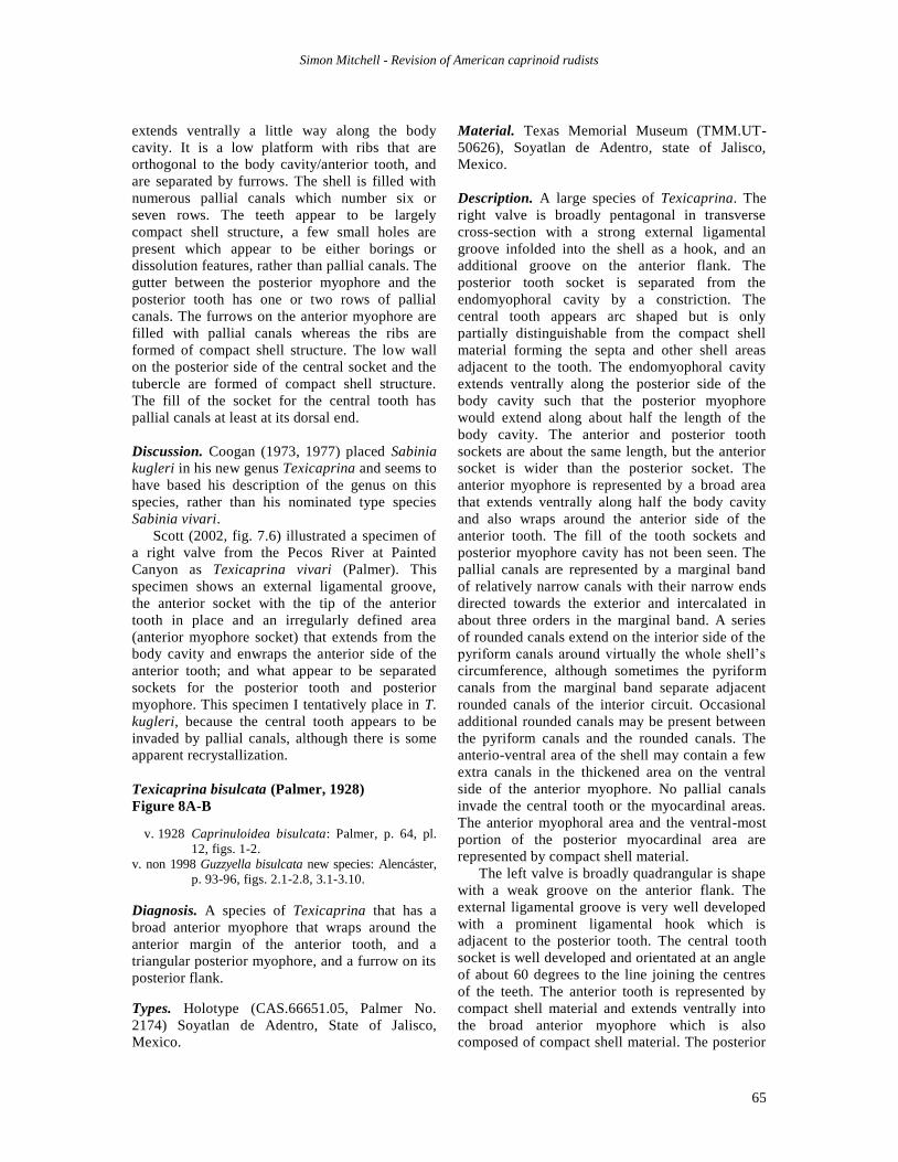
Figure 8. A-B, Texicaprina bisulcata (Palmer 1928). A, holotype (CAS.), transverse section of right valve, adapical
view, note thickened anterior myophore that wraps around the cavity for the anterior tooth; the posterior
myophore is as in Caprinuloidea. B, transverse section of a left valve (TMM.UT.50626), abapical view, note
thickened anterior myophore that wraps around the anterior tooth. C-E, Texicaprina acuminata (Alencáster 1998),
holotype (IGM.4590) of Guzzyella bisulcata Alencáster 1998: C, Right Valve, D, Left Valve, E, Bivalve.
Simon Mitchell - Revision of American caprinoid rudists
65
extends ventrally a little way along the body
cavity. It is a low platform with ribs that are
orthogonal to the body cavity/anterior tooth, and
are separated by furrows. The shell is filled with
numerous pallial canals which number six or
seven rows. The teeth appear to be largely
compact shell structure, a few small holes are
present which appear to be either borings or
dissolution features, rather than pallial canals. The
gutter between the posterior myophore and the
posterior tooth has one or two rows of pallial
canals. The furrows on the anterior myophore are
filled with pallial canals whereas the ribs are
formed of compact shell structure. The low wall
on the posterior side of the central socket and the
tubercle are formed of compact shell structure.
The fill of the socket for the central tooth has
pallial canals at least at its dorsal end.
Discussion. Coogan (1973, 1977) placed Sabinia
kugleri in his new genus Texicaprina and seems to
have based his description of the genus on this
species, rather than his nominated type species
Sabinia vivari.
Scott (2002, fig. 7.6) illustrated a specimen of
a right valve from the Pecos River at Painted
Canyon as Texicaprina vivari (Palmer). This
specimen shows an external ligamental groove,
the anterior socket with the tip of the anterior
tooth in place and an irregularly defined area
(anterior myophore socket) that extends from the
body cavity and enwraps the anterior side of the
anterior tooth; and what appear to be separated
sockets for the posterior tooth and posterior
myophore. This specimen I tentatively place in T.
kugleri, because the central tooth appears to be
invaded by pallial canals, although there is some
apparent recrystallization.
Texicaprina bisulcata (Palmer, 1928)
Figure 8A-B
v. 1928 Caprinuloidea bisulcata: Palmer, p. 64, pl.
12, figs. 1-2.
v. non 1998 Guzzyella bisulcata new species: Alencáster,
p. 93-96, figs. 2.1-2.8, 3.1-3.10.
Diagnosis. A species of Texicaprina that has a
broad anterior myophore that wraps around the
anterior margin of the anterior tooth, and a
triangular posterior myophore, and a furrow on its
posterior flank.
Types. Holotype (CAS.66651.05, Palmer No.
2174) Soyatlan de Adentro, State of Jalisco,
Mexico.
Material. Texas Memorial Museum (TMM.UT-
50626), Soyatlan de Adentro, state of Jalisco,
Mexico.
Description. A large species of Texicaprina. The
right valve is broadly pentagonal in transverse
cross-section with a strong external ligamental
groove infolded into the shell as a hook, and an
additional groove on the anterior flank. The
posterior tooth socket is separated from the
endomyophoral cavity by a constriction. The
central tooth appears arc shaped but is only
partially distinguishable from the compact shell
material forming the septa and other shell areas
adjacent to the tooth. The endomyophoral cavity
extends ventrally along the posterior side of the
body cavity such that the posterior myophore
would extend along about half the length of the
body cavity. The anterior and posterior tooth
sockets are about the same length, but the anterior
socket is wider than the posterior socket. The
anterior myophore is represented by a broad area
that extends ventrally along half the body cavity
and also wraps around the anterior side of the
anterior tooth. The fill of the tooth sockets and
posterior myophore cavity has not been seen. The
pallial canals are represented by a marginal band
of relatively narrow canals with their narrow ends
directed towards the exterior and intercalated in
about three orders in the marginal band. A series
of rounded canals extend on the interior side of the
pyriform canals around virtually the whole shell’s
circumference, although sometimes the pyriform
canals from the marginal band separate adjacent
rounded canals of the interior circuit. Occasional
additional rounded canals may be present between
the pyriform canals and the rounded canals. The
anterio-ventral area of the shell may contain a few
extra canals in the thickened area on the ventral
side of the anterior myophore. No pallial canals
invade the central tooth or the myocardinal areas.
The anterior myophoral area and the ventral-most
portion of the posterior myocardinal area are
represented by compact shell material.
The left valve is broadly quadrangular is shape
with a weak groove on the anterior flank. The
external ligamental groove is very well developed
with a prominent ligamental hook which is
adjacent to the posterior tooth. The central tooth
socket is well developed and orientated at an angle
of about 60 degrees to the line joining the centres
of the teeth. The anterior tooth is represented by
compact shell material and extends ventrally into
the broad anterior myophore which is also
composed of compact shell material. The posterior
Simon Mitchell - Revision of American caprinoid rudists
66
tooth extends ventrally into a triangular posterior
myophore which is truncated at its ventral end by
an endolithic boring, such that its ventral-most
morphology cannot be ascertained. The pallial
canals are largely filled with calcite and are not
very distinct; where best preserved on the
posterior side of the shell, they consist of a
marginal band of narrow pyriform canals with
their narrow ends pointing outwards and an inner
zone of rounded canals.
Discussion. The extension of the anterior
myophore around the anterior side of the anterior
tooth and the triangular posterior myophore,
which appears to be, at least slightly,
differentiated from the posterior tooth would place
this species in the genus Texicaprina. Texicaprina
bisculcata is distinguished from Caprinuloidea by
its broad anterior myophore that wraps around the
anterior margin of the anterior tooth. It differs
from Texicaprina acuminata (Alencáster)
described below by the form of the pallial canals,
which are elongate and in one or two rows in T.
bisulcata and small and rounded and in up to eight
rows in T. acuminata.
Stratigraphic and Geographical Distribution. This is a relatively rare species at Soyatlan de
Adentro, state of Jalisco, Mexico. The
Coalcomana-Caprinuloidea association is
generally attributed to the Lower Albian (Scott,
2002; Scott and Filkhorn 2007), and it would
appear that the most primitive forms of
Texicaprina first appear in the ?Lower Albian.
Texicaprina acuminata (Alencáster, 1998)
Figure 8C-E
non v. 1928 Caprinuloidea bisulcata: Palmer, p. 64, pl.
12, figs. 1-2.
v. 1998 Guzzyella bisulcata new species: Alencáster,
p. 93-96, figs. 2.1-2.8, 3.1-3.10.
v. 1998 Guzzyella acuminata new species:
Alencáster, p. 96-98, figs. 4, 5.1-5.6.
?. 1998 Guzzyella sp. 1: Alencáster, p. 98, figs. 6,
7.1-7.9.
?. 1998 Guzzyella sp. 2: Alencáster, p. 98-99, figs.
8.1-8.4.
? 1998a Caprinuloidea sp.; Chartrousse, fig. 5.1-4.
v. 2002 Guzzyella bisulcata Alencáster; Scott, fig.
3.8.
Discussion. Here, the name Texicaprina
acuminata (Alencáster, 1998) is used as a
replacement for the preoccupied name
Texicaprina bisulcata (Alencáster, 1998), which
is preoccupied because Caprinuloidea bisulcata
Palmer, 1928, is transferred here to Texicaprina
and therefore becomes a secondary senior
homonym of Texicaprina bisulcata (Alencáster,
1998).
Diagnosis. A species of Texicaprina with a
prominent tooth-like posterior myophore, a
wrinkled anterior myophore and pallial canals in
the wall structure but not in the myocardinal
(teeth, myophores) elements.
Type Series. Holotype (IGM-4590), Paratypes
(IGM-4591 to IGM-4593, IGM-4614 to IGM-
4621); all from the mid-late Albian of El Madroño,
state of Querétaro, central Mexico.
Description. See Alencáster (1998).
Stratigraphic and Geographic distribution. The
species has only been recorded in the late Albian
of El Madroño, state of Querétaro, central Mexico
(Alencáster, 1998).
Discussion. Alencáster (1898) placed two named
species and two further forms under open
nomenclature within the genus Guzzyella. All
these forms have essentially the same myocardinal
arrangement and the same distribution of pallial
canals and I recognize only a single species here
which is assigned to Texicaprina on the basis of its
myocardinal arrangement. Texicaprina acuminata
combines essentially primitive features (the lack of
pallial canals in the myocardinal elements) and
advanced features (a tooth-like posterior myophore
separated from the posterior tooth by a deep gutter.
Genus Youngicaprina gen. nov.
Type species. Youngicaprina gloria sp. nov. from
the mid-late Albian of El Madroño, state of
Querétaro, central Mexico.
Origin of name. From Keith Young for his work
on the Cretaceous of the North American region,
particularly his stratigraphic work on the
ammonites of the Texas region as well as his
stratigraphic studies on rudist bivalves of Texas
and Mexico (Young, 1985).
Diagnosis. A genus of Youngicaprininae with a
straight to arcuate curved to spiral-shaped Right
Valve and a gentle prosogyrally coiled left valve;
the ligament is internal and crescent-shaped in the
right valve, and represented by a pit in the left
valve; there is no external ligamental groove. The
right valve inner shell, including the central tooth,
Simon Mitchell - Revision of American caprinoid rudists
67
is filled with pallial canals, except the sockets for
the teeth and the body cavity which are filled with
tabulae. The anterior tooth is larger than the
posterior tooth, both teeth are solid, or contain a
few rudimentary irregular pallial canals in the Left
Valve. The posterior myophore of the left valve is
tooth-like and solid; it fits into a socket in the
right valve. The anterior myophore of the left
valve is composed of four to ten compact ribs
separated by furrows filled with lines of pallial
canals; it fits into a cavity and embayment in the
right valve.
Discussion. Youngicaprina gen. nov. shows an
advanced myocardinal arrangement and has lost
the external ligamental groove; in many ways
Youngicaprina gen. nov. is very similar to the
genus Texicaprina but differs in its lack of the
external ligamental groove. Specimens of
Youngicaprina have formerly been assigned to
Caprinuloidea (Scott and Weaver, 2010, fig. 3A-
B), Kimbleia (Coogan, 1973, 1977; Alencáster
and Oviedo-García, 1998) or to Texicaprina
(Alencáster and Oviedo-García, 1998).
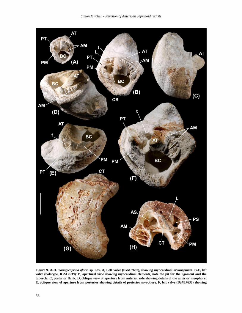
Youngicaprina gloria sp. nov.
Figure 9A-H
v. 1998 Texicaprina kugleri (Bouwman, 1937);
Alencáster and Oviedo-García, p. 167-171,
fig. 3 (1-2), fig. 4 (1-10).
?. 1998 Texicaprina vivari (Palmer, 1928);
Alencáster and Oviedo-García, p. 171, fig.
5.8-5.9 (but fig. 5.7-5.8 in the text in error).
Diagnosis. A species of Youngicaprina with a
short weakly coiled left valve, with eight or nine
blades in the anterior myophore of the left valve
that extend around the anterio-dorsal margin of
the anterior tooth.
Types. Holotype (IGM.7639), Paratypes
(IGM.7637, IGM.7638 and IGM.7643); all from
the mid-late Albian of El Madroño, state of
Querétaro, central Mexico.
Description. Bivalved specimens are not known,
so the relative sizes of the left and right valves are
unknown. The Right Valve is conical to
cylindrical, and relatively short. Transverse cross
sections are broadly triangular with rounded
angles. The central tooth is moderately broad and
filled with numerous small pallial canals. The
posterior tooth socket is deep and forms an angle
of about 60 to 70 degrees to the cardinal line (line
connecting the centres of the posterior and
anterior teeth). A slight restriction separates the
posterior tooth socket from the posterior myophore
cavity. The endomyophoral cavity is separated
from the body cavity by a septum which is solid
without pallial canals. The anterior tooth socket is
elongated dorso-ventrally. The cavity for the
anterior myophore is separated from the anterior
tooth socket by a septum containing pallial canals.
The anterior myophore cavity is triangular in form
and subdivided into three or four sectors by two or
three low septa, it extends into and embayment
adjacent to the anterior side of the anterior tooth.
The ligament is represented by a crescent shaped
structure which is concave to the dorsal margin;
the ligament is completely embedded within the
shell structure and there is no external ligamental
groove. No complete body cavity has been seen.
The nature of the fill of the body cavity and tooth
sockets is not known. Pallial canals have a uniform
size throughout the shell, and have a size of less
than 0.5 mm; the marginal layer of pallial canals is
poorly preserved. Pallial canals invade the entire
shell structure including the tooth and most of the
septa, pallial canals are also visible filling the
myophore sockets. Pallial canals have tabulae
spaced at distances of 1.5 to 3.5 mm.
The left valve has a transverse cross-section
which is triangular with rounded corners. Coiling
is slightly asymmetrical. There is no external
ligament groove; the dorsal side of the posterior
tooth having a depression corresponding to the
ligament in the right valve. The posterior tooth is
elongated anterio-posterioly, and is concave on the
dorsal side. The anterior tooth is much larger than
the posterior tooth and is quadrangular to broadly
rounded. The central tooth socket is narrow
between the teeth and expands a little posterio-
ventrally between the posterior tooth and the body
cavity, its bottom contains antero-posteriorly
arranged ribs, with the most dorsally positioned rib
rising to form a tubercle on dorsal side of the
central socket. The posterior myophore is
represented by a triangular projection that is
separated from the posterior tooth by a zone of
pallial canals. The posterior myophore extends a
little beyond the septum separating the central
socket from the body cavity. The anterior
myophore forms an arc that extends from the
dorsal extremity of the anterior tooth, around the
anterio-dorsal margin of the tooth and for a short
distance around the margin of the body cavity. It
consists of eight or nine projecting blades that are
separated by recessing areas with pallial canals;
the blades are arranged orthogonal to the anterior-
dorsal margin of the anterior tooth but become
broadly orthogonal to the body cavity at the
anterio-ventral margin of the anterior myophore.
Simon Mitchell - Revision of American caprinoid rudists
68
Figure 9. A-H. Youngicaprina gloria sp. nov. A, Left valve (IGM.7637), showing myocardinal arrangement. B-E, left
valve (holotype, IGM.7639): B, apertural view showing myocardinal elements, note the pit for the ligament and the
tubercle; C, posterior flank; D, oblique view of aperture from anterior side showing details of the anterior myophore;
E, oblique view of aperture from posterior showing details of posterior myophore. F, left valve (IGM.7638) showing
Simon Mitchell - Revision of American caprinoid rudists
69
The blades get progressively longer and higher
passing from the dorsal margin of the anterior
myophore to the anterio-ventral end of the
anterior myophore. The canals in the marginal
row of pallial canals in the right valve are slightly
radially elongated. The canals fill the inner shell
structure, but do not invade the two teeth, the
posterior myophore, the blades of the anterior
myophore, or the ribs and tubercle of the central
socket. The body cavity is broadly rounded, but its
fill is unknown.
Discussion. Youngicaprina gloria sp. nov. is
distinguished from Y. sangabrieli sp. nov. by: its
less strongly coiled left valve; its triangular cross-
section; the triangular form of the posterior
myophore separated from the posterior tooth by a
zone of pallial canals; the concave margin of the
posterior tooth; and the extension of the anterior
myophore, with its greater number of blades,
around the anterio-dorsal side of the anterior
tooth.
Youngicaprina sangabrieli gen. et sp. nov.
Figures 10, 11A-F
?pars 1888 Ichthyosaroclites anguis n. sp.: Roemer, p.
9-10, pl. I (XXXI), fig. 7a-b (Collected from
two miles above the mouth of Barton Creek
Austin, Travis Co., Texas, from somewhere
in the Edwards Limestone: Scott, 2002, p.
421)
v. 1973 Kimbleia sp.; Coogan, p. 58 (two LVs only),
pl. 1, fig. 3, pl 2, figs 5a-b.
v. 1977 Kimbleia sp.; Coogan, pl. 16, figs. 2a-b.
v. 1998 Kimbleia albrittoni (Perkins, 1960) pars;
Alencáster and Oviedo-García, p. 175-177,
fig. 8 (1), fig. 9 (1-2).
v. 2010 Caprinuloidea perfecta; Scott and Weaver,
fig. 3A-B, fig 5A, fig. 6C, fig. 7.
Diagnosis. A species of Youngicaprina with a
strongly coiled left valve, and with four or five ribs
on the anterior myophore of the left valve that are
arranged orthogonally to the anterior surface of the
anterior tooth, large pallial canals in 5 to 7 rows.
Types. Holotype (TMM.UT-36135.5); Paratypes
(TMM.UT-36135.1-4, BEG.2523.1, UT.3388,
UT.33871); all from the Edwards Limestone
(Middle Albian) of South San Gabriel River, Roy
Gunn’s Ranch, Texas (the locality now lies under
Lake Georgetown).
Description. The right valve is conical to
cylindrical, relatively short to relatively long,
straight. Transverse cross sections are quadrate
with an expanded, narrow ventral margin. The
posterior tooth is narrow and expands laterally
towards its ventral end. The posterior tooth socket
is deep and elongated in an anterior-posterior
direction; it forms an angle of about 60 to 70
degrees to the cardinal line. A slight restriction
separates the posterior tooth socket from the
endomyophoral cavity. The endomyophoral cavity
is separated from the body cavity by a septum
which is solid in small specimens but has a row of
pallial canals in larger specimens. The anterior
tooth socket is elongated dorso-ventrally. The
anterior myophore cavity is separated from the
anterior tooth socket by a low septum. The anterior
myophore cavity is triangular in form and
subdivided into three or four sectors by two or
three low septa. The ligament is represented by a
solid area that is concave to the dorsal margin; the
ligament is completely embedded within the shell
structure and there is no external ligamental
groove. The body cavity is broadly elliptical and
carries tabulae that are convex in an adapical
direction. The anterior tooth socket also carries
conical tabulae; the fill of the posterior tooth
socket is not known. Pallial canals have a uniform
size throughout the shell, they range from 0.5 to
1.5 mm in diameter; a marginal layer of elongated
pyriform canals with their narrow end directed
outwards encircles the entire shell; they lack
tabulae. Pallial canals invade the entire shell
structure including the central tooth and in larger
specimens also the septa. Pallial canals have
tabulae spaced at distances of 1.5 to 3.5 mm.
The left valve is coiled forming up to half a
revolution. It is much smaller than the right valve.
The left valve has a transverse cross-section which
is pear-shaped with the ventral side expanded to
form a rounded angle, and the anterior aspect
strongly flattened. Coiling is slightly
asymmetrical. There is no external ligament
groove, and no enclosed ligament has been seen.
The posterior tooth is elongated antero-posteriorly,
and is essentially a little flattened on the dorsal
side. The anterior tooth is elongated dorso-
ventrally. The central tooth socket is narrow
between the teeth and expands postero-ventrally
between the posterior tooth and the body cavity.
The posterior myophore is represented by a broad
[Figure 9 continued] myocardinal elements. G-H, Right valve (IGM.7643): G, posterior flank; H, apertural view
showing myocardinal elements, note the crescent-shaped ligament embedded in the interior of the shell. Scale
bar 20 mm. AT, anterior tooth; AS, anterior socket; PT, posterior tooth; PS, posterior socket; CT, central tooth;
CS, central socket; AM, anterior myophore; PM, posterior myophore; L, ligament; BC, body cavity; t, tubercle.
Simon Mitchell - Revision of American caprinoid rudists
70
Figure 10. Youngicaprina sangabrieli sp. nov. Right valve, (SI.USNM.547502), Edwards Limestone, Whitney Dam.
arc that extends from the posterior tooth past the
septum separating the central tooth socket from
the body cavity and a short distance around the
body cavity. The anterior myophore is a triangular
area formed by four or five dorso-ventral blades;
the blades are arranged parallel to the anterior
margin of the anterior tooth, with adjacent blades
separated by a row of pallial canals. The pallial
canals are the same as in the right valve; there is a
distinct marginal row of radially elongated pallial
canals; canals fill the remainder of the shell wall,
other than as described above for the myocardinal
elements. The body cavity is broadly elliptical, and
its upper part is filled with simple concave tabulae.
Simon Mitchell - Revision of American caprinoid rudists
71
Discussion. The type material comes from Texas
and is more robust that Y. Gloria gen. et sp. nov.
Alencáster and Oviedo-García (1998, fig. 9.1-2)
figured a large weathered left valve under the
name Kimbleia albrittoni (Perkins), but this
specimen has large pallial canals and a typical
Youngicaprina-like anterior myophore; for these
reasons, it is here placed in Y. sangabrieli gen. et
sp. nov. Two right valves were found associated
with Kimbleia in the Segovia Formation at Lopez
Ranch, Kimble County, Texas (Coogan, 1973,
1977; Plate 6F) and were tentatively assigned to
Kimbleia sp. by Coogan (1973, 1977). These
specimens, although coarsely silicified, show all
the characteristics of Youngicaprina and are here
attributed to Y. sangabreili gen. et sp. nov.
Stratigraphic and Geographic Distribution (Figure 12)
During the Hauterivian- early Barremian, Retha
tulae was inhabiting the carbonate platforms of
the Americas (Jamaica, ?Mexico) and gave rise to
the first evolutionary explosion of the
Amphitriscoelinae. The division of the gutters
between the myophores and shell margin gave rise
to pallial canals, and several distinct lineages
developed; Amphitriscoelus and Pantojaloria in
the Americas and Conchemipora in the Pacific
(Harris and Hodson, 1922; Alencáster, 1996;
Chartrousse and Masse, 1998; Skelton and Masse,
1998; Pantoja-Alor et al., 2004; Mitchell and
Brown, 2010). This diversification was wiped out
with the collapse of carbonate platforms in the
Early Aptian (Skelton and Gili, 2011).
By the Early Aptian, Huetamia the first
representative of the Caprinuloideinae had
appeared, and with the re-establishment of
extensive carbonate platforms in the Early Albian
of the Americas, a major diversification of the
Caprinuloieina occurred. The Early Albian saw
the appearance of Coalcomana, Planocaprina and
Caprinuloidea, the first two disappearing by the
end of the Early Albian, and the latter ranging into
the early Late Albian. Further diversification
occurred in the Late Albian with the appearance
of Mexicaprina, Kimbleia and Neokimbleia, with
Neokimbleia evolving from Kimbleia by the loss
of the external ligamental groove.
The Early Albian also saw the first
representatives of the Youngicaprininae, which
have a distinctive myocardinal arrangement.
Primitive Texicaprina appear in the Lower Albian
and are joined by Youngicaprina in the Middle
Albian and Jalpania in the upper Upper Albian.
Immanitas, which is also found in the upper Upper
Albian, may also belong here.
Acknowledgements. This work would have been
impossible without the help of numerous people who
have allowed access to collections, provided
photographs of specimens, or have discussed parts of
this work. Jann Thompson and Brian Huber facilitated
Peter Skelton and SFM at the Smithsonian Institution
for two weeks to study and sort the rudist collections in
2009. Ann Molineux (Non-Vertebrate Paleontology
Collections Manager, Texas Natural Science Center,
The University of Texas at Austin) facilitated my trip to
the Texas Memorial Museum in 2009, which was
supported by the National Science Foundation under
Grant No. 0646468 awarded to the Texas Natural
Science Center at the University of Texas. Ann.
Molineux and Jean DeMouthe facilitated the loan of
Palmer’s type specimens from the California Academy
of Sciences for study while I was in Texas. Dr. Jon Todd
facilitated my studies of the rudist bivalve collections at
The Natural History Museum, London. Dr. Reinaldo
Rojas-Consuegra kindly allowed me access and
permission to photograph the collections in the National
Natural History Museum, Havana, Cuba. I am indebted
to Dra. Ma del Carmenn Perrilliat and Violeta Romero
Mayen for allowing my access to the type collections of
the Museum of Paleontology, Institute of Geology,
Universidad Nacional Autónoma de México, Ciudad
Uiversitaria, México. Mr. W. J. Wildenberg allowed me
free access to work through the extensive Caribbean
rudist collections at Naturalis, Leiden, The Netherlands.
Hernán Santos facilitated my work in the Geological
Museum, University of Puerto Rico, Mayagüez and in
the field in Puerto Rico. Ian Brown and Dr. Sherene
James-Williamson allowed access to the collections at
the University of the West Indies Geological Museum,
Kingston, Jamaica. Peter Skelton took some of the
photographs in the Smithsonian during our joint trip
there. The photographs of the syntypes of Sabinia
kugleri preserved in the Naturhistorisches Museum
Basel were kindly taken by Dr. Walter Etter. I have
enjoyed discussion of the relationships of the Caprinoid
rudists with Peter W. Skelton, Bob Scott and Javier
Aguilar Pérez, and reviews by Peter W. Skelton and Bob
Scott greatly improved the original version.
REFERENCES
Aguilar Pérez, J. 2008. Rudistas del cretácico inferior y
medio. barremano-cenomaniano, noreste, ventro y
oeste de méxico. Tesis. Ciencias de la Tierra y del
Espacio, 139 pp.
Alencáster, G. 1999. New caprinid rudist genera
Guzzyella and Muellerriedia (Bivalvia-Hippuritacea)
from the Albian (Cretaceous) of central Mexico.
Revista Mexicana de Ciencias Geológicas, 15, 91-105.
Alencáster, G. and Aguilar-Pérez, J. 1996. Jalpania
queretana, a new rudist (Bivalvia-Hippuritacea) from
Simon Mitchell - Revision of American caprinoid rudists
72
Figure 11. A-F, Youngicaprina sangabrieli sp. nov. A, bivalve specimen (SI.USNM.547503 [CS3]), Edwards Limestone,
Whitney Dam. B-C, Left valve (Paratype, TMM.UT-33788), San Gabriel River, Roy Gunn’s Ranch, Texas, showing
large pallial canals and silicified myocardinal arrangement. D, Right valve (Paratype, TMM.UT-33871), San Gabriel
River, Roy Gunn’s Ranch, Texas, showing enclosed ligament and sockets for myophores separated from
Simon Mitchell - Revision of American caprinoid rudists
73
the mid-Cretaceous of central Mexioc. Revista Mexicana
de Ciencias Geológicas, 12(2), 117-122.
Alencáster, G. and Oviedo-García, A. 1998. Re-
examination of the genera Texicaprina Coogan,
Mexicaprina Coogan and Kimbleia Coogan (caprinid
rudists) from the Albian of central Mexico. Revista de
la Sociedad Mexicana de la Paleontologica, 8(2), 163-
179.
Alencáster, G. and Pantoja-Alora, J. 1996a. Coalcomana ramosa (Boehm) (Bivalvia,
Hippuritacea) del Albiano temprano del Cerro de
Tuxpan. Boletín de la Sociedad geológica mexicana,
47, 33-46.
Alencáster, G. and Pantoja-Alora, J. 1996b. New early
Aptian rudists (Bivalvia-Hippuritacea) from the
Huetamo area in southwestern Mexico. Revista
Mexicana de Ciencias Geológicas, 12 (for 1995), 123-
134.
Alencáster, G. and Pantoja-Alora, J. 1996c. The rudist
Amphitriscoelus (Bivalvia-Hippuritacea) in the Lower
Cretaceous of southwestern Mexico. Journal of
Paleontology, 70, 399-407.
Alencáster, G. and Pantoja-Alora, J. 1998. Two new
Lower Cretaceous rudists (Bivalvia-Hippuritacea)
from the Huetamo region of southwestern Mexico.
Geobios, Memoire spécial, 22, 15-28.
Boehm, G. 1898. Ueber Caprinidenkalke aus Mexico.
Zeitschrift der deutschen geologischen Gesellschaft,
50, 323-332, Berlin.
Bouwman, L.A.H. 1937. Sur une espéce nouvelle du
genere Sabinia (Caprininés). Koninklijke Akademie
van Wetenschappen te Amsterdam, Proceedings of the
Section of Sciences, 40(1 5), 449-453.
Brown, I. and Mitchell, S.F. 2010. Lithostratigraphy of
the Cretaceous succession in the Benbow Inlier,
Jamaica. Caribbean Journal of Earth Science, 41, 25-
37.
Carter, J.G., Altaba, C.R., Anderson, L.C., Araujo,
R., Biakov, A.S., Bogan, A.E., Campbell, D.C.,
Campbell, M., Jin-hua, C., Cope, J.C.W., Delvene,
G., Dijkstra, H.H., Zong-jie, F., Gavrilova, V.A.,
Goncharova, I., Guzhov, A.V., Harries, P.J.,
Hartman, J.H., Hautmann, M., Hoeh, W.R.,
Hylleberg, J., Bao-yu, J., Johnston, P., Kirkendale,
L., Kleemann, K., Koppka, J., Kříž, J., Machado,
D., Malchus, N., Márquez-Aliaga, A., Masse, J.-P.,
Middelfart, P.U., Mitchell, S., Nevesskaja, L.A.,
Özer, S., Pojeta, J. Jr., Polubotko, I.V., Pons, J.M.,
Popov, S., Sánchez, T., Sartori, A.F., Scott, R.W.,
Sey, I.I., Jin-geng, S., Signorelli, J.H., Silantiev,
V.V., Skelton, P.W., Steuber, T., Waterhouse, J.B.,
Wingard, G.L. and Yancey, T. 2011. A Synoptical
Classification of the Bivalvia (Mollusca).
Paleontological Contributions number 4, 48 pp.
University of Kansas.
Chartrousse, A. 1998a. The myocardinal organization of
coalcomaninid rudists revisted. Geobios, Memoire
spécial, 22, 75-85.
Chartrousse, A. 1998b. Les Caprinidae (Rudistes) du
Cretace Inferieur. Thèse de Doctorat, University de
Provence
Chartrousse, A. and Masse, J.-P. 1998. Coalcomaninae
(rudistes, Caprinidae) nouveaux de l’Aptien inferieur
des mid Pacific mountains. Geobios, Memoire Special
No. 22, 87-92.
Chartrousse, A. and Masse, J.-P. 2004. Revision of the
early Aptian Caprininae (rudist, bivalves) of the New
World, evolutionary and palaeobiogeographic
implications. Courier Forschungs-Institut
Senckenberg, 247, 19-34.
Chubb, L.J. 1971. Rudists of Jamaica.
Palaeontographica Americana, 7, 161-257.
Conrad, T.A. 1855. Descriptions of one Tertiary and
eight new Cretaceous fossils from Texas, in the
collection of Major Emory. Proceedings of the
Academy of Natural Sciences of Philidelphia, 7, 268-
269.
Conrad, T.A. 1857. Descriptions of Cretaceous and
Tertiary fossils. Pp. 141-174 in W.H. Emory. Report
of the United States and Mexican boundary survey,
U.S. 34th Congress, 1st Session, Senate Executive
Document 108, House Executive Document 135, 1(2).
Coogan, A.H. 1973. New rudists from the Albian and
Cenomanian of Mexico and south Texas. Revista del
Instituto Mexicano del Petroleo, 5(2), 51-83.
Coogan, A.H. 1977. Early and Middle Cretaceous
Hippuritacea (rudists) of the Gulf Coast. Pp. 32-70 in
D.G. Bebout and R.G. Loucks (Eds.), Cretaceous
carbonates of Texas & Mexico applications to
subsurface exploration. Report of Investigations No.
89, Bureau of Economic Geology, The University of
Texas at Austin, Austin, Texas, 332 pp.
Damestoy, G. 1971. Essai de classification phylogénique
des caprinidés (lamellibranches). Muséum National
d’Histoire Naturelle, Bulletin, (série 2) 42(5), 1003–
1008.
Douvillé, H. 1887. Sur quelques formes peu connues de
la famille des chamides. Bulletin de la Société
géologique de France, sér. 3, 15, 756-802.
Douvillé, H. 1900. Sur quelques rudistes Américains.
Bulletin de la Société géologique de France, 27(3),
205-221.
Felix, J. 1891. Versteinerungen aus der mexicanischen
Jura- und Kreide-Formation. Palaeontographica, 37(5-
6), 140-199, pls. 22-30.
Harris, G.D. and Hodson, F. 1922. The rudistids of
Trinidad. Palaeontographica Romeri, 1, 119-162.
Kutassy, A. 1934. Pachydonta mesozoica (rudistis
exclusis). Fossilium Catalogus, 1: Animalia, Paris 68.
Gustav Fellor, Neubrandenburg, 202 pp.
[Figure 11 continued] sockets for teeth. E, Left Valve (Holotype, TMM.UT-36135.5) San Gabriel River, Roy
Gunn’s Ranch, Texas, showing myocardinal structure. F, Left valve (IGM.2574), Segovia Formation at Lopez
Ranch, Kimble County, Texas, coarsely crystallized but showing features typical of this species. Note that all
specimens lack any indication of an external ligamental groove. Scale bar 20 mm. AT, anterior tooth; AS,
anterior socket; PT, posterior tooth; PS, posterior socket; CT, central tooth; CS, central socket; AM, anterior
myophore; PM, posterior myophore; L, ligament; BC, body cavity; t, tubercle.
Simon Mitchell - Revision of American caprinoid rudists
74
Figure 13. Evolution and biostratigraphic distribution of the Caprinuloideidae for the Lower Cretaceous of the
Americas.
Simon Mitchell - Revision of American caprinoid rudists
75
Mac Gillavry, H.J. 1937. Geology of the province of
Camaguey, Cuba, with revisional studies in rudist
paleontology. Geographische en geologische
Mededeelingen, 14, 168 pp., Utrecht.
Mitchell, S.F. 2013 (this volume). A new rudist bivalve
Curtocaprina clabaughikinsorum gen. et sp. nov. from
the mid Albian of Texas and its bearing on the origin
of the Ichthyosarcolitidae Douvillé. Caribbean
Journal of Earth Science, 46.
Mitchell, S.F. In press. A revision of the genera of the
rudist bivalve family Antillocaprinidae. Geobios.
Mitchell, S.F. and Green, R. 2011. Field Trip 1: Lower
Cretaceous rudists of the Benbow Inlier, central-north
Jamaica (Saturday, 18 June 2011). Pp. 29-36 in S.F.
Mitchell (Ed.), The Ninth International Congress on
Rudist Bivalves 18th to 25th June 2011 Kingston,
Jamaica. Abstracts, Articles and Field Guides. UWI
Contributions to Geology #6, 50 pp.
Mitchell, S.F. and Gunter, G.C. 2006. New tube-
bearing antillocaprinid rudist bivalves from the
Maastrichtian of Jamaica. Palaeontology, 49, 35-57.
Molineux, A., Scott, R.W., Ketcham, R.A. and
Maisano, J.A., 2007. Rudist Taxonomy Using X-Ray
Computed Tomorgraphy. Palaeontologia Electronica,
10(3); 13A:6p; http://palaeo-electronica.org/2007_3/
135/index.html
Myczyński, R. and Iturralde-Vinent, M. 2005. The
Late Lower Albian Invertebrate Fauna of the Río
Hatillo Formation of Pueblo Viejo, Dominican
Republic. Caribbean Journal of Science, 41, 782-796.
Newell, N.D. 1965. Classification of the Bivalvia.
American Museum Novitiates, 2206, 1-25.
d’Orbigny, Alcide. 1847. Sur les Brachiopodes ou
Palliobranches (deuxième mémoire). Comptes Rendus
Hebdomadaires des Séances de l’Académie des
Sciences, 25, 266–269.
Palmer, R.H. 1928. The rudistids of southern Mexico.
Occasional Papers of the California Academy of
Sciences, 14, 1-137.
Pantoja-Alor, J., Skelton, P.W. and Masse, J.-P. 2004. Barremian rudists of the San Lucas Formation around
San Lucas, Michoacan, SW Mexico. In: Richard
Höfling (Ed.), Contributions to the 5th International
Congress on Rudists, held in Erlangen, Germany,
1999 Dedicated to the memory of Erik Flügel (6 April
1934 - 14 April 2004). Courier Forschungsinstitut
Senckenberg, 247, 1-18.
Perkins, B.F. 1960. Biostratigraphic studies in the
Comanche (Cretaceous) series of northern Mexico and
Texas. Geological Society of America Memoir, 83, 138
pp.
Perkins, B.F. 1969. Rudist morphology. Pp. N751-N764
in R.C. Moore (Ed.), Treatise on Invertebrate
Paleontology Part N volume 2 Mollusca 6 Bivalvia,
Geological Society of America and University of
Kansas.
Roemer, F. 1888. Ueber eine durch die Häufigkeit
Hippuriten-artiger Chamiden ausgezeichnete Fauna der
oberturonen Kreide von Texas. Palaeontologische
Abhandlungen, 4, 281-296.
Scott, R.W. 2002. Albian caprinid rudists from Texas re-
evaluated. Journal of Paleontology, 76, 408-423.
Scott, R.W. and González-León, C. 1991. Paleontology
and biostratigraphy of Cretaceous rocks, Lampazos
area, Sonora, Mexico. Geological Society of America,
Special Paper 254, 51-67.
Scott, R.W. and Filkorn, H.F. 2007. Barremian-Albian
rudist zones, U.S. Gulf Coast. Pp. 167-180 in Scott,
R.W. (Ed.), Cretaceous rudists and carbonate
platforms: environmental feedback, SEPM Special
Publication No. 87, 257 pp.
Scott, R.W. and Weaver, M. 2010. Ontogeny and
functional morphology of a Lower Cretaceous Carpinid
{sic} rudist (Bivalvia, Hippuritoida). Turkish Journal
of Earth Sciences, 19, 527-542.
Skelton, P.W. 1996. IGCP Project 364: Correlation of
Caribbean ophiolites and volcanic arcs - field meeting.
Episodes, 19, 27-28; Herndon, Virginia.
Skelton, P.W. 2011. Rudist higher taxa for the revised
Bivalvia volumes of the Treatise on Invertebrate
Paleontology. Pp. 7-8 in S.F. Mitchell (Ed.), The Ninth
International Congress on Rudist Bivalves 18th to 25th
June 2011 Kingston, Jamaica. Abstracts, Articles and
Field Guides. UWI Contributions to Geology #6, 50 pp.
Skelton, P. W. 2013. Rudist classification for the revised
Bivalvia volumes of the ‘Treatise on Invertebrate
Paleontology.’ Caribbean Journal of Earth Science,
45, 9-33.
Skelton P.W. and Gili, E. 2011. Rudists and carbonate
platforms in the Aptian: a case study on biotic
interactions with ocean chemistry and climate.
Sedimentology, 59 (January 2012), 81–117.
Skelton, P.W. and Masse, J.-P. 1998. Revision of the
Lower Cretaceous rudist genera Pachytraga Paquier
and Retha Cox (Bivalvia: Hippuritacea) and the origins
of the Caprinidae. In: J.-P. Masse and P.W. Skelton
(Coordinators), Geobios – Mémoire spécial No. 22,
331-370.
Thiadens, A.A. 1936. On some caprinids and a
monopleurid from southern Santa Clara, Cuba.
Koninklijke Akademie van Wetenschappen te
Amsterdam, Proceedings of the Section of Sciences,
39(9), 1132-1141.
Young, K. 1985. Biogeography and stratigraphy of
selected middle Cretaceous rudists of southwestern
orth America. III Congreso atinoamericano de
Paleontolog a, In: Webber, W (Ed.), éxico simposio
sobre floras del tri sico tard o, su fitogeograf a y
paleoecolog a, 16 de octubre de 1984: memoria, 341-
3 0, México, AM, Instituto acional de Geolog a.
Accepted 15th March 2013