6
Ceramic Processing Research A study of the fabrication of cylindrical implants using hydroxyapatites Introduction Experimental

Journal of Ceramic Processing Research. Vol. 13, No. 2, pp. 117~122 (2012)
117
J O U R N A L O F
CeramicProcessing Research
A study of the fabrication of cylindrical implants using hydroxyapatites
Sang-Woo Chae and Su Chak Ryu*
Department of Nanomedical Engineering, Pusan National University, Miryang, Korea 627-706
Bioceramic hydroxyapatite (HAp) has excellent biocompatibility with bone tissue, and it exhibits good mechanical properties.We studied its mechanical properties and the effect on bone integration in a rabbit tibial defect model. HAp powder was
prepared by spray drying and compression forming. The HAp powder sintered at 1350 oC under a load of 1 tonne(9.8 KN)exhibited outstanding mechanical properties. In this test, the rabbit tibial defect was regenerated by using an HAp cylindrical
implant.
Key words: Hydroxyapatite, Spray dryer, Cylinder, Sintering, Implant.
Introduction
Calcium phosphate (CaP) ceramics, such as hydroxya-
patite, β-tricalcium phosphate, and biphasic calcium
phosphate, have been widely used as grafts for bone repair,
augmentation, or substitution. The wide usage of CaPs can
be attributed to the similarity between their composition
and that of bone mineral, bioactivity (the formation of
bone apatite-like material on their surfaces and a strong
bone-CaP biomaterial interface), and osteoconductivity
(an ability to provide the appropriate scaffold for bone
formation) [1-7].
Synthetic hydroxyapatite (HAp) has been recognized as
a suitable material for the fabrication of inorganic scaffolds
due to its close relationship with the mineral component
ofbone and because of its excellent osteophilic properties
[8-12]. It has also attracted considerable attention in the
field of material science and engineering because of its
unique biomimetic properties and osteoconductivity that
is similar to the osteoconductivity of natural biomaterials
[13-15].
In this study, a slurry of HAp nanoparticles was prepared
using an attrition mill to change the status of HAp powders.
HAp granules were prepared by a spray-drying method.
The HAp ceramic samples were prepared at different
temperatures and molding pressures. The mechanical
properties of these samples were evaluated to determine the
optimum conditions for their preparation. The bioactivities
of the HAp ceramic samples with varying compositions
were tested using a Simulated Body Fluid solution (hereafter
referred to as SBF). In the animal testing, a histological
analysis was conducted by implanting sintered HAp cylinders
into rabbit bones.
Experimental
HAp powder (≥ 98% purity), which was filtered through
a 200-mesh (≥ 75 µm) sieve, was considerately supplied
by the Bone Tech Inc. (Korea) and used without further
purification. After this filtering process, HAp powder with
particle sizes of less than 45 µm were obtained. The
properties of the HAp powder were characterized by
powder X-ray diffraction (XRD, Miniflex II, Rigaku, Co.
Ltd., Tokyo, Japan), inductively coupled plasma-optical
emission spectroscopy (ICP-OES, Optima 3300 DV,
Perkin-Elmer, Norwalk, CT), and energy-dispersive X-ray
spectroscopy (EDS, JEM-2011, Jeol Ltd., Tokyo, Japan).
Fig. 1 shows a schematic flowchart of the experimental
procedure for preparing a sintered HAp body.
Preparation of the HAp granular powderThe HAp slurry was prepared using HAp powder, distilled
water, and zirconia balls in an attrition mill. A dispersing
agent for 4 h disperse and then a leaving agent, antifoaming
agent, plasticizer, binder, and for 10 minute intervals into
the order of 12 h gives nano grinding and homogeneous
mixing. The HAp slurry was separated from the zirconia
ball using a 200-mesh sieve.
Manufacture of HAp cylinder samplesThe HAp granular powder was prepared by a spray-drying
method. The HAp cylindrical samples were prepared by
pressing the powder sample using compression weights
of 0.5, 1, and 1.5 tonne(9.8 KN). The HAp cylindrical
samples prepared from this powder had a diameter of
3.5 mm and a thickness of 4 mm. In order to test the bending
strength of the HAp samples, cylinders with a length, width,
and thickness of 21 mm, 4 mm, and 1.2 mm, respectively,
are fabricated using the granular powder. The samples
*Corresponding author: Tel : +82-51-350-5878Fax: +82-51-350-5839E-mail: [email protected]

118 Sang-Woo Chae and Su Chak Ryu
were sintered at temperatures of 1350 oC, 1400 oC, and
1450 oC for 2 h at the same heating rate of 55 oC/min.
The structures of the sintered HAp samples were determined
by powder X-ray diffraction (XRD, Miniflex II, Rigaku,
Co. Ltd., Tokyo, Japan). The mechanical properties of the
samples were determinedusing a universal testing machine
(SMTEST, SMB-001-5T, Korea), and the hardness was
measured using a micro Vickers Hardness tester (FM-
700, Future Tech, Japan).
Bioactivity testThe samples were in immersed an SBF solution to test
the bioactive property over 4 weeks. The temperature
of the samples in the SBF solution was similar to that
of human beings (37 oC). After 4 weeks, we observed the
HAp sample surface using a field-emission scanning electron
microscope (FE-SEM, HITACHI-S4700, HITACHI, Japan).
In vivo testIn this study, we used New Zealand white rabbits that
were approximately 15 weeks old at the beginning of the
experiment and weighed 2-2.3 kg each. The protocol for
the in vivo test was approved by the Institutional Animal
Care and Use Committee of Dong-A University, Busan,
Korea. All experimental procedures conformed to the life
science and humanitarian ethics code. The rabbits were
given ad libitum access to both sterile water and soft food.
The surgical procedures were performed after an intra-
muscular administration of 1.5 ml ketamine and 0.5 ml
Rompun (standard, 2.5-3 kg). Prior to the surgery, the skin
was shaved and then cleaned using a mixture of iodine
and 70% ethanol. The upper one-third of the tibia was
perforated by making a 2-cm midline incision through the
skin, fascia, and periosteum. One hole each was drilled on
the left and right side of the incision, bilaterally across the
near cortex; each hole had a diameter of 3.5 mm, and the
rabbits with four perforations were divided into four
treatment groups. The skin was sutured layer by layer after
the HAp cylinders were implanted into their respective
perforations. The rabbits were subcutaneously administered
0.05 mg/kg of buprenorphine every 12 h for the first 48 h
after the surgery. Within 2-3 days, the rabbits resumed
normal ambulation and did not show signs of pain or distress.
Radiologic analysis was performed every 2 weeks, and all
the rabbits were sacrificed 8 weeks after implantation
for a histological analysis. For each treatment group, the
volume of bone ingrowth and change in the bone mineral
density were statistically calculated five times 0, 2, 4, 6,
and 8 weeks after the implantation by computed tomography
or CT (FLEXTM for platform X-OTM, GMI, Northridge,
CA, USA). Histological analysis was performed 8 weeks
after implantation. The blocks were sectioned along a plane
parallel to the major axis of the HAp cylinder using a
micro-grinding machine (MG4000, EXAKT Apparatebau
GmbH, Norderstedt, Germany). The sections were stained
with Hematoxylin-Eosin. Routine histology and histomor-
phometric analyses were performed by transmission light
microscopy (Axioskop Carl Zeiss GmbH, Jena, Germany)
and image-analysis software (KS 300; Kontron Electronic
GmbH, Munchen, Germany), respectively.
Results and Discussion
Characterization of HApThe structural properties of the HAp powder used in
this study were determined using the XRD data, as shown
in Fig. 2. The XRD peaks of the HAp sample are found
at 2θ angles of 31.8, 32.25, and 32.91. The peak positions
and corresponding peak intensities were in good agreement
with the reference values provided by the JCPDS card
(#9-432). The EDS data given in Table 1 shows that
the atomic percentages of Ca and P were 19.25% and
11.50%, respectively, which leads to a Ca/P ratio of
1.674. This ratio was the same as the reference value of
Fig. 1. Flow chart of the experimental procedure.
Fig. 2. XRD patterns of the HAp powder: (A) prepared HAp and(B) standard peaks of HAp provided by JCPDS.

A study of the fabrication of cylindrical implants using hydroxyapatites 119
1.67. Table 2 shows the ICP-OES heavy metal analysis
results, and it shows that the total amount of heavy metal
is 0.02 ppm. The overall quantitative results indicate that
the HAp powder prepared was identical to natural HAp.
Measurement results of sample propertiesFig. 3 shows the results of XRD analysis of the HAp
body sintered at temperatures around 1350 oC to 1450 oC
for 2 h. When sintered at 1350 oC, only HAp crystalline
peaks were observed. However, β-TCP crystalline phase
peaks appeared when the sample was sintered at 1400 oC.
At 1450 oC, the number of β-TCP crystalline phase peaks
were found to increase with the temperature.
Figs. 4 and 5 show results of the compressive and
bending strengths of sintered HAp body at temperatures
ranging from 1350 oC to 1450 oC over a period of 2 h and
under formation loads ranging from 0.5 to 1.5 tonne(9.8 KN).
When the sample was fabricated under a pressure of 1 tonne
(9.8 KN) and subjected to heat treatment at 1350 oC, the
compressive strength and the bending strength were found
to be higher than those of the samples treated under the
other conditions. Therefore, the best conditions for HAp
sample preparation were found to be a pressure of 1 tonne
(9.8 KN) and a temperature of 1350 oC.
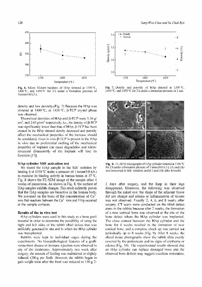
Fig. 6 shows the results of micro Vickers Hardness tests.
The samples were prepared by heat treatment at temperatures
around 1350 oC to 1450 oC and under a formation pressure
of 1 tonne(9.8 KN). The hardness of the sintered sample
at 1350 oC was higher than that of the samples prepared
under the other conditions.
According to the above results, the best mechanical
properties of the sintered HAp were obtained when the
sample was prepared under a formation pressure of 1 tonne
(9.8KN) and was sintered at a temperature of 1350 oC
for 2 h. The mechanical properties of HAp also depend
on the Ca/P ratio. HAp samples containing tricalcium
phosphate (TCP) showed poor mechanical properties [16].
The HAp samples sintered at 1350 oC showed a high
Table 1. EDS profile of HAp powder
Element Atomic%
O 69.25
P 11.50
Ca 19.25
Totals 100.00
Ca/P 1.674
Table 2. Heavy metal analysis of HAp powder by ICP-OES
Analysis Elements Analysis Date
As (ppm)Cd (ppm)Co (ppm)Cr (ppm)Hg (ppm)Mn (ppm)Ni (ppm)Sb (ppm)Se (ppm)V (ppm)Zn (ppm)
0.000.000.000.000.000.020.000.000.000.000.00
Total 0.02
Fig. 3. XRD patterns of HAp sintered at 1350 oC, 1400 oC, and1450 oC for 2 h.
Fig. 4. Compressive strength of HAp sintered at 1350 oC,1400 oC, and 1450 oC for 2 h under different loads.
Fig. 5. Binding strength of HAp sintered at 1350 oC, 1400 oC,and 1450 oC for 2 h under different loads.
120 Sang-Woo Chae and Su Chak Ryu
density and low porosity.(Fig. 7) Because the HAp was
sintered at 1400 oC, at 1450 oC, β-TCP crystal phase
was obtained.
Theoretical densities of HAp and β-TCP were 3.16 g/
cm3, and 2.65 g/cm3 respectively; i.e., the density of β-TCP
was significantly lower than that of HAp. β-TCP has been
created in the HAp sintered density decreased and porosity
affect the mechanical properties of the increase should
be considered. Even in vivo β-TCP is present in the HAp
in vivo due to preferential melting of the mechanical
properties of implants can cause degradation and micro-
structural disassembly of the implant will lose its
function [17].
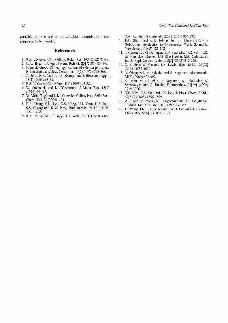
HAp cylinder SBF activation testWe tested the HAp sample in the SBF solution by
heating it at 1350 oC under a pressure of 1 tonne(9.8 KN)
to examine its binding activity in human bones at 37 oC.
Fig. 8 shows the FE-SEM image of the sample other 4
weeks of immersion. As shown in Fig. 8, the surface of
HAp samples exhibits changes. This result indirectly proves
that the HAp samples are bioactive in the human body.
We assumed on the basis of the concentration of Ca2+
ions that reactions between the Ca2+ ions and HAp occurred
at the sample surfaces.
Results of the in vivo testHAp cylinders were used in this study as a bone graft
material in order to determine the possibility of using the
right and left sides of the rabbit tibial defect that was
artificially generated in situ and in which the HAp cylinder
was transplanted.
Rabbits were kept in individual cages during the
experiments. No histopathological features of a graft-
versus-host disease or immune rejection were observed in
any of the treatments. Approximately one week after
surgery, the amount of feeding was maintained or slightly
reduced (200 g per feed). However, the rabbits began to
gain weight soon after the food was reduced to 150 g 2-
3 days after surgery, and the limp in their legs
disappeared. Moreover, the following was observed
through the naked eye: the shape of the adjacent tissue
did not change and edema or inflammation of tissues
was not observed. Exactly 2, 4, 6, and 8 weeks after
surgery, CT scans were conducted on the tibial defect
areas in the rabbits because after 2 weeks, the formation
of a new cortical bone was observed at the site of the
bone defect where the HAp cylinder was implanted.
The close contact between the HAp cylinder and the
bone for 4 weeks resulted in the formation of new
cortical bone, and a complete check up was carried out
periodically up to 8 weeks (Fig. 9). After 8 weeks, the
sliced tissue photographs show the rabbit tibia evenly
covered by the periosteum and no signs of erythema or
edema (Fig. 10). The experimental results showed that
an HAp cylinder can replace damaged bone and the
observed bone defects may suggest excellent restoration.
Fig. 6. Micro Vickers hardness of HAp sintered at 1350 oC,1400 oC, and 1450 oC for 2 h under a formation pressure of1tonne(9.8 KN).
Fig. 8. FE-SEM micrographs of HAp cylinder sintered at 1350 oCfor 2 h under a formation pressure of 1 tonne(9.8 KN). (A) and (B):non-immersed in SBF solution; and (C) and (D): after 4 weeks.
Fig. 7. Density and porosity of HAp sintered at 1350 oC,1400 oC, and 1450 oC for 2 h under a formation pressure of 1 ton.

A study of the fabrication of cylindrical implants using hydroxyapatites 121
Conclusions
The temperature and pressure changes for the sintering
HAp the powder were noted; a granular HAp powder
was prepared using the spray-drying method. The best
mechanical properties were obtained for a HAp prepared
under a formation load of 1 tonne(9.8 KN) and sintered at
a temperature of 1350 oC for 2 h. The results of the binding
activity were used to study the bioactivity of the various HAp
samples in an SBF solution over four weeks so that the
activity over the complete surface of the samples could be
estimated.
Animal test results on erythema or edema in the bone
marrow without osseointegration was a quick check on the
repair of damaged bone tissue which could be induced and
determined. The HAp cylinder used in this study can thus
be used as an excellent bone substitute, and HAp also
shows potential for use in artificial bones, artificial
teeth, and ceramic implants, e.g., of various human
organs to replace the use of other materials and thus be
Fig. 9. CT images showing tibia of a rabbit inserted with an HAp cylinder. (A) normal, after (B) 2 weeks, (C) 4 weeks, (D) 6 weeks, and (E)8 weeks.
Fig. 10. Photomicrograph of tibia that was regenerated using an HAp cylinder. After (A) 2 weeks, (B) 4 weeks, and (C) 8 weeks.
122 Sang-Woo Chae and Su Chak Ryu
possible, for the use of unfavorable materials for these
purposes to be avoided.
References
1. R.Z. LeGeros, Clin. Orthop. Relat. Res. 395 (2002) 81-98.2. K.A. Hing, Int. J. Appl. Ceram. Technol. 2[3] (2005) 184-199.3. Klaas de Groot. Clinical applications of calcium phosphate
biomaterials: a review. Ceram Int. 19[5] (1993) 363-366.4. A. John, H.K. Varma, T.V. Kumari and J. Biomater. Appl.,
18[1] (2003) 63-78.5. R.Z. LeGeros, Clin Mater. 4[1] (1993) 65-88.6. W. Suchanek and M. Yoshimura, J Mater Res. 13[1]
(1998) 94-117.7. M. Vallet-Reg and J. M. Gonzalez-Calbet, Prog Solid State
Chem. 32[1-2] (2004) 1-31.8. B.S. Chang, C.K. Lee, K.S. Hong, H.J. Youn, H.S. Ryu,
S.S. Chung and K.W. Park, Biomaterials. 21[12] (2000)1291-1298.
9. R.M. Pilliar, M.J. Filiaggi, J.D. Wells, M.D. Grynpas and
R.A. Kandel, Biomaterials. 22[9] (2001) 963-972.10. E.C. Shors and R.E. Holmes, in: L.L. Hench, J.Wilson
(Eds.), An Introduction to Bioceramics, World Scientific,New Jersey. (1993) 181-198
11. J. Cesarano, J.G. Dellinger, M.P. Saavedra, D.D. Gill, R.D.Jamison, B.A. Grosser, J.M. Sinn-Hanlon, M.S. Goldwasser,Int. J. Appl. Ceram. Technol. 2[3] (2005) 212-220.
12. S. Michna, W. Wu and J.A. Lewis, Biomaterials. 26[28](2005) 5632-5639.
13. Y. Ohbayashi, M. Miyake and S. Nagahata, Biomaterials.21[5] (2000) 501-509.
14. S. Itoha, M. Kikuchib, Y. Koyamac, K. Takakudac, K.Shinomiyaa and J. Tanaka, Biomaterials. 23[19] (2002)3919-3926.
15. Y.G. Kim, D.S. Seo and J.K. Lee, J. Phys. Chem. Solids.69[5-6] (2008) 1556-1559.
16. A. Royer. J.C. Viguie, M. Heughebaert and J.C. Heughbaert,J. Mater. Sci. Mat. Med. 4[1] (1993) 76-82.
17. H. Wang, J.K. Lee, A. Moursi and J. Lannutti, J. Biomed.Mater. Res. 68A[1] (2004) 61-71.
I


