145 ISSN 1392–1320 MATERIALS SCIENCE (MEDŽIAGOTYRA). Vol. 18, No. 2. 2012 Direct Laser Fabrication of Polymeric Implants for Cardiovascular Surgery Paulius DANILEVIČIUS 1 , Sima REKŠTYTĖ 1 , Evaldas BALČIŪNAS 2 , Antanas KRANIAUSKAS 3 , Raimondas ŠIRMENIS 3 , Daiva BALTRIUKIENĖ 2 , Mangirdas MALINAUSKAS 1 ∗ , Virginija BUKELSKIENĖ 2 , Roaldas GADONAS 1 , Vytautas SIRVYDIS 3 , Algis PISKARSKAS 1 1 Laser Research Center, Department of Quantum Electronics, Faculty of Physics, Vilnius University, Saulėtekio ave. 10, LT-10223 Vilnius, Lithuania 2 Vivarium, Institute of Biochemistry, Vilnius University, Mokslininkų str. 12, LT-08662 Vilnius, Lithuania 3 Heart Surgery Center, Faculty of Medicine, Vilnius University, Santariškių str. 2, LT-08661 Vilnius, Lithuania http://dx.doi.org/10.5755/j01.ms.18.2.1917 Received 28 October 2011; accepted 14 November 2011 In this work we present Multi-Photon Polymerization fabrication technique for biomedical applications. Optimal structuring parameters were defined in different polymeric materials using various lasers with different pulse durations (8 ps, 300 fs, 80 fs) and excitation wavelengths (532 nm, 515 nm and 800 nm), respectively. The applied photopolymers were: acrylate based AKRE, hybrid organic-inorganic Ormocore b59 and SZ2080, biodegradable PEG-DA-258 mixed with radical polymerization photoinitiators optimized for specific exposure conditions. It was defined that photoinitiators’ molecules did not affect materials cytotoxicity. Biocompatibilities of the used materials were investigated and showed positive results in vitro and in vivo. Furthermore, various in size and form artificial scaffolds were designed and fabricated as sample prototype implant structures for further experiments for tissue engineering applications in cardiovascular surgery. Keywords: laser non-linear lithography, 3D microstructures, photopolymerization, biocompatible polymers, biodegradable polymers, stem cells, tissue engineering. 1. INTRODUCTION ∗ Direct Laser Writing (DLW) is an attractive fabrication technology, which has evolved rapidly during past decades. Multi-Photon Polymerization (MPP) is a branch of DLW, which allows to modify polymeric materials in nano-scale. Due to non-linear nature, MPP can be easily employed for the fabrication of three-dimensional (3D) structures for number of applications, such as microfluidics [1], microoptics [2], photonics [3] as well as biomedicine and tissue engineering [4]. MPP has been first demonstrated in 1997 by S. Kawata group [5] and since then has expanded as a flexible technique that allows creation of microscopic objects with nano-scale resolution. Varieties of micro-structures have been fabricated since, ranging from complex microme- chanical components [1] to conductive metamaterials [6]. Lately it has been demonstrated that biocompatible polymeric, gelatin or even protein structures can be used for biomedical applications as well [7-9]. Polymers are attractive for the possibility to dope them and in this way to add desired functionality, for example: fluorescent dyes can help imaging of scaffold cell interaction [10]. One of the most promising biomedical applications of MPP is engineering of artificial tailor-made tissues, which could be transplanted into patients to cure diseases or traumas [11]. MPP has been employed for the fabrication of artificial scaffolds, which could serve as an Extra Cellular Matrix (ECM) and sustain stem cell growth in vitro. ∗ Corresponding author. Tel.: +370-600-02843; fax: +370-5-2366006. E-mail address: [email protected](M. Malinauskas) Controllable biomimetic and geometrical properties of the scaffolds can affect cell viability, adhesion and direct their differentiation and this can be used for constructing artifi- cial tissues of desirable form and functionality [12, 13]. Cardiovascular disease is an important issue in nowadays medicine. Artificial blood vessel implants fabricated via DLW out of acrylate based materials can be a competent replacement for currently used synthetic grafts (i.e. polytetrafluoroethylene or polyethylene terephthalate), because last-mentioned are usually suitable as large diameter (> 6 mm) blood vessel prostheses due to poor mechanical properties [14]. Controllable elastic modulus combined with high strain at break and high tear resistance are important features of biocompatible materials which are suitable for polymerization structuring. These properties can be applied for producing small size blood vessel implants [15]. Physical design of the scaffold also plays an important role as it significantly affects cell organization, proliferation and differentiation [16]. MPP satisfies the requirement to precisely control surface geometry, scaffold configuration and pore structure in micro-scale. In this case, it is a more reliable structuring technique than the traditional ones used for scaffold fabrication, such as fiber bonding [17], gas foaming [18], solvent casting/particulate leaching [19], phase separation [20]. However, to date there is a lack of knowledge of cell- matrix and cell-cell interactions at micro-scale. Therefore, the current research should be aimed to the production of synthetic ECM with suitable biological and chemical properties, which could mimic the native tissues and help to investigate cell behavior on 3D scaffolds.
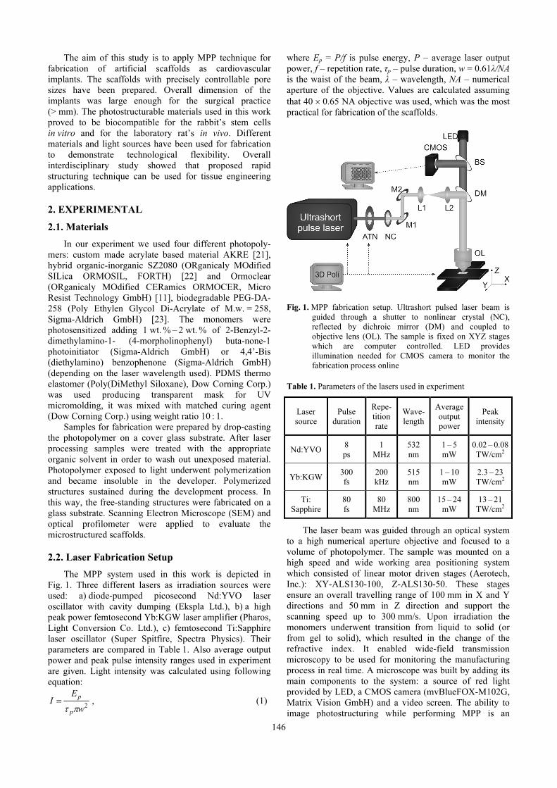
1 Laser Research Center, Department of Quantum Electronics, Faculty of Physics, Vilnius University,
Saulėtekio ave. 10, LT-10223 Vilnius, Lithuania 2 Vivarium, Institute of Biochemistry, Vilnius University, Mokslininkų str. 12, LT-08662 Vilnius, Lithuania 3 Heart Surgery Center, Faculty of Medicine, Vilnius University, Santariškių str. 2, LT-08661 Vilnius, Lithuania
http://dx.doi.org/10.5755/j01.ms.18.2.1917
Received 28 October 2011; accepted 14 November 2011
In this work we present Multi-Photon Polymerization fabrication technique for biomedical applications. Optimal
structuring parameters were defined in different polymeric materials using various lasers with different pulse durations
(8 ps, 300 fs, 80 fs) and excitation wavelengths (532 nm, 515 nm and 800 nm), respectively. The applied photopolymers
were: acrylate based AKRE, hybrid organic-inorganic Ormocore b59 and SZ2080, biodegradable PEG-DA-258 mixed
with radical polymerization photoinitiators optimized for specific exposure conditions. It was defined that
photoinitiators’ molecules did not affect materials cytotoxicity. Biocompatibilities of the used materials were
investigated and showed positive results in vitro and in vivo. Furthermore, various in size and form artificial scaffolds
were designed and fabricated as sample prototype implant structures for further experiments for tissue engineering
applications in cardiovascular surgery.
Keywords: laser non-linear lithography, 3D microstructures, photopolymerization, biocompatible polymers,
PDMS elastomer is placed on the original structure and
cured via thermal reaction, then transparent PDMS mold is
placed on the new monomer and the latter is cross-linked
by exposing to UV light.
By using this technique, we have successfully
replicated large area 2D scaffolds for stem-cell growth.
Fabrication time was reduced up to twenty times for
(15 × 15) mm2 2D scaffolds. We have shown that via UV
148
micromolding technique we can reproduce surface
roughness with 2 % inaccuracy (Fig. 5).
Fig. 4. Replication steps: a – original structure; b – PDMS
elastomer is poured onto original structure and cured
thermally; c – PDMS mold is removed from the substrate;
d – mold is placed on new monomer material;
e – monomer is polymerized with UV radiation; f – mold
is removed revealing replicated structure
Fig. 5. Profilometer image comparing original and molded
structures. It shows high quality reproduction possibilities
of micromolding technique
3.3. Biocompatibility of materials
Our experiments in vitro and in vivo showed that all
four of the used polymers (Ormoclear, SZ2080, PEG-DA-
258 and AKRE) are biocompatible. Adult myogenic stem
cells derived from rabbit muscle were seeded on non-
structured polymeric films in vitro for 48 h and their
viability was registered by staining with dye-mix solution
(Fig. 6, a). The results demonstrated that polymers were as
biocompatible as control polystyrene and glass surfaces.
Fig. 6. Alive rabbit stem cells growing in vitro on the non-
structured polymer SZ2080 surface (a). Section of
biocompatible polymer SZ2080 and surgical clip
implanted in rat’s muscle in vivo (b)
Furthermore, biocompatibility of polymeric samples
manufactured as shapeless granules were tested in vivo.
For comparison of tissue response, surgical suture was
taken as a control sample (Fig. 6, b). After three weeks of
implantation in rat’s muscle all tested materials were found
non-cytotoxic and as biocompatible as surgical suture,
showing them to be suitable for biomedical practice.
4. CONCLUSIONS
In conclusion, in present research biocompatibility of
four different photopolymers is stated by experiments in
vitro and in vivo. Three-dimensional scaffolds of scale
suitable for surgical practice were fabricated from these
materials having desired pore sizes and general porosity.
Additionally, replication technology of two-dimensional
scaffolds was applied and fabrication throughput of the
structures for stem cell growth is increased. Finally, multi-
photon polymerization of scaffolds is demonstrated with
three different laser sources, including low cost picosecond
laser, lowering technological costs comparing to
traditionally used femtosecond laser sources and opening
opportunities for practical applications in tissue
engineering. Future work is targeted to create and test in
vivo three-dimensional scaffolds for applications in tissue
engineering for cardiovascular surgery. Such biomedical
constructs could serve as biodegradable stents or vein
replacement implants with stem cells grown on them.
Acknowledgments
This work was financially supported by Lithuanian
Science Council grant MIP-10344 (BIOTISSUE). Authors
thank Mr. Rokas Smilingis (Ekspla Ltd.) for technical
assistance with picosecond Nd:YVO laser and
acknowledge company Altechna Ltd. for providing
assembled Aerotech stages.
REFERENCES
1. Schizas, C., Melissinaki, V., Gaidukevičiūtė, A., Reinhardt, C., Ohrt, C., Dedoussis, V., Chichkov, B., Fotakis, C., Farsari, M., Karalekas, D. On the Design and
Fabrication by Two-Photon Polymerization of a Readily
Assembled Micro-Valve International Journal of Advanced
2. Malinauskas, M., Žukauskas, A., Purlys, V., Belazaras, K., Momot, A., Paipulas, D., Gadonas, R., Piskarskas, A., Gilbergs, H., Gaidukevičiūtė, A., Sakellari, I., Farsari, M., Juodkazis, S. Femtosecond Laser Polymerization of
Hybrid/Integrated Micro-Optical Elements and Their
Characterization Journal of Optics 12 (12) 2010:
p. 124010.
3. Trull, J., Maigyte, L., Mizeikis, V., Malinauskas, M., Juodkazis, S., Cojocaru, C., Rutkauskas, M., Peckus, M., Sirutkaitis, V., Staliunas, K. Formation of Collimated
Beams behind the Woodpile Photonic Crystal Physical
Review A 84 (3) 2011: p. 033812.
4. Ovsianikov, A., Schlie, S., Ngezahayo, A., Haverich, A., Chichkov, B. N. Two-Photon Polymerization Technique for
Microfabrication of CAD-Designed 3D Scaffolds from
Commercially Available Photosensitive Materials Journal
of Tissue Engineering and Regenerative Medicine 1 (6)
2007: pp. 443 – 449.
5. Maruo, S., Nakamura, O., Kawata, S. Three-Dimensional
Microfabrication with Two-Photon-Absorbed Photo-
Polymerization Optics Letters 22 (2) 1997:
pp. 132 – 134.
http://dx.doi.org/10.1364/OL.22.000132
149
6. Terzaki, K., Vasilantonakis, N., Gaidukeviciute, A., Reinhardt, C., Fotakis, C., Vamvakaki, M., Farsari, M. 3D Conducting Nanostructures Fabricated Using Direct
7. Doraiswamy, A., Jin, C., Narayan, R. J., Mageswaran, P., Mente, P., Modi, R., Auyeung, R., Chrisey, D. B., Ovsianikov, A., Chichkov, B. Two Photon Induced
Polymerization of Organic–Inorganic Hybrid Biomaterials
for Microstructured Medical Devices Acta Biomaterialia
2 (3) 2006: pp. 267 – 275.
8. Ovsianikov, A., Deiwick, A., VanVlierberghe, S., Dubruel, P., Moller, L., Drager, G., Chichkov, B. Laser
Fabrication of Three-Dimensional CAD Scaffolds from
Photosensitive Gelatin for Applications in Tissue
Engineering Biomacromolecules 12 (4) 2011:
pp. 851 – 858.
9. Turunen, S., Kapyla, E., Terzaki, K., Viitanen, J., Fotakis, C., Kellomaki, M., Farsari, M. Pico- and
Femtosecond Laser-Induced Crosslinking of Protein
Microstructures: Evaluation of Processability and
Bioactivity Biofabrication 3 (4) 2011: p. 045002.
10. Žukauskas, A., Malinauskas, M., Kontenis, L., Purlys, V., Paipulas, D., Vengris, M., Gadonas, R. Organic Dye
Doped Microstructures for Optically Active Functional
Devices Fabricated via Two-Photon Polymerization
Technique Lithuanian Journal of Physics 50 (1) 2010:
pp. 55 – 61.
11. Schlie, S., Ngezahayo, A., Ovsianikov, A., Fabian, T., Kolb, H. A., Haferkamp, H., Chichkov, B. N. Three-
Dimensional Cell Growth on Structures Fabricated from
ORMOCER by Two-Photon Polymerization Technique
Journal of Biomaterials Applications 22 (3) 2007:
pp. 1 – 14.
http://dx.doi.org/10.1177/0885328207077590
12. Malinauskas, M., Danilevičius, P., Baltriukienė, D., Rutkauskas, M., Žukauskas, A., Kairytė, Ž., Bičkauskaitė, G., Purlys, V., Paipulas, D., Bukelskienė, V., Gadonas, R. 3D Artificial Polymeric Scaffolds for Stem
Cell Growth Fabricated by Femtosecond Laser Lithuanian
Journal of Physics 50 (1) 2010: pp. 75 – 82.
13. Psycharakis, S., Tosca, A., Melissinaki, V., Giakoumaki, A., Ranella, A. Tailor-Made Three-Dimensional Hybrid
Scaffolds for Cell Cultures Biomedical Materials 6 (4)
2011: p. 045008.
14. Hahn, M. S. Mechanical Stimulation and Biomimetic
Scaffolds for Tissue Engineered Vascular Grafts Topics in
Tissue Engineering 4 2008.
15. Baudis, S., Heller, C., Liska, R., Stampfl, J., Bergmeister, H., Weigel, G. (Meth)acrylate-Based Photoelastomers as
Tailored Biomaterials for Artificial Vascular Grafts
Journal of Polymer Science A1 47 (10) 2009:
pp. 2664 – 2676.
16. Li, Y., Yang, S. T. Effects of Three-Dimensional Scaffolds
on Cell Organization and Tissue Development
Biotechnology and Bioprocess Engineering 6 (5) 2011: pp.
311 – 325.
17. Mikos, A. G., Bao, Y., Cima, L. G., Ingber, D. E., Vacanti, J. P., Langer, R. Preparation of Poly(Glyco1ic
Acid) Bonded Fiber Structures for Cell Attachment and
Transplantation Journal of Biomedical Materials Research
27 (2) 1993: pp. 183 – 189.
18. Mooney, D. J., Baldwin, D. F., Suh, N. P., Vacanti, J. P., Langer, R. Novel Approach to Fabricate Porous Sponges of
Poly(D,L-Lactic-Co-Glycolic Acid) Without the Use of
Organic Solvents Biomaterials 17 (14) 1996:
pp. 1417 – 1422.
19. Mikos, A. G., Thorsen, A. J., Czerwonka, L. A., Bao, Y., Langer, R., Winslow, D. N., Vacanti, J. P. Preparation and
Characterization of Poly(L-Lactic Acid) Foams Polymer
35 (5) 1994: pp. 1068 – 1077.
http://dx.doi.org/10.1016/0032-3861(94)90953-9
20. Whang, K., Thomas, C. H., Healy, K. E., Nuber, G. A
Novel Method to Fabricate Bioabsorbable Scaffolds
Polymer 36 (4) 1995: pp. 837 – 842.
21. Malinauskas, M., Purlys, V., Rutkauskas, M., Gaidukevičiūtė, A., Gadonas, R. Femtosecond Visible
Light Induced Two-Photon Photopolymerization for 3D
Micro/Nanostructuring in Photoresists and Photopolymers
Lithuanian Journal of Physics 50 (2) 2010: pp. 201 – 207.
22. Ovsianikov, A., Viertl, J., Chichkov, B., Oubaha, M., MacCraith, B., Sakellari, I., Giakoumaki, A., Gray, D., Vamvakaki, M., Farsari, M., Fotakis, C. Ultra-Low
Shrinkage Hybrid Photosensitive Material for Two-Photon
23. Ovsianikov, A., Malinauskas, M., Schlie, S., Chichkov, B., Gittard, S., Narayan, R., Löbler, M., Sternberg, K., Schmitz, K. P., Haverich, A. Three-Dimensional Laser
Micro- and Nano-Structuring of Acrylated Poly(Ethylene
Glycol) Materials and Evaluation of Their Cytoxicity for


![Fabrication and Characterization of Polymeric Nerve Conduits and...represent a specific form of axonotmesis [6]. Associated with each category are specific symptoms ranging from](https://static.documents.pub/doc/80x56/5e66af864f35951ee24b5a4d/fabrication-and-characterization-of-polymeric-nerve-conduits-and-represent-a.jpg)


