58
Abdominal Trauma Dr.Janet Lee Department of Surgery
| Date post: | 27-Dec-2015 |
| Category: |
Documents |
| Upload: | brandon-henry |
| View: | 218 times |
| Download: | 0 times |

Abdominal Trauma
Dr.Janet Lee
Department of Surgery

Mechanism of Injury
Blunt injury
Penetrating injury
Blast injury
Iatrogenic injury

Blunt injury
Commonest mode
Frequently multi-system injury
Abdominal injury accounts for 10% blunt trauma death
Road traffic accident

Mechanism of blunt injury
Direct impact
Deceleration and rotational forces
Liver and spleen are the most commonly injured organs
Bowel injury (acute increase in intraluminal pressure / shearing at mesentery)

Penetrating injury
High velocity
Gunshot wounds
Low velocity
Stab wounds / low-velocity missiles

Mechanism of penetrating injury
Stab wounds
Injury confined to the tract of wounding
Gunshot wounds
Depends on the energy transferred
Penetration is accompanied by shock wave with cavitating effect (spiral path of motion)

Blast injury
Positive and negative pressure waves
Cause associated pressure changes in bowel gas (blowout)
Victim thrown by the force of pressure waves
Shrapnel

Iatrogenic injury
Uncommon
Laparoscopy
Endoscopy

Primary survey and resuscitation
Objectives of this phase:To identify and correct any immediate life-threatening conditionsTo anticipate problems
The activities are performed simultaneously with enough personnelA- Airway and cervical spine controlB- BreathingC- Circulation with haemorrhage controlD- DisabilityE- Exposure

Airway and C-spine control
C-spine injury should be assumedNo attempt should be made to turn the patient’s head to one side unless C-spine injury has been ruled outOxygen provided once airway cleared and securedBeware of aspiration

Breathing
Anticipate SIX immediately life-threatening thoracic conditions:1. Airway obstruction2. Tension pneumothorax3. Open chest wound4. Massive haemothorax5. Flail chest6. Cardiac tamponade
Respiratory rate and effort are both sensitive markers of underlying lung pathology (both should be monitored)

Circulation
Key objectives of circulatory care:
Stop haemorrhage
Assess hypovolaemia
Vascular assess
Appropriate fluid resuscitation

Stop haemorrhage
Direct pressure (external haemorrhage)Long bone fractures be splintedPelvic bindingPneumatic anti-shock garment (PASG)Pelvic fracture may need external fixationTry to avoid:Vessel clampingTourniquets (distal ischaemia)

Assessment for hypovolaemia
Skin (colour, clamminess and capillary refill)Heart rate and BPPulse pressureConscious levelECG monitoringSearch for common sites of occult bleeding:ChestAbdomen / RetroperitoneumPelvisLong bonesSplints and dressings

Vascular assess
Large bore IV catheter
20ml blood taken for grouping and x-match and for e- + full blood count
Femoral line / venous cut down / intra-osseous access (if peripheral IV assess failed)
Central venous line insertion is not essential for initial resuscitation

Fluid resuscitation
Initial fluid resuscitation:
2L warmed crystalloid
Responder: Give maintenance fluids once initial deficit replaced
Transient responder: Deteriorate due to continued haemorrhage, give blood and urgent surgical opinion
Non-responder: Ongoing haemorrhage at a greater rate, need urgent surgical opinion

Resuscitation end-point
Administer sufficient fluids to maintain perfusion of essential organsSBP 80mmHg (previously normotensive)Equivalent to a palpable radial pulsePermissive hypotension to minimizeOngoing haemorrhageDisruption of established thrombusDilution of clotting factorsMonitored vitals:Resp rate, SaO2, HR, BP, Pulse pressure, Cardiac monitoring, Temp, Urine output, GCS

Urethral injury
Far more common in male patients5-25% patients with pelvic fractures have an associated urethral injurySymptoms:Perineal painDysuriaFailure to voidSigns:Blood at urethral meatusBruising around scrotumHigh-riding prostate

Urethral injury
Urinary catheterization is contraindicated:
Conversion of partial to complete transection
Stricture formation
Introduce infection
Diagnosis confirmed by retrograde urethrogram

Disability
Baseline neurological examination:AVPU responseGlasgow comma scale (if time permits)Pupillary responseRepeated assessment to look for signs of deteriorationCommon causes of deterioration:HypoxiaHypovolaemiaHypoglycaemiaRaised ICP

Exposure
Trauma victims must be kept warm and covered with blankets when not examined
Log-roll
Assess the spine from skull base to coccyx
Examine the back for signs of injury
Rectal examination
Secondary survey (abdominal examination)
Key objective:To decide if laparotomy is neededDetailed examination of the abdomen, pelvis and perineumNote for bruising and woundsCover exposed bowel loops with warm NS soaked gauzeGastric tube to decompress distended stomach to facilitate abdominal examination and reduce risk of aspiration

Physical examination
Most alert patients will have abdominal tenderness
Initial PE in blunt abdominal trauma is only 65% accurate
Altered mental state (drugs, alcohol, HI, etc)
Sensory abnormalities (spinal cord injury)
Distracting injuries (extra-abdominal)
Serial examinations are often more important

Physical findings
Distension
Usually 20 to ileus or pneumoperitoneum or
haemoperitoneum
Bruising

Palpation
Lower ribs fractureAbdominal tenderness, guarding or reboundPelvic stabilityLumbar spine for tendernessRectal examinationAnal toneProstate position (?high riding)Blood over examination glove
Plain radiographs
CXR
The most important plain film
Obvious intra-thoracic and diaphragmatic injuries
Pelvis (AP view)
C-spine (Lat view) make sure C1-C7 are well shown
AXR seldom helpful (not routine)

Laboratory studies
Laboratory tests play limited role in the diagnosis of IAI (normal test never R/O IAI)
Baseline Hb level
Acid-base status
Amylase (not sensitive / specific)
Urinalysis (gross haematuria is the most consistent sign of serious renal injury)

Diagnostic peritoneal lavage
Before the introduction of DPL ~20% patient with abdominal trauma died of unrecognized injurySensitive 97-99%Fast (5-15 min)False +ve 1.4%Complication rate 1%No information on retroperitoneal organsNot sensitive to detect diaphragmatic or bladder injuries (these result in minimal bleeding)

Contraindication of PDL
Absolute
Obvious need for laparotomy
Evisceration
Relative
Pregnancy (>12 wks)
Previous abdominal surgery
Criticism of PDL
Overly sensitive
Non-bleeding solid organ injuries (which can be managed conservatively)
Non-therapeutic laparotomies
Best preserved for hypotensive, unstable, multi-injured patients

Techniques
Closed percutaneous
Semi-closed
Open
1 Liter of warm normal saline is instilled in adults
15 ml/kg in children
A minimum of 300 ml of lavage fluid must return to give a representative sample

Positive results of DPL
10ml gross blood or bowel contents with initial aspirationRBC count >100,000 cells/ml in blunt traumaRBC count >10,000 cells/ml in stab woundsRBC count >5000 cells/ml in penetrating chest traumaWBC count >500 cells/ml

Ultrasound
Kristensen et al first reported the use of USG in abdominal trauma in 1971Non-invasive and inexpensivePortable (bed side)No radiation / contrast requiredWell tolerated (excellent for unstable patients)Quick (within 3 mins in experienced hands)Serial examination easy to performBest screens for haemoperitoneum
FAST technique
Focused Abdomianl Sonography for Trauma (Rozycki et al)A standard approach which involves imaging a limited number of US windows to detect fluid:RUQ (Morison’s pouch)LUQ (to view the spleen)Pelvis (Douglas pouch)Pericardial window to assess for pericardial effusion (epigastric)
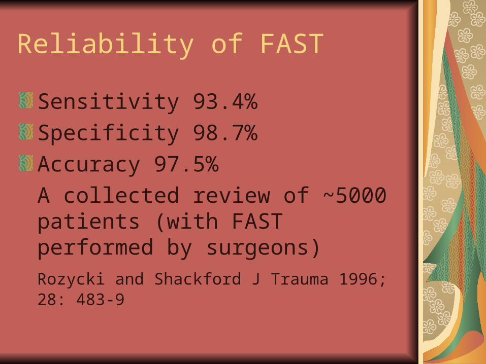
Reliability of FAST
Sensitivity 93.4%
Specificity 98.7%
Accuracy 97.5%
A collected review of ~5000 patients (with FAST performed by surgeons)Rozycki and Shackford J Trauma 1996; 28: 483-9

Results interpretation
Unstable patients with a +ve US requires laparotomy
Stable patients can be followed by serial US or employ CT for further evaluation

Limitations
Operator dependent
Uncooperative / agitated patients
Obesity
Surgical emphysema
Ileus
Cannot assess retroperitoneal organs
Like CT, US is insensitive for bowel injury
Poor sensitivity for penetrating trauma

Abdominal computed tomography
Introduced in late 1970s for trauma management
CT quantifies intraperitoneal blood and grades organ injury
IV and oral contrast
Accuracy is extremely reader-dependent
Modern spiral scan requires 3-5 mins
Dome of diaphragm to pelvis

Precautions
Haemodynamically stable
More time consuming than DPL / FAST
30-50 min
Adequate monitoring
Resuscitation facilities must be available in the CT room

Diagnostic laparoscopy
DL is a relatively new investigationLittle evidence to support its role in blunt traumaNot sensitive in Dx hollow viscus and retroperitoneal injuryPenetrating trauma (stab wounds) in stable patient100% sensitivity for identification of peritoneal penetrationMost effective for diagnosing ruptured diaphragm

Limitation of DL
Time consuming
Invasive
General anaesthetic
Difficult to exclude hollow viscus perforation

Management approach for blunt abdominal trauma
Unstable patient with abdominal signOperationUnstable patient with uncertain abdominal injuryDPL or FASTStable patient with associated severe injuriesDPL or FASTStable patient with associated minor injuries and equivocal abdomenCT scanStable patient with abdominal signsCT scan (allowing non-operative Tx if appropriate)

Stab wounds
Penetrates peritoneum in 2/3 casesOnly 50-70% of these have significant visceral or vascular injurySelective laparotomies to reduce morbidity and hospital stay in haemodynamically stable patientsDiagnostic aids:Wound explorationDPLLaparoscopySerial examinations

Lumbar and flank wounds
Significantly less risk (<15%) for intra-abdominal injuries than those with anterior woundsA more selected approach is warrantedContrast enhanced CT scan combined with serial examinations is recommendedRenal injuries occur in 6-8%
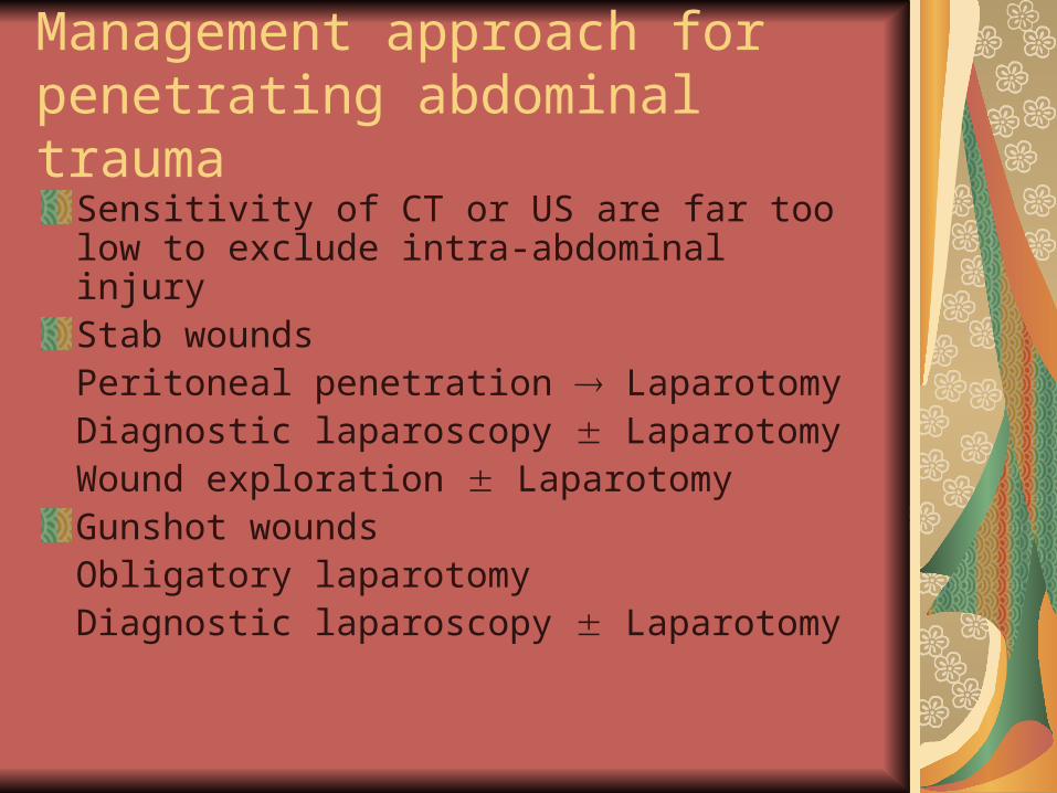
Management approach for penetrating abdominal trauma
Sensitivity of CT or US are far too low to exclude intra-abdominal injuryStab woundsPeritoneal penetration LaparotomyDiagnostic laparoscopy LaparotomyWound exploration LaparotomyGunshot woundsObligatory laparotomyDiagnostic laparoscopy Laparotomy

Incidence of IAI requiring exploratory laparotomy
Blunt % Penetrating %
Spleen 47 7
Liver 51 28
Pancreas / Duodenum
10 11
Colon 5 23
Stomach / Small bowel
9 42

Management “Prioritization”
Concurrent head injuries
An exsanguinating abdominal injury demands a laparotomy to control bleeding before assessment of the HI
Pelvic fracture
Rapid application of external fixator to stabilize the pelvis before laparotomy

Non-operative management of solid organ injury
Increasing evidence to support non-operative Mx
Parallels with the wide-spread use of CT
Clinical criteria (not CT grading) are used for decision making
Must be continuously monitored in HDU or ICU setting

Criteria for non-operative Mx
Solid organ injury shown on CT scan
Minimal abdominal signs
Haemodynamically stable
Requires <2 units of blood
HDU or ICU available
Surgeons committed for repeated evaluation

Success rate of non-operative Mx
Liver
50-80%
Spleen
93% for minor injuries
Renal
Majority can be Mx conservatively unless there is injury to renal pedicle or massive damage

Intervention radiology
Angiography embolization
Both diagnostic and therapeutic
Common use
Pelvic fracture with bleeding uncontrolled by fixation
Solid organ injury

Damage control surgery
10% trauma patients cannot tolerate definitive procedure at initial laparotomy
Survival benefit demonstrated with the use of “damage control” approach
Control bleeding
Injured bowel stapled without anastomosis
Solid organ injury packed
Abdomen rapidly closed with towel clips or plastic bag

Indications for damage control
Hypothermia 350C
Acidosis pH <7.2
Coagulopathy
Definitive surgery is deferred for 24-48 hrs when resuscitation in ICU has corrected these physiological parameters

Abdominal compartment syndrome
ACS: A group of adverse progressive physiological effects of raised intra-abdominal pressureAbdominal trauma is the commonest causePressure required to precipitate ACS is unknown (varies with individuals)Most will require decompression at 25-35 cmH2O

Predisposing factors in trauma patients
Massive intra-abdominal bleeding
Visceral edema (ischaemia-reperfusion)
Vigorous fluid resuscitation
Surgery
Packing

Pathophysiology
Diaphragmatic splinting (Resp)
Pressure on IVC (Decreases venous return and thus cardiac output)
Oliguria (Direct renal compression +/- reduced systemic blood flow)
The condition is fatal unless treated before irreversible physiological insult occurs

Major systems affected
Pulmonary
Cardiovascular
Renal

Treatment of ACS
Urinary manometry to monitor the intraabdominal pressureNasogastric decompressionAbdominal decompressionControl of haemorrhageEvacuation of gauze packs and bloodDelayed wound closure (temporary plastic wrap)Ventilatory support till definitive closure (optimally in 2-3 days time)


