(CANCER RESEARCH 52. 4787-4790. September 1. 1992]
Absence of p53 Gene Mutations in Primary Nasopharyngeal Carcinomas1
Charles H. Spruck HI, Yvonne C. Tsai, Dolly P. Huang, Allen S. Yang, William M. Rideout III,Mirella Gonzalez-Zulueta, Peter Choi, Kwok-Wai Lo, Mimi C. Yu, and Peter A. Jones2Kenneth Karris Jr. Comprehensive Cancer Center. University of Southern California, Los Angeles, California 90033 fC. H. S., Y. C. T., A. S. Y., W. M. R., M. G-Z.,M. C. Y., P. A. J.I, and the Chinese University of Hong Kong, Shatin. Hong Kong ¡D.P. H., P. C., K-W. L.]
ABSTRACT
Alterations in the p53 tumor suppressor gene and Epstein-Barr virusstatus were investigated in 15 nasopharyngeal carcinoma (NPC) biopsies, 4 xenografts, and 2 cell lines from the Cantonese region of southernChina. One other established NPC cell line obtained from a northernChinese patient was also studied. Restriction fragment length polymorphism analysis revealed a loss of heterozygosity for chromosome 17p.»herethep53 gene resides, in only one of 15 NPC biopsies. Polymerasechain reaction-single-stranded conformational polymorphism analysisand direct sequencing failed to detect sequence alterations in exons 5through 8 of the p53 gene in the 15 tumors and in the 4 NPC xenografts,all of which tested positive for Epstein-Barr virus. In contrast, the 3NPC cell lines were all negative for Epstein-Barr virus and containedG —¿�C transversions in thep53 gene, with cell lines CNE-1 and CNE-2harboring identical AGA (arginine) to ACA (threonine) changes atcodon 280. These results suggest that p53 inactivation is not a necessary-
component of nasopharyngeal carcinogenesis in Cantonese but may beimportant in the establishment of cell lines derived from these tumors.
INTRODUCTION
NPC3 is a rare malignancy in most parts of the world, where
the annual incidence of the disease is generally less than one per100,000 population in either sex (1). Among the handful ofpopulations that are known to deviate from this low-risk pattern, the highest incidence is observed in southern Chinese whoreside in central Guangdong Province and speak the Cantonesedialect; rates of NPC among men in this high-risk population(which are about 2-3 times the female rates) range from 30 to50/100,000 person-years ( 1, 2). Causative factors believed to beassociated with NPC include a genetically determined susceptibility that is correlated with specific histocompatibility locusantigen haplotypes (3), infection by EBV (4), and life-style factors, including diet (5). There is convincing epidemiological andexperimental evidence implicating diet, specifically the intakeof salted fish, as the primary cause for the high NPC incidencein the Cantonese. It is estimated that 90% of NPC cases occurring in Hong Kong (whose population is primarily Cantonese)can be attributed to the consumption of salted fish early in life(5).
Tumorigenesis is believed to involve the multistep accumulation of genetic alterations, resulting in the activation of on-cogenes and/or the inactivation of tumor suppressor genes (6).RFLP analysis has demonstrated frequent LOH on the shortarm of chromosome 3 in NPC tumors, suggesting a possible
Received 3/30/92; accepted 6/23/92.The costs of publication of this article were defrayed in part by the payment of
page charges. This article must therefore be hereby marked advertisement in accordance with 18 U.S.C. Section 1734 solely to indicate this fact.
1Supported by USPHS Grants ROÕCA40468 and R35 CA49758 from theNational Cancer Institute. NIH. Department of Health and Human Services, andthe Betty Lou Warren Research Fund.
2 To whom requests for reprints should be addressed, at the Kenneth Morris Jr.Comprehensive Cancer Center. University of Southern California. 1441 EastlakeAvenue. Los Angeles, CA 90033-0800.
3The abbreviations used are: NPC. nasopharyngeal carcinoma; EBV. Epstein-Barr virus; LOH. loss of heterozygosity; PCR, polymerase chain reaction; SSCP.single-strand conformation polymorphism; RFLP. restriction fragment lengthpolymorphism.
location for a putative tumor suppressor gene (7). The p53 genehas been implicated as a tumor suppressor gene in many typesof human cancers (8). Mutations in the gene are generally concentrated in the four highly conserved regions, within exons 5through 8, which are known to include the simian virus 40large-T binding domain (9).
Recently, specific G —¿�T substitutions at codon 249 in thep53 gene were observed in a high proportion of hepatocellularcarcinoma cases from regions of Africa and China wheredietary aflatoxin is suspected to be a major risk factor (10, 11).Contrary to these reports, hepatocellular carcinoma cases fromgeographic regions where aflatoxin exposure is rare do notdemonstrate the mutational hotspot at codon 249 (12). Theseobservations suggest a direct genetic alteration in humans as aresult of exposure to an environmental carcinogen.
The predominant role of salted fish exposure in NPC etiology in Hong Kong (being responsible for 90% of cases in thatcity) prompted us to search for possible structural alterations inexons 5 through 8 of the p53 gene in 15 NPC biopsies, fourxenografts, and one NPC cell line from this region by PCR-SSCP analysis (13). Two additional NPC cell lines establishedby investigators in the People's Republic of China were also
analyzed. One of the Chinese cell lines (CNE-2) was derivedfrom a Cantonese patient, while the other (CNE-1) came froma patient in the northern Chinese province of Jilin. Supplementary RFLP analysis for LOH on chromosome 17p was alsoperformed on the 15 NPC biopsy specimens.
MATERIALS AND METHODS
Tissues, Cell Lines, and DNA Extractions. Fifteen NPC biopsyspecimens and matching blood samples were obtained from patients atthe Prince of Wales Hospital, Hong Kong. A portion of the tumortissue was submitted for histopathologic examination, and the remainder was snap-frozen and kept at -70°C for subsequent DNA analysis.
AH NPC specimens were undifferentiated carcinomas according to theWHO classification (14). The percentages of tumors in the 15 NPCspecimens were not determined, but all 15 DNA samples in a previousstudy showed complete loss of heterozygosity at either the D3S3 locusor RAF-1 locus on chromosome 3, thus indicating negligible contamination by normal cells in the biopsy specimens (7). Four NPC xenografts (15) (NPC tumors grafted and growing m vivoon athymicnudemice) and one NPC cell line (NPC/HK-1) (16) derived from patients inHong Kong were established in one of our laboratories. Two other NPCcell lines (CNE-1 and CNE-2) were established by investigators in thePeople's Republic of China. One of the Chinese cell lines (CNE-2) was
derived from a Cantonese patient (17), while the other (CNE-1) camefrom a patient in the northern province of Jilin (18). Primary tumorDNA was not available for any of the xenografts or cell lines studied.High-molecular-weight DNA was prepared from tumor specimens,matching blood samples, and cell lines by proteinase K digestion andphenol/chloroform extraction as described (19. 20).
PCR. Oligonucleotide primers used for PCR amplification of exons5-8 of the p53 gene were prepared based on published sequences(21): PX5LT, 5'GGAATTCCTCTTCCTGCAGTACTC; PX5RT, 5'-
CCTGG.All primers have additional nucleotides creating EcoRi sites at their
5' ends. Two sets of primer pairs, PX5LT/PX6RT and PX7LT/
PX8RT. were used for amplification of exons 5-6 and 7-8, respectively,from genomic DNA. Amplified products were resolved on 1.8% agarosegels and isolated from the gel with Geneclean II (Bio 101, La Jolla,CA). Individual exons for SSCP analysis were amplified in secondaryPCR reactions using the Genecleaned products as templates. Primary'
PCR conditions were as follows. In a total volume of 50 M', l ^g ofgenomic DNA was incubated in 10 m.\i Tris-HCl (pH 8.3), 1.5 IÎIMMgClj, 50 niM KC1, 0.01% gelatin, 1 MMof each primer, 0.2 HIMdeox-ynucleotide triphosphates, and 1.0 unit of Taq polymerase. SecondaryPCR conditions were as follows. In a total volume of 25 ¿tl,1 M!DNApurified by Geneclean was incubated in 10 HIMTris-HCl (pH 8.3), 1.5mM MgCl2, 50 mM KC1, 0.01% gelatin, 1 MMof each primer, 0.2 rtiMdeoxynucleotide triphosphates, 2.5 MCi of [a-12P]dCTP (3000 Cimmol '), and 0.5 units of Taq polymerase. NPC biopsy specimens and
xenografts were tested for the presence of the EBV genome by PCR asdescribed by Telenti et al. (22).
SSCP Analysis. SSCP was performed essentially as described (13).Briefly, amplification products were diluted by adding 1 M'of the secondary PCR reaction to 9 M!of Sequenase stop solution (U.S. Biochem-icals, Cleveland, OH) and heat-denatured by boiling for 5 min, and2-3 n\ were rapidly loaded onto a nondenaturing polyacrylamide gel(6% acrylamide, 0.15% bis-acrylamide, 10% glycerol, lx Tris-Borate-EDTA). Electrophoresis was carried out at room temperature with acooling fan at 30 W constant power for 5-6 h, or at 6 W constant powerovernight. Gels were subjected to brief drying after electrophoresis andautoradiographed using Amersham Hyperfilm-MP film at -80°Cwith
an intensifying screen. DNA from a grade III transitional cell carcinoma of the bladder containing a known G —¿�Amutation at the secondposition of codon 1464 was used as a mutant control in exon 5 SSCP
determination.DNA Sequencing. Primary PCR products from NPC cell lines
(NPC/HK-1, CNE-1, and CNE-2) and NPC tumors (NPC 8 and NPC38) were digested with £coRI,cloned into M13 mpl9, and sequenced.In all cases, sequencing was performed in both directions using thedideoxy chain termination method by Sequenase version 2.0 (U.S. Bio-chemicals). Conditions were maintained according to the manufacturer's instructions.
RFLP Analysis for LOH. DNA from the tumors and the matchingWBC were digested with Taql and analyzed for LOH as described (7,23). The DNA probes used, their marker loci, and the correspondingchromosomal locations were pEW301 (D17S58) 17pll.2-pl 1.1,
pl44D6 (D17S34) 17pl3, and pYNH37.3 (D17S28) 17pl3.3, respectively.
Cell Line Fingerprinting. DNA from NPC cell lines CNE-1 andCNE-2 used in SSCP analysis was fingerprinted using a PCR protocoldetecting a GT polymorphism on chromosome 9q as described (24).PCR primers used (5'-AAGGGAATTCATCCCCTGCT and TTA-
CACTATACCAAGACTCC) were directed to locus D9S59 (24).
RESULTS
Fifteen NPC biopsy specimens, 4 xenografts, and 3 established cell lines were analyzed by PCR-SSCP for p53 genemutations. Our study concentrated on exons 5 through 8 of thep53 gene, since they have been shown to harbor up to 98% ofthe inactivating mutations of the gene in a variety of humantumors (25).
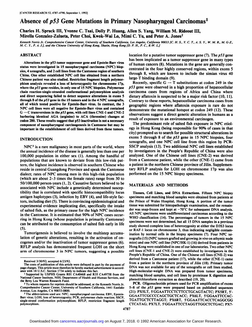
Fig. \A shows a representative PCR-SSCP gel of NPC xenografts and cell lines for exon 5 of the p53 gene. Mobilityshifts such as those observed in the mutant control (Fig. \A,Lane 2) and cell line NPC/HK-1 (Fig. IA, Lane 7) in compar-
' C. H. Spruck. unpublished data.
Bi
Fig. 1. A. PCR-SSCP analysis of NPC xenografts and cell lines for exon 5 ofthe p53 gene. Lanes I and 2, human WBC DNA as the normal control and abladder tumor DNA with known exon 5 mutation as the mutant control, respectively: Lanes 3-6, NPC xenograft DNA; Lanes 7-9, NPC cell line DNA NPC/HK1. CNE-1, and CNE-2. respectively. Arrow, mobility shift observed in NPC/HK1 (Lane 7). B. PCR-SSCP analysis of NPC tumor tissues, xenograft. and celllines for exon 8 of the p53 gene. Lanes 1-4, NPC tumor DNA S9 to S12,respectively; Lane 5. NPC xenograft DNA NPC/HK2117; Lanes 6-8, NPC cellline DNA NPC/HK1, CNE-1, CNE-2, respectively. Arrow, mobility shift observed in CNE-1 and CNE-2 (Lanes 7 and 8).
ison to the pattern of the normal control (Fig. \A, Lane 1)indicate the presence of p53 mutations. Direct sequencingshowed that cell line NPC/HK-1 carried a C —¿�G point mutation of CTC to GTC at codon 130, substituting a leucine witha valine in the p53 protein (data not shown). The presence of anSSCP band migrating with the mobility of the wild-type band inNPC/HK-1 indicates that this cell line contains both a normaland a mutant copy of the p53 gene. No p53 mutations weredetected in exon 5 in the remaining four xenografts and two celllines tested. The PCR-SSCP pattern of four NPC tumors, one
xenograft, and three cell lines for exon 8 of the p53 gene isshown in Fig. IB. The CNE-1 and CNE-2 cell lines demonstrated mobility shifts indicative of p53 mutations. Both celllines showed identical G —¿�C transversions at codon 280 of thep53 gene upon sequencing, changing AGA (arginine) to ACA(threonine) (data not shown).
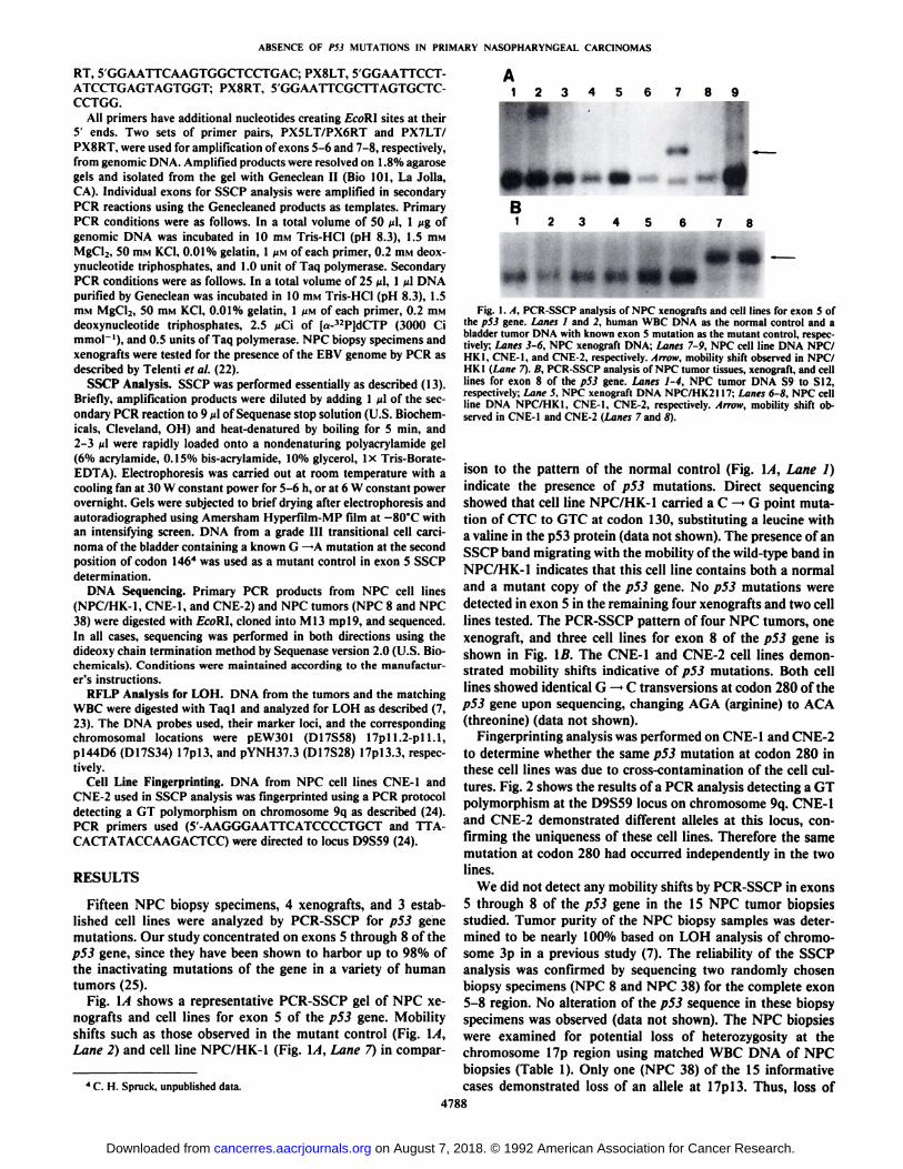
Fingerprinting analysis was performed on CNE-1 and CNE-2to determine whether the same p53 mutation at codon 280 inthese cell lines was due to cross-contamination of the cell cul
tures. Fig. 2 shows the results of a PCR analysis detecting a GTpolymorphism at the D9S59 locus on chromosome 9q. CNE-1and CNE-2 demonstrated different alÃelesat this locus, con
firming the uniqueness of these cell lines. Therefore the samemutation at codon 280 had occurred independently in the twolines.
We did not detect any mobility shifts by PCR-SSCP in exons
5 through 8 of the p53 gene in the 15 NPC tumor biopsiesstudied. Tumor purity of the NPC biopsy samples was determined to be nearly 100% based on LOH analysis of chromosome 3p in a previous study (7). The reliability of the SSCPanalysis was confirmed by sequencing two randomly chosenbiopsy specimens (NPC 8 and NPC 38) for the complete exon5-8 region. No alteration of the p53 sequence in these biopsy
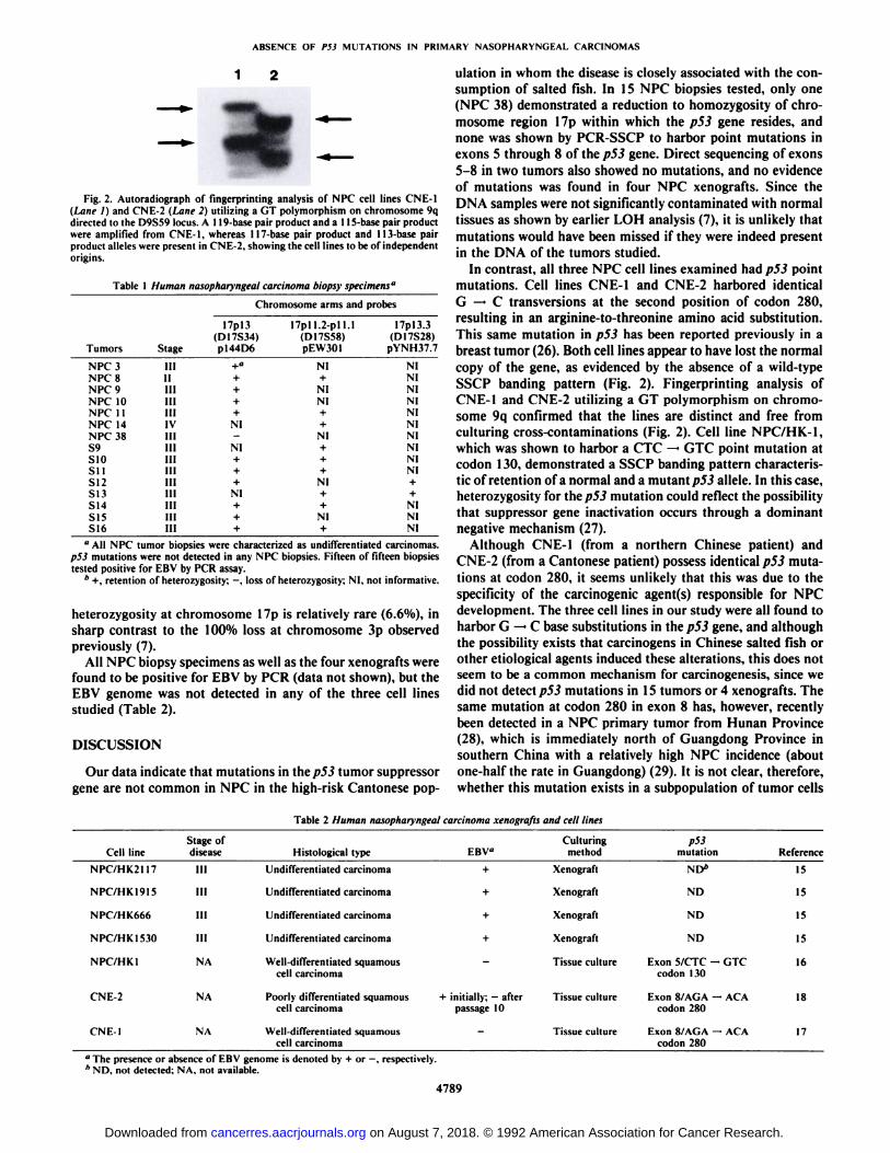
specimens was observed (data not shown). The NPC biopsieswere examined for potential loss of heterozygosity at thechromosome 17p region using matched WBC DNA of NPCbiopsies (Table 1). Only one (NPC 38) of the 15 informativecases demonstrated loss of an alÃeleat 17pl3. Thus, loss of
ABSENCE OF PS3 MUTATIONS IN PRIMARY NASOPI1ARYNGF.AL CARCINOMAS
Fig. 2. Autoradiograph of fingerprinting analysis of NPC cell lines CNE-1(Lane I) and CNE-2 (¡Mne2) utilizing a GT polymorphism on chromosome 9qdirected to the D9S59 locus. A 119-base pair product anda II 5-hase pair productwere amplified from CNE-I, whereas 117-base pair product and ll.Vbase pairproduct alÃeleswere present in CNE-2. showing the cell lines to be of independentorigins.
Table I Human nasopharyngeal carcinoma biopsy specimens"
Chromosome arms andprobesTumorsNPC
3NPC8NPC9NPC10NPC11NPC14NPC38S9S10SIIS12SI
3S14S15S16I7pl3(D17S34)Stage
pl44D6III+"11+HI+111+HI+IVNIIIIIII
NIIII+III+III+111NIIII+III+III
+17pll.2-pll.l(D17S58)pEW30lNI+NINI++NI+++NI++NI+I7pl3.3(D17S28)pYNH37.7NINININININININININI++NININI" All NPC tumor biopsies were characterized as undifferentiated carcinomas.
p53 mutations were not detected in any NPC biopsies. Fifteen of fifteen biopsiestested positive for EBV by PCR assay.
* +, retention of heterozygosity; -, loss of heterozygosity; NI. not informative.
heterozygosity at chromosome 17p is relatively rare (6.6%), insharp contrast to the 100% loss at chromosome 3p observedpreviously (7).
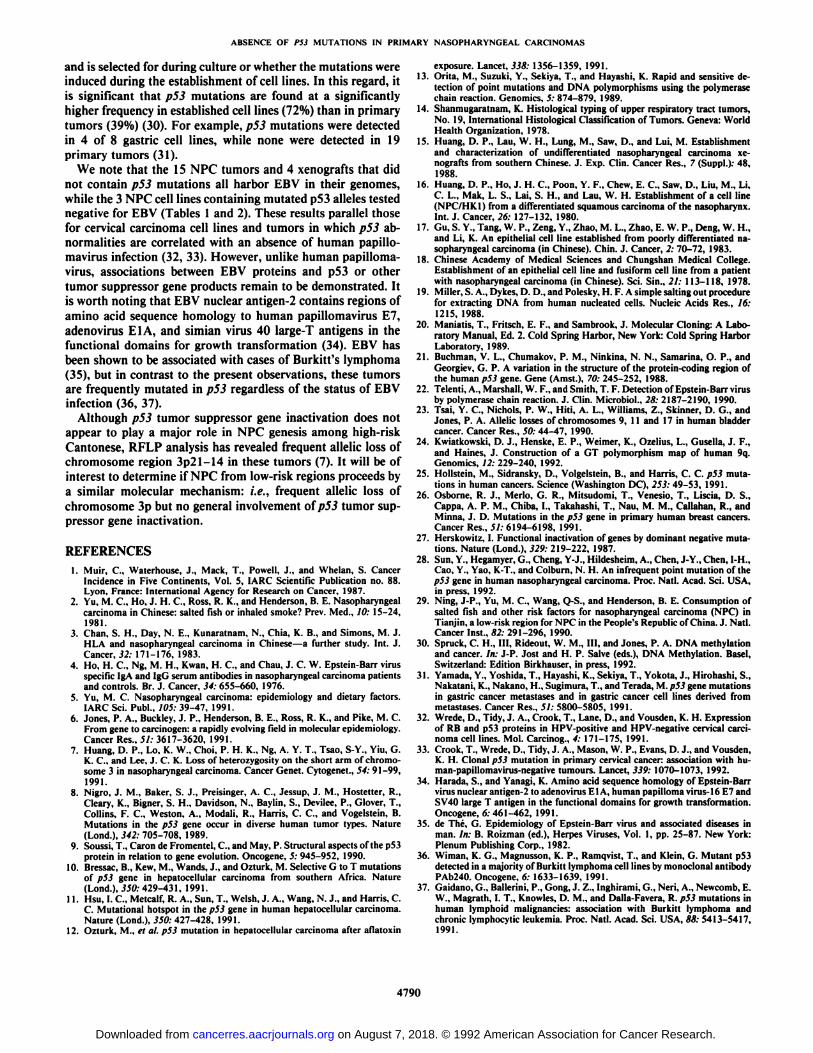
All NPC biopsy specimens as well as the four xenografts werefound to be positive for EBV by PCR (data not shown), but theEBV genome was not detected in any of the three cell linesstudied (Table 2).
DISCUSSION
Our data indicate that mutations in the p53 tumor suppressorgene are not common in NPC in the high-risk Cantonese pop
ulation in whom the disease is closely associated with the consumption of salted fish. In 15 NPC biopsies tested, only one(NPC 38) demonstrated a reduction to homozygosity of chromosome region 17p within which the p53 gene resides, andnone was shown by PCR-SSCP to harbor point mutations inexons 5 through 8 of ihe p53 gene. Direct sequencing of exons5-8 in two tumors also showed no mutations, and no evidenceof mutations was found in four NPC xenografts. Since theDNA samples were not significantly contaminated with normaltissues as shown by earlier LOH analysis (7), it is unlikely thatmutations would have been missed if they were indeed presentin the DNA of the tumors studied.
In contrast, all three NPC cell lines examined had p53 pointmutations. Cell lines CNE-1 and CNE-2 harbored identicalG —¿�C transversions at the second position of codon 280,resulting in an arginine-to-threonine amino acid substitution.This same mutation in p53 has been reported previously in abreast tumor (26). Both cell lines appear to have lost the normalcopy of the gene, as evidenced by the absence of a wild-type
SSCP banding pattern (Fig. 2). Fingerprinting analysis ofCNE-1 and CNE-2 utilizing a GT polymorphism on chromosome 9q confirmed that the lines are distinct and free fromculturing cross-contaminations (Fig. 2). Cell line NPC/HK-1,which was shown to harbor a CTC —¿�GTC point mutation atcodon 130, demonstrated a SSCP banding pattern characteristic of retention of a normal and a mutant p53 alÃele.In this case,heterozygosity for ihe p53 mutation could reflect the possibilitythat suppressor gene inactivation occurs through a dominantnegative mechanism (27).
Although CNE-1 (from a northern Chinese patient) andCNE-2 (from a Cantonese patient) possess identical p53 muta
tions at codon 280, it seems unlikely that this was due to thespecificity of the carcinogenic agent(s) responsible for NPCdevelopment. The three cell lines in our study were all found toharbor G —¿�C base substitutions in the p53 gene, and althoughthe possibility exists that carcinogens in Chinese salted fish orother etiological agents induced these alterations, this does notseem to be a common mechanism for carcinogenesis, since wedid not detect p53 mutations in 15 tumors or 4 xenografts. Thesame mutation at codon 280 in exon 8 has, however, recentlybeen detected in a NPC primary tumor from Hunan Province(28), which is immediately north of Guangdong Province insouthern China with a relatively high NPC incidence (aboutone-half the rate in Guangdong) (29). It is not clear, therefore,
whether this mutation exists in a subpopulation of tumor cells
Table 2 Human nasopharyngeal carcinoma xenografts and cell lines
ABSENCE OF PS3 MUTATIONS IN PRIMARY NASOPHARYNGEAL CARCINOMAS
and is selected for during culture or whether the mutations wereinduced during the establishment of cell lines. In this regard, itis significant that p53 mutations are found at a significantlyhigher frequency in established cell lines (72%) than in primarytumors (39%) (30). For example, p53 mutations were detectedin 4 of 8 gastric cell lines, while none were detected in 19primary tumors (31).
We note that the 15 NPC tumors and 4 xenografts that didnot contain p53 mutations all harbor EBV in their genomes,while the 3 NPC cell lines containing mutated p53 alÃelestestednegative for EBV (Tables 1 and 2). These results parallel thosefor cervical carcinoma cell lines and tumors in which p53 abnormalities are correlated with an absence of human pupillo-max irus infection (32, 33). However, unlike human papilloma-virus, associations between EBV proteins and p53 or othertumor suppressor gene products remain to be demonstrated. Itis worth noting that EBV nuclear antigen-2 contains regions ofamino acid sequence homology to human papillomavirus E7,adenovirus EIA, and simian virus 40 large-T antigens in thefunctional domains for growth transformation (34). EBV hasbeen shown to be associated with cases of Burkitt's lymphoma
(35), but in contrast to the present observations, these tumorsare frequently mutated in p53 regardless of the status of EBVinfection (36, 37).
Although p53 tumor suppressor gene inactivation does notappear to play a major role in NPC genesis among high-riskCantonese, RFLP analysis has revealed frequent allelic loss ofchromosome region 3p21-14 in these tumors (7). It will be ofinterest to determine if NPC from low-risk regions proceeds bya similar molecular mechanism: i.e., frequent allelic loss ofchromosome 3p but no general involvement ofp53 tumor suppressor gene inactivation.
REFERENCES
1. Muir. C., Waterhouse, J., Mack, T.. Powell, J.. and Whelan, S. CancerIncidence in Five Continents. Vol. 5, IARC Scientific Publication no. 88.Lyon. France: International Agency for Research on Cancer. 1987.
2. Yu, M. C, Ho, J. H. C, Ross, R. K., and Henderson, B. E. Nasopharyngealcarcinoma in Chinese: salted fish or inhaled smoke? Prev. Med., 10: 15-24,1981.
3. Chan, S. H.. Day, N. E.. Kunaratnam. N., Chia, K. B., and Simons, M. J.HLA and nasopharyngeal carcinoma in Chinese—a further study. Int. J.Cancer, 32: 171-176, 1983.
4. Ho. H. C., Ng, M. H., Kwan, H. C., and Chau, J. C. W. Epstein-Barr virusspecific IgA and IgG serum antibodies in nasopharyngeal carcinoma patientsand controls. Br. J. Cancer, 34: 655-660, 1976.
5. Yu, M. C. Nasopharyngeal carcinoma: epidemiology and dietary factors.IARC Sci. Pubi., 105: 39-47, 1991.
6. Jones, P. A., Buckley, J. P.. Henderson, B. E., Ross. R. K.. and Pike, M. CFrom gene to carcinogen: a rapidly evolving field in molecular epidemiology.Cancer Res., 5/: 3617-3620, 1991.
7. Huang, D. P., Lo, K. W., Choi, P. H. K., Ng, A. Y. T., Tsao, S-Y., Yiu, G.K. C., and Lee, J. C. K. Loss of heterozygosity on the short arm of chromosome 3 in nasopharyngeal carcinoma. Cancer Genet. Cytogenet., 54: 91-99,1991.
8. Nigro, J. M., Baker, S. J., Preisinger, A. C., Jessup, J. M., Hosteller, R.,Cleary. K., Bigner. S. H., Davidson, N., Baylin, S., Devilee, P., Glover, T.,Collins. F. C., Weston, A.. Modali, R., Harris, C. C., and Vogelstein, B.Mulalions in the p53 gene occur in diverse human tumor types. Nature(Lond.). 342: 705-708, 1989.
9. Scussi, T., Carónde Fromentel. C., and May, P. Structural aspects of the p53protein in relation to gene evolution. Oncogene. 5: 945-952. 1990.
10. Bressac, B., Kew, M., Wands, J., and Ozlurk, M. Selective G to T mutationsof p53 gene in hepatocellular carcinoma from southern Africa. Nature(Lond.), 350: 429-431, 1991.
11. Hsu, I. C., Metcalf, R. A.. Sun, T., Welsh, J. A.. Wang, N. J., and Harris, C.C. Mutational hotspot in the p53 gene in human hepatocellular carcinoma.Nature (Lond.), 350: 427-428. 1991.
12. Ozturk, M.. el al. p53 mutalion in hepatocellular carcinoma after aflatoxin
exposure. Lancet, 338: 1356-1359, 1991.13. Orita, M., Suzuki, Y., Sekiya, T., and Hayashi, K. Rapid and sensitive de
tection of point mutations and DNA polymorphisms using the polymerasechain reaction. Genomics, 5: 874-879. 1989.
14. Shanmugaratnam, K. Histological typing of upper respiratory tract tumors,No. 19, Internalional Histological Classification of Tumors. Geneva: WorldHealth Organization. 1978.
15. Huang, D. P., Lau, W. H., Lung, M., Saw, D., and Lui, M. Establishmentand characterization of undifferentiated nasopharyngeal carcinoma xenografts from southern Chinese. J. Exp. Clin. Cancer Res., 7 (Suppl.): 48.1988.
16. Huang, D. P., Ho, J. H. C.. Poon, Y. F., Chew, E. C., Saw, D., Liu, M., Li,C. L., Mak, L. S., Lai, S. H.. and Lau, W. H. Establishment of a cell line(NPC/HK1) from a differentiated squamous carcinoma of the nasopharynx.Int. J. Cancer, 26: 127-132, 1980.
17. Gu, S. Y.. Tang, W. P., Zeng, Y., Zhao, M. L., Zhao, E. W. P., Deng, W. H.,and Li, K. An epithelial cell line established from poorly differentiated nasopharyngeal carcinoma (in Chinese). Chin. J. Cancer, 2: 70-72, 1983.
18. Chinese Academy of Medical Sciences and Chungshan Medical College.Eslablishment of an epithelial cell line and fusiform cell line from a patientwith nasopharyngeal carcinoma (in Chinese). Sci. Sin., 21: 113-118, 1978.
19. Miller, S. A.. Dykes, D. D., and Polesky. H. F. A simple salting out procedurefor extracting DNA from human nucleated cells. Nucleic Acids Res., 16:1215, 1988.
20. Maniatis. T.. Fritsch, E. F., and Sambrook, J. Molecular Cloning: A Laboratory Manual, Ed. 2. Cold Spring Harbor, New York: Cold Spring HarborLaboratory, 1989.
21. Buchman, V. L., Chumakov, P. M., Ninkina, N. N., Samarina, O. P., andGeorgiev, G. P. A variation in the structure of the protein-coding region ofthe human p53 gene. Gene (Amst.). 70: 245-252, 1988.
22. Telenti, A., Marshall, W. F., and Smith, T. F. Detection of Epstein-Barr virusby polymerase chain reaction. J. Clin. Microbio!.. 28: 2187-2190, 1990.
23. Tsai, Y. C, Nichols, P. W., Hiti, A. L., Williams, Z., Skinner, D. G.. andJones, P. A. Allelic losses of chromosomes 9, 11 and 17 in human bladdercancer. Cancer Res., 50: 44-47, 1990.
24. Kwiatkowski, D. J., Henske, E. P., Weimer, K., Ozelius, L., Gusella, J. F.,and Haines, J. Construction of a GT polymorphism map of human 9q.Genomics, 12: 229-240, 1992.
25. Hollstein, M., Sidransky, D., Volgelslein, B., and Harris, C. C. p53 mutations in human cancers. Science (Washington DC). 253: 49-53, 1991.
28. Sun, Y., Hegamyer, G., Cheng, Y-J., Hildesheim, A., Chen, J-Y., Chen, I-H.,Cao, Y., Yao, K-T., and Colburn. N. H. An infrequent point mutation of thep53 gene in human nasopharyngeal carcinoma. Proc. Nati. Acad. Sci. USA,in press, 1992.
29. Ning, J-P., Yu, M. C., Wang, Q-S., and Henderson, B. E. Consumption ofsalted fish and other risk factors for nasopharyngeal carcinoma (NPC) inTianjin, a low-risk region for NPC in the People's Republic of China. J. Nati.Cancer Insl., 82: 291-296, 1990.
30. Spruck, C. H., III. Rideout, W. M., III. and Jones, P. A. DNA methylalionand cancer. In: J-P. Jost and H. P. Salve (eds.), DNA Methylation. Basel,Switzerland: Edition Birkhauser, in press, 1992.
32. Wrede, D., Tidy, J. A., Crook, T., Lane, D., and Vousden, K. H. Expressionof RB and p53 proleins in HPV-positive and HPV-negative cervical carcinoma cell lines. Mol. Carcinog., 4: 171-175, 1991.
33. Crook. T., Wrede, D., Tidy, J. A., Mason, W. P., Evans, D. J., and Vousden,K. H. Clonal p53 mutalion in primary cervical cancer: associalion with hu-man-papillomavirus-negative tumours. Lancel, 339: 1070-1073, 1992.
34. Harada, S., and Yanagi, K. Amino acid sequence homology of Epstein-Barrvirus nuclear antigen-2 to adenovirus E1 A, human papilloma virus-16 E7 andSV40 large T antigen in the functional domains for growth transformation.Oncogene, 6:461-462, 1991.
36. Wiman. K. G., Magnusson, K. P., Ramqvist, T., and Klein, G. Mutant p53detected in a majority of Burkitt lymphoma cell lines by monoclonal antibodyPAb240. Oncogene, 6: 1633-1639, 1991.
37. Caldano. G., Ballerini, P.. Gong. J. Z., Inghirami. G., Neri, A., Newcomb, E.W., Magrath, I. T., Knowles, D. M., and Dalla-Favera, R. p53 mutations inhuman lymphoid malignancies: association with Burkitt lymphoma andchronic lymphocytic leukemia. Proc. Nati. Acad. Sci. USA, **: 5413-5417,1991.