ABSORPTION AND CIRCULAR DICHROISM STUDlES References Barondes, S. H., and Nirenberg, M. W. (1962), Science 138, 810. Basilio, C., Wahba, A. J., Lengyel, P., Speyer, J. F., and Ochoa, S. (1962), Proc. Nat. Acad. Sci. U. S. 48,613. Bobst, A. M., Cerutti, P. A., and Rottman, F. (1969a), J. Amer. Chem. SOC. 91, 1246. Bobst, A. M., Rottman, F., and Cerutti, P. A. (1969b), J. Amer. Chem. Soc. 91,4603. Bobst, A. M., Rottman, F., and Cerutti, P. A. (1969c), J. Mol. Bid. 46, 221. Bollum, F. J. (1966), in Procedures in Nucleic Acid Research, Cantoni, G. L., and Davies, D. R., Ed., New York, N. Y., Harper & Row, p 296. Davies, J. (1966), Cold Spring Hurbor Symp. Quant. Biol. 31, 665. Di Mauro, E., Snyder, L., Marino, P., Lamberti, A., Coppo, A., and Tocchini-Valentini, G. P. (1969), Nature (London) 222, 533. Hall, R. H. (1964), Biochemistry 3, 876. Janion, C., Zmudzka, B., and Shugar, D. (1970), Acta Bio- Jones, 0. W., Townsend, E. E., Sober, H. A., Heppel, L. A. chim. Polon. 17, 31. Absorption and Circular Dichroism Studies on Nucleohistone IV" H. J. Li, I. IsenbergJ and W. C. Johnson, Jr. ABSrRAcr: Reconstituted calf thymus nucleohistone IV was studied by absorption and circular dichroism spectroscopy. The binding of histone IV to DNA does not change either the absorption or the circular dichroism spectrum of DNA at a detectable level. Based on this fact both the absorption and circular dichroism spectrum of histone IV, in its conformation as part of nucleohistone IV were obtained, and found to be independent of coverage. The absorption spectrum of com- plexed histone 1V shows an anomalously large 70% hypo- chromism for the 190-nm K + K* band of the amide linkage. In contrast, the n --f K* absorption near 220 nm of the car- bonyl of the amide group is not significantly affected by com- H istones complexed with DNA repress the latter in RNA transcription (Huang and Bonner, 1962; Barr and Butler, 1963; Allfrey et ai., 1963). Histone-DNA interaction is there- fore biologically interesting, and this physical interaction has been cxtensively investigated (Zubay and Doty, 1959; Bonner _____ ..- ' From the Department of Biochemistry and Biophysics, Oregon State Uiiivcr.iity, Corvallis, Orcgon 9733 I. Receired Februarj' 3, 1971. The Lvork of H. J. L, and I. I. was supported by Public Health Service Grant CA 10872 and the work of W. C. J. by National Science Founda- tion Grcint GB-8647 and Public Health Service Research Career De- velopniciit Awtird GM-32784. t I-0 hlioni to addrcss corrcspoiidence. (1964), Biochemistry 3,238. Mol. Biol. I, 837. R. J. (1951), J. Biol. Chem. 193,265. Cold Spring Harbor Symp. Quant. Biol. 31,683. J. Mol. Bioi. 26,477. Knorre D. G., Sirotyuk, V. I., Stefanovich, L. E. (1967). Lowry, 0. H., Rosenbrough, N. J., Farr, A. L., and Randall, McCarthy, B. J., Holland, J. J., and Buck, C. A. (1966), Morgan, A. R., Wells, R. D., and Khorana, H. G. (1967), Nakada, D. (1965), J. Mol. Biol. 12,695. Nirenberg, M. W., and Matthaei, J. H. (1961), Proc. Nut. Price, A. R., and Rottman, F. (1970), Biochemistry 9, 4524. Rottman, F., and Heinlein, K. (1968), Biochemistry 7, 2634. Rottman, F., and Johnson, K. L. (1969), Biochemistry 8, Smith, J. D., and Dunn, D. B. (1959), Biochim. Biophys. Acta Starr, J. L., and Sells, B. H. (1969), Physiol. Reo. 49, 623. Szer, W., andOchoa, S. (1964), J. Mol. Biol. 8,823. Wagner, E. K., Penman, S., and Ingram, V. M. (1967), Zmudzka, B., Janion, C., and Shugar, D. (1969), Biochem. Acud. Sci. U. S. 47, 1588. 4354. 31, 573. J. Mol. Bioi. 29, 371. Bwph s. Res. Commun. 37, 895. plex formation. The circular dichroism spectrum of histone IV in the complex is also anomalous, and is not typical of CY helix, 3 structure, or random coil, or a linear combination of these. The implications of these spectral observations are discussed. In addition, studies are presented which show that histone IV binding to DNA does not protect the DNA from salt perturbation. Molar extinction coefficients for both free and complexed histone IV are reported here, for the first time, through the amide absorbance region, down to 180 nm. There is a high probability that the amides interact with the phos- phates or sugars rather than the bases. and Ts'o, 1964; Huang et ul., 1964; Akinrimisi et al., 1965; Ohlenbusch et al., 1967; Olins, 1969; Shih and Bonner, 1970; Fasman et ai., 1970a,b; Li and Bonner, 1971; Adler et ai., 1971). In addition, polypeptides such as polylysine and poly- arginine have been used as model molecules for the study of basic protein-DNA interaction (Tsuboi et ai., 1966; Leng and Felsenfeld, 1966; Olins et nl., 1967, 1968; Evett and Isenberg, 1969; Evett etal., 1970). Among the classes of histones, histone IV is particularly interesting because the primary sequence is known (DeLang et al., 1969; Ogawa et ai., 1969). However very little physical information on histone IV is as yet known. A proton magnetic resonance study of histone IV (Boublik et al., 1970) indicates UIOCHEM~STKY, VOL. 10, YO. 13, 1971 2587
Transcript
A B S O R P T I O N A N D C I R C U L A R D I C H R O I S M S T U D l E S
References
Barondes, S. H., and Nirenberg, M. W. (1962), Science 138, 810.
Basilio, C., Wahba, A. J., Lengyel, P., Speyer, J. F., and Ochoa, S. (1962), Proc. Nat. Acad. Sci. U. S. 48,613.
Bobst, A. M., Cerutti, P. A., and Rottman, F. (1969a), J . Amer. Chem. SOC. 91, 1246.
Bobst, A. M., Rottman, F., and Cerutti, P. A. (1969b), J. Amer. Chem. Soc. 91,4603.
Bobst, A. M., Rottman, F., and Cerutti, P. A. (1969c), J. Mol. Bid . 46, 221.
Bollum, F. J. (1966), in Procedures in Nucleic Acid Research, Cantoni, G. L., and Davies, D. R., Ed., New York, N. Y . , Harper & Row, p 296.
Davies, J. (1966), Cold Spring Hurbor Symp. Quant. Biol. 31, 665.
Di Mauro, E., Snyder, L., Marino, P., Lamberti, A., Coppo, A., and Tocchini-Valentini, G. P. (1969), Nature (London) 222, 533.
Hall, R. H. (1964), Biochemistry 3, 876. Janion, C. , Zmudzka, B., and Shugar, D. (1970), Acta Bio-
Jones, 0. W., Townsend, E. E., Sober, H. A., Heppel, L. A. chim. Polon. 17, 31.
Absorption and Circular Dichroism Studies on Nucleohistone IV"
H. J. Li, I . IsenbergJ and W. C. Johnson, Jr.
ABSrRAcr: Reconstituted calf thymus nucleohistone IV was studied by absorption and circular dichroism spectroscopy. The binding of histone IV to DNA does not change either the absorption or the circular dichroism spectrum of D N A at a detectable level. Based on this fact both the absorption and circular dichroism spectrum of histone IV, in its conformation as part of nucleohistone IV were obtained, and found to be independent of coverage. The absorption spectrum of com- plexed histone 1V shows a n anomalously large 70% hypo- chromism for the 190-nm K + K* band of the amide linkage. In contrast, the n --f K* absorption near 220 nm of the car- bonyl of the amide group is not significantly affected by com-
H istones complexed with DNA repress the latter in RNA transcription (Huang and Bonner, 1962; Barr and Butler, 1963; Allfrey et ai., 1963). Histone-DNA interaction is there- fore biologically interesting, and this physical interaction has been cxtensively investigated (Zubay and Doty, 1959; Bonner
_____ ..-
' From the Department of Biochemistry and Biophysics, Oregon State Uiiivcr.iity, Corvallis, Orcgon 9733 I . Receired Februarj' 3, 1971. The Lvork of H. J. L , and I . I . was supported by Public Health Service Grant C A 10872 and the work of W. C. J. by National Science Founda- tion Grcint GB-8647 a n d Public Health Service Research Career De- velopniciit Awtird GM-32784.
t I-0 hlioni to addrcss corrcspoiidence.
(1964), Biochemistry 3,238.
Mol. Biol. I , 837.
R. J. (1951), J . Biol. Chem. 193,265.
Cold Spring Harbor Symp. Quant. Biol. 31,683.
J . Mol. Bioi. 26,477.
Knorre D. G., Sirotyuk, V. I . , Stefanovich, L. E. (1967).
Lowry, 0. H., Rosenbrough, N. J., Farr, A. L., and Randall,
McCarthy, B. J., Holland, J. J., and Buck, C. A. (1966),
Morgan, A. R., Wells, R. D., and Khorana, H. G. (1967),
Nakada, D. (1965), J . Mol. Biol. 12,695. Nirenberg, M. W., and Matthaei, J. H. (1961), Proc. Nut.
Price, A. R., and Rottman, F. (1970), Biochemistry 9, 4524. Rottman, F., and Heinlein, K. (1968), Biochemistry 7, 2634. Rottman, F., and Johnson, K. L. (1969), Biochemistry 8,
Smith, J. D., and Dunn, D. B. (1959), Biochim. Biophys. Acta
Starr, J. L., and Sells, B. H. (1969), Physiol. Reo. 49, 623. Szer, W., andOchoa, S. (1964), J . Mol. Biol. 8,823. Wagner, E. K., Penman, S., and Ingram, V. M. (1967),
Zmudzka, B., Janion, C., and Shugar, D. (1969), Biochem.
Acud. Sci. U. S. 47, 1588.
4354.
31, 573.
J . Mol. Bioi. 29, 371.
Bwph s. Res. Commun. 37, 895.
plex formation. The circular dichroism spectrum of histone IV in the complex is also anomalous, and is not typical of CY helix, 3 structure, or random coil, or a linear combination of these.
The implications of these spectral observations are discussed. In addition, studies are presented which show that histone IV binding to D N A does not protect the D N A from salt perturbation. Molar extinction coefficients for both free and complexed histone IV are reported here, for the first time, through the amide absorbance region, down to 180 nm. There is a high probability that the amides interact with the phos- phates or sugars rather than the bases.
and Ts'o, 1964; Huang et ul., 1964; Akinrimisi et al . , 1965; Ohlenbusch et al., 1967; Olins, 1969; Shih and Bonner, 1970; Fasman et ai., 1970a,b; Li and Bonner, 1971; Adler et ai., 1971). In addition, polypeptides such as polylysine and poly- arginine have been used as model molecules for the study of basic protein-DNA interaction (Tsuboi et ai., 1966; Leng and Felsenfeld, 1966; Olins et nl., 1967, 1968; Evett and Isenberg, 1969; Evett etal., 1970).
Among the classes of histones, histone IV is particularly interesting because the primary sequence is known (DeLang et al., 1969; Ogawa et ai., 1969). However very little physical information on histone IV is as yet known. A proton magnetic resonance study of histone IV (Boublik et al., 1970) indicates
U I O C H E M ~ S T K Y , V O L . 1 0 , Y O . 1 3 , 1 9 7 1 2587
L I , I S E N B E R G , A N D J O H N S O N
r I I I I I 8 0 1 1201 i
X ( n r n )
FIGURE 1 : Absorption spectrum of histone IV in 5 X IO+ M phos- phate buffer (pH 7.4). (-) Measured on Cary 14 in 1-cm cell; (A---A) measured on vacuum ultraviolet spectrometer in 1-mm cell; (-----) computed spectrum in the complex (from Figure 2.).
that histone IV tends to aggregate a t the carboxylic terminal end which contains mainly hydrophobic amino acid residues. However, the arginine residues, which are mainly located in the amino-terminal end, are also involved in such intermolec- ular interactions (H. J. Li et al., 1971, manuscript in prepara- tion).
Optical rotation and circular dichroism spectroscopy are sensitive tools which have been used in the study of the con- formation of proteins and nucleic acids. They have also been applied to nucleohistones (Bradbury et a[., 1965; Jirgensons and Hnilica, 1965; Olins, 1969; Tuan and Bonner, 1969; Fasman et al., 1970a,b; Permogorov et al., 1970; Simpson and Sober, 1970; Shih and Fasman, 1970).
Here we have used both absorption and circular dichroism spectroscopy to study histone IV-DNA complexes (nucleo- histone IV). We report here, for the first time, the molar ex- tinction coefficient of histone IV through the amide absorp- tion region, down to 180 nm. This is measured for histone IV in solution and calculated for the complex. The results of our investigation, reported here, indicate that the binding of his- tone IV t o D N A has little effect, if any a t all, on the D N A structure. In contrast to this, the contact between histone IV and D N A changes both the absorption and circular dichroism spectra of histone IV to a great extent. Salt effects on the cir- cular dichroism spectra of nucleohistone IV and D N A are also described.
Materials and Methods
Histone IV from calf thymus was prepared and purified by the methodology of Ogawa et al. (1969). Our samples were electrophoretically pure and their amino acid composition agreed with the published sequence (Ogawa et al., 1969; DeLange et a/., 1969).
Calf thymus DNA, purchased from Worthington Bio- chemical Corp., was further purified by standard ethanol and
2588 B I O C H E M I S T R Y , V O L . 1 0 , N O . 1 3 , 1 9 7 1
'---I- t- *\
I I
220 260 300 A ( n m )
FIGURE 2: Upper curves: absorption spectrum of histone IV-DNA complex. (-) Number of amino acid residues per nucleotide, a = 0.0; (----) a = 0.56; (-,--I a = 1.5. Lower curve: absorption spectrum of histone IV in the complex, CH. (0 ) a = 0.56; (A) a = 1.5. (A curve for a = 1.0 for the complex was not included, to avoid clutter, but it leads to an absorption spectrum for histone IV in the complex which is the same as the other two curves.) Molar ex- tinctions are per peptide residue.
2-propanol precipitation procedures. The D N A concentration in the free state, or in nucleohistone IV, was determined spectrophotometrically, using €260 6500 M- cm-l/nucleotide.
We used the molar extinction values of Shih and Bonner (1970) in the 220- to 230-nm region to normalize our histone IV curves. The values agree with the extinction coefficients re- ported by Ohlenbusch etal . (1967).
Nucleohistone IV was prepared by continuous salt gradient dialysis from 2.0 to 0.05 M NaCl in the presence of 5 !VI urea (Li and Bonner, 1971) which is a modification of the step- wise salt gradient dialysis in urea (Huang and Huang, 1969; Bekhor et al., 1969; Shih and Bonner, 1970). Urea was then continuously dialyzed out against 5 x M Tris (pH 8.0). It was finally dialyzed into 5 X M phosphate buffer (pH 7.4) for absorption and circular dichroism studies. The input ratios of amino acid residues of histone IV to nucleotides of DNA, denoted by a , are shown where appropriate.
Absorption spectra above 190 nm were measured on a Cary 14 spectrophotometer. Circular dichroism spectra above 210 nm were measured on a Model CD-SP Durrum-Jasco circular dichroism recorder. The absorption spectra down to 180 nm were measured on a McPherson 225 double-beam spectrom- eter and the circular dichroism spectra down to 180 nm were measured on a vacuum ultraviolet circular dichroism spectrometer built in the laboratory of W. C. J. (Johnson, 1971).
Results
The absorption spectrum of histone IV in 5 X M
phosphate buffer (pH 7.4) is shown in Figure 1. The spectrum between 230 and 190 nm, measured on the Cary Model 14 spectrophotometer using a cell of I-cm path length, agrees
A B S O R P T I O N A N D C I R C U L A R D I C H R O I S M S T U D I E S
E u 0
E \u a
21c 230 250 270 240 X ( n m )
F I G U R E 3 : Circular dichroism spectrum of histone IV-DNA com- plex measured on the Durrum-Jasco circular dichroism recorder with I-cm path length. ( 0 - ~ 0 ) = 0.0; (A--A) o = 0.56; (C- ”) ( I : l .O, (X-- -X)cr = 1 . 5 . J ~ ~ i s e 1 - t , i i ih i - lcm-l inresidues.
well with that taken on the vacuum absorption spectrometer, using a cell of 0.063-mm path length. Beer’s law was found to be obeyed for all wavelengths from 300 to 190 nni.
The weak absorption peak at 278 nm is due to the four tyrosyl residues in histone IV. The strong and sharp absorption peak at 190 nm is essentially due to the absorpticn of amide chromophores in histone IV. Contributions to the 190-nm absorption, from aromatic residues, are weak because there are only 4 tyrosines and 2 phenylalanines on histone IV, which has 102 amino acid residues. We find the molar extinc- tion coefficient at 190 nm to be 7500 per peptide residue for histone IV . This may be compared to 7340 for the coil form of polylysine, or 7200 for the coil form of polyglutamic acid (Rosenheck and Doty, 1961 ; Tinoco e / ai., 1962).
The absorption spectra of DNA and nucleohistone 1V arc shown in Figure 2. We note first that the molar ahsorbance of histone I V is less than 1 % of that of D N A at 260 nm. Of greater importance is the observation that the histone does not change the DNA spectrum. DNA and nucleohistone IV have the same spectrum above 240 nni, the D N A absorbance region, except for slight scattering at the higher coverages of histone I V or DNA. It will also he shown below that the bind- ing of histone 1V to DNA does not change the circular di- chroism of DNA to any significant extent. Scattering correc- tions were not made. The scattering is essentially negligible for nucleohistone IV at low coverage ( ( I = 0.56) and is less than 1 0 % of real absorbance at 260 nm even at the high cover- agrofct =- 1.5 .
With the assumptions that scattering may be neglected and the DNA absorbance is unchanged, the absorption spec- trum of histone I V in the complex can be computed from the following eq LI;I t ion
where e , , , , c I , . and t i l are respectively, the molar extinction codlicient measured per nucleotide, that of D N A per nucleo- tide, and that of histone 1V in the complex per amino acid residue. The ratio of amino acid residues to nucleotide in nucleohistone I V i s denoted by u.
The absorption spectra of histone 1V in the complexes, e l , ; were so computed for various coverages. These are shown in
24.0 1
180 200 220 240 260 280 300
X (nm) F I G U R E 4: Circular dicliroism spectrum of histone IV-DNA com- plex Measured on vacuum ultraviolet circular dichroism spectrom- eter with 1-mm path length. (0) N = 0.0; ( X ) ci = 0.56; (A) u =
1.0; (E) N = 1.5. Lern is defined in Figure 3 legend.
Figure 2. Note that they are independent of the extent of coverage of histone IV on D N A with a equal to 0.56, 1.0, or 1.5. Coniparing it to that of free histone IV in Figure 1, we see that the absorption spectrum of histone IV in the complex is much broader and has a 70% reduction in absorption inten- sity a t 190 nm. This large hypochromism is more than twice the 30% intensity reduction for a typical polypeptide going from a random coil t o an a helix (Rosenheck and Doty, 1961 ; Tinoco rt a/ . , 1962). The 70% hypochromism when histone IV is complexed with D N A could result from a strong interaction between the amides of histone IV and the D N A in nucleo- histone IV.
The circular dichroism spectra of D N A and nucleohistone IV are shown in Figures 3 and 4. The spectra above 250 nm are essentially identical with one another and independent of histone IV binding. Since circular dichroism spectra of DNA are sensitive to both the twisting and tilting of base pairs (Tinoco, 1968; Johnson and Tinoco, 196Y), the results in Figure 3 indicate that histone IV binding to D N A does not twist or tilt the D N A base pairs to a significant extent. Wagner (1970) also has presented data indicating that the positive circular dichroism band of D N A is not altered upon the bind- ing of histone.
Figure 4 shows circular dichroism spectra of D N A and nu- cleohistone IV down to 182 nm as measured on the vacuuni ultraviolet circular dichroism spectrometer with a cell of 1-mm path length. The spectra above 210 nm are identical with those obtained on the Durrum-Jasco circular dichroism recorder with cells of 1-cm path length. The measurements a t shorter wave- lengths reveal a big positive circular dichroism band of D N A or nucleohistone IV at about 186 nm. The position of this peak is close to the absorption peaks of both DNA and nucleo- histone IV (Figure 2 ) .
According to the results in Figure 3 it can be reasonably assumed that the circular dichroism spectrum of D N A is not
B I O C H E M I S T R Y , V O L . I O , N O . 1 3 , 1 9 7 1 2589
L I , I S E N B E R G , A N D J O H N S O N
L L A 200 2 20 240 -2'0180
A (nm)
180 200 220 240
X ( n m )
FIGURE 5: Circular dichroism spectrum of histone IV. (a) In histone 1V-DNA complex (see the text). (A) A = 0.56: ( X ) a = 1.0; (0) a = 1.5. (b) Measured on vacuum ultraviolet circular dichroism spectrometer with 0.5-mm path length. (----) I n hater, pH 5.0: (--) in 5 X 10' M phosphate buffer, pH 7.3; (- .-) in nucleohistone I V , from Figure 5a. See text for discussion of insensitivity to pH of circular dichroism In water.
changed by histone binding even down to 182 nm. Accepting this assumption, the circular dichroism spectrum of histone IV in the complex may then be computed by an equation analogous t o the one used for the absorption work
where Ae = €1 - er . Aem, A m , and Atl l refer to measured, DNA, and histone IV A€, in the complex, in analogy to the symbols in eq 1.
The calculated results for Acll are shown in Figure 5a. Within experimental error, they indicate that the circular
NaCl ( M I
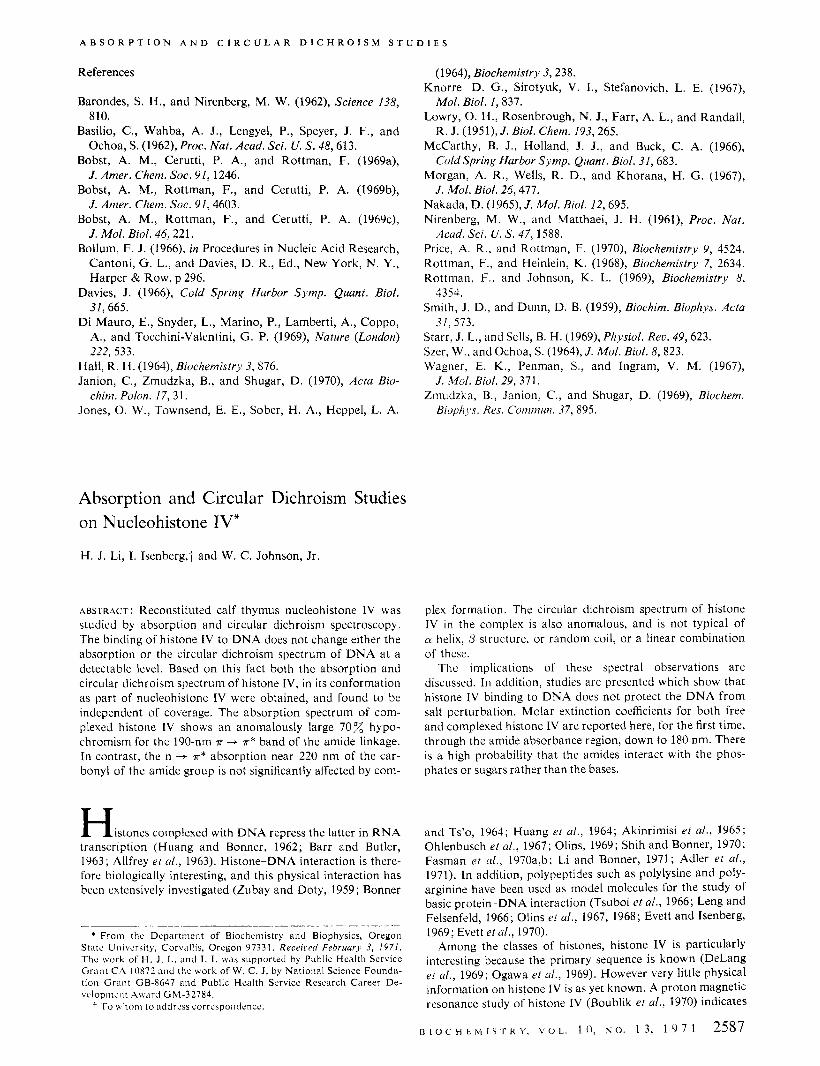
FIGURE 7: Comparison of NaCl effect on the circular dichroism of DNA and histone IV-DNA complex. (A) DNA; (0) histone 1V- DNA complex with u = 1.5.
2590 B I O C H E M I S T R Y , V O L . 1 0 , N O . 1 3 , 1 9 7 1
dichroism spectrum of histone IV in the complex is indepen- dent of the coverage, when a is varied from 0.56 to 1.5. This spectrum is compared to those of histone IV in water and in phosphate buffer in Figure 5'0. Histone IV in water has a strong negative band at 198 nm which indicates a random coil configuration. Random coil configurations have also been found in other histone preparations in water (Bradbury et al., 1965; Jirgensons and Hnilica, 1965; Fasman et al., 1970a,b). The circular dichroism spectrum of histone IV in phosphate buffer is quite different from that in water. There is a positive band at 190 nm and a broad negative band a t 214 nm. The shape of these bands suggests that histone IV has a higher cy
helix or /3 sheet, or both, in phosphate buffer than in water. The spectral difference in water and in phosphate buffer is not a p H effect (pH 5.0 in water and pH 7.4 in phosphate buffer) because histone IV in water, at pH 7.2, has the same circular dichroism spectrum as it does at pH 5.0.
The circular dichroism spectrum of histone IV, complexed with DNA, is quite difrerent from that of free histone in solu- tion. There is a positive band near 195 nm and a negative band at 220 nm. This spectrum is also different from the circu- lar dichroism spectrum of polypeptides in cy helix, /3 sheet, or random coil (Holzwarth and Doty, 1965; Grecnfield and Fasman, 1969). We have not been able to fit the circular di- chroism spectrum even by assuming that the spectrum of histone 1V in the complex is a linear combination of those three spectra.
In any case, we note that the circular dichroism spectrum of histone IV, as well as the absorption spectrum, is changed to a considerable extent when i t is complexed to DNA.
DNA structure in solution is perturbed by salt (Tunis and Hearst, 1968) and by ethylene glycol (Nelson and Johnson, 1970). Salt tends to change the DNA conformation from a B form toward a C form (Nelson and Johnson, 1970). The ques-
A B S O R P T I O N A N D C I R C U L A R D I C H R O I S M S T U D l E S
W c,
I I I I L . 1 .- i ~ 1 - - ..I- . ~ .---I__.Ll-.J
X ( n r n ) x ( n n ) 210 230 250 270 290 210 230- ~ 2 50 2 70 290
FIGURE 6: (a) NaCl effect on circular dichroism spectrum of histone IV-DNA complex with u = 1.5. (0-0) NaCl = 0.0, 0.07, and 0.2 M ; (A---A) NaCl = 0.59 M ; (x-X) NaCl = 1.17 M; (u-u) NaCl = 2.92 M. (b) NaCl effect on the circular dichroism spectrum of DNA. (O--O)NaCl = O.O,O. l , and0 .24~ ; (X- - -X)NaCl = 0.64hi;(A--A)NaCl = 1,28bt:(O---C)NaCI = 3.2 M.
tion we asked was whether the shielding of D N A by histone IV binding can afford protection to the D N A against salt effects. The results are shown in Figures 6a,b and 7. NaCl effects on the circular dichroism spectrum above 250 nm for D N A are essentially identical with those for nucleohistone IV. The addition of salt shifts the positive peak from 278 to 282 nm and reduces the intensity. NaCl from 0 to 3 M does not change the circular dichroism of D N A in the 220-nm region while it increases the negative circular dichroism of nucleohistone IV in this wavelength region.
Discussion
The conformation of D N A in native nucleohistone differs from uncomplexed DNA, as judged by X-ray studies (Wilkins et a/., 1959; Pardon et al., 1967), optical rotation (Tuan and Bonner, 1969), and circular dichroism (Permogorov et UI., 1970; Simpson and Sober, 1970; Shih and Fasman, 1970). Circular dichroism studies indicate a slight base tilting of D N A in native nucleohistones, which is possibly due to the binding of slightly lysine-rich histone I1 (Simpson and Sober, 1970). Reconstituted nucleohistone I shows no change in the circular dichroism spectrum of D N A if the complex is pre- pared by salt gradient dialysis down to very low salt (Olins, 1969), while there is big change in the circular dichroism spectrum of DNA if the salt gradient is stopped a t 0.14 M N a F or higher (Fasrnan et u/., 1970a). This difference may be due to a n irreversible conformational change of the DNA when salt goes, for example, from 0.14 M to a very low concen- tration.
We have studied the optical properties of nucleohistone IV prepared by salt gradient dialysis down to around 0.05 hi
NaCl in the presence of 5 M urea. We find, for a wide range of coverage, that both the absorption and circular dichroism spectrum of the complexed DNA is essentially the same as that of the uncomplexed DNA in the 300- to 240-nm region, where there is little interference from protein bands (Figures 2 and 3). This indicates that histone IV does not alter the conformation of the DNA in nucleohistone IV. It implies further that histone IV may not be the main component alter- ing DNA structure in native nucleohistone, confirming the suggestion of Simpsonand Sober (1970).
We have obtained both the absorption and circular di-
chroism spectrum of histone IV in its conformation in nucleo- histone IV (Figures 2 and 5 ) . Since neither the absorption nor the circular dichroism spectrum of D N A changes in the 300- to 240-nm region, it is reasonable to assume that these spectra are unchanged a t shorter wavelengths, down to 180 nm. We make this assumption, and subtract the contribution of the DNA to the nucleohistone IV spectrum recorded to 180 nm, to obtain histone IV spectra. For both absorption and circular dichroism the spectrum so computed is independent of cover- age. Note that this type of spectral calculation is similar to one made earlier in a study of dye binding to DNA (Li and Crothers, 1969).
There is a strong ionic interaction between the basic residues of histones and the phosphates of DNA (Ohlenbusch et ui., 1967; Olins, 1969; Shih and Bonner, 1970; Li and Bonner, 1971). Proton magnetic resonance studies on the histone I-DNA complex (Lee et a/., 1971, manuscript in preparation) also show a strong interaction between the D N A and both hydrophobic and glutamic acid residues of histones. When histones are brought into close interaction with D N A in a complex, the amides of the protein may have a strong interaction with DNA. Our computed absorption and circular dichroism spectrum for histone IV in the complex both in- dicate strong amino acid-DNA interaction in nucleohistone IV.
Upon helix formation, the 190-nm a -+ a* transition of polypeptides splits into a perpendicularly polarized band at around 188 nm and a parallel polarized band at around 204 nm (Gratzer et d., 1961; Tinoco er nl., 1962; 1963; Holzwarth and Doty, 1965). The weak absorption band at 220 nm is due to an n -+ a* transition of the carbonyl group of the amide (Schellman and Oriel, 1962; Woody and Tinoco, 1967; Holzwarth and Doty, 1965).
Our results in Figures 1 and 2 show the biggest percentage reduction of absorption intensity a t 190 nm, a smaller reduc- tion of 205 nm and only a slight reduction a t 220 nm when histone IV in the free and complexed states are compared. Hypochrornism is therefore biggest for the perpendicular a -+
K*, smaller for the parallel a + K* and weakest for the n -+
K* transition. The order is similar to the hypochromism for a-helix formation of polypeptide from random coil con- formation (Tinoco et a/., 1962).
Hypochromism a t 190 nm, due to a-helix formation in
BIOC'HE?vlISTRY, V O L . 1 0 , N O . 1 3 , 1 9 7 1 2591
LI, I S E N B E R G , A N D J O H N S O N
the complex, could contribute to the hypochromism observed here, Nevertheless, this contribution is probably minor be- cause the formation of a n CY helix from a random coil leads to only a 30-40% hypochromism (Rosenheck and Doty, 1961; Tinoco et al., 1962). In contrast, we observe a 70% hypochromism in the absorption of histone IV on forming the complex (Figures 1 and 2), a n anomalously large reduction in intensity.
Before discussing the hypochromism further, let us consider the computed circular dichroism spectrum of histone IV in the complex (Figure 5). For polypeptides, three different secondary structures have been commonly considered, CY helix, 0 sheet, and random coil. However, our computed spectrum does not match any of these three structures, and we have been unable to fit it t o a linear combination of the three as well. O n closer inspection this should perhaps not be sur- prising.
A true random coil may be difficult t o define in proteins, or even polypeptides, because side-chain interaction through electrostatic forces, hydrophobic forces, or hydrogen bonding will prohibit a segment from assuming a random coil con- figuration even though it is a nonhelical and non-0 structure. Also, the circular dichroism parameters of polypeptides such as polylysine or polyglutamic acid can only approximate analogous parameters in proteins and, as Fasman et al., (1970a,b) have shown, the circular dichroism of unordered polylysine and a number of proteins differ from one another. Furthermore, both the basic and less basic halves of histone molecules are bound to different regions of the DNA, and have different thermal stabilizations (Li and Bonner, 1971). This implies that the residues of the histone are in close contact with the D N A along the phosphate lattice or in the grooves. The interaction between the amino acid residues and the D N A , as well as steric hindrance provided by DNA, must play a major role in determining the configuration of the amide backbone of the histone in the complex.
It appears to be premature to present a model that is too detailed. However, certain general considerations may be made.
Basically there are only two possible types of explanations for the observed spectral changes. One is that histone IV complexed with DNA, has a conformation in which the amide-amide interaction is even stronger than it is in a n CY
helix. The other is that strong interaction between the amides of histone IV and the D N A moieties directly contribute t o the hypochromism and anomalous circular dichroism. Of course, a combination of these possibilities is also reason- able.
We may comment on the possibility of amide interaction with the bases, in contrast to interaction with the phosphates or sugars. First consider the central assumption of this paper. We believe it is extremely unlikely that histone IV could change the optical properties of D N A between 240 and 180 nm in the complex and not change the absorption, or the circular dichroism, of the D N A above 240 nm. Accept- ing this assumption, then any interaction between histone and D N A moieties which changes the optical properties of the histone cannot be a n interaction with the bases. If the spectral changes of histone IV were due to amide-base interaction, theoretical considerations (Tinoco et a/., 1962) state that the spectra of the bases must also be altered. In- deed, this suggests that the histone IV may be in a con- formation which excludes interaction of the amides with the bases.
There is no apparent relationship between our work and
2592 B I O C H E M I S T R Y , V O L . 1 0 , N O . 1 3 , 1 9 7 1
recent circular dichroism studies of nucleohistone (Wilhelm et al., 1970; Henson and Walker, 1970). We believe that it would be premature to attempt to correlate such studies a t the present time.
Before ending this discussion, we may note the close relationship between the absorbance and circular dichroism spectra, near 220 nm, of histone IV in nucleohistone. The ab- sorption spectrum of histone IV, complexed with DNA, shows a shoulder near 220 nm (Figures 1 and 2). Presumably this shoulder is a n + P* transition of the carbonyl group of the amide (Schellman and Oriel, 1962; Woody and Tinoco, 1967). Histone IV in the complex has a negative circular dichroism band a t 220 nm (Figures 5a,b) which is similar to the nonconservative and negative circular dichroism band a t this wavelength for the n + P* transition in a n CY helix (Holzwarth and Doty, 1965). Therefore, both the absorption and circular dichroism spectra give strong support t o the assignment of the 220-nm band of histone IV in the complex as a n + x* transition.
Tunis and Hearst (1968) have examined salt-induced changes in the optical rotation of DNA. They show that the effective- ness of various ions in perturbing the optical rotatory dis- persion of D N A does not correlate either with perturbations of water structure or with the lowering of the melting tem- perature of DNA. They propose that it appears likely that changes are due to a perturabtion of the D N A structure. Our results suggest that such a perturbation would be the same for D N A and histone IV-DNA complexes. One could argue that salt simultaneously removes histone IV and perturbs the DNA. Indeed, histone IV in nucleohistone IV is dissociated by NaCl from D N A in concentrations from around 0.4 to 2 M, with a midpoint for dissociation of 0.95 M NaCl (H. J. Li and I. Isenberg, 1971, unpublished data). This, it may be noted, agrees with the NaCl dissociation of histone IV from native nucleohistone (Fambrough and Bonner, 1968). However the excellent quantitafiue agreement over the entire range of salt measured (Figure 7), between the changes in D N A and in the complex, indicates that salt simply does not dissociate the complex and then perturb the uncomplexed DNA. Instead, it appears most likely that histone IV simply does not protect the DNA against salt perturba- tion.
In summary we find: (1) the binding of histone IV to D N A does not change the conformation of the D N A in complexes prepared by gradient dialysis down to low ionic strength. (2) This observation allows us to compute the absorbance and circular dichroism spectrum of histone IV in the complex. The computed spectra are independent of coverage. (3) Both the absorption and circular dichroism of histone IV change greatly upon binding to DNA. This indicates that either the histone IV structure is altered, or the electronic perturba- tion of the histone by the D N A changes the spectral properties of the protein. We emphasize that one cannot make this distinction on the basis of circular dichroism data alone. How- ever, it is probable that amide-base interaction does not occur. (4) The binding of histone IV to DNA does not protect the D N A from salt perturbation.
Acknowledgments
We thank Professor I. Tinoco, Jr . , for discussions and Professor K . E. Van Holde for the use of his Durrum-Jasco circular dichroism spectrometer. We thank Linda Haley for her skillful preparation of histone IV and Robert Howard for amino acid analyses.
A B S O R P T I O N A N D C I R C U L A R D I C H R O I S M S T U D I E S
References
Adler, A. J., Schaffhausen, B., La.ngan, T. A., and Fasman, G. (1971), Biochemistry (in press).
Akinrimisi, E. O., Bonner, J . , and Ts’o, P. 0. P. (1965), J . Mol. B id . 11, 128.
Allfrey, V. G.? Littau, V. C., and Mirsky, A. E. (1963), Proc. Nut. Acad. Sci. U. S. 49,414.
Barr, G . C., and Butler, J . A. V. (1963), Nature (London) 199,1170.
Bekhor, I. , Kung, G. M. , and Bonner, J. (1969), J . Mol. B i d . 39,351.
Bonner, J., and Ts’o, P. 0. P., Ed. (1964), Nucleohistones, San Francisco, Calif., Holden Day.
Boublik, M., Bradbury, E. M., and Crane-Robinson, C. (1970), Eiir.. J . Biochetii. 14,486.
Bradbury, E. M., Crane-Robinson, C., Phillips, D. M., Johns, E. W., and Murray, K . (1965), Nature (London) 205,1315.
DeLange, R . J., Fambrough, D. M . ? Smith, E. L., and Bonner, J. (1969), J . Biol. Cheni. 244, 319.
Evett, J., and Isenberg. I. (1969), Ann. N . 1’. Acud. Sci. 158, 210.
Evett, J . , McKenzie, R . L.? and Isenberg. 1. (1970), Bio- clieriiisfrj. 9,451 3.
Fambrough, D., and Bonner. J. (1968). Biocliini. Biop/ij,s. Actu 154, 601,
Fasman, G. D., Hoving, H., and Timasheff, S. N. (1970b), Bioclieiiiistr,~ 9, 3316.
Fasinan, G. D., Schaffhausen, B., Goldsmith, L. , and Adler. A. (1 970a), Biochemisrrj, 9: 2814.
Gratzer, W. B., Holzwarth, G . M., and Doty, P. (1961). Proc. N n f . Acad. Sci. U. S . 47, 1785.
Greenfield. N., and Fasnian, G. D. (196Y), Rioclieriiistrj. 8, 4108.
Henson, P., and Walker, I. 0. (197O), Ew. J . Bioc/ie/ii. 16, 524. Holzwarth, G . , and Doty, P. (1965), J . Ainer. Chern. Soc.
Huang, R. C. C., and Bonner, J . (1962), Proc. Nut. Acrid. Sci.
Htiang, R . C. C., Bonner, J., and Murray, K . (1964), J . Mol.
Huang, R . C. C., and Huang, P. C. (196Y), J . Mol. Biol. 3 9 ,
Jirgensons, B., and Hnilica, L. (1965), Biocliim. Bio/di.rs.
Johnson, W. C. , Jr. (1971), Rer. Sci. Insfrurii. (in press).
87,218.
C’. s. 48,1216.
Biol. 8, 54.
365.
Acta 109,241.
Johnson, W . C., Jr., and Tinoco, I . , Jr. (1969), Biopolj.iiiers
Leng, M., and Felsenfeld, G. (1966), Proc. Nut. Acad. Sci.
Li, H. J., and Bonner, J. (1971), Biochernistr), (in press). Li, H. J., and Crothers, D. M. (1969), Biopolyniers 8, 217. Nelson, R. G . , and Johnson, W. C., Jr. (1970), Biochmi.
Biop/ij,s. Res. Coriimun. 41, 21 1. Ogawa, Y . , Quagliarotti, G., Jordan, J., Taylor, C. W.,
Starhuck, W. C.. and Busch, H. (1969), J . Biol. Chen7. 244, 4387.
Ohlenbusch, H. H., Olivera, B. M. , Tuan, D. , and Davidson, N. ( I 967), J . Mol. B id . 25, 299.
O h , D. E . (1969), J . Mol. Biol. 43,439. O h , D. E. , Olins, A . L.. and von Hippel, P. H . (1967),
J . Mol. Biol. 24, 157. Olins, D. E.. Olins. A. L. and von Hippel, P. H. (1968),
J . Mol. Biol. 33, 265. Pardon, J. F., Wilkins, M. H. F., and Richards, B. M. ,
(1 967). Nufure (London) 215, 508. Permogorov, U. I.. Debabov, U. G. , Sladkova, I. A,, and
Rcbentish, B. A. (1970). Biochim. Biop/i~,s. Acta 199, 556. Rosenheck, K . , and Doty, P. (1961), Proc. Nur. Acad. Sci.
C. S . 47, 1775. Schellman, J. A., and Oricl. P. (1962), J . CIiem. Phys. 37, 2114. Shih, T. Y . , and Bonner, J . (1970), J . Mol. Biol. 48,469. Shih, T. Y. , and Fasnian. G . D. (1970), J . Mol. Biol. 52, 125. Simpson, R . B., and Sober, H. (1970). Biochemistry 9, 3103. Tinoco, I.. Jr. (1968), J . Chini. P/i.~s. 65, 91. Tinoco. I . . Jr.. Halptlrn. A, , and Simpson, W. T. (1962),
in Polyamino Acids, Polypeptides and Proteins, Stahmann, M . . Ed.. Madison. M’is., Univmity of Wisconsin Press, p 1-17,
Tinoco. I . . Jr.. Woodq. R . W.. and Bradley, D . F. (1963), J . Cl i~ni . Plij,s. 38, 1317.
Tsuboi. hl.. Matsuo. K . . and Ts’o, P. 0. P. (1966), J . Mol. B id . 15. 256.
Tuan, D.. and Bonncr, J . (1Y69), J . Mol. Bid. 45, 59. Tunis: M . B., and Hearsi. J . E. (1968), Biopo/j3iriers 6 , 1218. Wagner. T. E. (1970), Nltiwe (Lo17don) 227, 65. Wilhclm. F. X.. Champagne, M. H., and Daunt:, M. P.
Wilkins, M . H. F., Z~ibay, G., and Wilson, H . R. (1959),
Woody, R . W., and Tinoco, I . . Jr. (lY67), J . Cheni. P/i)..s.
Zubay, G.. and Doty, P. (1959), J . Mol. Biol. I , 1.
7, 727.
U. S. 56,1325.
(1970), ELII.. J . Biociicni. 15, 321.
J . Mol. Bid. 1. 179.
46,4927.
B I O C H E M I S T R Y , V O L . I O , N O . 1 3 , 1 9 7 1 2593