CHAPTER 6 Absorption and Distribution of Toxicants RONALD E. BAYNES and ERNEST HODGSON 6.1 INTRODUCTION As illustrated in the previous chapter, the human body can be exposed to a variety of toxicants that may be present in various environmental media such as air, soil, water, or food. However, just simply being exposed to these hazardous chemicals does not necessarily translate into a toxicological response. The mammalian body has several inherent defense mechanisms and membrane barriers that tend to prevent the entry or absorption and distribution of these toxicants once an exposure event has occurred. However, if the toxicant is readily absorbed into the body, there are still other anatomical and physiological barriers that may prevent distribution to the target tissue to elicit a toxic response. As the toxicological response is often related to the exposed dose, interactions between the toxicant and the body’s barriers and defense mechanisms will have an effect on toxicant movement in the body, and ultimately modulate the rate and extent of toxicant absorption and distribution to the target tissue. The skin represents the largest organ in the human body, and one of its primary functions can be seen as a physical barrier to absorption of toxicants. The other major routes of toxicant entry into the body are through the respiratory and gastrointestinal tract, which can be seen to offer less resistance to toxicant absorption than the skin. In general, the respiratory tract offers the most rapid route of entry, and the dermal the least rapid. One reason for this major difference is primarily because membrane thickness, which is really the physical distance between the external environment (skin surface, air in the lung, or lumen of the gut) and the blood capillaries, varies across these portals of entry. The overall entry depends on both the amount present and the saturability of the transport processes involved. Liver metabolism will have the most significant effect on toxicant bioavailability following gastrointestinal absorption, but microbial activity and various enzymes in the gastrointestinal tract and the skin can play a significant role in oral and dermal absorption, respectively. Physicochemical characteristics of the toxicant such as the chemical form can be a useful indicator of whether the toxicant will be absorbed and distributed in the body. In this regard toxicant molecular weight, ionization (pKa), and octanol/water partition coefficient (log P ) are useful indexes of predicting chemical A Textbook of Modern Toxicology, Third Edition, edited by Ernest Hodgson ISBN 0-471-26508-X Copyright 2004 John Wiley & Sons, Inc. 77
Transcript
CHAPTER 6
Absorption and Distribution of Toxicants
RONALD E. BAYNES and ERNEST HODGSON
6.1 INTRODUCTION
As illustrated in the previous chapter, the human body can be exposed to a varietyof toxicants that may be present in various environmental media such as air, soil,water, or food. However, just simply being exposed to these hazardous chemicalsdoes not necessarily translate into a toxicological response. The mammalian body hasseveral inherent defense mechanisms and membrane barriers that tend to prevent theentry or absorption and distribution of these toxicants once an exposure event hasoccurred. However, if the toxicant is readily absorbed into the body, there are stillother anatomical and physiological barriers that may prevent distribution to the targettissue to elicit a toxic response. As the toxicological response is often related to theexposed dose, interactions between the toxicant and the body’s barriers and defensemechanisms will have an effect on toxicant movement in the body, and ultimatelymodulate the rate and extent of toxicant absorption and distribution to the target tissue.
The skin represents the largest organ in the human body, and one of its primaryfunctions can be seen as a physical barrier to absorption of toxicants. The other majorroutes of toxicant entry into the body are through the respiratory and gastrointestinaltract, which can be seen to offer less resistance to toxicant absorption than the skin.In general, the respiratory tract offers the most rapid route of entry, and the dermalthe least rapid. One reason for this major difference is primarily because membranethickness, which is really the physical distance between the external environment (skinsurface, air in the lung, or lumen of the gut) and the blood capillaries, varies acrossthese portals of entry. The overall entry depends on both the amount present and thesaturability of the transport processes involved.
Liver metabolism will have the most significant effect on toxicant bioavailabilityfollowing gastrointestinal absorption, but microbial activity and various enzymes inthe gastrointestinal tract and the skin can play a significant role in oral and dermalabsorption, respectively. Physicochemical characteristics of the toxicant such as thechemical form can be a useful indicator of whether the toxicant will be absorbed anddistributed in the body. In this regard toxicant molecular weight, ionization (pKa), andoctanol/water partition coefficient (log P ) are useful indexes of predicting chemical
A Textbook of Modern Toxicology, Third Edition, edited by Ernest HodgsonISBN 0-471-26508-X Copyright 2004 John Wiley & Sons, Inc.
77
Dr. Le Quoc Tuan
Rectangle
78 ABSORPTION AND DISTRIBUTION OF TOXICANTS
transport from an environmental media across biological membranes to the bloodstream. The reader should also be aware that for those toxicants that are readily ion-ized, the pH gradient across membranes can determine the extent of toxicant transportand accumulation in tissues.
Once the toxicant has been absorbed, the toxicant molecules can move around thebody in two ways: (1) by bulk flow transfer (i.e., in the blood stream) and (2) by dif-fusional transfer (i.e., molecule-by-molecule over short distances). Disposition is theterm often used to describe the simultaneous effects of distribution and elimination pro-cesses subsequent to absorption. The cardiovascular system provides distribution of alltoxicants, regardless of their chemical nature, to various organs and tissues with variouslevels of affinities for toxicants. It should be remembered that organ mass and bloodperfusion can vary, which can account for differential distribution of toxicants. Toxi-cant disposition can also be influenced by plasma protein binding in the blood stream.The nature of this toxicant-protein interaction is dependent on the chemical nature ofthe toxicant, the presence of other toxicants or drugs in the blood stream, as well asplasma protein levels. However, what distinguishes one toxicant pharmacokineticallyfrom another is its diffusional characteristics. That is, its ability to cross nonaqueousdiffusional barriers (e.g., cell membranes) from an aqueous compartment. This usuallyinvolves movement across several compartments separated by lipid membranes. It istherefore important to understand the mechanisms by which drugs cross membranes andthe physicochemical properties of molecules and membranes that influence the move-ment of drugs from the environment to the body via either oral, inhalation, or dermalroutes. These factors also influence movement from one compartment to another withinthe body during distribution as well as metabolism, and excretion.
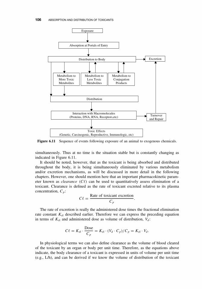
We can quantitate this movement or transport from one compartment to anotherusing mathematical models to describe transport rates. This in fact is what we do inpharmacokinetic analysis and modeling. Pharmaco- or toxicokinetics is therefore thequantitation of the time course of toxicants in the body during the various processes ofabsorption, distribution, and elimination or clearance (metabolism and/or excretion) ofthe toxicant. Stated differently, this is a study of how the body “handles” the toxicant asit is reflected in the plasma concentration at various time points. The two most importantpharmacokinetic parameters that describe the disposition of a chemical are volume ofdistribution and systemic (body) clearance. Pharmaco- and toxicodynamics is the studyof the biochemical and physiological effects of drugs and toxicants and determinestheir mechanism of action. Physiologically based pharmaco- or toxicokinetic modelsare used to integrate this information and to predict disposition of toxicants for a givenexposure scenario. These concepts will be introduced at the end of this chapter.
6.2 CELL MEMBRANES
During absorption, distribution, and elimination processes the toxicant will encountervarious cell membranes before interacting with the target tissue. Each step of theseprocess involves translocation of the chemical across various membrane barriers, fromthe skin or mucosa through the capillary membranes, and through the cellular andorganelle membranes (Figure 6.1). These membrane barriers vary from the relativelythick areas of the skin to the relatively thin lung membranes. In all cases, however,the membranes of tissue, cell, and cell organelle are relatively similar.
CELL MEMBRANES 79
EnvironmentInterstitial
FluidInterstitial
FluidIntracellular
FluidPlasma
Mucosaor
Skin
CapillaryMembrane
CapillaryMembrane
Target-CellMembrane
SubcellularOrganelleMembrane
Intra-organelleFluid
(Mitochondria,Liposome,Nucleus)
Figure 6.1 Schematic showing membranes that a chemical may need to cross during passagefrom the environment to the site of action. (Adapted from E. Hodgson and P. E. Levi, eds.Introduction to Biochemical Toxicology, 2nd ed., Appleton and Lange, 1994, p. 12.)
The cell membranes are predominantly a lipid matrix or can be considered a lipidbarrier with an average width of a membrane being approximately 75 A. The membraneis described as the fluid mosaic model (Figure 6.2) which consist of (1) a bilayer ofphospholipids with hydrocarbons oriented inward (hydrophobic phase), (2) hydrophilicheads oriented outward (hydrophilic phase), and (3) associated intra- and extracellularproteins and transverse the membrane. The ratio of lipid to protein varies from 5:1 forthe myelin membrane to 1:5 for the inner structure of the mitochondria. However, 100%of the myelin membrane surface is lipid bilayer, whereas the inner membrane of themitochondria may have only 40% lipid bilayer surface. In this example the proportionof membrane surface that is lipid will clearly influence distribution of toxicants ofvarying lipophilicity.
The lipid constituents in the membrane permit considerable movement of macromole-cules, and membrane constituents may move appreciably within membranes. Membranefluidity, a function of lipid composition, can be altered by temperature and chemicals
Figure 6.2 Schematic diagram of biological membrane. Head groups of lipids represented byspheres, tail ends by zigzag lines. Black, white, or stippled spheres indicate different kinds oflipids and illustrate asymmetry in certain cases. Large bodies are membrane-associated proteins.(Adapted from Singer and Nicolson, Science 175:720, 1972.)
80 ABSORPTION AND DISTRIBUTION OF TOXICANTS
(e.g., anesthetics). Several types of lipids are found in membranes, with phospholipidsand cholesterol predominating. Sphingolipids comprise the primary minor component.Phosphatidylcholine, phosphatidylserine, and phosphatidylethanolamine are the primaryphosphatides, and their two fatty acid hydrocarbon chains (typically 16 to 18, but varyingfrom 12 to 22) comprise the nonpolar region. Some of the fatty acids are unsaturatedand contribute appreciably to the fluidity of the membrane.
Proteins, which have many physiological roles in normal cell function, are intimatelyassociated with lipids and may be located throughout lipid bilayers. These proteins maybe located on either the surface or traverse the entire structure. Hydrophobic forcesare responsible for maintaining the structural integrity of proteins and lipids withinmembranes, but movement within the membranes may occur. External and internalmembrane proteins can function as receptors. Many proteins that traverse the membraneare transport proteins, and are involved in translocation of ligands; that is, they areinvolved in active and facilitated transport.
Complexes of intrinsic membrane proteins and lipids can form hydrophilic or hydro-phobic channels that allow transport of molecules with different physicochemicalcharacteristics. The amphipathic nature of the membrane creates a barrier for ion-ized, highly polar drugs, although it does not completely exclude them. The presenceof pores of approximately 4 A are believed to allow for ready movement of smallmolecules such as water. Thus certain molecules that ordinarily would be excludedcan rapidly traverse the highly lipid membrane barrier.
It is worth noting that differences among membranes, such as the presence of dif-ferent lipids, the amount of surface lipid, differences in size and shape of proteins,or physical features of bonding, may cause differences in permeability among mem-branes. These biochemical and biophysical differences are thought to be responsiblefor permeability differences in skin from different anatomical regions of the body.
6.3 MECHANISMS OF TRANSPORT
In general, there are four main ways by which small molecules cross biologicallipid membranes:
1. Passive diffusion. Diffusion occurs through the lipid membrane.2. Filtration. Diffusion occurs through aqueous pores.3. Special transport. Transport is aided by a carrier molecule, which act as a “ferry-
boat.”4. Endocytosis. Transport takes the form of pinocytosis for liquids and phagocytosis
for solids.
The first and third routes are important in relation to pharmacokinetic mechanisms.The aqueous pores are too small in diameter for diffusion of most drugs and toxicant,although important for movement of water and small polar molecules (e.g., urea).Pinocytosis is important for some macromolecules (e.g., insulin crossing the blood-brain barrier).
6.3.1 Passive Diffusion
Most drugs and toxicant pass through membranes by simple diffusion down a concentra-tion gradient. The driving force being the concentration gradient across the membrane.
MECHANISMS OF TRANSPORT 81
This diffusion process can continue until equilibrium, although in reality there isalways movement but the net flux is zero. Eventually the concentration of unionizedor unbound (free) toxicant is the same on either side of the membrane. In other words,there is no competition of molecules and there is generally a lack of saturation. Sol-ubility in the lipid bilayer is important, and the greater the partition coefficient, thehigher is the concentration in the membrane, and the greater is the rate of diffusionacross the membrane. For ionized toxicants the steady state concentration is dependenton the differences in pH across the membrane. Most membranes are relatively perme-able to water either by diffusion or by flow that results from hydrostatic or osmoticdifferences across the membrane, and bulk flow of water can also carry with it smalland water soluble molecules by this mechanism. These substances generally have amolecular weight of less than 200. Although inorganic ions are small and will readilydiffuse across some membranes, their hydrated ionic radius is relatively large. In suchcases active transport is required (see below). Specific ion fluxes are also controlledby specific channels that are important in nerves, muscles, and signal transduction.
We can now quantitate the rate at which a toxicant can be transported by passivediffusion, and this can be described by Fick’s law of diffusion as follows:
Rate of diffusion = D × Sa × Pc
d(CH − CL),
where D is the diffusion coefficient, Sa is the surface area of the membrane, Pc
is the partition coefficient, d is the membrane thickness, and CH and CL are theconcentrations at both sides of the membrane (high and low, respectively). The firstpart of this equation (DPc/d) represents the permeability coefficient of the drug. Thepermeability expresses the ease of penetration of a chemical and has units of velocity,distance/time (cm/h).
The diffusion coefficient or diffusivity of the toxicant, D, is primarily dependenton solubility of the toxicant in the membrane and its molecular weight and molecularconformation. Depending on the membrane, there is a functional molecular size and/orweight cutoff that prevents very large molecules from being passively absorbed acrossany membrane. One would expect small molecular weight molecules to diffuse morerapidly than larger molecular weight toxicants. Therefore the magnitude of a toxicant’sdiffusion coefficient really reflects the ease with which it is able to diffuse through themembrane. The reader should also be aware that as a toxicant crosses from the donoror aqueous medium and through the membrane medium, there are really two diffusionenvironments and thus two diffusion coefficients to consider. Another important factorthat can influence the diffusion coefficient is membrane viscosity. This physicochemicalcharacteristic should remain constant in biological systems but can be modified in skinmembranes exposed to various pharmaceutical or pesticide formulations. Formulationadditives or excipients may enter the membrane barrier and reversibly or irreversiblychange viscosity and thus diffusion coefficient of the drug or pesticide in the barriermembranes of the skin.
The partition coefficient, which will be described in more detail later in this chapter,is the relative solubility of the compound in lipid and water, and the compound’ssolubility really reflects the ability of the toxicant to move from a relatively aqueousenvironment across a lipid membrane. It is this factor that is often manipulated inpesticide and drug formulations to create a vehicle. Membrane permeability is thereforestrongly correlated to the lipid solubility of the toxicant in the membrane as well as
82 ABSORPTION AND DISTRIBUTION OF TOXICANTS
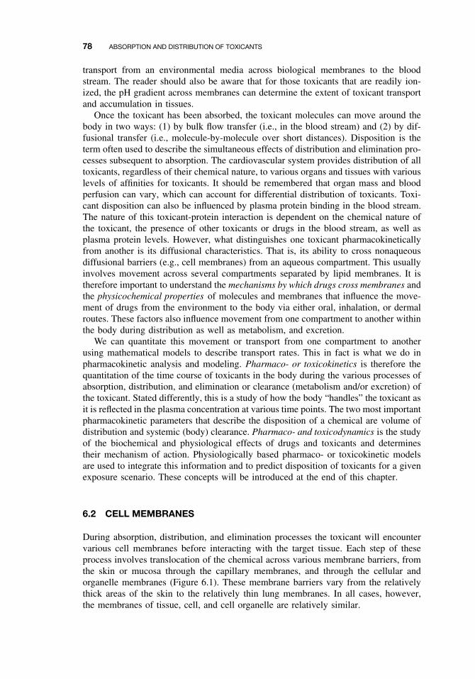
the aqueous environment surrounding the membrane. Please be aware that there areinstances where partition coefficient or lipid solubility of the toxicant may be very large,and there may be a tendency for the drug to sequester in the membrane. Membranesurface area and membrane thickness can also vary across different organs in the body,but one does not expect these two factors in Fick’s equation to vary considerably. Thefinal component of Fick’s equation is the concentration gradient (CH − CL) acrossthe membrane, which is the driving force for diffusion, and as will be demonstratedbelow in our discussion on first-order kinetics, is the most important factor dictatingthe rate of transport across most biological membranes.
First-Order Kinetics. When the rate of a process is dependent on a rate constant anda concentration gradient, a linear or first-order kinetic process will be operative. Thereader should be aware that there are numerous deviations from the first-order processwhen chemical transport in vivo is analyzed, and this can be deemed an approximationsince, in many barriers, penetration is slow and a long period of time is required toachieve steady state.
The rate of movement of a toxicant across a membrane may be expressed as thechange in amount of toxicant, A, (dA) or toxicant concentration, C, (dC) per unit oftime (dt), which equals dA/dt. Calculus can be used to express instantaneous rates oververy small time intervals (dt). Thus rate processes may then be generally expressed as
dAdt
= KAn
where dA/dt is the rate of chemical (X) movement (e.g. absorption, distribution, elim-ination), K is the rate constant of the process, and n is the kinetic order of the transportprocess (e.g., absorption). The n either equals 1 (first order) or 0 (zero order). Thusthe first-order rate equation is written as
dAdt
= KA1 = KA,
and the zero-order rate equation as
dAdt
= KA0 = K.
We know from Fick’s law that the rate of diffusion (now expressed as dA/dt) is
dAdt
= DžSažPc(A1 − A2)
d.
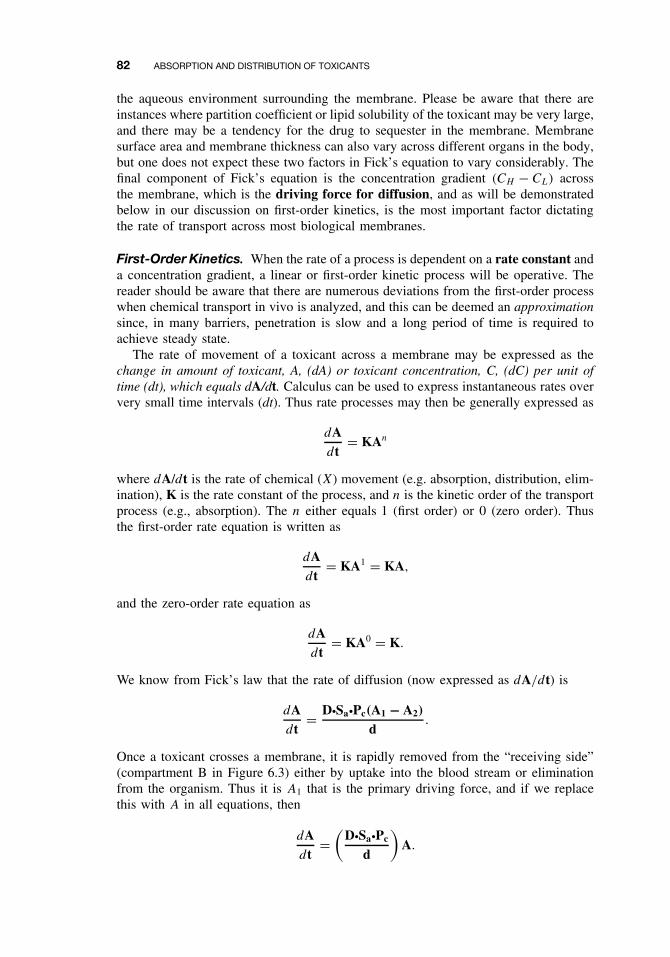
Once a toxicant crosses a membrane, it is rapidly removed from the “receiving side”(compartment B in Figure 6.3) either by uptake into the blood stream or eliminationfrom the organism. Thus it is A1 that is the primary driving force, and if we replacethis with A in all equations, then
dAdt
=(
DžSažPc
d
)A.
MECHANISMS OF TRANSPORT 83
Figure 6.3 Illustration of concentration gradient generated by administration of a drug thatcan travel down this gradient from area A and across a biological membrane to area B.
If we let K = (DžSažPc/d), then, since A is present in the equation, n must equal 1,so we have a first-order rate process. Fick’s law of diffusion, which is important forquantitating rates of absorption, distribution, and elimination, is thus the basis for usingfirst-order kinetics in most pharmacokinetic models.
Therefore in a first-order process, the rate of drug movement is directly proportionalto the amount of drug (A) in the body, which is usually a function of the dose. K isthe first-order fractional rate constant with units of liters/time (time−1) and representsthe fraction of drug that is transported per unit of time. Thus in a first-order process,the rate of drug movement is proportional to dose but the fraction moved per unit oftime is constant and independent of dose.
When first-order kinetics hold, a simple relationship exists between the penetrationrate constant, K , and t0.5 (time necessary for one-half of the applied dose to penetrate):
K = 0.693
t0.5,
where the units of K are a percentage of the change/time unit. We can also derive theconcentration of the toxicant if we know the volume or volume of distribution (Vd) ofthe toxicant compartment as
Vd(volume) = A(mass)
C(mass/volume).
(Vd is discussed in more detail later in this chapter.)
6.3.2 Carrier-Mediated Membrane Transport
This mechanism is important for compounds that lack sufficient lipid solubility to moverapidly across the membrane by simple diffusion. A membrane-associated protein isusually involved, specificity, competitive inhibition, and the saturation phenomenon andtheir kinetics are best described by Michaelis-Menton enzyme kinetic models. Membranepenetration by this mechanism is more rapid than simple diffusion and, in the case ofactive transport, may proceed beyond the point where concentrations are equal on both
84 ABSORPTION AND DISTRIBUTION OF TOXICANTS
sides of the membrane. Generally, there are two types of specialized carrier-mediatedtransport processes:
Passive facilitated diffusion involves movement down a concentration gradientwithout an input of energy. This mechanism, which may be highly selective for spe-cific conformational structures, is necessary for transport of endogenous compoundswhose rate of transport by simple diffusion would otherwise be too slow. The classicalexample of facilitated diffusion is transport of glucose into red blood cells.
Active transport requires energy, and transport is against a concentration. Main-tenance against this gradient requires energy. It is often coupled to energy-producingenzymes (e.g., ATPase) or to the transport of other molecules (e.g., Na+, Cl−, H+)that generate energy as they cross the membranes. Carrier-mediated drug transport canoccur in only a few sites in the body, and the main sites are
There are instances in which toxicants have chemical or structural similarities toendogenous chemicals that rely on these special transport mechanisms for normal phys-iological uptake and can thus utilize the same system for membrane transport. Usefulexamples of drugs known to be transported by this mechanism include levodopa, whichis used in treating Parkinson’s disease, and fluorouracil, a cytotoxic drug. Levodopais taken up by the carrier that normally transports phenylalanine, and fluorouracil istransported by the system that carries the natural pyrimidines, thymine, and uracil. Ironis absorbed by a specific carrier in the mucosal cells of the jejunum, and calcium by avitamin D-dependent carrier system. Lead may be more quickly moved by a transportsystem that is normally involved in the uptake of calcium.
For carrier-mediated transport, the rate of movement across a membrane will nowbe constant, since flux is dependent on the capacity of the membrane carriers and notthe mass of the chemical to be transported. These processes are described by zero-orderkinetic rate equations of the form:
dXdt
= KX0 = K0.
K0 is now the zero-order rate constant and is expressed in terms of mass/time. Inan active carrier-mediated transport process following zero-order kinetics, the rate ofdrug transport is always equal to K once the system is fully loaded or saturated. Atsubsaturation levels, the rate is initially first order as the carriers become loaded withthe toxicant, but at concentrations normally encountered in pharmacokinetics, the ratebecomes constant. Thus, as dose increases, the rate of transport does not increase inproportion to dose as it does with the fractional rate constant seen in first-order process.This is illustrated in the Table 6.1 where it is assumed that the first-order rate constantis 0.1 (10% per minute) and the zero-order rate is 10 mg/min.
In the case of first order, these amounts will subsequently diminish (10% of 900is 90, etc.). In the case of zero order, the amount transported does not vary with time(constant rate of transport).
PHYSICOCHEMICAL PROPERTIES RELEVANT TO DIFFUSION 85
Table 6.1 Amount of Toxicant (mg) Transportedin One Minute
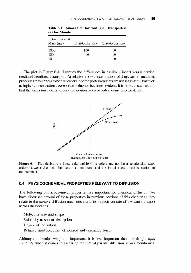
The plot in Figure 6.4 illustrates the differences in passive (linear) versus carrier-mediated (nonlinear) transport. At relatively low concentrations of drug, carrier-mediatedprocesses may appear to be first order since the protein carriers are not saturated. However,at higher concentrations, zero-order behavior becomes evident. It is in plots such as thisthat the terms linear (first order) and nonlinear (zero order) come into existence.
Linear
Non-linear
Mass or Concentration(Dependent upon Experiment)
Flux
Figure 6.4 Plot depicting a linear relationship (first order) and nonlinear relationship (zeroorder) between chemical flux across a membrane and the initial mass or concentration ofthe chemical.
6.4 PHYSICOCHEMICAL PROPERTIES RELEVANT TO DIFFUSION
The following physicochemical properties are important for chemical diffusion. Wehave discussed several of these properties in previous sections of this chapter as theyrelate to the passive diffusion mechanism and its impacts on rate of toxicant transportacross membranes.
Molecular size and shapeSolubility at site of absorptionDegree of ionizationRelative lipid solubility of ionized and unionized forms
Although molecular weight is important, it is less important than the drug’s lipidsolubility when it comes to assessing the rate of passive diffusion across membranes.
86 ABSORPTION AND DISTRIBUTION OF TOXICANTS
The permeability, P(P = Pc × D), of a nonpolar substance through a cell membrane isdependent on two physicochemical factors: (1) solubility in the membrane (Pc), whichcan be expressed as a partition coefficient of the drug between the aqueous phase andmembrane phase, and (2) diffusivity or diffusion coefficient (D), which is a measure ofmobility of the drug molecules within the lipid. The latter may vary only slightly amongtoxicants, but the former is more important. Lipid solubility is therefore one of the mostimportant determinants of the pharmacokinetic characteristics of a chemical, and it isimportant to determine whether a toxicants is readily ionized or not influenced by pHof the environment. If the toxicant is readily ionized, then one needs to understand itschemicals behavior in various environmental matrices in order to adequately assess itstransport mechanism across membranes.
6.4.1 Ionization
For the purposes of this discussion on membrane transport, chemicals can be broadlycategorized into those that are ionized and those that are not ionized. Many drugs (e.g.,antibiotics) and several toxicants (e.g., strychnine) are either weak acids or weak basesand can exist in solution as a mixture of nonionized and ionized forms. Generally, thesedrugs and toxicants must be in the uncharged or nonionized form to be transported bypassive diffusion across biological membranes. This is because biological membranesare of a lipid nature and are less permeable to the ionized form of the chemical. ThepH of the environment (e.g., lumen of the gastrointestinal tract and renal tubules)can influence transfer of toxicant that are ionizable by increasing or decreasing theamount of nonionized form of the toxicant. Aminoglycosides (e.g., gentamicin) arethe exception to this general rule in that the uncharged species is insufficiently lipidsoluble to cross the membrane appreciably. This is due to a preponderance of hydrogen-bonding groups in the sugar moiety that render the uncharged molecule hydrophilic.Note that some amphoteric drugs (e.g., tetracyclines) may be absorbed from both acidicand alkaline environments. In essence, the amount of drug or toxicant in ionized ornonionized form depends on the pKa (pH at which 50% of the drug is ionized) ofthe drug and the pH of the solution in which the drug is dissolved. The pKa, which isthe negative logarithm of the dissociation constant of a weak acid or weak base, is aphysicochemical characteristic of the drug or toxicant. When the pH of the solution isequal to the pKa, then 50% of the toxicant is in the ionized form and 50% is in thenonionized form. The ionized and nonionized fractions can be calculated according tothe Henderson-Hasselbach equations listed below:
For an organic acid (RCOOH ↔ RCOO− + H+), acidic conditions (pH less than thepKa of the compound) will favor the formation of the nonionized RCOOH, whereasalkaline conditions (pH greater than pKa) will shift the equilibrium to the right. Foran organic base (RNH2 + H+ ⇔ RNH3
+), the reverse is true, and decreasing the pH(increasing the concentration of H+) will favor formation of the ionized form, whereasincreasing the pH (decreasing the concentration of H+) will favor formation of thenonionized form.
PHYSICOCHEMICAL PROPERTIES RELEVANT TO DIFFUSION 87
Table 6.2 Amount of Toxicant Absorbed at VariouspH Values (%)
Memory aid: In general, weak organic acids readily diffuse across a biologicalmembrane in an acidic environment, and organic bases can similarly diffuse in a basicenvironment. This is illustrated quite well in Table 6.2 for the chemical in rat intes-tine. There are the usual exceptions to the generalizations concerning ionization andmembrane transport, and some compounds, such as pralidoxime (2-PAM), paraquat,and diquat, are absorbed to an appreciable extent even in the ionized forms. Themechanisms allowing these exceptions are not well understood.
Ion trapping can occur when at equilibrium the total (ionized + nonionized) con-centration of the drug will be different in each compartment, with an acidic drug ortoxicant being concentrated in the compartment with the relatively high pH, and viceversa. The pH partition mechanism explains some of the qualitative effects of pHchanges in different body compartment on the pharmacokinetics of weakly basic oracidic drugs or toxicant as it relates to renal excretion and penetration of the blood-brainbarrier. Alkalization of urine in the lumen of renal tubules can enhance eliminationof weak acids. However, this phenomenon is not the main determinant of absorptionof drugs or toxicants from the gastrointestinal tract. In the gastrointestinal tract theenormous absorptive surface area of the villi and microvilli in the ileum, compared tothe smaller absorptive area of the stomach, is of overriding importance.
6.4.2 Partition Coefficients
A second physicochemical parameter influencing chemical penetration through mem-branes is the relative lipid solubility of the potential toxicant that can be ascertainedfrom its known partition coefficient. The partition coefficient is a measure of the abilityof a chemical to separate between two immiscible phases. The phases consist of anorganic phase (e.g., octanol or heptane) and an aqueous phase (e.g., water). The lipidsolvent used for measurement is usually octanol because it best mimics the carbonchain of phospholipids, but many other systems have been reported (chloroform/water,ether/water, olive oil/water). The lipid solubility and the water solubility characteristicsof the chemical will allow it to proportionately partition between the organic and waterphase. The partition coefficients can be calculated using the following equation:
P = Vw
Vo
[(Cwo − Cw)
Cw
],
88 ABSORPTION AND DISTRIBUTION OF TOXICANTS
where P is the partition coefficient and usually expressed in terms of its logarithmicvalue (log P ), Vw and Vo are the volumes of aqueous and oil or organic phase, respec-tively, and Cwo and Cw are drug or toxic concentrations in the aqueous phase beforeand after shaking, respectively.
The lower the partition coefficient, the more water soluble, and the least permeablethe toxicant is across a membrane. Regarding dermal absorption, partition coefficientscan be predictive of absorption. However, toxicants with extremely high partition coef-ficients tend to remain in the membrane or skin. This explains why a strong correlationbetween permeability and the partition coefficient can exist for a hypothetical seriesof analogous chemicals for a specific range of partition coefficients, but the correla-tion does not exists for log P values greater than 6 in many instances. A log P ofaround 1 is often taken as desirable for skin penetration. The reader should also recallthat this parameter is operative as the chemical diffuses across membranes (Figure 6.1)of varying lipid content during absorption, distribution, and elimination processes.
6.5 ROUTES OF ABSORPTION
Primary routes of entry of toxicants to the human body are dermal, gastrointestinal,and respiratory. Methods for studying these different routes are numerous, but they areperhaps best developed for the study of dermal absorption because this route is subjectto more direct methodology, whereas methods for studying respiratory or gastroin-testinal absorption require more highly specialized instrumentation. Additional routesencountered in experimental studies include intraperitoneal, intramuscular, and subcu-taneous routes. When direct entry into the circulatory system is desired, intravenous(IV) or intra-arterial injections can be used to bypass the absorption phase. Informationfrom this more direct route of entry (e.g., IV) should, however, be used in additionto data from the extravascular route of interest to adequately assess the true extent ofabsorption of a toxicant.
6.5.1 Extent of Absorption
It is often useful to determine how much of the drug actually penetrates the membranebarrier (e.g., skin or gastrointestinal tract) and gets into the blood stream. This isusually determined experimentally for oral and dermal routes of administration. Thearea under the curve (AUC) of the concentration-time profiles for oral or dermal routesis compared with the AUC for IV routes of administration. The AUC is determinedby breaking the curve up into a series of trapezoids and summing all of the areas withthe aid of an appropriate computer program (Figure 6.5).
The intravenous correction is very important if absolute bioavailability is desired.The ratio of these AUC values is absolute bioavailability, F :
F = (AUC)route
(AUC)IV.
The relationship above holds if the same doses are used with both routes, but thebioavailability should be corrected if different doses are used:
F = AUCroute × DoseIV
AUCIV × Doseroute.
ROUTES OF ABSORPTION 89
T1
C6
C1
C2
C5
C3
C4
T2 T3 T4 T5 T6
Time
Con
cent
ratio
n
Slope
Figure 6.5 Plasma concentration time profile for oral exposure to a toxicant and depiction ofAUCs determined by summation of trapezoids at several time periods.
Another technique is to monitor drug or toxicant excretion rather than blood con-centrations, especially when blood or plasma concentrations are very low. Using thesame equations, the AUC is now replaced by chemical concentrations in urine, feces,and expired air. Some chemicals are primarily excreted by the kidney and urine dataalone may be necessary. The rate and extent of absorption are clearly important fortherapeutic and toxicological considerations. For example, different formulations of thesame pesticide can change the absorption rate in skin or gastrointestinal tract, and notbioavailability, but can result in blood concentrations near the toxic dose. Also differentformulations can result in similar absorption rates but different bioavailability.
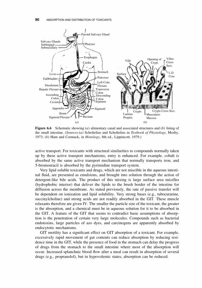
6.5.2 Gastrointestinal Absorption
The gastrointestinal tract (GIT) is a hollow tube (Figure 6.6a) lined by a layer ofcolumnar cells, and usually protected by mucous, which offers minimal resistance totoxicant penetration. The distance from the outer membrane to the vasculature is about40 µm, from which point further transport can easily occur. However, the cornifiedepithelium of the esophagus prevents absorption from this region of the GIT. Most ofthe absorption will therefore occur in the intestine (pH = 6), and to some extent in thestomach (pH = 1–3). Buccal and rectal absorption can occur in special circumstances.Note that secretions from the lachrymal duct, salivary gland, and nasal passages canenter the GIT via the buccal cavity. Therefore, following IV administration, a toxicantcan enter the GIT if the drug is in these secretions.
The intestine can compensate the 2.5 log units difference between it and the stom-ach by the increased surface area in the small intestines. The presence of microvilli(Figure 6.6b) in the intestine is an increase of 600-fold in surface area compared to ahollow tube of comparable length. Note that there is no absorption, except for water,in the large intestine.
Most of the absorption in the GIT is by passive diffusion, except for nutrients;glucose, amino acids, and drugs that look like these substances are taken up by
90 ABSORPTION AND DISTRIBUTION OF TOXICANTS
Parotid Salivary Gland
Pharynx
Esophagus
Cardia
Stomach
Pancreas
Left ColicFlexure
TransverseColonDescendingColonJejunum
Sigmoid
RectumSigmoid FlexureIleum
Appendix
Cecum
AscendingColon
Hepatic FlexureDuodenum
Gallbladder
Liver
SubmaxillarySublingualSalivary Glands
(a) (b)
Villi
Surface L.DCore
VillusCrossSect.
Opening ofCrypt
Crypts Crypts Cross Sect.LaminaPropria
MuscularisMucosa
Figure 6.6 Schematic showing (a) alimentary canal and associated structures and (b) lining ofthe small intestine. (Sources:(a) Scholtelius and Scholtelius in Textbook of Physiology, Mosby,1973; (b) Ham and Cormack, in Histology, 8th ed., Lippincott, 1979.)
active transport. For toxicants with structural similarities to compounds normally takenup by these active transport mechanisms, entry is enhanced. For example, cobalt isabsorbed by the same active transport mechanism that normally transports iron, and5-bromouracil is absorbed by the pyrimidine transport system.
Very lipid soluble toxicants and drugs, which are not miscible in the aqueous intesti-nal fluid, are presented as emulsions, and brought into solution through the action ofdetergent-like bile acids. The product of this mixing is large surface area micelles(hydrophobic interior) that deliver the lipids to the brush border of the intestine fordiffusion across the membrane. As stated previously, the rate of passive transfer willbe dependent on ionization and lipid solubility. Very strong bases (e.g., tubocurarine,succinylcholine) and strong acids are not readily absorbed in the GIT. These musclerelaxants therefore are given IV. The smaller the particle size of the toxicant, the greateris the absorption, and a chemical must be in aqueous solution for it to be absorbed inthe GIT. A feature of the GIT that seems to contradict basic assumptions of absorp-tion is the penetration of certain very large molecules. Compounds such as bacterialendotoxins, large particles of azo dyes, and carcinogens are apparently absorbed byendocytotic mechanisms.
GIT motility has a significant effect on GIT absorption of a toxicant. For example,excessively rapid movement of gut contents can reduce absorption by reducing resi-dence time in the GIT, while the presence of food in the stomach can delay the progressof drugs from the stomach to the small intestine where most of the absorption willoccur. Increased splanchnic blood flow after a meal can result in absorption of severaldrugs (e.g., propranolol), but in hypovolemic states, absorption can be reduced.
ROUTES OF ABSORPTION 91
Biotransformation in the GIT prior to absorption can have a significant impact onbioavailability of a toxicant. The resident bacterial population can metabolize drugs inthe GIT. Because of microbial fermentation in the rumen of ruminants and large intes-tine and cecum of horses and rabbits, its is often difficult to compare drug absorptionprofiles with carnivores (e.g., dogs) and omnivores (e.g., humans, pigs). Acid hydrol-ysis of some compounds can also occur, and enzymes in the intestinal mucosa canalso have an effect on oral bioavailability. If the toxicant survive these microbial andchemical reactions in the stomach and small intestine, it is absorbed in the GIT andcarried by the hepatic portal vein to the liver, which is the major site of metabolism.Chapters 7, 8, and 9 will discuss liver metabolism of toxicants in more detail. In brief,this activity in the liver can result in detoxification and/or bioactivation. Some drugsand toxicant that are conjugated (e.g., glucuronidation) in the liver are excreted viathe biliary system back into the GIT. Once secreted in bile by active transport andexcreted from the bile duct into the small intestine, this conjugated toxicant can besubjected to microbial beta-glucuronidase activity that can result in regeneration of theparent toxicant that is more lipophilic than the conjugate. The toxicant can now bereabsorbed by the GIT, prolonging the presence of the drug or toxicant in the systemiccirculation. This is called enterohepatic circulation, which will be covered in greaterdetail in subsequent chapters.
6.5.3 Dermal Absorption
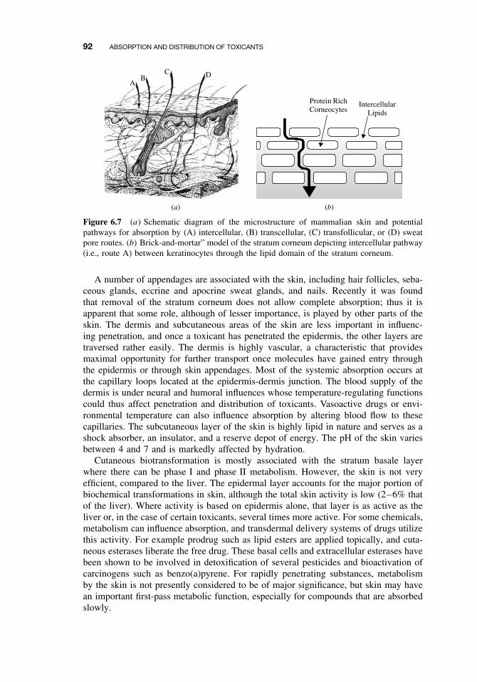
The skin is a complex multilayered tissue with a large surface area exposed to theenvironment. Skin anatomy, physiology, and biochemistry vary among species, withinspecies, and even between anatomic sites within an individual animal or human. Logi-cally these biological factors alone can influence dermal absorption. What is consistentis that the outer layer, the stratum corneum (SC), can provide as much as 80% of theresistance to absorption to most ions as well as aqueous solutions. However, the skinis permeable to many toxicants, and dermal exposure to agricultural pesticides andindustrial solvent can result in severe systemic toxicity.
The anatomy of the skin is depicted in the schematic diagram of Figure 6.7. Inmammalian skin there are really three distinct layers, which are the epidermis, dermis,and hypodermis or subcutaneous fat layer. Human skin is 3 mm thick, but it is theepidermis, which is only 0.1 to 0.8 mm, that provides the greatest resistance to toxicantpenetration. The five layers of the epidermis, starting from the outside, are the stratumcorneum, stratum lucidum, stratum granulosum, stratum spinosum, and stratum basale.The basal cells of the epidermis proliferate and differentiate as they migrate outwardtoward the surface of the skin. It requires about 2 to 28 days for cells to migratefrom the basal layer to the stratum corneum, where they are eventually sloughedoff. These dead, keratinized cells are, however, very water absorbant (hydrophilic),a property that keeps the skin soft and supple. Sebum, a natural oil covering theskin, functions in maintaining the water-holding ability of the epidermis. The stratumcorneum is the primary barrier to penetration, and it consists primarily of these deadkeratin-filled keratinocytes embedded in an extracellular lipid matrix. The lipids areprimarily sterols, other neutral lipids, and ceramides. This association between lipidsand dead keratinized cells, which is often referred to as the “brick and mortar” modelas depicted in Figure 6.7b, is used to simplify the composition of the stratum corneumthat is integral to chemical transport through skin.
92 ABSORPTION AND DISTRIBUTION OF TOXICANTS
(b)(a)
AB
C D
Protein RichCorneocytes
IntercellularLipids
Figure 6.7 (a) Schematic diagram of the microstructure of mammalian skin and potentialpathways for absorption by (A) intercellular, (B) transcellular, (C) transfollicular, or (D) sweatpore routes. (b) Brick-and-mortar” model of the stratum corneum depicting intercellular pathway(i.e., route A) between keratinocytes through the lipid domain of the stratum corneum.
A number of appendages are associated with the skin, including hair follicles, seba-ceous glands, eccrine and apocrine sweat glands, and nails. Recently it was foundthat removal of the stratum corneum does not allow complete absorption; thus it isapparent that some role, although of lesser importance, is played by other parts of theskin. The dermis and subcutaneous areas of the skin are less important in influenc-ing penetration, and once a toxicant has penetrated the epidermis, the other layers aretraversed rather easily. The dermis is highly vascular, a characteristic that providesmaximal opportunity for further transport once molecules have gained entry throughthe epidermis or through skin appendages. Most of the systemic absorption occurs atthe capillary loops located at the epidermis-dermis junction. The blood supply of thedermis is under neural and humoral influences whose temperature-regulating functionscould thus affect penetration and distribution of toxicants. Vasoactive drugs or envi-ronmental temperature can also influence absorption by altering blood flow to thesecapillaries. The subcutaneous layer of the skin is highly lipid in nature and serves as ashock absorber, an insulator, and a reserve depot of energy. The pH of the skin variesbetween 4 and 7 and is markedly affected by hydration.
Cutaneous biotransformation is mostly associated with the stratum basale layerwhere there can be phase I and phase II metabolism. However, the skin is not veryefficient, compared to the liver. The epidermal layer accounts for the major portion ofbiochemical transformations in skin, although the total skin activity is low (2–6% thatof the liver). Where activity is based on epidermis alone, that layer is as active as theliver or, in the case of certain toxicants, several times more active. For some chemicals,metabolism can influence absorption, and transdermal delivery systems of drugs utilizethis activity. For example prodrug such as lipid esters are applied topically, and cuta-neous esterases liberate the free drug. These basal cells and extracellular esterases havebeen shown to be involved in detoxification of several pesticides and bioactivation ofcarcinogens such as benzo(a)pyrene. For rapidly penetrating substances, metabolismby the skin is not presently considered to be of major significance, but skin may havean important first-pass metabolic function, especially for compounds that are absorbedslowly.
ROUTES OF ABSORPTION 93
The intercellular pathway is now accepted as the major pathway for absorption.Recall that the rate of penetration is often correlated with the partition coefficient. Infact this is a very tortuous pathway, and the h (skin thickness) in Fick’s first lawof diffusion is really 10× the measured distance. By placing a solvent (e.g., ether,acetone) on the surface or tape stripping the surface, the stratum corneum (SC) isremoved, and absorption can be significantly increased by removing this outer barrier.This may not be the case for very lipophilic chemical. This is because the viableepidermis and dermis are regarded as aqueous layers compared to the SC. Note thatthe more lipophilic the drug, the more likely it will form a depot in the SC and beslowly absorbed over time and thus have a prolonged half-life.
The transcellular pathway has been discredited as a major pathway, although somepolar substances can penetrate the outer surface of the protein filaments of hydratedstratum corneum. The transfollicular pathway is really an invagination of the epidermisinto the dermis, and the chemical still has to penetrate the epidermis to be absorbedinto the blood stream. This is also a regarded as minor route. Sweat pores are notlined with the stratum corneum layer, but the holes are small, and this route is stillconsidered a minor route for chemical absorption. In general, the epidermal surface is100 to 1000 times the surface area of skin appendages, and it is likely that only verysmall and/or polar molecules penetrate the skin via these appendages.
Variations in areas of the body cause appreciable differences in penetration of tox-icants. The rate of penetration is in the following order:
Scrotal > Forehead > Axilla >= Scalp > Back = Abdomen > Palm and plantar.
The palmar and plantar regions are highly cornified and are 100 to 400 times thickerthan other regions of the body. Note that there are differences in blood flow and to alesser extent, hair density, that may influence absorption of more polar toxicants.
Formulation additives used in topical drug or pesticide formulations can alter thestratum corneum barrier. Surfactants are least likely to be absorbed, but they canalter the lipid pathway by fluidization and delipidization of lipids, and proteins withinthe keratinocytes can become denatured. This is mostly likely associated with for-mulations containing anionic surfactants than non-ionic surfactants. Similar effectscan be observed with solvents. Solvents can partition into the intercellular lipids,thereby changing membrane lipophilicity and barrier properties in the following order:ether/acetone > DMSO > ethanol > water. Higher alcohols and oils do not damagethe skin, but they can act as a depot for lipophilic drugs on the skin surface. Thepresence of water in several of these formulations can hydrate the skin. Skin occlu-sion with fabric or transdermal patches, creams, and ointments can increase epidermalhydration, which can increase permeability.
The reader should be aware of the animal model being used to estimate dermalabsorption of toxicants in humans. For many toxicants, direct extrapolation from arodent species to human is not feasible. This is because of differences in skin thickness,hair density, lipid composition, and blood flow. Human skin is the least permeablecompared to skin from rats, mice, and rabbits. Pig skin is, however, more analogousto human skin anatomically and physiologically, and pig skin is usually predictive ofdermal absorption of most drugs and pesticides in human skin. Human skin is thebest model, followed by skin from pigs, primates, and hairless guinea pigs, and thenrats, mice, and rabbits. In preliminary testing of a transdermal drug, if the drug does
94 ABSORPTION AND DISTRIBUTION OF TOXICANTS
not cross rabbit or mice skin, it is very unlikely that it will cross human skin. Thereare several in vitro experimental techniques such as static diffusion (Franz) cells orflow-through diffusion (Bronough) cells. There are several ex vivo methods includingthe isolated perfused porcine skin flap (IPPSF), which with its intact microvasculaturemakes this model unique. In vivo methods are the golden standard, but they are veryexpensive, and there are human ethical and animal rights issues to be considered.
There are other factors that can influence dermal absorption, and these can includeenvironmental factors such as air flow, temperature, and humidity. Preexisting skindisease and inflammation should also be considered. The topical dose this is usuallyexpressed in per unit surface area can vary, and relative absorption usually decreaseswith increase in dose.
6.5.4 Respiratory Penetration
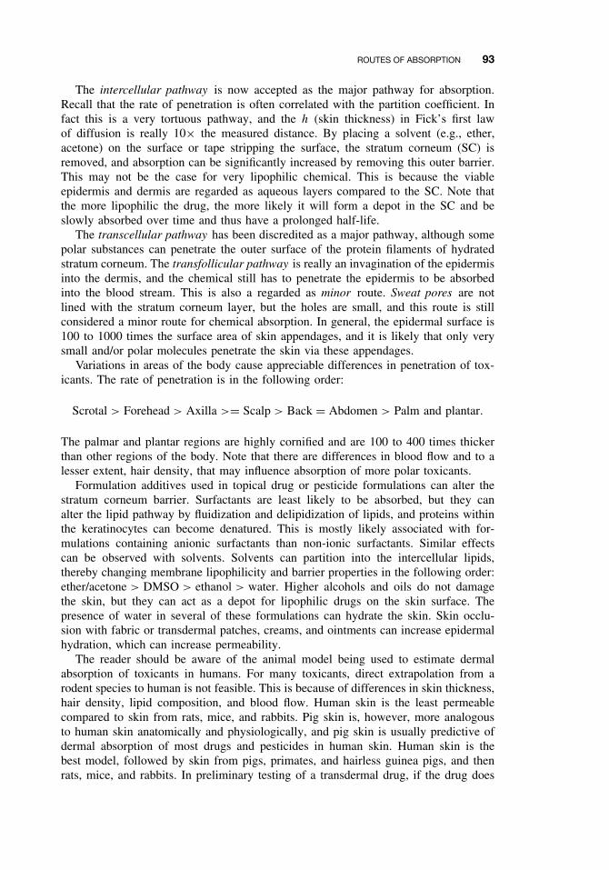
As observed with the GIT and skin, the respiratory tract can be regarded as an externalsurface. However, the lungs, where gas/vapor absorption occurs, are preceded by pro-tective structures (e.g., nose, mouth, pharynx, trachea, and bronchus), which can reducethe toxicity of airborne substances, especially particles. There is little or no absorptionin these structures, and residual volume can occur in these sites. However, cells liningthe respiratory tract may absorb agents that can cause a toxicological response. Theabsorption site, which is the alveoli-capillary membrane, is very thin (0.4–1.5 µm).The membranes to cross from the alveolar air space to the blood will include: type Icells to basement membrane to capillary endothelial cells (Figure 6.8). This short dis-tance allows for rapid exchange of gases/vapors. The analogous absorption distance inskin is 100 to 200 µm, and in GIT it is about 30 µm. There is also a large surfacearea (50 times the area of skin) available for absorption as well as significant bloodflow, which makes it possible to achieve rapid adjustments in plasma concentration.
Respiratory Bronchiole
Alveolar Duct
Alveolar Sac
Alveolus
Atrium
Atrium
Pore
Figure 6.8 Schematic representation of the respiratory unit of the lung. (From Bloom andFawcett, in A Textbook of Histology, Philadelphia: Saunders, 1975.)
ROUTES OF ABSORPTION 95
Gases/vapors must get into solution in the thin fluid film in the alveoli for systemicabsorption to occur. For this reason doses are often a measurement of partial pressures,which is important for gases/vapors.
The process of respiration involves the movement and exchange of air throughseveral interrelated passages, including the nose, mouth, pharynx, trachea, bronchi,and successively smaller airways terminating in the alveoli, where gaseous exchangeoccurs. These alveoli consist mainly of type I pneumocytes, which represent 40% ofall cells but cover > 90% of surface area, and type II pneumocytes, which represent60% of all cells but cover 5% of surface area. Macrophages make up 90% of cellsin alveolar space. The amount of air retained in the lung despite maximum expiratoryeffort is known as the residual volume. Thus toxicants in the respiratory air may notbe cleared immediately because of slow release from the residual volume. The rateof entry of vapor-phase toxicants is controlled by the alveolar ventilation rate, withthe toxicant being presented to the alveoli in an interrupted fashion approximately20 times/min.
Airborne toxicants can be simplified to two general types of compounds, namelygases and aerosols. Compounds such as gases, solvents, and vapors are subject to gaslaws and are carried easily to alveolar air. Much of our understanding of xenobioticbehavior is with anesthetics. Compounds such as aerosols, particulates, and fumes arenot subject to gas laws because they are in particulate form.
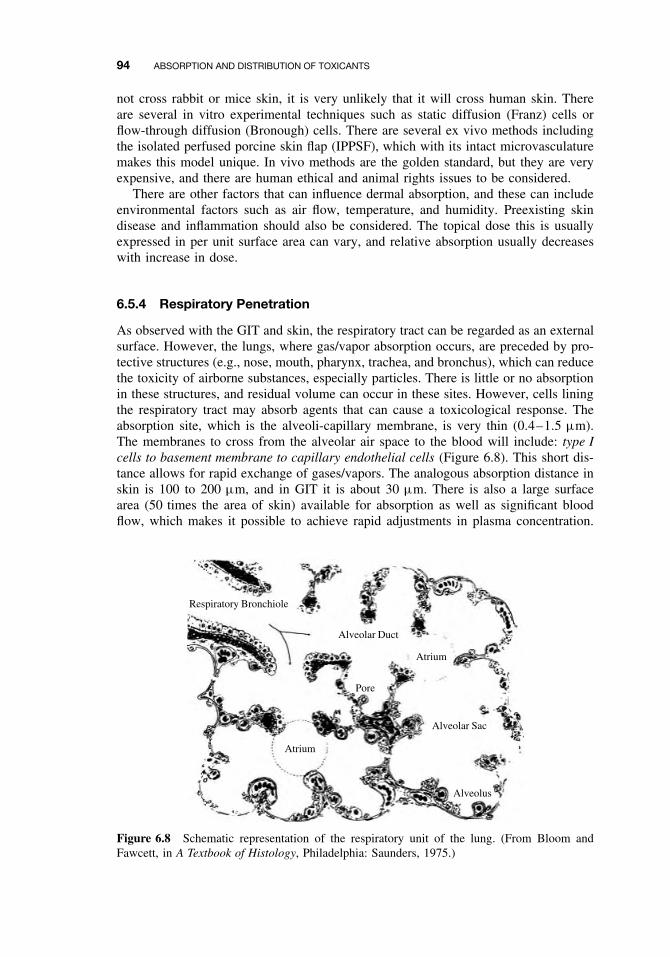
The transfer of gas from alveoli to blood is the actual absorption process. Amongthe most important factors that determine rate and extent of absorption of a gas inlungs is the solubility of that gas. Therefore it is not the membrane partition coefficientthat necessarily affects absorption as has been described for skin and GIT membranes,but rather the blood: gas partition coefficient or blood/gas solubility of the gas. A highblood: gas partition coefficient indicates that the blood can hold a large amount of gas.Keep in mind that it is the partial pressure at equilibrium that is important, so the moresoluble the gas is in blood, the greater the amount of gas that is needed to dissolve inthe blood to raise the partial pressure or tension in blood. For example, anesthetics suchas diethyl ether and methoxyflurane, which are soluble (Table 6.3), require a longerperiod for this partial pressure to be realized. Again, the aim is to generate the sametension in blood as in inspired air. Because these gases are very soluble, detoxificationis a prolonged process. In practice, anesthetic induction is slower, and so is recoveryfrom anesthesia. For less soluble gases (e.g., NO, isoflurane, halothane), the partialpressure or tension in blood can be raised a lot easier to that of inspired gases, anddetoxification takes less time than those gases that are more soluble.
There are several other important factors that can determine whether the gas willbe absorbed in blood and then transported from the blood to the perfused tissue. Theconcentration of the gas in inspired air influences gas tension, and partial pressurecan be increased by overventilation. In gas anesthesiology we know that the effects of
Table 6.3 Blood: Gas Partition Coefficient in Humans
respiratory rate on speed of induction are transient for gases that have low solubilityin blood and tissues, but there is a significant effect for agents that are more solubleand take a longer time for gas tensions to equilibrate. In determining how much of thegas is absorbed, its important to consider what fraction of the lung is ventilated andwhat fraction is perfused. However, one should be aware that due to diseased lungs,there can be differences between these fractions. For example, decreased perfusion willdecrease absorption, although there is agent in the alveoli, and vice versa. The rateat which a gas passes into tissues is also dependent on gas solubility in the tissues,rate of delivery of the gas to tissues, and partial pressures of gas in arterial blood andtissues. After uptake of the gas, the blood takes the gas to other tissues. The mixedvenous blood returned to the lungs progressively begins to have more of the gas, anddifferences between arterial (or alveolar) and mixed venous gas tensions decreasescontinuously.
While gases are more likely to travel freely through the entire respiratory tract tothe alveoli, passage of aerosols and particles will be affected by the upper respiratorytract, which can act as an effective filter to prevent particulate matter from reachingthe alveoli. Mucous traps particles to prevent entry to alveoli, and the mucociliaryapparatus in the trachea traps and pushes particles up the trachea to the esophaguswhere they are swallowed and possibly absorbed in the GI tract.
In addition to upper pathway clearance, lung phagocytosis is very active in bothupper and lower pathways of the respiratory tract and may be coupled to the mucuscilia. Phagocytes may also direct engulfed toxicants into the lymph, where the toxicantsmay be stored for long periods. If not phagocytized, particles ≤1 µm may penetrate tothe alveolar portion of the lung. Some particles do not desequamate but instead form adust node in association with a developing network of reticular fibers. Overall, removal
Nose
Mouth
Pharynx
5 – 30 µm
Trachea
Bronchi
Bronchioli
1 – 5 µm
Alveoli1 µm
Figure 6.9 Schematic illustration of the regions where absorption may occur in the respira-tory tract.
TOXICANT DISTRIBUTION 97
of alveolar particles is markedly slower than that achieved by the directed upper pul-monary mechanisms. This defense mechanism is not important for vapors/gases. Theefficiency of the system is illustrated by the fact that on average, only 100 g of coal dustis found postmortem in the lungs of coal miners, although they inhale approximately6000 g during their lifetime.
The deposition site of particles in the respiratory tract is primarily dependent on theaerodynamic behavior of the particles. The particle size, density, shape, hygroscopicity,breathing pattern, and lung airway structure are also important factors influencing thedeposition site and efficiency. The aerodynamic-equivalent diameter (for particle >
0.5 µm) and diffusion-equivalent diameter (< 0.5 µm) are defined as the diameter ofa unit density sphere having the same settling velocity (aerodynamic-equivalent) or thesame diffusion rate (diffusion-equivalent) as the irregularly shaped particle of interest.Deposition occurs by five possible mechanisms: electrostatic precipitation, interception,impaction, sedimentation, impaction, and diffusion. The latter three are most important.Only particle sizes less than 10 to 20 µm that get pass the nasopharyngeal regions andreach the alveoli are of medical concern. As particle size decreases below 0.5 µm, theaerosol begins to behave like a gas (Figure 6.9). For these particles, diffusion becomesthe primary mechanism of deposition in the respiratory tract before it finally reachesthe alveoli.
6.6 TOXICANT DISTRIBUTION
6.6.1 Physicochemical Properties and Protein Binding
Absorption of toxicants into the blood needs to be high enough so that it will havea significant effect at the site of action in other areas of the body. The distributionprocess that takes the absorbed drug to other tissues is dependent on various physio-logical factors and physicochemical properties of the drug. This process is therefore areversible movement of the toxicant between blood and tissues or between extracellularand intracellular compartments. There are, however, several complicating factors thatcan influence the distribution of a toxicant. For example, perfusion of tissues is animportant physiological process, as some organs are better perfused (e.g., heart, brain)than others (e.g., fat). There can also be significant protein binding that affects deliv-ery of drug to tissues. To further complicate the issue, elimination processes such asexcretion and biotransformation (discussed at a later time) is occurring simultaneouslyto remove the toxicant from the blood as well as the target site.
There are several physiochemical properties of the toxicant that can influence itsdistribution. These include lipid solubility, pKa, and molecular weight, all of whichwere described earlier in this chapter (Section 6.4) and will not be described here. Formany toxicants, distribution from the blood to tissues is by simple diffusion down aconcentration gradient, and the absorption principles described earlier also apply here.The concentration gradient will be influenced by the partition coefficient or ratherthe ratio of toxicant concentrations in blood and tissue. Tissue mass and blood flowwill also have a significant effect on distribution. For example, a large muscle masscan result in increased distribution to muscle, while limited blood flow to fat or bonetissue can limit distribution. The ratio of blood flow to tissue mass is also a usefulindicator of how well the tissue is perfused. The well perfused tissues include liver,
98 ABSORPTION AND DISTRIBUTION OF TOXICANTS
kidney, and brain, and the low perfused tissues include fat and bone where there isslow elimination from these tissues. Initial distribution to well-perfused tissues (e.g.,heart, brain) occurs within the first few minutes, while delivery of drug to other tissues(e.g., fat, skin) is slower.
If the affinity for the target tissue is high, then the chemical will accumulate orform a depot. The advantage here is that if this is a drug, there is no need to loadup the central compartment to get to the active site. However, if the reservoir for thedrug has a large capacity and fills rapidly, it so alters the distribution of the drug thatlarger quantities of the drug are required initially to provide a therapeutic effectiveconcentration at the target organ. If this is a toxicant, this may be an advantageousfeature as toxicant levels at the target site will be reduced. In general, lipid-insolubletoxicants stay mainly in the plasma and interstitial fluids, while lipid-soluble toxicantsreach all compartments, and may accumulate in fat. There are numerous examples ofcellular reservoirs for toxicants and drugs to distribute. Tetracycline antibiotics have ahigh affinity for calcium-rich tissues in the body. The bone can become a reservoir forthe slow release of chemicals such as lead, and effects may be chronic or there may beacute toxicity if the toxicant is suddenly released or mobilized from these depots. Theantimalaria drug quinacrine accumulates due to reversible intracellular binding, andthe concentration in the liver can be several thousand times that of plasma. Anotherantimalaria drug, chloroquine, has a high affinity for melanin, and this drug can betaken up by tissues such as the retina, which is rich in melanin granules, and can causeretinitis with a drug overdose. Lipophilic pesticides and toxicants (e.g., PCBs) andlipid soluble gases can be expected to accumulate in high concentration in fat tissue.
There are unique anatomical barriers that can limit distribution of toxicants. Aclassical example of such a unique barrier is the blood-brain barrier (BBB), whichcan limit the distribution of toxicants into the CNS and cerebrospinal fluid. There arethree main processes or structures that keep drug or toxicant concentrations low inthis region: (1) The BBB, which consist of capillary endothelial tight junctions andglial cells, surrounds the precapillaries, reduces filtration, and requires that the toxicantcross several membranes in order to get to the CSF. (Note that endothelial cells inother organs can have intercellular pores and pinocytotic vesicles.) (2) Active transportsystems in the choroid plexus allow for transport of organic acids and bases from theCSF into blood. (3) The continuous process of CSF production in the ventricles andvenous drainage continuously dilutes toxicant or drug concentrations. Disease processessuch as meningitis can disrupt this barrier and can allow for penetration of antibiotics(e.g., aminoglycosides) that would not otherwise readily cross this barrier in a healthyindividual. Other tissue/blood barriers include prostate/blood, testicles/blood, and globeof eye/blood, but inflammation or infection can increase permeability of these barriers.Toxicants can cross the placenta primarily by simple diffusion, and this is most easilyaccomplished if the toxicants are lipid-soluble (i.e., nonionized weak acids or bases).The view that the placenta is a barrier to drugs and toxicants is inaccurate. The fetusis, at least to some extent, exposed to essentially all drugs even if those with low lipidsolubility are taken by the mother.
As was indicated earlier, the circulatory system and components in the blood streamare primarily responsible for the transport of toxicants to target tissues or reservoirs.Erythrocytes and lymph can play important roles in the transport of toxicants, butcompared to plasma proteins, their role in toxicant distribution is relatively minor formost toxicants. Plasma protein binding can affect distribution because only the unbound
TOXICANT DISTRIBUTION 99
toxicant is free or available to diffuse across the cell membranes. The toxicant-proteinbinding reaction is reversible and obeys the laws of mass action:
Toxicant(free)
+ Protein k1↔k2
Toxicant-Protein(bound)
Usually the ratio of unbound plasma concentration (Cu) of the toxicant to total toxicantconcentration in plasma (C) is the fraction of drug unbound, fu , that is,
fu = Cu
C.
The constants k1 and k2 are the specific rate constants for association and dissociation,respectively. The association constant Ka will be the ratio k1/k2, and conversely, thedissociation constant, Kd will be k2/k1. The constants and parameters are often usedto describe and, more important, to compare the relative affinity of xenobiotics forplasma proteins.
The are many circulating proteins, but those involved in binding xenobiotics includealbumin, α1-acid glycoprotein, lipoproteins, and globulins. Because many toxicantsare lipophilic, they are likely to bind to plasma α- and β-lipoproteins. There aremainly three classes of lipoproteins, namely high-density lipoprotein (HDL), low-density lipoprotein (LDL), and very low density lipoprotein (VLDL). Iron and copperare known to interact strongly with the metal-binding globulins transferin and ceru-loplasmin, respectively. Acidic drugs bind primarily to albumin, and basic drugs arebound primarily to α1-acid glycoprotein and β-globulin. Albumin makes up 50% oftotal plasma proteins, and it reacts with a wide variety of drugs and toxicants. Theα1-acid glycoprotein does not have as many binding sites as albumin, but it has onehigh-affinity binding site. The amount of toxicant drug that is bound depends on freedrug concentration, and its affinity for the binding sites, and protein concentration.Plasma protein binding is nonselective, and therefore toxicants and drugs with similarphysicochemical characteristics can compete with each other and endogenous sub-stances for binding sites. Binding to these proteins does not necessarily prevent thetoxicant from reaching the site of action, but it slows the rate at which the toxicantreaches a concentration sufficient to produce a toxicological effect. Again, this is relatedto what fraction of the toxicant is free or unbound (fu).
Toxicants complex with proteins by various mechanisms. Covalent binding mayhave a pronounced effect on an organism due to the modification of an essentialmolecule, but such binding is usually a very minor portion of the total dose. Becausecovalently bound molecules dissociate very slowly, if at all, they are not consideredfurther in this discussion. However, we should recognize that these interactions areoften associated with carcinogenic metabolites. Noncovalent binding is of primaryimportance to distribution because the toxicant or ligand can dissociate more readilythan it can in covalent binding. In rare cases the noncovalent bond may be so stable thatthe toxicant remains bound for weeks or months, and for all practical purposes, the bondis equivalent to a covalent one. Types of interactions that lead to noncovalent bindingunder the proper physiological conditions include ionic binding, hydrogen bonding, vander Waals forces, and hydrophobic interactions. There are, however, some transitionmetals that have high association constants and dissociation is slow.
100 ABSORPTION AND DISTRIBUTION OF TOXICANTS
We know more about ligand-protein interactions today because of the numerousprotein binding studies performed with drugs. The major difference between drugsand most toxicants is the frequent ionizability and high water solubility of drugs ascompared with the non-ionizability and high lipid solubility of many toxicants. Thusexperience with drugs forms an important background, but one that may not alwaysbe relevant to other potentially toxic compounds.
Variation in chemical and physical features can affect binding to plasma constituents.Table 6.4 shows the results of binding studies with a group of insecticides with greatlydiffering water and lipid solubilities. The affinity for albumin and lipoproteins isinversely related to water solubility, although the relation may be imperfect. Chlo-rinated hydrocarbons bind strongly to albumin but even more strongly to lipoproteins.Strongly lipophilic organophosphates bind to both protein groups, whereas more water-soluble compounds bind primarily to albumin. The most water-soluble compoundsappear to be transported primarily in the aqueous phase. Chlordecone (Kepone) haspartitioning characteristics that cause it to bind in the liver, whereas DDE, the metabo-lite of DDT, partitions into fatty depots. Thus the toxicological implications for thesetwo compounds may be quite different.
Although highly specific (high-affinity, low-capacity) binding is more common withdrugs, examples of specific binding for toxicants seem less common. It seems probablethat low-affinity, high-capacity binding describes most cases of toxicant binding. Thenumber of binding sites can only be estimated, often with considerable error, becauseof the nonspecific nature of the interaction. The number of ligand or toxicant moleculesbound per protein molecule, and the maximum number of binding sites, n, define thedefinitive capacity of the protein. Another consideration is the binding affinity Kbinding
(or 1/Kdiss). If the protein has only one binding site for the toxicant, a single value,Kbinding, describes the strength of the interaction. Usually more than one binding site ispresent, each site having its intrinsic binding constant, k1, k2, . . . , kn. Rarely does onefind a case where k1 = k2 = . . . = kn, where a single value would describe the affinity
Table 6.4 Relative Distribution of Insecticides intoAlbumin and Lipoproteins
Source: Adapted from B. P. Maliwal and F. E. Guthrie, ChemBiol Interact 35:177–188, 1981.Note: LOL, low-density lipoprotein; HOL, high-density lipo-protein.
TOXICANT DISTRIBUTION 101
constant at all sites. This is especially true when hydrophobic binding and van derWaals forces contribute to nonspecific, low-affinity binding. Obviously the chemicalnature of the binding site is of critical importance in determining binding. The three-dimensional molecular structure of the binding site, the environment of the protein, thegeneral location in the overall protein molecule, and allosteric effects are all factorsthat influence binding. Studies with toxicants, and even more extensive studies withdrugs, have provided an adequate elucidation of these factors. Binding appears to betoo complex a phenomenon to be accurately described by any one set of equations.
There are many methods for analyzing binding, but equilibrium dialysis is the mostextensively used. Again, the focus of these studies is to determine the percentage oftoxicant bound, the number of binding sites (n), and the affinity constant (Ka). Theexamples presented here are greatly simplified to avoid the undue confusion engenderedby a very complex subject.
Toxicant-protein complexes that utilize relatively weak bonds (energies of the orderof hydrogen bonds or less) readily associate and dissociate at physiological tempera-tures, and the law of mass action applies to the thermodynamic equilibrium:
Kbinding = [T P ]
[T ][P ]= 1
Kdiss,
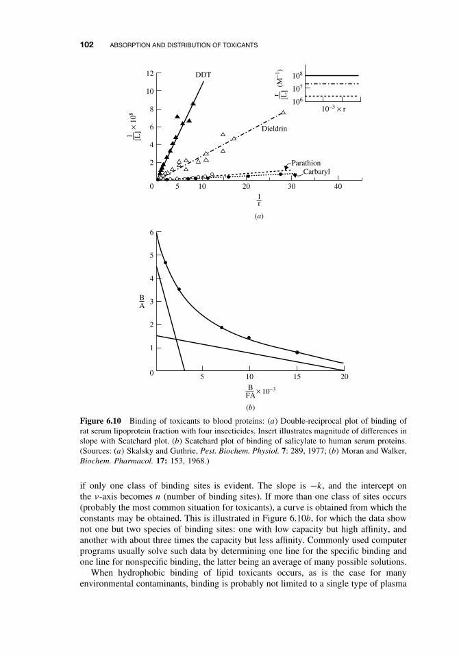
where Kbinding is the equilibrium constant for association, [TP] is the molar concen-tration of toxicant-protein complex, [T ] is the molar concentration of free toxicant,and [P ] is the molar concentration of free protein. This equation does not describe thebinding site(s) or the binding affinity. To incorporate these parameters and estimate theextent of binding, double-reciprocal plots of 1/[TP] versus 1/[T ] may be used to testthe specificity of binding. The 1/[TP] term can also be interpreted as moles of albuminper moles of toxicant. The slope of the straight line equals 1/nKa and the intercept ofthis line with the x-axis equals −Ka . Regression lines passing through the origin implyinfinite binding, and the validity of calculating an affinity constant under these circum-stances is questionable. Figure 6.10 illustrates one such case with four pesticides, andthe insert illustrates the low-affinity, “unsaturable” nature of binding in this example.
The two classes of toxicant-protein interactions encountered may be defined as(1) specific, high affinity, low capacity, and (2) nonspecific, low affinity, high capacity.The term high affinity implies an affinity constant (Kbinding) of the order of 108 M−1,whereas low affinity implies concentrations of 104 M−1. Nonspecific, low-affinity bind-ing is probably most characteristic of nonpolar compounds, although most cases arenot as extreme as that shown in Figure 6.10.
An alternative and well-accepted treatment for binding studies is the Scatchardequation especially in situations of high-affinity binding:
ν = nk[T ]
1 + k[T ],
which is simplified for graphic estimates to
ν
[T ]= k(n − ν),
where ν is the moles of ligand (toxicant) bound per mole of protein, [T ] is the con-centration of free toxicant, k is the intrinsic affinity constant, and n is the number ofsites exhibiting such affinity. When ν[T ] is plotted against ν, a straight line is obtained
102 ABSORPTION AND DISTRIBUTION OF TOXICANTS
12
10
8
6
4
2
0
DDT
Dieldrin
ParathionCarbaryl
403020105
10−3 × r
108
107
106
1r
BA
6
5
4
3
2
1
0 5 201510
1 [L]
× 10
8
r [L]
(M−1
)
10−3BFA
×
(a)
(b)
Figure 6.10 Binding of toxicants to blood proteins: (a) Double-reciprocal plot of binding ofrat serum lipoprotein fraction with four insecticides. Insert illustrates magnitude of differences inslope with Scatchard plot. (b) Scatchard plot of binding of salicylate to human serum proteins.(Sources: (a) Skalsky and Guthrie, Pest. Biochem. Physiol. 7: 289, 1977; (b) Moran and Walker,Biochem. Pharmacol. 17: 153, 1968.)
if only one class of binding sites is evident. The slope is −k, and the intercept onthe ν-axis becomes n (number of binding sites). If more than one class of sites occurs(probably the most common situation for toxicants), a curve is obtained from which theconstants may be obtained. This is illustrated in Figure 6.10b, for which the data shownot one but two species of binding sites: one with low capacity but high affinity, andanother with about three times the capacity but less affinity. Commonly used computerprograms usually solve such data by determining one line for the specific binding andone line for nonspecific binding, the latter being an average of many possible solutions.
When hydrophobic binding of lipid toxicants occurs, as is the case for manyenvironmental contaminants, binding is probably not limited to a single type of plasma
TOXICANT DISTRIBUTION 103
protein. For example, the binding of the chlorinated hydrocarbon DDT is strongest forlipoproteins and albumin, but other proteins account for a significant part of overalltransport. Similar results have been observed for several compounds with a range ofphysiochemical properties.
The presence of another toxicant and/or drug that can bind at the same site can alsoincrease the amount of free or unbound drug. This is an example of drug interactionthat can have serious toxicological or pharmacological consequences. In general, whenbound concentrations are less than 90% of the total plasma concentrations, plasmaprotein binding has little clinical importance. Plasma protein binding becomes importantwhen it is more than 90%. For example, if a toxicant is 99% bound to plasma proteins,then 1% is free, but if there is toxicant interaction (e.g., competitive binding) thatresults in 94% bound, 6% is now free. Note that because of this interaction, theamount of available toxicant to cause a toxicological response has increased sixfold.Such a scenario may result in severe acute toxicity. Extensive plasma protein bindingcan influence renal clearance if glomerular filtration is the major elimination processin the kidney, but not if it is by active secretion in the kidney. Binding can also affectdrug clearance if the extraction ratio (ER) in the liver is low, but not if the ER ishigh for that toxicant. Plasma protein binding can vary between and within chemicalclasses, and it is also species specific. For example, humans tend to bind acidic drugsmore extensively than do other species.
There are several other variables that can alter plasma protein concentrations. Theseinclude malnutrition, pregnancy, cancer, liver abscess, renal disease, and age canreduce serum albumin. Furthermore α1-glycoprotein concentrations can increase withage, inflammation, infections, obesity, renal failure, and stress. Small changes in bodytemperature or changes in acid-base balance may alter chemical protein-binding charac-teristics. Although termination of drug or toxicant effect is usually by biotransformationand excretion, it may also be associated with redistribution from its site of actioninto other tissues. The classical example of this is when highly lipid-soluble drugsor toxicants that act on the brain or cardiovascular system are administered by IV orby inhalation.
6.6.2 Volume of Distribution (Vd)
Usually after a toxicant or drug is absorbed it can be distributed into various physiologicfluid compartments. The total body water represents 57% of total body mass (0.57 L/kg)(Table 6.5). The plasma, interstitial fluid, extracellular fluid, and intracellular fluidrepresent about 5, 17, 22, and 35% body weight, respectively. The extracellular fluidcomprises the blood plasma, interstitial fluid, and lymph. Intracellular fluid includes
Table 6.5 Volume of Distribution into Physiological Fluid Compartments
Compartment Volume of Distribution in L/kg Body Weight (Ls/70 kg Body weight)
Plasma 0.05(3.5 L)Interstitial fluid 0.18(12.6 L)Extracellular fluid 0.23(16.1 L)Intracellular fluid 0.35(24.5 L)Total body water 0.55(39 L)
104 ABSORPTION AND DISTRIBUTION OF TOXICANTS
the sum of fluid contents of all cells in the body. There is also transcellular fluid thatrepresents 2% body weight, and this includes cerebrospinal, intraocular, peritoneal,pleural, and synovial fluids, and digestive secretions. Fat is about 20% body weight,while the GIT contents in monogastrics make up 1% body weight, and in ruminants itcan constitute 15% body weight.
Its sometimes useful to quantitate how well a drug or toxicant is distributed into thesevarious fluid compartments, and in this context the apparent volume of distributioncan be a useful parameter. The apparent volume of distribution, Vd , is defined as thevolume of fluid required to contain the total amount, A, of drug in the body at thesame concentration as that present in plasma, Cp,
Vd = A
Cp
.
In general, the Vd for a drug is to some extent descriptive of its distribution pat-tern in the body. For example, drugs or toxicants with relatively small Vd values maybe confined to the plasma as diffusion across the capillary wall is limited. There areother toxicants that have a slightly larger Vd (e.g., 0.23 L/kg), and these toxicantsmay be distributed in the extracellular compartment. This includes many polar com-pounds (e.g., tubocurarine, gentamicin, Vd = 0.2–0.4 L/kg). These toxicants cannotreadily enter cells because of their low lipid solubility. If the Vd for some of thesetoxicants is in excess of the theoretical value, this may be due to limited degree ofpenetration into cells or from the extravascular compartment. Finally there are manytoxicants distributed throughout the body water (Vd ≥ 0.55 L/kg) that may have Vd
values much greater than that for total body water. This distribution is achieved byrelatively lipid-soluble toxicants and drugs that readily cross cell membranes (e.g.,ethanol, diazepam; Vd = 1 to 2 L/kg). Binding of the toxicant anywhere outside of theplasma compartment, as well as partitioning into body fat, can increase Vd beyond theabsolute value for total body water. In general, toxicants with a large Vd can even reachthe brain, fetus, and other transcellular compartments. In general, toxicants with largeVd are a consequence of extensive tissue binding. The reader should be aware that weare talking about tissue binding, and not plasma protein binding where distribution islimited to plasma for obvious reasons.
The fraction of toxicant located in plasma is dependent on whether a toxicant bindsto both plasma and tissue components. Plasma binding can be measured directly, butnot tissue binding. It can, however, be inferred from the following relationship:
Amount in body = Amount in plasma + Amount outside plasma
Vd × C = Vp × C + VT W × CT W
where Vd is the apparent volume of distribution, Vp the volume of plasma, VT W theapparent volume of tissue, and CT W the tissue concentration. If the preceding equationis divided by C, it now becomes
Vd = Vp + VT W × CT W
C
Recall that fu = Cu/C occurs with plasma, and also that the fraction unbound intissues is fuT = CuT /CT W .
TOXICOKINETICS 105
Assuming at equilibrium that unbound concentration in tissue and plasma are equal,then we let the ratio of fu/fuT replace CT W/C and determine the volume of distributionas follows:
Vd = Vp + VT W ×(
fu
fuT
).
It is possible to predict what happens to Vd when fu or fuT changes as a resultof physiological or disease processes in the body that change plasma and/or tissueprotein concentrations. For example, Vd can increase with increased unbound toxicantin plasma or with a decrease in unbound toxicant tissue concentrations. The precedingequation explains why: because of both plasma and tissue binding, some Vd valuesrarely correspond to a real volume such as plasma volume, extracellular space, or totalbody water. Finally interspecies differences in Vd values can be due to differences inbody composition of body fat and protein, organ size, and blood flow as alluded toearlier in this section. The reader should also be aware that in addition to Vd , thereare volumes of distribution that can be obtained from pharmacokinetic analysis of agiven data set. These include the volume of distribution at steady state (Vd,ss), volumeof the central compartment (Vc), and the volume of distribution that is operative overthe elimination phase (Vd,area). The reader is advised to consult other relevant textsfor a more detailed description of these parameters and when it is appropriate to usethese parameters.
6.7 TOXICOKINETICS
The explanation of the pharmacokinetics or toxicokinetics involved in absorption, dis-tribution, and elimination processes is a highly specialized branch of toxicology, andis beyond the scope of this chapter. However, here we introduce a few basic conceptsthat are related to the several transport rate processes that we described earlier in thischapter. Toxicokinetics is an extension of pharmacokinetics in that these studies areconducted at higher doses than pharmacokinetic studies and the principles of pharma-cokinetics are applied to xenobiotics. In addition these studies are essential to provideinformation on the fate of the xenobiotic following exposure by a define route. Thisinformation is essential if one is to adequately interpret the dose-response relationshipin the risk assessment process. In recent years these toxicokinetic data from laboratoryanimals have started to be utilized in physiologically based pharmacokinetic (PBPK)models to help extrapolations to low-dose exposures in humans. The ultimate aim inall of these analyses is to provide an estimate of tissue concentrations at the target siteassociated with the toxicity.
Immediately on entering the body, a chemical begins changing location, concentra-tion, or chemical identity. It may be transported independently by several componentsof the circulatory system, absorbed by various tissues, or stored; the chemical mayeffect an action, be detoxified, or be activated; the parent compound or its metabo-lite(s) may react with body constituents, be stored, or be eliminated—to name someof the more important actions. Each of these processes may be described by rate con-stants similar to those described earlier in our discussion of first-order rate processesthat are associated with toxicant absorption, distribution, and elimination and occur
106 ABSORPTION AND DISTRIBUTION OF TOXICANTS
Metabolism toMore ToxicMetabolites
Metabolism toLess ToxicMetabolites
Metabolism toConjugation
Products
Interaction with Macromolecules(Proteins, DNA, RNA, Receptors,etc)
Figure 6.11 Sequence of events following exposure of an animal to exogenous chemicals.
simultaneously. Thus at no time is the situation stable but is constantly changing asindicated in Figure 6.11.
It should be noted, however, that as the toxicant is being absorbed and distributedthroughout the body, it is being simultaneously eliminated by various metabolismand/or excretion mechanisms, as will be discussed in more detail in the followingchapters. However, one should mention here that an important pharmacokinetic param-eter known as clearance (C�) can be used to quantitatively assess elimination of atoxicant. Clearance is defined as the rate of toxicant excreted relative to its plasmaconcentration, Cp:
C� = Rate of toxicant excretion
Cp
.
The rate of excretion is really the administered dose times the fractional eliminationrate constant Kel described earlier. Therefore we can express the preceding equationin terms of Kel and administered dose as volume of distribution, Vd :
C� = Kel · Dose
Cp
= Kel · (Vd · Cp)/Cp = Kel · Vd.
In physiological terms we can also define clearance as the volume of blood clearedof the toxicant by an organ or body per unit time. Therefore, as the equations aboveindicate, the body clearance of a toxicant is expressed in units of volume per unit time(e.g., L/h), and can be derived if we know the volume of distribution of the toxicant
TOXICOKINETICS 107
and fractional rate constant. In many instances this can only be derived by appropriatepharmacokinetic analysis of a given data set following blood or urine sample collectionand appropriate chemical analyses to determine toxicant concentrations in either ofthese biological matrices.
Each of the processes discussed thus far—absorption, distribution, and elimina-tion—can be described as a rate process. In general, the process is assumed to be firstorder in that the rate of transfer at any time is proportional to the amount of drug in thebody at that time. Recall that the rate of transport (dC/dt) is proportional to toxicantconcentration (C) or stated mathematically:
dC
dt= KC,
where K is the rate constant (fraction per unit time). Many pharmacokinetic analysesof a chemical are based primarily on toxicant concentrations in blood or urine samples.It is often assumed in these analyses that the rate of change of toxicant concentrationin blood reflects quantitatively the change in toxicant concentration throughout thebody (first-order principles). Because of the elimination/clearance process, which alsoassumed to be a first-order rate process, the preceding rate equation now needs anegative sign. This is really a decaying process that is observed as a decline of toxicantconcentration in blood or urine after intravenous (IV) administration. The IV routeis preferred in these initial analyses because there is no absorption phase, but onlychemical depletion phase. However, one cannot measure infinitesimal change of C
or time, t ; therefore there needs to be integration after rearrangement of the equationabove: −dC
C= kdt becomes
∫ −dC
C= k
∫dt,
which can be expressed asC = C0e−kt ,
where e is the base of the natural logarithm. We can remove e by taking the ln ofboth sides:
ln Ct = ln C0 − kt.
Note that K is the slope of the straight line for a semilog plot of toxicant concentrationversus time (Figure 6.12). In the preceding equation it is the elimination rate constantthat is related to the half-life of the toxicant described earlier in this chapter. Thederived C0 can be used to calculate the volume of distribution (Vd) of the toxicantas follows:
Vd = Dose
C0.
However, toxicokinetic data for many toxicants do not always provide a straightline when plotted as described above. More complicated equations with more than oneexponential term with rate constants may be necessary to mathematically describe theconcentration-time profile. These numerous rate constants are indicative of chemicaltransport between various compartments in the body and not only to a single cen-tral compartment as suggested in the simple equation and semilog plot described in
108 ABSORPTION AND DISTRIBUTION OF TOXICANTS
Ka
Kel
(a) (b)
Central
Time
lnC
p
Slope = −Kel
Cp0
↓
Figure 6.12 (a) Semilog plot of plasma concentration (Cp) versus time. Cp0 is the intercept
on the y-axis, and Kel is the elimination rate constant. (b) Single compartment model with rateconstants for absorption, Ka and for elimination, Kel.
(a) (b)
Central(1)
Peripheral(2)
Time
lnC
p
Cp0
K12
K21
Kel = K10
b = l2A2 = B
A1 = A
a = l1
Figure 6.13 (a) Semilog plot of plasma concentration for (Cp) versus time representative ofa two-compartment model. The curve can be broken down into an α or λ1 distribution phaseand β or λ2 elimination phase. (b) Two-compartment model with transfer rate constants, K12
and K21, and elimination rate constant, Kel.
Figure 6.12. In some instances the data may fit to a bi-exponential concentration-timeprofile (Figure 6.13). The equation to describe this model is
C = Ae−αt + Be−βt .
In other instances, complex profiles may require a three- or multi-exponentialconcentration-time profile (Figure 6.14). The equation to describe the three-profilecase is
C = Ae−αt + Be−βt + Ce−γ t .
In the physiological sense, one can divide the body into “compartments” thatrepresent discrete parts of the whole-blood, liver, urine, and so on, or use a math-ematical model describing the process as a composite that pools together parts oftissues involved in distribution and bioactivation. Usually pharmacokinetic compart-ments have no anatomical or physiological identity; they represent all locations withinthe body that have similar characteristics relative to the transport rates of the par-ticular toxicant. Simple first-order kinetics is usually accepted to describe individual
SUGGESTED READING 109
(a) (b)
Time (hrs)
K12
K21
K31
K13
Kel = K10
l1
l2
l1l2
l3
0
0 5 10
10
100
20 40 60 80
100
10
1
0.1
0.01
Con
cent
ratio
n (µ
g/m
l)
2 1 3
Figure 6.14 (a) Semilog plot of plasma concentration for (Cp) versus time representativeof a three- or multi-compartment model. The curve can be broken down into three phases,λ1, λ2, and λ3. (b) Three-compartment model with transfer rate constants, K12,K21,K13, K31,and elimination rate constant, Kel. As these models can get more complicated, the α, β, and γ
nomenclature may get replaced with λn as indicated in the profile.
rate processes for the toxicant after entry. The resolution of the model necessitatesmathematical estimates (as a function of time) concerning the absorption, distribution,biotransformation, and excretion of the toxicant.
Drugs and toxicants with multi-exponential behavior depicted in Figure 6.14 requirecalculation of the various micro constants. An alternative method involves using model-independent pharmacokinetics to arrive at relevant parameters. Very briefly, it involvesdetermination of the area under the curve (AUC) of the concentration-time profiles.The emergence of microcomputers in recent years has greatly facilitated this approach.
In conclusion, pharmacokinetics is a study of the time course of absorption, distri-bution, and elimination of a chemical. We use pharmacokinetics as a tool to analyzeplasma concentration time profiles after chemical exposure, and it is the derived ratesand other parameters that reflect the underlying physiological processes that determinethe fate of the chemical. There are numerous software packages available today toaccomplish these analyses. The user should, however, be aware of the experimen-tal conditions, the time frame over which the data were collected, and many of theassumptions embedded in the analyses. For example, many of the transport processesdescribed in this chapter may not obey first-order kinetics, and thus may be nonlinearespecially at toxicological doses. The reader is advised to consult other texts for moredetailed descriptions of these nonlinear interactions and data analyses.
SUGGESTED READING
R. Bronaugh and H. Maibach, eds. Percutaneous Absorption. New York: Dekker, 1989.
A. Goodman Gilman, T. W. Rall, A. S. Nies, and P. Taylor, eds. Goodman and Gilman’s ThePharmacological Basis of Therapeutics, 8th edn. Elmsford, NY: Pergamon Press, 1990.
P. Grandjean, ed. Skin Penetration: Hazardous Chemicals at Work. London: Taylor and Francis,1990.
110 ABSORPTION AND DISTRIBUTION OF TOXICANTS
R. Krieger, ed. Handbook of Pesticide Toxicology, 2nd edn. San Diego: Academic Press, 2001.
M. Rowland and T. N. Tozer, eds. Clinical Pharmacokinetics. Concepts and Applications, 3rdedn. Philadelphia: Lea and Febiger, 1995.
L. Shargel and A. B. C. Yu, eds. Applied Biopharmaceutics and Pharmacokinetics, 4th edn.Norwalk, CT: Appleton and Lange, 1999.