Case Report Acute bacterial endophthalmitis after intravitreal bevacizumab injection: Case report and literature review Saba Al Rashaed, MD ⇑ ; Abdulaziz Rushood, MD Abstract A case report of a 52 year old male who received intravitreal bevacizumab and developed culture positive endophthalmitis. Vit- reous culture indicated that endophthalmitis was caused by Staphylococcus epidermidis. The patient was initially managed with intravitreal injection of ceftazidime and vancomycin, followed by pars plana lensectomy, pars plana vitrectomy with intravitreal injection of 1 mg/0.1 ml vancomycin, 2.25 mg/0.1 ml ceftazidime, 5 mg/0.1 ml fortified amphotericin-B and 4 mg/0.1 ml dexa- methasone. Postoperatively the patient improved significantly. However, vision improved from hand motion to counting fingers secondary to severe retinal ischemia. Acute endophthalmitis can develop after intravitreal bevacizumab injections and cause pro- found visual loss. A review of literature was also performed for similar cases. Keywords: Endophthalmitis, Bevacizumab, Staphylococcus, Pars plana vitrectomy, Visual loss, Infection Ó 2012 Saudi Ophthalmological Society, King Saud University. All rights reserved. http://dx.doi.org/10.1016/j.sjopt.2012.04.002 Introduction The introduction of vascular endothelial growth factor antagonists has lead to a dramatic increase in intravitreal injections worldwide. Currently the most common intravitreal injections are performed with triamcinolone acetonide, bev- acizumab (Avastin Ò ; Genentech Inc., San Francisco, CA, USA) and ranibizumab (Lucentis Ò ; Genentech Inc., San Fran- cisco, CA, USA) for a variety of conditions including exudative age related macular degeneration (AMD) and macular ede- ma of various etiologies. 1–5 Complications after intravitreal injection include infectious and sterile endophthalmitis, rhegmatogenous retinal detach- ment, a transient increase in intraocular pressure and iatro- genic injury to the eye. 1–5 Infectious endophthalmitis is a rare complication of intravitreal injection, but is relevant due to the increased frequency and number of injections being performed. Clinically, endophthalmitis can cause pro- found loss of vision, which may be permanent despite prompt and appropriate management. In this case report we present a case of acute endophthal- mitis after intravitreal injection of bevacizumab for prolifera- tive diabetic retinopathy with macular edema that resulted in with poor vision despite prompt management. We also performed a literature review of similar cases of endophthalmitis. Case report A 52 years old insulin dependent male diabetic was pre- sented to the emergency room with loss of vision, pain, and redness in his left eye. Three days prior to presentation, the patient received an intravitreal injection of bevacizumab elsewhere for diabetic macular edema. Ophthalmic examination revealed visual acuity was 20/80 in the right eye and hand motion in the left eye. There were no significant Peer review under responsibility of Saudi Ophthalmological Society, King Saud University Production and hosting by Elsevier Access this article online: www.saudiophthaljournal.com www.sciencedirect.com Received 5 March 2012; received in revised form 26 March 2012; accepted 16 April 2012; available online 28 April 2012. From Vitreoretinal Division, King Khaled Eye Specialist Hospital, Riyadh, Saudi Arabia q The authors have no proprietary interests in the materials presents in this paper. ⇑ Corresponding author. Address: King Khaled Eye Specialist Hospital, P.O. Box 7191, Riyadh 11462, Saudi Arabia. Tel./fax: +966 1 482 1234x1908. e-mail address: [email protected](S.A. Rashaed). Saudi Journal of Ophthalmology (2013) 27, 55–57
Transcript
Saudi Journal of Ophthalmology (2013) 27, 55–57
Case Report
Acute bacterial endophthalmitis after intravitreal bevacizumabinjection: Case report and literature review
Saba Al Rashaed, MD ⇑; Abdulaziz Rushood, MD
Abstract
A case report of a 52 year old male who received intravitreal bevacizumab and developed culture positive endophthalmitis. Vit-reous culture indicated that endophthalmitis was caused by Staphylococcus epidermidis. The patient was initially managed withintravitreal injection of ceftazidime and vancomycin, followed by pars plana lensectomy, pars plana vitrectomy with intravitrealinjection of 1 mg/0.1 ml vancomycin, 2.25 mg/0.1 ml ceftazidime, 5 mg/0.1 ml fortified amphotericin-B and 4 mg/0.1 ml dexa-methasone. Postoperatively the patient improved significantly. However, vision improved from hand motion to counting fingerssecondary to severe retinal ischemia. Acute endophthalmitis can develop after intravitreal bevacizumab injections and cause pro-found visual loss. A review of literature was also performed for similar cases.
Keywords: Endophthalmitis, Bevacizumab, Staphylococcus, Pars plana vitrectomy, Visual loss, Infection
� 2012 Saudi Ophthalmological Society, King Saud University. All rights reserved.http://dx.doi.org/10.1016/j.sjopt.2012.04.002
Introduction
The introduction of vascular endothelial growth factorantagonists has lead to a dramatic increase in intravitrealinjections worldwide. Currently the most common intravitrealinjections are performed with triamcinolone acetonide, bev-acizumab (Avastin�; Genentech Inc., San Francisco, CA,USA) and ranibizumab (Lucentis�; Genentech Inc., San Fran-cisco, CA, USA) for a variety of conditions including exudativeage related macular degeneration (AMD) and macular ede-ma of various etiologies.1–5
Complications after intravitreal injection include infectiousand sterile endophthalmitis, rhegmatogenous retinal detach-ment, a transient increase in intraocular pressure and iatro-genic injury to the eye.1–5 Infectious endophthalmitis is arare complication of intravitreal injection, but is relevantdue to the increased frequency and number of injectionsbeing performed. Clinically, endophthalmitis can cause pro-
Peer review under responsibilityof Saudi Ophthalmological Society,King Saud University
Received 5 March 2012; received in revised form 26 March 2012; accepted 16
From Vitreoretinal Division, King Khaled Eye Specialist Hospital, Riyadh, Saudq The authors have no proprietary interests in the materials presents in this paper.
found loss of vision, which may be permanent despiteprompt and appropriate management.
In this case report we present a case of acute endophthal-mitis after intravitreal injection of bevacizumab for prolifera-tive diabetic retinopathy with macular edema that resultedin with poor vision despite prompt management. We alsoperformed a literature review of similar cases ofendophthalmitis.
Case report
A 52 years old insulin dependent male diabetic was pre-sented to the emergency room with loss of vision, pain,and redness in his left eye. Three days prior to presentation,the patient received an intravitreal injection of bevacizumabelsewhere for diabetic macular edema. Ophthalmicexamination revealed visual acuity was 20/80 in the righteye and hand motion in the left eye. There were no significant
Production and hosting by Elsevier
Access this article online:www.saudiophthaljournal.comwww.sciencedirect.com
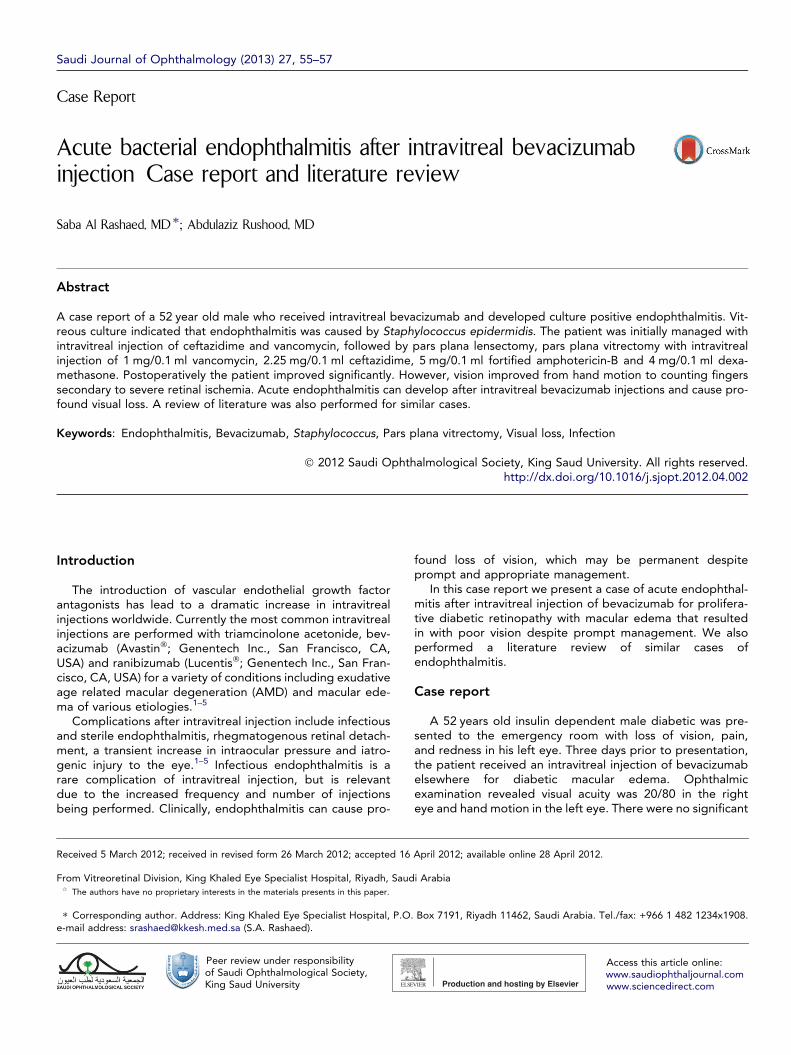
Figure 1. Progress of an eye that underwent intravitreal bevacizumab injection and subsequently developed culture positive endophthalmitis. (A) Atpresentation, 48 h after intravitreal injection showed no improvement with ciliary vessel injection, hypopyon and absence of a red reflex. (B) Three daysafter the surgery. Note the disorganized hypopyon with corneal edema. (C) Two weeks after surgery showed a quiet eye and remnants of a disorganizedhypopyon. (D) Two months follow up showed central corneal haze with a quiet eye and resolution of the hypopyon.
56 S.A. Rashaed, A. Rushood
findings in the right eye other than severe non-proliferativediabetic retinopathy. The left eye had marked lid edema withciliary injection, corneal edema with a 3 ml hypopyon, poster-ior synechia and a white fundus reflex.
B-scan showed marked vitreal and subvitreal opacitieswith diffuse retino-choroidal layers. The patient was diag-nosed with acute endophthalmitis after intravitreal bev-acizumab. An urgent vitreous tap was performed alongwith intravitreal injection of vancomycin and ceftazidime.The patient was placed on oral moxifloxacin once a dayand instructed to instill topical fortified cephalexin and ceft-azidime drops hourly. Two days later, there was no clinicalimprovement. The patient underwent synechiolysis, parsplana lensectomy, pars plana vitrectomy with intravitrealinjection of 1 mg/0.1 ml vancomycin, 2.25 mg/0.1 ml ceftazi-dime, 5 mg/0.1 ml amphotericin-B and 4 mg/0.1 ml dexa-methasone. Postoperatively there was significant clinicalimprovement. However, the vision improved from hand mo-tion to counting by fingers secondary to severe retinal ische-mia caused by the disease process. Fig. 1 presents theprogress of the eye from presentation onward. A culturefrom the vitreous sample was positive for Staphylococcusepidermis.
Discussion
Bevacizumab (Avastin�) is a recombinant humanized fulllength monoclonal antibody which binds to all biologicallyactive isoforms of a key mediator of angiogenesis – vascularendothelial growth factor A (VEGF A). Bevacizumab has beenused off label to treat neovascular age related maculardegeneration (AMD) since 2005.3,6–9 Several groups have re-ported the use of intravitreal bevacizumab for a number ofposterior segment conditions including central retinal veinocclusion in proliferative diabetic retinopathy,10–19 pseud-
The incidence of (sterile or infectious) endophthalmitis re-ported in most studies remains rare. For example, Funget al.28 analyzed 7113 cases of intravitreal bevacizumabworldwide and found 1 case (0.01%) of endophthalmitis.However Fung et al. did not differentiate between sterileand infectious endophthalmitis. We believe the outcomesfrom Fung et al’s28 and similar studies may underestimate ad-verse events due to survey design, specifically the self report-ing of complications.
The development of infectious endophthalmitis after anintravitreal bevacizumab injection has been previously re-ported. Fintak et al.1 reported an incidence of 0.02% forinfectious endophthalmitis following 12,585 cases of intravi-treal bevacizumab. Two of the cases were caused by Strepto-coccus viridans and one by S. epidermidis.1 All cases in Fintaket al’s1 study ended with hand motion vision.1 The incidenceof endophthalmitis following intravitreal bevacizumab injec-tion in a large case series ranges from 0.019% to0.099%.1,29–31 The organism isolated in one study was Serra-tia marcescens in two cases with a final visual outcome of nolight perception.29 However in30,31 two separate series,reporting on cases of Haemophilus influenzae, or S. epide-rmidis (2 cases) the patients had a final visual acuity of 20/400 vision. Our case resulted in a final visual outcome of 4/200.
In a report32 of 5 cases of severe acute intraocular inflam-mation from 1278 cases that received intravitreal bev-acizumab, 2 cases of Propionibacterium acnes, 1 case ofcoagulase-negative Staphylococcus and 2 cases with nomicrobial growth were detected from the vitreous aspirate.In our case, Staphylococcus epidermidis was identified.
The risk of infectious endophthalmitis is always a concernwith any intraocular procedure. The most common sources
Acute bacterial endophthalmitis after intravitreal bevacizumab 57
of bacteria are from patient’s flora originating from the lids,lashes, and conjunctiva.33 Hence, thorough, standardizedcleansing techniques should help mitigate the incidence ofinfectious endophthalmitis. Precautions against infectiontend to vary between studies and practitioners. The onlystandard prophylactic measure is the use of preoperativepovidone–iodine drops and the use of a lid speculum. Preop-erative povidone–iodine is recommended due to a previousreport of a significant reduction in endophthalmitis after pre-operative use of povidone–iodine for intraocular surgery.34,35
The importance of a lid speculum, was determined by theVISION36 trial that found two thirds of the 12 cases ofendophthalmitis were associated with protocol violations,the most common being the lack of a lid speculum. The lidspeculum retracts the lid margin and lashes away from theinjection site, the general consensus is that lid speculumuse is an important step in reducing the risk of endophthalmi-tis. Other prophylactic measures include instillation of topicalantibiotic postoperatively, wearing sterile gloves, and wear-ing a mask during the procedure. In conclusion, although pre-vious studies report a low incidence rate of endophthalmitisafter intravitreal injection of bevacizumab, it is still a concerndue to the potential for profound visual loss as our and pre-vious reported cases have demonstrated. Informed consentshould incorporate the possibility of profound visual lossafter the procedure.
References
1. Fintak DR, Shah GK, Blinder KJ, Regillo CD, Pollack J, Heier JS, et al..Incidence of endophthalmitis related to intravitreal injection ofbevacizumab and ranibizumab. Retina 2008;28:1395–9.
2. Stepien KE, Eaton AM, Jaffe GJ, Davis JL, Raja J, Feuer W. Increasedincidence of sterile endophthalmitis after intravitreal triamcinoloneacetonide in spring 2006. Retina 2009;29:207–13.
3. Rosenfeld PJ, Moshfeghi AA, Puliafito CA. Optical coherenceTomography findings after an intravitreal injection of bevacizumab(Avastin) for neovascular age-related macular degeneration.Ophthalmic Surg Lasers Imaging 2005;36:331–5.
4. Greenberg PB, Martidis A, Rogers AH, Duker JS, Reichel E.Intravitreal triamcinolone for the treatment of macular oedema dueto central retinal vein occlusion. Br J Ophthalmol 2002;86:247–8.
5. Heier JS, Antoszyk AN, Pavan PR, Leff SR, Rosenfeld PJ, Ciulla TA,et al.. Ranibizumab for treatment of neovascular age-related maculardegeneration: a phase I/II multicenter, controlled, multidose study.Ophthalmology 2006;113:633–42.
7. Spaide RF, Laud K, Fine HF, Klancnik Jr JM, Meyerle CB, et al..Intravitreal bevacizumab treatment of choroidal neovascularizationsecondary to age-related macular degeneration. Retina2006;26:383–90.
8. Rich R, Rosenfeld PJ, Puliafito CA, Dubovy SR, Davis JL, Flynn Jr HW,et al.. Short-term safety and efficacy of intravitreal bevacizumab(Avastin) for neovascular age-related macular degeneration. Retina2006;26:495–511.
9. Emerson MV, Lauer AK, Flaxel CJ, Wilson DJ, Francis PJ, Stout JT,et al.. Intravitreal bevacizumab (Avastin) treatment of neovascularage-related macular degeneration. Retina 2007;27:439–44.
10. Costa RA, Jorge R, Calucci DC, Melo Jr LA, Cardillo JA, Scott IU.Intravitreal bevacizumab (Avastin) for central and hemicentral retinalvein occlusions: IBEVO Study. Retina 2007;27:141–9.
12. Rosenfeld PJ, Fung AE, Puliafito CA. Optical coherence tomographyfindings after an intravitreal injection of bevacizumab (Avastin) for
macular edema from central retinal vein occlusion. Ophthalmic SurgLasers Imaging 2005;36:336–9.
13. Gregori NZ, Rosenfeld PJ, Puliafito CA, et al.. One-year safety andefficacy of intravitreal triamcinolone acetonide for the managementof macular edema secondary to central retinal vein occlusion. Retina2006;26:889–95.
15. Jorge R, Costa R, Calluci D, Cintra LP, Scott IU. Intravitrealbevacizumab (Avastin) for persistent new vessels in diabeticretinopathy (IBEPE Study). Retina 2006;26:1006–13.
16. Isaacs T, Barry C. Rapid resolution of severe disc new vessels inproliferative diabetic retinopathy following a single intravitrealinjection of bevacizumab (Avastin). Clin Exp Ophthalmol2006;34:802–3.
18. Chen E, Park CH. Use of intravitreal bevacizumab as a preoperativeadjunct for tractional retinal detachment repair in severe proliferativediabetic retinopathy. Retina 2006;26:699–700.
19. Avery RL, Pearlman J, Pieramici DJ, Rabena MD, Castellarin AA, NasirMA, et al.. Intravitreal bevacizumab (Avastin) in the treatment ofproliferative diabetic retinopathy. Ophthalmology2006;113:1695–705.
20. Mason 3rd JO, Albert MA, Vail R. Intravitreal bevacizumab (Avastin)for refractory pseudophakic cystoid macular edema. Retina2006;26:356–7.
21. Mason 3rd JO, Albert MA, Mays A, Vail R. Regression of neovasculariris vessels by intravitreal injection of bevacizumab. Retina2006;26:839–41.
22. Davidorf FH, Mouser JG, Derick RJ. Rapid improvement of rubeosisiridis from a single bevacizumab (Avastin) injection. Retina2006;26:354–6.
23. Avery RL. Regression of retinal and iris neovascularization afterintravitreal bevacizumab (Avastin) treatment. Retina 2006;26:352–4.
24. Yazdani S, Hendi K, Pakravan M. Intravitreal bevacizumab (Avastin)injection for neovascular glaucoma. J Glaucoma 2007;16:437–9.
25. Chilov MN, Grigg JR, Playfair TJ. Bevacizumab (Avastin) for thetreatment of neovascular glaucoma. Clin Exp Ophthalmol2007;35:494–6.
26. Kahook MY, Schuman JS, Noccker RJ. Intravitreal bevacizumab in apatient with neovascular glaucoma. Ophthalmic Surg Lasers Imaging2006;37:144–6.
27. Haritoglou C, Kook D, Neubauer A, Wolf A, Priglinger S, Strauss R,et al.. Intravitreal bevacizumab (Avastin) therapy for persistent diffusediabetic macular edema. Retina 2006;26:999–1005.
28. Fung AE, Rosenfeld PJ, Reichel E. The international intravitrealbevacizumab safety survey. Using the internet to assess drug safetyworldwide. Br J Ophthalmol 2006;90:1334–49.
29. Lee SH, Woo SJ, Park KH, Kim JH, Song JH, Park KU, et al.. Serratiamarcescens endophthalmitis associated with intravitreal injections ofbevacizumab. Eye 2010;24(2):226–32.
30. Artunay O, Yuzbasioglu E, Rasier R, Sengül A, Bahcecioglu H.Incidence and management of acute endophthalmitis afterintravitreal bevacizumab (Avastin) injection. Eye 2009;23(2):2187–93.
31. Mason 3rd JO, White MF, Feist RM, Thomley ML, Albert MA, PersaudTO, et al.. Incidence of acute onset endophthalmitis followingintravitreal bevacizumab (Avastin) injection. Retina 2008;28(4):564–7.
32. Wickremasinghe SS, Michalova K, Gilhotra J, Guymer RH, Harper CA,Wong TY, et al.. Acute intraocular inflammation after intravitreousinjections of bevacizumab for treatment of neovascular age-relatedmacular degeneration. Ophthalmology 2008;115:1911–5.
33. Ta CN. Minimizing the risk of endophthalmitis following intravitreousinjections. Retina 2004;24:699–705.
35. Speaker MG, Milch FA, Shah MK, Eisner W, Kreiswirth BN. Role ofexternal bacterial flora in the pathogenesis of acute postoperativeendophthalmitis. Ophthalmology 1991;98(11):639–50.
36. Singerman LJ, Masonson H, Patel M, Adamis AP, Buggage R,Cunningham E, et al.. Pegaptanib sodium for neovascular age-related macular degeneration: third-year safety results of the VEGFInhibition Study in Ocular Neovascularisation (VISION) trial. Br JOphthalmol 2008;92:1606–11.