AD_________________ Award Number: DAMD17-02-1-0666 TITLE: Biological Basis for Chemoprevention of Ovarian Cancer PRINCIPAL INVESTIGATOR: Andrew Berchuck, M.D. CONTRACTING ORGANIZATION: Duke University Medical Center Durham, NC 27710 REPORT DATE: October 2005 TYPE OF REPORT: Annual PREPARED FOR: U.S. Army Medical Research and Materiel Command Fort Detrick, Maryland 21702-5012 DISTRIBUTION STATEMENT: Approved for Public Release; Distribution Unlimited The views, opinions and/or findings contained in this report are those of the author(s) and should not be construed as an official Department of the Army position, policy or decision unless so designated by other documentation.
Transcript
AD_________________ Award Number: DAMD17-02-1-0666 TITLE: Biological Basis for Chemoprevention of Ovarian Cancer PRINCIPAL INVESTIGATOR: Andrew Berchuck, M.D. CONTRACTING ORGANIZATION: Duke University Medical Center Durham, NC 27710 REPORT DATE: October 2005 TYPE OF REPORT: Annual PREPARED FOR: U.S. Army Medical Research and Materiel Command Fort Detrick, Maryland 21702-5012 DISTRIBUTION STATEMENT: Approved for Public Release; Distribution Unlimited The views, opinions and/or findings contained in this report are those of the author(s) and should not be construed as an official Department of the Army position, policy or decision unless so designated by other documentation.
REPORT DOCUMENTATION PAGE Form Approved
OMB No. 0704-0188 Public reporting burden for this collection of information is estimated to average 1 hour per response, including the time for reviewing instructions, searching existing data sources, gathering and maintaining the data needed, and completing and reviewing this collection of information. Send comments regarding this burden estimate or any other aspect of this collection of information, including suggestions for reducing this burden to Department of Defense, Washington Headquarters Services, Directorate for Information Operations and Reports (0704-0188), 1215 Jefferson Davis Highway, Suite 1204, Arlington, VA 22202-4302. Respondents should be aware that notwithstanding any other provision of law, no person shall be subject to any penalty for failing to comply with a collection of information if it does not display a currently valid OMB control number. PLEASE DO NOT RETURN YOUR FORM TO THE ABOVE ADDRESS. 1. REPORT DATE (DD-MM-YYYY)01-10-2005
7. PERFORMING ORGANIZATION NAME(S) AND ADDRESS(ES)
8. PERFORMING ORGANIZATION REPORT NUMBER
Duke University Medical Center Durham, NC 27710
9. SPONSORING / MONITORING AGENCY NAME(S) AND ADDRESS(ES) 10. SPONSOR/MONITOR’S ACRONYM(S)U.S. Army Medical Research and Materiel Command Fort Detrick, Maryland 21702-5012 11. SPONSOR/MONITOR’S REPORT NUMBER(S)
12. DISTRIBUTION / AVAILABILITY STATEMENT Approved for Public Release; Distribution Unlimited
13. SUPPLEMENTARY NOTES 14. ABSTRACT The rationale for ovarian cancer prevention is highlighted by the observations that pregnancy and birth control pill use are strongly protective. To achieve a better understanding of the etiology of ovarian cancer, which can then be translated into effective prevention strategies, we have initiated a case-control study that considers genetic susceptibility, epidemiologic risk factors and acquired genetic alterations. Subjects are interviewed in their homes and about 800 cases and 850 controls have been accrued thus far. Blood and cancer samples have been collected and molecular analyses of genetic polymorphisms (BRCA1/2, progesterone receptor, vitamin D receptor, transforming growth factor-beta receptor, BRAF) have been performed. In addition to analyses of polymorphism data within the North Carolina Study, we are performing joint analyses with other groups to validate positive associations. An initial ovarian cancer chemoprevention trial with levoneorestrel in chickens demonstrated a protective effect and we have shown that progestin mediated apoptosis in the ovarian epithelium is mediated by transforming growth factor-beta. In vitro data has suggested that vitamin D analogues may also represent appealing chemopreventives. A chemoprevention trial incorporates both progestins and vitamin D analogues is being initiated. These studies have the potential to increase our ability to identify high-risk women and to lead to the development of chemoprevention strategies that might decrease mortality from this disease.
15. SUBJECT TERMSOvarian Cancer
16. SECURITY CLASSIFICATION OF:
17. LIMITATION OF ABSTRACT
18. NUMBER OF PAGES
19a. NAME OF RESPONSIBLE PERSONUSAMRMC
a. REPORT U
b. ABSTRACTU
c. THIS PAGEU
UU
46
19b. TELEPHONE NUMBER (include area code)
Standard Form 298 (Rev. 8-98)Prescribed by ANSI Std. Z39.18
Key Research Accomplishments………………………………………….……….…20
Reportable Outcomes………………………………………………………………...20
Conclusions……………………………………………………………………………21
Appendices…………………………………………………………………………….22
4
4
Introduction Ovarian cancer is the fourth leading cause of cancer deaths among women in the United States. There are three potential approaches to decreasing ovarian cancer mortality: screening and early detection, more effective treatment and prevention. All of these avenues should be explored, but we believe that prevention represents the most feasible approach. The rationale for prevention is derived from epidemiologic studies that have examined the relationship between reproductive history, hormone use and ovarian cancer. It has been convincingly demonstrated that reproductive events which reduce lifetime ovulatory cycles are protective. Although most women are unaware of this protective effect, those who use oral contraceptive pills for more than 5 years or have 3 children decrease their risk of ovarian cancer by greater than 50%. The biological mechanisms that underlie the association between ovulation and ovarian cancer are poorly understood, however.
Our multidisciplinary ovarian cancer research group has been actively involved in studies that seek to elucidate the etiology of ovarian cancer and to translate this knowledge into effective preventive strategies. Joint consideration of genetic susceptibility, reproductive/hormonal and other exposures, acquired alterations in oncogenes and tumor suppressor genes and protective mechanisms such as apoptosis is required to accomplish this goal. We have initiated a molecular epidemiologic study of ovarian cancer in North Carolina that focuses on the identification of genetic polymorphisms that affect susceptibility to ovarian cancer. Over 1,600 subjects have been accrued thus far in this case-control study. We have examined several polymorphisms and also have forged a collaboration with a group in Australia that is also conducting a DOD funded case-control study of ovarian cancer. This will facilitate progress by allowing us to confirm positive results. In addition, we will pool polymorphism data to increase statistical power to examine relationships with less common histologic types (eg. borderline and non-serous) and gene-gene and gene-environment interactions.
We also are actively involved in development of chemopreventive strategies. We have performed a study in primates that suggests that the oral contraceptive has a potent apoptotic effect on the ovarian epithelium, mediated by the progestin component. In addition, in subsequent studies performed in vitro, we have induced apoptosis in epithelial cells treated with the progestin levonorgestrel. Progestin mediated apoptotic effects may be a major mechanism underlying the protection against ovarian cancer afforded by OCP use. This forms the basis for an investigation of the progestin class of drugs as chemopreventive agents for epithelial ovarian cancer. Initial studies to test the progestin levonorgestrel in an avian model of ovarian cancer have been undertaken and demonstrated a striking protective effect. In the present study, we are exploring the potential use of vitamin D compounds to enhance the apoptotic effect of progestins on the ovarian epithelium and to enhance the protection against ovarian cancer in the avian model. In addition, we are exploring the molecular pathways (most notably the TGF-beta pathway) that mediate progestin/vitamin D induced apoptosis in the ovarian epithelium. Finally, in an “idea project” we are exploring new pharmacologic approaches to targeting the progesterone receptor for ovarian chemoprevention. Over the past seven years with support from the DOD Ovarian Cancer Research Program we have made considerable progress. This report focuses on the most recent progress in the past 12 months.
5
5
Body Epidemiology and Tissue Core and Project 1: Genetic susceptibility to ovarian cancer
With the support of the Department of Defense Ovarian Cancer Research Program we initiated a molecular epidemiologic study of ovarian cancer to work towards the goal of a better understanding of the etiology of ovarian cancer. Drs. Andrew Berchuck (Gynecologic Oncologist) and Joellen Schildkraut (Epidemiologist) are working together to lead this study. Our initial plan was to accrue frozen tumor tissue and blood from 500 epithelial ovarian cancer cases treated at Duke University, the University of North Carolina at Chapel Hill and East Carolina University. In addition, 500 age and race-matched control subjects were to be accrued and both cases and controls were to be interviewed by telephone regarding known risk factors for ovarian cancer. After funding to support this project was received from the Department of Defense in 1998 with Dr Berchuck as PI, additional funding was received to support this project in the form of an RO1 grant from the NCI with Dr Schildkraut as PI. The additional funding has allowed us to increase the scope of the study such that nurse interviewers are visiting the homes of all the cases and controls to administer the study questionnaire. Research subjects are now accrued from hospitals in a 48 county region of central and eastern North Carolina using a rapid case ascertainment mechanism established through the state tumor registry. Prior to initiating the study, we had to go through the process of IRB approval in each of the various hospitals involved. The second DOD Ovarian Cancer Program Project which began in 2002 provides funding to increase our accrual to 820 ovarian cancer cases and an equal number of controls. Thus far about 800 women with ovarian cancer and 800 age and race-matched controls have been entered in the study and interviewed. The investigators have project meetings every month with all the research staff to review progress and address ongoing issues and at this point we are pleased with the accrual rate and other procedural aspects of the study. We continue to obtain blood specimens on over 99% of our study subjects. All clinical, epidemiologic and molecular data are stored as they are obtained in a computerized database. Paraffin blocks of tumor tissue are also obtained and these tissues are being used to assess alterations in cancer causing genes such as p53, cyclin E and HER-2/neu. We are continuing to test the hypothesis proposed in the first DOD program project grant that alterations in specific genes may represent molecular signatures that characterize distinct molecular epidemiological pathways of causation of ovarian cancer.
During the study interview a thorough history of the menstrual cycle and reproductive experiences of the study participants is obtained from each subject assisted by the use a life-time calendar method. In addition, information on oral contraceptives and hormone replacement therapy is obtained. Data on the family history of cancer, other risk factors, and potential confounders is also collected. The interview takes 60-90 minutes to complete. The interactions between the nurses and subjects has been uniformly positive. The women with ovarian cancer are highly motivated to talk about their history and have a high level of interest in supporting a study aimed at increasing our understanding of the causes of ovarian cancer. They greatly appreciate the opportunity to talk with a nurse who is truly interested in hearing all the details of their life experience. Although most of the genes responsible for dominant hereditary ovarian cancer syndromes (BRCA1/2, MSH2/MLH1) likely have been discovered, there is evidence to suggest that polymorphisms in other genes may also affect cancer susceptibility in a more weakly penetrant fashion. In project 1, we are examining the role of genetic susceptibility in the development of ovarian cancer. These studies focus on genes involved in pathways implicated in the development of ovarian cancer. Since the effect of cancer susceptibility genes may be modified by other genes and exposures, he also will determine whether gene-gene and gene-environment interactions affect ovarian cancer susceptibility. Because of the low
6
6
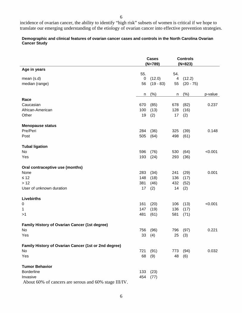
incidence of ovarian cancer, the ability to identify “high risk” subsets of women is critical if we hope to translate our emerging understanding of the etiology of ovarian cancer into effective prevention strategies. Demographic and clinical features of ovarian cancer cases and controls in the North Carolina Ovarian Cancer Study Cases Controls (N=789) (N=823) Age in years
mean (s.d) 55.
0 (12.0) 54.
4 (12.2) median (range) 56 (19 - 83) 55 (20 - 75) n (%) n (%) p-value Race Caucasian 670 (85) 678 (82) 0.237 African-American 100 (13) 128 (16) Other 19 (2) 17 (2) Menopause status Pre/Peri 284 (36) 325 (39) 0.148 Post 505 (64) 498 (61) Tubal ligation No 596 (76) 530 (64) <0.001 Yes 193 (24) 293 (36) Oral contraceptive use (months) None 283 (34) 241 (29) 0.001 ≤ 12 148 (18) 136 (17) > 12 381 (46) 432 (52) User of unknown duration 17 (2) 14 (2) Livebirths 0 161 (20) 106 (13) <0.001 1 147 (19) 136 (17) >1 481 (61) 581 (71) Family History of Ovarian Cancer (1st degree) No 756 (96) 796 (97) 0.221 Yes 33 (4) 25 (3) Family History of Ovarian Cancer (1st or 2nd degree) No 721 (91) 773 (94) 0.032 Yes 68 (9) 48 (6) Tumor Behavior Borderline 133 (23) Invasive 454 (77) About 60% of cancers are serous and 60% stage III/IV.
7
7
BRCA1/2: Since inherited BRCA1or BRCA2 mutations strikingly increase ovarian cancer risk, polymorphisms in these genes could represent low penetrance susceptibility alleles. Prior studies of the BRCA2 N372H polymorphism suggested that HH homozygotes have a modestly increased risk of both breast and ovarian cancer. We have examined whether BRCA2 N372H or common amino acid-changing polymorphisms in BRCA1 predispose to ovarian cancer in the North Carolina ovarian cancer study. Cases included 312 women with ovarian cancer (76% invasive, 24% borderline) and 401 age- and race- matched controls. Blood DNA from subjects was genotyped for BRCA2 N372H and BRCA1 Q356R and P871L. There was no association between BRCA2 N372H and risk of borderline or invasive epithelial ovarian cancer. The overall odds ratio for HH homozygotes was 0.8 (95% CI = 0.4-1.5) and was similar in all subsets including invasive serous cases. In addition, neither the BRCA1 Q356R (OR = 0.9, 95% CI 0.5-1.4) nor P871L (OR = 0.9, 95% CI 0.6-1.9) polymorphisms were associated with ovarian cancer risk. There was a significant racial difference in allele frequencies of the P871L polymorphism (P = 0.64 in Caucasians, L = 0.76 in African Americans, p<0.0001). In this population-based, case-control study, common amino acid changing BRCA1 and 2 polymorphisms were not found to affect the risk of developing ovarian cancer. These results were published in Clinical Cancer Research in 2003. Progesterone receptor: In view of the protective effect of a progestin dominant hormonal milieu (OC use, pregnancy), progesterone receptor variants with altered biological activity might affect ovarian cancer susceptibility. A German group reported that an intronic insertion polymorphism in the progesterone receptor was associated with a 2.1-fold increased ovarian cancer risk. It subsequently was shown that this Alu insertion is in linkage disequilibrium with SNPs in exons 4 and 5. However, several subsequent studies by our group and others failed to confirm an association between these polymorphisms and ovarian cancer. In addition, there is little evidence that this complex of polymorphisms, termed PROGINS, alters progesterone receptor function. More recently, sequencing of the progesterone receptor gene has revealed several additional polymorphisms, including one in the promoter region (+331G/A). The +331A allele creates a unique transcriptional start site that favors production of the progesterone receptor B (PR-B) isoform over progesterone receptor A (PR-A). The PR-A and PR-B isoforms are ligand-dependent members of the nuclear receptor family that are structurally identical except for an additional 164 amino acids at the N-terminus of PR-B, but their actions are distinct. The full length PR-B functions as a transcriptional activator and in the tissues where it is expressed it is a mediator of various responses, including the proliferative response to estrogen or the combination of estrogen and progesterone. PR-A is a transcriptionally inactive dominant-negative repressor of steroid hormone transcription activity that is thought to oppose estrogen-induced proliferation. An association has been reported between the +331A allele of the progesterone receptor promoter polymorphism and increased susceptibility to endometrial and breast cancers. It was postulated that upregulation of PR-B in carriers of the +331A allele might enhance formation of these cancers due to an increased proliferative response. The +331G/A polymorphism in the progesterone receptor promoter was examined in cases and controls from the North Carolina Ovarian Cancer Study. A second, independent, case-control study from Australia (Dr. Chenevix-Trench) that is also funded by the DOD was examined to confirm associations seen in the North Carolina study. Data from the two studies was then pooled to increase statistical power. The +331G/A single nucleotide polymorphism in the promoter of the progesterone receptor was genotyped using a TaqMan assay. Allelic discrimination was performed using the MGB primer/probe TaqMan assay on the ABI Prism 7700 system. Some samples were sequenced using the ABI 3100 system to confirm the accuracy of the Taqman assay. The +331A allele was found in 59/504 (11.7%) Caucasian controls and the distribution of genotypes was in Hardy-Weinberg Equilibrium (χ2 = 0.391, p = 0.53). Only 1/81 (1.2%) African American controls and none of 67 African American women with ovarian cancer carried the +331A allele. In view of the rarity of the +331A allele in African Americans,
8
8
these subjects were excluded from further analyses. The +331AA homozygotes were combined with heterozygotes in calculating odds ratios. The +331A allele was associated with a modest reduction in risk of ovarian cancer. Analysis by histologic type revealed that there was a slight trend towards protection against the common serous histologic type (OR = 0.80, 95% CI 0.49–1.29) but there was a more striking protection against endometrioid and clear cell cancers (OR = 0.30, 95% CI 0.09–0.97).
PR promoter polymorphism (left) TaqMan assay (green = GA heterozygotes, red = GG homozygotes) (right) GA heterozygote
Relationship between PR promoter polymorphism and ovarian cancer risk in histologic types of ovarian cancer
In view of the potential for false-positive results in genetic association studies, confirmation was sought using an independent study population from Australia. The frequency of the +331A allele among Caucasian controls varied by less than 1% between the Australian and North Carolina studies. The Australian study was not a population-based case-control study and fewer data were available regarding risk factors. Nevertheless, the results of the Australian study were similar to those of the North Carolina study, with a modest overall protective effect that was most pronounced for endometrioid cancers (OR = 0.51, 95% CI = 0.17–1.53). The Breslow-Day chi-square test was used to assess homogeneity of the results from the two study populations. Analyses involving the combined data set showed a significant association between the +331A allele and decreased risk of endometrioid/clear cell cases. In combining the two studies there was a significant risk reduction (OR = 0.46, 95% CI = 0.23–0.92) (P = 0.027). These types represent 21% of invasive ovarian cancer cases. Endometriosis is known to increase risk of endometrioid and clear cell ovarian cancers, many of which may arise in ovarian deposits of endometriosis. In this study, endometriosis was associated with an increased risk of endometrioid/clear
9
9
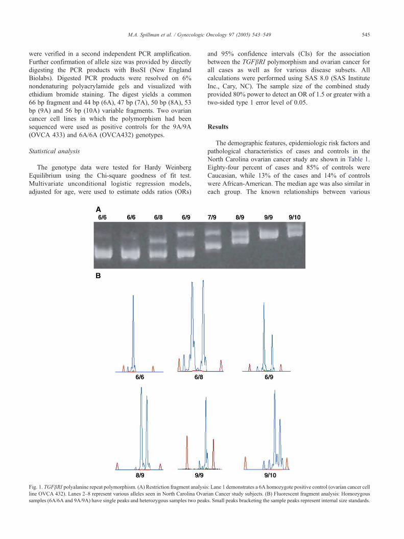
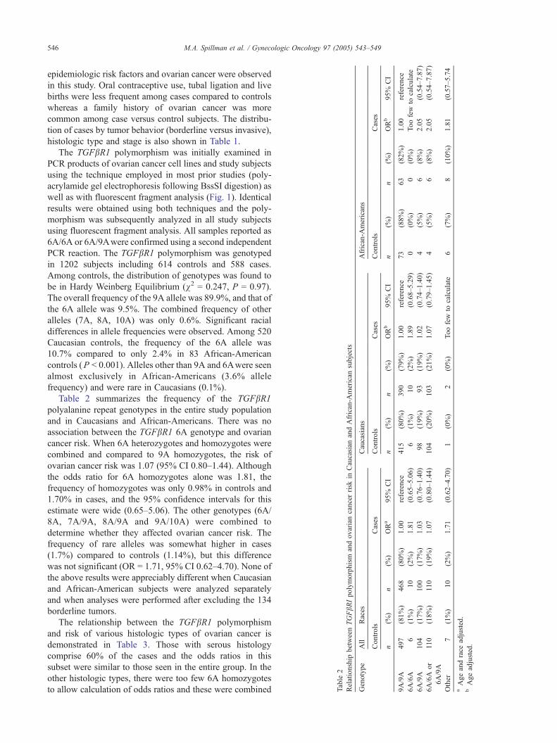
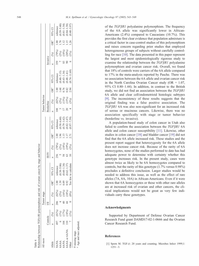
cell cancers (OR = 3.87, 95% CI = 2.09-7.17. The +331A allele appeared to be strongly protective against endometriosis (OR = 0.19, 95% CI 0.03 – 1.38), but this study was under powered to prove this conclusively. The literature is fraught with false-positive association studies of genetic susceptibility polymorphisms, but several features mitigate the likelihood of this in the present study. First, the known protective benefit of progestins against ovarian cancer provides a preexisting biologic plausibility for the observed association. In addition, the finding that the +331A allele is protective against both endometrioid/clear cell cancers and their precursor lesion (endometriosis) also is supportive. Confirmation of the positive association obtained in North Carolina study by the Australian study also represents an additional critical validation step. Finally, unlike many polymorphisms that lack known functional significance, the +331A allele increases transcription of PR-B in vitro. This study provides evidence for the existence of low penetrance ovarian cancer susceptibility polymorphisms. If multiple polymorphisms are identified that either increase or decrease the risk of various histologic types of ovarian cancer, this might be used in the future for risk stratification that would facilitate screening and prevention strategies. The paper describing the relationship between the progesterone receptor promoter polymorphism and ovarian cancer was published in the December 2004 issue of Cancer, Epidemiology, Biomarkers and Prevention (see appendix). Meta-analysis of the progesterone receptor promoter polymorphism (+331 G/A) confirms its protective effect against endometrioid/clear cell ovarian cancers Because of the potential for false-discovery in genetic association studies we have conducted a meta-analysis of several ongoing case-control studies to confirm this association. The +331G/A PR polymorphism was genotyped in blood DNA of 4,614 Caucasian subjects from population-based, case-control studies in the North Carolina Ovarian Cancer Study, Australia (Dr Trench), Massachusetts (Dr Daniel Cramer at Harvard) and Southern California (Dr. Leigh Pearce at USC). There were 2,269 subjects with invasive or borderline ovarian cancer (1,430 serous, 538 endometrioid/clear cell, 301 mucinous) and 2,345 controls. We conducted a meta-analysis using a fixed effects model to produce summary Mantel-Hanzel odds ratios (OR) for the four studies. The +331A allele (AA or GA) was present overall in 10.6% (151/1,430) of serous cases, 5.4% (34/538) of endometrioid/clear cell cases, 10.3% (31/301) of mucinous cases and 10.7% (251/2,345) of controls. The distribution of alleles in the controls conformed to Hardy-Weinberg equilibrium. There was no relationship between the +331A allele and serous or mucinous ovarian cancers in any of the individual studies or in the meta-analysis (serous OR = 0.98, 95% CI 0.79 - 1.22, mucinous OR = 0.91, 95% CI 0.59 - 1.38). In contrast, a protective effect against endometrioid/clear cell cancers was noted in each study (North Carolina OR = 0.45, Australia OR = 0.66, Massachusetts OR = 0.69 and Southern California OR = 0.30) and in the meta-analysis of all four studies (OR = 0.56, 95% CI 0.39 - 0.82) (p<0.003). These findings provide further evidence that the A allele of the +331G/A PR promoter polymorphism is carried by about 11% of Caucasians and is protective against endometrioid and clear cell ovarian cancers. Efforts to identify other common ovarian cancer susceptibility polymorphisms are ongoing, and if successful could allow screening and prevention strategies to be focused on populations at increased risk. TGF-β receptor 1: Progestin induced apoptosis in the ovarian epithelium may be mediated by the TGF-β pathway, and this pathway is the target for chemopreventive efforts in Project 2. In project 1, we are investigating the possibility that TGF-β receptors are appealing candidate ovarian cancer susceptibility genes. A polymorphism in the TGF-β I receptor has been described that involves deletion of 3 alanines from a 9 alanine tract (TβR1(6A)). IT has been suggested that the 6A allele might predispose to the development of ovarian cancer and other cancer types. In addition, there is some evidence that the
10
10
TβR1(6A) variant may be functionally significant and may confer an impaired ability to mediate TGF-β anti-proliferative effects. In view of the evidence that the TGFβR1 polyalanine polymorphism may affect ovarian cancer risk, this polymorphism was genotyped in 588 ovarian cancer cases and 614 controls from the North Carolina study (see tables below). Significant racial differences in the frequency of the 6A allele were observed between Caucasian (10.7%) and African American (2.4%) controls (p<0.001). One or two copies of the 6A allele of the TGFβR1 polyalanine polymorphism were carried by 18% of all controls and 19% of cases, and there was no association with ovarian cancer risk (OR = 1.07, 95% CI 0.80 – 1.44). The odds ratio for 6A homozygotes was 1.81 (95% CI 0.65 – 5.06), but these comprised only 0.98% of controls and 1.70% of cases. The 6A allele of the TGFβR1 polyalanine polymorphism does not appear to increase ovarian cancer risk. Larger studies are needed to exclude the possibility that the small fraction of individuals who are 6A homozygotes have an increased risk of ovarian or other cancers. Polymorphisms in other members of the TGF-β family of ligands, receptors and downstream effectors also are appealing candidates. This data was communicated as an oral presentation at the 2004 meeting of the International Gynecologic Cancer Society in Scotland and was published in the journal Gynecologic Oncology in 2005 (see appendix). Vitamin D Receptor pathway: High circulating levels of vitamin D may protect against ovarian cancer, since mortality rates are higher in northern latitudes where there is less sunlight. The most biologically active form of vitamin D, 1,25 (OH)2D3, is produced in the skin through sunlight exposure and vitamin D exhibits significant antineoplastic properties. Several factors, both dietary and genetic regulate the production of 1,25 (OH)2D3 from its precursor. A recent study suggested that about 22% of the variation may be accounted for by a putative major gene effect. Highly polymorphic loci involved in the metabolism and function of vitamin D include the vitamin D binding protein and vitamin D receptor genes. It has been suggested that a polymorphism in the vitamin D receptor gene involving a shared haplotype that includes a change in the 3’ untranslated region that alters transcriptional activity may be associated with increased prostate cancer risk. This has not been a uniform finding in all studies, however. Vitamin D receptor polymorphisms are being examined in the North Carolina Ovarian Cancer Study to test the hypothesis that vitamin D biosynthesis in the skin can protect susceptible individuals from developing ovarian cancer and that genetic variation in the vitamin D pathway may modify this protective effect. Seven haplotype tagging SNPs that include three functional variants have been genotyped and analyses are being performed to examine the relationship between genetic variation, sunlight exposure and ovarian cancer risk. BRAF polymorphisms Mutations in the BRAF gene, which is part of the RAS pathway, occur in some borderline serous ovarian tumors. In view of this, polymorphisms in the BRAF gene are appealing candidates that might affect susceptibility to borderline ovarian cancer. Dr Chenevix-Trench organized a multicenter collaborative study of BRAF polymorphisms with each center contributing their borderline cases and matched controls. These polymorphisms were not found to affect susceptibility to borderline serous tumors and this data was published in the journal Gynecologic Oncology in 2005 (see appendix). Illumina array In the last few years since our grant was funded, high throughput techniques for SNP genoyping have been developed. Presently, we are designing an Illumina array experiment that will allow us to genotype 1,536 SNPs in candidate genes in all 1,600 of our samples. We will include haplotype tagging SNPs as
11
11
well as nonsynonymous SNPs that result in amino acid changes. This experiment will focus on the hormonal pathway genes as well as DNA repair and inflammation pathway genes. The advent of this high throughput technology will allow us to generate vastly more genotype data in the next year than we have generated in the past years combined. Ovarian Cancer Association Consortium Although case-control studies of some polymorphisms have reported positive associations, these generally have not been confirmed in subsequent studies. Groups from the US, UK and Australia met in at Cambridge University in April 2005 to review results of various ongoing ovarian cancer association studies. There was a consensus that many of the challenges inherent in this field can best be addressed by collaborative efforts. In view of this, the group elected to establish an ovarian cancer association consortium (OCAC). Dr.Berchuck successfully applied to the Ovarian Cancer Research Fund for a $900,000 grant to fund the first three years of biannual meetings and other activities, and he will serve as the head of the steering committee. Dr Georgia Chenevix-Trench, who also is funded by the DOD Ovarian Cancer Research Program also is a leading member of the organizing group. The aims of the consortiuim are an outgrowth of the North Carolina and Australian DOD funded studies and reflect the successful translation of the DOD funding into a continued and expanded effort. The second meeting of the ovarian cancer association consortium took place in Salt Lake City in late October in concert with the American Society of Human Genetics annual meeting. Aim #1 - To develop an ovarian cancer association consortium (OCAC) that is dedicated to working together to identify and validate common low penetrance ovarian cancer susceptibility polymorphisms. The OCAC will meet each fall in concert with the American Society of Human Genetics meeting, and an annual spring meeting will be hosted by an OCAC member institution. This will provide the opportunity for face-to-face interactions that are critically important in sustaining the momentum of the OCAC. Aim #2 – To perform a comprehensive review of the existing ovarian cancer susceptibility polymorphism literature. This effort will produce a review article and will serve as a marker of the state of the field as the OCAC begins its work. Aim #3 – To determine whether polymorphisms in the progesterone receptor affect ovarian cancer risk. Polymorphisms in the progesterone receptor (PR) gene have been the most frequently examined. Several studies have suggested that polymorphisms in this gene affect risk, but not all studies have not confirmed these findings. The OCAC members will genotype PR polymorphisms in several thousand cases and controls and the data will be analyzed centrally to resolve the issue of whether PR variants affect ovarian cancer risk. Aim #4 – To examine associations between other promising candidate genetic variants and risk of ovarian cancer. In keeping with the goal of the OCAC to provide definitive evidence of genetic associations, the most promising candidate variants being studied by OCAC members will be genotyped in a collaborative manner as described above for the progesterone receptor. Aim #5 – To assign groups to write additional grant proposals that focus either on specific molecular pathways using a comprehensive approach or methodological issues for association studies. The groups in the ovarian cancer association consortium are funded to study specific genes and/or gene pathways. This includes various steroid hormone, DNA repair and inflammation related pathways as well as others. The goal will be to assign groups to seek additional funding to study these pathways in the OCAC. In addition, the group will be uniquely positioned to study methodological issues related to genetic association studies and the statistical geneticists in the group will have the opportunity to apply for funding to use OCAC data for this purpose. Aim #6 – To examine the interaction between major epidemiological risk factors and genetic polymorphisms. Because of the moderate size of most ovarian cancer association studies it has not been possible to perform analyses of gene-environment interactions. The OCAC will establish a common data
12
12
sheet that includes basic information relating to major epidemiological risk factors. This will focus mainly on family history and reproductive risk factors. Central analyses will be performed to examine interactions between factors such as OC use, genetic polymorphisms and ovarian cancer risk. Relevance: Presently, ovarian cancer risk stratification is not used to guide clinical surveillance or interventions in the vast majority of women, other than in rare individuals with BRCA/HNPCC mutations. This must change in the future if we are to decrease ovarian cancer incidence and mortality. The long term goal of the OCAC is to identify a panel of ovarian cancer susceptibility polymorphisms that can be used in combination with known epidemiological risk factors such as parity and OC use to better stratify ovarian cancer risk. This would greatly facilitate implementation of screening and prevention strategies by allowing these to be focused on higher-risk populations. The newly formed ovarian cancer association consortium includes essentially all of the leading groups in this field. We are eminently well positioned to achieve this goal. The OCRF can make a major impact in our ability to stratify ovarian cancer risk and to reduce mortality from the disease by providing support for the first three years of OCAC activities.
13
13
Project 2: Chemoprevention of Ovarian Cancer Project 2 is under the direction of Gustavo Rodriguez, M.D. (Gynecologic Oncologist). The prevention strategy outlined in our proposal focuses on the potential use of a combined approach incorporating both progestins and Vitamin D for the chemoprevention of ovarian cancer. The studies outlined in our prevention grant are designed to add further support to the notion that progestins and Vitamin D are potent apoptotic agents on human ovarian epithelial cells and to directly test the hypothesis in an animal model that these agents confer preventive effects against ovarian cancer. The aims in the grant are: (1) to evaluate the apoptotic effect of progestins and vitamin D analogues on the human ovarian epithelium in vitro, (2) elucidate the molecular mechanisms by which they induce apoptosis in ovarian epithelial cells, and (3) to directly test the hypothesis that progestins/vitamin D analogues confer preventive effects against ovarian cancer in a chemoprevention trial in the chicken, the only animal species with a high incidence of ovarian cancer. There is significant potential to decrease ovarian cancer incidence and mortality through prevention. Epidemiological evidence has shown that routine use of the combination estrogen–progestin oral contraceptive pill (OCP) confers a 30-50% reduction in the risk of developing subsequent epithelial ovarian cancer, suggesting that an effective ovarian cancer preventive approach using hormones is possible. Investigations by our group have elucidated a mechanism that we believe is responsible for the ovarian cancer preventive effects of the OCP. Specifically, we have discovered that the progestin component of the OCP is functioning as a classic chemopreventive agent, by activating potent molecular pathways known to be associated with cancer prevention in the ovarian surface epithelium. We have discovered that progestins markedly induce programmed cell death (apoptosis) and differentially regulate expression of Transforming Growth Factor Beta (TGF-β) in the ovarian epithelium. These two molecular events have been strongly implicated in cancer prevention in vivo, and are believed to underlie the protective effects of other well-known chemopreventive agents such as the retinoids and Tamoxifen. Our laboratory and animal research findings are supported by human data demonstrating that progestin-potent OCPs confer twice the ovarian cancer protection as newer weak-progestin OCPs. These human data provide proof of principle that progestins are effective chemopreventive agents for ovarian cancer, and suggest that a regimen that has enhanced chemopreventive biologic potency in the ovarian epithelium will be more effective than a lower potency regimen for ovarian cancer prevention.
The finding that progestins activate these molecular pathways in the ovarian epithelium opens the door toward a further investigation of progestins as chemopreventive agents for ovarian cancer, and raises the possibility that other agents that similarly activate cancer preventive pathways in ovarian epithelial cells may be attractive ovarian cancer preventives. Among the non-progestins, there is environmental, epidemiologic, laboratory and animal evidence in support of Vitamin D as a potent ovarian cancer preventive. In addition, results from a prevention trial that we have performed in the chicken ovarian cancer animal model suggest an additive ovarian cancer protective effect of Vitamin D when added to progestin.
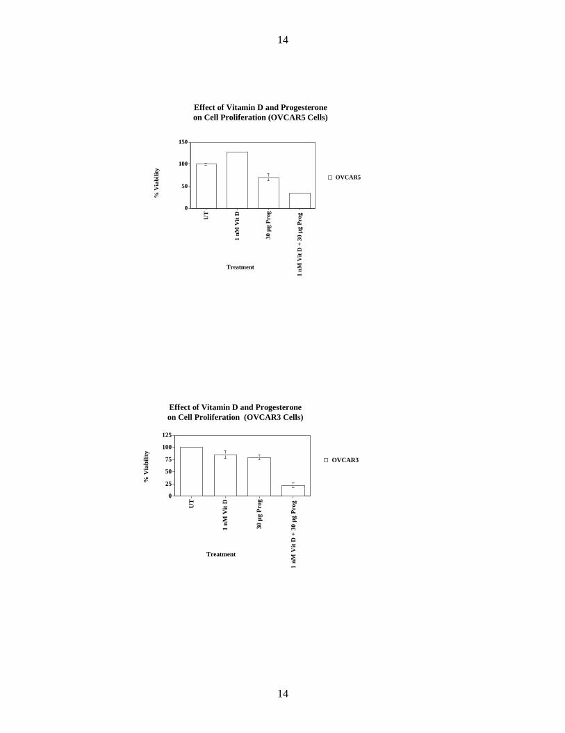
In our last annual summary, we presented evidence showing that the combination of a progestin and Vitamin D had a more potent biologic effect on cells derived from the human ovarian epithelium than either agent alone. We have expanded our studies to include immortalized cells derived from the normal human ovarian epithelium. Both drug classes markedly inhibited cell viability in a dose response fashion. The figures below demonstrate a marked impact on cell viability when the two agents are combined, and administered at a dosage that has a marginal impact for each agent given alone.
14
14
Effect of Vitamin D and Progesteroneon Cell Proliferation (OVCAR5 Cells)
0
50
100
150
% V
iabi
lity
UT
1 nM
Vit
D
30 µ
g Pr
og
1 nM
Vit
D +
30
µg P
rog
Treatment
OVCAR5
Effect of Vitamin D and Progesterone on Cell Proliferation (OVCAR3 Cells)
0
25
50
75
100
125
% V
iabi
lity
UT
1 nM
Vit
D
30 µ
g Pr
og
1 nM
Vit
D +
30
µg P
rog
Treatment
OVCAR3
15
15
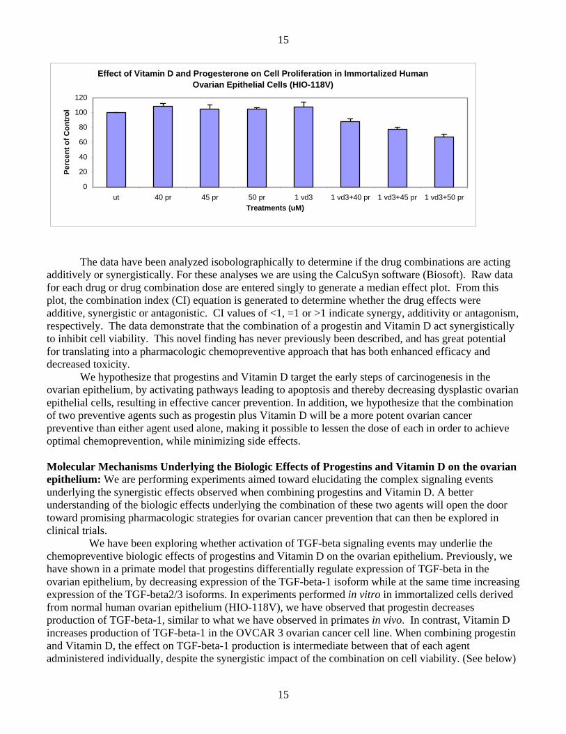
Effect of Vitamin D and Progesterone on Cell Proliferation in Immortalized Human Ovarian Epithelial Cells (HIO-118V)
The data have been analyzed isobolographically to determine if the drug combinations are acting additively or synergistically. For these analyses we are using the CalcuSyn software (Biosoft). Raw data for each drug or drug combination dose are entered singly to generate a median effect plot. From this plot, the combination index (CI) equation is generated to determine whether the drug effects were additive, synergistic or antagonistic. CI values of <1, =1 or >1 indicate synergy, additivity or antagonism, respectively. The data demonstrate that the combination of a progestin and Vitamin D act synergistically to inhibit cell viability. This novel finding has never previously been described, and has great potential for translating into a pharmacologic chemopreventive approach that has both enhanced efficacy and decreased toxicity.
We hypothesize that progestins and Vitamin D target the early steps of carcinogenesis in the ovarian epithelium, by activating pathways leading to apoptosis and thereby decreasing dysplastic ovarian epithelial cells, resulting in effective cancer prevention. In addition, we hypothesize that the combination of two preventive agents such as progestin plus Vitamin D will be a more potent ovarian cancer preventive than either agent used alone, making it possible to lessen the dose of each in order to achieve optimal chemoprevention, while minimizing side effects. Molecular Mechanisms Underlying the Biologic Effects of Progestins and Vitamin D on the ovarian epithelium: We are performing experiments aimed toward elucidating the complex signaling events underlying the synergistic effects observed when combining progestins and Vitamin D. A better understanding of the biologic effects underlying the combination of these two agents will open the door toward promising pharmacologic strategies for ovarian cancer prevention that can then be explored in clinical trials. We have been exploring whether activation of TGF-beta signaling events may underlie the chemopreventive biologic effects of progestins and Vitamin D on the ovarian epithelium. Previously, we have shown in a primate model that progestins differentially regulate expression of TGF-beta in the ovarian epithelium, by decreasing expression of the TGF-beta-1 isoform while at the same time increasing expression of the TGF-beta2/3 isoforms. In experiments performed in vitro in immortalized cells derived from normal human ovarian epithelium (HIO-118V), we have observed that progestin decreases production of TGF-beta-1, similar to what we have observed in primates in vivo. In contrast, Vitamin D increases production of TGF-beta-1 in the OVCAR 3 ovarian cancer cell line. When combining progestin and Vitamin D, the effect on TGF-beta-1 production is intermediate between that of each agent administered individually, despite the synergistic impact of the combination on cell viability. (See below)
16
16
Experiments are underway to evaluate the effect of progestins and Vitamin D on the other TGF-beta isoforms, and also on downstream signaling effects within the TGF-beta pathway. Effects of Hormone Treatments on TGF-beta-1 Production Cells were incubated in low serum conditions with the hormonal interventions labeled below. The supernatant was collected and examined for production of TGF-beta using a TGF-beta ELISA. In the HIO-118V immortalized ovarian epithelial cell line, results demonstrate down-regulation of TGF-b1 secretion in response to progestin, with VitD3 having a minimal effect and abrogating the progestin effect. Results have been normalized using MTS assay results, thereby correcting for cell number.
Effect of Vitamin D and Progesterone on TGF-B1 Production in HIO-118V cells
0
100
200
300
ut 35 uM pr 100 nM vd3 35 pr+100 vd3Treatments
pg/m
l TG
F-B
1
Similar trends are observed in the OVCAR-3 ovarian cancer cell line; however, Vitamin D up regulates TGF-beta production.
Effect of Vitamin D and Progesterone on TGFb1 Production in OVCAR 3 cells (96 hrs)
0
5
10
15
20
25
30
35
ut 15 PR 1 VD3 15 PR+1 VD3
Treatments (uM)
pg/m
l TG
Fb1
17
17
Apoptosis and Cell Cycle Cells were incubated for 28 and 48 hours in the hormonal treatments as indicated below and assessed for TUNEL reactivity and cell cycle. In these experiments, Apoptosis (TUNEL) data and cell cycle data shown are analyzed from the same experiment; in each cell line’s respective medium. MTS data are also in each cell line’s respective media. OVCAR 3 cells undergo a 7-fold increase in apoptosis at 48 hrs when treated with a combination of progesterone and vitamin D. HIO-118V cells show modest 1.7-fold increase in apoptosis with 45 uM progesterone alone and a 1.9-fold increase with the combination of 1 uM Vitamin D and 45 uM progesterone.
Effect of Vitamin D and Progesterone on Apoptosis in HIO-118V cells
Effect of Vitamin D and Progesterone on Apoptosis in Ovcar 3 cells
0123456789
ut 30 PR 35 PR 1 VD3 30PR+1VD3
35PR+1VD3
Treatments (uM)
Fold
incr
ease
in T
UN
EL (+
) cel
ls
28 hrs48 hrs
18
18Effect of Vitamin D and Progesteroneon Apoptosis (OVCAR 3)
untreated 35 uM PR
1 uM VD3 35 uM PR 1 uM VD3
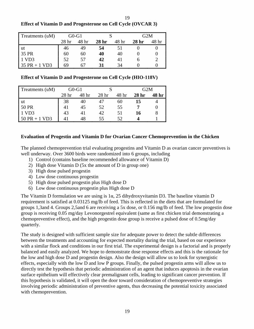
The cell cycle results confirm the apoptosis results. In the OVCAR 3, there is a modest reduction in S-phase due to progesterone and Vitamin D administered alone and a more robust reduction in S-phase with the two agents combined. In the HIO-118V, there is a progesterone-induced block of cells going from S-phase to G2M at 28 hours; however, the effect from Vitamin D is inconclusive.
19
19
Effect of Vitamin D and Progesterone on Cell Cycle (OVCAR 3) Treatments (uM) G0-G1 S G2M 28 hr 48 hr 28 hr 48 hr 28 hr 48 hr ut 46 49 54 51 0 0 35 PR 60 60 40 40 0 0 1 VD3 52 57 42 41 6 2 35 PR + 1 VD3 69 67 31 34 0 0 Effect of Vitamin D and Progesterone on Cell Cycle (HIO-118V) Treatments (uM) G0-G1 S G2M 28 hr 48 hr 28 hr 48 hr 28 hr 48 hrut 38 40 47 60 15 4 50 PR 41 45 52 55 7 0 1 VD3 43 41 42 51 16 8 50 PR + 1 VD3 41 48 55 52 4 1 Evaluation of Progestin and Vitamin D for Ovarian Cancer Chemoprevention in the Chicken The planned chemoprevention trial evaluating progestins and Vitamin D as ovarian cancer preventives is well underway. Over 3600 birds were randomized into 6 groups, including
1) Control (contains baseline recommended allowance of Vitamin D) 2) High dose Vitamin D (5x the amount of D in group one) 3) High dose pulsed progestin 4) Low dose continuous progestin 5) High dose pulsed progestin plus High dose D 6) Low dose continuous progestin plus High dose D
The Vitamin D formulation we are using is 1α, 25 dihydroxyvitamin D3. The baseline vitamin D requirement is satisfied at 0.03125 mg/lb of feed. This is reflected in the diets that are formulated for groups 1,3and 4. Groups 2,5and 6 are receiving a 5x dose, or 0.156 mg/lb of feed. The low progestin dose group is receiving 0.05 mg/day Levonorgestrel equivalent (same as first chicken trial demonstrating a chemopreventive effect), and the high progestin dose group is receive a pulsed dose of 0.5mg/day quarterly.
The study is designed with sufficient sample size for adequate power to detect the subtle differences between the treatments and accounting for expected mortality during the trial, based on our experience with a similar flock and conditions in our first trial. The experimental design is a factorial and is properly balanced and easily analyzed. We hope to demonstrate dose response effects and this is the rationale for the low and high dose D and progestin design. Also the design will allow us to look for synergistic effects, especially with the low D and low P groups. Finally, the pulsed progestin arms will allow us to directly test the hypothesis that periodic administration of an agent that induces apoptosis in the ovarian surface epithelium will effectively clear premalignant cells, leading to significant cancer prevention. If this hypothesis is validated, it will open the door toward consideration of chemopreventive strategies involving periodic administration of preventive agents, thus decreasing the potential toxicity associated with chemoprevention.
20
20
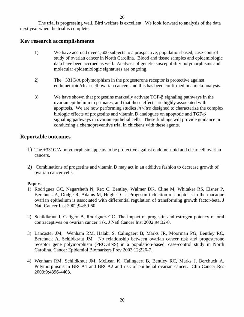
The trial is progressing well. Bird welfare is excellent. We look forward to analysis of the data next year when the trial is complete. Key research accomplishments
1) We have accrued over 1,600 subjects to a prospective, population-based, case-control study of ovarian cancer in North Carolina. Blood and tissue samples and epidemiologic data have been accrued as well. Analyses of genetic susceptibility polymorphisms and molecular epidemiologic signatures are ongoing.
2) The +331G/A polymorphism in the progesterone receptor is protective against
endometrioid/clear cell ovarian cancers and this has been confirmed in a meta-analysis. 3) We have shown that progestins markedly activate TGF-β signaling pathways in the
ovarian epithelium in primates, and that these effects are highly associated with apoptosis. We are now performing studies in vitro designed to characterize the complex biologic effects of progestins and vitamin D analogues on apoptotic and TGF-β signaling pathways in ovarian epithelial cells. These findings will provide guidance in conducting a chemopreventive trial in chickens with these agents.
Reportable outcomes
1) The +331G/A polymorphism appears to be protective against endometrioid and clear cell ovarian cancers.
2) Combinations of progestins and vitamin D may act in an additive fashion to decrease growth of
ovarian cancer cells. Papers 1) Rodriguez GC, Nagarsheth N, Rex C. Bentley, Walmer DK, Cline M, Whitaker RS, Eisner P,
Berchuck A, Dodge R, Adams M, Hughes CL: Progestin induction of apoptosis in the macaque ovarian epithelium is associated with differential regulation of transforming growth factor-beta. J Natl Cancer Inst 2002;94:50-60.
2) Schildkraut J, Caligert B, Rodriguez GC. The impact of progestin and estrogen potency of oral
contraceptives on ovarian cancer risk. J Natl Cancer Inst 2002;94:32-8.
3) Lancaster JM, Wenham RM, Halabi S, Calingaert B, Marks JR, Moorman PG, Bentley RC, Berchuck A, Schildkraut JM. No relationship between ovarian cancer risk and progesterone receptor gene polymorphism (PROGINS) in a population-based, case-control study in North Carolina. Cancer Epidemiol Biomarkers Prev 2003:12;226-7.
4) Wenham RM, Schildkraut JM, McLean K, Calingaert B, Bentley RC, Marks J, Berchuck A.
Polymorphisms in BRCA1 and BRCA2 and risk of epithelial ovarian cancer. Clin Cancer Res 2003;9:4396-4403.
21
21
5) Wenham RM, Calingaert B, Ali S, McLean K, Whitaker RS, Bentley RC, Lancaster JM, Schildkraut JM, Marks J, Berchuck A. Matrix metalloproteinase-1 gene promoter polymorphism and risk of ovarian cancer. J Soc Gynecologic Invest 2003;10:381-87.
6) Berchuck A, Schildkraut JM, Wenham RM, Calingaert B, Ali S, Henriott A, Halabi S, Rodriguez
GC, Gertig D, Purdie DM, Kelemen L, Spurdle AB, Marks J, Chenevix-Trench G. Progesterone receptor promoter +331A polymorphism is associated with a reduced risk of endometrioid and clear cell ovarian cancers. Cancer Epidemiol Biomarkers Prev 2004;13:2141-47.
7) Moorman PG, Berchuck A, Calingaert B, Halabi S, Schildkraut, JM. Antidepressant medication
use for and risk of ovarian cancer. Obstet Gynecol 2005;105:725-30.
8) Spillman MA, Schildkraut JM, Halabi S, Moorman P, Calingaert B, Bentley RC, Marks JR, Murphy S, Berchuck A. Transforming Growth Factor β Receptor I polyalanine repeat polymorphism does not increase ovarian cancer risk. Gynecol Oncol 2005;97:543-9.
9) Moorman PG, Schildkraut JM, Calingaert B, Halabi S, Berchuck A. Menopausal hormones and
risk of ovarian cancer. Am J Obstet Gynecol 2005;193:76-82.
10) Hoyo C, Berchuck A, Halabi S, Bentley RC, Moorman P, Calingaert B, Schildkraut JM. Anthropometric measurements and epithelial ovarian cancer risk in african american and white women. Cancer Causes and Control 2005;16:955-63.
11) Kelemen L, James M, Spurdle A, Campbell I, Chang-Claude J, Peel D, Anton-Culver H, Berchuck
A, Schildkraut J, Whittemore A, McGuire V, DiCioccio RA, Duffy D, Chenevix-Trench G. BRAF polymorphisms and the risk of ovarian cancer of low malignant potential. Gynecol Oncol 2005 97:807-12.
12) Schildkraut JM, Moorman P, Halabi S, Calingaert B, Marks JR, Havrilesky L, Berchuck A.
Analgesic drug use is associated with a decreased risk of ovarian cancer. Epidemiology (in press).
Conclusions
The studies initiated by our program will enable us to define more homogeneous subsets of ovarian cancer based on epidemiologic and molecular characteristics, to identify women who are at increased risk for this disease and to develop chemopreventive strategies designed to decrease ovarian cancer incidence and mortality. We anticipate that much of our data will grow to maturity in the coming few years with continued support from the DOD Ovarian Cancer Research Program.
22
22
Appendices
Progesterone Receptor Promoter +331A Polymorphism isAssociated with a Reduced Risk of Endometrioid andClear Cell Ovarian Cancers
Andrew Berchuck,1 Joellen M. Schildkraut,2 Robert M. Wenham,1 Brian Calingaert,2 Shazia Ali,1
Amy Henriott,1 Susan Halabi,2 Gustavo C. Rodriguez,1 Dorota Gertig,4 David M. Purdie,5
Livia Kelemen,6 Amanda B. Spurdle,6 Jeffrey Marks,3 and Georgia Chenevix-Trench6
Departments of 1Obstetrics and Gynecology/Division of Gynecologic Oncology, 2Community and Family Medicine, and 3Surgery,Duke University Medical Center, Durham, North Carolina; 4 Centre for Genetic Epidemiology, University of Melbourne, Melbourne,Victoria, Australia; and 5Population and Clinical Sciences Division and 6Cancer and Cell Biology Division, Queensland Institute ofMedical Research, Brisbane, Queensland, Australia
Abstract
Objective: The progestagenic milieu of pregnancy andoral contraceptive use is protective against epithelialovarian cancer. A functional single nucleotide poly-morphism in the promoter of the progesterone receptor(+331A) alters the relative abundance of the A and Bisoforms and has been associated with an increasedrisk of endometrial and breast cancer. In this study, wesought to determine whether this polymorphism affectsovarian cancer risk.Methods: The +331G/A polymorphism was genotypedin a population-based, case-control study from NorthCarolina that included 942 Caucasian subjects (438cases, 504 controls) and in a confirmatory group fromAustralia (535 cases, 298 controls). Logistic regressionanalysis was used to calculate age-adjusted oddsratios (OR).Results: There was a suggestion of a protective effectof the +331A allele (AA or GA) against ovarian cancerin the North Carolina study [OR, 0.72; 95% confidenceinterval (95% CI), 0.47-1.10]. Examination of genotypefrequencies by histologic type revealed that this was
due to a decreased risk of endometrioid and clear cellcancers (OR, 0.30; 95% CI, 0.09-0.97). Similarly, in theAustralian study, there was a nonsignificant decreasein the risk of ovarian cancer among those with the+331A allele (OR, 0.83; 95% CI, 0.51-1.35) that wasstrongest in the endometrioid/clear cell group (OR,0.60; 95% CI, 0.24-1.44). In the combined U.S.-Austra-lian data that included 174 endometrioid/clear cellcases (166 invasive, 8 borderline), the +331A allelewas significantly associated with protection againstthis subset of ovarian cancers (OR, 0.46; 95% CI, 0.23-0.92). Preliminary evidence of a protective effect of the+331A allele against endometriosis was also noted incontrol subjects (OR, 0.19; 95% CI, 0.03-1.38).Conclusions: These findings suggest that the +331G/Aprogesterone receptor promoter polymorphism maymodify the molecular epidemiologic pathway thatencompasses both the development of endometriosisand its subsequent transformation into endometrioid/clear cell ovarian cancer. (Cancer Epidemiol Bio-markers Prev 2004;13(12):2141–7)
Introduction
Epidemiologic studies have shown that both pregnancyand use of oral contraceptives dramatically reduceovarian cancer incidence (1). Reduction in numbers oflifetime ovulations due to pregnancy or oral contracep-tive use may decrease risk by reducing gonadotropinlevels, oxidative stress, DNA replication errors, andinclusion cyst formation in the ovarian epithelium. Inaddition, whereas estrogens and androgens have beenshown to increase ovarian cancer risk, both pregnancyand oral contraceptive use are characterized by a
protective progestagenic hormonal milieu (1, 2). Wehave previously reported that oral contraceptives withhigh progestin potency were associated with a greaterovarian cancer risk reduction than those with lowprogestin potency (3). In addition, we have shown thatprogestins may reduce ovarian cancer risk by stimulatingthe apoptosis of genetically damaged ovarian epithelialcells that otherwise might eventually evolve a fullytransformed phenotype (4, 5). This may account for theobservation that the protective effect of pregnancy andoral contraceptives is far greater than the extent to whichlifetime ovulatory cycles are reduced (1).
In view of the protective effect of progestins againstovarian cancer, progesterone receptor variants withaltered biological activity may affect ovarian cancersusceptibility. A German group reported that an inser-tion polymorphism in intron G of the progesteronereceptor was associated with a 2.1-fold increased ovariancancer risk (6, 7). It was subsequently shown that thisintronic AluI insertion is in linkage disequilibrium with
Received 2/3/04; revised 6/29/04; accepted 7/6/04.
Grant support: NIH grant 1-R01-CA76016, Department of Defense grant DAMD17-02-1-0666, Society for Gynecologic Investigation, and National Health and MedicalResearch Council of Australia.
The costs of publication of this article were defrayed in part by the payment of pagecharges. This article must therefore be hereby marked advertisement in accordancewith 18 U.S.C. Section 1734 solely to indicate this fact.
Requests for reprints: Andrew Berchuck, Division of Gynecologic Oncology, DukeUniversity Medical Center, Box 3079, Durham, NC 27710. Phone: 919-684-3765;Fax: 919-684-8719. E-mail: [email protected]
Copyright D 2004 American Association for Cancer Research.
2141
Cancer Epidemiol Biomarkers Prev 2004;13(12). December 2004
Cancer Epidemiology, Biomarkers & Prevention
polymorphisms in exons 4 and 5. However, severalsubsequent studies have failed to confirm an associationbetween these polymorphisms and ovarian cancer risk(8-12). In addition, there is little published evidence thatthis complex of polymorphisms, termed PROGINS, altersprogesterone receptor function.
More recently, sequencing of the progesterone receptorgene has revealed several additional polymorphisms,including one in the promoter region (+331G/A ; ref. 13).The +331A allele creates a unique transcriptional start sitethat favors the production of progesterone receptor B (PR-B) isoform over progesterone receptor A (PR-A; ref. 13).The PR-A and PR-B isoforms are ligand-dependentmembers of the nuclear receptor family that are structur-ally identical, except for an additional 164 amino acids atthe NH2 terminus of PR-B, but their actions are distinct(14, 15). The full-length PR-B functions as a transcriptionalactivator, and in the tissues where it is expressed, it is amediator of various responses, including the proliferativeresponse to estrogen or the combination of estrogen andprogesterone (16). PR-A is a transcriptionally inactivedominant-negative repressor of steroid hormone tran-scription activity that is thought to oppose estrogen-induced proliferation. An association has been reportedbetween the +331A allele and increased susceptibility toendometrial (13) and breast cancers (17). It was postulatedthat up-regulation of PR-B in carriers of the +331A allelemight enhance formation of these cancers due to anincreased proliferative response.
We used a case-control study design to explore whetherthe +331G/A polymorphism in the progesterone receptorpromoter affects susceptibility to various histologic typesof ovarian cancer in North Carolina. A second, indepen-dent, case-control study from Australia was examinedto confirm associations seen in the North Carolina study.
Materials and Methods
Subjects
North Carolina Ovarian Cancer Study. Primary ovariancancer cases enrolled in the study were identifiedthrough the North Carolina Central Cancer Registry, astatewide, population-based tumor registry, using rapidcase ascertainment. Eligibility criteria for ovarian cancercases include diagnosis since January 1, 1999, ages 20 to74 years at diagnosis, no prior history of ovarian cancer,and residence in a 48-county area of North Carolina.Physician permission was obtained before an eligiblecase was contacted. The diagnosis of epithelial ovariancancer (borderline or invasive) was confirmed by thestudy pathologist. The response rate among eligible caseswas 82%. Nonresponders were classified as patientrefusal (6.7%), inability to locate the patient (4.0%),physician refusal (3.5%), death (2.6%), or debilitatingillness (1.6%). Population-based controls were identifiedfrom the same 48-county region as the cases and werefrequency matched to the ovarian cancer cases based onrace (Black and non-Black) and age (5-year age catego-ries) using list-assisted random digit dialing. Potentialcontrols were screened for eligibility and were requiredto have at least one intact ovary and no prior diagnosis ofovarian cancer. Seventy-three percent of controls identi-fied by random digit dialing who passed the eligibility
screening agreed to be contacted and were sent addi-tional study information. Among those sent additionalstudy information, the response rate was 68%. The studyprotocol was approved by the Duke University MedicalCenter Institutional Review Board and the humansubjects committees at the North Carolina Central CancerRegistry and each of the hospitals where cases wereidentified. Trained nurse interviewers obtained writteninformed consent from study subjects at the time of theinterview, usually in the home of the study subject. A 90-minute questionnaire was given to obtain information onknown and suspected ovarian cancer risk factorsincluding family history of cancer in first- and second-degree relatives, menstrual characteristics, pregnancyand breast-feeding history, hormone use, and lifestylecharacteristics such as smoking, alcohol consumption,physical activity, and occupational history. A life eventscalendar, including marriage and education, was used toimprove recall. Additionally, anthropometric descriptors(height, weight, waist, and hip circumference) weremeasured and blood samples (30 mL) were collected.Germ line DNA was extracted using PureGene DNAisolation reagents according to the manufacturer’sinstructions (Gentra Systems, Minneapolis, MN). Analy-sis of data from the North Carolina study was limited toWhites. Data from 81 African American controls and 67cases were excluded because of the low frequency of thepolymorphism. Data were collected from 16 non-Black,non-Caucasian cases and 10 controls but were excludedbecause of the significant racial diversity and small sizeof this group.
Australian Study. Details of cases and controls includ-ed in the Australian study have been describedpreviously (18). Briefly, the case sample consisted of553 women with primary epithelial ovarian cancerascertained as incident case subjects as part of a largepopulation-based, case-control study from major gyne-cologic-oncology treatment centers in New South Wales,Victoria, and Queensland from 1990 to 1993 (n = 363)and from the Royal Brisbane Hospital, Queensland from1985 to 1996 (n = 190). Histopathologic informationregarding tumor behavior (low malignant potential orinvasive), histology, stage, and grade was available forall women; information on potential or known ovariancancer risk factors was ascertained by detailed question-naire for the subset of cases in the population-basedstudy and included age, ethnicity, country of birth,parity, oral contraceptive use, tubal ligation, hysterecto-my, and age at menarche. Limited information ascer-tained from hospital records was also available for theRoyal Brisbane Hospital patients and included age,ethnicity, and country of birth. Because blood sampleswere not collected from controls who participated in theovarian cancer case-control study, an additional group ofwomen, selected based on date-of-birth distribution tobest match cases, were included in the analyses. Thecontrol sample consisted of 300 adult female unrelatedmonozygotic twins (one per pair), ages 30 to 90 years,recruited through the volunteer Australian Twin Regis-try for the Semistructured Assessment for the Genetics ofAlcoholism study. This study reported participationrates of f70% for monozygotic female twins andrecruited individuals nationally from major cities inthe eastern states of Australia. Limited information
Progesterone Receptor Polymorphism2142
Cancer Epidemiol Biomarkers Prev 2004;13(12). December 2004
ascertained by detailed questionnaires as part of the Semi-structured Assessment for the Genetics of Alcoholismstudy was available for these women to assess con-founding and included age, ethnicity, country of birth,parity, and age at menarche. More than 90% of case andcontrol subject groups were of northern Europeandescent, and all subjects were from major cities in theeastern Australian states. Approvals were obtained fromthe ethics committees of the University of Melbourne,New South Wales Cancer Council, Anti-Cancer Councilof Victoria, and Queensland Institute of Medical Re-search in Australia. Written informed consent wasobtained from each participant. DNA isolation methodshave been detailed elsewhere (18). Fourteen Australiancases ages <30 years were excluded from this analysisbecause no controls were ages <30 years. Additionally,four cases and two controls were excluded because theydid not have +331G/A polymorphism results. Thus, theAustralian sample used for this analysis consisted of 535cases and 298 controls.
Genotyping of +331G/A Polymorphism. Allelic dis-crimination was done using the MGB primer/probeTaqman assay on the ABI Prism 7700 system. Details ofthe methods are described in the following sections.
North Carolina Study. Each 20 AL PCR reactioncontained 18 pmol of forward primer 5V-CACGAGTTT-GATGCCAGAGAAA-3V, 18 pmol of reverse primer 5V-GCGACGGCAATTTAGTGACA-3V, 4 pmol of G-alleleprobe (VIC)-CGGCTCcTTTATC-(MGBNFQ)-3V, 4 pmolof A-allele probe (FAM)-CGGCTCtTTTATCTC-(MGBNFQ)-3V (200 nmol/L), 10 AL of 2� TaqmanUniversal Master Mix without AmpErase UNG (AppliedBiosystems, Foster City, CA), and 25 ng of extractedleukocyte DNA. Cycling conditions were 958C for 10minutes followed by 40 cycles of 928C for 15 seconds and608C for 60 seconds. Allelic discrimination was done in96-plate format in the ABI Prism 7700 and analyzedusing the ABI Prism 7700 software. Some samples in theNorth Carolina ovarian cancer study were subjected tosequencing to confirm results obtained using the Taqmanassay. To do this, a 50 AL PCR reaction was done usingforward primer 5V-AACTCAGCGAGGGACTGAGA-3Vand reverse primer 5V-GAGGACTGGAGACGCAGAGT-3V, 0.5 ng/AL genomic DNA, 0.5 nmol/L forward primer,0.5 nmol/L reverse primer, 0.2 mmol/L deoxynucleotidetriphosphate, 1.5 mmol/L MgCl2 (Applied Biosystems),1� Applied Biosystems PCR buffer, and 0.025 units/ALAmpliTaq Gold DNA polymerase (Applied Biosystems).PCR conditions consisted of an initial denaturing step at958C for 12 minutes, 32 cycles of 948C for 60 seconds,55.08C for 60 seconds, and 728C for 3 minutes, and anextension step at 728C for 10 minutes. Samples were heldat 4.08C until they were purified using QIAquick 96vacuum filter plates (Qiagen, Germantown, MD) andfinally eluted in 150 AL of 10 mmol Tris-HCl (pH 8.5). Asequencing reaction was done using 1 AL of purifiedproduct and 4.4 pmol of unlabeled forward primer in aBigDye Terminator Cycle Sequencing Reaction as de-scribed by the supplier (Applied Biosystems). Sampleswere analyzed on the ABI 3100 system and sequencesdetermined using GeneScan software (Applied Biosys-tems).
Australian Study. Genotyping was done with Taqmanmethodology using identical probes as the NorthCarolina study. For detection and sequence confirmation
of positive controls, a 381-bp product was amplifiedusing the forward primer 5V-GTACGGAGCCAGCA-GAAGTC-3V and reverse primer 5V-ATCCTGTCGT-CAGGGGAACT-3V. Denaturing high-performance liquidchromatography (Helix System, Varian ChromatographySystems, Walnut Creek, CA) was used to identifyheterozygous GA individuals at 628C recommended bythe MELT program (http://insertion.stanford.edu/melt.html). Genotypes were confirmed by sequencing.Heterozygous GA PCR product was subcloned using thepGEM-T system to obtain G and A clones to use as controlstandards for the SDS allelic discrimination assay. The15 AL PCR reaction contained 900 nmol/L of forwardprimer 5V-GCGACGGCAATTTAGTGACA-3V, 900 nmol/L ofreverse primer 5V-TGCACGAGTTTGATGCCAGA-3V (givinga 68-bp product), 150 nmol/L of A-allele probe, 200 nmol/L ofG-allele probe, 1� Platinum Quantitative PCR SuperMixUDG (including passive reference ROX dye, Invitrogen,Melbourne, Victoria, Australia), and 15 ng of genomic orcontrol sample that had been dried in 96-well plates. PCR wasdone using the ABI 7700 SDS PCR machine for 2 minutes at508C and 2 minutes at 958C followed by 45 two-step cycles of15 seconds at 928C and 1 minute at 608C.
Statistical Analysis. The genotype data were tested forHardy-Weinberg equilibrium using the m2 goodness-of-fit test. Multivariate unconditional logistic regressionmodels, adjusted for age, were used to estimate oddsratio (OR) and 95% confidence interval (95% CI) for theassociation between polymorphism and epithelial ovar-ian cancer for all cases as well as for various diseasecategories. Potential confounders including menopausalstatus, tubal ligation, oral contraceptive use, body massindex, family history of breast or ovarian cancer in first-and second-degree relatives, and parity were individu-ally adjusted for in the North Carolina data to determineif they changed the crude OR by z10%. Analysisstratified by each of these factors was also conducted toassess potential effect modification. We found noevidence of confounding by these factors and thereforefelt it appropriate to combine the Australian and NorthCarolina data despite limited epidemiologic data in theAustralian sample. The Breslow-Day m2 test was used toassess homogeneity of the results from the two studypopulations. Analyses involving the combined data setwere based on a reanalysis of the raw data and wereadjusted for study as well as age. All calculations weredone using SAS 8.0 (SAS Institute, Inc., Cary, NC).
Results
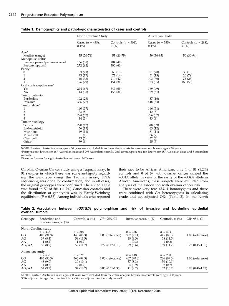
The demographic features, epidemiologic risk factors,and pathologic characteristics of cases and controls in theNorth Carolina (Caucasians only) and Australian studiesare shown in Table 1. Of note, the median ages of thecases and controls in both North Carolina and Australianstudies are similar. Caucasian women with ovariancancer in North Carolina were more likely to have usedoral contraceptives compared with Australian womenwith ovarian cancer (67% and 49%, respectively).Invasive ovarian cancer cases comprised 77% of theNorth Carolina cases compared with 84% of theAustralian cases. The +331G/A single nucleotide poly-morphism in the promoter of the progesterone receptorinitially was genotyped in samples from the North
Cancer Epidemiology, Biomarkers & Prevention 2143
Cancer Epidemiol Biomarkers Prev 2004;13(12). December 2004
Carolina Ovarian Cancer study using a Taqman assay. In91 samples in which there was some ambiguity regard-ing the genotype using the Taqman assay, DNAsequencing was done for confirmation, and in all cases,the original genotypes were confirmed. The +331A allelewas found in 59 of 504 (11.7%) Caucasian controls andthe distribution of genotypes was in Hardy-Weinbergequilibrium (P = 0.53). Among individuals who reported
their race to be African American, only 1 of 81 (1.2%)controls and 0 of 67 with ovarian cancer carried the+331A allele. In view of the rarity of the +331A allele inAfrican Americans, these subjects were excluded fromanalyses of the association with ovarian cancer risk.
There were very few +331A homozygotes and thesewere combined with GA heterozygotes in calculatingcrude and age-adjusted ORs (Table 2). In the North
Table 1. Demographics and pathologic characteristics of cases and controls
NOTE: Fourteen Australian cases ages <30 years were excluded from the entire analysis because no controls were ages <30 years.*Parity use not known for 187 Australian cases and 298 Australian controls. Oral contraceptive use not known for 187 Australian cases and 5 Australiancontrols.cStage not known for eight Australian and seven NC cases.
Table 2. Association between +331G/A polymorphism and risk of invasive and borderline epithelialovarian tumors
Genotype Borderline andinvasive cases, n (%)
Controls, n (%) OR* 95% CI Invasive cases, n (%) Controls, n (%) OR* 95% CI
North Carolina studyn = 438 n = 504 n = 336 n = 504
NOTE: Fourteen Australian cases ages <30 years were excluded from the entire analysis because no controls were ages <30 years.*ORs adjusted for age. For combined data, ORs are adjusted for the study as well.
Progesterone Receptor Polymorphism2144
Cancer Epidemiol Biomarkers Prev 2004;13(12). December 2004
Carolina sample, there was a suggestion that the +331Aallele was associated with a modest reduction in risk ofboth borderline tumors and invasive ovarian cancers(OR, 0.72; 95% CI, 0.47-1.10). Samples from the Austra-lian study were genotyped independently and 10.7% ofcontrols were found to carry the +331A allele. Thedistribution of genotypes in controls was found to be inHardy-Weinberg equilibrium (P = 0.27). Although notstatistically significant, a similar inverse association withinvasive ovarian cancer risk was observed (OR, 0.83; 95%CI, 0.51-1.35; Table 2). Excluding the borderline ovariancancers revealed little change in the point estimates of theassociation between the +331A allele and ovarian cancerfor either North Carolina or Australian comparisons(Table 2).
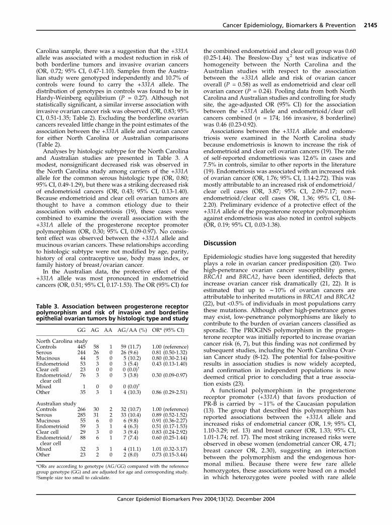
Analyses by histologic subtype for the North Carolinaand Australian studies are presented in Table 3. Amodest, nonsignificant decreased risk was observed inthe North Carolina study among carriers of the +331Aallele for the common serous histologic type (OR, 0.80;95% CI, 0.49-1.29), but there was a striking decreased riskof endometrioid cancers (OR, 0.43; 95% CI, 0.13-1.40).Because endometrioid and clear cell ovarian tumors arethought to have a common etiology due to theirassociation with endometriosis (19), these cases werecombined to examine the overall association with the+331A allele of the progesterone receptor promoterpolymorphism (OR, 0.30; 95% CI, 0.09-0.97). No consis-tent effect was observed between the +331A allele andmucinous ovarian cancers. These relationships accordingto histologic subtype were not modified by age, parity,history of oral contraceptive use, body mass index, orfamily history of breast/ovarian cancer.
In the Australian data, the protective effect of the+331A allele was most pronounced in endometrioidcancers (OR, 0.51; 95% CI, 0.17-1.53). The OR (95% CI) for
the combined endometrioid and clear cell group was 0.60(0.25-1.44). The Breslow-Day m2 test was indicative ofhomogeneity between the North Carolina and theAustralian studies with respect to the associationbetween the +331A allele and risk of ovarian canceroverall (P = 0.58) as well as endometrioid and clear cellovarian cancer (P = 0.24). Pooling data from both NorthCarolina and Australian studies and controlling for studysite, the age-adjusted OR (95% CI) for the associationbetween the +331A allele and endometrioid/clear cellcancers combined (n = 174; 166 invasive, 8 borderline)was 0.46 (0.23-0.92).
Associations between the +331A allele and endome-triosis were examined in the North Carolina studybecause endometriosis is known to increase the risk ofendometrioid and clear cell ovarian cancers (19). The rateof self-reported endometriosis was 12.6% in cases and7.5% in controls, similar to other reports in the literature(19). Endometriosis was associated with an increased riskof ovarian cancer (OR, 1.76; 95% CI, 1.14-2.72). This wasmostly attributable to an increased risk of endometrioid/clear cell cases (OR, 3.87; 95% CI, 2.09-7.17; non–endometrioid/clear cell cases OR, 1.36; 95% CI, 0.84-2.20). Preliminary evidence of a protective effect of the+331A allele of the progesterone receptor polymorphismagainst endometriosis was also noted in control subjects(OR, 0.19; 95% CI, 0.03-1.38).
Discussion
Epidemiologic studies have long suggested that heredityplays a role in ovarian cancer predisposition (20). Twohigh-penetrance ovarian cancer susceptibility genes,BRCA1 and BRCA2 , have been identified, defects thatincrease ovarian cancer risk dramatically (21, 22). It isestimated that up to f10% of ovarian cancers areattributable to inherited mutations in BRCA1 and BRCA2(22), but <0.5% of individuals in most populations carrythese mutations. Although other high-penetrance genesmay exist, low-penetrance polymorphisms are likely tocontribute to the burden of ovarian cancers classified assporadic. The PROGINS polymorphism in the proges-terone receptor was initially reported to increase ovariancancer risk (6, 7), but this finding was not confirmed bysubsequent studies, including the North Carolina Ovar-ian Cancer study (8-12). The potential for false-positiveresults in association studies is now widely accepted,and confirmation in independent populations is nowdeemed critical prior to concluding that a true associa-tion exists (23).
A functional polymorphism in the progesteronereceptor promoter (+331A) that favors production ofPR-B is carried by f11% of the Caucasian population(13). The group that described this polymorphism hasreported associations between the +331A allele andincreased risks of endometrial cancer (OR, 1.9; 95% CI,1.10-3.29; ref. 13) and breast cancer (OR, 1.33; 95% CI,1.01-1.74; ref. 17). The most striking increased risks wereobserved in obese women (endometrial cancer OR, 4.71;breast cancer OR, 2.30), suggesting an interactionbetween the polymorphism and the endogenous hor-monal milieu. Because there were few rare allelehomozygotes, these associations were based on a modelin which heterozygotes were pooled with rare allele
Table 3. Association between progesterone receptorpolymorphism and risk of invasive and borderlineepithelial ovarian tumors by histologic type and study
*ORs are according to genotype (AG/GG) compared with the referencegroup genotype (GG) and are adjusted for age and corresponding study.cSample size too small to calculate.
Cancer Epidemiology, Biomarkers & Prevention 2145
Cancer Epidemiol Biomarkers Prev 2004;13(12). December 2004
homozygotes. It was postulated that the rare allele of thispolymorphism may increase endometrial and breastcancer risks by enhancing PR-B-mediated proliferationin response to estrogen.
In the population-based North Carolina OvarianCancer Study, risk analyses were confined to Caucasiansubjects because of the rarity of +331A allele in AfricanAmerican women. Among Caucasian women, weobserved a weak protective effect of the +331A alleleagainst ovarian cancer (borderline and invasive). Histo-logic subtype analysis revealed that there was a weak,nonsignificant decrease in risk of serous cancers, whichare the most common subtype, whereas a strongerdecreased risk for endometrioid cancers was observed.This association became even stronger and statisticallysignificant after combining endometrioid and clear cellcancers, with about a two-thirds reduction in risk (OR,0.30; 95% CI, 0.09-0.97) in carriers of the +331A allele,although the 95% CIs are wide suggesting the instabilityof the estimate. In view of the potential for false-positive results in association studies of genetic poly-morphisms, we sought to confirm our findings in theAustralian study. The frequency of the +331A alleleamong Caucasian controls varied by <1% betweenAustralian and North Carolina studies and controlsreported in the Nurses’ Health Study (13, 17). TheAustralian study was not a population-based, case-control study and fewer data were available regardingrisk factors. Nevertheless, the results of the Australianstudy were similar to those of the North Carolina study,with a modest overall protective effect that was mostpronounced for endometrioid cancers (OR, 0.51; 95% CI,0.17-1.53). Age was not associated with genotype andadjusting for age had minimal effect on the ORsreported in this article.
Serous and endometrioid/clear cell ovarian cancersshare many of the same risk factors, such as parity andoral contraceptive use, but there is evidence to suggestthat differences exist in their etiology, molecular patho-genesis, and clinical behavior. For example, there aredifferences between these histologic subtypes withrespect to behavior (borderline versus invasive) andstage that likely reflect etiologic heterogeneity. Inaddition, mutations in BRCA1 and BRCA2 predisposeprimarily to serous cancers (24), which arise fromepithelial cells that line the ovarian surface or underlyinginclusion cysts. In contrast, it is thought that some, if notall, endometrioid and clear cell cancers arise fromdeposits of ovarian endometriosis (19). Coexistent endo-metriosis is commonly noted in women with ovarianendometrioid/clear cell cancers, and a strong associationbetween endometriosis and these cancers has beenreported in epidemiologic studies. Because endometri-osis is likely to be underdiagnosed, the relationshipbetween endometriosis and clear cell/endometrioidovarian cancers may be stronger than noted in case-control studies.
The finding that the +331A allele was associatedwith a decreased risk of endometrioid and clear cellovarian cancers was somewhat unexpected in view ofprior reports of an increased risk of endometrial andbreast cancers in carriers of the +331A allele (13, 17).However, these three diseases differ with respect toassociated risk factors and predisposing hormonalmilieu. Endometriosis is associated with endometrioid
and clear cell ovarian cancers (19) but does notincrease endometrial or breast cancer risk. In contrast,oral contraceptives are protective against all histologictypes of epithelial ovarian cancer as well as endome-trial cancers (1) but may increase breast cancer risk(25). In view of these significant differences in etiology,it is not surprising that predisposition to these cancersis affected differentially by the progesterone receptorpromoter polymorphism.
PR-A and PR-B are both expressed in the ovarian (26),endometrial (27), and breast epithelium (28), and therelative expression of the isoforms is frequently alteredduring malignant transformation. In the present study,the +331A allele of the progesterone receptor promoterpolymorphism was protective against endometrioid andclear cell ovarian cancers. We also observed preliminaryevidence that this polymorphism may protect againstendometriosis, the precursor of many of these cancers.Endometriotic implants have been shown to express onlythe PR-A isoform (27), and it has been suggested that theabsence of PR-B may account for the lack of appropriatecycling of these glands. In normal cycling endometrium,PR-A expression is predominant during the proliferativephase, whereas a shift toward PR-B occurs withdifferentiation in the early secretory phase (29). Becausethe +331A allele of the progesterone receptor promoterpolymorphism favors production of the PR-B isoform,it is possible that this might prevent the PR-A/PR-Bimbalance in endometriotic implants and protect againstthe growth and spread of endometriosis to the extent thatit becomes clinically apparent. The reduced risk ofendometrioid and clear cell cancers in women with the+331A allele might be attributable to a lower likelihoodof carriers developing more extensive endometriosis,which serves as a precursor for these cancers. In contrastto the pathogenic model proposed for endometriosis inwhich the +331A allele counters an abnormal imbalancein the PR-A/PR-B ratio in normal breast and endometrialtissues, the polymorphism may create an imbalance thatenhances both the proliferative response to estrogen andcancer risk.
The literature is fraught with false-positive associationstudies of genetic susceptibility polymorphisms, butseveral features mitigate the likelihood of this in thepresent study. First, the known protective benefit ofprogestins against ovarian cancer provides a preexistingbiological plausibility for the observed association. Inaddition, the finding that the +331A allele is protectiveagainst both endometrioid/clear cell cancers and theirprecursor lesion (endometriosis) is also supportive.Confirmation of the positive association obtained in theNorth Carolina study by the Australian study alsorepresents an additional critical validation step. Finally,unlike many polymorphisms that lack known functionalsignificance, the +331A allele is known to increasetranscription of PR-B in vitro (13).
Despite the agreement between North Carolina andAustralian data, the 95% CIs of the latter study arerelatively wide. Furthermore, the control subjects in theAustralian study were not collected in the context of anovarian cancer study. However, allele frequencies in theAustralian controls were similar to those seen inCaucasian controls in the North Carolina study. Anotherlimitation of this study is that the number of cases of theless common histologic types was relatively modest,
Progesterone Receptor Polymorphism2146
Cancer Epidemiol Biomarkers Prev 2004;13(12). December 2004
limiting the power to draw definitive conclusions.Additional studies are needed to confirm the protectiveeffect of the +331A allele against endometrioid and clearcell ovarian cancers.
In summary, the +331A allele of the progesteronereceptor promoter polymorphism is carried by about onein nine Caucasian women and is associated with adecrease in risk of endometrioid and clear cell ovariancancers. We also obtained preliminary evidence insupport of a protective effect against endometriosis.These findings suggest that the +331G/A progesteronereceptor promoter polymorphism may modify themolecular epidemiologic pathway that encompasses boththe growth of endometriosis and its subsequent trans-formation into endometrioid/clear cell cancers. Thisstudy provides evidence for the existence of low-penetrance ovarian cancer susceptibility polymorphisms.If multiple polymorphisms are identified that eitherincrease or decrease the risk of various histologic types ofovarian cancer, this might be used in the future for riskstratification that would facilitate screening and preven-tion strategies.
AcknowledgmentsWe thank the North Carolina Central Tumor Registry; the staffof the North Carolina Ovarian Cancer Study; Regina Whitakerfor expert technical assistance; Rex C. Bentley, MD, for review ofthe pathology; and Adele Green for access to ovarian case-subject DNA and Nicholas Martin for access to control-subjectDNA (Australian study).
References1. Whittemore AS, Harris R, Itnyre J. Characteristics relating to ovarian
cancer risk. Collaborative analysis of twelve US case-control studies.II. Invasive epithelial ovarian cancers in white women. Am JEpidemiol 1992;136:1184 – 203.
2. Risch HA. Hormonal etiology of epithelial ovarian cancer, with ahypothesis concerning the role of androgens and progesterone. J NatlCancer Inst 1998;90:1774 – 86.
3. Schildkraut JM, Calingaert B, Marchbanks PA, Moorman PG,Rodriguez GC. Impact of progestin and estrogen potency in oralcontraceptives on ovarian cancer risk. J Natl Cancer Inst 2002;94:32 – 8.
4. Rodriguez GC, Walmer DK, Cline M, et al. Effect of progestin on theovarian epithelium of macaques: cancer prevention through apop-tosis? J Soc Gynecol Investig 1998;5:271 – 6.
5. Rodriguez GC, Nagarsheth NP, Lee KL, et al. Progestin-inducedapoptosis in the macaque ovarian epithelium: differential regulationof transforming growth factor-h. J Natl Cancer Inst 2002;94:50 – 60.
6. McKenna NJ, Kieback DG, Carney DN, Fanning M, McLinden J,Headon, DRA. Germline TaqI restriction fragment length polymor-phism in the progesterone receptor gene in ovarian carcinoma. Br JCancer 1995;71:451 – 5.
7. Rowe SM, Coughlan SJ, McKenna NJ, et al. Ovarian carcinoma-associated Taq I restriction fragment length polymorphism in intronG of the progesterone receptor gene is due to an Alu sequenceinsertion. Cancer Res 1995;55:2743 – 5.
8. Lancaster JM, Berchuck A, Carney M, Wiseman RW, Taylor JA.Progesterone receptor gene polymorphism and risk of breast andovarian cancer. Br J Cancer 1998;78:277.
9. Manolitsas TP, Englefield P, Eccles DM, Campbell IG. No associa-tion of a 306-bp insertion polymorphism in the progesterone re-ceptor gene with ovarian and breast cancer. Br J Cancer 1997;75:1398 – 9.
10. Spurdle AB, Webb PM, Purdie DM, Chen X, Green A, Chenevix-Trench G. No significant association between progesterone receptorexon 4 Val660Leu G/T polymorphism and risk of ovarian cancer.Carcinogenesis 2001;22:717 – 21.
11. Tong D, Fabjani G, Heinze G, Obermair A, Leodolter S, Zeillinger R.Analysis of the human progesterone receptor gene polymorphismPROGINS in Austrian ovarian carcinoma patients. Int J Cancer 2001;95:394 – 7.
12. Lancaster JM, Wenham RM, Halabi S, et al. No relationship betweenovarian cancer risk and progesterone receptor gene polymorphism ina population-based, case-control study in North Carolina. CancerEpidemiol Biomarkers Prev 2003;12:226 – 7.
13. De Vivo I, Huggins GS, Hankinson SE, et al. A functionalpolymorphism in the promoter of the progesterone receptor geneassociated with endometrial cancer risk. Proc Natl Acad Sci U S A2002;99:12263 – 8.
14. Jacobsen BM, Richer JK, Schittone SA, Horwitz KB. New humanbreast cancer cells to study progesterone receptor isoform ratioeffects and ligand-independent gene regulation. J Biol Chem 2002;277:27793 – 800.
15. Richer JK, Jacobsen BM, Manning NG, Abel, MG, Wolf DM,Horwitz KB. Differential gene regulation by the two progesteronereceptor isoforms in human breast cancer cells. J Biol Chem 2002;277:5209 – 18.
16. Giangrande PH, Kimbrel EA, Edwards DP, McDonnell DP. Theopposing transcriptional activities of the two isoforms of the humanprogesterone receptor are due to differential cofactor binding. MolCell Biol 2000;20:3102 – 15.
17. DeVivo I, Hankinson SE, Colditz GA, Hunter DJA. A functionalpolymorphism in the progesterone receptor gene is associated withan increase in breast cancer risk. Cancer Res 2003;63:5236 – 8.
18. Spurdle AB, Purdie D, Chen X, Chenevix-Trench G. The prohibitin 3Vuntranslated region polymorphism is not associated with risk ofovarian cancer. Cancer Res 2003;90:145 – 9.
19. Ness RB. Endometriosis and ovarian cancer: thoughts on sharedpathophysiology. Am J Obstet Gynecol 2003;89:280 – 94.
20. Schildkraut JM, Thompson WD. Familial ovarian cancer: a popula-tion-based case-control study. Am J Epidemiol 1988;128:456 – 66.
21. Whittemore AS, Gong G, Itnyre J. Prevalence and contribution ofBRCA1 mutations in breast cancer and ovarian cancer: results fromthree U.S. population-based case-control studies of ovarian cancer.Am J Hum Genet 1997;60:496 – 504.
22. Risch HA, McLaughlin JR, Cole DE, et al. Prevalence andpenetrance of germline BRCA1 and BRCA2 mutations in apopulation series of 649 women with ovarian cancer. Am J HumGenet 2001;68:700 – 10.
23. Thomas DC, Witte JS. Point: population stratification: a problem forcase-control studies of candidate-gene associations? Cancer Epide-miol Biomarkers Prev 2002;11:505 – 12.
24. Boyd J, Rubin SC. Hereditary ovarian cancer: molecular genetics andclinical implications. Cancer Res 1997;64:196 – 206.
25. Petitti DB. Clinical practice. Combination estrogen-progestin oralcontraceptives. N Engl J Med 2003;349:1443 – 50.
26. Li AJ, Baldwin RL, Karlan BY. Estrogen and progesterone receptorsubtype expression in normal and malignant ovarian epithelial cellcultures. Am J Obstet Gynecol 2003;189:22 – 7.
27. Attia GR, Zeitoun K, Edwards D, Johns A, Carr BR, Bulun SE.Progesterone receptor isoform A but not B is expressed inendometriosis. J Clin Endocrinol Metab 2000;85:2897 – 2902.
28. Mote PA, Bartow S, Tran N, Clarke CL. Loss of co-ordinateexpression of progesterone receptors A and B is an early event inbreast carcinogenesis. Breast Cancer Res Treat 2002;72:163 – 72.
29. Mote PA, Balleine RL, McGowan EM, Clarke CL. Colocalization ofprogesterone receptors A and B by dual immunofluorescenthistochemistry in human endometrium during the menstrual cycle.J Clin Endocrinol Metab 1999;84:2963 – 71.
Cancer Epidemiology, Biomarkers & Prevention 2147
Cancer Epidemiol Biomarkers Prev 2004;13(12). December 2004
www.elsevier.com/locate/ygyno
Gynecologic Oncology
BRAF polymorphisms and the risk of ovarian
cancer of low malignant potential
Livia Kelemena, Michael Jamesa, Amanda Spurdlea, Ian Campbellb, Jenny Chang-Claudec,
David Peeld, Hoda Anton-Culverd, Andrew Berchucke, Joellen Schildkraute, Alice Whittemoref,
Valerie McGurief, Richard A. DiCiocciog, David Duffyb, Georgia Chenevix-Trencha,*
aQueensland Institute of Medical Research, 300 Herston Rd, Herston QLD 4006, AustraliabPeter MacCallum Cancer Institute, Melbourne, AustraliacDeutsches Krebsforschungszentrum, Heidelberg, Germany
dGenetic Epidemiology Research Institute, University of California, Irvine, CA 92717, USAeDuke University Medical Center, Durham, NC 27706, USA
fStanford University School of Medicine, Stanford, CA 94305, USAgRoswell Park Cancer Institute, Buffalo, NY 14263, USA
Received 28 October 2004
Abstract
Objective. The object of this study was to test the hypothesis that BRAF is a low-risk susceptibility gene for low malignant potential
(LMP) ovarian cancer. A recent study of the relationship between BRAF polymorphisms and malignant melanoma identified strong linkage
disequilibrium across the BRAF gene with one of the three most common haplotypes (haplotype C) having a population attributable risk of
approximately 1.6%. We therefore hypothesized that the same BRAF haplotype may confer an increased risk of serous ovarian tumors of low
malignant potential.
Methods. We genotyped 383 cases of LMP ovarian cancer, including 234 of serous histology, and 987 controls for seven SNPs,
representative of the most common BRAF gene haplotypes, using MALDI-TOF mass spectrometry.
Results. Haplotype information was obtained for 369 LMP ovarian cancer cases and 983 healthy controls. None of the haplotypes were
found to be associated with risk of LMP ovarian cancer (OR for haplotype C 0.81, 95% CI = 0.54–1.22), or with the risk of serous LMP
ovarian cancer (OR for haplotype C 0.90, 95% CI = 0.56–1.45).
Conclusion. We found no evidence to suggest that BRAF is a low-risk LMP ovarian cancer susceptibility gene.
Transforming growth factor h receptor I polyalanine repeat polymorphism
does not increase ovarian cancer risk
Monique A. Spillmana, Joellen M. Schildkrautb, Susan Halabib, Patricia Moormanb,
Brian Calingaertb, Rex C. Bentleyc, Jeffrey R. Marksd, Susan Murphya, Andrew Berchucka,TaDepartment of Obstetrics and Gynecology, Division of Gynecologic Oncology, Duke University, DUMC Box 3079, Durham, NC 27710, USA
bDepartment of Community and Family Medicine, Division of Cancer Control, Duke University, Durham, NC 27710, USAcDepartment of Pathology, Duke University, Durham, NC 27710, USAdDepartment of Surgery, Duke University, Durham, NC 27710, USA
Received 16 November 2004
Available online 22 March 2005
Abstract
Objectives. It has been suggested that the 6A allele of the type I TGFh receptor (TGFbR1) polyalanine repeat tract polymorphism may
increase susceptibility to various types of cancer including ovarian cancer.
Methods. The TGFbR1 polyalanine polymorphism was genotyped in 588 ovarian cancer cases and 614 controls from a population-based
case-control study in North Carolina.
Results. Significant racial differences in the frequency of the 6A allele were observed between Caucasian (10.7%) and African-American
(2.4%) controls (P b 0.001). One or two copies of the 6A allele of the TGFbR1 polyalanine polymorphism was carried by 18% of all
controls and 19% of cases, and there was no association with ovarian cancer risk (OR = 1.07, 95% CI 0.80–1.44). The odds ratio for 6A
homozygotes was 1.81 (95% CI 0.655.06), but these comprised only 0.98% of controls and 1.70% of cases.
Conclusions. The 6A allele of the TGFbR1 polyalanine polymorphism does not appear to increase ovarian cancer risk. Larger studies
would be needed to exclude the possibility that the small fraction of individuals who are 6A homozygotes have an increased risk of ovarian or
other cancers.
D 2005 Elsevier Inc. All rights reserved.
Keywords: TGFbRI; Ovarian cancer; 6A allele; Case-control study
Introduction
The transforming growth factor h (TGFh) family of
peptide growth factors have pleiomorphic effects on cellular
signaling, growth, differentiation and apoptosis and are
potent negative regulators of cell growth [1–3]. Decreased
TGFh activity due to dysregulation of elements of its
associated signal transduction pathways facilitates unre-
strained proliferation and a propensity for malignant trans-
formation. Conversely, the TGFh pathway is upregulated by
some agents such as anti-estrogens and retinoids that decrease
cancer risk [4]. Thus, inherited or acquired alterations in
0090-8258/$ - see front matter D 2005 Elsevier Inc. All rights reserved.
members of the TGFh pathway could affect cancer suscept-
ibility and the process of malignant transformation.
TGFh signaling is initiated by three cell surface receptors,
the type I and II serine/threonine kinase receptors [1,5] and
the type III betaglycan receptor [6,7]. A repetitive micro-
satellite sequence in the coding region of the type II receptor
is the target for inactivating mutations in some cancers with
defective DNA mismatch repair [5]. The type I TGFhreceptor (TGFbRI) is not a target of microsatellite instability
[5]; however, it has been suggested that a polyalanine repeat
polymorphism in TGFhRI increases susceptibility to color-
ectal and other cancers [8–10]. The most common allele of
this polymorphism encodes 9 alanine (9A) amino acid
residues. The next most common allele is nine base pairs
shorter producing a TGFbRI with 6 alanines (6A). Other
97 (2005) 543–549
M.A. Spillman et al. / Gynecologic Oncology 97 (2005) 543–549544
polymorphic alleles with 5, 7, 8, 10, 11 and 12 alanine
encoding GCC repeats have been described as well [8,11].
Most prior studies of the TGFbR1 polyalanine repeat
polymorphism have not focused specifically on ovarian
cancer, but a meta-analysis of published case-control studies
has suggested a protective effect [10]. We sought to confirm
this finding in a large population-based case-control study of
ovarian cancer performed in North Carolina.
Table 1
Demographic and clinical features of ovarian cancer cases and controls in
the North Carolina Ovarian Cancer Study
Cases (N = 588) Controls (N = 614)
Age in years
Mean (SD) 54.1 (11.5) 54.8 (12.3)
Median (range) 54 (20–74) 54 (20–75)
n (%) n (%) P value
Race
Caucasian 495 (84) 520 (85)
African-American 77 (13) 83 (14)
Other 16 (3) 11 (2)
Menopause status
Pre/Peri 226 (39) 248 (40) 0.55
Post 361 (61) 366 (60)
Tubal ligation
No 443 (75) 403 (66) b0.001
Yes 144 (25) 211 (34)
Oral contraceptive
use (months)
None 208 (35) 196 (32) 0.09
V12 101 (17) 100 (16)
N12 265 (45) 309 (50)
User of unknown
duration
13 (2) 9 (1)
Livebirths
0 123 (21) 81 (13) b0.001
1 105 (18) 94 (15)
N1 359 (61) 439 (71)
Family history of
ovarian cancer
No 562 (96) 596 (97) 0.04
Yes 25 (4) 17 (3)
Tumor behavior
Borderline 133 (23)
Invasive 454 (77)
Histologic subtype
Serous 353 (60)
Endometrioid 71 (12)
Mucinous 70 (12)
Clear Cell 37 (6)
Other 57 (10)
Stage
I 208 (35)
II 42 (7)
III 310 (53)
IV 19 (3)
Unknown 9 (2)
Odds ratios are age and race adjusted.
1 missing tumor behavior and 5 missing stage.
Materials and methods
Case-control study design
Epithelial ovarian cancer cases and controls were
enrolled in a continuing population-based case-control study
approved by the Duke University IRB. The 588 cases were
identified from the North Carolina Central Cancer Registry
and included women aged 20–74 with newly diagnosed
epithelial ovarian cancer residing in a 48 county region of
North Carolina. The ovarian cancer diagnosis was con-
firmed by the study pathologist. The 614 controls were
identified by either random digit dialing or Health Care
Financing Administration phone lists. Controls were
matched for 5 year age intervals and race (black or non-
black) from the same 48 county area of North Carolina. All
controls were required to have at least one intact ovary. Both
cases and controls participated in an extensive in-home
interview conducted by study nurses. Epidemiologic data
related to known and suspected ovarian cancer risk factors
were collected. Laboratory investigators were blinded to the
identity of cases and controls.
DNA extraction
A 30 ml peripheral blood sample was drawn from each
woman at the time of the nurse interview. Genomic DNA
was extracted from leukocytes using a Puregene DNA
Isolation kit (Gentra Systems, Minneapolis, MN) according
to the manufacturer’s protocol. An aliquot of the stock DNA
was diluted to a PCR concentration of 30 ng/Al and the
remainder of the stock frozen at �70C. PCR DNA dilutions
were arranged in 96 well microtiter plates.
PCR
Genomic DNA was amplified using the Advantage
Genomic PCR kit (BD Biosciences) and previously
published primer sequences FOR: 5V CCACAGGCGGTG-GCGGCGGGACCATG3V and REV: 5V [12]. 60 ng of DNA
was amplified with 1� PCR buffer, 4� dNTPs, 1 AM GC-
Melt, 0.5 � Advantage Taq polymerase mix and 10 AMeach forward and reverse primer. The forward primer was 5Vlabeled with FAM (Sigma) and HPLC purified. Amplifica-
tion conditions were as follows: 958, 4 min; 948, 30 s, 708, 8min times 30 cycles.
Genotyping
Fluorescent fragment analysis was performed by the
Duke University DNA Analysis core facility. Fluorescently
labeled PCR products were diluted 250-fold in sterile water
and then run on an Applied Biosystems 3100 Genetic
Analyzer with a 50 cm capillary array, POP6 polymer and
ROX400HD size standards. Allele peak sizes were assigned
to each sample after comparison with sequence verified
TGFbR1 6A and 9A standards. All samples with 6A alleles
M.A. Spillman et al. / Gynecologic Oncology 97 (2005) 543–549 545
were verified in a second independent PCR amplification.
Further confirmation of allele size was provided by directly
digesting the PCR products with BssSI (New England
Biolabs). Digested PCR products were resolved on 6%
nondenaturing polyacrylamide gels and visualized with
ethidium bromide staining. The digest yields a common
66 bp fragment and 44 bp (6A), 47 bp (7A), 50 bp (8A), 53
bp (9A) and 56 bp (10A) variable fragments. Two ovarian
cancer cell lines in which the polymorphism had been
sequenced were used as positive controls for the 9A/9A
(OVCA 433) and 6A/6A (OVCA432) genotypes.
Statistical analysis
The genotype data were tested for Hardy Weinberg
Equilibrium using the Chi-square goodness of fit test.