AD_________________ Award Number: W81XWH-11-1-0302 TITLE: Yin and Yang of heparanase in breast tumor initiation PRINCIPAL INVESTIGATOR: Xiulong Xu, Ph.D. CONTRACTING ORGANIZATION: Rush University Medical Center, Chicago, IL 60612 REPORT DATE: April 2013 TYPE OF REPORT: Revised Annual PREPARED FOR: U.S. Army Medical Research and Materiel Command Fort Detrick, Maryland 21702-5012 DISTRIBUTION STATEMENT: Approved for Public Release; Distribution Unlimited The views, opinions and/or findings contained in this report are those of the author(s) and should not be construed as an official Department of the Army position, policy or decision unless so designated by other documentation.
Transcript
AD_________________
Award Number: W81XWH-11-1-0302 TITLE: Yin and Yang of heparanase in breast tumor initiation PRINCIPAL INVESTIGATOR: Xiulong Xu, Ph.D. CONTRACTING ORGANIZATION: Rush University Medical Center, Chicago, IL 60612 REPORT DATE: April 2013 TYPE OF REPORT: Revised Annual PREPARED FOR: U.S. Army Medical Research and Materiel Command Fort Detrick, Maryland 21702-5012 DISTRIBUTION STATEMENT: Approved for Public Release; Distribution Unlimited The views, opinions and/or findings contained in this report are those of the author(s) and should not be construed as an official Department of the Army position, policy or decision unless so designated by other documentation.
1
REPORT DOCUMENTATION PAGE Form Approved
OMB No. 0704-0188 Public reporting burden for this collection of information is estimated to average 1 hour per response, including the time for reviewing instructions, searching existing data sources, gathering and maintaining the data needed, and completing and reviewing this collection of information. Send comments regarding this burden estimate or any other aspect of this collection of information, including suggestions for reducing this burden to Department of Defense, Washington Headquarters Services, Directorate for Information Operations and Reports (0704-0188), 1215 Jefferson Davis Highway, Suite 1204, Arlington, VA 22202-4302. Respondents should be aware that notwithstanding any other provision of law, no person shall be subject to any penalty for failing to comply with a collection of information if it does not display a currently valid OMB control number. PLEASE DO NOT RETURN YOUR FORM TO THE ABOVE ADDRESS. 1. REPORT DATE April 2013
2. REPORT TYPERevised Annual
3. DATES COVERED 1 April 2012-31 March 2013
4. TITLE AND SUBTITLE Yin and Yang of heparanase in breast tumor initiation
7. PERFORMING ORGANIZATION NAME(S) AND ADDRESS(ES) Rush University Medical Center, Chicago, IL 60612
8. PERFORMING ORGANIZATION REPORT NUMBER
9. SPONSORING / MONITORING AGENCY NAME(S) AND ADDRESS(ES) 10. SPONSOR/MONITOR’S ACRONYM(S)U.S. Army Medical Research and Materiel Command Fort Detrick, Maryland 21702-5012 11. SPONSOR/MONITOR’S REPORT NUMBER(S) 12. DISTRIBUTION / AVAILABILITY STATEMENT Approved for Public Release; Distribution Unlimited
13. SUPPLEMENTARY NOTES
14. ABSTRACT Heparanase (HPR1) is an endoglycosidase that specifically degrades heparan sulfate proteoglycans, a main constituent on the cell surface and in the extracellular matrix and basement membrane. The role of heparanase in breast cancer tumorigenesis remains unclear. In particular, whether HPR1 enzymatic activity is required for its stimulatory effect on tumor growth and initiation are not fully understood. Here we report that the C-terminus of HPR1, which lacks the enzymatic activity, was able to accelerate breast cancer formation in a somatic breast cancer mouse model since mice infected with RCAS-Neu virus plus RCAS-8C (a vector encoding the C terminus of HPR1), developed breast cancer faster than that infected with RCAS-Neu plus RCAS-GFP control virus. Our results suggest that HPR1 may promote tumor growth independent of its enzymatic activity.
Heparanase-1 (HPR1) is an endoglycosidase overexpressed in many malignancies including breast
cancer (1; 2). Previous studies suggest that the enzymatic activity of HPR1 can promote tumor angiogenesis
and growth by degrading extra cellular matrix and releasing the growth factors. Since the C-terminus of HPR1
can activate the PI-3 kinase pathway and induce endothelial and tumor cell migration independent of its
enzymatic activity, it is not clear whether its enzymatic activity or C-terminus or both contribute to breast tumor
initiation and growth. The goal of this project is to dissect the opposing effect of HPR1 enzymatic activity and
HPR1 C-terminus epitope on breast tumor initiation in a clinically relevant mouse breast cancer model. We
proposed to determine if HPR1 knockdown will suppress or accelerate breast tumor initiation mediated by three
oncogenes, PyMT, Neu and Wnt, and whether HPR1 C-terminus or an enzymatically dead HPR1 can
stimulates breast tumor initiation, whereas full-length HPR1 has no effect or is less effective in stimulating
breast tumor initiation and progression.
Experimental procedures and results
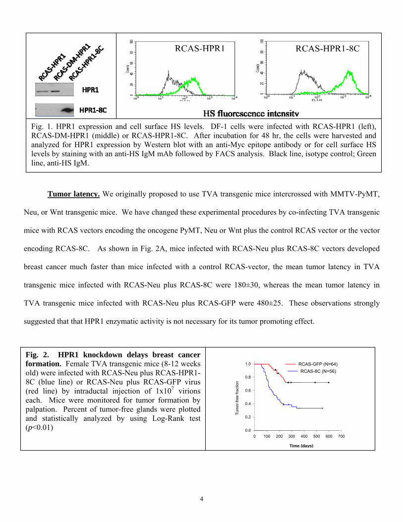
Preparation of RCAS vectors. Three RCAS vectors containing a full-length HPR1 gene, an enzymatic
activity-dead HPR1 gene (RCAS-DM-HPR1, double mutations at amino acid residues 225 & 343), and a C-
terminus gene fragment (RCAS-8C, with a fusion of 8-kDa and the C-terminus of HPR1 including amino acid
residues from 415-543). All inserts were tagged with a Myc epitope. This allowed us to titrate virus
concentrations and monitor the expression levels in vivo. Western blot analysis with an anti-Myc tag antibody
revealed that HPR1 was detected as a 18-kDa protein in DF-1 cells transfected with RCAS-8C vector, whereas
the full-length HPR1 was detected as 50-kDa protein. Immunofluorescence staining revealed that RCAS-HPR1
virus-infected DF-1 cells had lower cell surface heparan sulfate levels, compared to RCAS-8C-infected DF-1
cells (. These results confirmed that the C terminus of HPR1 did not have HPR1 enzymatic activity.
Neu,
mice
enco
brea
trans
TVA
sugg
Fig.formold)8C (redeachpalpand (p<0
FiRCanlevlin
Tumor l
, or Wnt tran
e with RCAS
oding RCAS
st cancer m
sgenic mice
A transgenic
gested that th
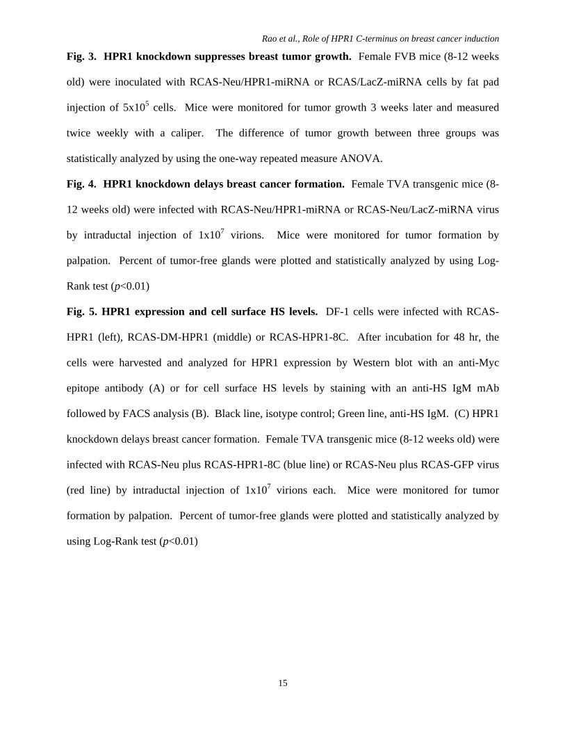
. 2. HPR1mation. Fem) were infect(blue line)
d line) by ih. Mice wepation. Perc
statistically0.01)
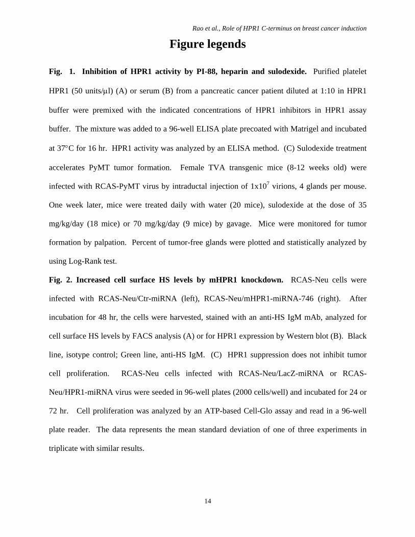
g. 1. HPR1 CAS-DM-Hnalyzed for Hvels by stainne, anti-HS I
latency. We
nsgenic mice
S vectors enc
S-8C. As sh
much faster t
infected wi
mice infect
hat HPR1 en
1 knockdowmale TVA trted with RCAor RCAS-N
intraductal iere monitorecent of tumoy analyzed
expression HPR1 (middl
HPR1 exprening with an IgM.
originally p
e. We have
coding the o
hown in Fig
than mice in
ith RCAS-N
ted with RC
nzymatic acti
wn delays ransgenic miAS-Neu plus
Neu plus RCinjection ofed for tumoor-free gland
by using
and cell sue) or RCAS
ession by Wanti-HS IgM
proposed to
changed the
oncogene PyM
g. 2A, mice i
nfected with
Neu plus RC
CAS-Neu plu
ivity is not n
breast canice (8-12 wes RCAS-HPCAS-GFP vf 1x10⁷ virior formationds were ploLog-Rank
urface HS leS-HPR1-8C.
Western blot M mAb follo
4
use TVA tra
ese experime
MT, Neu or
infected with
h a control R
CAS-8C wer
us RCAS-G
necessary for
ncer eeks R1-irus ions
n by tted test
evels. DF-1 After incu
with an antiowed by FAC
ansgenic mi
ental procedu
Wnt plus th
h RCAS-Ne
RCAS-vecto
re 180±30, w
GFP were 48
r its tumor pr
0
Tum
or-fr
ee fr
actio
n
0.0
0.2
0.4
0.6
0.8
1.0
1 cells were ubation for 4i-Myc epitopCS analysis.
ce intercross
ures by co-in
he control RC
eu plus RCA
or, the mean
whereas the
80±25. The
romoting eff
Time (
100 200 300 4
RR
infected wi48 hr, the cepe antibody . Black line
sed with MM
nfecting TV
CAS vector
AS-8C vector
n tumor late
e mean tumo
se observati
fect.
(days)
400 500 600 7
RCAS-GFP (N=64)RCAS-8C (N=56)
ith RCAS-Hells were har or for cell , isotype con
MTV-PyMT
VA transgenic
or the vecto
rs developed
ency in TVA
or latency in
ions strongly
700
HPR1 (left), rvested and surface HS
ntrol; Green
T,
c
r
d
A
n
y
5
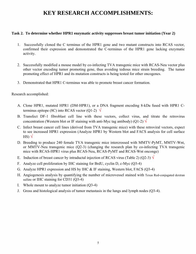
KEY RESEARCH ACCOMPLISHMENTS: Task 2. To determine whether HPR1 enzymatic activity suppresses breast tumor initiation (Year 2)
1. Successfully cloned the C terminus of the HPR1 gene and two mutant constructs into RCAS vector,
confirmed their expression and demonstrated the C-terminus of the HPR1 gene lacking enzymatic activity.
2. Successfully modified a mouse model by co-infecting TVA transgenic mice with RCAS-Neu vector plus
other vector encoding tumor promoting gene, thus avoiding tedious mice strain breeding. The tumor promoting effect of HPR1 and its mutation constructs is being tested for other oncogenes.
3. Demonstrated that HPR1 C-terminus was able to promote breast cancer formation.
Research accomplished:
A. Clone HPR1, mutated HPR1 (DM-HPR1), or a DNA fragment encoding 8-kDa fused with HPR1 C-terminus epitope (8C) into RCAS vector (Q1-2) √
B. Transfect DF-1 fibroblast cell line with these vectors, collect virus, and titrate the retrovirus concentration (Western blot or IF staining with anti-Myc tag antibody) (Q1-2) √
C. Infect breast cancer cell lines (derived from TVA transgenic mice) with these retroviral vectors, expect to see increased HPR1 expression (Analyze HPR1 by Western blot and FACS analysis for cell surface HS) √
D. Breeding to produce 240 female TVA transgenic mice intercrossed with MMTV-PyMT, MMTV-Wnt, or MMTV-Neu transgenic mice (Q2-3) (changing the research plan by co-infecting TVA transgenic mice with RCAS-HPR1 virus plus RCAS-Neu, RCAS-PyMT and RCAS-Wnt onconge)
E. Induction of breast cancer by intraductal injection of RCAS virus (Table 2) (Q2-3) √ F. Analyze cell proliferation by IHC staining for BrdU, cyclin D, c-Myc (Q3-4) G. Analyze HPR1 expression and HS by IHC & IF staining, Western blot, FACS (Q3-4) H. Angiogenesis analysis by quantifying the number of microvessel stained with Texas Red-conjugated dextran
sulfate or IHC staining for CD31 (Q3-4) I. Whole mount to analyze tumor initiation (Q3-4) J. Gross and histological analysis of tumor metastasis in the lungs and lymph nodes (Q3-4).
6
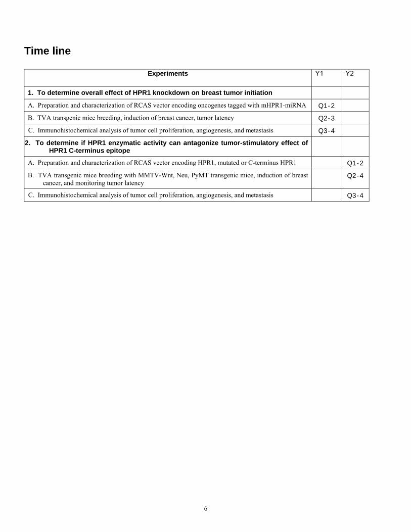
Time line
Experiments Y1 Y2
1. To determine overall effect of HPR1 knockdown on breast tumor initiation
A. Preparation and characterization of RCAS vector encoding oncogenes tagged with mHPR1-miRNA Q1-2
B. TVA transgenic mice breeding, induction of breast cancer, tumor latency Q2-3
C. Immunohistochemical analysis of tumor cell proliferation, angiogenesis, and metastasis Q3-4
2. To determine if HPR1 enzymatic activity can antagonize tumor-stimulatory effect of HPR1 C-terminus epitope
A. Preparation and characterization of RCAS vector encoding HPR1, mutated or C-terminus HPR1 Q1-2
B. TVA transgenic mice breeding with MMTV-Wnt, Neu, PyMT transgenic mice, induction of breast cancer, and monitoring tumor latency
Q2-4
C. Immunohistochemical analysis of tumor cell proliferation, angiogenesis, and metastasis Q3-4
7
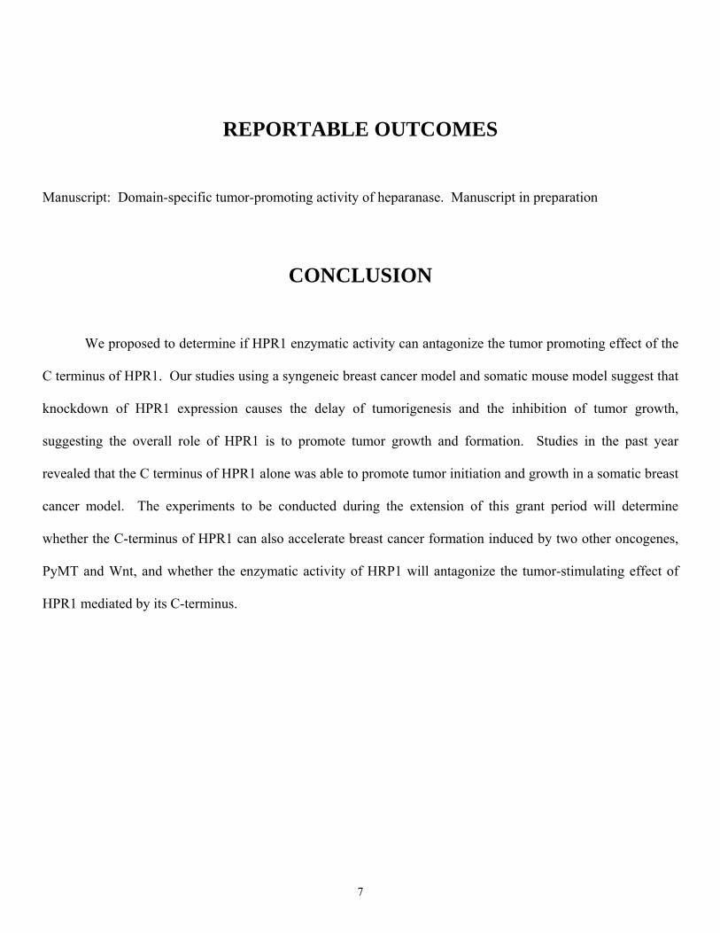
REPORTABLE OUTCOMES
Manuscript: Domain-specific tumor-promoting activity of heparanase. Manuscript in preparation
CONCLUSION
We proposed to determine if HPR1 enzymatic activity can antagonize the tumor promoting effect of the
C terminus of HPR1. Our studies using a syngeneic breast cancer model and somatic mouse model suggest that
knockdown of HPR1 expression causes the delay of tumorigenesis and the inhibition of tumor growth,
suggesting the overall role of HPR1 is to promote tumor growth and formation. Studies in the past year
revealed that the C terminus of HPR1 alone was able to promote tumor initiation and growth in a somatic breast
cancer model. The experiments to be conducted during the extension of this grant period will determine
whether the C-terminus of HPR1 can also accelerate breast cancer formation induced by two other oncogenes,
PyMT and Wnt, and whether the enzymatic activity of HRP1 will antagonize the tumor-stimulating effect of
HPR1 mediated by its C-terminus.
8
REFERENCES
1. Ilan N, Elkin M, Vlodavsky I: Regulation, function and clinical significance of heparanase in cancer
metastasis and angiogenesis. Int J Biochem Cell Biol 38:2018-2039, 2006
2. Gotte M, Yip GW: Heparanase, hyaluronan, and CD44 in cancers: a breast carcinoma perspective. Cancer
Res 66:10233-10237, 2006
3. Xu X, Quiros RM, Gattuso P, Ain KB, Prinz RA: High prevalence of BRAF gene mutation in papillary
thyroid carcinomas and thyroid tumor cell lines. Cancer Res 63:4561-4567, 2003
4. Xu X, Rao G, Quiros RM, Kim AW, Miao HQ, Brunn GJ, Platt JL, Gattuso P, Prinz RA: In vivo and in vitro
degradation of heparan sulfate (HS) proteoglycans by HPR1 in pancreatic adenocarcinomas. Loss of cell
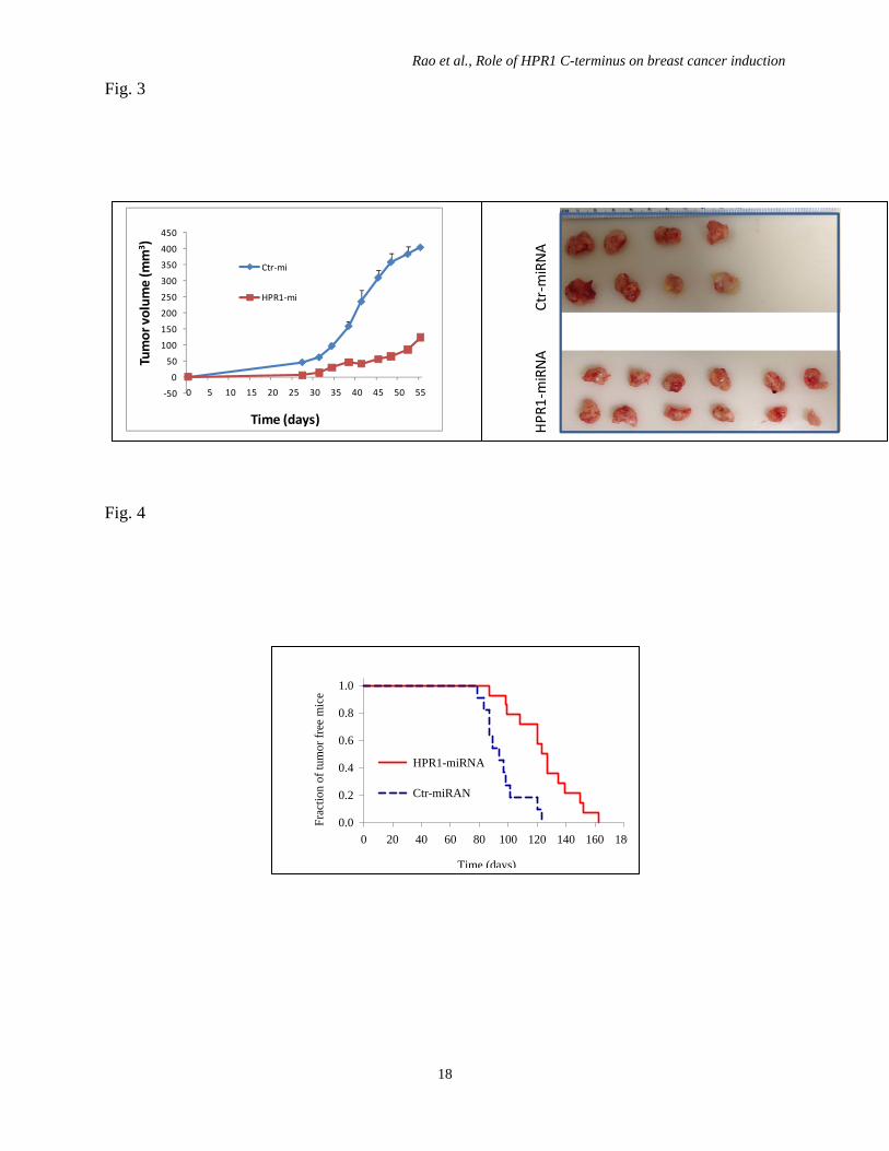
old) were inoculated with RCAS-Neu/HPR1-miRNA or RCAS/LacZ-miRNA cells by fat pad
injection of 5x10⁵ cells. Mice were monitored for tumor growth 3 weeks later and measured
twice weekly with a caliper. The difference of tumor growth between three groups was
statistically analyzed by using the one-way repeated measure ANOVA.
Fig. 4. HPR1 knockdown delays breast cancer formation. Female TVA transgenic mice (8-
12 weeks old) were infected with RCAS-Neu/HPR1-miRNA or RCAS-Neu/LacZ-miRNA virus
by intraductal injection of 1x10⁷ virions. Mice were monitored for tumor formation by
palpation. Percent of tumor-free glands were plotted and statistically analyzed by using Log-
Rank test (p<0.01)
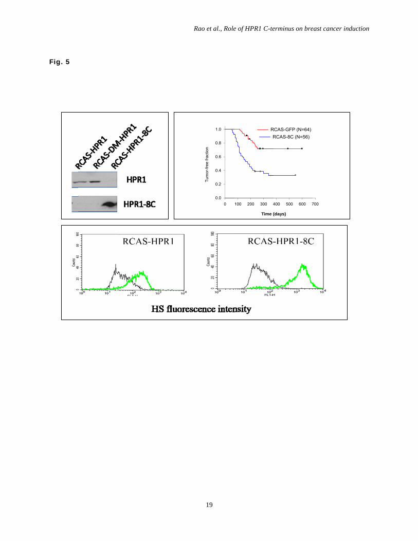
Fig. 5. HPR1 expression and cell surface HS levels. DF-1 cells were infected with RCAS-
HPR1 (left), RCAS-DM-HPR1 (middle) or RCAS-HPR1-8C. After incubation for 48 hr, the
cells were harvested and analyzed for HPR1 expression by Western blot with an anti-Myc
epitope antibody (A) or for cell surface HS levels by staining with an anti-HS IgM mAb
followed by FACS analysis (B). Black line, isotype control; Green line, anti-HS IgM. (C) HPR1
knockdown delays breast cancer formation. Female TVA transgenic mice (8-12 weeks old) were
infected with RCAS-Neu plus RCAS-HPR1-8C (blue line) or RCAS-Neu plus RCAS-GFP virus
(red line) by intraductal injection of 1x10⁷ virions each. Mice were monitored for tumor
formation by palpation. Percent of tumor-free glands were plotted and statistically analyzed by
using Log-Rank test (p<0.01)
Rao et al., Role of HPR1 C-terminus on breast cancer induction
16
Fig. 1
Days
0 20 40 60 80
Tum
or fr
ee e
vent
s
0.0
0.2
0.4
0.6
0.8
1.0 Sulodexide (35 mg/kg/day)(N=64)
Placebo (N=80)
Sulodexide (70 mg/kg/day) (N=36)
0
20
40
60
80
100
120
None PI-88 Heparin Sulodexide
% C
ontr
ol
0
0.51
1.52
2.5
0 0.2 1 5 25
Concentration (μg/ml)
OD
405
valu
e
PI-88 Heparin SulodexideA B
Rao et al., Role of HPR1 C-terminus on breast cancer induction
17
Fig. 2.
RCAS neu ctr RCAS neu 746 B
HS fluorescence intensity
0.00
20.00
40.00
60.00
80.00
100.00
24 hr 72 hr
RL
U (x
103 )
Ctr‐miRNA
mHPR1‐miRNA
Rao et al., Role of HPR1 C-terminus on breast cancer induction
18
Fig. 3
Fig. 4
‐50
0
50
100
150
200
250
300
350
400
450
0 5 10 15 20 25 30 35 40 45 50 55
Tumor volum
e (m
m3 )
Time (days)
Ctr‐mi
HPR1‐mi
HPR
1‐miRNA Ctr‐m
iRNA
Time (days)
0 20 40 60 80 100 120 140 160 180
Frac
tion
of tu
mor
free
mic
e
0.0
0.2
0.4
0.6
0.8
1.0
HPR1-miRNA
Ctr-miRAN
Fig. 5
R
Rao et al., Role
19
0Tu
mor
-free
frac
tion
0.0
0.2
0.4
0.6
0.8
1.0
e of HPR1 C-te
Time (
100 200 300 4
RR
erminus on bre
(days)
400 500 600 7
RCAS-GFP (N=64)RCAS-8C (N=56)
east cancer ind
700
duction
Rao et al., Role of HPR1 C-terminus on breast cancer induction
20
References 1. Vreys V and David G Mammalian heparanase: what is the message? J Cell Mol Med
2007; 11:427-52. 2. Gotte M and Yip GW Heparanase, hyaluronan, and CD44 in cancers: a breast carcinoma
perspective. Cancer Res 2006; 66:10233-7. 3. Ilan N, Elkin M, and Vlodavsky I Regulation, function and clinical significance of
heparanase in cancer metastasis and angiogenesis. Int J Biochem Cell Biol 2006; 38:2018-39.
4. Maxhimer JB, Pesce CE, Stewart RA, Gattuso P, Prinz RA, and Xu X Ductal carcinoma in situ of the breast and heparanase-1 expression: A molecular explanation for more aggressive subtypes. J Am Coll Surg 2005; 200:328-35.
5. Maxhimer JB, Quiros RM, Stewart R, Dowlatshahi K, Gattuso P, Fan M, et al. Heparanase-1 expression is associated with the metastatic portential of breast cancer. Surgery 2002; 132:326-333.
6. Vlodavsky I, Friedmann Y, Elkin M, Aingorn H, Atzmon R, Ishai-Michaeli R, et al. Mammalian heparanase: gene cloning, expression and function in tumor progression and metastasis. Nat Med 1999; 5:793-802.
7. Vlodavsky I and Friedmann Y Molecular properties and involvement of heparanase in cancer metastasis and angiogenesis. J Clin Invest 2001; 108:341-7.
8. Hulett M, Freeman C, Hamdorf B, Baker R, Harris M, and Parish C Cloning of mammalian heparanase, an important enzyme in tumor invasion and metastasis. Nature Medicine 1999; 5:803-809.
9. Parish CR, Freeman C, and Hulett MD Heparanase: a key enzyme involved in cell invasion. Biochim Biophys Acta 2001; 1471:M99-108.
10. Elkin M, Cohen I, Zcharia E, Orgel A, Guatta-Rangini Z, Peretz T, et al. Regulation of heparanase gene expression by estrogen in breast cancer. Cancer Res 2003; 63:8821-6.
11. Cohen I, Pappo O, Elkin M, San T, Bar-Shavit R, Hazan R, et al. Heparanase promotes growth, angiogenesis and survival of primary breast tumors. Int J Cancer 2005.
12. Kelly T, Suva LJ, Huang Y, Macleod V, Miao HQ, Walker RC, et al. Expression of heparanase by primary breast tumors promotes bone resorption in the absence of detectable bone metastases. Cancer Res 2005; 65:5778-84.
13. Edovitsky E, Elkin M, Zcharia E, Peretz T, and Vlodavsky I Heparanase gene silencing, tumor invasiveness, angiogenesis, and metastasis. J Natl Cancer Inst 2004; 96:1219-30.
14. Goldshmidt O, Zcharia E, Abramovitch R, Metzger S, Aingorn H, Friedmann Y, et al. Cell surface expression and secretion of heparanase markedly promote tumor angiogenesis and metastasis. Proc Natl Acad Sci U S A 2002; 3:3.
15. Zetser A, Bashenko Y, Miao HQ, Vlodavsky I, and Ilan N Heparanase affects adhesive and tumorigenic potential of human glioma cells. Cancer Res 2003; 63:7733-41.
16. Goldshmidt O, Zcharia E, Cohen M, Aingorn H, Cohen I, Nadav L, et al. Heparanase mediates cell adhesion independent of its enzymatic activity. Faseb J 2003; 17:1015-25.
17. Zetser A, Bashenko Y, Edovitsky E, Levy-Adam F, Vlodavsky I, and Ilan N Heparanase induces vascular endothelial growth factor expression: correlation with p38 phosphorylation levels and Src activation. Cancer Res 2006; 66:1455-63.
18. Cohen-Kaplan V, Naroditsky I, Zetser A, Ilan N, Vlodavsky I, and Doweck I Heparanase induces VEGF C and facilitates tumor lymphangiogenesis. Int J Cancer 2008; 123:2566-73.
Rao et al., Role of HPR1 C-terminus on breast cancer induction
21
19. Cohen-Kaplan V, Doweck I, Naroditsky I, Vlodavsky I, and Ilan N Heparanase augments epidermal growth factor receptor phosphorylation: correlation with head and neck tumor progression. Cancer Res 2008; 68:10077-85.
20. Lai NS, Simizu S, Morisaki D, Muroi M, and Osada H Requirement of the conserved, hydrophobic C-terminus region for the activation of heparanase. Exp Cell Res 2008; 314:2834-45.
21. Fux L, Feibish N, Cohen-Kaplan V, Gingis-Velitski S, Feld S, Geffen C, et al. Structure-function approach identifies a COOH-terminal domain that mediates heparanase signaling. Cancer Res 2009; 69:1758-67.
22. Liu CJ, Lee PH, Lin DY, Wu CC, Jeng LB, Lin PW, et al. Heparanase inhibitor PI-88 as adjuvant therapy for hepatocellular carcinoma after curative resection: A randomized phase II trial for safety and optimal dosage. J Hepatol 2009.
23. Lewis KD, Robinson WA, Millward MJ, Powell A, Price TJ, Thomson DB, et al. A phase II study of the heparanase inhibitor PI-88 in patients with advanced melanoma. Invest New Drugs 2008; 26:89-94.
24. Xu X, Ding J, Rao G, Shen J, Prinz RA, Rana N, et al. Estradiol induces heparanase-1 expression and heparan sulphate proteoglycan degradation in human endometrium. Hum Reprod 2007; 22:927-37.
25. Xu X, Rao G, Quiros RM, Kim AW, Miao HQ, Brunn GJ, et al. In vivo and in vitro degradation of heparan sulfate (HS) proteoglycans by HPR1 in pancreatic adenocarcinomas. Loss of cell surface HS suppresses fibroblast growth factor 2-mediated cell signaling and proliferation. J Biol Chem 2007; 282:2363-73.
26. Maxhimer JB, Somenek M, Rao G, Pesce CE, Baldwin D, Jr., Gattuso P, et al. Heparanase-1 gene expression and regulation by high glucose in renal epithelial cells: a potential role in the pathogenesis of proteinuria in diabetic patients. Diabetes 2005; 54:2172-8.
27. Quiros RM, Rao G, Plate J, Harris JE, Brunn GJ, Platt JL, et al. Elevated serum heparanase-1 levels in patients with pancreatic carcinoma are associated with poor survival. Cancer 2006; 106:532-40.
28. Xu X, Quiros RM, Gattuso P, Ain KB, and Prinz RA High prevalence of BRAF gene mutation in papillary thyroid carcinomas and thyroid tumor cell lines. Cancer Res 2003; 63:4561-4567.
29. Xu X, Quiros RM, Maxhimer JB, Jiang P, Marcinek R, Ain KB, et al. Inverse correlation between heparan sulfate deposition and heparanase-1 gene expression in thyroid papillary carcinomas: a potential role in tumor metastasis. Clinical Cancer Research 2003; 9:5968-5979.
30. Guy CT, Webster MA, Schaller M, Parsons TJ, Cardiff RD, and Muller WJ Expression of the neu protooncogene in the mammary epithelium of transgenic mice induces metastatic disease. Proc Natl Acad Sci U S A 1992; 89:10578-82.
31. Smith GH Stem cells and mammary cancer in mice. Stem Cell Rev 2005; 1:215-23. 32. Xiao L, Yuan X, and Sharkis SJ Activin A maintains self-renewal and regulates
fibroblast growth factor, Wnt, and bone morphogenic protein pathways in human embryonic stem cells. Stem Cells 2006; 24:1476-86.