288


Aerobic exercise and cognitive
behavioral therapy in FSHD:
A MODEL BASED APPROACH
Nicoline B.M. Voet

Acknowledgement:
The Prinses Beatrix Spierfonds (the Dutch Public Fund for Neuromuscular Disorders),
the Netherlands Organization for Health Research and Development (ZonMw),
Revalidatiefonds, Revalidatie Nederland and Global FSH funded the research in this
thesis. The FACTS-2-FSHD trial is part of the FACTS-2-NMD project, along with the
FACTS-2-PPS and FACTS-2-ALS trials.
Printing of this thesis was financially supported by:
Rehabilitation center Klimmendaal, Allergan, Ipsen Farmaceutica, Livit orthopedie and
ProReva orthopedisch maatwerk.
ISBN 978-94-6284-067-6
Cover design, artwork and layout by Erica Verlaan, xeri, Leersum
Propositions printed by Eveline Imminkhuizen, ei-design, Leersum
Thesis and invitations printed by Drukkerij Haveka, Alblasserdam
© N.B.M. Voet, Nijmegen, the Netherlands
All rights reserved. No part of this publication may be reproduced or transmitted in any
form or by any means, electronic or mechanical, including photocopy, recording, or any
information storage or retrieval system, without permission in writing from the author.
Articles are reprinted with permission of respective journals.

Aerobic exercise and cognitive behavioral therapy in FSHD:
A MODEL BASED APPROACH
Proefschrift
ter verkrijging van de graad van doctor
aan de Radboud Universiteit Nijmegen
op gezag van de rector magnificus prof. dr. J.H.J.M. van Krieken
volgens besluit van het college van decanen
in het openbaar te verdedigen
op vrijdag 14 oktober 2016
om 10.30 uur precies
door
Nicoline Berendina Maria Voet
geboren op 15 april 1983 te Nijmegen

Promotores:
Prof. dr. A.C.H. Geurts
Prof. dr. B.G.M. van Engelen
Prof. G. Bleijenberg
Manuscriptcommissie:
Prof. dr. M.T.E. Hopman (voorzitter)
Prof. dr. J.B. Prins
Prof. dr. F. Nollet (UvA)
Paranimfen:
Jos IJspeert
Erica Verlaan
CONTENTS
Chapter 1 General introduction 8
PART 1 FATIGUE IN NEUROMUSCULAR DISORDERS
Chapter 2 Muscle fatigue in muscular dystrophies 26
Chapter 3 Pain and fatigue in neuromuscular disorders 58
Chapter 4 Strength training and aerobic exercise training for muscle disease 70
PART 2 FACTS-2-FSHD STUDY
Chapter 5 Effect of aerobic exercise training and cognitive behavioral therapy on reduction of chronic fatigue in patients with facioscapulohumeral dystrophy: protocol of the FACTS-2-FSHD trial
118
Chapter 6 Both aerobic exercise and cognitive behavioral therapy reduce fatigue in FSHD: a RCT
140
Chapter 7 Distinct disease phases in muscles of facioscapulohumeral dystrophy patients identified by MR detected fat infiltration
164
Chapter 8 Quantitative MRI reveals decelerated fatty infiltration in muscles of active FSHD patients
190
Chapter 9 Summary and General discussion 210
Glossary of terms 246
Samenvatting 252
Dankwoord 262
Curriculum Vitae 270
List of publications 276
Donders Graduate School for Cognitive Neuroscience Series 284

Voor papa en mama


CHAPTER 1GENERAL INTRODUCTION

Mr. C is a 58-year old man with FSHD diagnosed at the age of 22. Apart from this
muscle disease, he has always been healthy. He has worked fulltime most of his
life but, since five years, he has been declared unfit for work. He used to live in
a home with a garden, together with his wife. Because he was not able to walk
stairs anymore, they were forced to move to an apartment with a small balcony.
Since gardening was his hobby, he lost his main activity which he replaced by
taking a nap every afternoon. At night, he is frequently awake and in the morning
he is already fatigued from the beginning of the day. His wife wants him to go with
her to family and friends, but he is reluctant to do so because he hates talking
about his disease and getting all sorts of well meant advice. He is afraid that
exercise might damage his muscles, as he regularly experiences muscle pain after
physical activity. As a consequence, he has stopped his daily cycling sessions
on a home trainer. His maximal walking distance has decreased to just a couple
of hundred meters, which makes him increasingly home-bound. All together, his
changing condition and circumstances have drawn him into a vicious circle of
physical inactivity and fatigue, with a great impact on his quality of life.
For patients with facioscapulohumeral muscular dystrophy (FSHD), medical
involvement often stops after receiving the diagnosis but, from their perspective,
the need for medical attention has just begun. Patients, clinicians and researchers
are searching for a curative treatment but, meanwhile, care for the consequences
of the disease is just as important, especially in the short turn. Many patients with
FSHD try to keep up their participation in social life and work. Citing a patient with
FSHD: “You just want to live your life like everyone else. That should be the aim of
medical research”. Yet, being physically active is difficult for patients due to muscle
weakness. The resulting reduction in aerobic capacity further restricts social
participation. Moreover, more than 60% of the patients with FSHD are severely
fatigued (1). In the past, fatigue in FSHD has received little attention as it was
regarded as an untreatable problem patients “just had to live with”. Consequently,
patients did not often spontaneously complain of fatigue. Still, recognition of
fatigue in FSHD is important for patients, whereas understanding and treating
fatigue is a great challenge for researchers and clinicians. Fortunately, medical
attention for fatigue is increasing.
FSHD
FSHD is the third-most common muscu lar dystrophy. The estimated prevalence is
one in 8,000 persons (2). FSHD is an autosomal dominant disease. It is associated
with subtelomeric contraction of the D4Z4 repeat region at chromosome 4q, with Cha
pte
r 1
Gen
eral
intr
oduc
tion
10

loss of tandem repeat units and toxic expression of the DUX4 gene in muscle
cells (3). In unaffected individuals, the D4Z4 array consists of 11 to 150 repeats,
whereas FSHD patients have only 1 to 10 repeats. In general, the disorder is more
severe in patients with lower numbers of repeats (Figure 1). Individuals genetically
determined to have FSHD, however, show a wide range of clinical severity, age of
onset, and rate of disease progression, including some who remain asymptomatic
throughout their lives. This variability suggests that the disease has a strong
epigenetic component.
Figure 1 FSHD is linked to the 4qQ subtelomere and the epigenetic status of
the 4q35 D4Z4 array
When D4Z4 is composed of many DUX4 copies the DNA becomes ‘locked’. As a result, the
DUX4 gene is switched off or ‘silenced’. However, if there are only a few DUX4 copies, the
DNA ‘relaxes’ and becomes accessible. When this happens, the DUX4 gene is switched
on resulting in carbon copies of the gene being made – called RNA. These contain the
instructions to build a DUX4 protein. Figure courtesy of Andreas Leidenroth (4).
Epigenetics concerns the mechanisms other than DNA sequence that
influence gene expression. An example of an epigenetic mechanism is DNA
methylation, a process by which methyl groups are added to DNA. The more
methylation, the tighter the chromatin is compacted and the less the gene inside
is expressed. Conversely, reduced methylation (hypomethylation) relaxes the
chromatin and increases the likelihood of gene expression. Healthy individuals have
numerous D4Z4 repeats which are highly methylated (Figure 1) (4). FSHD1-affected
individuals have few repeats and these are hypomethylated. FSHD1 asymptomatic
or unaffected individuals also have few repeats, but these have a higher degree of
methylation (5). Recently, a new subtype of FSHD, type 2, (FSHD2) was identified
(6). The symptoms of FSHD1 and FSHD2 are similar; the difference between
11

the conditions is their genetic locus and frequency of occurrence (7). FSHD2
individuals have many D4Z4 repeats, like healthy individuals, but they are severely
hypomethylated (6).
FSHD2 is much less prevalent than FSHD1. Current research projects try to identify
and manipulate the epigenetic regulators of the DUX4 gene expression in both
FSHD1 and FSHD2 in order to decrease the symptoms of the disease.
FSHD derives its name from the muscle groups that are affected first: facial and
shoulder girdle muscles. While the disease progresses, humeral, abdominal, pelvic
girdle and foot dorsiflexor muscles often become involved as well (Figure 2) (8).
Lower abdominal muscles are weaker than the upper abdominal muscles, causing
a ‘Beevor’s sign’, a physical finding specific for FSHD (Figure 3) (9).
Figure 2 A visual representation of the muscle groups ordered by degree
of fatty infiltration from red (most often affected) to yellow (least
affected) (8)
The most commonly described extramuscular manifestations are hearing loss
and retinal telangiectasias, occurring in 75% and 60% of the affected individuals,
respectively (10).Cha
pte
r 1
Gen
eral
intr
oduc
tion
12

The heart is not affected in most cases, although asymptomatic arrhythmias
and conduction defects have been described (11). The median age of onset
is around 17 years, but the onset of clinical symptoms varies from infancy to
the seventh decade. The course of FSHD is usually slowly progressive, but the
severity among patients is extremely variable, even within families, ranging from
isolated facial weak ness to severe generalized weakness, with approximately 20%
of patients eventually becoming wheelchair-dependent. Many patients report a
relapsing course, with long periods of quiescence interrupted by periods of rapid
deterioration involving a particular muscle group, often heralded by pain in the
affected limb. Most of the patients have a normal life expectancy (10).
Figure 3 Beevor’s sign
Many FSHD patients have a protruding abdomen because the lower abdominal muscles
are more severely affected than the upper abdominal muscles. This asymmetrical weakness
leads to Beevor’s sign: upward displacement of the navel while flexing the neck. It is a typical
finding for FSHD on clinical examination.
MUSCLE IMAGING IN FSHD
A typical characteristic of FSHD is the asymmetric and individual involvement
of different skeletal muscles. In the last decade, substantial progress has been
made in the understanding of the molecular genetics of FSHD (12). However,
it is still unknown why the weakening of different muscles and muscle groups
occurs at different rates and times. Moreover, there are no biomarkers for an
objective assessment of the severity and progression of FSHD and to establish the
effectiveness of treatments. Currently, muscle ultrasound is predominantly used
as a screening tool for patients with suspected neuromuscular disorders, as it can
13
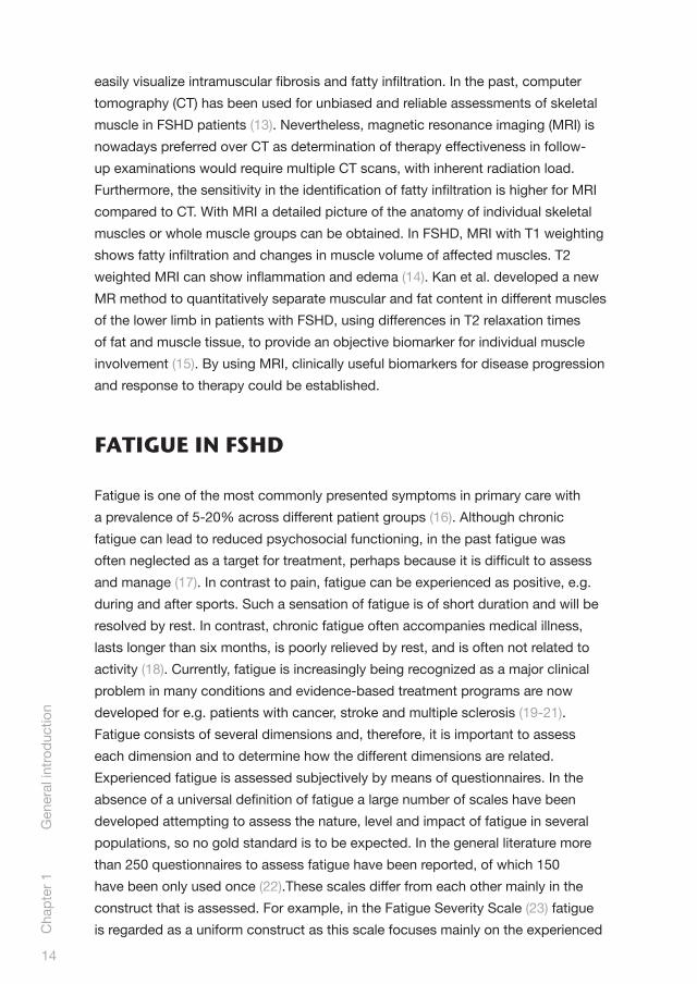
easily visualize intramuscular fibrosis and fatty infiltration. In the past, computer
tomography (CT) has been used for unbiased and reliable assessments of skeletal
muscle in FSHD patients (13). Nevertheless, magnetic resonance imaging (MRI) is
nowadays preferred over CT as determination of therapy effectiveness in follow-
up examinations would require multiple CT scans, with inherent radiation load.
Furthermore, the sensitivity in the identification of fatty infiltration is higher for MRI
compared to CT. With MRI a detailed picture of the anatomy of individual skeletal
muscles or whole muscle groups can be obtained. In FSHD, MRI with T1 weighting
shows fatty infiltration and changes in muscle volume of affected muscles. T2
weighted MRI can show inflammation and edema (14). Kan et al. developed a new
MR method to quantitatively separate muscular and fat content in different muscles
of the lower limb in patients with FSHD, using differences in T2 relaxation times
of fat and muscle tissue, to provide an objective biomarker for individual muscle
involvement (15). By using MRI, clinically useful biomarkers for disease progression
and response to therapy could be established.
FATIGUE IN FSHD
Fatigue is one of the most commonly presented symptoms in primary care with
a prevalence of 5-20% across different patient groups (16). Although chronic
fatigue can lead to reduced psychosocial functioning, in the past fatigue was
often neglected as a target for treatment, perhaps because it is difficult to assess
and manage (17). In contrast to pain, fatigue can be experienced as positive, e.g.
during and after sports. Such a sensation of fatigue is of short duration and will be
resolved by rest. In contrast, chronic fatigue often accompanies medical illness,
lasts longer than six months, is poorly relieved by rest, and is often not related to
activity (18). Currently, fatigue is increasingly being recognized as a major clinical
problem in many conditions and evidence-based treatment programs are now
developed for e.g. patients with cancer, stroke and multiple sclerosis (19-21).
Fatigue consists of several dimensions and, therefore, it is important to assess
each dimension and to determine how the different dimensions are related.
Experienced fatigue is assessed subjectively by means of questionnaires. In the
absence of a universal definition of fatigue a large number of scales have been
developed attempting to assess the nature, level and impact of fatigue in several
populations, so no gold standard is to be expected. In the general literature more
than 250 questionnaires to assess fatigue have been reported, of which 150
have been only used once (22).These scales differ from each other mainly in the
construct that is assessed. For example, in the Fatigue Severity Scale (23) fatigue
is regarded as a uniform construct as this scale focuses mainly on the experienced Cha
pte
r 1
Gen
eral
intr
oduc
tion
14

impact of fatigue on daily life. However, unidimensional fatigue measures do not
capture the full spectrum of fatigue as a multidimensional phenomenon. To assess
fatigue more extensively, especially in research, multidimensional questionnaires
are applied. An example of a multidimensional instrument is the Checklist Individual
Strength (CIS), consisting of four subscales: subjective fatigue experience,
concentration, motivation and subjective physical activity. The subscale
experienced fatigue of the CIS (CIS-fatigue) assesses the level of experienced
fatigue and has been frequently used in clinical studies. The CIS-fatigue consists
of eight questions that have to be answered on a seven-point Likert scale (range
7-56). Severe fatigue is defined by a cut-off score of 35 or higher (24). The CIS-
fatigue has been used to assess the level of fatigue in patients with FSHD in a
cross-sectional study by Kalkman et al. It was found that more than 60% of the
patients with FSHD experienced severe fatigue (1). In addition, being severely
fatigued was associated with a lower level of social participation. Apparently,
fatigue is a prevalent and a relevant problem in patients with FSHD.
REHABILITATION OF FATIGUE IN FSHD
Rehabilitation of people with neurological disabilities is a process aimed at
enabling them to reach and maintain their optimal physical, sensory, intellectual,
psychological and social activity level. Rehabilitation provides disabled people with
the tools they need to attain and maintain independence and self-efficacy (25).
In FSHD, muscle function is impaired and declines over time. A progressive loss
of muscle strength and muscle endurance often leads to loss of functional abilities
and mobility. Patients with FSHD identify poor mobility, fatigue and the emotional
and social burden of the disease as the factors with the greatest impact on their
lives (26). Fatigue may result in patients altering their lifestyles to avoid activities.
Low physical activity levels may lead to even deconditioining, greater weakness
and atrophy of skeletal muscles, which causes a vicious circle of disuse and
increased fatigue (27).
In a longitudinal study Kalkman et al (28) built a model of perpetuating factors for
fatigue in patients with FSHD using structural equation modelling. A total of 60
ambulatory patients were studied twice during an 18-months period. Experienced
fatigue was assessed with the CIS-fatigue (24), while a multidimensional functional
assessment was used to identify various dimensions relevant for fatigue: pain,
muscle strength, physical activity, neuropsychological impairments, psychological
distress, sleep disturbances, concentration problems, social functioning and
social support, and quality of life. It appeared that lack of physical activity, sleep
15

disturbances and pain all contributed to experienced fatigue. Loss of muscle
strength contributed to experienced fatigue through a lower level of physical
activity. In addition, pain contributed to physical inactivity. Ultimately, experienced
fatigue and physical inactivity both determined the level of social dependence and
loss of participation. The model, presented in Figure 4, served as a basis for the
treatment protocol used in this thesis.
Figure 4 Model of perpetuating factors of fatigue for patients with FSHD
Source: Adapted from Kalkman et al. (28)
It was hypothesized that, in order to preserve functioning at the highest achievable
level and to prevent the vicious circle of inactivity, two different therapeutic
approaches can be followed: aerobic exercise therapy to promote physical activity
and cognitive behavioral therapy to stimulate an active lifestyle yet avoiding
excessive physical strain.
Aerobic exercise aims at maintaining muscle function and improving
cardiorespiratory status to optimize physical capacity as a prerequisite for
executing many activities in daily life. For a long time, individuals with muscle
degeneration were discouraged to perform physical exercise based on fear for
exacerbation of disease activity and damage to muscle fibers. However, recent
studies have shown that exercise in patients with neuromuscular disorders is safe
and, thus, applicable to patients with FSHD (29). Although the number of exercise
studies in patients with neuromuscular disorders is increasing, the overall amount
of studies is still scarce.
FSHD has a strong impact on psychosocial functioning as patients have to
periodically re-adapt their daily life activities to living with a progressive illness.
Illness cognitions and coping style influence the choice and level of activities and,
hence, quality of life.
Because a cognitive-behavioral approach influencing illness cognitions and coping
strategies has been proven successful for chronic fatigue syndrome (30) and post-
cancer fatigue (19), it was expected to be efficacious for chronic fatigue in patients
with FSHD as well. No previous studies used cognitive behavioral therapy to treat
fatigue in FSHD.
Cha
pte
r 1
Gen
eral
intr
oduc
tion
16

THE FACTS-2-FSHD STUDY
Since there is still a long way to go before a treatment is expected that will
decelerate or perhaps even cease disease progression in FSHD, interventions
to treat the consequences of the disease are particularly important. This thesis
reports the results of the FACTS-2-FSHD study (acronym for Fitness And Cognitive
behavioral TherapieS for Fatigue and ACTivitieS in FSHD) which is the first model-
based randomized clinical trial that evaluates the effects of aerobic exercise
training (AET) and cognitive behavioral therapy (CBT) on chronic fatigue in patients
with FSHD. These interventions are based on the above-mentioned model of
chronic fatigue. The primary objective of this study was to evaluate the effect of
both interventions on chronic fatigue in patients with FSHD as assessed with the
subscale fatigue of the Checklist Individual Strength. The secondary objective
was to evaluate the effects of each intervention on the known perpetuating factors
of chronic fatigue in FSHD based on secondary outcome measures covering all
domains of the International Classification of Functioning, Disability and Health
(ICF). In addition, it was aimed to find clinically useful MRI biomarkers of disease
progression and response to therapy in patients with FSHD.
The FACTS-2-FSHD study is one of the studies conducted within the FACTS-2-
NMD consortium. FACTS-2-NMD stands for Fitness And Cognitive behavioral
TherapieS for Fatigue and ACTivitieS in NeuroMuscular Diseases (www.facts2nmd.
nl), a consortium funded by the Dutch Public Fund for Neuromuscular Disorders
(Prinses Beatrix Spierfonds) and the Netherlands Organization for Health Research
and Development (ZonMw) (grant nr 89000003).
17

AIMS AND OUTLINE OF THE THESIS
This thesis consists of two parts. The first part gives an overview of the
prevalence, measurement and treatment of fatigue in neuromuscular disorders.
The second part presents the results of the FACTS-2-FSHD study.
The following research questions will be addressed:
PART 1 FATIGUE IN NEUROMUSCULAR DISORDERS
1. What is the prevalence and relevance of fatigue in patients with muscular
dystrophy?
Chapter 2 gives an overview of the prevalence of fatigue and consequences in
muscular dystrophies.
2. How can we assess fatigue in patients with neuromuscular disorders?
Chapter 3 provides a core set of instruments for measuring fatigue in patients
with neuromuscular disorders.
3. What is the evidence for exercise in muscle disease?
A Cochrane review regarding the effects of strength training and aerobic
exercise therapy in patients with muscle disease is reported in chapter 4.
PART 2 FACTS-2-FSHD STUDY
4. What are the effects of aerobic exercise therapy and cognitive behavioral
therapy on chronic fatigue in patients with FSHD?
The protocol of the FACTS-2-FSHD trial, which aims to decrease experienced
fatigue by AET and CBT, is described in Chapter 5. The main results of the trial
are presented in Chapter 6.
5. Can we discover structural abnormalities in skeletal muscle of FSHD patients
that may serve as biomarkers for disease progression and response to
therapy?
Cha
pte
r 1
Gen
eral
intr
oduc
tion
18

In chapter 7, MRI measurements are used to provide more information about
the underlying pathobiology of FSHD. Chapter 8 describes the effects of AET
and CBT on the progression of fatty infiltration in the thigh muscles of patients
with FSHD.
19
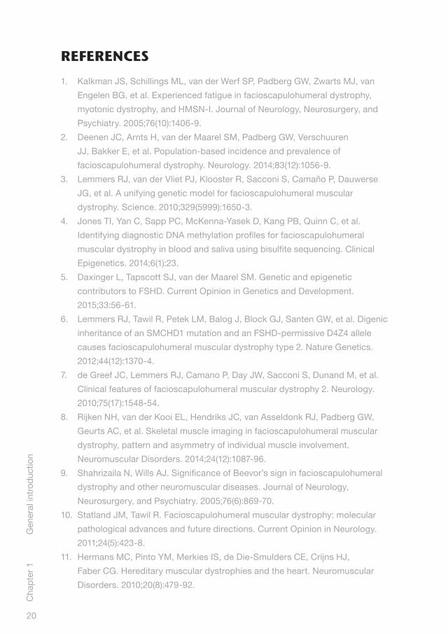
REFERENCES 1. Kalkman JS, Schillings ML, van der Werf SP, Padberg GW, Zwarts MJ, van
Engelen BG, et al. Experienced fatigue in facioscapulohumeral dystrophy,
myotonic dystrophy, and HMSN-I. Journal of Neurology, Neurosurgery, and
Psychiatry. 2005;76(10):1406-9.
2. Deenen JC, Arnts H, van der Maarel SM, Padberg GW, Verschuuren
JJ, Bakker E, et al. Population-based incidence and prevalence of
facioscapulohumeral dystrophy. Neurology. 2014;83(12):1056-9.
3. Lemmers RJ, van der Vliet PJ, Klooster R, Sacconi S, Camaño P, Dauwerse
JG, et al. A unifying genetic model for facioscapulohumeral muscular
dystrophy. Science. 2010;329(5999):1650-3.
4. Jones TI, Yan C, Sapp PC, McKenna-Yasek D, Kang PB, Quinn C, et al.
Identifying diagnostic DNA methylation profiles for facioscapulohumeral
muscular dystrophy in blood and saliva using bisulfite sequencing. Clinical
Epigenetics. 2014;6(1):23.
5. Daxinger L, Tapscott SJ, van der Maarel SM. Genetic and epigenetic
contributors to FSHD. Current Opinion in Genetics and Development.
2015;33:56-61.
6. Lemmers RJ, Tawil R, Petek LM, Balog J, Block GJ, Santen GW, et al. Digenic
inheritance of an SMCHD1 mutation and an FSHD-permissive D4Z4 allele
causes facioscapulohumeral muscular dystrophy type 2. Nature Genetics.
2012;44(12):1370-4.
7. de Greef JC, Lemmers RJ, Camano P, Day JW, Sacconi S, Dunand M, et al.
Clinical features of facioscapulohumeral muscular dystrophy 2. Neurology.
2010;75(17):1548-54.
8. Rijken NH, van der Kooi EL, Hendriks JC, van Asseldonk RJ, Padberg GW,
Geurts AC, et al. Skeletal muscle imaging in facioscapulohumeral muscular
dystrophy, pattern and asymmetry of individual muscle involvement.
Neuromuscular Disorders. 2014;24(12):1087-96.
9. Shahrizaila N, Wills AJ. Significance of Beevor’s sign in facioscapulohumeral
dystrophy and other neuromuscular diseases. Journal of Neurology,
Neurosurgery, and Psychiatry. 2005;76(6):869-70.
10. Statland JM, Tawil R. Facioscapulohumeral muscular dystrophy: molecular
pathological advances and future directions. Current Opinion in Neurology.
2011;24(5):423-8.
11. Hermans MC, Pinto YM, Merkies IS, de Die-Smulders CE, Crijns HJ,
Faber CG. Hereditary muscular dystrophies and the heart. Neuromuscular
Disorders. 2010;20(8):479-92.
Cha
pte
r 1
Gen
eral
intr
oduc
tion
20

12. Lemmers RJ, O’Shea S, Padberg GW, Lunt PW, van der Maarel SM. Best
practice guidelines on genetic diagnostics of Facioscapulohumeral muscular
dystrophy: workshop 9th June 2010, LUMC, Leiden, The Netherlands.
Neuromuscular Disorders. 2012;22(5):463-70.
13. van der Kooi EL, de Leeuw GE, Vlak MH, Hendriks JC, Padberg GW, Vogels
OJ. An unbiased and efficient computerised tomography method to quantify
muscle and adipose tissue volume in neuromuscular patients. Neurological
Sciences. 2006;26(6):423-9.
14. Mercuri E, Pichiecchio A, Allsop J, Messina S, Pane M, Muntoni F. Muscle
MRI in inherited neuromuscular disorders: past, present, and future. Journal
of Magnetic Resonance Imaging. 2007;25(2):433-40.
15. Kan HE, Scheenen TW, Wohlgemuth M, Klomp DW, van Loosbroek-
Wagenmans I, Padberg GW, et al. Quantitative MR imaging of individual
muscle involvement in facioscapulohumeral muscular dystrophy.
Neuromuscular Disorders. 2009;19(5):357-62.
16. Sharpe M, Wilks D. Fatigue. British Medical Journal. 2002;325(7362):480-3.
17. Dittner AJ, Wessely SC, Brown RG. The assessment of fatigue: a practical
guide for clinicians and researchers. Journal of Psychosomatic Research.
2004;56(2):157-70.
18. Krupp L. Fatigue: Butterworth Heinemann; 2003.
19. Gielissen MF, Verhagen S, Witjes F, Bleijenberg G. Effects of cognitive
behavior therapy in severely fatigued disease-free cancer patients compared
with patients waiting for cognitive behavior therapy: a randomized controlled
trial. Journal of Clinical Oncology. 2006;24(30):4882-7.
20. van Kessel K, Moss-Morris R, Willoughby E, Chalder T, Johnson MH,
Robinson E. A randomized controlled trial of cognitive behavior therapy for
multiple sclerosis fatigue. Psychosomatic Medicine. 2008;70(2):205-13.
21. Zedlitz AM, Rietveld TC, Geurts AC, Fasotti L. Cognitive and graded activity
training can alleviate persistent fatigue after stroke: a randomized, controlled
trial. Stroke. 2012;43(4):1046-51.
22. Hjollund NH, Andersen JH, Bech P. Assessment of fatigue in chronic disease:
a bibliographic study of fatigue measurement scales. Health and Quality of
Life Outcomes. 2007;5:12.
23. Krupp LB, LaRocca NG, Muir-Nash J, Steinberg AD. The fatigue severity
scale. Application to patients with multiple sclerosis and systemic lupus
erythematosus. Archives of Neurology. 1989;46(10):1121-3.
24. Vercoulen JH, Alberts M, Bleijenberg G. The Checklist Individual Strength.
Gedragstherapie. 1999;32:131–6.
21

25. WHO. Definition of Rehabilitation: World Health Association; 2013 [Available
from: http://www.who.int/topics/rehabilitation/en/].
26. Johnson NE, Quinn C, Eastwood E, Tawil R, Heatwole CR. Patient-identified
disease burden in facioscapulohumeral muscular dystrophy. Muscle & Nerve.
2012;46(6):951-3.
27. McDonald CM. Physical activity, health impairments, and disability
in neuromuscular disease. American journal of physical medicine &
rehabilitation. 2002;81(11 Suppl):S108-S20.
28. Kalkman JS, Schillings ML, Zwarts MJ, van Engelen BG, Bleijenberg G. The
development of a model of fatigue in neuromuscular disorders: a longitudinal
study. Journal of Psychosomatic Research. 2007;62(5):571-9.
29. Cup EH, Pieterse AJ, Ten Broek-Pastoor JM, Munneke M, van Engelen BG,
Hendricks HT, et al. Exercise therapy and other types of physical therapy
for patients with neuromuscular diseases: a systematic review. Archives of
Physical Medicine and Rehabilitation. 2007;88(11):1452-64.
30. Prins JB, Bleijenberg G, Bazelmans E, Elving LD, de Boo TM, Severens JL, et
al. Cognitive behaviour therapy for chronic fatigue syndrome: a multicentre
randomised controlled trial. Lancet. 2001;357(9259):841-7.
Cha
pte
r 1
Gen
eral
intr
oduc
tion
22

23

Training & Exercise
Medicine
MRI
Fatigue
ResearchFatigue in FSHD. . .
DNA
PART 1FATIGUE IN NEUROMUSCULAR DISORDERS
Fatigue

CHAPTER 2MUSCLE FATIGUE IN MUSCULAR DYSTROPHIES
Nicoline B.M. Voet
Alexander C.H. Geurts
Gijs Bleijenberg
Machiel J. Zwarts
George W. Padberg
Baziel G.M. van Engelen
Published in: Williams C., Ratel S. Human muscle fatigue.
Oxon: Routledge. 285-312 (2009)

OBJECTIVES The aim of this chapter is to provide an overview on:
the prevalence and assessment of fatigue in muscular dystrophies
the pathophysiological determinants of fatigue in muscular dystrophies
the possible treatment options of fatigue in muscular dystrophies
INTRODUCTION The aim of this chapter is to provide an update on the prevalence, relevance,
causes and treatment of fatigue in muscular dystrophies. In a study by McDonald
et al., the three problems most frequently cited as “very significant” by patients
with slowly progressive neuromuscular disease (n = 811) were muscle weakness
(57%), difficulty exercising (43%), and fatigue (40%) (1). In a study by Kalkman
et al., 61% of patients with facioscapulohumeral dystrophy (n = 139) and 74%
of patients with myotonic dystrophy (n = 322) were “severely fatigued” (2). The
muscular dystrophies are an inherited group of more than 30 distinct progressive
disorders resulting from defects in a number of genes required for normal muscle
structure and function. They are characterized by progressive loss of muscle
strength and integrity and they have a variable distribution and severity (3). We
will, however, limit our review to those main types of diseases which are most
frequent: Duchenne and Becker muscular dystrophy, myotonic dystrophy type 1,
facioscapulohumeral muscular dystrophy and the limb girdle muscular dystrophies.
A more extensive overview of muscular dystrophies can be found in Engel and
Franzini-Armstrong (4).
Cha
pte
r 2
Mus
cle
fatig
ue in
mus
cula
r d
ystr
ophi
es
28
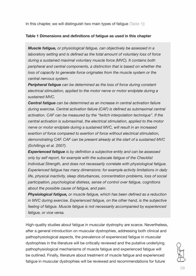
In this chapter, we will distinguish two main types of fatigue (Table 1):
Table 1 Dimensions and definitions of fatigue as used in this chapter
Muscle fatigue, or physiological fatigue, can objectively be assessed in a
laboratory setting and is defined as the total amount of voluntary loss of force
during a sustained maximal voluntary muscle force (MVC). It contains both
peripheral and central components, a distinction that is based on whether the
loss of capacity to generate force originates from the muscle system or the
central nervous system.
Peripheral fatigue can be determined as the loss of force during constant
electrical stimulation, applied to the motor nerve or motor endplate during a
sustained MVC.
Central fatigue can be determined as an increase in central activation failure
during exercise. Central activation failure (CAF) is defined as submaximal central
activation. CAF can be measured by the “twitch interpolation technique”. If the
central activation is submaximal, the electrical stimulation, applied to the motor
nerve or motor endplate during a sustained MVC, will result in an increased
exertion of force compared to exertion of force without electrical stimulation,
demonstrating CAF. CAF can be present already at the start of a sustained MVC
(Schillings et al. 2007).
Experienced fatigue is by definition a subjective entity and can be assessed
only by self report, for example with the subscale fatigue of the Checklist
Individual Strength, and does not necessarily correlate with physiological fatigue.
Experienced fatigue has many dimensions: for example activity limitations in daily
life, physical inactivity, sleep disturbances, concentration problems, loss of social
participation, psychological distress, sense of control over fatigue, cognitions
about the possible cause of fatigue, and pain.
Physiological fatigue, or muscle fatigue, which has been defined as a reduction
in MVC during exercise. Experienced fatigue, on the other hand, is the subjective
feeling of fatigue. Muscle fatigue is not necessarily accompanied by experienced
fatigue, or vice versa.
High-quality studies about fatigue in muscular dystrophy are scarce. Nevertheless,
after a general introduction on muscular dystrophies, addressing both clinical and
pathophysiological aspects, the prevalence of experienced fatigue in muscular
dystrophies in the literature will be critically reviewed and the putative underlying
pathophysiological mechanisms of muscle fatigue and experienced fatigue will
be outlined. Finally, literature about treatment of muscle fatigue and experienced
fatigue in muscular dystrophies will be reviewed and recommendations for future
29

research will be made. Throughout the chapter, the scientific knowledge will be
illustrated by a clinical case report that describes the experienced fatigue of a 59
year old man with facioscapulohumeral dystrophy, Mr. A.
DYSTROPHINOPATHIES
Duchenne muscular dystrophy (DMD) is the most common form of the human
muscular dystrophies. Becker muscular dystrophy (BMD) is a less frequent and
more benign form of the disease. The incidence of DMD is approximately 1 in
3.500 live male births. By comparison, BMD is found in 1 in 30,000 male births (5).
Both are X-linked recessive disorders and are caused by a mutation in the DMD
gene which is located on chromosome Xp21 and encodes for the production of
dystrophin. One third of the cases are due to spontaneous mutations (6).
The primary abnormality in DMD is the lack of dystrophin. In BMD, the protein
is reduced in amount or abnormal in size. Dystrophin is a 427 kiloDalton protein
normally found at the cytoplasmic face of the muscle cell surface membrane,
functioning as a component of a large, tightly associated glycoprotein complex (7).
In its absence, the glycoprotein complex is digested by proteases. This may initiate
the degeneration of muscle fibers, resulting in muscle weakness and potential
mechanical injury from tissue stress in rest and during exercise (8-10).
Diagnosis is suspected by characteristic clinical findings among which progressive
symmetrical muscle weakness is the most important, affecting proximal limb
muscles more than the distal muscles. Initially, only lower limb muscles are affected
accompanied by pseudohypertrophy of the calf muscles. Common musculoskeletal
complications are kyphoscoliosis and muscle contractures. Because dystrophin
is also found in the heart, brain, and the smooth muscles, frequent concomitant
manifestations are cardiomyopathy and mental retardation (11).
In DMD, the clinical symptoms first present between 3 and 5 years of age, and
patients generally lose ambulation between 7 and 12 years. In the past, death
usually occurred from cardiac or respiratory causes in the late teens or early
twenties. But recently, respiratory support can prolong survival into the fourth
decade (12). The diagnosis is supported by a family history suggestive of X-linked
recessive inheritance (4) or by dystrophin immunostaining of muscle tissue (6,
13, 14) Serum creatine phosphokinase (CK) level is generally increased to levels
that are 50-100 times the reference range (i.e. as high as 20,000 mU•mL-1). The
diagnosis is confirmed by identifying abnormalities in the dystrophin gene by
mutation analysis of DNA from peripheral blood leukocytes (6).
Cha
pte
r 2
Mus
cle
fatig
ue in
mus
cula
r d
ystr
ophi
es
30

In BMD, the distribution of muscle wasting and weakness is closely similar to that
in DMD, but the course of the disease is more benign and far less predictable,
with first clinical symptoms presenting around 12 years. Many patients remain
ambulatory into adult life (3, 6).
No curative treatment is available for both diseases, although first attempts are
made: the gene transfer technique by intramuscular injection of an antisense
oligonucleotide is under development (15), but several hurdles still need to be taken
(16). Therefore, emphasis currently is on respiratory care, treatment of cardiological
complications and optimizing the quality of life by symptomatic physiotherapeutic
and medical treatments (17). There is evidence that corticosteroid therapy in DMD
can reduce speed of decline of muscle strength and function (18, 19).
MYOTONIC DYSTROPHY
Myotonic dystrophy (DM) is the second most common muscular dystrophy.
There are two major forms: DM1, also known as Steinert’s disease, and DM2, a
multisystem disease, also known as Proximal Myotonic Myopathy (PROMM). In this
chapter, we will limit the discussion to DM1, which is more frequent.
DM1 is divided into congenital, classical, and minimal phenotypes according to the
age of the symptom onset and disease severity. The congenital form of DM1 will
not be further considered in this chapter, see Engel and Franzini-Armstrong (4). The
prevalence of DM1 is approximately 1 in 8,000 in the general population (20). DM1
is an autosomal-dominant disorder, of which the molecular basis is expansion of an
unstable repeat sequence in a non-coding part of the dystrophia myotonica protein
kinase gene (DMPK gene) on chromosome 19. The repeat expansion enlarges with
each generation, which leads to earlier onset and increased severity of symptoms
with each affected generation, a phenomenon which is known as “anticipation”
(21). There is increasing support for the theory that disruption of RNA metabolism,
which has effects on many other genes, explains the multisystemic nature of the
disease (22).
DM1 is clinically characterized by muscle weakness of the distal limbs, progressing
to the proximal limbs with gradual occurrence of myotonia (delayed relaxation
after muscle contraction). Weakness occurs most frequently in facial muscles, the
distal muscles of the forearm, and the ankle dorsiflexors with onset of symptoms in
the second, third or fourth decade (23). Associated findings include muscle pain,
cognitive and psychological changes, cataract, cardiac conduction defects and
endocrine disorders (20, 24, 25). Excessive daytime sleepiness is found in about
one-third of patients (26, 27).
31

The diagnosis can be suspected clinically by a positive family history and
by identifying the symptoms mentioned above. Specific genetic testing to
demonstrate the presence of an expanded CTG repeat in the DMPK gene is the
gold standard for the diagnosis of DM1 (20). Life expectancy is reduced for patients
with DM1. Respiratory insufficiency and cardiac diseases are the most common
causes of death (28-30). There is no disease-modifying therapy available for the
treatment of DM1. Therefore, treatment is symptomatic (31).
FACIOSCAPULOHUMERAL DYSTROPHY
Facioscapulohumeral dystrophy (FSHD) is the third most common muscular
dystrophy. The estimated prevalence is 1 in 20,000 persons (32). FSHD is an
autosomal dominant disease. It is associated with subtelomeric contraction of
chromosome 4q, with loss of tandem repeat-units. In general, the disorder is more
severe in a patient with a lower number of repeats. The pathogenetic mechanisms
in FSHD are unknown. The presence of some extramuscular manifestations in
FSHD suggests the involvement of a gene with pleiotropic effects or, alternatively,
the involvement of multiple genes (33). FSHD derives its name from the muscle
groups that are mainly affected first: facial and shoulder girdle muscles. During
disease progression humeral, abdominal, pelvic girdle and foot-extensor muscles
can become involved as well (32). Lower abdominal muscles are weaker than the
upper abdominal muscles, causing a “Beevor’s sign”, a physical finding specific
for FSHD (34). Most commonly described extramuscular manifestations are the
high-frequency hearing loss and retinal telangiectasias, occurring in 75% and 60%
of affected individuals, respectively (33). The heart is not affected in most cases,
though arrhythmias and conduction defects have been described (3).
The median age of onset is around 17 years, but the onset of clinical symptoms
varies from infancy to the seventh decade (32).
Although the exact gene defect or genetic mechanism is not yet known, a DNA test
is available for FSHD which detects a specific deletion in chromosome 4q35. This
diagnostic test is abnormal in 95 to 98 percent of typical FSHD cases (35-37).
The course of FSHD is usually slowly progressive but the severity among patients
is extremely variable, ranging from isolated facial weakness to severe generalized
weakness, with approximately 20 % of patients eventually becoming wheelchair-
dependent (32). Many patients report a relapsing course with long periods of
quiescence interrupted by periods of rapid deterioration involving a particular
muscle group, often heralded by pain in the affected limb. Most of the patients
have a normal life expectancy (33). Cha
pte
r 2
Mus
cle
fatig
ue in
mus
cula
r d
ystr
ophi
es
32

Currently, there is no genetic or pharmaceutical curative treatment available for
FSHD. Only two randomized controlled trials have been published. Recent trials
of albuterol, also known as salbutamol (38, 39), folic acid and methionine (40), and
creatine, a dietary supplement for building muscle (41), did not confirm or refute a
significant effect of either of these treatments (42). The mainstay of management
is, therefore, treatment of symptoms, prevention of secondary problems, and
improvement of functional abilities and quality of life (33).
LIMB GIRDLE MUSCULAR DYSTROPHIES
The limb girdle muscular dystrophies (LGMD) are a group of disorders which are
historically grouped together because of the shared clinical feature of predominant
involvement of the “limb-girdle” (pelvic and shoulder) musculature. However,
it is recognized that there is a broad heterogeneity of presentation and muscle
involvement in the LGMD group (43). The overall frequency has been estimated to
be 1: 14,000 – 1: 200,000 (5). Most cases of LGMD are inherited in an autosomal
recessive fashion (44). However, families with an autosomal dominant pattern of
inheritance have also been described, which probably account for about 10% of
all LGMDs (45). The emergence of a LGMD phenotype can result from mutations
in any of, at least, 19 different genes (46). The discovery of genetically distinct
subtypes has redefined the classification of LGMD and has led to a nomenclature
designating the autosomal dominant form as LGMD1A, 1B, 1C, etc, and the
autosomal recessive form as LGMD2A, 2B, 2C, etc (45). The proteins causing
LGMD have a wide range of localization across the muscle fiber, from sarcolemma
to nuclear envelope, with various functions (43, 46).
Weakness may affect proximal muscles of the shoulder girdle (scapulohumeral
type), the pelvic girdle (pelvifemoral type), or both. Neck flexor and extensor
muscles may be concurrently involved. Facial weakness, when present, is usually
mild and, in most cases, totally absent. Even in mild cases, there is preferential
weakness and atrophy of the biceps muscle.
Distal muscle strength is usually preserved, even at the late stage of the disease.
Selectivity of muscle involvement and clinical characteristics such as hypertrophy
of the calves or tongue, and late stage cardiac complications are associated more
or less specifically with each of the different forms (4).
The single constant biochemical abnormality in LGMD is the elevation of the CK
level. In autosomal recessive types of LGMD, serum CK is always increased, up
to 200 times the normal range. DNA analysis to detect a mutation in the affected
gene(s) is the gold standard of diagnosis (47). Reported age of onset of LGMDs
33
varies among the different mutations and is between 1 and 50 years, although
some patients may be asymptomatic. Compared with the autosomal dominant
type, autosomal recessive LGMD is usually associated with earlier age of onset,
more rapid progression, and relatively high CK values. Morbidity and mortality rates
vary, but with early onset the course is generally rapid (4). Treatment is supportive
and consists of physical therapy, assistive devices and monitoring of respiratory
function and cardial complications. Treatment is generally aimed at prolonging
survival and improving quality of life (46).
Clinical case: disease description
Mr. A is a 59 year old man who broke his clavicle in a football game when he was
18 years old. A year after the accident he went back to his general practitioner,
because symptoms of pain and decreased functioning of his shoulder did not
disappear. He was referred to a neurologist, who clinically diagnosed a “muscle
disease” when he was 19 years old. At that time, he knew that his mother, who
was wheelchair-dependent, had a “muscle disease”, but neither the diagnosis,
nor the prognosis of her condition was known. Decades later, his neurologist
told him he had a muscle disease which was known as “Landouzy–Dejerine”,
the former name of FSHD. The diagnosis FSHD was genetically confirmed
30 years later. At that time, an autosomal dominant inheritance pattern could
be recognized in his family. Many persons in every generation appeared to be
affected by the disease.
Mr. A experiences a relapsing course of FSHD with long stable periods followed
by periods of clear deterioration. Currently, facial, shoulder girdle, humeral,
abdominal, pelvic girdle and foot-dorsiflexor muscles are involved. He is still
ambulant but his unaided walking distance is restricted to approximately 100
meters. Outdoor he uses a rollator, which increases his walking distance to 250
meters. He is very afraid of becoming wheelchair-dependent, just like his mother.
Mr. A lives together with his wife in an apartment at ground level. He works four
days a week as an IT specialist and spends a lot of time in volunteer activities.
He plays the saxophone in a band.
EXPERIENCED FATIGUE
ASSESSMENT OF EXPERIENCED FATIGUE
Distinguishing experienced fatigue from muscle weakness, the key feature
in muscular dystrophy, may be difficult. Asking patients to describe their Cha
pte
r 2
Mus
cle
fatig
ue in
mus
cula
r d
ystr
ophi
es
34
fatigue will lead to several descriptions, varying from sleepiness, weakness,
exercise intolerance to exhaustion. Hence, experienced fatigue is, therefore, a
multidimensional concept with possible contributions of, for example physical,
cognitive and motivational factors (Table 1). Although experienced fatigue is
difficult to define, it still is a valuable concept which can be reliably measured by
using questionnaires. An often used questionnaire for the experience of fatigue
and its behavioral consequences is the Checklist Individual Strength (CIS). The
CIS is consists of four subscales: one scale for experienced fatigue, so called
“CIS-fatigue”, and three scales for reduction in motivation, physical activity and
concentration, respectively. Higher scores indicate higher levels of fatigue, more
concentration problems, a greater decrease in motivation and lower levels of
activities (48). The Abbreviated Fatigue Questionnaire (AFQ) is another short,
reliable, and easy-to-use instrument to determine the intensity of a patient’s
experienced fatigue. It consists of four questions that have to be answered on a
7-point Likert scale. A lower total score indicates a higher degree of fatigue (49).
PREVALENCE AND IMPACT OF EXPERIENCED FATIGUE
Kalkman et al. measured the prevalence of “severe experienced fatigue” in of 598
neuromuscular patients, among which 139 patients with FSHD and 322 patients
with DM. Both patient groups experienced high levels of fatigue (2). The mean CIS-
fatigue score in the FSHD group was 36.5 (SD 12.5) and in the DM group 40.4 (SD
11.8). In the FSHD group 61% of patients were “severely fatigued” (determined by
a CIS-fatigue score equal or above 35). In the DM group, this percentage was 74.
In both groups, age showed low but significant correlations with fatigue severity,
indicating that, in general, older patients experienced somewhat greater fatigue.
Severely fatigued patients scored lower on all Short Form-36 (SF-36) scales than
the non-severely fatigued patients, suggesting a relation between experienced
fatigue and activity limitations. There appeared to be several differences between
DM and FSHD patients. Patients with DM had higher scores for experienced
fatigue, reported greater problems with concentration, and had more difficulties
with initiative and planning than patients with FSHD. In FSHD patients and DM
patients, social functioning was related to fatigue severity.
Irrespective of its cause, fatigue has a major impact on daily functioning and
quality of life (50, 51). For example, in a study by van der Werf in patients with DM
(n = 32) and FSHD or LGMD (n = 20), severe fatigue was associated with greater
levels of psychological distress and more physical and psychosocial limitations, as
measured with the Sickness Impact Profile (SIP), the Symptom Checklist-90 (SCL-
90) and the Beck Depression Inventory Primary Care (BDI-PC) (52). In the study by
Kalkman et al. severely fatigued patients with FSHD or DM also had lower scores
35

on all subscales of the SF-36, which monitors disease burden.
This suggests a relation between experienced fatigue and the level of activity and
social participation (4). Apparently, fatigue is not only a frequent, but also a relevant
problem in muscular dystrophy.
Clinical case: experienced fatigue
Mr A has suffered from fatigue since the age of 40. He considers his fatigue and
muscle pain to be the most relevant and disabling consequences of his disease.
He defines his fatigue as a lack of energy which restrains him from activities.
After walking approximately 100 m, he has to stop because of severe fatigue
and muscle pain. These symptoms are comparable with the exhaustion he felt
after playing football in his younger years. That type of exhaustion, however, felt
positive, in contrast to the negative feeling associ ated with the present fatigue.
Fatigue has a significant and deleterious impact on his life that goes beyond the
other symptoms of FSHD. It takes almost two hours to prepare himself for work
every morning. After he has dressed, he often falls asleep due to exhaustion. He
can only travel by car because other forms of transport are too strenuous. He
describes himself as a “healthy mind in an aged body”.
DETERMINANTS OF FATIGUE
As fatigue in muscular dystrophy is a multidimensional concept, (see assessment
of experienced fatigue and Table 1), it is important to understand factors that
contribute to fatigue. Based on such an analysis, preventive and therapeutic
interventions can be developed. The critical pathophysiological determinants of
muscle fatigue and experienced fatigue will, therefore, be described in the next
section.
PATHOPHYSIOLOGICAL STUDIES OF MUSCLE FATIGUE
Because of practical reasons, pathophysiological studies depend to a large extent
on animal models. A review of Wineinger et al. summarizes the literature regarding
the physiological fatigue characteristics of skeletal muscles in animal models
of muscular dystrophy (53). Muscle fatigue in animal studies was expressed as
a percentage of initial force, i.e. physiological fatigue. Force was measured by
recording the action potential (AP) of muscles and muscle-evoked tension. Two
rodent models (mdx mouse and dystrophic hamster) have been studied most
extensively. The dystrophic hamster, lacking normal sarcoglycan, was used as a Cha
pte
r 2
Mus
cle
fatig
ue in
mus
cula
r d
ystr
ophi
es
36

model for LGMD. The mdx mouse lacks dystrophin, and was therefore considered
a model for DMD.
Significant variability has been observed before in studies of muscle fatigue
in dystrophic animals, which may be due to different experimental conditions.
Because of this variability, it is difficult to evaluate muscle fatigue in animal models
of muscular dystrophy. Still, some trends can be recognized (Table 2).
Table 2 The difference in fatigability of dystrophic animal muscles compared to healthy animal muscles can be explained by differences in muscle fiber types
Muscle fiber type Fatigability dystrophic animal muscles compared to healthy animal muscles.
Type I (slow-twitch, oxidative) ↓/ =
Type IIA ( fast-twitch, oxidative) =
Type IIB (fast-twitch, glycolytic) ↑
The dystrophic soleus muscle fatigued more slowly or at the same rate as that of
healthy animals. The soleus is largely composed of slow-twitch type I oxidative
muscle fibers and is considered to be fatigue resistant. Histological studies showed
an increase in the proportion of type I muscle fibers in the dystrophic soleus
muscle, which could explain the increased resistance to fatigue. The dystrophic
extensor digitorum longus (EDL) was weaker than in healthy animals and generally
more fatigable. The EDL muscle has a majority of type IIB fibers which are easily
fatigable. Pagala et al. described that type IIB dystrophic muscle fibers are more
susceptible to degeneration, in contrast to type I muscle fibers (54). No difference
was found in fatigability between healthy and dystrophic diaphragm muscles. The
diaphragm is composed of fast oxidative IIA muscle fibers, which are relatively
fatigue resistant. It appears that difference in fatigability of dystrophic animal
muscles compared to muscles of healthy animals can largely be explained by
differences in muscle fiber types (Table 2).
Apparently, type I, in contrast to type II muscle fibers of dystrophic animal muscles
have the potential to regenerate. Because aerobic training increases the proportion
of type I muscle fibers and, with that, fatigue resistance of healthy muscles, aerobic
training could be effective in decreasing fatigability of dystrophic muscles, as well
through the same mechanism (55).
37

Increased muscle fatigue has often been attributed to a decrease in the metabolic
potential of the individual muscle fibers. It is known that the levels of some energy
metabolites like creatine are decreased in muscular dystrophies such as DMD
(56, 57) which may aggravate muscle weakness and muscle fatigue. Interestingly,
in some types of LGMD, in which muscles are less severely affected, creatine
does not seem to be decreased, indicating that the level of creatine may serve
as a biomarker for the severity of muscle weakness and muscle fatigue (58).
Furthermore, the decrease in concentrations of other metabolites such as choline
and lactate was less severe in LGMD compared to DMD suggesting that these
metabolites could also be potential biomarkers (57).
In summary, these reports indicate that abnormal metabolite profiles could serve as
specific biomarkers to characterize the severity of muscular dystrophies.
PERIPHERAL VERSUS CENTRAL FATIGUE
Until recently, the emphasis in clinical research in muscular dystrophies was on
peripheral fatigue (Table 1). However, not only peripheral impairments, but also
changes within the central nervous system could be responsible for increased
fatigue. Schillings et al. first investigated central aspects of physiological fatigue
in patients with muscular dystrophy (59). Both peripheral and central aspects of
fatigue were determined during a sustained maximum voluntary contraction (MVC)
of elbow flexion in patients with FSHD (n=65) and DM (n=79) (Figure 1).
Unexpectedly, overall physiological fatigue and peripheral fatigue were smaller in
neuromuscular patients compared with healthy controls. Moreover, in patients with
FSHD and DM, physiological fatigue did not correlate with the level of experienced
fatigue. In contrast, Schulte-Mattler et al. described excessive peripheral fatigue in
a mixed group of neuromuscular disorders, among which FSHD and DM (60). This
discrepancy may be explained by a difference in the exercises. The type of exercise
in the study by Schillings et al., i.e. isometric contraction at maximal force level, is
hardly ever required in daily life and may also decrease blood supply.
Schulte-Mattler et al. elicited fatigue by intermittent and non-tetanic contractions
to avoid blood vessel occlusion. This type of exercise may be clinically more
relevant and valid for measuring physiological fatigue. CAF and central fatigue in
the study by Schillings et al. were measured by the twitch interpolation technique
(See chapter 2) (61). Central fatigue was minimal in all groups and did not differ
between groups nor did it have any relation with experienced fatigue. Remarkably,
CAF at the start of sustained MVC was enlarged in patients compared to controls.
CAF in patients was related to the level of experienced fatigue. An increased Cha
pte
r 2
Mus
cle
fatig
ue in
mus
cula
r d
ystr
ophi
es
38

CAF further decreases the maximal voluntary force in patients with muscular
dystrophy. The cause of this decreased central activation cannot be determined by
currently available techniques. It could be that the activation pattern of the central
nervous system is not able to compensate for the peripheral problems in muscular
dystrophies. The increased CAF could also be considered a beneficial adaptation,
which prevents the affected muscles from excessive fatigue.
Figure 1 Schematic representation of peripheral and central fatigue
The figure shows the decline over time (within 2 min) of the maximum voluntary force (on
the Y-axis), which is peripheral fatigue. The arrows indicate the moments of superimposed
electrical endplate stim ulation. The twitch interpolation may induce increments in muscle
force with examples of a negligible (†) and a large CAF (#). A (near) absent response indicates
a full voluntary activa tion of the muscle. The “at-rest twitches” are visible before (*) and
following (**) the contrac tion, with the post-experimental twitch being clearly lower, indicative
of peripheral fatigue. Source: Adapted from Zwarts et al., 2008. This figure is not the
registration of an individual patient.
VICIOUS CIRCLE OF PHYSICAL INACTIVITY
Fatigue may result in patients altering their life-styles to avoid activities. Low
physical activity levels may lead to even greater weakness and atrophy of skeletal
muscles, which causes a vicious circle of disuse and weakness. Physical inactivity
in turn can lead to chronic cardiovascular and muscle deconditioning and
increased cardiovascular health risks (1). For example, the maximal oxygen uptake
(VO2max) is abnormally low in patients with muscular dystrophy (62).
39
Body-composition measurements in muscular dystrophy patients by various
methods indicate reduced fat-free mass (FFM) and increased adiposity in these
patients relative to able-bodied control subjects of comparable ages and body
weights (63, 64). The excess body fat of muscular dystrophy patients additionally
impairs mobility and further increases the risk of cardiovascular disease.
In a study by McCrory et al., resting energy expenditure (REE) and total daily
energy expenditure (TEE) were measured by indirect calorimetry and heart rate
monitoring, respectively (65). Relatively active muscular dystrophy patients (FSHD,
LGMD, DM and BMD) did not differ in REE, but had a lower estimated TEE. They
also had a higher energy cost of physical activity than able-bodied subjects of the
same gender who were similar in age and weight, even after adjustment for FFM
differences. It is possible that the lower amount of time spent in physical activity
by muscular dystrophy patients can be attributed to the higher energy cost. An
alternative explanation is that persons with muscular dystrophy avoid physical
activity, because of the widespread belief that too much strain on the muscles will
accelerate the disease process (overwork weakness). Fear of physical activity, or
fear to damage the muscle, may also contribute to the reduced central activation
in patients with muscular dystrophy as described in the section peripheral versus
central fatigue. Irrespective of its cause, physical inactivity should be discouraged
in muscular dystrophy patients, because of an increasing risk of cardiovascular
disease and muscle deconditioning.
PERPETUATING FACTORS OF EXPERIENCED FATIGUE
Experienced fatigue can be regarded as a multimodal concept, with a wide
variety of contributing factors in patients with muscular dystrophy. These factors
can be categorized into predisposing, precipitating and perpetuating factors.
Predisposing factors include the presence of muscular dystrophy, whereas
precipitating factors include acute physical stresses such as a concomitant disease
or a period of relatively deterioration of muscle function. These factors cannot be
treated, in contrast to perpetuating factors, which contribute to the continuation of
experienced fatigue. Kalkman et al. used a longitudinal design to investigate the
perpetuating factors of experienced fatigue in patients with FSHD (n = 60) and DM
(n = 70) (66). Structural equation techniques, also referred to as “causal modeling”
were used. Based on longitudinal data, separate models for FSHD and DM were
developed. The model of perpetuating factors of experienced fatigue in FSHD
differed from the model for DM, the main difference being physical (in)activity and
pain. The model fit was best for FSHD (Figure 2).
Cha
pte
r 2
Mus
cle
fatig
ue in
mus
cula
r d
ystr
ophi
es
40

Figure 2 Adjusted model of perpetuating factors of experienced fatigue in
patients with FSHD (n = 60)
Severe muscle strength, (self-reported) physical inactivity, pain and sleep disturbances were
significantly associated with the level of experienced fatigue.
In FSHD, the level of physical (in)activity has a central place in the model. Lower
levels of physical activity contribute to higher levels of experienced fatigue and,
through that, to restrictions in social participation. The level of physical activity
is directly and negatively influenced by loss of muscle strength. In addition, pain
complaints influence levels of experienced fatigue both directly and indirectly by
decreasing physical activity. In contrast, in DM, physical activity and pain did not
differ between patients with and without severe experienced fatigue and, therefore,
did not significantly contribute to experienced fatigue.
Yet, sleep disturbances lead to higher levels of experienced fatigue in both FSHD
and DM patients. The observed patterns of perpetuating factors are unique
for FSHD and DM, and are different from the model of experienced fatigue in
chronic fatigue syndrome (48). They can be used as a basis to develop evidence-
based interventions to reduce fatigue. Specific attention should be paid to
sleep disturbances in both patient groups. Specifically in FSHD, treatment of
fatigue should also be directed at increasing physical activity and reducing pain
complaints.
EXPERIENCED FATIGUE AND PSYCHIATRIC DISORDERS
Fatigue is a characteristic of a number of affective disorders, and an association
between experienced fatigue and psychiatric symptoms has been reported in
a number of central nervous system disorders, including Parkinson’s disease
and multiple sclerosis (67, 68). In this perspective, it is relevant to know whether
psychiatric comorbidity is associated with fatigue severity in muscular dystrophies.
Although in the study by Kalkman et al. (see section on prevalence and impact
of experienced fatigue) severe experienced fatigue was related to higher levels of
psychological distress in both patients with FSHD and DM, most of the severely
fatigued patients did not fulfill the operational criteria of depression (4). The authors
41

argued that severe experienced fatigue can, therefore, not be seen as merely a
sign of depression. In a later study by Kalkman et al. using the Structured Clinical
Interview for DSM-IV axis 1 disorders, and Beck Depression Inventory (BDI)
lifetime and current psychiatric disorders (mood disorders, anxiety disorders and
substance-related disorders) were equally prevalent in a large cohort of DM and
FSHD patients, and were equally or even less present than these disorders in the
general (Dutch) population (69, 70). The most common psychiatric disorders were
depression and phobias. Psychiatric comorbidity was not associated with fatigue
severity or muscle strength in the various neuromuscular disorders. In conclusion,
psychiatric comorbidity is not an explanation for experienced fatigue in FSHD and
DM.
Clinical case: perpetuating factors of fatigue
Mr A experiences fatigue during activities of daily life, work and leisure, but also
when reading a book due to concentration problems. He easily falls asleep by
day. At night, his sleep is often disturbed by muscle pain. In the past, when
he was less physically disabled, he exercised at a low intensity. In his youth,
he played football. In adult life, he practised swimming once a week and, later
on, physical fitness, but avoided excessive training because of fear of overuse.
Nevertheless, many years ago he altered his lifestyle. He stopped swimming
and playing football. Currently, he is physically inactive and in a vicious circle of
disuse and weakness. His physical inactivity results in muscle and cardiovascular
deconditioning and obesity. Altogether, he is at risk for cardiovascular disease.
Seven years ago, he experienced an ischemic cerebrovascular incident, which
further increased his experienced fatigue.
TREATMENT OF FATIGUE
Most treatment studies in patients with muscular dystrophy do not describe the
efficacy of the intervention in terms of decreasing muscle fatigue or experienced
fatigue. Nevertheless, we will provide a critical overview of the possible treatment
options with respect to fatigue in these patients. Treatment strategies that will
be reviewed include physical exercise training, drug treatment and cognitive
behavioral therapy.
Cha
pte
r 2
Mus
cle
fatig
ue in
mus
cula
r d
ystr
ophi
es
42

TRAINING STUDIES IN ANIMALS
Extrapolating data from animal studies to humans must be done with caution,
because there are large differences in biomechanical properties and phenotypic
expression of the dystrophic disorder between humans and, for example, the mdx
mouse. Nevertheless, it may still be valuable to consider animal studies first, since
unique information can be obtained (53, 71). Exercise training in animals mainly
consisted of high-repetition aerobic-type activities like swimming, treadmill running
or voluntary-wheel running. Two reviews described that dystrophic animals had a
normal (and beneficial) adaptation to mild, voluntary submaximal aerobic exercise,
which generally included an increase in muscle strength per cross-sectional area
of muscle tissue and a reduction in muscle degeneration. The oxidative capacity
and the proportion of oxidative fibers were increased, especially in slow-twitch
muscles and in the muscles that were not severely affected by the dystrophy (53,
72). Aerobic training apparently increases the amount of type I muscle fibers, as
hypothesized earlier in this chapter (see section on pathophysiological studies of
muscle fatigue). Younger animals tend to benefit more from exercise studies than
older animals. The muscles of young dystrophic mdx mice have a greater rate
of recovery of force production than those of older mdx mice. Histological and
contractile studies suggest that this difference is due to an increased regenerative
capacity in young dystrophin-deficient mdx mice, which is lost in older mdx mice
(71, 73).
Carter et al. reviewed studies of exercise training and contraction-induced
muscle-injury in animal models of muscular dystrophy (72). A majority of the
studies in both normal and dystrophic animals showed that untrained eccentric
exercise (lengthening of the muscle during contraction) may injure the contractile
and cytoskeletal components of the muscle fibers. During eccentric exercise,
sarcomeres are stretched and the actin and myosin filaments are pulled apart,
leading to disruption of the thick and thin filament array and subsequent damage
to cytoskeletal proteins. The inability to quickly repair a disruption of the membrane
causes an elevation in intracellular calcium concentration, which triggers calcium-
activated degradation pathways and further structural damage. This damage
results in fiber degeneration followed by inflammation and, eventually, fiber
regeneration. Probably because of their increased regenerative capacity, muscles
of younger mdx mice recovered more rapidly than those of older mdx mice (74-
77). Based on these animal studies, one can conclude that submaximal aerobic
exercise training can be beneficial. However, eccentric exercise training should be
avoided.
43

TRAINING STUDIES IN MUSCULAR DYSTROPHY PATIENTS
In the past, many patients with muscular dystrophy were advised not to exercise
because of the belief that too much exercise might lead to “overuse weakness”
(78-80). Yet, in their Cochrane review on muscle strength training and aerobic
exercise training for patients with muscle diseases, van der Kooi et al. concluded
that moderate intensity strength training in DM and FSHD appeared not to be
harmful, although there was insufficient evidence to establish its benefit (81). This
conclusion was based on merely two randomized clinical trials (RCTs) (38, 82).
When RCTs are scarce, evidence from nonrandomized studies and other designs,
such as pre-post studies or case-control studies, may be particularly relevant (83).
For this reason, Cup et al. reviewed not only RCTs, but also controlled clinical trials
and other designs of sufficient quality, using the list by van Tulder et al. (84, 85).
All types of exercise therapy and other physical therapy modalities were included
for patients with muscular dystrophy, among which patients with FSHD, LGMD,
DM and DMD. Cup et al. also concluded that exercise training is not harmful in
muscular dystrophies (85). However, based on the reviewed studies, there was
insufficient evidence for the effectiveness of muscle strengthening exercises,
although there were some indications that aerobic exercises may have a positive
effect on body functions as well as on activities and participation.
There are several limitations to consider when reviewing training studies in
muscular dystrophies. First of all, there are only very few randomized controlled
trials, each small in sample size. Second, studies are not immediately comparable
because they have used training protocols which differ regarding the intensity
and duration of the training, targeted muscle groups, type of strength training,
i.e. isometric or isokinetic, and type of controls. The majority of exercise training
studies have evaluated non-supervised home programs of relatively short duration,
using submaximal, low-intensity training levels. The short duration of most
strengthening studies does not allow differentiation between neural training effects
versus muscle fiber hypertrophy, which generally occurs after six weeks. Third,
the compliance of patients, especially during non-supervised home protocols, is a
possible confounding factor in all training studies. Fourth, because of the scarcity
of patients of each muscular dystrophy, studies have often grouped together
several disorders. Persons with different types of muscular dystrophy may however
respond very differently to exercise (86). Fifth, some studies used the contralateral
non-exercised muscle as a control in muscle strengthening interventions (87-89).
The problem with this study design is that there may be confounding cross-over
effect in the non-exercised muscles. Moreover, one can hardly expect meaningful
Cha
pte
r 2
Mus
cle
fatig
ue in
mus
cula
r d
ystr
ophi
es
44

effects of a single-limb training program on a patient’s activities, participation
and well-being (86). Olsen et al. (90) investigated the effect of aerobic training in
8 patients with FSHD. Twelve weeks of low-intense aerobic exercise improved
maximal oxygen uptake and workload with no signs of muscle damage. The
authors conclude that aerobic training is a safe method to increase exercise
performance in patients with FSHD. Most importantly, only one study described the
effect of strength training for experienced fatigue (39) (see section on medication
for muscle fatigue and experienced fatigue).
To conclude, aerobic exercise training appears not to be harmful in muscular
dystrophies and could have a positive effect on functioning, activities and
participation, but the number of high quality studies is low.
MEDICATION FOR MUSCLE FATIGUE AND EXPERIENCED FATIGUE
No curative pharmacological interventions are available, nevertheless, many
agents have been proposed as a potential pharmacological treatment for
decreasing muscle fatigue in muscular dystrophies. Creatine and β2- agonists
have been studied most frequently. Creatine is a well known nutritional supplement
among athletes because it increases muscle force and lean body mass (91).
Supplementation of creatine monohydrate might enhance muscle performance
in patients with muscular dystrophy, as they tend to have lower skeletal muscle
creatine levels (see section on pathophysiological studies off muscle fatigue).
A recent systematic review on creatine for treating muscle disorders concluded
that short- to intermediate-term supplementation with creatine monohydrate in
patients with muscular dystrophy may result in a significant, but minimal increase
in maximal isometric force of approximately 8.5% in quantitative muscle testing.
Most of the potentially clinically relevant effects in muscular dystrophies were seen
in dystrophinopathies (92).
Other investigators expected a positive effect of β2- agonists in decreasing muscle
fatigue as high doses of β2- agonists have muscle anabolic properties. In animals
and healthy volunteers, β2-adrenergic agonists, such as clenbuterol and albuterol,
increase muscle strength and muscle mass, in particular when combined with
strength training (93, 94). Based on the model by Kalkman et al. (see section on
perpetuating factors of experienced fatigue) we might expect that these drugs
can decrease experienced fatigue, as many of them are aimed to increase muscle
strength. However, in the study by van der Kooi et al., albuterol and a strength
training program did not have any effect on experienced fatigue (39). Moreover,
45

β2- agonists are often associated with numerous undesirable side effects including
increased heart rate and muscle tremor, factors that have limited their therapeutical
potential. Consequences of prolonged use are presently unclear (38, 39).
Thus, although creatine and albuterol appear to be effective in increasing muscle
strength in healthy subjects, they seem to have little effect on muscle strength and
experienced fatigue in muscular dystrophies.
COGNITIVE BEHAVIORAL THERAPY
Muscular dystrophies have a large impact on psychosocial functioning as patients
must continuously adapt to their progressive illness. Illness cognitions and coping
styles influence the level of physical activity and, consequently, experienced fatigue
and restrictions in social participation. Hence, changing illness cognitions and
coping style may lead to a better quality of life. A cognitive-behavioral approach
has been proven successful in the chronic fatigue syndrome (95, 96) and for post
cancer fatigue (97, 98) and may be effective in patients with muscular dystrophy as
well. Cognitive behavioral therapy in FSHD should, for instance, be focused on the
known perpetuating factors of experienced fatigue as described by Kalkman et al.,
i.e. sleep disturbances, pain complaints and, physical inactivity (66, 99) (see section
on perpetuating factors of experienced fatigue). Therapy should be adapted to the
life of each individual, resulting in an individualized treatment approach. Altogether,
cognitive behavioral therapy seems a rational, promising treatment for fatigue in
muscular dystrophies.
Clinical case: treatment of fatigue
Mr A. has never used any drug for alleviating his fatigue. It has simply not been
mentioned by any clinician. He believes that physical exercise is beneficial for
decreasing his fatigue. In this perspective, he regrets that it is very difficult for
him to exercise because of his obesity and poor cardiovascular condition. He
is convinced that exercise at a maximum level will lead to overuse. He regards
cognitive behavioral therapy as a promising intervention for him.
Clinical case: Experienced fatigue in clinical setting
Mr. A. regrets that his experienced fatigue has never been asked for, nor treated
by health workers. He regards his fatigue as an essential disability, whereas his
physician has focused on the genetical aspects and diagnosis of the disease. He
is happy that fatigue is now on the research and on the medical agenda.Cha
pte
r 2
Mus
cle
fatig
ue in
mus
cula
r d
ystr
ophi
es
46

CONCLUSION
Fatigue is not only a frequent, but also a very relevant symptom in patients
with muscular dystrophy. Based on the content of this chapter, several
recommendations for clinical practice and research can be made. Clinicians
should actively ask their patients about the presence of fatigue and its individual
characteristics. The nature of the experienced fatigue gives directions to its primary
cause. In particular, affective disorders need to be diagnosed or ruled out. The CIS-
fatigue questionnaire can be used to measure the severity of experienced fatigue.
The history taking should cover the possible perpetuating factors of fatigue and the
perceived disabilities in daily life.
Based on these perpetuating factors, a therapeutic intervention can be proposed.
Both in DM and FSHD, specific attention should be paid to sleep disturbances.
Specifically in FSHD, the level of physical activity should improve.
Aerobic exercise training appears to be a safe intervention to increase physical
activity. It may also be effective in maximizing functional ability and preventing
chronic physical complications of inactivity. High-resistance and eccentric strength
training should be avoided, particularly in muscular dystrophies caused by defects
in structural proteins in the dystrophin-glycoprotein complex, i.e. LGMD, BMD and
DMD.
In the future, research on fatigue in muscular dystrophies needs to investigate
each type of muscular dystrophy separately, both pathophysiologically and
therapeutically. All treatment strategies will ultimately depend on pathophysiology,
but the absence of hard evidence should not prevent clinicians and researchers
from investigating treatment options based on credible hypotheses.
47
FIVE KEY PAPERS THAT SHAPED THE TOPIC AREA
Study 1. Wineinger, M.A., Walsh, S.A. and Abresch, T. (2002). Muscle fatigue in
animal models of neuromuscular disease. American Journal of Physical Medicine
and Rehabilitation, 81, S81-S98.
A review of Wineinger et al. showed that difference in fatigability between
dystrophic animal muscles and muscles of healthy animals can largely be explained
by differences in muscle fiber types. The dystrophic soleus muscle, which is largely,
composed of slow-twitch type I oxidative muscle fibers, fatigued more slowly or
at the same rate as that of healthy animals. Yet, the dystrophic extensor digitorum
longus (EDL), which has mainly type IIB fibers, was weaker than in healthy animals
and generally more fatigable. No differences in fatigability were found between
healthy and dystrophic diaphragm muscles. The diaphragm is composed of fast
oxidative IIA muscle fibers, which are relatively fatigue resistant (Table 2).
Study 2. Kalkman, J.S., Schillings, M.L., Zwarts, M.J., van Engelen, B.G. and
Bleijenberg, G. (2007b). The development of a model of fatigue in neuromuscular
disorders: a longitudinal study. Journal of Psychosomatic Research, 62, 571-579.
Kalkman et al. investigated the perpetuating factors of experienced fatigue in
patients with FSHD (n = 60) and DM (n = 70) (Figure 2). Only in FSHD, the level
of physical (in)activity had a central place in the model. Lower levels of physical
activity contributed to higher levels of experienced fatigue and, through that, to
restrictions in social participation. The level of physical activity was directly and
negatively influenced by loss of muscle strength. In addition, pain complaints
influenced levels of experienced fatigue, both directly and indirectly, by decreasing
physical activity.
Study 3. Vignos, P.J., and Watkins, M.P. (1966). The effect of exercise in muscular
dystrophy. The Journal of the American Medical Association, 197, 843-848.
The first controlled study of strengthening exercise in muscular dystrophies used
a regimen with gradual increase in weight resistance in patients with DMD (n=14),
FSHD (n=4) and LGMD (n=6) over a one-year period. The authors reported strength
improvement throughout the first four months of exercise regardless of type of
dystrophy. The degree of improvement was related to the initial strength of the
exercised muscle. Therefore, they concluded that exercise programs should begin
early in the course of the disease.
Cha
pte
r 2
Mus
cle
fatig
ue in
mus
cula
r d
ystr
ophi
es
48
Study 4. Florence, J.M. and Hagberg, J.M. (1984). Effect of training on the exercise
responses of neuromuscular disease patients. Medicine and Science in Sports and
Exercise, 16, 460-465.
Because the hallmark of muscular dystrophy is motor weakness, more studies
have been conducted looking at muscle strength training than at aerobic exercise
training. Only in 1984, Florence et al. first described a positive effect of aerobic
exercise training on the exercise responses of neuromuscular disease patients
among which patients with LGMD (n=3) and FSHD (n=1). All patients completed a
12-week cycle ergometry training program, three days per week. The increase in
VO2max in the patients was almost the same as that of the healthy subjects (n=4).
The authors concluded that patients can develop relatively normal adaptations to
training. No definitive deleterious effects of training were demonstrated in these
patients.
Study 5. Lindeman, E., Leffers, P., Spaans, F., Drukker, J., Reulen, J., Kerckhoffs,
M. and Koke, A. (1995). Strength training in patients with myotonic dystrophy and
hereditary motor and sensory neuropathy: a randomized clinical trial. Archives of
Physical Medicine and Rehabilitation, 76, 612-620.
The number of recent studies on the effect of training in muscular dystrophy
lacking a randomized controlled design is striking. Lindeman et al. first conducted
a randomized clinical trial on the effects of strength training in muscular dystrophy.
This trial compared the effect of 24 weeks of strength training of the thigh muscles
versus no training in 36 adult patients with myotonic dystrophy. The participants
trained 3 times a week for 24 weeks with weights adjusted to their force. In the
DM patients, none of the outcomes showed any training effect. No serious side
effects of the training occurred. Training loads could be gradually increased in
all patients, because the repetition maximum improved. Three rather weak DM
patients were unable to perform the exercises according to the training instructions.
Most of the differences in muscle strength outcomes (isometric, dynamic and
endurance) between groups showed small, non-significant positive effects in
favor of the training group. Only changes in the endurance measure (13.1 s longer
maximum duration of an isometric contraction; 95% CI 2.2 to 24.0) reached
statistical significance. No signs of overuse, such as a decline in strength or a rise
in parameters of muscle membrane permeability were seen. However, this study
imposed merely a controlled strain for a relatively short period.
49

REFERENCES1. McDonald CM. Physical activity, health impairments, and disability in
neuromuscular disease. American journal of physical medicine & rehabilitation.
2002;81(11 Suppl):S108-S20.
2. Kalkman JS, Schillings ML, van der Werf SP, Padberg GW, Zwarts MJ, van
Engelen BG, et al. Experienced fatigue in facioscapulohumeral dystrophy,
myotonic dystrophy, and HMSN-I. Journal of Neurology, Neurosurgery, and
Psychiatry. 2005;76(10):1406-9.
3. Emery AE. The muscular dystrophies. Lancet. 2002;359(9307):687-95.
4. Engel AG, Franzini-Armstrong C. Myology 2004.
5. Emery AE. Population frequencies of inherited neuromuscular diseases-a world
survey. Neuromuscular Disorders. 1991;1(1):19-29.
6. Aartsma-Rus A, van Deutekom JC, Fokkema IF, van Ommen GJ, den Dunnen
JT. Entries in the Leiden Duchenne muscular dystrophy mutation database: an
overview of mutation types and paradoxical cases that confirm the reading-
frame rule. Muscle & Nerve. 2006;34(2):135-44.
7. Monaco AP, Bertelson CJ, Liechti-Gallati S, Moser H, Kunkel LM. An
explanation for the phenotypic differences between patients bearing partial
deletions of the DMD locus. Genomics. 1988;2(1):90-5.
8. Petrof BJ, Stedman HH, Shrager JB, Eby J, Sweeney HL, Kelly AM.
Adaptations in myosin heavy chain expression and contractile function in
dystrophic mouse diaphragm. American Journal of Physiology. 1993;265(3 Pt
1):C834-C41.
9. Muntoni F, Torelli S, Ferlini A. Dystrophin and mutations: one gene, several
proteins, multiple phenotypes. Lancet Neurology. 2003;2(12):731-40.
10. Lapidos KA, Kakkar R, McNally EM. The dystrophin glycoprotein complex:
signaling strength and integrity for the sarcolemma. Circulation Research.
2004;94(8):1023-31.
11. Muntoni F. Cardiomyopathy in muscular dystrophies. Current Opinion in
Neurology. 2003;16(5):577-83.
12. Eagle M, Bourke J, Bullock R, Gibson M, Mehta J, Giddings D, et al. Managing
Duchenne muscular dystrophy-the additive effect of spinal surgery and
home nocturnal ventilation in improving survival. Neuromuscular Disorders.
2007;17(6):470-5.
13. Griggs RC, Bushby K. Continued need for caution in the diagnosis of
Duchenne muscular dystrophy. Neurology. 2005;64(9):1498-9.
14. Muntoni F. Is a muscle biopsy in Duchenne dystrophy really necessary?
Neurology. 2001;57(4):574-5.
Cha
pte
r 2
Mus
cle
fatig
ue in
mus
cula
r d
ystr
ophi
es
50
15. van Deutekom JC, Janson AA, Ginjaar IB, Frankhuizen WS, artsma-Rus
A, Bremmer-Bout M, et al. Local dystrophin restoration with antisense
oligonucleotide PRO051. The New England Journal of Medicine.
2007;357(26):2677-86.
16. Cossu G, Sampaolesi M. New therapies for Duchenne muscular dystrophy:
challenges, prospects and clinical trials. Trends in Molecular Medicine.
2007;13(12):520-6.
17. Bogdanovich S, Perkins KJ, Krag TO, Khurana TS. Therapeutics for Duchenne
muscular dystrophy: current approaches and future directions. Journal of
Molecular Medicine. 2004;82(2):102-15.
18. de Groot I. [Guideline on the use of corticosteroids in Duchenne muscular
dystrophy from paediatric neurologists, neurologists and rehabilitation
physicians]. Nederlands Tijdschrift voor Geneeskunde . 2006;150(12):684-5.
19. Manzur AY, Kuntzer T, Pike M, Swan A. Glucocorticoid corticosteroids for
Duchenne muscular dystrophy. Cochrane Database of Systematic Reviews.
2008(1):CD003725.
20. Machuca-Tzili L, Brook D, Hilton-Jones D. Clinical and molecular aspects of
the myotonic dystrophies: a review. Muscle & Nerve. 2005;32(1):1-18.
21. Howeler CJ, Busch HF, Geraedts JP, Niermeijer MF, Staal A. Anticipation in
myotonic dystrophy: fact or fiction? Brain. 1989;112 ( Pt 3):779-97.
22. Schara U, Schoser BG. Myotonic dystrophies type 1 and 2: a summary on
current aspects. Seminars in Pediatric Neurology. 2006;13(2):71-9.
23. Day JW, Ricker K, Jacobsen JF, Rasmussen LJ, Dick KA, Kress W, et al.
Myotonic dystrophy type 2: molecular, diagnostic and clinical spectrum.
Neurology. 2003;60(4):657-64.
24. George A, Schneider-Gold C, Zier S, Reiners K, Sommer C. Musculoskeletal
pain in patients with myotonic dystrophy type 2. Archives of Neurology.
2004;61(12):1938-42.
25. Kalkman JS, Schillings ML, Zwarts MJ, van Engelen BG, Bleijenberg
G. Influence of relatives on fatigue experienced by patients with
facioscapulohumeral dystrophy, myotonic dystrophy and HMSN-I. European
Neurology. 2006;56(1):24-30.
26. Culebras A. Sleep and neuromuscular disorders. Neurologic Clinics.
1996;14(4):791-805.
27. Phillips MF, Steer HM, Soldan JR, Wiles CM, Harper PS. Daytime somnolence
in myotonic dystrophy. Journal of Neurology. 1999;246(4):275-82.
28. Reardon W, Newcombe R, Fenton I, Sibert J, Harper PS. The natural history
of congenital myotonic dystrophy: mortality and long term clinical aspects.
Archives of Disease in Childhood. 1993;68(2):177-81.
51

29. de Die-Smulders CE, Howeler CJ, Thijs C, Mirandolle JF, Anten HB, Smeets
HJ, et al. Age and causes of death in adult-onset myotonic dystrophy. Brain.
1998;121 ( Pt 8):1557-63.
30. Mathieu J, Allard P, Potvin L, Prevost C, Begin P. A 10-year study of mortality in
a cohort of patients with myotonic dystrophy. Neurology. 1999;52(8):1658-62.
31. Harper PS, van Engelen BG, Eymard B, Rogers M, Wilcox D. 99th ENMC
international workshop: myotonic dystrophy: present management, future
therapy. 9-11 November 2001, Naarden, The Netherlands. Neuromuscular
Disorders. 2002;12(6):596-9.
32. Padberg GW. Facioscapulohumeral disease: Univeristy of Leiden, Leiden, the
Netherlands; 1982.
33. Tawil R, van der Maarel SM. Facioscapulohumeral muscular dystrophy. Muscle
& Nerve. 2006;34(1):1-15.
34. Shahrizaila N, Wills AJ. Significance of Beevor’s sign in facioscapulohumeral
dystrophy and other neuromuscular diseases. Journal of Neurology,
Neurosurgery, and Psychiatry. 2005;76(6):869-70.
35. Kohler J, Rohrig D, Bathke KD, Koch MC. Evaluation of the
facioscapulohumeral muscular dystrophy (FSHD1) phenotype in correlation
to the concurrence of 4q35 and 10q26 fragments. Clinical Genetics.
1999;55(2):88-94.
36. Ricci E, Galluzzi G, Deidda G, Cacurri S, Colantoni L, Merico B, et al. Progress
in the molecular diagnosis of facioscapulohumeral muscular dystrophy and
correlation between the number of KpnI repeats at the 4q35 locus and clinical
phenotype. Annals of Neurology. 1999;45(6):751-7.
37. Vitelli F, Villanova M, Malandrini A, Bruttini M, Piccini M, Merlini L, et al.
Inheritance of a 38-kb fragment in apparently sporadic facioscapulohumeral
muscular dystrophy. Muscle & Nerve. 1999;22(10):1437-41.
38. van der Kooi EL, Vogels OJ, van Asseldonk RJ, Lindeman E, Hendriks JC,
Wohlgemuth M, et al. Strength training and albuterol in facioscapulohumeral
muscular dystrophy. Neurology. 2004;63(4):702-8.
39. van der Kooi EL, Kalkman JS, Lindeman E, Hendriks JC, van Engelen
BG, Bleijenberg G, et al. Effects of training and albuterol on pain and
fatigue in facioscapulohumeral muscular dystrophy. Journal of Neurology.
2007;254(7):931-40.
40. van der Kooi EL, de Greef JC, Wohlgemuth M, Frants RR, van Asseldonk RJ,
Blom HJ, et al. No effect of folic acid and methionine supplementation on
D4Z4 methylation in patients with facioscapulohumeral muscular dystrophy.
Neuromuscular Disorders. 2006;16(11):766-9.
Cha
pte
r 2
Mus
cle
fatig
ue in
mus
cula
r d
ystr
ophi
es
52

41. Walter MC, Lochmuller H, Reilich P, Klopstock T, Huber R, Hartard M, et al.
Creatine monohydrate in muscular dystrophies: A double-blind, placebo-
controlled clinical study. Neurology. 2000;54(9):1848-50.
42. Rose MR, Tawil R. Drug treatment for facioscapulohumeral muscular
dystrophy. Cochrane Database of Systematic Reviews. 2004(2):CD002276.
43. Bushby K, Norwood F, Straub V. The limb-girdle muscular dystrophies-
diagnostic strategies. Biochimica et Biophysica Acta. 2007;1772(2):238-42.
44. Zatz M, de PF, Starling A, Vainzof M. The 10 autosomal recessive limb-girdle
muscular dystrophies. Neuromuscular Disorders. 2003;13(7-8):532-44.
45. Kirschner J, Bonnemann CG. The congenital and limb-girdle muscular
dystrophies: sharpening the focus, blurring the boundaries. Archives of
Neurology. 2004;61(2):189-99.
46. Daniele N, Richard I, Bartoli M. Ins and outs of therapy in limb girdle
muscular dystrophies. International Journal of Biochemistry and Cell Biology.
2007;39(9):1608-24.
47. Norwood F, de VM, Eymard B, Lochmuller H, Bushby K. EFNS guideline on
diagnosis and management of limb girdle muscular dystrophies. European
Journal of Neurology. 2007;14(12):1305-12.
48. Vercoulen JH, Swanink CM, Fennis JF, Galama JM, van der Meer JW,
Bleijenberg G. Dimensional assessment of chronic fatigue syndrome. Journal
of Psychosomatic Research. 1994;38(5):383-92.
49. Alberts M, Smets EM, Vercoulen JH, Garssen B, Bleijenberg G. [‘Abbreviated
fatigue questionnaire’: a practical tool in the classification of fatigue].
Nederlands Tijdschrift voor Geneeskunde. 1997;141(31):1526-30.
50. Sharpe M, Wilks D. Fatigue. BMJ. 2002;325(7362):480-3.
51. Gielissen MF, Knoop H, Servaes P, Kalkman JS, Huibers MJ, Verhagen S, et
al. Differences in the experience of fatigue in patients and healthy controls:
patients’ descriptions. Health and Quality of Life Outcomes. 2007;5:36.
52. van der Werf SP, Kalkman J, Bleijenberg G, van EB, Schillings M, Zwarts M.
The relation between daytime sleepiness, fatigue, and reduced motivation
in patients with adult onset myotonic dystrophy. Journal of Neurology,
Neurosurgery, and Psychiatry. 2003;74(1):138-9.
53. Wineinger MA, Walsh SA, Abresch T. Muscle fatigue in animal models of
neuromuscular disease. American journal of physical medicine & rehabilitation.
2002;81(11 Suppl):S81-S98.
54. Pagala MK, Venkatachari SA, Nandakumar NV, Ravindran K, Kerstein J, Namba
T, et al. Peripheral mechanisms of fatigue in muscles of normal and dystrophic
mice. Neuromuscular Disorders. 1991;1(4):287-98.
53

55. Ingjer F. Effects of endurance training on muscle fibre ATP-ase activity, capillary
supply and mitochondrial content in man. Journal of Physiology. 1979;294:419-
32.
56. Griffin JL, Williams HJ, Sang E, Clarke K, Rae C, Nicholson JK. Metabolic
profiling of genetic disorders: a multitissue (1)H nuclear magnetic resonance
spectroscopic and pattern recognition study into dystrophic tissue. Analytical
Biochemistry. 2001;293(1):16-21.
57. Sharma U, Atri S, Sharma MC, Sarkar C, Jagannathan NR. Skeletal muscle
metabolism in Duchenne muscular dystrophy (DMD): an in-vitro proton NMR
spectroscopy study. Magnetic Resonance Imaging. 2003;21(2):145-53.
58. Sharma U, Atri S, Sharma MC, Sarkar C, Jagannathan NR. Biochemical
characterization of muscle tissue of limb girdle muscular dystrophy: an 1H and
13C NMR study. NMR in Biomedicine. 2003;16(4):213-23.
59. Schillings ML, Kalkman JS, Janssen HM, van Engelen BG, Bleijenberg G,
Zwarts MJ. Experienced and physiological fatigue in neuromuscular disorders.
Clinical Neurophysiology. 2007;118(2):292-300.
60. Schulte-Mattler WJ, Muller T, Deschauer M, Gellerich FN, Iaizzo PA, Zierz
S. Increased metabolic muscle fatigue is caused by some but not all
mitochondrial mutations. Archives of Neurology. 2003;60(1):50-8.
61. Gandevia SC. Spinal and supraspinal factors in human muscle fatigue.
Physiological Reviews. 2001;81(4):1725-89.
62. Lewis SF, Haller RG. Skeletal muscle disorders and associated factors that limit
exercise performance. Exercise and Sport Sciences Reviews. 1989;17:67-113.
63. Kilmer DD, Abresch RT, McCrory MA, Carter GT, Fowler WM, Jr., Johnson
ER, et al. Profiles of neuromuscular diseases. Facioscapulohumeral muscular
dystrophy. American journal of physical medicine & rehabilitation. 1995;74(5
Suppl):S131-S9.
64. Johnson ER, Abresch RT, Carter GT, Kilmer DD, Fowler WM, Jr., Sigford BJ, et
al. Profiles of neuromuscular diseases. Myotonic dystrophy. American journal
of physical medicine & rehabilitation. 1995;74(5 Suppl):S104-S16.
65. McCrory MA, Kim HR, Wright NC, Lovelady CA, Aitkens S, Kilmer DD. Energy
expenditure, physical activity, and body composition of ambulatory adults
with hereditary neuromuscular disease. American Journal of Clinical Nutrition.
1998;67(6):1162-9.
66. Kalkman JS, Schillings ML, Zwarts MJ, van Engelen BG, Bleijenberg G. The
development of a model of fatigue in neuromuscular disorders: a longitudinal
study. Journal of Psychosomatic Research. 2007;62(5):571-9.
67. Surtees PG, Wainwright NW, Khaw KT, Day NE. Functional health status,
chronic medical conditions and disorders of mood. British Journal of
Psychiatry. 2003;183:299-303.Cha
pte
r 2
Mus
cle
fatig
ue in
mus
cula
r d
ystr
ophi
es
54

68. Schrag A. Psychiatric aspects of Parkinson’s disease--an update. Journal of
Neurology. 2004;251(7):795-804.
69. De Graaf R, Bijl RV, Smit F, Ravelli A, Vollebergh WA. Psychiatric and
sociodemographic predictors of attrition in a longitudinal study: The
Netherlands Mental Health Survey and Incidence Study (NEMESIS). American
Journal of Epidemiology. 2000;152(11):1039-47.
70. Kalkman JS, Schillings ML, Zwarts MJ, van Engelen BG, Bleijenberg G.
Psychiatric disorders appear equally in patients with myotonic dystrophy,
facioscapulohumeral dystrophy, and hereditary motor and sensory neuropathy
type I. Acta Neurologica Scandinavica. 2007;115(4):265-70.
71. Abresch RT, Walsh SA, Wineinger MA. Animal models of neuromuscular
diseases: pathophysiology and implications for rehabilitation. Physical
Medicine and Rehabilitation Clinics of North America. 1998;9(1):285-99.
72. Carter GT, Abresch RT, Fowler WM, Jr. Adaptations to exercise training
and contraction-induced muscle injury in animal models of muscular
dystrophy. American journal of physical medicine & rehabilitation. 2002;81(11
Suppl):S151-S61.
73. Carter GT, Wineinger MA, Walsh SA, Horasek SJ, Abresch RT, Fowler WM,
Jr. Effect of voluntary wheel-running exercise on muscles of the mdx mouse.
Neuromuscular Disorders. 1995;5(4):323-32.
74. McArdle A, Edwards RH, Jackson MJ. Accumulation of calcium by normal and
dystrophin-deficient mouse muscle during contractile activity in vitro. Clinical
Science (London). 1992;82(4):455-9.
75. Sacco P, Jones DA, Dick JR, Vrbova G. Contractile properties and
susceptibility to exercise-induced damage of normal and mdx mouse tibialis
anterior muscle. Clinical Science (London). 1992;82(2):227-36.
76. Dick J, Vrbova G. Progressive deterioration of muscles in mdx mice induced by
overload. Clinical Science (London). 1993;84(2):145-50.
77. Brussee V, Tardif F, Tremblay JP. Muscle fibers of mdx mice are more
vulnerable to exercise than those of normal mice. Neuromuscular Disorders.
1997;7(8):487-92.
78. Johnson EW, Braddom R. Over-work weakness in facioscapulohumeral
muscular dystrophy. Archives of Physical Medicine and Rehabilitation.
1971;52(7):333-6.
79. Fowler WM, Jr. Importance of overwork weakness. Muscle & Nerve.
1984;7(6):496-9.
80. Petrof BJ. The molecular basis of activity-induced muscle injury in Duchenne
muscular dystrophy. Molecular and Cellular Biochemistry.1998;179(1-2):111-23.
55
81. van der Kooi EL, Lindeman E, Riphagen I. Strength training and aerobic
exercise training for muscle disease. Cochrane Database of Systematic
Reviews. 2005(1):CD003907.
82. Lindeman E, Leffers P, Spaans F, Drukker J, Reulen J, Kerckhoffs M, et al.
Strength training in patients with myotonic dystrophy and hereditary motor and
sensory neuropathy: a randomized clinical trial. Archives of Physical Medicine
and Rehabilitation. 1995;76(7):612-20.
83. Hartling L, McAlister FA, Rowe BH, Ezekowitz J, Friesen C, Klassen TP.
Challenges in systematic reviews of therapeutic devices and procedures.
Annals of Internal Medicine. 2005;142(12 Pt 2):1100-11.
84. van Tulder MW, Assendelft WJ, Koes BW, Bouter LM. Method guidelines for
systematic reviews in the Cochrane Collaboration Back Review Group for
Spinal Disorders. Spine. 1997;22(20):2323-30.
85. Cup EH, Pieterse AJ, Ten Broek-Pastoor JM, Munneke M, van Engelen BG,
Hendricks HT, et al. Exercise therapy and other types of physical therapy
for patients with neuromuscular diseases: a systematic review. Archives of
Physical Medicine and Rehabilitation. 2007;88(11):1452-64.
86. Fowler WM, Jr. Role of physical activity and exercise training in neuromuscular
diseases. American journal of physical medicine & rehabilitation. 2002;81(11
Suppl):S187-S95.
87. Aitkens SG, McCrory MA, Kilmer DD, Bernauer EM. Moderate resistance
exercise program: its effect in slowly progressive neuromuscular disease.
Archives of Physical Medicine and Rehabilitation. 1993;74(7):711-5.
88. Kilmer DD, McCrory MA, Wright NC, Aitkens SG, Bernauer EM. The effect of a
high resistance exercise program in slowly progressive neuromuscular disease.
Archives of Physical Medicine and Rehabilitation. 1994;75(5):560-3.
89. Tollbäck A, Eriksson S, Wredenberg A, Jenner G, Vargas R, Borg K, et al.
Effects of high resistance training in patients with myotonic dystrophy.
Scandinavian Journal of Rehabilitation Medicine. 1999;31(1):9-16.
90. Olsen DB, Orngreen MC, Vissing J. Aerobic training improves exercise
performance in facioscapulohumeral muscular dystrophy. Neurology.
2005;64(6):1064-6.
91. Bemben MG, Lamont HS. Creatine supplementation and exercise
performance: recent findings. Sports Medicine. 2005;35(2):107-25.
92. Kley RA, Tarnopolsky MA, Vorgerd M. Creatine treatment in muscle disorders:
a meta-analysis of randomised controlled trials. Journal of Neurology,
Neurosurgery, and Psychiatry. 2008;79(4):366-7.
93. Yang YT, McElligott MA. Multiple actions of beta-adrenergic agonists on
skeletal muscle and adipose tissue. Biochemical Journal. 1989;261(1):1-10.
Cha
pte
r 2
Mus
cle
fatig
ue in
mus
cula
r d
ystr
ophi
es
56

94. Maltin CA, Delday MI. Satellite cells in innervated and denervated muscles
treated with clenbuterol. Muscle & Nerve. 1992;15(8):919-25.
95. Prins JB, Bleijenberg G, Bazelmans E, Elving LD, de Boo TM, Severens JL,
et al. Cognitive behaviour therapy for chronic fatigue syndrome: a multicentre
randomised controlled trial. Lancet. 2001;357(9259):841-7.
96. Chambers D, Bagnall AM, Hempel S, Forbes C. Interventions for the treatment,
management and rehabilitation of patients with chronic fatigue syndrome/
myalgic encephalomyelitis: an updated systematic review. Journal of the Royal
Society of Medicine. 2006;99(10):506-20.
97. Gielissen MF, Verhagen CA, Bleijenberg G. Cognitive behaviour therapy for
fatigued cancer survivors: long-term follow-up. British Journal of Cancer.
2007;97(5):612-8.
98. Gielissen MF, Verhagen S, Witjes F, Bleijenberg G. Effects of cognitive behavior
therapy in severely fatigued disease-free cancer patients compared with
patients waiting for cognitive behavior therapy: a randomized controlled trial.
Journal of Clinical Oncology. 2006;24(30):4882-7.
99. Della MG, Frusciante R, Vollono C, Dittoni S, Galluzzi G, Buccarella C, et
al. Sleep quality in Facioscapulohumeral muscular dystrophy. Journal of
Neurological Science. 2007;263(1-2):49-53
57

Research
CHAPTER 3184TH ENMC WORKSHOP REPORT: PAIN AND FATIGUE IN NEUROMUSCULAR DISORDERS
Imelda J.M. de Groot
Nicoline B.M. Voet
Henriet van Middendorp
Hans Knoop
Jes Rahbek
Baziel G.M. van Engelen
Published in: Neuromuscular Disorders 23 (2013); 1028-1032

INTRODUCTION This European Neuromuscular Centre (ENMC) care workshop was attended by
19 professionals working in the field of neuromuscular disorders (NMD) (pediatric
and adult), pain or fatigue from 8 different countries (USA, Great Britain, Germany,
France, Italy, Sweden, Denmark and the Netherlands), and 3 patient representatives
coming from the Netherlands. This ENMC organized workshop aimed to achieve
consensus on the definition of pain and fatigue in NMD, to define a core set of
measurement instruments for pain and fatigue in NMD, and to discuss possible
interventions. Prior to the workshop, all participants received a questionnaire on the
assessment instruments and interventions that they regularly used to, respectively,
assess and treat pain and fatigue. Two experts on (chronic) pain and fatigue in
general (Henriët van Middendorp and Hans Knoop) were invited to present an
overview of definitions, mechanisms, and measurement instruments for pain
and fatigue. Next to that, several participants presented data on the prevalence
of pain and fatigue in the various NMD populations. Through group discussions
with all participants, consensus was reached on the definition and core set of
measurement instruments for use in future research and clinical practice in NMD.
BACKGROUND
Pain and fatigue are common symptoms in neuromuscular disorders (NMD) with a
prevalence of 30–90%, present in all types of NMD, both in adults and children (1-
6). Pain and fatigue have a strong impact on many activities of daily life, including
mobility, work, school, leisure, and sleep (4, 7). The differences in reported
prevalence could be due to different definitions of pain and fatigue, and various
types of measurements. In order to develop effective treatment approaches, both
definitions and methods to measure pain and fatigue should be agreed on.
QUESTIONNAIRES
Questionnaires were sent to all participants prior to the workshop to get an
overview of the questionnaires on pain and fatigue that are currently used in
research and practice in NMD (8). In addition, the various interventions used to treat
pain and fatigue were investigated. To assess pain in adults, nearly all participants
used the SF-36 (items related to pain), a Likert scale, visual analog scale (=VAS),
numeric rating scale (=NRS), and the McGill Pain Questionnaire. For neuropathic
pain, more specific scales were mentioned, such as the Douleur Neuropathic 4
(=DN4), Neuropathic Pain Symptom Inventory (=NPSI), or ID-pain. In children, all Cha
pte
r 3
184t
h E
NM
C w
orks
hop
rep
ort:
Pai
n an
d fa
tigue
in n
euro
mus
cula
r d
isor
der
s
60
used the VAS or the Faces Pain Scale (of which there are many different versions),
few used body images to indicate the site of pain or the PedsQol (pain items), and
one used the San Salvadour scale for severely handicapped children or the Douleur
Enfant Gustav Roussy Scale for children between 2 and 6 years of age.
For fatigue in adults, most of the participants used the Fatigue Severity Scale
(=FSS), Borg CR-10 scale, NRS for fatigue, and some used the Modified Fatigue
Impact Scale or the Checklist Individual Strength (=CIS). Specific for Myotonic
Dystrophy type 1 (DM1), the following sleepiness scales were reported: Daytime
Sleepiness Scale (DSS), Pittsburgh Sleep Quality Index (PSQI), Epworth Sleepiness
Scale (ESS), and Unnalinna Narcolepsy Scale (UNS). For children, the (modified)
Borg scales were reported and the six minute walking test for ambulant children.
As possible interventions for pain, all participants mentioned guidance and
advice for daily activities with special attention for overuse, physical therapy, and
possible drugs such as pregabalin, citalopram, duloxetine, amitriptyline, creatine
monohydrate, and paminodrate.
For fatigue, possible interventions that were reported included polysomnography
to evaluate nocturnal hypoventilation and treatment, guidance and advice for
daily activities with special attention for overuse, physical therapy, and cognitive
behavioral therapy. Only for DM1 drugs to treat sleepiness were mentioned such as
modafinil, citalopram, and Ritalin.
The level of evidence for the efficacy of the interventions is low; available evidence
is restricted to drug treatment for neuropathic pain and sleepiness and fatigue in
DM (see Cochrane reviews).
PARTICIPANTS’ RESEARCH ON PAIN AND FATIGUE IN NMD
Baziel van Engelen reported on several studies in NMD populations, consisting of
cross-sectional data of adult DM1 (n = 322), DM type 2 (n = 29), OculoPharyngeal
Muscular Dystrophy (OPMD) (n = 35), Chronic Progressive External
Ophthalmoplegia (CPEO) (n = 28), Facioscapulohumeral muscular dystrophy
(FSHD) (n = 139), and Charcot Marie Tooth (CMT) (n = 137), as well as longitudinal
data of adult DM1 (n = 79), FSHD (n = 65), and CMT (n = 73). To assess pain and
fatigue in these studies, the McGill pain questionnaire and VAS were used for pain,
and the CIS and SF-36 for fatigue. The reported fatigue was between 54% (OPMD)
and 68% (CPEO), with significant impact on daily functioning. The pain scores were
between 47% (DM1) and 96% (CPEO), with a different distribution pattern for each
specific type of NMD. Pain scores were independent of age, impairments, physical
activity level, or muscle force. The longitudinal study in FSHD (n = 60) and DM1
61

(N = 75) resulted in an FSH health status model and a DM1 health status model.
Pain and fatigue were main determinants of health status in both diseases (7).
These models enable to rationally develop treatment strategies in FSHD and DM1
and form the starting point of the study presented by Nicoline Voet: the protocol
of the FACTS-2-FSHD trial. In this recently finished study, aerobic exercise training
is compared to cognitive behavioral therapy (data currently in analysis). Pain and
fatigue are measured by CIS, VAS for pain, 6 min walking test, and SF36.
Birgit Steffensen reported on a study of self-reported pain and fatigue in patients
with FSHD and their healthy relatives or caregivers. The FSS and VAS were used
to assess fatigue; pain was measured on a 1–7 scale as adapted from the FSS, by
VAS, and by localization on a body image.
Pain and fatigue scores were significantly higher in patients than in healthy
persons, there was no statistical difference between patients who were still able to
walk and those who were not, and there was a tendency towards higher pain and
fatigue scores in patients with less muscle strength.
Marie Kierkegaard reported the results of a cross-sectional study on 70 persons
with DM1. The ICF checklist was used for self-rating of perceived pain. Fatigue and
excessive daytime sleepiness was evaluated with the FSS and ESS, as well as with
the ICF checklist. Fatigue, daytime sleepiness and pain were rated as a problem
in 76%, 80% and 51%, respectively. The impact of the cut-off level for fatigue and
excessive sleepiness when using the FSS and ESS was stressed. For example,
with a score equal or above 4 in FSS 52% are classified as experiencing fatigue
compared to 17% when a cut-off score equal or above 5 is used.
Cornelia Kornblum discussed her study of 22 patients with DM1, 22 with DM2, and
22 controls. The FSS, DSS, ESS, PSQI, and UNS were used to assess fatigue and
sleepiness as part of a more extensive psychological test set. DM1 showed more
sleepiness and fatigue in all measurements compared to the other groups; only
the mean KFSS-, DSS-, and PSQI-scores were above the cutoff for pathological
performing. The DM2-patients showed more fatigue and a lower sleep quality
(mean KFSS- and PSQI-scores were above the cut-off for pathological performing);
no differences were found in daytime sleepiness. DM1 and DM2 patients were both
more fatigued than healthy controls, and DM1 more than DM2.
Luca Padua showed the results of a study in 392 patients with peripheral nerve
diseases, in which the Neuropathic Pain Symptom Inventory (NPSI), VAS, ID-pain,
and DN4 were assessed. In 60% of the patients, the VAS-pain was 3 or higher,
and approximately 30% had a VAS of 5 or higher. On ID-pain and DN4, 50–60%
62
Cha
pte
r 3
184t
h E
NM
C w
orks
hop
rep
ort:
Pai
n an
d fa
tigue
in n
euro
mus
cula
r d
isor
der
s

had symptoms of neuropathic pain. The NPSI showed no statistical difference
between the different symptoms of neuropathic pain, in which paraesthesias had
the highest score. In another study on 65 patients with FSHD, the SF-36, VAS-
pain, and ID-pain were assessed. According to the ID-pain, 7% of the patients had
very probable neuropathic pain, 20% probable neuropathic pain, 24% possible
neuropathic pain, 33% no pain, and 16% joint pain exclusively.
Bernard Wuyam showed the preliminary results of an ongoing study on the
assessment of neuromuscular dysfunction by means of magnetic nerve stimulation
in muscular dystrophies and CMT, and the effect of exercise.
Ulla Werlauff showed results of studies on the perception of pain and fatigue in
persons with spinal muscular atrophy (SMA) (52 persons: 7.8–72.6 years) and
congenital myopathies (95 persons: 5.5–75.2 years). The PedsQoL (generic,
specific), SF-36, Egen classification 2 scale, FSS, and Modified Fatigue Impact
Scale were used to measure pain and fatigue. In congenital myopathies, pain was a
problem in all muscles (overload?) whereas in SMA-type 2 it was not, or related to
an event like surgery.
In this study, fatigue was a problem in congenital myopathies but not in SMA. From
her results, Ulla Werlauf concluded that PedsQoL, SF-36 and Modified fatigue
impact scale were not very sensitive or conclusive. The recommended scale would
be the FSS.
Imelda de Groot showed results of former studies in adults with SMA and ALS and
an ongoing study in DMD. In an open questionnaire study in 99 adult SMA patients,
divided into two groups (SMA type1–2 and type 3), neck pain was most prevalent
in both groups (38% in SMA1-2, and 34% in SMA3); fatigue was reported in 64%
in SMA3, in contrast to 34% in SMA1-2. In a study in 74 persons with ALS, no
correlations were found between functional abilities (measured with ALS-FRS), and
pain or fatigue (measured with SF-36). In the ongoing study in boys with DMD (N
= 30) and training, the preliminary results show that boys with DMD have similar
adaptations to physical exercise (heart rate and EMG-adaptations) as healthy age-
matched controls (N = 99) and comparable subjective fatigue (measured by OMNI
scale), but that some boys can perceive/ experience fatigue in rest.
Yaacov Anziska showed preliminary results of two studies: one study in children
with NMD (7 with myopathy/limb girdle muscular dystrophy, 9 with DMD, 3
with CMT, and 2 with myasthenia gravis; age range 5– 19 years). The Epworth
Sleepiness Scale, the Vignos, and Brooke ratings scales, the Children’s Depression
Inventory, and the PedsQoL 3.0 Neuromuscular Module for Patients and Parents
63
were used. Except for the myopathy/DMD-group, there was no correlation between
PedsQOL-scores of the patients and those of their parents. There was evidence of
mild abnormal mood disorders and mild-moderate sleepiness. A study of 31 adults
(5 patients with DM1-2, 14 with LGMD, 5 with CMT, 5 with motor neuron diseases,
2 with myasthenia gravis) in which the SF-36 was used gave similar results.
Carole Bérard showed results of a study in 22 boys with DMD with pain and
osteopenia (7 ambulant, and 15 non-ambulant). In 21 of the 22 boys, a moderate
to severe pain was measured with NRS, most of the boys (21) experienced
pain during physiotherapy; 11 also spontaneously, 9 during the night and 5
(nonambulant) during transfers. There was no correlation between the NRS-pain
score and osteopenia.
Anna-Karin Kroksmark shared some practical experiences on pain and fatigue
in children with DMD from studies performed at her centre. In these studies,
PedsQoL, Multidimensional Fatigue Scale, Sleep Quality Index, and questions
about pain and tiredness were used. Pain has an increasing interest in the network
in Sweden and a national survey is now being performed. Adapted Faces Scales
were shown.
Caron Coleman showed her work on establishing how to measure pain in children:
one size does not fit all! Due to the different developmental stages the following
measurements were recommended for clinical utility: 0–5 years and cognitive or
learning difficulties observation and parent/caretaker report; 5–7 years-faces and
questions to child and parents regarding location, frequency, quality (if possible)
and impact on function; 8+ years-combination of faces/NRS and questions as
above; 13+ years Brief Pain Inventory/faces/NRS.
Heinz Jungbluth gave an overview of different childhood NMD in which exertial
myalgia with or without rhabdomyolysis is a common primary or secondary feature.
One of the open questions that need to be addressed in future research is whether
the different mechanisms underlying these features in distinct disorders will require
different treatment approaches.
Marion Main shared some thoughts on pain and fatigue in children with NMD. Pain
can be caused by secondary complications of the disorder, such as contractures,
cramps, muscle soreness etc. A warning was given that the physiotherapist can be
a cause of pain due to treatments like stretching.
Cha
pte
r 3
184t
h E
NM
C w
orks
hop
rep
ort:
Pai
n an
d fa
tigue
in n
euro
mus
cula
r d
isor
der
s
64

She pointed at the lack of knowledge on the topic of fatigue with regard to the role
of weakness in the development of fatigue and the important role of exercise in the
management of pain and fatigue.
Finally, Jo Auld gave advices on the psychometric properties that outcome
measures on pain and fatigue should encompass.
CONCLUSIONS
Pain as well as fatigue are currently measured by various methods and several
NMDs have been studied so far. It can be concluded that pain and fatigue are
highly prevalent and important problems, both in adults and children. It is important
to differentiate pain and fatigue from sleepiness, depression, de-conditioning, and
side effects of drugs. Specific types of pain and fatigue, related to the underlying
NMD and/or stage of the NMD can be distinguished. These should be differentiated
from aspecific pain and fatigue, which are not primary related to NMD.
Fatigue can have physiological features, such as loss of voluntary force or
endurance during exercise/activity (both of peripheral and central origin). This type
of fatigue is activity-related. This aspect of fatigue is also applicable for cognitive
fatigue, defined as a reduction in cognitive performance over time.
65

PROPOSED DEFINITIONS
As presented by the experts on pain and fatigue, both pain and fatigue are
multidimensional concepts and include the following aspects:
Characteristics: acute or chronic (“long lasting”), intensity, frequency,
location, quality.
Cause(s).
Consequences or impact on: body function(s), activities of daily life,
and participation.
Cognitive behavioral factors (predisposing and/or perpetuating).
The proposed definition of pain in NMD is from the International Association of
Study on Pain (IASP): pain is an unpleasant sensory and emotional experience
associated with actual or potential tissue damage, or described in terms of such
damage.
Fatigue can have many definitions. The proposed one is: experienced/subjective
fatigue is lack of energy or the existence of weakness or exhaustion – mentally,
physically or both.
PROPOSED MEASUREMENT CORE-SETS
Several instruments to measure pain and fatigue in NMD were discussed with
experts in the research field (8). Two types of measurement core-sets are proposed:
one to use in daily practice and one for research purposes.
For pain the following core-sets are proposed:
Core-set (daily practice):
Numeric Rating Scales (faces for children)/Face Legs Activity Cry Consolability
(FLACC) in young children.
Modified Brief Pain Inventory.
Hospital Anxiety and Depression Scale (HADS).
Pay attention to organic/biomechanical causes/ neuropathic pain (if the latter is
suspected: use ID-pain or DN4).
66
Cha
pte
r 3
184t
h E
NM
C w
orks
hop
rep
ort:
Pai
n an
d fa
tigue
in n
euro
mus
cula
r d
isor
der
s

Research (extended screening):
Pain Disability Index (PDI) or Brief Pain Inventory (BPI).
SF-36.
HADS and/or Beck’s Depression Index (BDI).
Cognitive behavioral factors.
In case of neuropathic pain, use ID-pain or NPSI.
For fatigue:
Core set (daily practice):
NRS.
Checklist Individual Strength (CIS) (norms, multiple dimensions), or if CIS is
not available fatigue severity scale (FSS) (impact of fatigue).
Look for organic/biomechanical causes/forced vital capacity.
Extended/research:
Physical activity (e.g. actography, activlim).
Disability scale (e.g. SIP, SF-36).
Psychological distress (e.g. Hospital Anxiety and Depression Scale).
Sleep disturbances.
Social support scale.
Fatigue-related cognitions.
Pain.
FUTURE PLANS
This workshop only covers discussions and proposals for definitions of pain and
fatigue in NMD and the core set of measurements for daily practice and research.
The management of pain and fatigue is still a point to discuss in the future. It
was decided to collaborate on the use of the core-sets and share data with each
other in order to get more insight in the possible contributing factors of pain and
fatigue in NMD. Based on these results, possible management approaches can be
discussed in a next workshop on Management of pain and fatigue in NMD.
Participants:
Yaacov Anzsika, SUNY Downstate medical center, Brooklyn NY, USA.
Jo Auld, King’s College Hospital, London, GB.
Carole Bérard, Centre Hospitalier, Lyon, Fr.
67

Caron Coleman, Evelina Childrens Hospital, London, GB.
Baziel van Engelen, Radboud University Nijmegen medical centre,
Nijmegen, NL.
Imelda de Groot, Radboud University Nijmegen medical center, Nijmegen, NL.
Heinz Jungbluth, King’s College, London, GB.
Marie Kierkegaard, Karolinska Institute, Stockholm, Sw.
Hans Knoop, Radboud University Nijmegen medical center, Nijmegen, NL.
Cornelia Kornblum, University Hospital of Bonn, Bonn, Ger.
Anne-Karin Kroksmark, Queen Silvia’s Children’s hospital, Gothenburg, Sw.
Marion Main, Great Ormond street Children’s hospital, London, GB.
Henriët van Middendorp, Radboud University Nijmegen medical center,
Nijmegen, NL.
Luca Padua, Universita Cattolica del Sacro Cuore, Rome, It.
Jes Rahbek, Rehabiliteringscenter for Muskelsvind/ Muskelvindfonden,
Aarhus, Dk.
Birgit Steffensen, Rehabiliteringscenter for Muskelsvind/Muskelvindfonden,
Aarhus, Dk.
Nicole Voet, Radboud University Nijmegen medical center, Nijmegen, NL.
Ulla Werlauff, Rehabiliteringscenter for Muskelsvind/ Muskelvindfonden,
Aarhus, Dk.
Bernard Wuyam, Université Joseph Fourier, Grenoble Fr.
Patient representatives:
Paul Hölzenspies, NL.
Joanna Kleiverda NL.
Stephanus Kuijpers NL.
ENMC:
Annette Boersen
Annelies Zittersteijn
Maarten Rector
68
Cha
pte
r 3
184t
h E
NM
C w
orks
hop
rep
ort:
Pai
n an
d fa
tigue
in n
euro
mus
cula
r d
isor
der
s

ACKNOWLEDGEMENTS
This workshop was made possible by the financial support of the European
Neuromuscular Centre (ENMC) and ENMC main sponsors: Deutsche
Gesellschaft für Muskelkranke (Germany), Muscular Dystrophy Campaign (UK),
Muskelsvindfonden (Denmark), Prinses Beatrix Spierfonds (The Netherlands),
Schweizerische Stiftung für die Erforschung der Muskelkrankheiten (Switzerland),
Telethon Foundation (Italy), and Spierziekten Nederland (The Netherlands), and
associated members: Association Française contre les Myopathies (France) and
Muscular Dystrophy Association of Slovenia (Slovenia). With special thanks to
Sanquin Pasmaprodukten for their support.
REFERENCES
1. Abresch RT, Carter GT, Jensen MP, Kilmer DD. Assessment of pain and health-
related quality of life in slowly progressive neuromuscular disease. American
Journal of Hospice & Palliative Medicine . 2002;19(1):39-48.
2. de Groot IJ, de Witte LP. Physical complaints in ageing persons with spinal
muscular atrophy. Journal of Rehabilitation Medicine. 2005;37(4):258-62.
3. Rekand T, Kõrv J, Farbu E, Roose M, Gilhus NE, Langeland N, et al. Lifestyle
and late effects after poliomyelitis. A risk factor study of two populations. Acta
Neurologica Scandinavica. 2004;109(2):120-5.
4. Jensen MP, Abresch RT, Carter GT, McDonald CM. Chronic pain in persons
with neuromuscular disease. Archives of Physical Medicine and Rehabilitation.
2005;86(6):1155-63.
5. Kalkman J, Schillings M, Zwarts M, van Engelen B, Bleijenberg G. Pain and the
relation with fatigue in patients with Facioscapulohumeral Dystrophy, Myotonic
Dystrophy and HMSN-1. Thesis: Radboud University, Nijmegen, (chapter 8);
2006.
6. Zebracki K, Drotar D. Pain and activity limitations in children with Duchenne
or Becker muscular dystrophy. Developmental Medicine and Child Neurology.
2008;50(7):546-52.
7. Kalkman JS, Schillings ML, Zwarts MJ, van Engelen BG, Bleijenberg G. The
development of a model of fatigue in neuromuscular disorders: a longitudinal
study. Journal of Psychosomatic Research. 2007;62(5):571-9.
8. Treat-NMD. For outcome measures, see the Register of Outcome Measures.
69
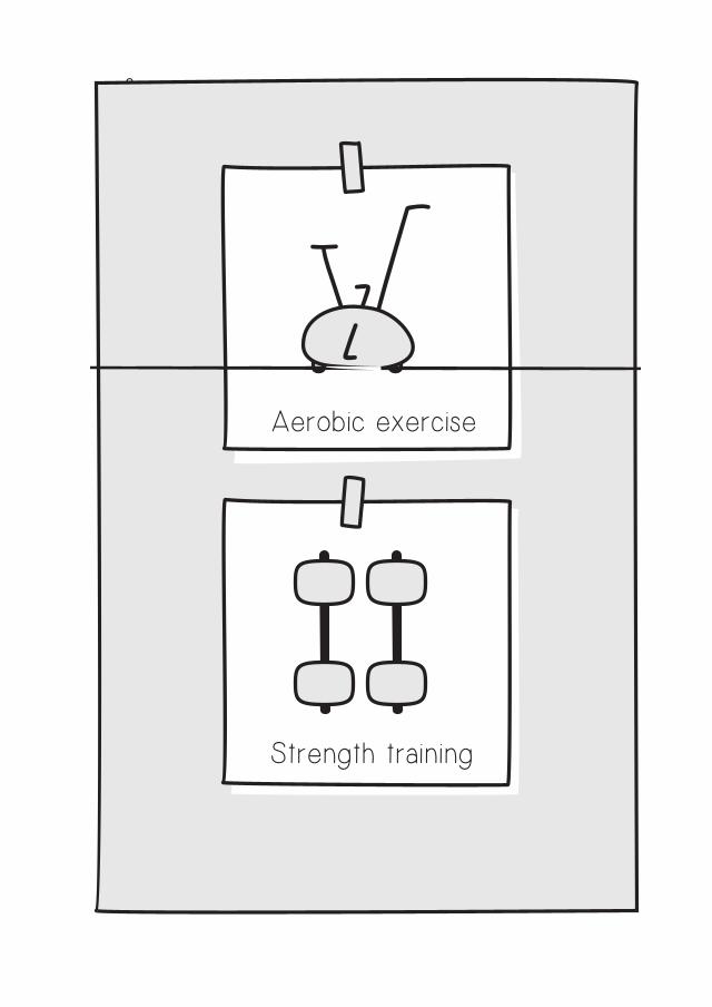
9.
Aerobic exercise
Strength training

CHAPTER 4STRENGTH TRAINING AND AEROBIC EXERCISE TRAINING FOR MUSCLE DISEASE
Nicoline B.M. Voet
Elly L. van der Kooi
Ingrid L. Riphagen
Eline Lindeman ƚBaziel G.M. van Engelen
Alexander C.H. Geurts
Published in: Cochrane Database of Systematic Reviews 2013, Issue 6.
Art. No.: CD003907. DOI:10.1002/14651858.CD003907.pub4.

ABSTRACT
BackgroundStrength training or aerobic exercise programs might optimize muscle
and cardiorespiratory function and prevent additional disuse atrophy and
deconditioning in people with a muscle disease. This is an update of a review first
published in 2004.
Objectives To examine the safety and efficacy of strength training and aerobic exercise training
in people with a muscle disease.
Search methods We searched the Cochrane Neuromuscular Disease Group Specialized Register
(July 2012), CENTRAL (2012 Issue 3 of 4), MEDLINE (January 1946 to July 2012),
EMBASE (January 1974 to July 2012), EMBASE Classic (1947 to 1973) and
CINAHL (January 1982 to July 2012).
Selection criteria Randomized or quasi-randomized controlled trials comparing strength training or
aerobic exercise programs, or both, to no training, and lasting at least six weeks,
in people with a well-described diagnosis of a muscle disease. We did not use the
reporting of specific outcomes as a study selection criterion.
Data collection and analysis Two authors independently assessed trial quality and extracted the data obtained
from the full text-articles and from the original investigators. We collected adverse
event data from included studies.
Main results We included five trials (170 participants). The first trial compared the effect of
strength training versus no training in 36 people with myotonic dystrophy. The
second trial compared aerobic exercise training versus no training in 14 people
with polymyositis and dermatomyositis. The third trial compared strength training
versus no training in a factorial trial that also compared albuterol with placebo, in
65 people with facioscapulohumeral muscular dystrophy (FSHD). The fourth trial
compared combined strength training and aerobic exercise versus no training in 18
people with mitochondrial myopathy. The fifth trial compared combined strength
training and aerobic exercise versus no training in 35 people with myotonic
Cha
pte
r 4
Str
engt
h tr
aini
ng a
nd a
erob
ic e
xerc
ise
trai
ning
for
mus
cle
dis
ease
72

dystrophy type 1. In both myotonic dystrophy trials and the dermatomyositis
and polymyositis trial there were no significant differences between training and
non-training groups for primary and secondary outcome measures. The risk of
bias of the strength training trial in myotonic dystrophy and the aerobic exercise
trial in polymyositis and dermatomyositis was judged as uncertain, and for the
combined strength training and aerobic exercise trial, the risk of bias was judged
as adequate. In the FSHD trial, for which the risk of bias was judged as adequate, a
+1.17 kg difference (95% confidence interval (CI) 0.18 to 2.16) in dynamic strength
of elbow flexors in favor of the training group reached statistical significance. In
the mitochondrial myopathy trial, there were no significant differences in dynamic
strength measures between training and non-training groups. Exercise duration
and distance cycled in a submaximal endurance test increased significantly in the
training group compared to the control group. The differences in mean time and
mean distance cycled till exhaustion between groups were 23.70 min (95% CI 2.63
to 44.77) and 9.70 km (95% CI 1.51 to 17.89), respectively. The risk of bias was
judged as uncertain. In all trials, no adverse events were reported.
Authors’ conclusions Moderate-intensity strength training in myotonic dystrophy and FSHD and aerobic
exercise training in dermatomyositis and polymyositis and myotonic dystrophy
type I appear to do no harm, but there is insufficient evidence to conclude that
they offer benefit. In mitochondrial myopathy, aerobic exercise combined with
strength training appears to be safe and may be effective in increasing submaximal
endurance capacity. Limitations in the design of studies in other muscle diseases
prevent more general conclusions in these disorders.
PLAIN LANGUAGE SUMMARY
STRENGTH TRAINING OR COMPREHENSIVE AEROBIC EXERCISE TRAINING FOR MUSCLE DISEASE
Strength training, which is performed to improve muscle strength and muscle
endurance, or aerobic exercise programs, which are designed to improve
cardiorespiratory endurance, might optimize physical fitness and prevent
additional muscle wasting in people with muscle disease. However, people with
muscle disease and some clinicians are still afraid of overuse and have a cautious
approach to training. This updated review (most recent date of search 2 July 2012)
included two eligible trials of strength training in people with facioscapulohumeral
73
muscular dystrophy (FSHD) and myotonic dystrophy (101 participants), two trials
of strength training combined with aerobic exercise in people with mitochondrial
myopathy (18 participants) and myotonic dystrophy type I (35 participants) and
one trial of aerobic exercise in people with polymyositis and dermatomyositis
(14 participants). These trials showed that moderate-intensity strength training in
people with myotonic dystrophy or with FSHD, and aerobic exercise training in
people with dermatomyositis or polymyositis appear not to harm muscles. Strength
training combined with aerobic exercise appears to be safe in myotonic dystrophy
type I and may be effective in increasing endurance in people with mitochondrial
myopathy. Evidence suggests that strength training is not harmful in people in
FSHD, myotonic dystrophy, mitochondrial disorders and dermatomyositis and
polymyositis, but further research is needed to determine potential benefit.
BACKGROUND
The term ’muscle disease’ comprises a large group of conditions. Skeletal muscles
are primarily affected but in some disorders other organ systems may also be
involved. Most conditions are progressive, causing the muscles to gradually
weaken over time. When a person is diagnosed as having a muscle disease,
questions arise about the prognosis, possible interventions and genetics. However,
people with muscle disease are usually also concerned about everyday issues
such as participation in sports, work and hobbies. We cannot give evidence-based
advice about these issues, because we do not know how physical exercise affects
the diseased muscular system or the cardiorespiratory system. To answer these
questions, controlled trials of aerobic exercise and strength training in people with
a muscle disease are needed.
Weakness and impaired cardiorespiratory function are common in people with
muscle disease; pain and fatigue may also be common symptoms, all of which
contribute to a decreased quality of life. In healthy persons the best intervention
to improve strength and cardiorespiratory function is physical training. Strength
training or aerobic exercise programs in people with muscle disease might
maximise muscle and cardiorespiratory function and prevent additional disuse
atrophy (1). However, reports of progression of weakness after exercise in people
with myopathies have encouraged a cautious approach to training (2-4). Therefore,
many people with a muscle disease were advised to avoid physical exertion (3).
Thus the benefit from strength training or aerobic exercise training in muscle
diseases is still not clear (5).
Cha
pte
r 4
Str
engt
h tr
aini
ng a
nd a
erob
ic e
xerc
ise
trai
ning
for
mus
cle
dis
ease
74

The relative rarity of many muscle diseases has led researchers to group
participants with different neuromuscular disorders together in one study,
including myopathies, neuropathies and motor neuron disease (6-13). As the
pathophysiology of these disorders differs, their reaction to an intervention might
also be different. Therefore, conclusions about the effect of training derived from
these mixed populations cannot readily be extrapolated to people with specific
muscular disorders (14).
In this review we systematically analyzed randomized controlled trials (RCTs) of
these interventions for people with specified muscle diseases. This review was first
published in 2004, with the most recent update of the searches in 2012.
Objectives To examine the safety and efficacy of strength training and aerobic exercise training
in people with a muscle disease.
METHODS
CRITERIA FOR CONSIDERING STUDIES FOR THIS REVIEW
Types of studies
We included all RCTs or quasi-RCTs that made any of the following comparisons:
strength training versus no training;
aerobic exercise training versus no training;
combined strength training and aerobic exercise versus no training.
Quasi-RCTs are trials that allocate participants to experimental or control groups
based on a method that is not truly random, for example, hospital record number or
date of birth.
Types of participants
We selected all trials that included participants with a well-described diagnosis
of a muscle disease, such as inflammatory myopathies, metabolic myopathies,
muscular dystrophies, muscle diseases with myotonia and other well-defined
myopathies. We decided not to include studies looking at strength training or
aerobic exercise training for people in whom muscle weakness was not the primary
feature, but might have been secondary to chronic renal insufficiency, chronic
75

heart failure, renal or heart transplantation, or corticosteroid use. We did not review
the effects of respiratory muscle training. We did not include studies regarding
aerobic exercise training for McArdle disease because there is a separate Cochrane
review available for this metabolic myopathy (15). We excluded studies in which
participants had a variety of muscle diseases if we could not obtain results for each
condition separately. We assessed the diagnostic criteria of each study; diagnosis
has to be confirmed by muscle biopsy or genetic testing.
Types of interventions
To date, there is no evidence or recommendation for a minimum duration of training
in muscle disease. However, in the first six weeks, the change in muscle strength or
aerobic capacity is generally caused by neural adaptation. Therefore, we included
all forms of strength training and aerobic exercise training lasting at least six weeks.
We excluded all studies using a within-subjects design with the non-exercised limb
as a control. If exercises are performed to increase muscle strength on one side of
the body, voluntary strength can increase on the contralateral side. This concept is
called cross-education, and has been described with different forms of exercises. A
meta-analysis of 16 randomized studies concluded that, on average, the magnitude
of cross-education is eight per cent of the initial strength of the untrained limb (16).
Neural adaptations to training and learning effects due to testing are postulated
as explanations (17-20). Moreover, the results may well be confounded by the
presence of asymmetric weakness of both limbs, as the absolute gain in muscle
strength resulting from strength training is related to pre-exercise muscle weakness
(21). For this reason, a non-exercised limb is not an appropriate control, even if
training is randomly assigned. For this reason, we have excluded studies using
such a within-subjects design.
Definitions
Training or physical fitness training: a planned, structured regimen of regular
physical exercise deliberately performed to improve one or more of the
following components of physical fitness: cardiorespiratory fitness, body
composition, muscle strength and endurance, and flexibility (22).
Strength training: a systematic program of exercises designed to increase an
individual’s ability to exert or resist force using, for example, weights, weight
machines or elastic cords (22).
Aerobic exercise training or cardiorespiratory fitness training: training that is
designed to improve the capacity and efficiency of aerobic energy-producing
systems and is effective for improving cardiorespiratory endurance. It consists Cha
pte
r 4
Str
engt
h tr
aini
ng a
nd a
erob
ic e
xerc
ise
trai
ning
for
mus
cle
dis
ease
76

of an activity or combination of activities that uses large muscle groups, that
can be maintained continuously, and is rhythmical and aerobic in nature, for
example walking, running, cycling, aerobic dance exercise or swimming (22).
TYPES OF OUTCOME MEASURES
Primary outcomes
The primary outcome measure for strength training was:
change in muscle strength, expressed in measures of static (that is, isometric)
or dynamic strength between baseline and six weeks.
The primary outcome measure for aerobic exercise training was:
change in aerobic capacity, expressed in measures of work capacity between
baseline and six weeks.
Secondary outcomes
The secondary outcome measure specific to strength training was:
change in muscle endurance muscle endurance or muscle fatigue between
baseline and six weeks.
The secondary outcome measure specific to aerobic exercise training was:
change in aerobic capacity, expressed in measures of oxygen consumption,
parameters of cardiac function or parameters of respiratory function between
baseline and six weeks.
Secondary outcome measures applicable to both strength training and aerobic
exercise training showing a change from baseline and six weeks were:
timed-scored functional assessments of muscle performance, such as a six-
minute walk test (23);
quality of life measures, such as the Short Form 36 (SF-36) Health Survey (24);
parameters of muscle membrane permeability (serum creatine kinase level,
myoglobin level) to assess safety; pain assessed by an analogue pain scale
(25);
experienced fatigue assessed by questionnaires, e.g. Checklist Individual
Strength (CIS-fatigue) (26);
adverse effects requiring withdrawal of the participant from the study: acute
rhabdomyolysis, increasing muscle pain, injury, etc;
77

We compared data on outcome measures at baseline with those obtained after at
least six weeks of training. When there were assessments at more than one time
(during the intervention, after cessation of the intervention), our preference was for
data on outcome measures obtained at the end of the intervention.
SEARCH METHODS FOR IDENTIFICATION OF STUDIES
We searched the following databases: the Cochrane Neuromuscular Disease Group
Specialized Register (July 2012), the Cochrane Central Register of Controlled Trials
(CENTRAL, in The Cochrane Library 2012, Issue 7 of 12), MEDLINE (January 1946
to July 2012), EMBASE (January 1974 to July 2012), EMBASE Classic (1947 to
1973) and CINAHL (January 1982 to July 2012). We reviewed the bibliographies
of the trials identified and other reviews of the subject, and contacted some of the
authors in the field to identify additional published and unpublished data.
DATA COLLECTION AND ANALYSIS
Selection of studies
Two review authors (Voet, van der Kooi) checked the references identified by
the search strategy. We obtained the full text of all potentially relevant studies
for independent assessment by both authors. We decided which trials fitted the
inclusion criteria.
Data extraction and management
Two review authors (Voet, van der Kooi) independently extracted the data from the
included trials onto a specially designed data extraction form, and graded the risk
of bias and certain other aspects of the design of the included trials.
Assessment of risk of bias in included studies
We assessed the risk of bias and other aspects according to the Cochrane
approach using the updated guidance in the Cochrane Handbook for Systematic
Reviews of Interventions (27).
We assessed the included studies for randomization sequence generation,
allocation concealment, blinding (participants and outcome assessors), incomplete
outcome data, selective outcome reporting and other sources of bias. When there
was uncertainty, we contacted authors for clarification. We resolved disagreements
Cha
pte
r 4
Str
engt
h tr
aini
ng a
nd a
erob
ic e
xerc
ise
trai
ning
for
mus
cle
dis
ease
78

about fulfillment of inclusion or quality criteria by discussion between the two
authors. We made a judgement on each of the ’Risk of bias’ criteria, of “High risk of
bias”, “Low risk of bias” or “Unclear risk of bias”. Whenever characteristics of study
design or drop-out rates were likely to cause a higher risk of bias, we planned to
make a note of this and investigate the possibility of differences in treatment effects
varying with the degree of this problem.
Data synthesis
We intended to combine trial results for appropriate pairings of treatments by
calculating a mean of the difference between their effects using the Cochrane
statistical package Review Manager 5 (RevMan) (28). Because pooling of the
results of trials in different muscle diseases is usually not appropriate, we
expressed, when possible, the results per muscle disease as mean differences
(MDiff) with 95% confidence intervals (CI) for continuous outcomes, and risk ratios
(RR) with 95% CI for dichotomous outcome measures. The intended testing for
heterogeneity, and consequent actions, turned out to be unnecessary.
Subgroup analysis and investigation of heterogeneity
We decided, in advance, not to perform subgroup analyses based on sex or
age because we anticipated that the differences in muscle disease severity
would have a much bigger influence on outcome than sex or age. Moreover, the
American College of Sports Medicine stated in their Position Stand (22) that relative
improvements resulting from aerobic and strength training are similar for young and
old, male and female. We presented data for individual muscle diseases separately.
As the pathophysiology of each muscle disease differs, we considered that their
reaction to training might be different. If in future data are available for meta-
analysis, we will consider investigating the effect of different durations of exercise
or training intervention.
RESULTS
DESCRIPTION OF STUDIES
In this review, the search retrieved approximately 7400 records. After assessing the
titles and abstracts, we identified 61 studies for potential inclusion: 26 completed
trials that studied strength training as an intervention, 20 trials studying aerobic
79

exercise training, and 15 trials studying combined strength training and aerobic
exercise, sometimes incorporated in more comprehensive rehabilitation programs.
Most strength training trials included people with the following muscle diseases:
slowly progressive dystrophies (mostly myotonic dystrophy, limb-girdle dystrophies,
facioscapulohumeral muscular dystrophy (FSHD)) and in the older studies, non-
specified progressive muscular dystrophies and inflammatory myopathies.
Studies on the effects of aerobic exercise training included mainly people with
slowly progressive dystrophies and metabolic myopathies (mostly unspecified
mitochondrial myopathies).
Studies have generally been limited by small sample sizes. We excluded 48 studies
because there was no randomized controlled comparison between training and
non-training participants and six studies because of a within-subjects design (see
Table 2: Characteristics of excluded studies).
Table 1 Characteristics of included studies
Cejudo 2005
Methods Parallel group RCT
Participants 20 adults with mitochondrial myopathy, diagnosed on the basis of clinical, familial and muscle biopsy data
Interventions Strength training and aerobic exercise training versus no training Type of training and exercise Endurance bicycle training, dynamic isotonic with weights Intensity Aerobic training: individualized work rate, 30 min leg exercise on an ergo cycle, 70% of the peak work rate; strength training: one set dynamic and isotonic of 10 to 15 repetitions at 50% 1RM load, to 2 or 3 sets. Adjustments on workload changed every 2 weeks Frequency 3 times/week Duration Session: approximately 60 min. Program: 24 weeks Muscle groups Shoulder, upper back, arm, pectoralis major, biceps brachii and brachialis muscles Supervision Supervised training program by specialized nurses and a physiatrist specialist in a rehabilitation unit on an outpatient basis
Outcomes Primary: exercise capacity - expressed in measures of oxygen uptake (ie. VO2 max), endurance time and distance walked in the shuttle walking test. Secondary outcomes were: peripheral muscle strength (1RMtest), quality of life, symptoms of myalgia, cramps and fatigability and functional exercise capacity
Notes -
Cha
pte
r 4
Str
engt
h tr
aini
ng a
nd a
erob
ic e
xerc
ise
trai
ning
for
mus
cle
dis
ease
80
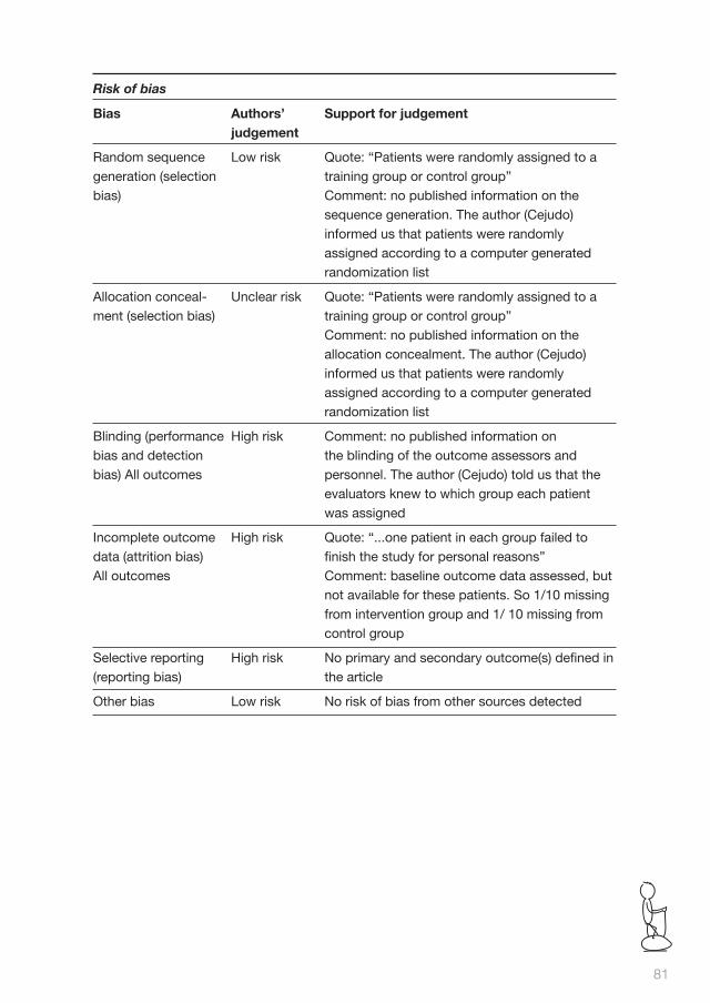
Risk of bias
Bias Authors’ judgement
Support for judgement
Random sequence generation (selection bias)
Low risk Quote: “Patients were randomly assigned to a training group or control group” Comment: no published information on the sequence generation. The author (Cejudo) informed us that patients were randomly assigned according to a computer generated randomization list
Allocation conceal-ment (selection bias)
Unclear risk Quote: “Patients were randomly assigned to a training group or control group” Comment: no published information on the allocation concealment. The author (Cejudo) informed us that patients were randomly assigned according to a computer generated randomization list
Blinding (performance bias and detection bias) All outcomes
High risk Comment: no published information on the blinding of the outcome assessors and personnel. The author (Cejudo) told us that the evaluators knew to which group each patient was assigned
Incomplete outcome data (attrition bias) All outcomes
High risk Quote: “...one patient in each group failed to finish the study for personal reasons” Comment: baseline outcome data assessed, but not available for these patients. So 1/10 missing from intervention group and 1/ 10 missing from control group
Selective reporting (reporting bias)
High risk No primary and secondary outcome(s) defined in the article
Other bias Low risk No risk of bias from other sources detected
81

Kierkegaard 2011
Methods Evaluator blind, parallel group RCT
Participants 35 adults with myotonic dystrophy type 1, genetically confirmed
Interventions Strength training and aerobic exercise training versus no training Type of training and exercise Strength training, aerobic exercise, balance exercises Intensity Strength exercises for arm, leg, back and abdominal muscles 16-20 repetitions, for 6- 7 min, balance exercises for 3-4 min, aerobic activities for 11-12 min at 60-80% of maximum heart rate. Once a week a 30-min brisk walk Frequency 2 times/week and once a week a brisk walk Duration Session: 60 min and a 30-min walk. Program: 14 weeks Muscle groups Arm, leg, back and abdominal muscles Supervision All sessions were supervised by a specialized physiotherapist
Outcomes Primary: distance walked in the 6-min walk test Secondary: timed-stands test, timed up-and-go test
Notes Participants were stratified before randomization by their results in the 6-min walk test
Risk of bias
Bias Authors’ judgement
Support for judgement
Random sequence generation (selection bias)
Low risk Quote: “The lots were drawn by a person who was not involved in any other part of the study”
Allocation concealment (selection bias)
Low risk Quote: “Patients were randomly assigned to a training group or control group” Comment: no published information on the allo-cation concealment.
Blinding (performance bias and detection bias) All outcomes
Low risk Quote: “Data was collected before and after the intervention by two independent experienced physiotherapists, blinded to group allocation and each assessing the same participants on both occasions”
Incomplete outcome data (attrition bias) All outcomes
Low risk Quote: “one person in the control group did not attend the data collection after the intervention”
Selective reporting (reporting bias)
Low risk No evidence found for selective reporting
Other bias Low risk No risk of bias from other sources detected
Cha
pte
r 4
Str
engt
h tr
aini
ng a
nd a
erob
ic e
xerc
ise
trai
ning
for
mus
cle
dis
ease
82

Lindeman 1995
Methods Evaluator blind, matched-control RCT
Participants 36 adults with myotonic dystrophy (2 congenital form, 34 classical adult type), diagnosis not verified
Interventions Strength training versus no training Type of training and exercise Dynamic strength training with weightsIntensity Individualized progressive overload, 3 sets from 25 repetitions at 60% of 1RM, via 15 repetitions at 70%, to 10 repetitions at 80% Frequency 3 times/week Duration Session: within 30 min. Program: 24 weeks Muscle groups Knee extensors and flexors, hip extensors and abductors Supervision Supervised home training program
Outcomes Primary: muscle strength by isokinetically measured knee torques and isometrically as MVIC. Main secondary outcomes were: endurance by maximum duration of contraction at 80% of MVIC, functional performance by timed motor performance tests and by questionnaires. Serum myoglobin levels to detect changes in muscle fiber membrane permeability
Notes Participants were matched based on muscle strength (knee ex-tension torque/bodyweight) and on performance in a stair-climb-ing test. Only complete pairs were analyzed
Risk of bias
Bias Authors’ judgement
Support for judgement
Random sequence generation (selection bias)
Low risk Comment: there was no published information on the sequence generation but the author (Lindeman) informed us that 2 independent persons drew a sealed lot per matched pair and allocated it by tossing a coin to the training or non-training group
Allocation concealment (selection bias)
Low risk Comment: there was no published information on the method of allocation concealment but the author (Lindeman) informed us that 2 independent persons allocated the training, after tossing the coin, to the training or nontraining group
83
Blinding (performance bias and detection bias) All outcomes
Low risk Quote: “observers of the outcome measure-ments were blinded for treatment allocation” Comment: approximately 20% of the myotonic dystrophy participants revealed information to the clinical evaluators that resulted in unblinding during the course of the trial
Incomplete outcome data (attrition bias) All outcomes
High risk 3 of the initially 36 randomized participants with-drew before disclosure of treatment allocation. The 33 participants starting the trial made 15 matched pairs. During the trial 1 person dropped out. Because of the matched pair design only complete pairs were analyzed, therefore eventu-ally 28 of the initial 36 randomized participants were analyzed. Follow-up was therefore incom-plete and analysis was not by intention-to-treat. However, the flow path of participants was well documented
Selective reporting (reporting bias)
Low risk No evidence found for selective reporting
Other bias Low risk No risk of bias from other sources detected
Van der Kooi 2004
Methods Evaluator blind, parallel group, RCT
Participants 65 adults with FSHD, genetically confirmed
Interventions Strength training versus no training (and as add-on in a double blind randomized controlled design albuterol or placebo) Type of training and exercise Dynamic and isometric strength training with weightsIntensity Individualized progressive overload, 2 sets dynamic from 10 repetitions at 10RM, via 8 repetitions at 8RM, to 5 repe-titions at 5RM, and 30s isometric with same weight Frequency 3 times/week Duration Session: Within 30 min. Program: 52 weeks Muscle groups Elbow flexors, ankle dorsiflexors Supervision Supervised home training program
Outcomes Primary: difference in muscle strength of elbow flexors and an-kle dorsiflexors after 52 weeks using the MVIC. Main secondary outcomes were muscle endurance (MVIC Force-Time Integral) and dynamic muscle strength (1RM). Other measures included functional tests and timed motor performance tasks
Cha
pte
r 4
Str
engt
h tr
aini
ng a
nd a
erob
ic e
xerc
ise
trai
ning
for
mus
cle
dis
ease
84

Notes Outcomes are presented for the 4 treatment groups (i.e. the 4 combinations of training versus non-training, and albuterol versus placebo). Effect sizes are presented by intervention as well
Risk of bias
Bias Authors’ judgement
Support for judgement
Random sequence generation (selection bias)
Low risk Quote: “...participants were randomly assigned to one of the four treatment groups according to a computer generated randomization list”
Allocation concealment (selection bias)
Unclear risk Quote: “information on the assignment to training or non-training was disclosed to the participants by the physical therapist”
Blinding (performance bias and detection bias) All outcomes
Low risk Quote: “The RM measurements were performed by the physical therapist, who was not blinded for the allocation to training or non-training, as this specific measurement carried too great a risk of unblinding the clinical evaluator” Comment: adequate although one of the main secondary outcome measures, the 1RM measurement for assessing dynamic strength, was performed by the physical therapist, who supervised the training, and was therefore not blinded to the allocation to training or non-train-ing. Unblinding during the trial was adequately registered. Allocation to training or non-training was unmasked in 3 cases, due to unintentional remarks
Incomplete outcome data (attrition bias) All outcomes
Low risk Quote: “One patient stopped training because of recurring, training-related muscle soreness and fatigue. Four participants stopped using their study medication because of side effects. Data for the participants who discontinued an intervention were analyzed in the assigned treatment group” Comment: complete follow-up of all participants
Selective reporting (reporting bias)
Low risk No evidence found for selective reporting
Other bias Low risk No risk of bias from other sources detected
85

Wiesinger 1998
Methods Parallel group RCT
Participants 9 adults with dermatomyositis and 5 adults with polymyositis Diagnosis of primary inflammatory muscle disease was defined by the criteria of Bohan and Peter
Interventions Aerobic exercise training versus no training Type of training and exercise Endurance bicycle training, endurance step aerobics Intensity Bicycle training: 30min, slowly increased on an individual basis. Resistance was increased until a heart rate of 60% of maximum. Step aerobics: 30 minFrequency During the first 2 weeks, twice weekly, during the remaining 4 weeks, 3 times weekly Duration Session: 60 min. Program: 6 weeks Muscle groups Not applicable Supervision Supervised by a physiotherapist
Outcomes No primary outcome or secondary outcomes defined. Study outcomes: activities of daily living score, peak isometric torque of knee extensors and hip flexors, peak oxygen consumption and creatine kinase and aldolase levels
Notes Outcomes are not presented separately for the dermatomyositis and polymyositis patients
Risk of bias
Bias Authors’ judgement
Support for judgement
Random sequence generation (selection bias)
Unclear risk Quote: “Distinct randomization lists were used”. Comment: there was no information about the generation of the list. It is not clear what is meant by “distinct randomization lists”
Allocation concealment (selection bias)
Unclear risk Comment: there was no published information on the method of allocation concealment
Blinding (performance bias and detection bias) All outcomes
Unclear risk Quote: “Muscle strength assessments were carried out by the same person who was unaware of the group to which the individual patients belonged”. Comment: there was no published information about blinding of the assessor of the other measurements
Incomplete outcome data (attrition bias) All outcomes
Low risk Comment: complete follow-up of all participants
Cha
pte
r 4
Str
engt
h tr
aini
ng a
nd a
erob
ic e
xerc
ise
trai
ning
for
mus
cle
dis
ease
86

Selective reporting (reporting bias)
High risk Comment: no primary or secondary outcomes are defined
Other bias Low risk No risk of bias from other sources detected. Outcomes are not presented for dermatomyositis and polymyositis separately
MVIC: maximum voluntary isometric strength, RCT: randomized controlled trial
RM: repetition maximum, VO2 max: maximal oxygen uptake
Only seven studies were RCTs making a comparison between training and
non-training participants (7, 14, 29-33). Regrettably, the extension of the
initially randomized, controlled six-week aerobic exercise study in people with
dermatomyositis and polymyositis by Wiesinger et al (32) lost its randomized
controlled design due to a decision of the ethics committee. The randomized
controlled strength training combined with aerobic exercise trial which compared
eight weeks of walking and strengthening exercises versus no training in 20
participants with different muscle diseases (7) has been excluded as both study
groups consisted of participants with various muscle diseases and the outcome
measures were not presented for each muscle disease separately. As the
pathophysiology of each muscle disease differs, their reaction to training might
be different. It is not known if the effect of strength training and aerobic exercise
training is the same for every muscle disease. Therefore, data should be presented
and analyzed for each disease individually, and the power should be sufficient for
each individual disorder. For this reason, no conclusions can be drawn with regard
to the effect of exercise training for each specific muscle disease in the trial. Finally,
no specific details about the exercise program were provided and the risk of bias of
the trial was high.
In conclusion, we included two strength training trials (14, 31), one aerobic exercise
trial (33) and two strength training combined with aerobic exercise trials (29, 30)
(see Table 1 Characteristics of included studies).
87

Table 2 Characteristics of excluded studies
Study Reason for exclusion
Abramson 1952 Not a RCT
Aitkens 1993 Not a RCT. Exercised versus non-exercised control limb (randomly assigned) and patients versus healthy volunteers
Aldehag 2005 Not a RCT
Alexanderson 1999 Pilot study. Not a RCT
Alexanderson 2000 Extension of a pilot study. Not a RCT
Alexanderson 2007 Not a RCT
Arnardottir 2003 Not a RCT
Chung 2007 No non-exercising control group
Dastmalchi 2007 Not a RCT
Dawes 2006 Excluded because of serious insufficiencies in the study design
De Lateur 1979 Not a RCT. Exercised versus non-exercised control limb (randomly assigned)
Escalante 1993 Not a RCT
Florence 1984a Not a RCT
Florence 1984b Not a RCT
Fowler 1965 Not a RCT. Exercise combined with medication
Heikkila 2001 Not a RCT. Training program duration of 3 weeks
Hicks 1989 Not a RCT. Training program duration of 1 month
Hoberman 1955 Not a RCT. 3 drugs added to a comprehensive regimen of therapies, including breathing and resistive exercises
Jeppesen 2006 Not a RCT
Jeppesen 2009a Not a RCT
Johnson 2007 Not a RCT
Johnson 2009 Not a RCT
Kelm 2001 Not a RCT
Kilmer 1994 Not a RCT. Exercised versus non-exercised control limb (randomly assigned) and patients versus healthy volunteers
Kilmer 2005 Not a RCT. Training program duration for participants with muscle disorders ranged from approximately 1 to 21 months
Lenman 1959 Not a RCT. Training program duration for participants with muscle disorders ranged from approximately 1 to 21 months
Mate-Munoz 2007 Not a RCT
McCartney 1988 Not a RCT. Exercised versus non-exercised control limb (randomly assigned)C
hap
ter
4 S
tren
gth
trai
ning
and
aer
obic
exe
rcis
e tr
aini
ng fo
r m
uscl
e d
isea
se
88

Mielke 1990 Not a RCT
Milner-Brown 1988a Not a RCT. Training program duration for participants with muscle disorders ranged from approximately 2 to 48 months
Milner-Brown 1988b Not a RCT. Intervention is not training versus non-training, but training added to electric stimulation or electric stimulation only in 1 limb versus a non-stimulated, non-exercised control limb
Milner-Brown 1990 Not a RCT. Intervention is not training versus no training, but amitriptyline added to strength training
Murphy 2008 Not a RCT
Na 1996 Not a RCT. Intervention is not training versus non-training, but training and daily quinine sulfate
Nader 2010 Not a RCT
Olsen 2005 Not a RCT
Omori 2010 Not a RCT
Orngreen 2005 Not a RCT
Scott 1981 A RCT that makes a comparison between 2 different training regimes. No comparison of training versus non-training participants
Siciliano 2000 Not a RCT
Spector 1997 Not a RCT
Sunnerhagen 2004 Not a RCT
Sveen 2007 Not a RCT
Sveen 2008 Not a RCT
Taivassalo 1998 Not a RCT
Taivassalo 1999 Not a RCT
Taivassalo 2001 Not a RCT
Taivassalo 2006 Not a RCT
Tollbäck 1999 Not a RCT. Exercised versus non-exercised control limb (randomly assigned)
Trenell 2006 Not a RCT
Varju 2003 Not a RCT. Training program duration of 3 weeks
Vignos 1966 Not a RCT
Wiesinger 1998b A non-randomized extension of a RCT (Wiesinger 1998a)
Wright 1996 Not a RCT
Yildirim 2007 Not a RCT
RCT: randomized controlled trial
89

The first strength training trial compared the effect of 24 weeks of training versus
no training in 36 adults with myotonic dystrophy and 30 adults with hereditary
motor and sensory neuropathy types I or II (14). As this review is concerned
with muscle disease, we will not discuss the results of the hereditary motor and
sensory neuropathy participant group. The aerobic exercise trial compared six
weeks of cycle and step aerobics exercise with no training in nine adults with
dermatomyositis and five adults with polymyositis (33). The second strength
training trial compared 52 weeks of strength training versus no training in a factorial
trial that also compared albuterol with placebo after the first 26 weeks of training in
65 adult participants with FSHD (30). Only the results for the comparison strength
training versus no training will be discussed in this review. The first combined
aerobic exercise and strength training trial compared 12 weeks of cycle exercises
and dynamic and isokinetic strength training versus no training in 18 people with
mitochondrial myopathy (29) (see Table 1 Characteristics of included studies). The
second combined aerobic exercise and strength training trial compared 14 weeks
of balance exercises, aerobic activities, flexibility exercises, strength exercises and
a brisk walk versus no training in 35 people with myotonic dystrophy type I. (30).
RISK OF BIAS IN INCLUDED STUDIES
Strength training trial in myotonic dystrophy
In the first myotonic dystrophy trial (14), participants with myotonic dystrophy were
individually matched for muscle strength and performance in a stair-climbing test.
Within each matched pair, participants were randomly assigned to the training or
control group.
There was no published information on the method of randomization or on
allocation concealment but the first author (Lindeman) informed us that two
independent persons drew one sealed name per matched pair and allocated it
to the training or non-training group by tossing a coin. We graded the intention
to blind the clinical evaluators as adequate, although approximately 20% of the
myotonic dystrophy participants revealed information to the clinical evaluators that
resulted in unblinding during the course of the trial. The authors considered the
baseline comparability of the groups as suboptimal because the training group had
longer time scores for stair climbing (a measure of functional ability) and had higher
knee torques (a measure of muscle strength). They argued that the first three items
could have resulted in an underestimation of the training effect, whereas the last
item could have resulted in an overestimation of the training effect. They concluded
that the differences in experimental group composition did not seem to explain the
absence of differences in outcomes between treatment groups. We considered the Cha
pte
r 4
Str
engt
h tr
aini
ng a
nd a
erob
ic e
xerc
ise
trai
ning
for
mus
cle
dis
ease
90

way the authors presented and discussed the baseline differences as adequate.
Three of the initially 36 randomized participants withdrew before disclosure of
treatment allocation. The 33 participants starting the trial made 15 matched pairs.
During the trial one person dropped out because of knee problems. Because of
the matched pair design only complete pairs were analyzed, thus eventually 28
of the initial 36 randomized participants were analyzed. Follow-up was therefore
incomplete and analysis was not by intention-to-treat. However, the flow path of
participants was well documented.
Dermatomyositis and polymyositis trial
In the dermatomyositis and polymyositis trial (33), nine people with
dermatomyositis and five with polymyositis were randomly assigned to the training
or control group using distinct randomization lists. The training group received six
weeks of bicycle exercises and step aerobics. Participants in the control group did
not undergo any training and continued their previous way of life. There was no
published information on allocation concealment and our attempts to obtain further
information on this were not successful. During the strength measurements, the
clinical evaluator was blinded to the treatment allocation. The success of blinding
of assessors was not formally checked as blinding of participants is not possible
in an exercise study. There was no published information on blinding during the
other measurements. Baseline characteristics were presented for both groups.
The authors considered the two groups to be well balanced with respect to most
baseline characteristics. There was complete follow-up of all participants.
Facioscapulohumeral muscular dystrophy (FSHD)
In the FSHD trial (31), 65 participants were stratified into two groups based on
muscle strength. Participants in both strata were randomly assigned to one of the
four treatment groups according to a computer-generated randomization list. The
treatments consisted of training plus albuterol, training plus placebo, non-training
plus albuterol, or non-training plus placebo. Training or non-training was the first
intervention, starting just after the baseline visit until after the final visit at 52 weeks.
Information on the assignment to training or non-training was disclosed to the
participants by the physical therapist (supervising the training program) after
their baseline visit. The clinical evaluator was blinded for the assignment to both
interventions. The participants, physical therapist and the neurologist evaluating
side effects were blinded to the treatment allocation. The blinding of the clinical
evaluator was considered adequate, although one of the main secondary outcome
measures, the one-repetition maximum (1RM) measurement for assessing dynamic
91

strength, was performed by the physical therapist who supervised the training,
and who was therefore not blinded to the allocation to training or non-training.
Allocation to the training or non-training group was unmasked in three cases,
due to unintentional remarks. The success of blinding was not formally checked.
Baseline characteristics were presented for all treatment groups. One participant
stopped training but still attended all trial visits, resulting in complete follow-up
of all participants. Data analysis was by the intention-to-treat principle. As no
statistically significant interactions between the two interventions (that is, training
versus non-training) could be detected, the effect sizes, being the differences
in mean change from baseline, were presented for each intervention. In the
mitochondrial myopathy trial (29), 20 participants were randomly assigned to the
training or control group. There was no published information on the method of
randomization, allocation concealment, or blinding of the evaluators. The author
(Cejudo) informed us that participants were randomly assigned according to a
computer generated randomization list. The evaluators were not blinded to the
intervention allocation, but knew to which group each participant was assigned.
One participant in each group failed to finish the study for personal reasons.
Baseline characteristics were presented for both groups, except for the participants
lost to follow-up. Follow-up was therefore incomplete and analysis was not done
by intention-to-treat. No flow path of participants was documented. The authors
considered both groups as comparable with respect to age and gender, as well as
to each measured variable at baseline.
Combined aerobic exercise and strength training trial in myotonic dystrophy type 1
In the second myotonic dystrophy trial (30), the median value of the results of
the six-minute walk test was used to divide the 35 participants into two strata
from which they were divided into the training or the control group. The lots
consisted of folded pieces of paper with the name of the participant and were
drawn by a person not involved in any part of the study. Since participants were
recruited before randomization, concealed allocation procedures were applied.
An intention-to-treat analysis was applied. Three participants had missing data for
perceived exertion at baseline and one person in the control group did not attend
the measurement after the intervention but still completed the questionnaires.
There was no significant difference in sex or age of participants between groups
in the study; however, the mean muscular impairment scale (MIRS) grade was
higher in the exercise group, indicating that participants in the exercise group were
more severely impaired than participants in the control group. The training group
received a comprehensive group exercise training program, they were also asked Cha
pte
r 4
Str
engt
h tr
aini
ng a
nd a
erob
ic e
xerc
ise
trai
ning
for
mus
cle
dis
ease
92

to perform an active 30min walk every week. The participants in the control group
were advised to live their normal lives and to maintain their degree of physical
activity during the study period.
The degree of activity of both groups was not objectively checked.
We ranked each criterion using the Cochrane ’Risk of bias’ tool. The review
authors’ judgements about each risk of bias item for included studies are presented
in Figure 1.
Ran
dom
seq
uenc
e ge
nera
tion
(sel
ectio
n b
ias)
Allo
catio
n co
ncea
lmen
t (s
elec
tion
bia
s)
Blin
din
g (p
erfo
rman
ce b
ias
and
d
etec
tion
bia
s)
Inco
mp
lete
out
com
e d
ata
(att
ritio
n b
ias)
Sel
ectiv
e re
por
ting
(rep
ortin
g b
ias)
Otz
er b
ias
Cejudo 2005 + ? - - - +
Kierkegaard 2011 + + + + + +
Lindeman 1995 + + + - + +
Van der Kooi 2004 + ? + + + +
Wiesinger 1998 ? ? ? + - +
Figure 1 Risk of bias summary: review authors’ judgements about each risk of
bias item for each included study
QUALITY OF DIAGNOSTIC CRITERIA
This assessment took into account if and how diagnoses were verified. In the
first myotonic dystrophy trial (14), participants were recruited via neurologists,
physiatrists and the Dutch association for neuromuscular diseases (Vereniging
Spierziekten Nederland) on clinical grounds and without genetic verification.
We therefore considered the quality of the diagnostic criteria as inadequate. In
the second myotonic dystrophy (type I) trial (30), the diagnosis was genetically
confirmed in all participants and the diagnostic criteria are therefore adequate.
In the dermatomyositis and polymyositis trial (33), all the participants had an
established diagnosis of primary inflammatory muscle disease as defined by
the established criteria of Bohan and Peter, with a disease duration of at least
six months (34, 35). In all participants, muscle biopsies, electromyograms and
93
laboratory studies had been performed to establish the diagnosis. We therefore
considered the quality of the diagnostic criteria to be adequate.
In the FSHD trial (31), participants or a first-degree relative had the associated
deletion at chromosome 4 (36). The quality of the diagnosis was therefore
adequate. In the mitochondrial myopathy trial (29) participants were recruited
from a larger group of patients followed at the university hospital of Sevilla, Spain.
Diagnosis was based on clinical and muscle biopsy data. Biopsy findings were
determined by biochemical and histological techniques without genetic verification.
One participant in each group had only a probable diagnosis of mitochondrial
myopathy. The quality of the diagnostic criteria is therefore uncertain.
QUALITY OF TRAINING PROGRAM
The training programs of the first myotonic dystrophy (14), FSHD (31),
mitochondrial myopathy (37) and dermatomyositis and polymyositis (33) trials
fulfilled most of the minimum requirements as defined by the American College of
Sports Medicine (ACSM) Position Stand (22). In the second myotonic dystrophy
type I trial (30), the intervention consisted of a comprehensive group exercise
training program supported by music. The author could not give the exact training
load of each strength training exercise as a percentage of repetition maximum (RM)
as it was not tested that way. However, all major muscle groups were trained: arm,
back, leg and abdominal muscles (30). The training scheme for the other strength
training trials was inadequate only with respect to the number of muscle groups
trained, as the ACSM recommends eight to 10 exercises of all the major muscle
groups. Only four muscle groups were trained in the first myotonic dystrophy trial
(14), two in the FSHD trial (31) and three in the mitochondrial myopathy trial (29).
All studies except the combined aerobic exercise and strength training in myotonic
dystrophy type I trial (30), focused on a limited number of muscle groups for
reasons of effect evaluation, safety and time restraints per training session. In the
dermatomyositis and polymyositis trial (33), the training frequency was only twice
a week in the first two weeks, but increased to three times a week in the remaining
four weeks. In the mitochondrial myopathy trial (29), there was no published
information regarding supervision.
In the other trials (14, 30, 31), a physiotherapist supervised training. A description
of the training programs is given in the Characteristics of included studies (Table 1).
Cha
pte
r 4
Str
engt
h tr
aini
ng a
nd a
erob
ic e
xerc
ise
trai
ning
for
mus
cle
dis
ease
94
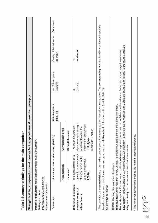
95
*The
bas
is fo
r th
e as
sum
ed r
isk
(e.g
. the
med
ian
cont
rol g
roup
ris
k ac
ross
stu
die
s) is
pro
vid
ed in
foot
note
s. T
he c
orr
esp
ond
ing
ris
k (a
nd it
s 95
% c
onfid
ence
inte
rval
) is
bas
ed o
n th
e as
sum
ed r
isk
in t
he c
omp
aris
on g
roup
and
the
rel
ativ
e ef
fect
of t
he in
terv
entio
n (a
nd it
s 95
% C
I).
CI:
confi
den
ce in
terv
al
Str
eng
th t
rain
ing
co
mp
ared
to
usu
al c
are
for
faci
osc
apul
ohu
mer
al m
uscu
lar
dys
tro
phy
Tab
le 3
Sum
mar
y o
f fi
ndin
gs
for
the
mai
n co
mp
aris
on
Pat
ient
or
po
pul
atio
n: fa
cios
cap
uloh
umer
al m
uscu
lar
dys
trop
hy
Set
ting
s: a
t ho
me
Inte
rven
tio
n: s
tren
gth
trai
ning
Co
mp
aris
on:
usu
al c
are
1 T
he lo
wer
con
fiden
ce li
mit
cros
ses
the
min
imal
imp
orta
nt d
iffer
ence
.
GR
AD
E W
orki
ng G
roup
gra
des
of e
vid
ence
Hig
h q
ualit
y: F
urth
er r
esea
rch
is v
ery
unlik
ely
to c
hang
e ou
r co
nfid
ence
in t
he e
stim
ate
of e
ffect
.
Mo
der
ate
qua
lity:
Fur
ther
res
earc
h is
like
ly t
o ha
ve a
n im
por
tant
imp
act
on o
ur c
onfid
ence
in t
he e
stim
ate
of e
ffect
and
may
cha
nge
the
estim
ate.
Low
qua
lity:
Fur
ther
res
earc
h is
ver
y lik
ely
to h
ave
an im
por
tant
imp
act
on o
ur c
onfid
ence
in t
he e
stim
ate
of e
ffect
and
is li
kely
to
chan
ge t
he e
stim
ate.
Very
low
qua
lity:
We
are
very
unc
erta
in a
bou
t th
e es
timat
e.
Out
com
esIll
ustr
ativ
e co
mp
arat
ive
risk
s* (9
5% C
I)
Ass
umed
ris
k
Usu
al c
are
Co
rres
po
ndin
g r
isk
Str
eng
th t
rain
ing
Rel
ativ
e ef
fect
(95%
CI)
No
of P
artic
ipan
ts
(stu
die
s)
Qua
lity
of t
he e
vid
ence
(GR
AD
E)
Com
men
ts
The
mea
n d
iffer
ence
in
dyn
amic
mus
cle
stre
ngth
of e
lbow
flex
ors
in t
he
cont
rol g
roup
s w
as
1.39
Nm
•••
mo
der
ate1
Diff
eren
ce in
dyn
amic
mus
cle
stre
ngth
of
elb
ow
flex
ors
65 (1 s
tud
y)
The
mea
n d
iffer
ence
in
dyn
amic
mus
cle
stre
ngth
of e
lbow
flex
ors
in t
he
inte
rven
tion
grou
ps
was
1.17
hig
her
(0.1
8 to
2.1
6 hi
gher
)

Cha
pte
r 4
Str
engt
h tr
aini
ng a
nd a
erob
ic e
xerc
ise
trai
ning
for
mus
cle
dis
ease
96
Aer
ob
ic e
xerc
ise
and
str
eng
th t
rain
ing
co
mp
ared
to
usu
al c
are
for
mit
och
ond
rial
myo
pat
hy
Tabl
e 4
Add
itio
nal s
umm
ary
of fi
ndin
gs
Pat
ient
or
po
pul
atio
n: m
itoch
ond
rial m
yop
athy
Set
ting
s: u
ncle
ar
Inte
rven
tio
n: a
erob
ic e
xerc
ise
and
str
engt
h tr
aini
ng
Co
mp
aris
on:
usu
al c
are
Out
com
esIll
ustr
ativ
e co
mp
arat
ive
risk
s* (9
5% C
I)
Ass
umed
ris
k
Usu
al c
are
Co
rres
po
ndin
g r
isk
Str
eng
th t
rain
ing
Rel
ativ
e ef
fect
(95%
CI)
No
of P
artic
ipan
ts
(stu
die
s)
Qua
lity
of t
he e
vid
ence
(GR
AD
E)
Com
men
ts
The
mea
n d
iffer
ence
in
wor
k ca
pac
ity -
mea
n
time
until
exh
aust
ion
in
cycl
e te
st in
the
con
trol
grou
ps
was
-2.7
min
The
mea
n d
iffer
ence
in
wor
k ca
pac
ity-
mea
n
dis
tanc
e un
til e
xhau
stio
n
in c
ycle
tes
t in
the
cont
rol g
roup
s w
as
-0.9
km
•••
mo
der
ate1
•••
mo
der
ate1
Diff
eren
ce in
wo
rk
cap
acit
y- m
ean
tim
e un
til e
xhau
stio
n in
cy
cle
test
ele
ctro
nica
lly
bra
ked
erg
o cy
cle
Follo
w-u
p: m
ean
12
wee
ks
Diff
eren
ce in
wo
rk
cap
acit
y- m
ean
tim
e un
til e
xhau
stio
n in
cy
cle
test
ele
ctro
nica
lly
bra
ked
erg
o cy
cle
Follo
w-u
p: m
ean
12
wee
ks
18 (1 s
tud
y)
18 (1 s
tud
y)
The
mea
n d
iffer
ence
in
wor
k ca
pac
ity -
mea
n
time
until
exh
aust
ion
in
cycl
e te
st in
the
inte
rven
tion
grou
ps
was
23.7
hig
her
(2.6
3 to
44.
77 h
ighe
r)
The
mea
n d
iffer
ence
in
wor
k ca
pac
ity-
mea
n
dis
tanc
e un
til e
xhau
stio
n
in c
ycle
tes
t in
the
inte
rven
tion
grou
ps
was
9.7
hig
her
(1.5
1 to
17.
89 h
ighe
r)

97
*The
bas
is fo
r th
e as
sum
ed r
isk
(e.g
. the
med
ian
cont
rol g
roup
ris
k ac
ross
stu
die
s) is
pro
vid
ed in
foot
note
s. T
he c
orr
esp
ond
ing
ris
k (a
nd it
s 95
% c
onfid
ence
inte
rval
) is
bas
ed o
n th
e as
sum
ed r
isk
in t
he c
omp
aris
on g
roup
and
the
rel
ativ
e ef
fect
of t
he in
terv
entio
n (a
nd it
s 95
% C
I).
CI:
confi
den
ce in
terv
al
1 In
thi
s tr
ial,
clin
ical
eva
luat
ors
wer
e no
t b
lind
ed, w
hich
may
hav
e le
d t
o an
ove
rest
imat
ion
of t
he t
rain
ing
effe
ct o
n m
uscl
e st
reng
th a
nd a
erob
ic c
apac
ity. A
naly
sis
in t
his
tria
l was
not
by
inte
ntio
n-to
-tre
at.
GR
AD
E W
orki
ng G
roup
gra
des
of e
vid
ence
Hig
h q
ualit
y: F
urth
er r
esea
rch
is v
ery
unlik
ely
to c
hang
e ou
r co
nfid
ence
in t
he e
stim
ate
of e
ffect
.
Mo
der
ate
qua
lity:
Fur
ther
res
earc
h is
like
ly t
o ha
ve a
n im
por
tant
imp
act
on o
ur c
onfid
ence
in t
he e
stim
ate
of e
ffect
and
may
cha
nge
the
estim
ate.
Low
qua
lity:
Fur
ther
res
earc
h is
ver
y lik
ely
to h
ave
an im
por
tant
imp
act
on o
ur c
onfid
ence
in t
he e
stim
ate
of e
ffect
and
is li
kely
to
chan
ge t
he e
stim
ate.
Very
low
qua
lity:
We
are
very
unc
erta
in a
bou
t th
e es
timat
e.

Cha
pte
r 4
Str
engt
h tr
aini
ng a
nd a
erob
ic e
xerc
ise
trai
ning
for
mus
cle
dis
ease
98
EFFECTS OF INTERVENTIONS
See: Summary of findings for the main comparison (Table 3); Strength training
compared to usual care for facioscapulohumeral muscular dystrophy; Additional
Summary of findings 2 (Table 4). Aerobic exercise and strength training compared
to usual care for mitochondrial myopathy. We intended to combine trial results for
appropriate pairings of treatments by calculating a mean of the difference between
their effects using the Cochrane statistical package RevMan. Because we could not
obtain the original data for the mitochondrial myopathy (29), dermatomyositis and
polymyositis (33) and myotonic dystrophy trials, we describe the results of these
trials as published in the article. We were unable to produce MDiffs and 95% CIs
for the myotonic dystrophy trial (14) because of the matched pair design. We report
the findings of the study as given in the paper.
Primary outcome measure for strength training: muscle strength, expressed in measures of static (i.e. isometric) or dynamic strength
Muscle strength was the primary outcome measure for the first myotonic dystrophy
(14) and FSHD trials (31). In the first myotonic dystrophy trial (14), differences in
muscle strength were measured isokinetically on a dynamometer as maximum
concentric knee torques at three velocities, and isometrically as maximum
voluntary contraction. Knee torques of the myotonic dystrophy group did not show
any statistically significant difference between the training and control groups, as
found with a paired t-test. After 24 weeks, mean change in isokinetic knee torque
extension was 1.4 Nm (SD 8.2) for the control group and 5.3 Nm (SD 12.9) for the
training group, p = 0.34. Mean change in isokinetic knee torque flexion was 3.7 Nm
(SD 8.6) for the control group and 7.4 (SD 11.4) for the training group, p = 0.34 and
mean change in maximum isometric voluntary contraction was 6.6 Nm (SD 11.0) for
the control group and 8.7 Nm (SD 14.71) for the training group, p = 0.67.
The primary outcome measure in the FSHD trial (31) was a change in maximum
voluntary isometric strength of the elbow flexors and ankle dorsiflexors, measured
on a Quantitative Muscle Assessment fixed myometry testing system. After
52 weeks the isometric strength of the elbow flexors did not differ significantly
between the training and non-training group, for the right side the difference in the
means was 0.54 kgF (95% CI - 0.38 to 1.46), with the better score being for the
training group. Dynamic strength was evaluated using the one repetition maximum
(1RM), the weight a person can lift once, but not twice, at a steady controlled
pace through the full range of joint motion. The 1RM of the elbow flexors showed
a significantly larger increase in the training group compared to the non-training
group (for the right side the difference in the means was 1.17 kg (95% CI 0.18 to
99
2.16). Both strength measures of the ankle dorsiflexors decreased significantly
and markedly in all treatment groups. This decrease was not influenced by training
(on the right side the difference in the means in maximum voluntary isometric
contraction (MVIC) was 0.43 kgF (95% CI -1.62 to 2.48) more for the training group,
in 1RM the difference was -0.44 kg (95% CI -1.77 to 0.89) less for the training
group). Differences between groups for the left sided trained muscles did not
materially differ from those for the right side.
Muscle strength was a secondary outcome in the mitochondrial myopathy trial (29).
In this trial, weight-lifting capacity was measured as the heaviest weight that could
be lifted throughout the complete range of movement (1RM test). After the study
period, all participants showed increases in all 1RM tests. After 12 weeks, weight-
lifting capacity did not differ significantly between the training and non-training
group. The differences in mean 1RM between groups were -5.00 kg (95% CI -14.71
to 4.71) less for the training group for the shoulder press exercise, 6.40 kg (95% CI
-2.89 to 15.69) in favor of the training group for the butterfly exercise and 7.30 kg
(95% CI -2.91 to 17.51) in favor of the training group for the biceps curls exercise.
Primary outcome measure for aerobic exercise training: aerobic capacity, expressed in measures of work capacity
This outcome was published in the mitochondrial myopathy trial (29) and was a
primary outcome in the combined aerobic exercise and strength training trial in
myotonic dystrophy (30).
In the inflammatory muscle disease trial (33), no primary outcome measure was
defined and aerobic capacity was not measured.
In the mitochondrial myopathy trial (29), work capacity was measured in a cycle
test and in the shuttle walking test. Endurance time was measured in a submaximal
cycling test at a constant workload of 70% of the maximum power output achieved
during the baseline incremental cycle test. After 12 weeks, the differences in
mean time and distance cycled till exhaustion and leg fatigue or breathlessness
exhaustion differed significantly between groups. The differences in mean time and
distance cycled till exhaustion between groups were 23.70 min (95% CI 2.63 to
44.77) and 9.70 km (95% CI 1.51 to 17.89), respectively. The distance walked until
exhaustion was measured in the shuttle walking test and was 78.00 m more for
the training group (95%CI -144.86 to 300.86). The primary outcome in the second
myotonic dystrophy type I trial (30) was the distance walked in the six-minute walk
test. A difference above or equal to 6% in distance walked between the baseline
Cha
pte
r 4
Str
engt
h tr
aini
ng a
nd a
erob
ic e
xerc
ise
trai
ning
for
mus
cle
dis
ease
100
measurement and the measurement after the intervention period of 14 weeks
was considered as a minimally clinically important change. After 14 weeks, the
differences in mean distance walked in the six-minute walk test was 11.00 m (95%
CI -66.92 to 88.92), in favor of the training group.
Secondary outcome measures for aerobic exercise or strength training, or both Aerobic capacity, expressed in measures of oxygen uptake (i.e. VO2 max)
This outcome was available for the mitochondrial myopathy (29) and inflammatory
muscle disease trial (33). In the inflammatory muscle disease trial (33), work
capacity was measured during an incremental cycle test on a cycle ergometer.
Maximal oxygen uptake (VO2max) was defined as the highest O2 consumption
obtained during the symptom-limited exercise test. After six weeks, the difference
in mean VO2 max (ml/ min/kg) was 14.6% higher for the training group (95%
CI -0.96 to 30.16). In the mitochondrial myopathy trial (29), VO2 max was
noninvasively determined in a maximal incremental cycle exercise test. After 12
weeks, the difference in mean VO2 max was 400 ml/ min (95% CI 61.97 to 861.97)
in favor of the training group.
Muscle strength, expressed in measures of endurance or fatigue
This outcome was published for the first myotonic dystrophy (14) and FSHD (31)
studies. In the myotonic dystrophy trial (14) endurance was measured as maximum
duration of contraction at 80%ofMVIC on an isokinetic dynamometer. After 24
weeks, the difference in MVIC for the control group was -7.4 s (SD 12.0) and for
the training group 5.7 s (SD 17.0), p = 0.09. This difference was mainly due to a
decrease in endurance in the non-training group.
In the FSHD trial (31), muscle endurance was expressed as a Force-Time
Integral (FTI30) of a sustained 30 s maximal isometric contraction measured on a
Quantitative Muscle Assessment fixed myometry testing system. After 52 weeks,
the FTI30 of the elbow flexors did not differ significantly between the training and
non-training group. The FTI30 of the ankle dorsiflexors decreased significantly and
markedly in all treatment groups. This decrease was not influenced by training (for
the right side the difference in the means was -1 kgF.s (95% CI -42 to 41). Changes
in FTI30 for the left-sided trained muscle groups did not differ significantly from the
right-sided results.

101
(Time-scored) functional assessments of muscle performance
This outcome was available for all trials (14, 30, 31, 33) except the mitochondrial
myopathy trial (29). In the first myotonic dystrophy trial (14), functional assessments
comprised the following time-scored activities: ascending and descending stairs,
rising from a chair, rising from supine, walking 50 m as fast as possible, and
walking 6 m at natural speed. In the inflammatory muscle disease trial (33), the
modified Functional Assessment Screening Questionnaire was used for evaluating
disability (38).
In the FSHD trial (31) the functional tests consisted of the assessment of a
functional upper extremity grade and functional lower extremity grade (39), and
the following timed-scored tasks: standing from lying supine, standing from sitting,
walking 30 feet (9.14 m), and climbing three standard stairs (39). In the combined
aerobic exercise and strength training trial in myotonic dystrophy type 1 (30), the
timed-stands test, and the timed up-and-go test were used for evaluation of effects
of the exercises.
In all trials (14, 30, 31, 33), no differences between groups in functional
assessments were reported.
Quality of life
This outcome was assessed in the FSHD trial (31) using the Sickness Impact Profile
(SIP) and the Symptom-Checklist (SCL-90-R). The mean total of the SIP and its
subscales did not demonstrate relevant or significant changes for either the training
or non-training groups. In addition, for both groups the mean SCL total did not
change between the baseline and final visit.
In the mitochondrial myopathy trial (29), the Nottingham Health Profile (NHP)
questionnaire was used. Scores ranged from 0 (no problem) to 100 (maximum
problem). The MDiff in overall mean score between both groups was -9.80 (95%CI
-25.70 to 6.14).
In the aerobic exercise and strength training trial in myotonic dystrophy type I
(30), quality of life was measured by the SF-36 Health Survey. The scores on all
subscales of the SF- 36 did not demonstrate relevant or significant changes for
either the training or non-training group.

Parameters of muscle membrane permeability (serum creatine kinase level, serum myoglobin level, serum aldolase level)
This outcome was available for the first myotonic dystrophy trial (14), mitochondrial
myopathy trial (29) and inflammatory muscle disease trial (33). In the myotonic
dystrophy trial (14), serum myoglobin levels were assessed just before and one
hour after the measurement session at the baseline visit and at the final visit.
Changes in serum myoglobin activity one hour after a standardised test should
reflect changes in muscle fiber permeability due to muscle damage. The mean rise
in serum myoglobin levels did not differ significantly between the training and the
non-training group (-21.00 ng/l, 95% CI -48.35 to 6.35). In the inflammatory muscle
disease trial (33), serum levels of creatine kinase and aldolase were measured
weekly on Monday after a weekend recovery phase without exercise. There was no
statistically significant change in serum creatine kinase level and serum aldolase
level during the observation period either in the control group (mean - 13.9%, 95%
CI -41.34 to 13.54) or in the training group (mean -6%, 95% CI -22.66 to 10.66).
In the mitochondrial myopathy trial (29), the authors state that the participants’
serum creatine kinase levels remained unaltered after the intervention period.
However, data for the serum creatine kinase level were not published. In the FSHD
trial (31), one participant stopped training because of recurring, training-related
muscle soreness and fatigue. A diagnostic work-up revealed a mitochondrial
myopathy as well as FSHD. In the mitochondrial myopathy trial (29), cancellations
of exercise sessions by participants happened because of muscle soreness
associated with the exercise activity. However, every participant was able to
tolerate the exercise training regimen without complications. In the first myotonic
dystrophy trial (14), a few participants complained of muscle soreness and transient
strength reduction after eight weeks. However, no signs of muscle damage were
found at the final visit after 24 weeks. In the second myotonic dystrophy trial (30),
one person had periods of atrial arrhythmia; however, this was not in connection
with the training and the participant was allowed to complete the study by a
cardiologist. No other adverse effects were reported. In all trials no other signs of
overuse, such as a decline in strength measures (14, 29, 31) or training-related
increase in pain or fatigue (31) were reported.
Pain
This outcome was available in both the FSHD (31) and mitochondrial myopathy
trials (29). In the FSHD trial (31), 11 out of 34 participants in the training group
Cha
pte
r 4
Str
engt
h tr
aini
ng a
nd a
erob
ic e
xerc
ise
trai
ning
for
mus
cle
dis
ease
102

reported pain in the neck and shoulder region to the physical therapist during
home visits. Five people mentioned a period with elbow complaints. However,
the number of people with neck-shoulder and elbow complaints did not differ
between treatment groups at baseline nor at the final visit. Moreover, the number
of participants with neck-shoulder and elbow complaints slightly decreased in
both groups. RR at the final visit was 1.02 (95%CI 0.66 to 1.58) for neck-shoulder
and 1.82 (95%CI 0.17 to 19.13) for elbow complaints in favor of the non-training
group. Although not formally quantified, the authors mentioned that participants
experienced no notable muscle soreness after training.
At the final visit, scores on the VAS for pain and the mean daily rated pain scores
did not demonstrate significant changes for either group.
In the mitochondrial myopathy trial (29), participants’ arm and leg myalgia was
recorded by a simple questionnaire and scored as mild, moderate or severe. Two
people in the exercise group and three people in the control group reported severe
myalgia in arms and legs. Seven people in the exercise group and five people in
the control group reported moderate myalgia in arms and legs. After the 12-week
training program no participants in the exercise group and five participants in the
control group still reported symptoms of myalgia.
Experienced fatigue
In the FSHD trial (31), experienced fatigue was measured by the subscale “fatigue
severity” of the Checklist Individual Strength (CIS-fatigue). At the final visit, the
mean score on the CIS-fatigue did not change significantly between the baseline
and final visit for either group. The mean daily rated fatigue score of the participants
in the training group slightly decreased, whereas the score in the non-training group
showed a small increase.
In the mitochondrial myopathy trial (29), participants’ usual fatigability was
recorded in a simple questionnaire and scored as mild, moderate or severe. Three
participants in the exercise group and five participants in the control group reported
severe fatigue in arms and legs. At the end of the study period, no participants
in the exercise group and five participants in the control group reported severe
fatigue in arms and legs. Six participants in the exercise group and two participants
in the control group reported moderate fatigue. After the intervention period, five
participants in the exercise group and two participants in the control group still
reported moderate fatigue.
103

Adverse events
There were no serious adverse effects related to strength or aerobic training.
DISCUSSION
Only six out of the 60 identified studies on the effect of training in people with
muscle disease used a randomized controlled design (7, 14, 29-31, 33). The
randomized controlled strength training combined with aerobic exercise trial which
compared eight weeks of walking and strengthening exercises versus no training
in 20 participants with different muscle diseases (7) has been excluded because
the outcome measures were not presented separately for each different muscle
disease. Moreover, no specific details about the exercise program were provided
and the risk of bias of the trial was judged as ’high’.
The strength training trial in FSHD participants (31) had minor methodological
shortcomings. One of the main secondary outcome measures, the 1RM strength
measurement, was performed by a physical therapist not blinded to the allocation
to training or non-training. The overall risk of bias was, therefore, judged as ’low’.
The dermatomyositis and polymyositis trial (33) had several uncertainties regarding
the generation of the randomization list, allocation concealment and blinding of the
assessor. No primary or secondary outcome measures were defined. The overall
risk of bias was, therefore, judged as ’unclear’. In the myotonic dystrophy strength
training trial (14) diagnoses were not adequately verified. Furthermore, analysis was
not by intention-to-treat partly due to the matched-pair design. Because of these
major methodological shortcomings, we judged the overall risk of bias as ’unclear’.
In the mitochondrial myopathy trial (29), clinical evaluators were not blinded, which
may have led to an overestimation of the training effect on muscle strength and
aerobic capacity. Analysis in this trial was not by intention-to-treat. The overall
risk of bias was therefore judged as ’unclear’. Most differences in mean muscle
strength outcomes (isometric, dynamic and endurance) between groups in all trials
showed small, non-significant beneficial effects in favur of the training groups.
In the first myotonic dystrophy trial (14), only changes in the endurance measure
(13.10 s longer maximum duration of an isometric contraction (95%CI 2.20 to
24.00)) and in the FSHD trial (31) only the dynamic strength measure for the elbow
flexors (concentric contraction with 1.20 kg heavier weight (95% CI 0.18 to 2.16))
reached statistical significance. However, no adjustments were made for multiple
comparisons. Cha
pte
r 4
Str
engt
h tr
aini
ng a
nd a
erob
ic e
xerc
ise
trai
ning
for
mus
cle
dis
ease
104

The absent or limited positive effects of strength training on muscle strength could
reflect the inability of the diseased muscular system to respond with normal neural
and trophic adaptations to the applied training stimuli. However, part of this lack
of response could be due to the specificity of the training (14). All adaptations to
training are specific to the stimuli applied. Specific strength training essentially
involves exercising the muscles in the same manner as the expected use (40). This
means that a training program with dynamic exercises increases dynamic strength
more than isometric strength, and vice versa. This phenomenon of specificity of
training has implications for the sensitivity of the outcome measures; for example,
the positive effect of a dynamic strength training program may be captured by
using a dynamic evaluation technique, but might be missed using an isometric
strength measure. The size of the carry-over effect from, for example, dynamic
strength to isometric strength cannot be predicted and it may be that there is a
diminished ability of the diseased muscular system to transfer effects of a specific
training program from one strength modality to another (31).
In the FSHD trial (31), training did not influence strength of the ankle dorsiflexors, in
contrast to the elbow flexors.
The authors thought that a difference in grade of muscle weakness at baseline
between elbow and ankle dorsiflexors might provide the explanation for the
difference in their response to training. In this study elbow flexors were eligible for
testing and training when strength according to the MRC scale grade was three
or more, whereas ankle dorsiflexors were eligible when the muscles moved the
ankle joint in a position between dorsiflexion and plantarflexion, which potentially
includes MRC grades less than three (41). Therefore, pre-exercise weakness
might have been more severe in ankle dorsiflexors compared to elbow flexors. In
people with a muscle disease, it is assumed that absolute gain in muscle strength
resulting from strength training is probably related to pre-exercise muscle strength,
and that severely weakened muscles (< 10% of normal strength) may not be able
to improve. However, this widely reported assumption is based on one published
observation only (12).
In the mitochondrial myopathy trial (29), the MDiff in aerobic capacity as measured
in a submaximal cycle test differed significantly between the training and non-
training group after the study period. Participants in the training group cycled on
average 23.70 min (95% CI 2.63 to 44.77) and 9.70 km longer (95% CI 1.51 to
17.89) than participants in the control group. The distance walked in the shuttle
walking test did not differ between groups. This could be explained by the
specificity of training, because training consisted of cycling rather than walking
exercises.
105

The timed-scored functional assessments did not demonstrate any relevant or
significant changes between treatment groups in the two myotonic dystrophy
trials (14, 30), the dermatomyositis and polymyositis trial (33) or the FSHD trial
(31). This may be due to the small number of muscle groups trained, the absent
or limited effects on muscle strength, and the specificity of the training stimuli
applied. In all trials no signs of overuse were reported. This is of major clinical
importance because these findings do not support the notion of increased risk
of muscle strain in slowly progressive muscular dystrophies. However, adverse
events were only mentioned in general and not compared between groups. Only
in the dermatomyositis and polymyositis trial (33), were serum levels of enzymes
mentioned for both groups. Moreover, several participants in all trials experienced
muscle soreness. An enhanced liability for overwork weakness in more severely
affected FSHD patients cannot be excluded, because patients unable to walk
independently were not included in the FSHD trial (31). Furthermore, all training
studies, including the studies included in this review, imposed a controlled strain
for a relatively short period. Hence, exertion of longer duration may still have an
undetermined effect on disease progression.
Based on the evidence of the five selected RCTs in this review concerning
myotonic dystrophy (14) and myotonic dystrophy type I (30), dermatomyositis and
polymyositis (33), FSHD (31) and mitochondrial myopathy (29), people with these
specific disorders can be advised that ’normal’ participation in sports and work
appears not to harm their muscles.
Yet there is still insufficient evidence for general prescription of strength training
and aerobic exercise programs in myotonic dystrophy, polymyositis and
dermatomyositis and FSHD. Nevertheless, there is some evidence for training
effects in mitochondrial myopathy. Unfortunately, no clearly defined exercise
protocols can be drawn from the current research evidence.
Evidence from non-randomized studies and other designs, such as pre-post
studies or case-control studies showed that aerobic exercise training appears
to be safe and effective in adults with various muscle diseases and that strength
training appears to be safe and effective in adults with slowly progressive muscle
diseases (42, 43) but limitations in the design of these studies prevent valid
conclusions. The number of recent studies lacking a randomized controlled design
is striking. At least for the relatively frequent muscle diseases, one should aim for
randomized controlled training studies. Preferably, homogeneous groups of people
with the same muscle disease should be included. When people with different
neuromuscular disorders but with similar distribution and severity of muscle
weakness participate in the same study, the data should also be presented for each Cha
pte
r 4
Str
engt
h tr
aini
ng a
nd a
erob
ic e
xerc
ise
trai
ning
for
mus
cle
dis
ease
106
major type of muscle disease separately to detect possible disease-specific trends.
Because we cannot pool the results of the trials in different muscle diseases in this
review, it is not possible to define the optimal exercise duration for people with a
specific muscle disease.
Specific diagnostic criteria should be given for all muscle diseases included.
Information on the severity of the muscle disease in participants should also be
presented so as to allow readers to assess the generalisability of the results to
other people with the similar type and severity of muscle disease. In trials with a
small sample size, participants should be stratified for disease severity. Another
related characteristic that may influence outcome is the level of activity (sedentary
versus active) at baseline, because in the healthy population untrained persons
respond with higher percentages and rates of gain in strength, compared to trained
individuals (22). Activity level and change in activity level for each participant should
be monitored objectively during the trial period, for example with an accelerometer.
Participants in an active training group may experience additional non-specific
benefits (that is, Hawthorne effects), for instance from regular interaction with a
skilled therapist, in contrast to those in a non-treatment or usual care group. As it
is well known that such Hawthorne effects may affect outcome (44), future studies
should preferably have an appropriate control intervention rather than ’no training’
in order to assess the specific benefits of aerobic exercise and strength training.
For example, the control group might receive weekly counseling sessions with
general information about exercise.
In strength training and aerobic exercise intervention studies, the training program
should be described in detail, just as the prescription of drugs would be.
Authors should provide information about the type(s) of exercises, the intensity
(including progression rate), frequency, duration per exercise session, the duration
of the entire program, as well as the trained muscle groups, and the supervision of
training.
The recommendations from the ACSM Position Stand on ’The recommended
quantity and quality of exercise for developing and maintaining cardiorespiratory
and muscular fitness, and flexibility in healthy adults’ (22) can be used as
requirements for an effective, safe and individualized exercise prescription,
taking into account the pre-training level of fitness. The ACSM recommendations
were almost all adhered to by most of the included and excluded studies in this
review. The only criterion that was rarely met was that eight to 10 major muscle
groups should be exercised in strength training programs. This is probably partly
due to limitations in time available to evaluate the effects of training by multiple
107

assessments covering the different outcome measures. In addition, expenses for
(adjusted) training equipment can be high. Thirdly, investigators were perhaps too
cautious in order not to strain participants too much. Moreover, strength training
for fewer than eight muscle groups could be adequate in people with a muscle
disease, who are generally untrained.
More studies that evaluate the level of basic muscle function and aerobic capacity
are needed on the effects of aerobic exercise and strength training programs in
people with specific muscle diseases. There are well-validated outcome measures
that are able to assess positive and, at least equally important, negative effects on
the diseased muscular system. The expertise to deliver training programs in healthy
individuals is already present in sports medicine and experts in exercise physiology
should be consulted.
If strength training and aerobic exercise training programs prove to be effective for
people with a muscle disease, we can then aim to develop and evaluate programs
adjusted to each different muscle disease. In people with muscular disorders,
combinations of muscle weakness, fatigue, pain and difficulty exercising can all
lead to reduced physical activity and a sedentary lifestyle (45). Physical inactivity
negatively impacts quality of life and health outcomes (45).
In healthy young adults, in the elderly, and in cardiac patients, increasing physical
activity and participation by comprehensive exercise programs incorporating
aerobic activities, strength training and flexibility exercises has been shown to
reduce the risk of several chronic diseases (for example, coronary heart disease,
obesity, diabetes and osteoporosis) (22). Therefore, indicators of chronic disease
risk such as blood pressure, resting heart rate, body mass, glucose tolerance and
bone density could be useful as additional outcome measures (46), although little is
known about the risks of comorbidity in people with a muscle disease. Cost-benefit
analyses are only relevant if the benefit of training is much higher than studies have
shown so far.
In summary, the authors’ recommendations for future studies are as follows.
Participants with different muscle disorders can participate in one study, but
data should be presented for each major type of muscle disease separately.
Randomized controlled comparisons should be made with participants having
the same muscle disease. The effect of training in people with a muscle
disease should be compared to a non-exercising control group of people with
Cha
pte
r 4
Str
engt
h tr
aini
ng a
nd a
erob
ic e
xerc
ise
trai
ning
for
mus
cle
dis
ease
108

the same muscle disease and not to healthy individuals, or to contralateral
nonexercised limbs.
An appropriate placebo intervention is recommended in order to measure
exercise-specific benefits.
Stratified randomization is strongly advised with regard to disease severity,
particularly in studies with a small sample size. It should also be considered
for pre-training level of activity (sedentary versus active), particularly in aerobic
intervention studies.
The following aspects of the training intervention should be specified: type(s)
of exercise training, intensity and progression rate, frequency, duration per
exercise session and of the entire program, trained muscle groups, and
supervision of training. Duration of the training intervention should be at least
six weeks.
Outcomes should at least include measures of muscle function (for example,
strength, endurance measured by the maximum duration of contraction) and
aerobic capacity (for example, work capacity measured by an incremental
cycle test), and functional assessments such as a six-minute walk test.
Researchers should be aware of the specificity of training effects in their
choice of outcome measures. The following evaluations are strongly advised:
measures of quality of life, pain and experienced fatigue.
Outcomes assessors should be blinded to interventions, to avoid measurement
bias.
Activity level of participants in the control group should be monitored
objectively in order to assess the specific benefits of aerobic exercise and
strength training exercise.
AUTHORS‘ CONCLUSIONS
IMPLICATIONS FOR PRACTICE
Based on the evidence from five RCTs in this review, moderate-intensity strength
training in myotonic dystrophy (14) and FSHD (31), aerobic exercise therapy in
dermatomyositis and polymyositis (33) and a combination of strength and aerobic
exercise training in myotonic dystrophy type I (30) show no harm, but there is
insufficient evidence to conclude that they offer benefit. A combination of aerobic
exercise and strength training in mitochondrial myopathy shows no harm and could
be beneficial for aerobic capacity (29). The small number of included studies and
limitations in study design of the other studies prevent general conclusions in other
muscle diseases.
109

IMPLICATIONS FOR RESEARCH
There is a need for more research to establish whether strength training and
aerobic exercise training is beneficial in all forms of muscle disease, and to define
the optimal exercise programs for people with a muscle disease.
Cha
pte
r 4
Str
engt
h tr
aini
ng a
nd a
erob
ic e
xerc
ise
trai
ning
for
mus
cle
dis
ease
110

ACKNOWLEDGEMENTS
The Netherlands Organization for Scientific Research (NWO), The Netherlands
Organization for Health Research and Development (ZonMw) and the Princess
Beatrix Fund (the Dutch Public Fund for Neuromuscular Disorders) for supporting
three of the authors (Voet, van der Kooi, Lindeman) in related neuromuscular
research projects. Editorial support from the Cochrane Neuromuscular Disease
Group for this update was supported by the MRC Centre for Neuromuscular
Diseases and the Muscular Dystrophy Campaign.
REFERENCES
1. Vignos Pj Jr. Physical models of rehabilitation in neuromuscular disease.
Muscle and Nerve. 1983;6(5):323-38.
2. Brouwer OF, Padberg GW, van der Ploeg RJ, Ruys CJ, Brand R. The influence
of handedness on the distribution of muscular weakness of the arm in
facioscapulohumeral muscular dystrophy. Brain. 1992;115(Pt 5):1587-98.
3. Fowler WM, Jr. Importance of overwork weakness. Muscle & Nerve.
1984;7(6):496-9.
4. Johnson EW, Braddom R. Over-work weakness in facioscapulohumeral
muscular dystrophy. Archives of Physical Medicine and Rehabilitation.
1971;52(7):333-6.
5. Kilmer DD. The role of exercise in neuromuscular disease. Physical Medicine
and Rehabilitation Clinics of North America. 1998;9(1):115-25, vi.
6. Aitkens SG, McCrory MA, Kilmer DD, Bernauer EM. Moderate resistance
exercise program: its effect in slowly progressive neuromuscular disease.
Archives of Physical Medicine and Rehabilitation. 1993;74(7):711-5.
7. Dawes H, Korpershoek N, Freebody J, Elsworth C, van TN, Wade DT, et al. A
pilot randomised controlled trial of a home-based exercise programme aimed
at improving endurance and function in adults with neuromuscular disorders.
Journal of Neurology, Neurosurgery, and Psychiatry. 2006;77(8):959-62.
8. Kilmer DD, McCrory MA, Wright NC, Aitkens SG, Bernauer EM. The effect of a
high resistance exercise program in slowly progressive neuromuscular disease.
Archives of Physical Medicine and Rehabilitation. 1994;75(5):560-3.
9. Kilmer DD, Wright NC, Aitkens S. Impact of a home-based activity and dietary
intervention in people with slowly progressive neuromuscular diseases.
Archives of Physical Medicine and Rehabilitation. 2005;86(11):2150-6.
111

10. McCartney N, Moroz D, Garner SH, McComas AJ. The effects of strength
training in patients with selected neuromuscular disorders. Medicine & Science
in Sports & Exercise. 1988;20(4):362-8.
11. Milner-Brown HS, Miller RG. Muscle strengthening through electric stimulation
combined with low-resistance weights in patients with neuromuscular
disorders. Archives of Physical Medicine and Rehabilitation. 1988;69(1):20-4.
12. Milner-Brown HS, Miller RG. Muscle strengthening through high-resistance
weight training in patients with neuromuscular disorders. Archives of Physical
Medicine and Rehabilitation. 1988;69(1):14-9.
13. Wright NC, Kilmer DD, McCrory MA, Aitkens SG, Holcomb BJ, Bernauer
EM. Aerobic walking in slowly progressive neuromuscular disease: effect
of a 12-week program. Archives of Physical Medicine and Rehabilitation.
1996;77(1):64-9.
14. Lindeman E, Leffers P, Spaans F, Drukker J, Reulen J, Kerckhoffs M, et al.
Strength training in patients with myotonic dystrophy and hereditary motor and
sensory neuropathy: a randomized clinical trial. Archives of Physical Medicine
and Rehabilitation. 1995;76(7):612-20.
15. Quinlivan R, Vissing J, Hilton-Jones D, Buckley J. Physical training for McArdle
disease. Cochrane Database of Systematic Reviews. 2011(12).
16. Munn J, Herbert RD, Gandevia SC. Contralateral effects of unilateral resistance
training: a meta-analysis. Journal of Applied Physiology. 2004;96(5):1861-6.
17. Lee M, Carroll TJ. Cross education: possible mechanisms for the contralateral
effects of unilateral resistance training. Sports Medicine. 2007;37(1):1-14.
18. Munn J, Herbert RD, Hancock MJ, Gandevia SC. Training with unilateral
resistance exercise increases contralateral strength. Journal of Applied
Physiology. 2005;99(5):1880-4.
19. Sale DG. Neural adaptation to resistance training. Medicine & Science in
Sports & Exercise. 1988;20(5 Suppl):S135-S45.
20. Shima N, Ishida K, Katayama K, Morotome Y, Sato Y, Miyamura M. Cross
education of muscular strength during unilateral resistance training and
detraining. European Journal of Applied Physiology. 2002;86(4):287-94.
21. Kilmer DD. Response to resistive strengthening exercise training in humans
with neuromuscular disease. American journal of physical medicine &
rehabilitation. 2002;81(11 Suppl):S121-S6.
22. Garber CE, Blissmer B, Deschenes MR, Franklin BA, Lamonte MJ, Lee IM, et
al. American College of Sports Medicine position stand. Quantity and quality
of exercise for developing and maintaining cardiorespiratory, musculoskeletal,
and neuromotor fitness in apparently healthy adults: guidance for prescribing
exercise. Medicine & Science in Sports & Exercise. 2011;43(7):1334-59.
Cha
pte
r 4
Str
engt
h tr
aini
ng a
nd a
erob
ic e
xerc
ise
trai
ning
for
mus
cle
dis
ease
112
23. Florence JM, van der Ploeg A, Clemens PR, Escolar DM, Laforet P,
Rosenbloom B, et al. Use of the 6min walk test as an endpoint in clinical trials
for neuromuscular diseases. Neuromuscular Disorders. 2008;18(9):738-9.
24. Ware Je Jr. SF-36 health survey update. Spine. 2000;25(24):3130-9.
25. Kahl C, Cleland JA. Visual analogue scale, numeric pain rating scale and the
McGill pain questionnaire: an overview of psychometric properties. Physical
Therapy Reviews. 2005;10(2):123–8-–8.
26. Vercoulen JH, Alberts M, Bleijenberg G. The Checklist Individual Strength.
Gedragstherapie. 1999;32:131–6.
27. Higgins JPT, Green S, editors. The Cochrane Collaboration 2008. Available
from www.cochrane-handbook.org; 2011.
28. Review Manager (RevMan). Copenhagen: The Nordic Cochrane Centre. The
Cochrane Collaboration; 2012.
29. Cejudo P, Bautista J, Montemayor T, Villagomez R, Jimenez L, Ortega F, et al.
Exercise training in mitochondrial myopathy: a randomized controlled trial.
Muscle & Nerve. 2005;32(3):342-50.
30. Kierkegaard M, Harms-Ringdahl K, Edström L, Widén Holmqvist L, Tollbäck
A. Feasibility and effects of a physical exercise programme in adults with
myotonic dystrophy type 1: a randomized controlled pilot study. Journal of
Rehabilitation Medicine. 2011;43(8):695-702.
31. van der Kooi EL, Vogels OJ, van Asseldonk RJ, Lindeman E, Hendriks JC,
Wohlgemuth M, et al. Strength training and albuterol in facioscapulohumeral
muscular dystrophy. Neurology. 2004;63(4):702-8.
32. Wiesinger GF, Quittan M, Graninger M, Seeber A, Ebenbichler G,
Sturm B, et al. Benefit of 6 months long-term physical training in
polymyositis/dermatomyositis patients. British Journal of Rheumatology.
1998;37(12):1338-42.
33. Wiesinger GF, Quittan M, Aringer M, Seeber A, Volc-Platzer B, Smolen J,
et al. Improvement of physical fitness and muscle strength in polymyositis/
dermatomyositis patients by a training programme. British Journal of
Rheumatology. 1998;37(2):196-200.
34. Bohan A, Peter JB. Polymyositis and dermatomyositis (first of two parts). New
England Journal of Medicine. 1975;292(7):403-7.
35. Bohan A, Peter JB. Polymyositis and dermatomyositis (second of two parts).
New England Journal of Medicine. 1975;292(7):344-7.
36. Deidda G, Cacurri S, Piazzo N, Felicetti L. Direct detection of 4q35
rearrangements implicated in facioscapulohumeral muscular dystrophy (FSHD).
Journal of Medical Genetics. 1996;33(5):361-5.
113
37. Cejduo P, Bautista J, Montemayor T, Villagomez R, Jimenez L, Ortega F, et al.
Exercise training in mitochondrial myopathy: a randomized clinical trial. Muscle
and Nerve. 2005;32(3):342-50.
38. Millard RW. The functional assessment screening questionnaire: application
for evaluating pain-related disability. Archives of Physical Medicine and
Rehabilitation. 1989;70(4):303-7.
39. Personius KE, Pandya S, King WM, Tawil R, McDermott MP.
Facioscapulohumeral dystrophy natural history study: standardization of
testing procedures and reliability of measurements. The FSH DY Group.
Physical Therapy. 1994;74(3):253-63.
40. Kraemer WJ, Adams K, Cafarellil E, Dudley GA, Dooly C, Feigenbaum MS,
et al. American College of Sports position stand. Progression models in
resistance training for healthy adults. Medicine and Science in Sports and
Exercise. 2002;34(2):364-80.
41. Council MR. Aids to investigation of peripheral nerve injuries. London: Her
Majesty’s Stationary Office 1981.
42. Cup EH, Pieterse AJ, Ten Broek-Pastoor JM, Munneke M, van Engelen BG,
Hendricks HT, et al. Exercise therapy and other types of physical therapy
for patients with neuromuscular diseases: a systematic review. Archives of
Physical Medicine and Rehabilitation. 2007;88(11):1452-64.
43. Ansved T. Muscle training in muscular dystrophies. Acta Physiol Scand.
2001;171(3):359-66.
44. Parsons HM. What happened at Hawthorne? New evidence suggests the
Hawthorne effect resulted from operant reinforcement contingencies. Science.
1974;183(4128):922-32.
45. McDonald CM. Physical activity, health impairments, and disability in
neuromuscular disease. American journal of physical medicine & rehabilitation.
2002;81(11 Suppl):S108-S20.
46. Kilmer DD. Response to aerobic exercise training in humans with
neuromuscular disease. American journal of physical medicine & rehabilitation.
2002;81(11 Suppl):S148-S50.
Cha
pte
r 4
Str
engt
h tr
aini
ng a
nd a
erob
ic e
xerc
ise
trai
ning
for
mus
cle
dis
ease
114

115
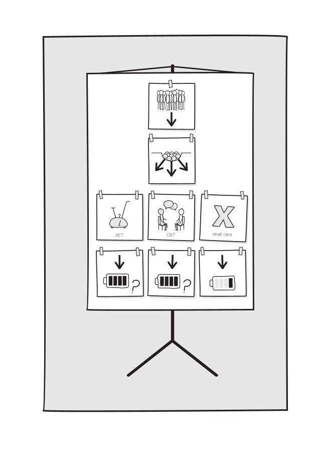
AET CBT usual care
? ?

PART 2FACTS-2-FSHD STUDY

?

CHAPTER 5EFFECT OF AEROBIC EXERCISE TRAINING AND COGNITIVE BEHAVIORAL THERAPY ON REDUCTION OF CHRONIC FATIGUE IN PATIENTS WITH FACIOSCAPULOHUMERAL DYSTROPHY: PROTOCOL OF THE FACTS-2-FSHD TRIAL
Nicoline B.M. Voet
Gijs Bleijenberg
George W. Padberg
Baziel G.M. van Engelen*
Alexander C.H. Geurts*
Published in: BMC Neurology. 2010 Jun 30;10:56.
*: contributed equally

ABSTRACT
Background: In facioscapulohumeral dystrophy (FSHD) muscle function is impaired and declines
over time. Currently there is no effective treatment available to slow down this
decline. We have previously reported that loss of muscle strength contributes to
chronic fatigue through a decreased level of physical activity, while fatigue and
physical inactivity both determine loss of societal participation. To decrease chronic
fatigue, two distinctly different therapeutic approaches can be proposed: aerobic
exercise training (AET) to improve physical capacity and cognitive behavioral
therapy (CBT) to stimulate an active life-style yet avoiding excessive physical
strain. The primary aim of the FACTS-2-FSHD (acronym for Fitness And Cognitive
behavioral TherapieS / for Fatigue and ACTivitieS in FSHD) trial is to study the
effect of AET and CBT on the reduction of chronic fatigue as assessed with the
Checklist Individual Strength subscale fatigue (CIS-fatigue) in patients with FSHD.
Additionally, possible working mechanisms and the effects on various secondary
outcome measures at all levels of the International Classification of Functioning,
Disability and Health (ICF) are evaluated.
Methods/Design: A multi-centre, assessor-blinded, randomized controlled trial is conducted. A
sample of 75 FSHD patients with severe chronic fatigue (CIS-fatigue ≥ 35) will be
recruited and randomized to one of three groups: (1) AET + usual care, (2) CBT +
usual care or (3) usual care alone, which consists of no therapy at all or occasional
(conventional) physical therapy. After an intervention period of 16 weeks and a
follow-up of 3 months, the third (control) group will as yet be randomized to either
AET or CBT (approximately 7 months after inclusion). Outcomes will be assessed at
baseline, immediately post intervention and at 3 and 6 months follow up.
Discussion: The FACTS-2-FSHD study is the first theory-based randomized clinical trial
which evaluates the effect and the maintenance of effects of AET and CBT on the
reduction of chronic fatigue in patients with FSHD. The interventions are based on
a theoretical model of chronic fatigue in patients with FSHD. The study will provide
a unique set of data with which the relationships between outcome measures at all
levels of the ICF could be assessed.
Trial registration: Dutch Trial Register, NTR1447.
Cha
pte
r 5
Pro
toco
l of t
he F
AC
TS-2
-FS
HD
tria
l
120

BACKGROUND
Facioscapulohumeral dystrophy (FSHD) is the third most common inherited
neuromuscular disorder. It is an autosomal dominant slowly progressive myopathy
with a variable age of onset, mostly in the second or third decade of life. Its yearly
incidence rate is approximately 1:20,000 (1). The disease primarily affects the facial
muscles, the muscles of the shoulder girdle (most typically the scapula stabilizers)
and various leg muscles, while pelvic and trunk muscles are eventually affected as
well (2-4). The pattern of muscle weakness is often asymmetrical, and the rate and
extent of progression may vary considerably with sudden periods of unexplained
rapid disease progression. In a small percentage of the patients, even respiratory
insufficiency may occur (5). Only very recently, evidence became available that
there may be a selective involvement of the central nervous system as well, in
terms of decreased grey matter volume (6) and reduced intracortical inhibition (7).
Although FSHD is associated with a partial deletion of a critical number of repetitive
elements (D4Z4) on chromosome 4q35, to date no causal gene has been identified
and no curative treatment is available (3, 8). FSHD may eventually lead to serious
disabilities of speech, swallowing, reaching, standing and walking, even in early
adulthood. Twenty percent of the patients become wheelchair bound. Since no
cure is available, rehabilitation is the mainstay of treatment (2, 3, 9).
Only recently it was shown by our group that severe fatigue, defined as a score
equal as or higher than 35 on the subscale fatigue of the Checklist Individual
Strength (CIS-fatigue), was reported by 61% of the patients with FSHD. These
severely fatigued patients had more problems with physical and social functioning
as well as with mental and general health than similar patients without a severe
fatigue. They also had more problems with concentration, initiating and planning
(10). As such, experienced fatigue should be clearly distinguished from muscle
weakness, which is probably the most common and characteristic symptom of
FSHD (11). In a longitudinal study, we built a model of perpetuating factors for
fatigue in patients with FSHD (Figure 1). It appeared that lack of physical activity,
sleep disturbances and pain all contributed to experienced fatigue. In addition, loss
of muscle strength and pain contributed to fatigue through a lower level of physical
activity. Ultimately, experienced fatigue and physical inactivity both contributed
to the level of societal participation (12). Thus, theoretically, in order to improve
societal participation one should improve muscle strength, reduce pain, optimize
physical activity and alleviate experienced fatigue. In addition, falling appears to be
a major problem among FSHD patients. Our group was able to show that 65% of
the patients reported falling at least once a year (13). Since fall incidents often lead
121

to fear of falling and avoidance behavior, they have a serious negative impact on
physical activity and participation.
Improving muscle strength by strength training and/or (anabolic) medication has
shown not to be successful in patients with FSHD (14). Until now, only one trial
has investigated low-intensity aerobic exercises, indicating that aerobic training
is a safe method to increase exercise performance (14, 15). Although, in general,
physical activity does not appear harmful (16, 17), more research is needed to
establish whether AET is beneficial in patients with FSHD. Besides improving
physical (aerobic) capacity, it seems important to optimize physical activity and
change behavior in daily life. Indeed, symptoms and signs of muscle weakness
and fatigue as well as the anticipation of a (further) decline in physical capacity
may elicit an inactive life-style, which may disproportionally affect physical activity,
fatigue and societal participation. From this perspective, it might be beneficial to
alter illness cognitions and coping styles by means of CBT. However, evidence for
the effectiveness of CBT in patients FSHD is not yet available.
The primary objective of the FACTS-2-FSHD trial is:
to study the efficacy of AET and CBT for decreasing chronic fatigue in patients
with FSHD. It is hypothesized that both AET and CBT are more effective in
decreasing fatigue than usual care, which is no therapy at all or occasional
(conventional) physical therapy. The improvement by AET may be obtained
through enhancement of physical (aerobic) capacity, whereas beneficial effects
of CBT may be achieved through changes in daily activities and behavior. By
changing illness cognitions and improving coping style, the balance between
actual behavior and physical capacity will be optimized. Since changes
achieved by CBT are more ‘intrinsic’, possible beneficial effects of CBT may
last longer than those of AET.
Secondary objectives of the FACTS-2-FSHD trial are:
to evaluate the effects of AET and CBT on bodily functions and structures
as defined by the International Classification of Functioning, Disability and
Health (ICF): lower extremity muscle strength, pain, psychological well being,
cardiovascular risk factors, aerobic exercise tolerance, sleeping pattern, as well
as biomarkers in blood and urine and structural and metabolic muscle tissue
characteristics.
to evaluate the effects of AET and CBT on the ICF level of activities: physical
activity in daily life, self perceived functional status, and fall incidence.
to evaluate the effects of AET and CBT on the ICF level of participation:
limitations in participation and autonomy and quality of life.Cha
pte
r 5
Pro
toco
l of t
he F
AC
TS-2
-FS
HD
tria
l
122

to evaluate the effects of AET and CBT on environmental and personal factors
as defined by the ICF: coping style, illness cognitions, concentration problems,
motivation, caregiver strain, experienced fatigue of the caregiver, social support
and coping of the caregiver.
Figure 1 Model of perpetuating factors of experienced fatigue in patients with
FSHD
METHODS
STUDY POPULATION
It is intended to include 75 FSHD patients, diagnosed on both clinical and genetic
grounds, aged 18 years and older. All patients who participated in previous FSHD
studies at the Radboud University Nijmegen Medical Centre (10, 12, 14, 18)
are approached by the primary investigator (NV). In addition, all patients known
at the departments of Neurology and Rehabilitation of the Radboud University
Nijmegen Medical Centre, University Medical Centre of Utrecht, Amsterdam
University Medical Centre or any of the affiliated rehabilitation centers are invited
to participate as well. In addition, patients who are registered in the Dutch
neuromuscular “computer registry of all myopathies and polyneuropathies”
(CRAMP) database (19) and/or who are member of the Dutch patient support
organization “vereniging spierziekten Nederland” (VSN) will be invited by the
primary investigator (NV) and a member of the VSN, respectively, to take part in the
study. If the patient is willing to participate, the primary investigator (NV) will check
the inclusion and exclusion criteria (Table 1) and estimate the disease severity using
the Ricci score (20). When a patient meets all the criteria, oral and written informed
consent are obtained according to the declaration of Helsinki. Separate consent is
asked to (i) obtain blood and urine samples, and/or (ii) undergo magnetic resonance
imaging (MRI) / spectroscopy (MRS), and/or (iii) undergo muscle ultrasound. The
study protocol was approved by the Dutch ethical committee CCMO (Centrale
commissie mensgebonden onderzoek) and all participating centers granted
(ethical) approval to participate.
123

Table 1 Inclusion and exclusion criteria
Inclusion criteria
(1) age 18 years and older
(2) suffering from severe fatigue (CIS-fatigue ≥ 35) (21)
(3) ability to walk independently (ankle-foot orthoses and canes are accepted)
(4) being able to exercise on a bicycle ergometer
(5) being able to complete either type of intervention
Exclusion criteria
(1) cognitive impairment
(2) insufficient mastery of the Dutch language
(3) neurological or orthopedic co-morbidity interfering with the interventions or possibly
influencing outcomes
(4) pregnancy
(5) use of psychotropic drugs (except simple sleeping medication)
(6) severe cardiopulmonary disease (chest pain, arrhythmia, pacemaker, cardiac surgery,
severe exertional dyspnea, emphysema)
(7) epileptic seizures
(8) poorly regulated diabetes mellitus or hypertension
(9) clinical depression, as diagnosed with the Beck Depression Inventory for primary
care (BDI-PC) (22, 23)
RANDOMIZATION AND BLINDING
Participants fulfilling all inclusion and exclusion criteria are randomized to one of
three groups by creating computer-generated ‘blocks’ ensuring that the same
number of participants is allocated to each group. Experimental group 1 (E1)
receives AET and usual care for 3 times a week during 16 weeks. Experimental
group 2 (E2) receives CBT and usual care once time a week during 16 weeks.
Thereafter, both groups are followed up for 6 months. A third group (C) receives
usual care only for 16 weeks and serves as a waiting list control group. After
another 3-months follow-up (i.e. 7 months after inclusion), this group will as yet
be randomized to either AET or CBT for 16 weeks and followed up until 6 months
later (total time in study 17 months) (Figure 2). All outcomes are assessed by
blinded and independent physical therapists. At the beginning of each assessment,
patients are always instructed not to reveal their group allocation to the blinded
assessor.Cha
pte
r 5
Pro
toco
l of t
he F
AC
TS-2
-FS
HD
tria
l
124

Figure 2 Study design
125

INTERVENTIONS
Usual care
All participants receive usual care. In the Netherlands, patients with FSHD typically
receive no therapy at all, or occasional (conventional) physical therapy. Patients are
not restricted in any activities, but all co-interventions are monitored throughout the
study by diaries and at every measurement.
Aerobic Exercise Training (AET)
The AET consists of aerobic cycling exercise on a bicycle ergometer. The training
program has a duration of 16 weeks and comprises home training twice a week
and a supervised training once a week. Training sessions consist of a 30-minute
aerobic exercise period with a warming-up and cooling-down period of 5 and
3 minutes, respectively. The cardiovascular load during the training period is
individually adjusted and increased from a level of 60% to 75% of the heart rate
reserve (HRR). HRR is the difference between the predicted maximum heart rate
and the measured resting heart rate. The HRR is equivalent to the difference
between the maximum and resting maximal oxygen consumption (VO2max). Each
participant is learned how to adjust the physical load to the preferred individual
heart rate. Participants are supplied with a Monark 827E bicycle ergometer, a
Garmin forerunner 50 heart rate watch with breast belt, and a log book with training
instructions at home for the duration of the intervention. During each training, the
heart rate is monitored continuously by the breast belt. The number of training
sessions, the total time spent on AET, possible adverse effects and the training
parameters (physical load, heart rate) are recorded in the individual log book. Once
a week, individually supervised training is given by trained physical therapists
during one-hour sessions in small groups in a nearby rehabilitation centre. During
these sessions, therapy compliance in the home situation is verified by reading out
the heart rate watches and checking the log books. In addition, instructions for the
next week are provided. The (unblinded) primary investigator and physician (NV)
gives instructions to the physical therapists and performs integrity checks at each
treatment location.
Cognitive Behavioral Therapy (CBT)
CBT will be focused on the perpetuating factors of fatigue as established in
previous research (10, 12, 24) and based on experience in clinical practice. These
factors encompass insufficient coping with the disease, dysfunctional illness Cha
pte
r 5
Pro
toco
l of t
he F
AC
TS-2
-FS
HD
tria
l
126

cognitions, catastrophizing, dysregulation of sleep, dysregulation of activity, low
social support and negative social interactions (see appendix 1 for the various
modules). Because of large inter-individual differences, CBT will be adapted to the
needs of each patient. For instance, barriers to become more physically active are
explored and possibly alleviated in some patients, whereas overactivity is reduced
in others. To determine which modules are appropriate, each perpetuating factor
is assessed with specific tests, and within each module, the CBT approach is
standardized (see appendix 1). The precise number of sessions is dependent on
the number of modules.
Each session has a duration of one hour and is given at the most nearby
participating centre by a registered cognitive behavioral therapist, especially
trained in CBT for FSHD. The therapists have been specifically trained to use the
diagnostic tests and indicate the different modules and are regularly supervised by
one of the investigators (GB).
COMPLIANCE AND ATTRITION
Therapy compliance is assessed by recording the number of treatment sessions
(AET and CBT). For the participants randomized to AET, the total time spent on
the bicycle ergometer at home is recorded as well. When applicable, participants
are asked for their reasons for poor compliance. In the case of therapy drop out,
patients are asked for the reason of non compliance and are stimulated to continue
participation in the assessments until the last follow-up.
OUTCOME ASSESSMENT
Outcome measures are listed in Table 2. The primary outcome measure is fatigue
severity as assessed with the CIS-fatigue (21). Secondary outcome measures are
categorized according to the different ICF levels (25). Outcome measurements are
obtained at the Radboud University Nijmegen Medical Centre at the start of the
study period (T0), immediately after the intervention period of 16 weeks (T1) and
after 3 (T2) and 6 months of follow-up (T3). Observers of the secondary outcome
measurements are experienced physical therapists blinded for treatment allocation.
At the first measurement (T0), demographic data is obtained by the primary
investigator and physician (NV), as well as a general and FSHD related medical
history, anthropometric measures (diastolic and systolic blood pressure in mm,
resting pulse rate in beats per minute and auscultation of heart and lungs), to verify
eligibility. The baseline (T0) and post treatment (T1) visits consist of muscle strength
testing of the thigh and aerobic exercise tolerance testing. Participants are asked
127

to complete the questionnaires and to wear the actometer for 12 consecutive days.
An actometer is a motion sensing device that can register and quantify human
physical activity and has to be worn at the ankle (26, 27). Blood and urine analyses
and MRI, MRS and ultrasound measurements of the thigh muscles are performed.
Follow-up measurements (T2 and T3) consist of muscle strength testing of the
thigh, aerobic exercise tolerance testing, questionnaires are completed and the
actometer is provided.
At the level of bodily functions and structures, patients are asked to give separate
consent for several ‘invasive’ assessments at baseline and after the intervention
period. Blood and urine samples are collected by experienced nurses and will be
explored by nuclear magnetic resonance (NMR) for possible biomarkers of disease
and response to the interventions. Ultrasound measurements of the thigh muscles
are made by an experienced ultrasound professional, blinded for the treatment
allocation, and analyzed for muscle thickness and echo intensity. In addition,
Magnetic Resonance Imaging (MRI) and 31P and proton (1H) Magnetic Resonance
Spectroscopy (MRS) are performed by trained professionals, blinded for the
assignment to the intervention.
It has been shown that in vivo MRS is able to produce spectra of multiple
metabolites simultaneously and is well suited to study energy metabolism in
patients with muscular dystrophies (28, 29). The MR examinations start with
T1 and T2 weighted images of the thigh for detailed structural analysis. Muscle
involvement is specifically assessed by the presence of fatty infiltration on T1
weighted MR images. 1H MRS is used to assess muscle specific creatine as well
as extramyocellular lipids and intramyocellular lipids levels, whereas 31P MRS is
applied to get information about tissue pH and the level of high energy phosphates.
Cha
pte
r 5
Pro
toco
l of t
he F
AC
TS-2
-FS
HD
tria
l
128
Table 2 Outcome measures and tests
Tests
Primary outcome measure
Fatigue severity Checklist Individual Strength (CIS subscale fatigue) (30)
Secondary outcome measures
ICF: bodily functionsAerobic exercise tolerance Maximal oxygen consumption (VO2 max) using the
Åstrand protocol (31) 6-minutes walking test (32) Resting heart rate

Muscle strength of quadriceps, hamstrings and tibialis anterior muscles Cardiovascular risk factors
PainPsychological well-being and sleeping patternMetabolic biomarkers
Structural and metabolic muscle characteristics
Quantitative Muscle Assessment using fixed myometry testing system (QMA) (18)
Blood pressure*
Abdominal circumferenceWeight / Body Mass Index (BMI)Percentage body fatVisual Analogue Scale (VAS) (33)Daily Observed Pain (during a period of 2 weeks) (27, 34)Brief Symptom Inventory (BSI) (35)
Blood and urine analyses for creatine, glucose, creatine kinase, sodium, potassium, calcium, phosphorus, ureum, ALAT, ASAT, gamma-GT, bilirubine, AF and LDH***
T1 MRI, T2 MRI, 31P and 1H MRS analysis of muscle specific creatine, extramyocellular lipids, intramyocellular lipids, levels and phosphometabolites in thigh muscles; ultrasound of thigh muscles
ICF: activitiesPhysical activity in daily life
Self perceived functional status
Actometer, a motion sensing device (during a period of 2 weeks) (26, 27) Daily Observed Spontaneous physical activity (during a period of 2 weeks) (27, 34). Checklist Individual Strength (CIS subscale physical activity) (30)Sickness Impact Profile (SIP subscales mobility control and mobility range, social behavior) (36)
ICF: participationLimitations in participation and autonomy Quality of life
Fall incidence
The Impact on Participation and Autonomy Questionnaire (IPAQ) (37)36-Item Short-Form Health Survey (SF-36) (38)Telephone computer (weekly calls)**
ICF: personal factorsCoping
Illness cognitionsConcentration problems
Motivation
Coping Inventory for Stressful Situation (CISS) (30)ALCOS-16 (39)Ziekte cognitie lijst (ZCL) (40)Checklist Individual Strength (CIS subscale concentration) (21)Checklist Individual Strength (CIS subscale motivation) (21)
129
Table 2 Outcome measures and tests (Continued)

ICF: environmental factorsCaregiver strainExperienced fatigue of patient from perspective of relative.Experienced fatigue of partner.
Social support
Coping of partner
Caregiver Strain Index of partner/caregiver (CSI) (41)Checklist Individual Strength (CIS subscale fatigue, filled in by relative about patient) (21)Checklist Individual Strength (CIS subscale fatigue, filled in by relative about him/herself) (21)Sociale steunlijst-subschaal discrepantie (SSL-D verkort) (42)Coping Inventory for Stressful Situations (CISS) (30)
*Measured only at inclusion** Participants will be called weekly throughout the study by a telephone-computer to obtain fall incidence; in the case of fall incident(s), a subsequent telephone interview is held to obtain information about the cause and circumstances of falling, fall direction, possible injury and ability to get up from the floor*** In all groups, three extra urine samples will be obtained at week 4, 8 and 12 of the intervention period
An adverse event is defined as any undesirable experience or outcome. Specially
assigned site investigators are instructed to report all adverse events immediately
to the primary investigator (NV) and to evaluate each event for its date of onset,
possible relation to the interventions (based on clinical judgment), possible
treatment and course in time. In addition, adverse events can be reported by the
participants directly to the primary investigator and physician (NV). All adverse
events reported will be carefully monitored and registered until they have abated or
a stable situation has been reached.
STATISTICAL ANALYSIS
Generalized estimated equations analysis will be used to investigate differences
in the effects on primary and secondary outcome measures between the study
groups and to investigate the influence of possible effect modifiers. When
necessary, analyses will be adjusted for group differences in fatigue severity and
physical activity at baseline. Data will be analyzed according to the intention-to-
treat principle.
POWER
In order to detect a 10% group difference (E1 and E2 versus C) in change in
fatigue severity between the start and the end of the intervention period (assuming
difference in standard deviation between the start and the end of the intervention
Cha
pte
r 5
Pro
toco
l of t
he F
AC
TS-2
-FS
HD
tria
l
130
Table 2 Outcome measures and tests (Continued)

(SDdif) = 10%, α = .05, β = .80), 20 participants per group are required. With an
expected drop-out rate of maximally 25%, 25 participants will be recruited in each
group (n=75).
DISCUSSION
To the best of our knowledge, the FACTS-2-FSHD study is the first randomized
clinical trial which evaluates the effect of AET and CBT on the reduction of chronic
fatigue in patients with FSHD.
This study has several strengths. First, the selected interventions are based on a
theoretical model of chronic fatigue in patients with FSHD (12) and are compared
with usual care in a randomized design. Until now, only one randomized controlled
trial has been conducted that could not establish a beneficial effect of muscle
strength training compared to no training in FSHD (14, 18). In addition, one trial
has been conducted that investigated low-intensity aerobic exercises in FSHD.
Although this latter study reported improved maximal oxygen uptake and workload
as a result of training, this was an uncontrolled and unblinded trial of only 8
patients (15). The majority of the training studies in patients with muscle disorders
did not include a (no-training) control group or used healthy subjects as controls.
In addition, data are often presented for mixed groups of muscle disorder (17).
Second, the proposed study uses a broad arsenal of secondary outcome measures
at all levels of the ICF, including ‘invasive’ measurements of possible biomarkers
in blood and urine as well as measurements of structural and metabolic muscle
characteristics.
This approach will provide a unique set of data with which it should be possible
to accurately assess the relationships between disease characteristics, loss of
bodily functions, activity limitations and restrictions in societal participation in
patients with FSHD. Third, all patients will be followed up until 6 months after the
interventions, which will not only provide information about the maintenance of
effects, but also about any long-term adverse events.
A limitation of this study is that the sample size calculation was based on detection
of a 10% difference between the intervention groups and the control group,
presuming more or less equal effect sizes of AET and CBT. Detecting more subtle
differences in the effectiveness between both interventions would require a much
larger sample size. In the Netherlands alone, such a trial would not be feasible.
131

In conclusion, the FACTS-2-FSHD study will increase our insight into the
effectiveness of aerobic exercise training and cognitive behavioral therapy to
reduce chronic fatigue and to optimize physical activity and capacity in patients
with FSHD. A successful outcome of this study has the potential to change existing
(inter)national guidelines for physical training and to improve the quality of life in
patients with FSHD.
Cha
pte
r 5
Pro
toco
l of t
he F
AC
TS-2
-FS
HD
tria
l
132
APPENDIX 1 DIFFERENT MODULES OFCOGNITIVE BEHAVIORAL THERAPY
Perpetuating factors: insufficient coping with the disease
Insufficient coping with the disease is assessed with the Impact of event Scale (43,
44). A participant can continue to be occupied with the period of being diagnosed
with FSHD. By means of talking or writing about this experience (which can be
referred to as ‘exposure’), the participant will acquire better coping skills. Fear of
progression is assessed with a questionnaire especially designed for FSHD. The
therapist helps the participant to formulate explicit words to describe the thoughts
of fear of progression. These thoughts are challenged against reality (reality testing).
In this way, daily unhelpful thoughts about the disease progression are reduced and
put into perspective.
Perpetuating factors: dysfunctional cognitions regarding fatigue, activity and other symptoms
Dysfunctional cognitions relate to a variety of ideas, including a participant’s idea
of lack of control over symptoms, and dysfunctional cognitions about symptoms,
such as catastrophizing. The sense of control in relation to fatigue complaints will
be assessed with the self-efficacy scale (34, 45).
Perpetuating factors: catastrophizing
Catastrophizing will be assessed with the Jacobsen Fatigue Catastrophizing Scale
(46). These cognitions are disputed and more helpful ways of thinking are taught.
Perpetuating factors: dysregulation of sleep
Dysregulation of sleep is based on self-report in a sleep diary (47). An irregular
sleep-wake rhythm can perpetuate fatigue. To restore the biologic rhythm,
participants are encouraged to adhere to fixed bedtimes and wake-up times and
discouraged from sleeping during the day, or they are helped with adapting fixed
rest period(s).
133

Perpetuating factors: dysregulation of activity
Dysregulation of activity is based on activity (stepping) monitoring using an
actometer and a physical activity questionnaire (Physical Activity Scale for
Individuals with Physical Disabilities (44, 48). Some patients experience fluctuating
periods of activity with subsequent periods of rest during a longer period.
Others avoid activity because they are concerned that activity increases fatigue;
consequently, they are physically inactive. For patients with fluctuating activity
levels, a base level should be established by alternating rest and activities to
prevent bursts of activity. Once the participant has set a base level, the physical
activity program is started, usually twice a day, starting with 5 to 10 minutes of an
activity such as walking or cycling. The activity is increased by 1 minute a day each
time the activity is performed (ending at a maximum of 2 times 60 minutes minutes
per day). The inactive participant will start the activity program immediately.
Gradually, physical activities are replaced by other activities.
Perpetuating factors: low social support and negative social interactions
Low social support and negative social interactions are based on the discrepancy
subscale of the Social Support List (49). If a participant still has unrealistic
expectations of others or perceives a discrepancy between actual support and
desired support, the therapist helps to install more realistic expectations toward the
participant’s social support group. The partner or caregiver will be included in this
treatment module.
Cha
pte
r 5
Pro
toco
l of t
he F
AC
TS-2
-FS
HD
tria
l
134

ACKNOWLEDGEMENTS
This study is funded by the Prinses Beatrix Fonds (PBF) (The Dutch Public Fund
for Neuromuscular and Movement Disorders), the Netherlands Organization for
Health Research and Development (ID: ZonMw 89000003) and by the FSHD Global
Research Foundation.
AUTHORS‘ CONCLUSIONS
NV is primary investigator and responsible for data collection and analysis and for
drafting the manuscript. AG, BvE, GB, and GP designed and supervised the study.
AG and BvE obtained funding for the study. All authors helped in finalizing the
manuscript.
COMPETING INTERESTS
The authors declare that they have no competing interests.
REFERENCES
1. Kissel JT. Facioscapulohumeral dystrophy. Seminars in Neurology.
1999;19(1):35-43.
2. Padberg GW, Lunt PW, Koch M, Fardeau M. Diagnostic criteria for
facioscapulohumeral muscular dystrophy. Neuromuscular Disorders.
1991;1(4):231-4.
3. Tawil R, van der Maarel SM. Facioscapulohumeral muscular dystrophy. Muscle
& Nerve. 2006;34(1):1-15.
4. Padberg GW, van Engelen BG. Facioscapulohumeral muscular dystrophy.
Current Opinion in Neurology. 2009;22(5):539-42.
5. Wohlgemuth M, van der Kooi EL, van Kesteren RG, van der Maarel SM,
Padberg GW. Ventilatory support in facioscapulohumeral muscular dystrophy.
Neurology. 2004;63(1):176-8.
6. Quarantelli M, Lanzillo R, Del VW, Mollica C, Prinster A, Iadicicco L, et al.
Modifications of brain tissue volumes in facioscapulohumeral dystrophy.
Neuroimage. 2006;32(3):1237-42.
7. Di Lazzaro V, Oliviero A, Tonali PA, Felicetti L, De Marco MB, Saturno E, et
al. Changes in motor cortex excitability in facioscapulohumeral muscular
dystrophy. Neuromuscular Disorders. 2004;14(1):39-45.
135

8. van der Maarel SM, Frants RR, Padberg GW. Facioscapulohumeral muscular
dystrophy. Biochimica et Biophysica Acta. 2007;1772(2):186-94.
9. Lunt PW, Harper PS. Genetic counselling in facioscapulohumeral muscular
dystrophy. Journal of Medical Genetics. 1991;28(10):655-64.
10. Kalkman JS, Schillings ML, van der Werf SP, Padberg GW, Zwarts MJ, van
Engelen BG, et al. Experienced fatigue in facioscapulohumeral dystrophy,
myotonic dystrophy, and HMSN-I. Journal of Neurology, Neurosurgery, and
Psychiatry. 2005;76(10):1406-9.
11. Engel AG, Franzini-Armstrong C. Myology 2004.
12. Kalkman JS, Schillings ML, Zwarts MJ, van Engelen BG, Bleijenberg G. The
development of a model of fatigue in neuromuscular disorders: a longitudinal
study. Journal of Psychosomatic Research. 2007;62(5):571-9.
13. Horlings CG, Munneke M, Bickerstaffe A, Laverman L, Allum JH, Padberg
GW, et al. Epidemiology and pathophysiology of falls in facioscapulohumeral
disease. Journal of Neurology, Neurosurgery, and Psychiatry.
2009;80(12):1357-63.
14. van der Kooi EL, Kalkman JS, Lindeman E, Hendriks JC, van Engelen
BG, Bleijenberg G, et al. Effects of training and albuterol on pain and
fatigue in facioscapulohumeral muscular dystrophy. Journal of Neurology.
2007;254(7):931-40.
15. Olsen DB, Orngreen MC, Vissing J. Aerobic training improves exercise
performance in facioscapulohumeral muscular dystrophy. Neurology.
2005;64(6):1064-6.
16. Cup EH, Pieterse AJ, Ten Broek-Pastoor JM, Munneke M, van Engelen BG,
Hendricks HT, et al. Exercise therapy and other types of physical therapy
for patients with neuromuscular diseases: a systematic review. Archives of
Physical Medicine and Rehabilitation. 2007;88(11):1452-64.
17. Voet NB, van der Kooi EL, Riphagen II, Lindeman E, van Engelen BG, Geurts
AC. Strength training and aerobic exercise training for muscle disease.
Cochrane Database of Systematic Reviews. 2010(1):CD003907.
18. van der Kooi EL, Vogels OJ, van Asseldonk RJ, Lindeman E, Hendriks JC,
Wohlgemuth M, et al. Strength training and albuterol in facioscapulohumeral
muscular dystrophy. Neurology. 2004;63(4):702-8.
19. van Engelen BG, van VH, Van Doorn PA, Faber CG, van der Hoeven JH,
Janssen NG, et al. The Dutch neuromuscular database CRAMP (Computer
Registry of All Myopathies and Polyneuropathies): development and preliminary
data. Neuromuscular Disorders. 2007;17(1):33-7.
Cha
pte
r 5
Pro
toco
l of t
he F
AC
TS-2
-FS
HD
tria
l
136

20. Ricci E, Galluzzi G, Deidda G, Cacurri S, Colantoni L, Merico B, et al. Progress
in the molecular diagnosis of facioscapulohumeral muscular dystrophy and
correlation between the number of KpnI repeats at the 4q35 locus and clinical
phenotype. Annals of Neurology. 1999;45(6):751-7.
21. Vercoulen JH, Swanink CM, Fennis JF, Galama JM, van der Meer JW,
Bleijenberg G. Dimensional assessment of chronic fatigue syndrome. Journal
of Psychosomatic Research. 1994;38(5):383-92.
22. Arnau RC, Meagher MW, Norris MP, Bramson R. Psychometric evaluation of
the Beck Depression Inventory-II with primary care medical patients. Health
Psychology. 2001;20(2):112-9.
23. Beck AT, Guth D, Steer RA, Ball R. Screening for major depression disorders
in medical inpatients with the Beck Depression Inventory for Primary Care.
Behaviour Research and Therapy. 1997;35(8):785-91.
24. Kalkman JS, Schillings ML, Zwarts MJ, van Engelen BG, Bleijenberg
G. Influence of relatives on fatigue experienced by patients with
facioscapulohumeral dystrophy, myotonic dystrophy and HMSN-I. European
Neurology. 2006;56(1):24-30.
25. WHO. International Classification of Functioning, Disability and Health (ICF).
Geneva 2001.
26. van der Werf SP, Prins JB, Vercoulen JH, van der Meer JW, Bleijenberg
G. Identifying physical activity patterns in chronic fatigue syndrome
using actigraphic assessment. Journal of Psychosomatic Research.
2000;49(5):373-9.
27. Vercoulen JH, Bazelmans E, Swanink CM, Fennis JF, Galama JM, Jongen PJ,
et al. Physical activity in chronic fatigue syndrome: assessment and its role in
fatigue. Journal of Psychiatric Research. 1997;31(6):661-73.
28. Argov Z, Lofberg M, Arnold DL. Insights into muscle diseases gained
by phosphorus magnetic resonance spectroscopy. Muscle & Nerve.
2000;23(9):1316-34.
29. Kan HE, Scheenen TW, Wohlgemuth M, Klomp DW, van Loosbroek-
Wagenmans I, Padberg GW, et al. Quantitative MR imaging of individual muscle
involvement in facioscapulohumeral muscular dystrophy. Neuromuscular
Disorders. 2009;19(5):357-62.
30. Endler NS, Parker JD. Multidimensional assessment of coping: a critical
evaluation. Journal of Personality and Social Psychology. 1990;58(5):844-54.
31. Astrand PO, Ryhming I. A normogram for calculation for aerobic capacity
(physical fitness) from pulse rate during sub-maximal work. Journal of Applied
Physiology. 1954(7):218-22.
137

32. Enright PL. The six-minute walk test. Respiratory Care. 2003;48(8):783-5.
33. Folstein MF, Luria R. Reliability, validity, and clinical application of the Visual
Analogue Mood Scale. Psychological Medicine. 1973;3(4):479-86.
34. Vercoulen JH, Hommes OR, Swanink CM, Jongen PJ, Fennis JF, Galama
JM, et al. The measurement of fatigue in patients with multiple sclerosis. A
multidimensional comparison with patients with chronic fatigue syndrome and
healthy subjects. Archives of Neurology. 1996;53(7):642-9.
35. Derogatis LR, Melisaratos N. The Brief Symptom Inventory: an introductory
report. Psychological Medicine. 1983;13(3):595-605.
36. Jacobs HM, Luttik A, Touw-Otten FW, de Melker RA. [The sickness impact
profile; results of an evaluation study of the Dutch version]. Nederlands
Tijdschrift voor Geneeskunde. 1990;134(40):1950-4.
37. Cardol M, de Haan RJ, van den Bos GA, de Jong BA, de GI. The development
of a handicap assessment questionnaire: the Impact on Participation and
Autonomy (IPA). Clinical Rehabilitation. 1999;13(5):411-9.
38. Aaronson NK, Muller M, Cohen PD, Essink-Bot ML, Fekkes M, Sanderman R,
et al. Translation, validation, and norming of the Dutch language version of the
SF-36 Health Survey in community and chronic disease populations. Journal of
Clinical Epidemiology. 1998;51(11):1055-68.
39. Bosscher RJ, Smit JH. Confirmatory factor analysis of the General Self-Efficacy
Scale. Behaviour Research and Therapy. 1998;36(3):339-43.
40. Evers AW, Kraaimaat FW, van LW, Jongen PJ, Jacobs JW, Bijlsma JW. Beyond
unfavorable thinking: the illness cognition questionnaire for chronic diseases.
Journal of Consulting and Clinical Psychology. 2001;69(6):1026-36.
41. Post MW, Festen H, van dPI, Visser-Meily JM. Reproducibility of the
Caregiver Strain Index and the Caregiver Reaction Assessment in partners
of stroke patients living in the Dutch community. Clinical Rehabilitation.
2007;21(11):1050-5.
42. Van Sonderen EL. Sociale Steun Lijst Interacties (SSL-I) en Sociale Steun Lijst
Discrepanties (SSL-D). 1993.
43. Creamer M, Bell R, Failla S. Psychometric properties of the Impact of Event
Scale - Revised. Behaviour Research and Therapy. 2003;41(12):1489-96.
44. van der Ploeg E, Mooren TT, Kleber RJ, van der Velden PG, Brom D. Construct
validation of the Dutch version of the impact of event scale. Psychological
Assessment. 2004;16(1):16-26.
45. Prins JB, Bleijenberg G, Bazelmans E, Elving LD, de Boo TM, Severens JL,
et al. Cognitive behaviour therapy for chronic fatigue syndrome: a multicentre
randomised controlled trial. Lancet. 2001;357(9259):841-7.
Cha
pte
r 5
Pro
toco
l of t
he F
AC
TS-2
-FS
HD
tria
l
138

46. Andrykowski MA, Schmidt JE, Salsman JM, Beacham AO, Jacobsen PB.
Use of a case definition approach to identify cancer-related fatigue in women
undergoing adjuvant therapy for breast cancer. Journal of Clinical Oncology.
2005;23(27):6613-22.
47. Servaes P, Verhagen C, Bleijenberg G. Fatigue in cancer patients during and
after treatment: prevalence, correlates and interventions. European Journal of
Cancer. 2002;38(1):27-43.
48. Washburn RA, Zhu W, McAuley E, Frogley M, Figoni SF. The physical activity
scale for individuals with physical disabilities: development and evaluation.
Archives of Physical Medicine and Rehabilitation. 2002;83(2):193-200.
49. Ormel J, Kempen GI, Penninx BW, Brilman EI, Beekman AT, van Sonderen E.
Chronic medical conditions and mental health in older people: disability and
psychosocial resources mediate specific mental health effects. Psychological
Medicine. 1997;27(5):1065-77.
139
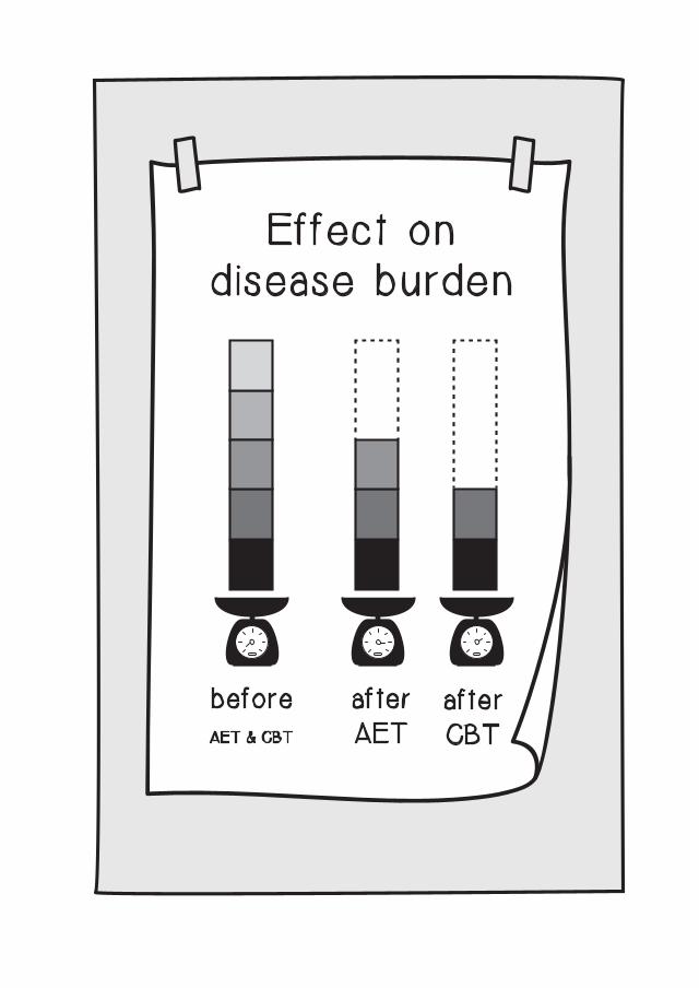
Ef fec t ond i sease burden
af terCBT
af terAET
beforeAET & CBT
CHAPTER 6BOTH AEROBIC EXERCISE AND COGNITIVE BEHAVIORAL THERAPY REDUCE FATIGUE IN FSHD: A RCT
Nicoline B.M. Voet
Gijs Bleijenberg
Jan C. Hendriks
Imelda J.M. de Groot
George W. Padberg
Baziel G.M. van Engelen*
Alexander C.H. Geurts*
Published in: Neurology. 2014;83:1-9.
*: contributed equally
ABSTRACT
Objective: To investigate the effect of aerobic exercise training (AET) and cognitive behavioral
therapy (CBT) on chronic fatigue in patients with facioscapulohumeral muscular
dystrophy (FSHD).
Methods: We performed a multicenter, assessor-blinded, randomized clinical trial (RCT).
Fifty-seven patients with FSHD type 1 with severe chronic fatigue were randomly
allocated to AET, CBT, or usual care (UC). Outcomes were assessed before
treatment, following 16 weeks of intervention, and after a 12-week follow-up. A
linear mixed model for repeated measurements was used to study the estimated
group differences.
Results: Following treatment, both the AET (28 participants) and CBT (25 participants)
intervention groups had less fatigue relative to the UC group (24 participants), with
a difference of -9.1 for AET (95% confidence interval {CI} -12.4 to -5.8) and -13.3
for CBT (95% CI -16.5 to -10.2). These beneficial effects lasted through follow-up,
with a difference of -8.2 for AET (95% CI -12.4 to -5.8) and -10.2 for CBT (95% CI
-14.0 to -6.3). The patients who received CBT had an increase in registered and
experienced physical activity, sleep quality, and social participation. The patients
who received AET had an increase in registered physical activity only. The increase
in registered physical activity in both groups and the improvement in social
participation following CBT were still present at follow-up.
Conclusions: This RCT shows that AET and CBT can ameliorate chronic fatigue in patients with
FSHD.
Classification of evidence: This study provides Class III evidence that, in patients with FSHD type 1 and severe
chronic fatigue, AET or CBT reduces the severity of chronic fatigue.
Cha
pte
r 6
Bot
h ae
rob
ic e
xerc
ise
and
cog
nitiv
e b
ehav
iora
l the
rap
y re
duc
e fa
tigue
in F
SH
D: a
RC
T
142

Facioscapulohumeral muscular dystrophy (FSHD) is an autosomal dominant,
slowly progressive myopathy, affecting approximately 1:8,000 individuals (1). We
previously reported that severe fatigue is experienced by 61% of patients with
FSHD and contributes considerably to reduced social participation (2). We also
developed a FSHD-specific model of fatigue-perpetuating factors (3). Because the
patient’s level of physical activity plays a central role in this model, it is a logical
target for therapeutic intervention. In theory, one can increase physical activity
either by improving muscle strength or by optimizing aerobic capacity. Althoug h
strength training in patients with FSHD appears to be safe, its beneficial effects are
limited (4, 5). In contrast, a pilot study provided evidence that patients with FSHD
can benefit from aerobic exercise training (AET) (6). Cognitive behavioral therapy
(CBT) is a different type of intervention that can be used to alleviate individually
relevant fatigue-perpetuating factors. Several published studies have provided
evidence that CBT can reduce fatigue in chronic fatigue syndrome, multiple
sclerosis, and cancer survivors (7-9). However, the effect of CBT on fatigue in
patients with muscular dystrophy has not been investigated.
METHODS
STUDY OBJECTIVES
The primary objective of this study was to evaluate the effect of 16 weeks of AET
or CBT on chronic fatigue in patients with FSHD type 1 compared to usual care
(UC) (10) (level of evidence: Class III). The secondary objective was to evaluate
the effects of each intervention on the known fatigue-perpetuating factors in these
patients (level of evidence: Class III). In addition, we were interested whether CBT,
as it focuses on all fatigue-perpetuating factors, might provide longer-lasting
benefits than AET.
PARTICIPANTS
All the adult patients with FSHD who participated in any previous study at our
center, (2-4) who were registered in a Dutch neuromuscular database, (11) or
who participated in a patient support organization were invited to participate. In
addition, they were asked to complete the Short Fatigue Questionnaire (SFQ) (12). If
a patient was willing to participate, N.V. checked the inclusion and exclusion criteria
(Table 1). A diagnosis of FSHD type 1 had to be confirmed by DNA testing (13).
143

Table 1 Inclusion and exclusion criteria
Inclusion criteria
(1) age 18 years and older
(2) suffering from severe fatigue (CIS-fatigue ≥ 35)
(3) ability to walk independently (ankle-foot orthoses and canes are accepted)
(4) being able to exercise on a bicycle ergometer
(5) being able to complete either type of intervention
Exclusion criteria
(1) cognitive impairment
(2) insufficient mastery of the Dutch language
(3) neurological or orthopedic co-morbidity interfering with the interventions or possibly influencing outcomes
(4) pregnancy
(5) use of psychotropic drugs (except simple sleeping medication)
(6) severe cardiopulmonary disease (chest pain, arrhythmia, pacemaker, cardiac surgery,severe exertional dyspnea, emphysema)
(7) epileptic seizures
(8) poorly regulated diabetes mellitus or hypertension
(9) clinical depression, as diagnosed with the BDI-PC
CIS-fatigue: subscale fatigue of the Checklist Individual Strength, BDI-PC: the Beck Depression Inventory for Primary CareSource: adapted with permission from Voet et al. (16)
DESIGN
The study was a multicenter, assessor-blinded, randomized clinical trial using
repeated measurements. An independent research assistant allocated each
participant to 16 weeks of AET, CBT, or UC using a computer-generated
randomization block list. The block sizes varied randomly in order to prevent
predictability of the allocation process. After 12 weeks of follow-up, after a total
of 28 weeks (i.e., period 1), the patients in the UC group were randomly allocated
to 16 weeks AET or CBT followed by another 12 weeks of follow-up (i.e., period
2: 28 weeks). The interventions were performed at 9 health care institutions for
convenience of the patients
Cha
pte
r 6
Bot
h ae
rob
ic e
xerc
ise
and
cog
nitiv
e b
ehav
iora
l the
rap
y re
duc
e fa
tigue
in F
SH
D: a
RC
T
144

STANDARD PROTOCOL APPROVALS, REGISTRATIONS, AND PATIENT CONSENTS
The study protocol was approved by the regional medical ethics committee
(CMO2008/228) and by each institution’s local committee. All patients provided
oral and written informed consent (14). The design of this study was published
previously (15) and was registered as the FACTS-2-FSHD trial in the Dutch Trial
Register (NTR1447).
INTERVENTIONS
AET consisted of 3 weekly sessions of aerobic cycling exercises on a Monark
(Varberg, Sweden) 827E ergometer; 2 sessions were performed in the patient’s
home, and the third was supervised by a physical therapist. N.V. performed
integrity checks at each study site. The participants cycled for 30 minutes with
additional warming-up and cooling-down periods of 5 and 3 minutes, respectively.
During the training period, cardiovascular load was continuously monitored with a
heart rate belt and watch (Garmin {Olathe, Kansas} Forerunner 50) and adjusted to
the individual participant’s level. It was aimed to achieve an increase of 50%–65%
in heart rate reserve (HRR) (16). Participants who were taking β-blockers were
instructed to exercise at a perceived exertion level of 12–14 on the Borg scale
(appendix 1) (17). The number and duration of the sessions, the Borg score, the
mean and maximum heart rate, and adverse effects were recorded in an individual
log book. During the weekly supervised sessions, the physical therapist verified
compliance by reading the data recorded by the heart rate watch and by reviewing
the log book. In addition, training instructions for the following week were given
to the participant. Acceptable compliance with the AET program was defined as
completion of a minimum of 40 training sessions.
CBT comprised 6 possible modules based on the known fatigue-perpetuating
factors (3) and previous research (8). These modules were directed at insufficient
coping with the disease; dysfunctional cognitions regarding fatigue, activity, pain,
or other symptoms; fatigue catastrophizing; dysregulation of sleep or activity;
poor social support; and negative social interactions (appendix 2). To account for
interindividual differences in these factors, the intervention was adapted to the
specific needs of each participant, including an individually tailored structured
activity program. Each session was 50 minutes in duration and was conducted
at the nearest participating center by a cognitive behavioral therapist. The total
number of sessions for each participant was based on the number of modules to
145
be addressed, which were identified by the therapist by performing an interview
and specific tests. Acceptable compliance with the CBT program was defined as
completion of a minimum of 3 sessions.
The participants in the UC group received no specific treatment for fatigue but
occasional physical therapy was allowed. Throughout the study, the participants
were not restricted in their activities. Any cointervention (e.g., physiotherapy
session) was noted in the log books.
OUTCOMES
The primary outcome measure was fatigue severity, assessed with the fatigue
subscale of the Checklist Individual Strength (CIS-fatigue). This scale consists of
8 questions regarding fatigue as experienced during the previous 2 weeks; each
question was scored on a 7-point Likert scale (18). A total score ≥35 indicates
severe fatigue (3). The CIS-fatigue has good internal consistency (Cronbach a
0.83–0.92), high discriminative validity, and high sensitivity to change in patients
with FSHD (3).
The secondary outcome measures were selected to evaluate each factor of the
described model (3).
Maximum voluntary isometric strength for the quadriceps was measured using
Quantitative Muscle Assessment (19); the highest value of 2 maximal isometric
contractions was collected and the average value of the left and right quadriceps
strength was calculated.
Aerobic exercise tolerance (VO2 peak, in L/min) was estimated with the Åstrand
test (20). Physical endurance was tested with the distance walked during a
6-minute walking test (21).
Participants were asked to wear an ankle actometer for 12 consecutive days
(throughout the day and night) (22). Registered physical activity was averaged over
the 12-day period and expressed as the average number of body accelerations
per 5-minute period. The physical activity subscale of the Checklist Individual
Strength (CIS-activity) was used as a measure of experienced physical activity. This
subscale consists of 3 questions regarding activity during the previous 2 weeks;
each question was scored on a 7-point Likert scale (22).
Pain intensity was assessed with a Visual Analogue Scale (VAS-pain, range 0–100)
(23).
Self-reported sleep disturbances were assessed using the 5- item sleep subscale
of the Nottingham Health Profile (NHPsleep, range 0–100) (24, 25). Cha
pte
r 6
Bot
h ae
rob
ic e
xerc
ise
and
cog
nitiv
e b
ehav
iora
l the
rap
y re
duc
e fa
tigue
in F
SH
D: a
RC
T
146

Social participation restrictions were assessed with the social behavior subscale of
the Sickness Impact Profile 68 (SIP68-sb, a weighted score ranging from 0 to 572)
(26).
For CIS-fatigue, CIS-activity, VAS-pain, NHP-sleep, and SIP68-sb, a higher score
indicates poorer outcome.
All adverse events reported by the participants or observed by the therapists were
recorded. Participants were called weekly throughout the study by a telephone-
computer to obtain fall incidence (27).
PROCEDURE
At baseline, demographic and clinical characteristics were collected, including the
Ricci score as a measure of disease severity (28) (Table 2).
147
Table 2 Baseline characteristics
UC (n=24) AET (n=20) CBT (n=13)
Characteristics n median range n median range n median range
Age (years) 24 52 (20-79) 20 59 (21-68) 13 49 (24-69)
Gender (male) 24 17 (71%) 20 12 (60%) 13 8 (62%)
Ricci score 24 3.0 (1.5-4.0) 20 3.0 (0.5-4.0) 13 2.0 (1.0-4.0)
Duration of illness (years)
24 16.7 (1.0-49.0) 20 13.0 (1.0-42.0) 13 7.0 (0.0-40.0)
Duration of fatigue (years)
18 5.0 (0.5-25.0) 17 10.0 (1.7-30.0) 12 4.5 (2.0-28.0)
Physical therapy (PT) (n/%)
24 10 (42%) 20 8 (40%) 13 4 (31%)
Sessions PT/patient/week
24 0 (0-2) 20 0 (0-4) 13 0 (0-2)
CIS-fatigue 24 42 (35-51) 20 41 (35-54) 13 43 (35-56)
UC: usual care, AET: aerobic exercise training, CBT: cognitive behavioral therapy, CIS-fatigue: subscale fatigue of the Checklist Individual Strength
The outcome data of the participants in the AET and CBT groups were collected
immediately before treatment, immediately following the 16-week intervention
period, and 12 weeks postintervention (period 1 and period 2). The outcome data
of the UC group were collected at baseline and after 16 and 28 weeks follow-up
(period 1). All measurements were performed at the Radboud University Medical
Center by 2 blinded physical therapists. At follow-up, N.V. recorded whether the
participants had continued their level of activity during the follow-up period.
POWER
The primary outcome is change in CIS-fatigue between the start and the end of the
treatment period (16 weeks). Assuming a difference (in change) between the groups
of 8 points, a SD of the change of 8 points, α = 0.05, β= 0.80, and applying a
Bonferroni adjustment for the number of comparisons, 20 patients are needed per
group using a 2-sided t test. We aimed to include 25 patients per group to allow a
dropout rate of 25%. We planned to randomize the patients of the UC group (after
28 weeks) to either intervention group, so that a total of 50 patients (instead of 75)
would be needed to evaluate 25 patients per group. No further (down) adjustment
for repeated measurements was applied to the already low number of patients per
group.
STATISTICAL ANALYSIS
A linear mixed model for repeated measurements was used to study group
differences in the change per week after treatment of each of the primary and
secondary outcomes. The dependent variable was the specific outcome. The
independent class variables were treatment (3 levels: AET, CBT, UC) and period
(2 levels: first, second). The independent continuous variables were time (weeks
since baseline) and the value of the specific outcome at baseline. The intercept
of each patient was treated as a random variable. This allows different levels for
different individuals. In addition, all first-order and second-order interaction terms
between group, time, and period were included in the linear part of the model. At
first, we found that, for all outcomes, the factor period and all interaction terms,
except the interaction term between time and treatment, never reached the level of
statistical significance and that the models were never significantly reduced when
these terms were omitted from the model (likelihood ratio test). Second, we also
used time as a class variable and found that this never significantly improved the
fit of the model to the data (likelihood ratio test). Consequently, in the final model,
period and the period-related interaction terms were omitted and time was treated
as a continuous variable, as presented above. Finally, the regression per week in Cha
pte
r 6
Bot
h ae
rob
ic e
xerc
ise
and
cog
nitiv
e b
ehav
iora
l the
rap
y re
duc
e fa
tigue
in F
SH
D: a
RC
T
148

the UC group was estimated using the data of all 3 points of measurement, and
the estimated mean group differences with 95% confidence intervals (CIs) were
calculated at 16 and 28 weeks. The difference in percentage of patients with
severe fatigue (CIS-fatigue score ≥35) was used as an endpoint to calculate the
absolute risk reduction (ARR) for both intervention groups compared to the UC
group. The number needed to treat (NNT) was calculated as the reciprocal of the
ARR. Statistical analyses were performed with SAS version 9.2 for Windows (SAS
Institute, Cary, NC). p < 0.05 was considered statistically significant.
RESULTS
The flowchart of the patients in the study is shown in the Figure.
In total, 377 patients were invited to participate. The most commonly cited reason
for unwillingness to participate (n = 199) was unknown (n = 153) or the long
distance required to travel to the training location (n = 33).
149
1 drop-out (trial too time consuming)
Invited to participate (n=377)
Excluded (n= 37) - No severe fatigue (n= 25) - Wheelchair-bounded (n=8) - Illiteracy (n=2) - Other reasons (n=2) Included n = 57
Randomization
FU (n=19)
Not willing to participate (n= 199) No reaction (84)
Assessed for eligibility n = 94
FU (n=13) FU (n=24)
Discontinued trial (n = 4)
Randomization
Period 2
Period 1
Pre-Treatment (n=20)
UC (n=24) AET (n=20 ) CBT (n=13)
Pre-Treatment (n=24) Pre-Treatment (n=13)
Post-Treatment (n=19) Post-Treatment (n=24) Post-Treatment (n=13)
FU (n=8)
Pre-Treatment (n=8)
AET (n=8 )
Post-Treatment (n=8)
FU (n=12)
Pre-Treatment (n=12)
AET (n= 12)
Post-Treatment (n=12)

Of the 94 patients who were willing to participate, 57 met the inclusion criteria
and were enrolled from January 2009 through February 2012. The nonparticipants
were more often female than the participants (50% vs 35%, respectively) and were
slightly less fatigued based on their SFQ scores (17.2 vs 19.7, respectively) (12).
One participant in the AET group withdrew due to time constraints.
In the UC group, 4 participants withdrew just before the second randomization
because they thought that the intervention would be too time-consuming. Thus,
after periods 1 and 2, the AET, CBT, and UC groups contained 28, 25, and 24
participants, respectively. During the entire study period, the number of participants
receiving co-interventions (physiotherapy only) and the number of physiotherapy
sessions remained constant in all groups.
In Table 2, the baseline characteristics are presented. Groups were similar
at baseline with respect to demographics, clinical characteristics, amount of
physiotherapy, and experienced fatigue. A Kruskal-Wallis one-way analysis of
variance indicated that there were no differences between groups either for
duration of fatigue (p = 0.37) or for duration of FSHD symptoms (p = 0.30). Post-
intervention, the median number of received AET sessions was 42 (range 0–48).
Eleven AET participants (39%) did not achieve the level of acceptable adherence.
The primary reason for non-adherence was time constraints. Across all AET
sessions, the mean exercise time was 30 minutes (SD 1), the mean Borg score was
12.8 (SD 0.3), the mean heart rate was 123 beats/min (SD 12), mean cardiovascular
load was 53% HRR (SD 10), and mean work load was 71 Watt (SD 28). Eight
participants reached the target training intensity of 65% HRR at the last session.
The median number of received CBT sessions was 5 (range 1–13). Six CBT
participants (24%) did not achieve the level of acceptable adherence. The most
commonly applied modules concerned dysregulation of activity and dysfunctional
cognitions.16 At follow-up, 20 AET and 19 CBT participants (71% and 76%,
respectively) reported that they had continued their level of activity during the
follow-up period.
In Table 3 the observed medians and ranges are presented by group and point
of measurement. Note that similar results are observed in period 2 compared to
the first period. Especially, the baseline values are similar irrespective of group
or period. Post-treatment, the mean experienced fatigue was lower in each
intervention group compared to the UC group (AET: mean difference -9.1 {95% CI
-12.4 to -5.8}, CBT: mean difference -13.3 {95% CI -16.5 to -10.2}). The beneficial
effects were still present at follow-up (AET: mean difference -8.2 {95% CI -12.1 to
-4.2}, CBT: mean difference -10.2 {95% CI -14.0 to -6.3}) (Table 4).
Cha
pte
r 6
Bot
h ae
rob
ic e
xerc
ise
and
cog
nitiv
e b
ehav
iora
l the
rap
y re
duc
e fa
tigue
in F
SH
D: a
RC
T
150

Post-treatment, 19 participants (76%) in the CBT group and 14 participants (50%)
in the AET group were no longer severely fatigued, as reflected by a CIS-fatigue
score, 35. The NNT for AET was 2.3 (95% CI 1.4– 3.1) with an ARR of 50% (95%
CI 32–69%). The NNT for CBT was 1.3 (95% CI 1.1–1.7) with an ARR of 76% (95%
CI 59–93%). Post-treatment, the mean sleep disturbances, the mean registered
physical activity, and the mean experienced physical activity were significantly
improved in the CBT group compared to the UC group. In the AET group, only
mean registered physical activity was improved compared to the UC group at post-
treatment. The beneficial effects regarding mean registered physical activity in the
AET and CBT groups were still present at follow-up. The level of social participation
restrictions had significantly decreased in the CBT group compared to the UC
group post-treatment and at follow-up (Table 4).
Fifteen participants who had received AET reported 1 to 5 adverse events: 4
participants had experienced knee pain, 9 saddle soreness, 7 neck and shoulder
pain, and 6 back pain. All these complaints resolved spontaneously during the
study period. No adverse events were reported by the participants who had
received CBT. For the AET participants, mean fall rate was 0.11 (SD 0.23) times per
week during the intervention period, and 0.08 (SD 0.16) times per week during the
follow-up. For the CBT participants, mean fall rate was 0.10 (SD 0.21) times per
week during the intervention period compared with 0.15 (SD 0.24) times per week
during the follow-up. For the UC group, mean fall rate was 0.15 (SD 0.27) times per
week. None of the falls occurred during an intervention session. No major injuries
occurred as a result of falling.
DISCUSSION
Consistent with our hypothesis, 16 weeks of CBT or AET was more effective than
UC for reducing fatigue in patients with FSHD. After a 12-week follow-up without
supervision, these beneficial effects remained, probably due to the fact that more
than 70% of the AET and CBT participants continued their adjusted level of activity
during the follow-up period. Remarkably, these 2 discrete interventions produced
quantitatively similar beneficial effects on fatigue, even though the median number
of therapy sessions was much lower in the CBT group than in the AET group. This
result suggests that CBT is potentially more feasible and cost-effective than AET.
As we expected, in the CBT group, all known fatigue-perpetuating factors (with the
exception of pain) were positively modified. This finding might explain the higher
social participation reported in the CBT group compared to the UC group.
151
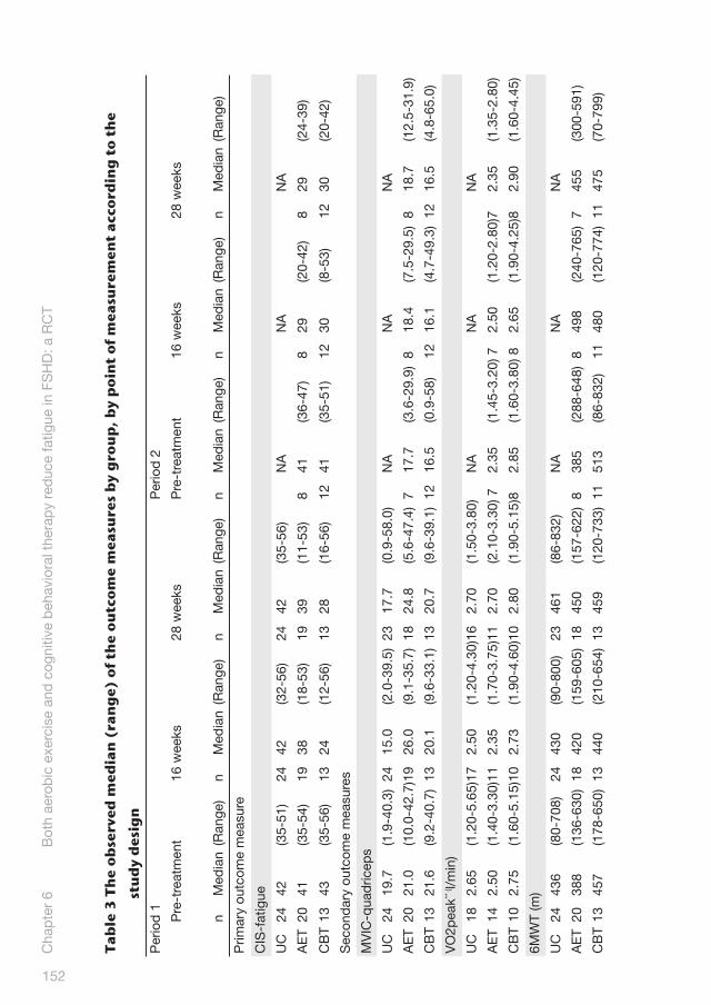
Tabl
e 3
The
obse
rved
med
ian
(ran
ge)
of t
he o
utco
me
mea
sure
s by
gro
up, b
y po
int
of m
easu
rem
ent
acco
rdin
g to
the
stud
y de
sign
Per
iod
1P
erio
d 2
Pre
-tre
atm
ent
16 w
eeks
28 w
eeks
Pre
-tre
atm
ent
16 w
eeks
28 w
eeks
nM
edia
n(R
ange
)n
Med
ian
(Ran
ge)
nM
edia
n(R
ange
)n
Med
ian
(Ran
ge)
nM
edia
n(R
ange
)n
Med
ian
(Ran
ge)
Prim
ary
outc
ome
mea
sure
CIS
-fat
igue
UC
2442
(35-
51)
2442
(32-
56)
2442
(35-
56)
NA
NA
NA
AE
T20
41(3
5-54
)19
38(1
8-53
)19
39(1
1-53
)8
41(3
6-47
)8
29(2
0-42
)8
29(2
4-39
)
CB
T13
43(3
5-56
)13
24(1
2-56
)13
28(1
6-56
)12
41(3
5-51
)12
30(8
-53)
1230
(20-
42)
Sec
ond
ary
outc
ome
mea
sure
s
MV
IC-q
uad
ricep
s
UC
2419
.7(1
.9-4
0.3)
2415
.0(2
.0-3
9.5)
2317
.7(0
.9-5
8.0)
NA
NA
NA
AE
T20
21.0
(10.
0-42
.7)1
926
.0(9
.1-3
5.7)
1824
.8(5
.6-4
7.4)
717
.7(3
.6-2
9.9)
818
.4(7
.5-2
9.5)
818
.7(1
2.5-
31.9
)
CB
T13
21.6
(9.2
-40.
7)13
20.1
(9.6
-33.
1)13
20.7
(9.6
-39.
1)12
16.5
(0.9
-58)
1216
.1(4
.7-4
9.3)
1216
.5(4
.8-6
5.0)
VO
2pea
k** ( l/
min
)
UC
182.
65(1
.20-
5.65
)17
2.50
(1.2
0-4.
30)1
62.
70(1
.50-
3.80
)N
AN
AN
A
AE
T14
2.50
(1.4
0-3.
30)1
12.
35(1
.70-
3.75
)11
2.70
(2.1
0-3.
30) 7
2.35
(1.4
5-3.
20) 7
2.50
(1.2
0-2.
80)7
2.35
(1.3
5-2.
80)
CB
T10
2.75
(1.6
0-5.
15)1
02.
73(1
.90-
4.60
)10
2.80
(1.9
0-5.
15)8
2.85
(1.6
0-3.
80) 8
2.65
(1.9
0-4.
25)8
2.90
(1.6
0-4.
45)
6MW
T (m
)
UC
2443
6(8
0-70
8)24
430
(90-
800)
2346
1(8
6-83
2)N
AN
AN
A
AE
T20
388
(136
-630
)18
420
(159
-605
)18
450
(157
-622
)8
385
(288
-648
)8
498
(240
-765
)7
455
(300
-591
)
CB
T13
457
(178
-650
)13
440
(210
-654
)13
459
(120
-733
)11
513
(86-
832)
1148
0(1
20-7
74)
1147
5(7
0-79
9)
Cha
pte
r 6
Bot
h ae
rob
ic e
xerc
ise
and
cog
nitiv
e b
ehav
iora
l the
rap
y re
duc
e fa
tigue
in F
SH
D: a
RC
T
152

Act
omet
er
UC
2438
(19-
74)
2336
(10-
72)
2332
(12-
70)
NA
NA
NA
AE
T17
32(6
-68)
1638
(15-
65)
1632
(1-6
0)8
30(1
2-42
)8
29(1
7-60
)8
29(1
5-40
)
CB
T13
43(2
1-69
)13
43(1
7-73
)13
41(1
7-65
)11
35(1
5-66
)11
40(1
9-74
)11
35(1
8-65
)
CIS
-act
ivity
UC
2414
(3-2
0)24
11(3
-18)
2413
(3-2
1)N
AN
AN
A
AE
T20
17(4
-21)
1917
(3-1
9)19
13(3
-21)
812
(5-1
8)8
14(5
-20)
811
(6-2
1)
CB
T13
14(5
-21)
139
(13-
19)
1313
(4-2
1)12
13(3
-21)
129
(3-1
7)12
10(3
-21)
VAS
-pai
n
UC
2418
.5(0
-59)
2421
(0-6
5)24
21(0
-69)
NA
NA
NA
AE
T20
19.5
(4-6
5)19
20(2
-65)
1916
(0-7
1)8
13(0
-47)
810
(0-7
2)8
10(0
-80)
CB
T13
15(0
-84)
1310
(0-7
2)13
10(0
-80)
1220
(0-5
6)12
20(0
-70)
1220
(0-6
9)
NH
P-s
leep
UC
240
(0-8
3.9)
2412
.57
(0-7
7.63
)24
12.5
7(0
-77.
63)
NA
NA
NA
AE
T20
6.28
5(0
-77.
63)
190
(0-8
7.43
)19
12.5
7(0
-72.
74)
80
(0-3
4.27
)8
0(0
-21.
7)8
12.5
7(0
-21.
7)
CB
T13
12.5
7(0
-61.
53)
130
(0-6
1.53
)13
0(0
-61.
53)
1212
.57
(0-7
7.63
)12
0(0
-34.
27)
120
(0-2
1.7)
SIP
68-s
b
UC
2413
0(0
-387
)24
140
(0-3
72)
2416
9(0
-572
)N
AN
AN
A
AE
T20
187
(0-5
72)
1914
4(0
-572
)19
152
(0-5
23)
814
9(0
-387
)8
112
(0-3
44)
813
7(4
3-38
6)
CB
T13
152
(0-4
76)
1314
9(0
-387
)13
100
(0-3
87)
1221
5(0
-572
)12
144
(0-4
20)
1210
9(0
-294
)
Per
iod
: firs
t-, s
econ
d a
lloca
tion,
Per
iod
1 r
epre
sent
s th
e d
ata
of t
he fi
rst
allo
catio
n an
d p
erio
d 2
the
dat
a of
the
allo
catio
n of
the
UC
gro
up a
fter
the
firs
t 28
wee
ks. N
A:
not
app
licab
le, U
C: u
sual
car
e, A
ET:
aer
obic
exe
rcis
e tr
aini
ng, C
BT:
cog
nitiv
e b
ehav
iora
l the
rap
y, C
IS-f
atig
ue: s
ubsc
ale
fatig
ue o
f the
Che
cklis
t In
div
idua
l Str
engt
h,
MV
IC-Q
uad
ricep
s: a
vera
ge v
alue
of m
axim
um v
olun
tary
mus
cle
stre
ngth
in k
g fo
r le
ft a
nd r
ight
qua
dric
eps,
VO
2pea
k: a
erob
ic e
xerc
ise
tole
ranc
e in
ml/m
in, 6
MW
T:
six-
min
utes
wal
king
tes
t in
met
ers,
phy
sica
l act
ivity
: the
ave
rage
num
ber
of b
ody
acce
lera
tions
per
5-m
in p
erio
d, C
IS-a
ctiv
ity: s
ubsc
ale
phy
sica
l act
ivity
of t
he C
heck
-lis
t In
div
idua
l Str
engt
h, V
AS
pai
n: p
ain
inte
nsity
on
a sc
ale
from
0-1
00, N
HP
-sle
ep: s
ubsc
ale
slee
p o
f the
Not
tingh
am H
ealth
Pro
file,
SIP
-68-
sb: s
ubsc
ale
soci
al b
ehav
ior
of t
he S
ickn
ess
Imp
act
Pro
file
**: V
O2
pea
k d
ata
wer
e no
t av
aila
ble
for
the
six
par
ticip
ants
who
did
not
rea
ch t
he t
arge
t he
art
rate
of 1
30-1
70 b
eats
/min
ute
in t
he 5
th-
6th m
inut
e of
the
Åst
rand
tes
t; in
ad
diti
on, V
O2
pea
k d
ata
wer
e no
t av
aila
ble
for
14 p
artic
ipan
ts w
ho t
ook
bet
a-b
lock
ers
(UC
(n=
5);
AE
T (n
=6)
; CB
T (n
=3)
).
153

Tabl
e 4
T
he e
stim
ated
mea
n ch
ange
sin
ce p
re-t
reat
men
t (9
5% C
I) a
nd t
he e
stim
ated
mea
n gr
oup
diff
eren
ces
(95%
CI)
in
all
outc
ome
mea
sure
s by
poi
nt o
f mea
sure
men
t, u
sing
a li
near
mix
ed m
odel
for
repe
ated
mea
sure
men
ts
wit
h ad
just
men
t fo
r pr
e-tr
eatm
ent
valu
es
Cha
nge
sinc
e p
re-t
reat
men
tD
iffer
ence
bet
wee
n gr
oup
s
UC
(n=
24)
AE
T (n
=28
)C
BT
(n=
25)
AE
T vs
UC
CB
T vs
UC
Mea
n(9
5%C
I)M
ean
(95%
CI)
Mea
n(9
5%C
I)M
ean
(95%
CI)
Mea
n(9
5%C
I)
Prim
ary
outc
ome
mea
sure
CIS
-fat
igue
16 w
eeks
-1.2
(-3.
2 to
0.9
)-8
.5(-
12.4
to
-4.6
)-1
3.7
(-17
.8 t
o -9
.5)
-9.1
(-12
.4 t
o -5
.8) *
-13.
3(-
16.5
to
-10.
2) *
28 w
eeks
0.0
(-1.
7 to
1.8
)-9
.1(-
13.4
to
-4.7
)-1
1.1
(-15
.6 t
o -6
.6)
-8.2
(-12
.1 t
o -4
.2) *
-10.
2(-
14.0
to-
6.3)
*
Sec
ond
ary
outc
ome
mea
sure
s
MV
IC-q
uad
ricep
s
16 w
eeks
-1.8
(-4.
6 to
1.0
)-1
.7(-
4.5
to 1
.0)
-1.5
(-3.
8 to
0.9
)2.
1(0
.5 t
o 4.
7)*
-0.2
(-2.
9 to
2.5
)
28 w
eeks
0.8
(-1.
2 to
2.8
) 1.
8(-
0.1
to 3
.6)
-0.1
(-2.
5 to
2.3
)2.
3(-
1.3
to 5
.9)
0.9
(-3.
0 to
4.8
)
VO
2pea
k** (l
/min
)
16 w
eeks
-0.4
(-1.
9 to
1.1
)0.
7(-
1.8
to 3
.2)
1.0
(-1.
0 to
3.0
)0.
1(-
0.1
to 0
.3)
0.2
(0.0
to
0.5)
28 w
eeks
-0.3
(-1.
8 to
1.2
)-0
.7(-
3.7
to 2
.2)
96(-
69 t
o 26
0)0.
2(-
0.1
to 0
.5)
0.3
(-0.
1 to
0.7
)
6MW
T (m
)
16 w
eeks
0(-
30 t
o 30
)31
(-22
to
84)
11(-
20 t
o 42
)18
(-12
to
48)
-9(-
38 t
o 20
)
28 w
eeks
9(-
18 t
o 36
) 26
(-15
to
66)
3(-
31 t
o 37
)15
(-19
to
50)
-12
(-43
to
20)
Act
omet
er
16 w
eeks
-1.3
(-5.
6 to
2.9
)2.
8(-
2.1
to 7
.7)
2.9
(0.0
to
5.8)
4.6
(1.4
to
7.8)
*5.
6(1
.7 t
o 9.
4)*
28 w
eeks
-5.3
(-9.
3 to
-1.
4)-1
.6(-
5.2
to 2
.0)
0.7
(-1.
8 to
3.2
)5.
5(1
.6 t
o 9.
3)*
7.1
(1.5
to
12.7
)*
Cha
pte
r 6
Bot
h ae
rob
ic e
xerc
ise
and
cog
nitiv
e b
ehav
iora
l the
rap
y re
duc
e fa
tigue
in F
SH
D: a
RC
T
154

CIS
-act
ivity
16 w
eeks
-1.5
(-3.
0 to
0.1
)0.
2(-
1.1
to 1
.5)
-3.6
(-5.
5 to
-1.
7)0.
6(-
0.8
to 2
.0)
-2.6
(-4.
1 to
-1.
2)*
28 w
eeks
-0.3
(-1.
8 to
1.2
)-0
.5(-
1.9
to 0
.8)
-1.1
(-2.
8 to
0.6
)0.
6(-
1.3
to 2
.5)
-1.1
(-3.
0 to
0.8
)
VAS
-pai
n
16 w
eeks
1(-
8 to
10)
0(-
6 to
5)
4.0
(-3.
9 to
11.
9)-2
.0(-
10.4
to
6.3)
-0.9
(-8.
5 to
6.7
)
28 w
eeks
1(-
6 to
8)
2(-
5 to
10)
4(-
4 to
12)
0.1
(-11
.3 t
o 11
.5)
0.0
(-9.
5 to
9.6
)
NH
P-s
leep
16 w
eeks
3.1
(-4.
2 to
10.
4)-0
.7(-
6.1
to 4
.7)
-6.7
(-17
.1 t
o 3.
6)-0
.6(-
6.3
to 5
.1)
-7.0
(-12
.5 t
o -1
.5)*
28 w
eeks
-3.1
(-14
.2 t
o 8.
1)0.
4(-
5.7
to 6
.4)
-8.4
(-17
.2 t
o 0.
4)1.
2(-
5.2
to 7
.7)
-4.5
(-10
.4 t
o 1.
3)
SIP
68-s
b
16 w
eeks
8(-
30 t
o 47
)-2
(-34
to
29)
-39
(-78
to
1)-3
(-36
to
31)
-61
(-94
to
-29)
*
28 w
eeks
45(4
to
86)
10(-
26 t
o 44
)-6
1(-
116
to -
6)-1
3(-
50 t
o 25
)-9
2(-
127
to 5
7)
*: S
tatis
tical
ly s
igni
fican
t d
iffer
ence
s ar
e m
arke
d w
ith a
n as
teris
k. A
ll ou
tcom
es a
re s
econ
dar
y ot
her
than
tha
t se
t in
bol
d. C
I: co
nfid
ence
inte
rval
, AE
T: a
erob
ic e
xerc
ise
trai
ning
CB
T: c
ogni
tive
beh
avio
ral t
hera
py,
CIS
-fat
igue
: sub
scal
e fa
tigue
of t
he C
heck
list
Ind
ivid
ual S
tren
gth,
MV
IC-Q
uad
ricep
s: a
vera
ge v
alue
of m
axim
um v
olun
tary
m
uscl
e st
reng
th in
kg
for
left
and
rig
ht q
uad
ricep
s, V
O2p
eak:
aer
obic
exe
rcis
e to
lera
nce
in m
l/min
, 6M
WT:
six
-min
utes
wal
king
tes
t in
met
ers,
phy
sica
l act
ivity
: the
av
erag
e nu
mb
er o
f bod
y ac
cele
ratio
ns p
er 5
-min
per
iod
, CIS
-act
ivity
: su
bsc
ale
phy
sica
l act
ivity
of t
he C
heck
list
Ind
ivid
ual S
tren
gth,
VA
Sp
ain:
pai
n in
tens
ity o
n a
scal
e fr
om 0
-100
, NH
P-s
leep
: sub
scal
e sl
eep
of t
he N
ottin
gham
Hea
lth P
rofil
e, S
IP-6
8-sb
: sub
scal
e so
cial
beh
avio
r of
the
Sic
knes
s Im
pac
t P
rofil
e
**: V
O2
pea
k d
ata
wer
e no
t av
aila
ble
for
the
six
par
ticip
ants
who
did
not
rea
ch t
he t
arge
t he
art
rate
of 1
30-1
70 b
eats
/min
ute
in t
he 5
th-6
th m
inut
e of
the
Åst
rand
tes
t;
in a
dd
ition
, VO
2 p
eak
dat
a w
ere
not
avai
lab
le fo
r 14
par
ticip
ants
who
too
k b
eta-
blo
cker
s (U
C (n
= 5
); A
ET
(n=
6); C
BT
(n=
3)).
155

In the AET group, the only change in fatigue-perpetuating factors was an increase
in registered physical activity. Unexpectedly, we found no improvement in
aerobic capacity. This lack of improved aerobic capacity might have been due to
insufficient therapy compliance or to the fact that VO2 peak data were not available
for the 6 and 14 participants who did not reach the target heart rate or were taking
β-blockers, respectively. In addition, the absence of any significant change in
isometric quadriceps strength or distance walked in the 6-minute walking test is
most likely due to the fact that these capacities were not specifically trained in
either treatment group (29).
In this study, only mild adverse effects were found, which, together with an
absence of pain increase during the intervention period, suggests that both AET
and CBT are safe interventions in patients with FSHD.
A limitation of this study is the potentially low generalizability as only 74 of the
377 invited patients took part. Nevertheless, our study sample was sufficiently
heterogeneous with respect to age, disease severity, physical endurance, and level
of social participation.
The adherence rate was lower than expected. Especially for AET, practical factors
like travel time to the study sites and the fit of the exercise regimen in daily life
accounted for the adherence rate being less than 100%. This study provides
evidence that AET and CBT can effectively reduce chronic fatigue in patients with
FSHD. Future research should focus on the specific factors that contribute to this
beneficial effect to optimize patient selection. These studies may also include AET
or CBT as part of a more comprehensive treatment program for patients with FSHD
and use a longer follow-up period. Tailoring such an intervention program to the
specific needs of each patient would meet one of the primary research priorities as
identified by patients with a neuromuscular disease (30, 31).
Cha
pte
r 6
Bot
h ae
rob
ic e
xerc
ise
and
cog
nitiv
e b
ehav
iora
l the
rap
y re
duc
e fa
tigue
in F
SH
D: a
RC
T
156

AUTHOR CONTRIBUTIONS
Alexander Geurts, Baziel van Engelen, and Gijs Bleijenberg designed the study
and obtained funding. Nicoline Voet included the participants, monitored the study
conduct, supervised the physical therapists for data collection, managed and
developed the databases, trained the physical therapists, and wrote the first draft.
Gijs Bleijenberg trained the cognitive behavioral therapists. Jan Hendriks gave
statistical advice and performed the data analysis. All authors contributed to data
analysis and interpretation and the writing and editing of the report.
ACKNOWLEDGEMENTS
The authors thank the patients who participated in this trial, the physicians at
the participating health care institutions, the outcome assessors, the physical
therapists, and the cognitive behavioral therapists.
STUDY FUNDING
Funded by Prinses Beatrix SpierFonds (PBF) (The Dutch Public Fund for
Neuromuscular Disorders), the Netherlands Organization for Health Research and
Development (ID: ZonMw 89000003), and the FSHD Global Research Foundation.
The FACTS-2-FSHD trial (Fitness and Cognitive Behavioral Therapies for Fatigue
and Activities in FSHD) is part of the FACTS-2-NMD project, together with the
FACTS-2-PPS and FACTS-2-ALS trials.
DISCLOSURE
N. Voet received grants from Prinses Beatrix SpierFonds (PBF) (The Dutch
Public Fund for Neuromuscular Disorders), the Netherlands Organization for
Health Research and Development (ID: ZonMw 89000003), and global FSH.
G. Bleijenberg, J. Hendriks, I. de Groot, and G. Padberg report no disclosures
relevant to the manuscript. B. van Engelen was research director of the European
Neuromuscular Centre (ENMC), received grants from global FSH, the Netherlands
Organization for Health Research and Development, Prinses Beatrix SpierFonds
(PBF) (The Dutch Public Fund for Neuromuscular Disorders), and the Dutch FSHD
Foundation. S. Geurts received grants from Prinses Beatrix SpierFonds (PBF) (The
Dutch Public Fund for Neuromuscular Disorders), the Netherlands Organization for
Health Research and Development (ID: ZonMw 89000003), and global FSH. Go to
Neurology.org for full disclosures.
157

APPENDIX 1: PROTOCOL AEROBIC EXERCISE TRAINING
The aerobic exercise training (AET) consisted of cycling exercise on an ergometer.
The training program had a duration of 16 weeks and comprised home training
twice a week and a supervised training by a physical therapist at the most nearby
healthcare institution once a week. A cycle ergometer (Monark 827E), a pulse watch
with breast belt (Garmin forerunner 50 heart rate), and a log book with training
instructions were provided to each participant for home use. Training sessions
consisted of a 30-minute aerobic exercise period with warming-up and cooling-
down periods of 5 and 3 minutes, respectively. The cardiovascular load during the
training period was individually adjusted. Every four weeks, the level was increased
with 5% from 50% to 65% of the heart rate reserve (HRR). HRR is the difference
between the predicted maximum heart rate and the measured resting heart rate.
Resting heart rate was measured during the baseline assessment. Maximum heart
rate (MHR) was estimated using the formula: MHR = 220 - age. Each participant
was taught how to adjust the physical load to the preferred individual heart rate.
During each training, the heart rate was monitored continuously by the breast
belt. Participants who were taking beta-blockers were instructed to exercise
at a perceived exertion level of 12-14 on the Borg scale. Level of exertion was
monitored by heart rate and the Borg visual analog scale. For patients with beta-
blocker medication, level of exertion was monitored by the Borg scale alone. The
number and duration of the sessions, the Borg score, the mean and maximum
heart rate, and any adverse effects were recorded in the individual log book. During
the weekly sessions at the healthcare institution, therapy compliance in the home
situation was verified by the physical therapist by reading out the heart rate watch
and checking the log book regarding the previous week. In addition, individually
tailored instructions for the next week were given. The primary investigator (NV)
gave instructions to the physical therapists and performed integrity checks at each
location. Acceptable compliance with the AET program was defined as a minimum
of 40 completed training sessions.
Cha
pte
r 6
Bot
h ae
rob
ic e
xerc
ise
and
cog
nitiv
e b
ehav
iora
l the
rap
y re
duc
e fa
tigue
in F
SH
D: a
RC
T
158

APPENDIX 2: THE VARIOUS MODULES OF COGNITIVE BEHAVIORAL THERAPY BASED ON THE FATIGUE-PERPETUATING FACTORS IN FSHD
Insufficient coping with the disease
Insufficient coping with the disease is assessed using the Impact of Event Scale
(33, 34). A participant can continue to be mentally occupied with the diagnosis
facioscapulohumeral muscular dystrophy (FSHD). By talking and writing about
this experience (‘exposure’), the participant can acquire better coping skills. Fear
of progression is assessed using a questionnaire designed specifically for FSHD
patients. The therapist helps the participant formulate his or her thoughts regarding
fear of progression. These thoughts are then challenged against reality (‘reality
testing’), thereby reducing daily unhelpful thoughts regarding disease progression
and putting these thoughts into perspective.
Dysfunctional cognitions regarding fatigue, activity, pain, and other symptoms
Dysfunctional cognitions are related to a variety of unhelpful thoughts, including
the participant’s feeling of a lack of control over the symptoms and inappropriate
thoughts regarding the symptoms. The sense of control in relation to fatigue
complaints is assessed using the self-efficacy scale (35, 36).
Catastrophizing
Catastrophizing is assessed using the Jacobsen Fatigue Catastrophizing Scale
(37). These thoughts are challenged, and more constructive thoughts are taught.
Dysregulation of sleep
Dysregulation of sleep is assessed using a self-reported sleep diary (38). An
irregular circadian rhythm can be a perpetuating factor of fatigue. To restore normal
biological rhythm, the participants are encouraged to maintain a fixed bed-time and
wake-up time and are discouraged from sleeping during the day.
159

Dysregulation of activity
Dysregulation of activity is based on activity (e.g. steps) and is monitored using
an actometer and a physical activity questionnaire (Physical Activity Scale
for Individuals with Physical Disabilities) (34,39). Some patients experience a
fluctuating activity pattern, with highly active periods followed by periods of
prolonged inactivity. Other patients avoid physical activity because they are
concerned that it will increase their fatigue. For participants with fluctuating
activity levels, a stable baseline level of alternating resting and active periods is
first established. Once the participant has achieved this baseline level, a physical
activity program is introduced, beginning with 5-10 minutes of walking or cycling.
This activity is gradually increased by 1 minute a day each time the activity is
performed, reaching a maximum of two 60-minute sessions per day. Inactive
participants are instructed to commence this activity program immediately.
Gradually, some physical activities can be replaced with other (preferred) physical
activities.
Poor social support and negative social interactions
Poor social support and negative social interactions are determined using the
discrepancy subscale of the Social Support List (40). If a participant still has
unrealistic expectations of others or perceives a discrepancy between actual
support and desired support, the therapist will attempt to instill more realistic
expectations with respect to the participant’s social support group. The partner or
caregiver is included in this treatment module.
REFERENCES
1. Deenen JC, Arnts H, van der Maarel SM, Padberg GW, Verschuuren JJ, Bakker
E, et al. Population-based incidence and prevalence of facioscapulohumeral
dystrophy. Neurology. 2014;83(12):1056-9.
2. Kalkman JS, Schillings ML, van der Werf SP, Padberg GW, Zwarts MJ, van
Engelen BG, et al. Experienced fatigue in facioscapulohumeral dystrophy,
myotonic dystrophy, and HMSN-I. Journal of Neurology, Neurosurgery, and
Psychiatry. 2005;76(10):1406-9.
3. Kalkman JS, Schillings ML, Zwarts MJ, van Engelen BG, Bleijenberg G. The
development of a model of fatigue in neuromuscular disorders: a longitudinal
study. Journal of Psychosomatic Research. 2007;62(5):571-9.
Cha
pte
r 6
Bot
h ae
rob
ic e
xerc
ise
and
cog
nitiv
e b
ehav
iora
l the
rap
y re
duc
e fa
tigue
in F
SH
D: a
RC
T
160

4. van der Kooi EL, Vogels OJ, van Asseldonk RJ, Lindeman E, Hendriks JC,
Wohlgemuth M, et al. Strength training and albuterol in facioscapulohumeral
muscular dystrophy. Neurology. 2004;63(4):702-8.
5. Colson SS, Benchortane M, Tanant V, Faghan JP, Fournier-Mehouas M, Benaïm
C, et al. Neuromuscular electrical stimulation training: a safe and effective
treatment for facioscapulohumeral muscular dystrophy patients. Archives of
Physical Medicine and Rehabilitation. 2010;91(5):697-702.
6. Olsen DB, Orngreen MC, Vissing J. Aerobic training improves exercise
performance in facioscapulohumeral muscular dystrophy. Neurology.
2005;64(6):1064-6.
7. Price JR, Mitchell E, Tidy E, Hunot V. Cognitive behaviour therapy for chronic
fatigue syndrome in adults. Cochrane Database of Systematic Reviews.
2008(3):CD001027.
8. Gielissen MF, Verhagen S, Witjes F, Bleijenberg G. Effects of cognitive behavior
therapy in severely fatigued disease-free cancer patients compared with
patients waiting for cognitive behavior therapy: a randomized controlled trial.
Journal of Clinical Oncology. 2006;24(30):4882-7.
9. van Kessel K, Moss-Morris R, Willoughby E, Chalder T, Johnson MH, Robinson
E. A randomized controlled trial of cognitive behavior therapy for multiple
sclerosis fatigue. Psychosomatic Medicine. 2008;70(2):205-13.
10. Cup EH, Pieterse AJ, Ten Broek-Pastoor JM, Munneke M, van Engelen BG,
Hendricks HT, et al. Exercise therapy and other types of physical therapy
for patients with neuromuscular diseases: a systematic review. Archives of
Physical Medicine and Rehabilitation. 2007;88(11):1452-64.
11. van Engelen BG, van Veenendaal H, Van Doorn PA, Faber CG, van der Hoeven
JH, Janssen NG, et al. The Dutch neuromuscular database CRAMP (Computer
Registry of All Myopathies and Polyneuropathies): development and preliminary
data. Neuromuscular Disorders. 2007;17(1):33-7.
12. Alberts M, Smets EM, Vercoulen JH, Garssen B, Bleijenberg G. [‘Abbreviated
fatigue questionnaire’: a practical tool in the classification of fatigue].
Nederlands Tijdschrift voor Geneeskunde. 1997;141(31):1526-30.
13. Lemmers RJ, O’Shea S, Padberg GW, Lunt PW, van der Maarel SM. Best
practice guidelines on genetic diagnostics of Facioscapulohumeral muscular
dystrophy: workshop 9th June 2010, LUMC, Leiden, The Netherlands.
Neuromuscular Disorders. 2012;22(5):463-70.
14. Alberts M, Vercoulen J, Bleijenberg G. Assessment of fatigue: the practical
utility of the subjective feeling of fatigue in research and clinical practice. In:
Vingerhoets A, ed. Assessment in Behavioral Medicine. East Sussex, UK:
Brunner-Routledge; 2001:301–327.
161

15. World Medical Association Declaration of Helsinki, Ethical Principles for
Medical Research involving human subjects. 2008. Available from: http://www.
wma.net/e/policy/b3.htm.
16. Voet NB, Bleijenberg G, Padberg GW, van Engelen BG, Geurts AC. Effect of
aerobic exercise training and cognitive behavioural therapy on reduction of
chronic fatigue in patients with facioscapulohumeral dystrophy: protocol of the
FACTS-2-FSHD trial. BMC Neurol. 2010;10:56.
17. Durstine JL. ACSM’s exercise management for persons with chronic diseases
and disabilities. 3rd ed. ed. Leeds: Human Kinetics; 2009.
18. Borg G. Perceived exertion as an indicator of somatic stress. Scandinavian
Journal of Rehabilitation Medicine. 1970;2(2):92-8.
19. Vercoulen JH, Swanink CM, Fennis JF, Galama JM, van der Meer JW,
Bleijenberg G. Dimensional assessment of chronic fatigue syndrome. Journal
of Psychosomatic Research. 1994;38(5):383-92.
20. Statland JM, McDermott MP, Heatwole C, Martens WB, Pandya S, van
der Kooi EL, et al. Reevaluating measures of disease progression in
facioscapulohumeral muscular dystrophy. Neuromuscular Disorders.
2013;23(4):306-12.
21. Macsween A. The reliability and validity of the Astrand nomogram and linear
extrapolation for deriving VO2max from submaximal exercise data. Journal of
sports medicine and physical fitness. 2001;41(3):312-7.
22. Enright PL. The six-minute walk test. RespirCare. 2003;48(8):783-5.
23. van der Werf SP, Prins JB, Vercoulen JH, van der Meer JW, Bleijenberg
G. Identifying physical activity patterns in chronic fatigue syndrome
using actigraphic assessment. Journal of Psychosomatic Research.
2000;49(5):373-9.
24. Folstein MF, Luria R. Reliability, validity, and clinical application of the Visual
Analogue Mood Scale. Psychological Medicine. 1973;3(4):479-86.
25. Hunt SM, McEwen J, McKenna SP. Measuring health status: a new tool for
clinicians and epidemiologists. J R Coll Gen Pract. 1985;35(273):185-8.
26. Erdman RA, Passchier J, Kooijman M, Stronks DL. The Dutch version of
the Nottingham Health Profile: investigations of psychometric aspects.
Psychological Reports. 1993;72(3 Pt 1):1027-35.
27. Post MW, de Bruin A, de Witte L, Schrijvers A. The SIP68: a measure of
health-related functional status in rehabilitation medicine. Archives of Physical
Medicine and Rehabilitation. 1996;77(5):440-5.
28. van der Marck MA, Overeem S, Klok PC, Bloem BR, Munneke M. Evaluation
of the falls telephone: an automated system for enduring assessment of falls. J
Am Geriatr Soc. 2011;59(2):340-4.
Cha
pte
r 6
Bot
h ae
rob
ic e
xerc
ise
and
cog
nitiv
e b
ehav
iora
l the
rap
y re
duc
e fa
tigue
in F
SH
D: a
RC
T
162

29. Ricci E, Galluzzi G, Deidda G, Cacurri S, Colantoni L, Merico B, et al. Progress
in the molecular diagnosis of facioscapulohumeral muscular dystrophy and
correlation between the number of KpnI repeats at the 4q35 locus and clinical
phenotype. Annals of Neurology. 1999;45(6):751-7.
30. Voet NB, van der Kooi EL, Riphagen II, Lindeman E, van Engelen BG, Geurts
AC. Strength training and aerobic exercise training for muscle disease.
Cochrane Database of Systematic Reviews. 2013;7:CD003907.
31. Nierse CJ, Abma TA, Horemans AM, van Engelen BG. Research priorities of
patients with neuromuscular disease. Disabil Rehabil. 2013;35(5):405-12.
32. Smith AE, McMullen K, Jensen MP, Carter GT, Molton IR. Symptom burden
in persons with myotonic and facioscapulohumeral muscular dystrophy.
American journal of physical medicine & rehabilitation. 2014;93(5):387-95.
33. Creamer M, Bell R, Failla S. Psychometric properties of the Impact of Event
Scale - Revised. Behaviour Research and Therapy 2003;41:1489-1496.
34. van der Ploeg E, Mooren TT, Kleber RJ, van der Velden PG, Brom D. Construct
validation of the Dutch version of the impact of event scale. Psychological
Assessment 2004;16:16-26.
35. Vercoulen JH, Hommes OR, Swanink CM, et al. The measurement of fatigue in
patients with multiple sclerosis. A multidimensional comparison with patients
with chronic fatigue syndrome and healthy subjects. Archives of Neurology
1996;53:642-649.
36. Prins JB, Bleijenberg G, Bazelmans E, et al. Cognitive behaviour therapy for
chronic fatigue syndrome: a multicentre randomised controlled trial. Lancet
2001;357:841-847.
37. Andrykowski MA, Schmidt JE, Salsman JM, Beacham AO, Jacobsen PB.
Use of a case definition approach to identify cancer-related fatigue in women
undergoing adjuvant therapy for breast cancer. Journal of Clinical Oncology
2005;23:6613-6622.
38. Servaes P, Verhagen C, Bleijenberg G. Fatigue in cancer patients during
and after treatment: prevalence, correlates and interventions. EurJCancer
2002;38:27-43.
39. Washburn RA, Zhu W, McAuley E, Frogley M, Figoni SF. The physical activity
scale for individuals with physical disabilities: development and evaluation.
Archives of Physical Medicine and Rehabilitation 2002;83:193-200.
40. Ormel J, Kempen GI, Penninx BW, Brilman EI, Beekman AT, van Sonderen E.
Chronic medical conditions and mental health in older people: disability and
psychosocial resources mediate specific mental health effects. Psychological
Medicine 1997;27:1065-1077.
163
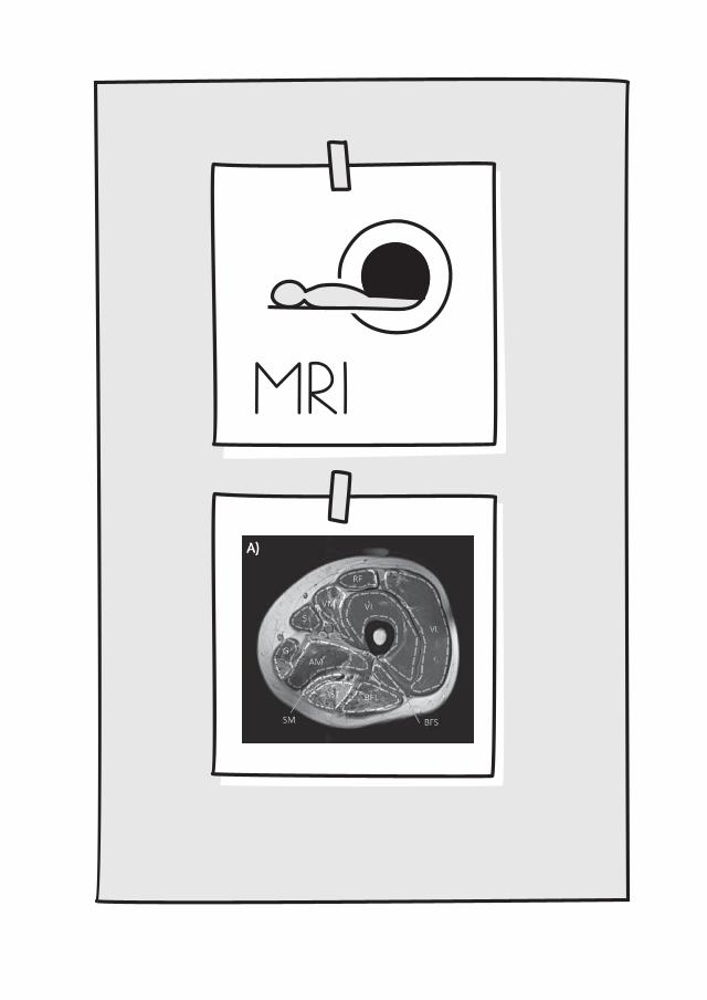
MRI

CHAPTER 7DISTINCT DISEASE PHASES IN MUSCLES OF FACIOSCAPULOHUMERAL DYSTROPHY PATIENTS IDENTIFIED BY MR DETECTED FAT INFILTRATION.
Barbara H. Janssen
Nicoline B.M. Voet
Christine I. Nabuurs
Hermien E. Kan
Jacky W.J. de Rooy
Alexander C.H. Geurts
George W. Padberg
Baziel G.M. van Engelen
Arend Heerschap
Published in: Neurology. 2014;83:1-9.

ABSTRACT
Facioscapulohumeral muscular dystrophy (FSHD) is an untreatable disease,
characterized by asymmetric progressive weakness of skeletal muscle with fatty
infiltration. Although the main genetic defect has been uncovered, the downstream
mechanisms causing FSHD are not understood. The objective of this study was
to determine natural disease state and progression in muscles of FSHD patients
and to establish diagnostic biomarkers by quantitative MRI of fat infiltration and
phosphorylated metabolites. MRI was performed at 3T with dedicated coils on
legs of 41 patients (28 men/13 women, age 34–76 years), of which eleven were
re-examined after four months of usual care. Muscular fat fraction was determined
with multi spin-echo and T1 weighted MRI, edema by TIRM and phosphorylated
metabolites by 3D 31P MR spectroscopic imaging. Fat fractions were compared
to clinical severity, muscle force, age, edema and phosphocreatine (PCr)/ATP.
Longitudinal intramuscular fat fraction variation was analyzed by linear regression.
Increased intramuscular fat correlated with age (p<0.05), FSHD severity score
(p<0.0001), inversely with muscle strength (p=0.0001), and also occurred
subclinically. Muscles were nearly dichotomously divided in those with high
and with low fat fraction, with only 13% having an intermediate fat fraction. The
intramuscular fat fraction along the muscle’s length, increased from proximal to
distal. This fat gradient was the steepest for intermediate fat infiltrated muscles
(0.07±0.01/cm, p<0.001). Leg muscles in this intermediate phase showed a
decreased PCr/ATP (p<0.05) and the fastest increase in fatty infiltration over
time (0.18±0.15/year, p<0.001), which correlated with initial edema (p<0.01), if
present. Thus, in the MR assessment of fat infiltration as biomarker for diseased
muscles, the intramuscular fat distribution needs to be taken into account. Our
results indicate that healthy individual leg muscles become diseased by entering a
progressive phase with distal fat infiltration.
Cha
pte
r 7
Dis
tinct
dis
ease
pha
ses
in m
uscl
es o
f FS
HD
pat
ient
s id
entifi
ed b
y M
R d
etec
ted
fat
infil
trat
ion
166
INTRODUCTION
Facioscapulohumeral muscular dystrophy (FSHD) is the third most common
hereditary muscular disorder (1). The disease is characterized by progressive
asymmetric weakness and fatty infiltration of skeletal muscles. In recent years it
was demonstrated that FSHD is associated with a contraction of D4Z4 repeats
on chromosome 4q35 (2), leading to lost repression of DUX4, a protein that exerts
toxic effects on muscle cells (3).
Even though the most important genetic event for the disease seems to be
identified, a causative treatment is not yet available (4). Progress is hampered
because the trigger for DUX4 expression and the further unfolding of disease
processes leading to fatty infiltration and muscle weakness are not known.
Thus clarification of the underlying mechanisms is expected to offer clues for
a more targeted approach in the search for treatment (5). Understanding these
mechanisms first requires that some key questions concerning the process of fatty
infiltration are addressed. What is the natural distribution of fatty infiltration? How is
this related to clinical severity, to muscle weakness and to energy metabolism? Is
there prevalence for specific muscles to be affected and does fatty infiltration vary
within muscles? What is the natural progression over time and what are predictive
signs of progression? To answer these questions and to evaluate treatment
effectiveness, the use of a non-invasive quantitative imaging method, such as MRI,
is essential. Unlike biopsies, MRI is not limited to a single location, and longitudinal
data can be collected without risk for the patient. MR of fatty infiltration in muscles
has been used to study muscular disorders like Duchenne muscular dystrophy (6,
7). We have introduced a quantitative MRI measure of fatty infiltration in muscles
based on T2 relaxation time analysis and demonstrated its value in a preliminary
study of FSHD patients (8). Phosphorus MR spectroscopy has been used
extensively to investigate the energy status of diseased muscles (9-15). Recently it
was also introduced in a pilot study with FSHD patients (16).
Until now quantitative MR imaging studies were performed in limited numbers
of patients. However, because of the variability in age of onset and in degree of
disease severity (17), a study of its pathophysiology requires the participation of
a relatively large number of well described patients. The main aim of this study
was to determine natural disease state and progression by quantitative MRI of
skeletal muscles in the legs of a large, well-characterized cohort of genetically
confirmed FSHD patients. In particular we wanted to address the aforementioned
pathophysiological questions to ultimately uncover clues on disease mechanism
and to establish MRI biomarkers with prognostic and predictive value for
personalized assessments.
167
MATERIALS AND METHODS
PATIENTS AND STUDY DESIGN
We recruited 41 FSHD patients from the local neurology department (28 men/13
women, age 21–81 years, see Table 1 for patients’ demographics). Of 36 patients
the upper leg (‘thigh’) was examined, they were selected from a group of patients
that were entering a clinical trial to assess the effects of rehabilitation intervention
(18). In addition we included the MR exams of the lower leg of five patients from a
previous study (8), which were reanalyzed in the exact same way as the MR exams
of the aforementioned patients (vide infra).
Eleven patients (8 men/3 women, age 34–76 years) were randomly selected,
from the group that underwent an MR examination of the thigh, for a follow-up
measurement after a period of four months. During this period these patients were
instructed to maintain a normal level of activity (‘usual care’).
All patients were clinically and genetically diagnosed with FSHD and able to walk
independently (ankle-foot orthoses and canes were accepted). Patients were all
unrelated except for one mother and son (patients #7 and #37). Disease severity
was assessed with the Ricci score (19) and maximum voluntary isometric extension
(quadriceps) and flexion (hamstrings) of the knee were measured with a quantitative
fixed myometry testing system (20). Ethical approval was obtained from the
Radboud university medical center review board, and written informed consent was
obtained from all subjects.
Table 1 Patient demographics
Patient nr. Ricci score Sex Age (years) FSHD duration (years)
1 0 F 21 15
2 1.5 F 25 2
3 1.5 M 31 5
4* 2 M 34 3
5* 3 M 34 9
6 1.5 M 38 17
7 2 M 38 16
8* 3 M 38 19
9 1 F 39 5
10 3.5 M 42 32
Cha
pte
r 7
Dis
tinct
dis
ease
pha
ses
in m
uscl
es o
f FS
HD
pat
ient
s id
entifi
ed b
y M
R d
etec
ted
fat
infil
trat
ion
168

11* 2 M 44 14
12 4 M 48 40
13 3.5 M 49 14
14 1.5 F 50 1
15 1.5 F 50 6
16* 4 M 51 19
17* 3.5 M 52 9
18 4.5 F 53 24
19* 3 F 54 1
20* 1.5 M 55 21
21 2 F 55 2
22 2 F 56 22
23 3.5 M 56 17
24 4 M 57 41
25 4 M 57 41
26* 2 F 58 22
27 3 M 59 41
28 4 M 60 9
29 3 M 61 22
30 3.5 M 61 17
31 4 F 63 31
32 4 M 64 15
33 4 M 66 30
34 3 M 66 9
35* 4.5 F 66 49
36 3 M 66 6
37 3 F 68 12
38 2 M 69 Na
39 3 M 69 19
40 3.5 M 76 4
41* 3.5 M 81 3
*Underwent two MR exams four months apart, na = not available,
MR METHODS
MR measurements were performed on a 3T MR system (TIM Trio, Siemens,
Erlangen, Germany). Subjects were positioned feet first supine inside the magnet
bore. Images were acquired with a home-built proton birdcage radiofrequency coil
(inner diameter 25 cm).
169

In 36 patients, the least affected thigh, according to the subject’s own experience
was examined, unless there were contraindications (e.g. a previous fracture or
recent injury). A fish oil capsule was positioned at one third of the distance between
the spina iliaca anterior superior and the patella and served as a landmark for exact
matching of the imaging slices between the baseline and follow-up measurements.
For the MR examinations of leg, the upper end of the proton coil was positioned at
the center of the patella.
MR imaging
Scout images were made in three orthogonal directions to position MRI slices for
subsequent scans. The transmit frequency was set on the water resonance and the
transmitter voltage was adjusted to the load.
All imaging was performed in the transversal plane centered on the middle femur
for the thigh, or the largest circumference for the leg.
T1 weighted spin echo (SE) MR images were acquired first (field of view (FOV) 175
x 175 mm; base resolution 384; repetition time (TR) 530 ms; echo time (TE) 16 ms;
slices 23; slice thickness 4 mm; gap 0.4 mm).
Turbo Inversion Recovery Magnitude (TIRM) images were collected with an
inversion time (IT) to null the fat signals (FOV 175 x 175 mm; base resolution 256;
TR 4000 ms; TE 41 ms; IT 22 ms; slices 23; slice thickness 4 mm; gap 0.4 mm) to
visualize edema (21-23). To avoid inflow artifacts from venous and arterial blood,
saturation bands were placed above the upper and below the lower slice (24).
Subsequently, multi SE MR images were acquired (FOV 175 x 175 mm; base
resolution 256; TR 3000 ms; 16 equally spaced TE’s 7.7–123.2 ms; slices 5–8,
limited by specific absorption rate; slice thickness 6 mm; gap 9 mm).
Phosphorus MR spectroscopic imaging (31P MRSI).
A 31P quadrature insert surface coil covered the quadriceps muscles of the thigh,
and for the leg measurements a circularly polarized half volume 31P coil covered
the calf musculature. A 3D 31P MRSI dataset was acquired after imaging (FOV 150
x 150 x 200 mm; matrix-size 14 x 14 x 8 quadriceps/10 x 10 x 8 calf, TR 1000 ms;
BIR45 adiabatic pulse for excitation; 12 averages; weighted k-space acquisition;
nominal voxel volume 8.6 ml quadriceps/ 16.6 ml calf). Datasets were interpolated
to a matrix size of 16 x 16 x 8. Cha
pte
r 7
Dis
tinct
dis
ease
pha
ses
in m
uscl
es o
f FS
HD
pat
ient
s id
entifi
ed b
y M
R d
etec
ted
fat
infil
trat
ion
170

DATA ANALYSIS
MR imaging
Each of the investigated muscles (see Figure 1) was analyzed separately. T1
weighted images were scored for fatty infiltration using the four grade scale of
Lamminen (25), by one experienced musculoskeletal radiologist (J.W.J.R). When a
different score was awarded to the proximal and distal images the average score
was used.
Muscle area was assessed by drawing regions of interest (ROI’s) for every muscle
on the center slice of the T2weighted MRI. Fat and muscle fractions were quantified
from the multi SE MR images as described earlier (8). Note that normal fat fraction
for healthy muscle does not exceed 10% (26). This method is not suitable when
edema is present, as it will affect the tissues transverse relaxation properties. In
those cases, T1 signal intensity (SI) and TIRM SI of the individual muscles were
quantified by carefully drawing ROI’s in ImageJ (http://rsb.info.nih.gov/ij/) and
normalized to bone marrow SI.
To assess natural progression fat fraction differences were normalized to a period
of one year for every patient (for every muscle) by dividing these fractions by the
exact number of days between the baseline and follow up measurement, multiplied
by 365.25.
31P MRSI.
From the middle slice of the 3D-MRSI dataset with the largest circumference,
representative voxels were assigned to a specific muscle according to the
corresponding T1weighted image overlaid with the MRSI grid. Only spectra with a
sufficient signal-to-noise-ratio (SNR) (Cramer Rao Lower Bound (ƔATP) <30%) were
included for further analysis.
Free induction decays were zero-filled to double the number of points and
apodized by 8 Hz with a Lorentzian line shape and manually phased using jMRUI
4.0 (27). Peak areas were obtained from inorganic phosphate (Pi), PCr (fitted
to a Lorentzian line shape), and ATP (fitted to a Gaussian line shape), using the
AMARES algorithm (28) with prior knowledge on the relative line width, frequency
and amplitude.
171

Figure 1 Typical transversal T1 weighted and TIRM MR images of FSHD
patients
(A) Transverse T1 weighted image of the thigh of a male FSHD patient (age 38), showing
fatty infiltration (hyperintense signal) in the semimembranosus and semitendinosus muscles.
(B) Transverse T1 weighted image of the leg of a male FSHD patient (age 66 year). Fatty
infiltration of the soleus muscles is clearly visible. (C) Transverse T1 weighted image of
the thigh of a 39-year-old male FSHD patient. (D) Corresponding TIRM image. The semi-
membranosus is clearly fat infiltrated (grey striped arrow), this results in a nulled signal on
the corresponding TIRM image. In contrast, the vastus lateralis and vastus intermedius show
hyperintense signal in the TIRM images (white arrows) reflecting edema or inflammation. The
different muscles in the thigh (Figure 1A) and calf (Figure 1B) are indicated by the following
abbreviations: rectus femoris (RF), vastus lateralis (VL), vastus intermedius (VI), vastus
medialis (VM), sartorius (S), adductor longus (AL), adductor magnus (AM), gracillis (G), semi
membranosus (SM), semitendinosus (ST), biceps femoris long head (BFL) and biceps femoris
short head (BFS), tibialis anterior (TA), extensor digitorum, longus (EDL), peroneus brevis (PB),
tibialis posterior (TP), soleus medialis (SOM), soleus lateralis (SL), gastrocnemius medialis
(GM) and gastrocnemius lateralis (GL).Cha
pte
r 7
Dis
tinct
dis
ease
pha
ses
in m
uscl
es o
f FS
HD
pat
ient
s id
entifi
ed b
y M
R d
etec
ted
fat
infil
trat
ion
172

Metabolite ratios: PCr/ATP and Pi/ATP were evaluated to avoid coil profile
variations. The pH was calculated from the Pi- PCr frequency shift (29). The value
of each parameter was averaged for all analyzed voxels in one muscle and this
value was used for further analysis.
STATISTICS
Statistical analyses were performed with Prism 5.0 (GraphPad Software, San Diego,
California, USA). Non-parametric one-way-ANOVA (Kruskal-Wallis test) was used
to investigate differences in the average fat fraction between muscles, with Dunn’s
Multiple Comparison Test as post-hoc test. One-tailed correlation analyses were
performed between fat fraction and patients’ age, duration of disease, radiological
score, Ricci-scores, maximum voluntary force, PCr/ATP, Pi/ATP, and pH. Linear
regression analysis was used to assess the distribution of fatty infiltration over the
length of the muscle. Outcome parameters in this analyses are the slope of the
line, indicating the direction of fatty infiltration over the length of the muscle, and
the coefficient of determination (R2), indicating to what extent fat fraction increases
or decreases linearly over the length of the muscle. One-way ANOVA was used to
investigate dependence of fat fraction progression on initial muscle fraction.
T1 SI difference was compared between muscles normal and hyperintense TIRM
images with a one-tailed t-test, and correlation was investigated with linear
regression.
RESULTS
MUSCULAR FAT INFILTRATION, EDEMA, CLINICAL GRADING AND MUSCLE STRENGTH IN FSHD
Fat infiltration in skeletal muscles is visible as hyperintense areas on T1 weighted
MR images (Figure 1A–C). This may be accompanied by edema, which can be
identified independently from fatty infiltration by TIRM images (Figure 1D). In 41
FSHD patients we investigated 446 leg muscles, of which 4.3% showed edema,
which was mostly present in the quadriceps muscles.
The quantitative assessment of muscular fat fraction revealed that 262 of the
remaining 427 muscles were normal or mildly fat infiltrated (<0.25 fat fraction),
54 were intermediately fat infiltrated (between 0.25 and 0.75 fat fraction) and 111
muscles were severely infiltrated (>0.75 fat fraction). A fat fraction distribution
plot resulted in a typical hourglass shape (Figure 2A). Significant differences
were observed in average fat fractions of thigh muscles (p<0.01), in particular the
173

semimembranosus had a significant higher fat fraction than the vastus lateralis and
vastus medialis (Figure 2B).
Average fat fraction correlated positively with patients’ age (p<0.05, R2 =0.15)
(Figure 3A) and FSHD duration (p<0.0001, R2 = 0.54), (Figure 3B). Slopes of the
correlations were not significantly different between muscles (Figure 4 and 5). The
average yearly increase in fatty infiltration was 0.8+/-0.4% for age and 1.9+/-0.3%
for FSHD duration.
The average fat fraction showed a strong correlation with radiological scores
(p<0.0001, R2 = 0.70) (Figure 3C) and with overall clinical Ricci score for FSHD
severity (p<0.0001, R2 = 0.90) (Figure 3D). The fat fraction deviated from normal at
Ricci score 2 (a subclinical event as this score excludes leg muscle involvement)
and further increased at higher Ricci scores. Muscle fraction multiplied by the
muscle area significantly correlated with muscle strength for the quadriceps and
hamstring (p<0.0001, R2= 0.57) (Figure 3E).
Figure 2 Distribution of naturally occurring fat fraction of the thigh muscles of
a large cohort of FSHD patients.
(A) Fat fraction distribution over all muscles. Fat fraction of 0 signifies 100% muscle, 1
indicates 100% fat. Muscles with an intermediary fat fraction (>0.25 and >0.75) are observed,
in ~13% of the investigated muscles. (B) Involvement of individual thigh muscles in FSHD.
Average fat fraction of 36 patients. Error bars (SEM) reflect the high variability in this fraction
between patients. The SM appears to be the most affected muscle of the upper leg, having
a significantly higher average fat fraction (0.54±0.41) compared to the VL or VI (0.20±0.29,
0.20±0.27, respectively). Note that fat fractions are not Gaussian distributed therefore
reporting only mean±error values is not a good representation of the data.
Cha
pte
r 7
Dis
tinct
dis
ease
pha
ses
in m
uscl
es o
f FS
HD
pat
ient
s id
entifi
ed b
y M
R d
etec
ted
fat
infil
trat
ion
174

Figure 3 Correlation of fat or muscle fraction, determined by quantitative MRI,
with clinical scores
(A) Correlation between age of the patient and average fat fraction of the thigh (p<0.05,
R2 = 0.15). (B) Average fat fraction of the thigh and FSHD duration are highly correlated
(p<0.0001, R2 = 0.54). (C) Fat fraction highly correlates with the radiological Lamminen score
of the corresponding muscle (p<0.0001, R2 = 0.70). (D) Quantitative fat fraction of lower limb
correlates with patients Ricci score (p<0.0001, R2 = 0.90). Fat fraction starts to increase above
normal levels at Ricci score 2. The high standard deviation depicted in the error bars signifies
the large variation in fat fraction determined in the limb and the appointed Ricci score. (E)
Correlation between muscle fraction (1-fat fraction) and force of quadriceps and hamstring
muscle groups (p<0.0001 and R2 = 0.76).
175

Fat fractions and age
Figure 4 Correlation between fat fractions and age for the individual thigh
muscles
Solid line gives best linear correlation with 95% confidence interval indicated by the dotted
lines. Slopes of the lines were statistically tested to identify possible differences between
the muscles. However analyses showed no significant differences. VL =vastus lateralis, VI
= vastus intermedius, RF = rectus femoris, VM= vastus medialis, BFS biceps femoris short
head, BFL= biceps femoris long head, S = sartorius, G= gracillis, ST= semitendinosus, SM=
semimembranosus, AM= adductor magnus, AL =adductor longus.
Cha
pte
r 7
Dis
tinct
dis
ease
pha
ses
in m
uscl
es o
f FS
HD
pat
ient
s id
entifi
ed b
y M
R d
etec
ted
fat
infil
trat
ion
176

Fat fractions and FSHD duration
Figure 5 Correlation between fat fractions and disease duration for the
individual thigh muscles
Solid line gives best linear correlation with 95% confidence interval indicated by the dotted
lines. Slopes of the lines were statistically tested to identify possible differences between
the muscles. However analyses showed no significant differences. VL= vastus lateralis, VI=
vastus intermedius, RF= rectus femoris, VM= vastus medialis, BFS biceps femoris short
head, BFL= biceps femoris long head, S= sartorius, G= gracillis, ST= semi tendinosus, SM=
semimembranosus, AM= adductor magnus, AL= adductor longus.
177

INTRAMUSCULAR FAT DISTRIBUTION
Visual inspection of MR images revealed that the fat fraction was often not evenly
distributed over the length of the muscle (Figure 6). Muscle with an intermediate fat
fraction showed the steepest fatty infiltration gradient over the length of the muscle
(7±1% cm-1, mean±SEM). This value was significantly higher compared to muscles
that were normal or mildly fat infiltrated (1.3±0.3% cm-1, p<0.0001) and those that
were heavily infiltrated by fat (1.1±0.1% cm-1, p<0.0001). Overall fat fraction rose
from proximal to distal.
Figure 6 Intramuscular distribution and progression of fatty infiltration
Transversal T1 weighted images at different positions of the thigh of a FSHD patient. Baseline
measurement (left panels) reveals an uneven distribution over the length of the muscle with an
increasing fat infiltration from proximal (top) to distal (bottom), especially prominent in the VM,
VI, AM. This fatty gradient was largest in intermediate fat infiltrated muscles, as was shown
by the linear regression analyses. These intermediately fat infiltrated muscles also showed the
largest increase in fatty infiltration over time. From the follow-up measurement (right panels) it
is clear to see that fat is increasing distally. AM= adductor magnus; BFL = biceps femoris long
head; VI = vastus intermedius; VL = vastus lateris; VM= vastus medialis.Cha
pte
r 7
Dis
tinct
dis
ease
pha
ses
in m
uscl
es o
f FS
HD
pat
ient
s id
entifi
ed b
y M
R d
etec
ted
fat
infil
trat
ion
178

NATURAL PROGRESSION OF FATTY INFILTRATION
The natural progression of fatty infiltration was investigated for 85 muscles of
eleven patients. An average increase in fat fraction of 0.054±0.12 per year was
observed. In intermediately affected muscles (n =12) the progression of fatty
infiltration was much faster (0.18±0.15 per year) as compared to heavily fat
infiltrated muscles (0.00±0.10 per year, n= 20) and to normal to mildly infiltrated
muscles (0.043±0.10 per year, n =53). Natural progression in fat infiltration
depended on the initial muscle fraction (p<0.01) and appeared to increase from
distal to proximal (Figure 6).
Six muscles, in two patients, showed hyperintensity on the baseline TIRM images,
indicating edema. The T1 SI difference between baseline and follow up exam,
representing fat infiltration, was significantly different in muscles with hyperintense
signal on baseline TIRM images compared to TIRM normal muscles (n= 14) of
the same patients (p<0.01) (Figure 7). Linear regression analysis showed a trend
between the TIRM SI and the difference in T1 SI (p<0.1, R2 = 0.1).
Figure 7 The presence of edema, as identified by TIRM imaging, correlates
with increased fatty infiltration, as reflected in changes in T1
weighted images
(A) TIRM and T1 weighted images of a 76 year-old male FSHD patient. (B) TIRM and T1
weighted images of a 39 year-old male FSHD patient. (A–B.1) VL(*) and VM(**) muscles of
two FSHD patients show hyperintensity on TIRM images, indicating edema. (A–B.2) Baseline
T1 weighted images. (A–B.3) Follow-up T1 weighted images showing an increase of fatty
infiltration after about 4 months in the VL(*) and VM(**) muscles. (C) SI difference between
baseline and follow-up T1 weighted images is significantly different in TIRM hyperintense
FSHD muscles (N = 6) compared to TIRM normal FSHD muscles (n = 14) (p<0.01).
179

HIGH-ENERGY PHOSPHATE METABOLITES
Analysis of phosphate metabolites by 31P MRS revealed that the PCr/ATP ratio
correlated with the fat fraction of the specific muscle (quadriceps p<0.05, R2
= 0.06, calf p<0.01, R2 = 0.33). This PCr/ATP ratio is significantly decreased in
the intermediately fat infiltrated muscles compared to muscles with a normal fat
fraction (p<0.05), but was not further decreased in muscles with a high fat fraction
(Figure 8A). The PCr/ATP ratio also correlated with muscle force (p<0.001, Figure
8B).
Figure 8 High-energy phosphates in the different stages of fatty infiltration and
correlation with muscle force
(A) Representative phosphorous MR spectra of VL muscle of FSHD patients, upper with a
normal fat fraction, lower with a high fat fraction. (B) PCr/ATP decreases with fat fraction
(mean+/-SD). In intermediately fat infiltrated muscles the PCr/ATP is already decreased
significantly from 4.15±1.00 to 3.57±0.88. Completely fat infiltrated muscles do not show
a further decrease of this ratio. (C) Significant correlation between PCr/ATP and muscle
strength (p<0.001, R2 = 0.29). Pi = inorganic phosphate; PCr = phosphocreatine; ATP =
adenosine triphosphate.Cha
pte
r 7
Dis
tinct
dis
ease
pha
ses
in m
uscl
es o
f FS
HD
pat
ient
s id
entifi
ed b
y M
R d
etec
ted
fat
infil
trat
ion
180

DISCUSSION
In this study we identified three distinct phases of fat infiltration in lower limb
muscles of FSHD patients by quantitative MR. An analysis of the average fat
fraction for all individual muscles uncovered an hourglass pattern of many muscles
with either very here low or high fat, and few muscles with an intermediate fat
fraction.
This quasi-binary distribution has not been reported for other muscular dystrophies
(7) and may be FSHD specific. The intermediate phase is most characteristic,
showing a relative steep fat gradient over the length of the muscles, an altered
energy metabolism and rapid progression of fatty infiltration.
For other dystrophies often average values of all subjects or muscles are
presented, which obscures the presence of a specific distribution. The average fat
fraction as calculated over all investigated muscles in this study (0.3±0.1), actually
only was present in 36 out of 427 muscles. Fat fractions were highest in the semi
membranosus, semitendinosus and adductor muscles as has been previously
described by Wattjes et al. (21). The vastus muscles were largely preserved.
We found that in leg muscles the intramuscular fat fraction increased linearly from
proximal to distal, as was also observed in a pilot study of only the lower leg (8).
The steepest fat gradient occurred in the intermediate affected muscles indicating
that these muscles are progressing towards a complete fat infiltrated state. This
interpretation is supported by the follow-up measurements, which revealed that
intermediate affected muscles were most prone to increase their fat-muscle ratio.
In these muscles the average increase of this ratio was about 10% in four months.
This may seem fast for a disease that is characterized by slow progression, but
we observed it in only a relative small fraction of muscles. The quasi-binary fat
distribution of muscles in FSHD patients mentioned above also indicates that
relative rapid transitions occur. Moreover, a sudden disease progression within
individual muscles is in accordance with the often reported observation in FSHD
patients of periods of rapid deterioration of single muscles or muscle groups,
interrupting long stable periods (30, 31). In some cases the lower performance of
a single muscle may be compensated by unaffected synergistic muscles, which
would clinically mask its dysfunction (32, 33).
Assuming that replacement of muscular tissue by fat occurs at a constant rate
after entering this intermediate phase, fat replacement of entire muscles will, on
the average, be completed within approximately three and a half years. This can be
relevant for prognostication and monitoring therapy effectiveness in FSHD. There
is no report on fat gradients over the length of muscles in other neuromuscular
181

disorders, which may be FSHD specific. Recently, a muscular fatty content gradient
was also found in inherited poly-neuropathy, but this was not associated with
disease progression (34).
The low percentage of muscles involved in a rapid progression towards complete
fatty infiltration indicates that this process is triggered by an infrequent event. The
nature of this event is unknown, but the initial relatively high distal level of fatty
infiltration and the differences between muscles suggests a local origin. This is in
agreement with findings that only 0.1% of muscle nuclei express DUX4 in FSHD
patients (35). A recent paper by Tassin and colleagues (36) describes a model of
initiation and propagation of a transcriptional cascade, which provides an elegant
explanation for our observation of a gradient of fatty infiltration and fast progression
in intermediate fat infiltrated muscles. In this model the activation of the DUX4
gene in (one or) few myonuclei yields DUX4 protein molecules that diffuse into the
cytoplasm towards neighboring nuclei where they activate target genes, which
causes expansion into a transcriptional cascade of dysregulation. Because of the
multinucleated nature of myofibers this model predicts a gradient of dysregulation
over the length of muscles. The amplification of DUX4 gene activation into a
transcriptional cascade may also explain the fast progression observed in the
intermediate fat infiltrated muscles. Preferential involvement of particular muscles
(e.g. semimembranosus) and distal initiation may hide clues towards the initial
DUX4 gene activation trigger. Our finding that MR visible fat content increases
more in the case of initial edema supports the involvement of inflammation in early
disease onset, as TIRM positive muscles are associated with muscle inflammation
(21-23, 37-39). However, whether inflammation is cause or consequence of DUX4
transcription in the initiation process remains unclear.
The correlation between increased fat fraction and lower strength of skeletal
muscles is coherent with the loss of muscle mass and also explains the (weak)
relation with the age of the patients. Clinical severity scores (Lamminen (25) and
Ricci (19) strongly correlated with fat fraction, but abnormal high fat fractions were
also present in lower limb muscles without clinical symptoms, as was observed
in patients with Ricci score 2 (excludes lower limb involvement). Thus, imaging
fatty infiltration is a potential tool to predict clinical muscle affliction (32, 33). The
extent of edema in our study (4.3%) is somewhat lower than reported in two recent
FSHD studies, that however, included more muscles per patient and more severely
affected patients (23, 39, 40).
The lower PCr/ATP ratios observed in intermediately fatty infiltrated muscles
suggest an early change in high-energy phosphate metabolism in disease Cha
pte
r 7
Dis
tinct
dis
ease
pha
ses
in m
uscl
es o
f FS
HD
pat
ient
s id
entifi
ed b
y M
R d
etec
ted
fat
infil
trat
ion
182

development. Lower steady state PCr/ATP ratios were also found in muscles
of Becker and Duchenne patients (10, 41). This may represent a lower cellular
(phospho)creatine pool due to a lower energy state. Alternatively, it may represent
a change in fiber type composition, if the fraction of oxidative fibers, which have
lower PCr/ATP ratio’s (42, 43), increase due to preferred involvement of type
II fibers. This is supported by histological findings of biopsies, showing more
dominant type I fibers among the remaining fibers in FSHD affected muscles (44)
and is also congruent with the correlation between PCr/ATP and muscle force.
Taking muscle biopsies remains the gold standard to examine muscular
dystrophies, but this is invasive, painful, restricted to a limited number of biopsy
sites and only provides focal information.
As observed in the present and a previous study (8) fatty infiltration is very
heterogeneous, both between and within muscles, which demonstrates the need
to know in advance which (part of a) muscle is affected, to acquire representative
tissue. Our study indicates that MRI guidance in taking muscle biopsies is needed.
Other common imaging techniques have disadvantages, such as radiation
exposure in computer tomography, or poor signal to noise and limited penetration
depth in ultrasound. In clinical trials muscle strength is often assessed to evaluate
treatment effects, but this may show a placebo effect (45). Muscular fat fraction
determined by MRI does not involve a placebo effect.
A limitation of our study was the lack of including a component for the presence
of edema in the T2 analysis. However, we identified muscles with edema by TIRM
and excluded the very small fraction of edematous muscles from this T2 analysis.
Progression of fatty infiltration in these muscles was then derived from T1 images.
Furthermore, we chose to investigate lower extremity muscles in these patients
even though FSHD is a disease known to first involve the facial and scapular
muscles. However, for this study we aimed for the highest image quality, which
could be achieved with a dedicated coil for the lower extremity. To compare
different disease phases we had to introduce fat fraction cut-off values, for which
we chose 25% and 75% of fatty infiltration. Shifting these values by 65% did not
change the main results of this study.
In conclusion, this study established fat fraction as assessed by MR imaging as
an objective quantitative and sensitive biomarker for muscular affliction in FSHD,
detecting even subclinical muscle involvement. This MR biomarker may serve
to predict disease progression, to guide biopsies and to evaluate treatments to
preserve or improve muscle performance. Importantly, in these applications the
intramuscular fat distribution may have to be taken into account. Our data suggest
183

a specific sequence of events that leads towards full muscle pathology in FSHD,
in which muscles first progress from normal to being distally fat infiltrated, with an
altered metabolic profile, after which fat rapidly infiltrates the whole muscles. This
process of disease unfolding may direct new treatment strategies.
Cha
pte
r 7
Dis
tinct
dis
ease
pha
ses
in m
uscl
es o
f FS
HD
pat
ient
s id
entifi
ed b
y M
R d
etec
ted
fat
infil
trat
ion
184
ACKNOWLEDGMENT
We thank Rob J.W. Arts for helping with data processing and Jos IJspeert for
performing the force measurements.
AUTHOR CONTRIBUTIONS
Conceived and designed the experiments: AG BE GP AH. Performed the
experiments: BJ NV CN HK. Analyzed the data: BJ NV CN HK JR. Wrote the paper:
BJ NV CN HK JR AG GP BE AH.
REFERENCES
1. Padberg GW. Facioscapulohumeral disease: University of Leiden, Leiden, the
Netherlands; 1982.
2. Lemmers RJ, van der Vliet PJ, Klooster R, Sacconi S, Camaño P, Dauwerse JG,
et al. A unifying genetic model for facioscapulohumeral muscular dystrophy.
Science. 2010;329(5999):1650-3.
3. van der Maarel SM, Tawil R, Tapscott SJ. Facioscapulohumeral muscular
dystrophy and DUX4: breaking the silence. Trends in Molecular Medicine.
2011;17(5):252-8.
4. Pandya S, King WM, Tawil R. Facioscapulohumeral dystrophy. Physical
Therapy. 2008;88(1):105-13.
5. Tawil R. Facioscapulohumeral muscular dystrophy. Neurotherapeutics.
2008;5(4):601-6.
6. Gaeta M, Messina S, Mileto A, Vita GL, Ascenti G, Vinci S, et al. Muscle fat-
fraction and mapping in Duchenne muscular dystrophy: evaluation of disease
distribution and correlation with clinical assessments. Preliminary experience.
Skeletal Radiology. 2012;41(8):955-61.
7. Wren TA, Bluml S, Tseng-Ong L, Gilsanz V. Three-point technique of fat
quantification of muscle tissue as a marker of disease progression in Duchenne
muscular dystrophy: preliminary study. American Journal of Roentgenology.
2008;190(1):W8-12.
8. Kan HE, Scheenen TW, Wohlgemuth M, Klomp DW, van Loosbroek-
Wagenmans I, Padberg GW, et al. Quantitative MR imaging of individual muscle
involvement in facioscapulohumeral muscular dystrophy. Neuromuscular
Disorders. 2009;19(5):357-62.
185

9. Heerschap A, den Hollander JA, Reynen H, Goris RJ. Metabolic changes
in reflex sympathetic dystrophy: a 31P NMR spectroscopy study. Muscle &
Nerve. 1993;16(4):367-73.
10. Banerjee B, Sharma U, Balasubramanian K, Kalaivani M, Kalra V, Jagannathan
NR. Effect of creatine monohydrate in improving cellular energetics and muscle
strength in ambulatory Duchenne muscular dystrophy patients: a randomized,
placebo-controlled 31P MRS study. Magnetic Resonance Imaging.
2010;28(5):698-707.
11. Barbiroli B, Funicello R, Iotti S, Montagna P, Ferlini A, Zaniol P. 31P-NMR
spectroscopy of skeletal muscle in Becker dystrophy and DMD/BMD carriers.
Altered rate of phosphate transport. Journal of Neurological Science.
1992;109(2):188-95.
12. Barnes PR, Kemp GJ, Taylor DJ, Radda GK. Skeletal muscle metabolism in
myotonic dystrophy A 31P magnetic resonance spectroscopy study. Brain.
1997;120 ( Pt 10):1699-711.
13. Grehl T, Müller K, Vorgerd M, Tegenthoff M, Malin JP, Zange J. Impaired aerobic
glycolysis in muscle phosphofructokinase deficiency results in biphasic post-
exercise phosphocreatine recovery in 31P magnetic resonance spectroscopy.
Neuromuscular Disorders. 1998;8(7):480-8.
14. Lodi R, Muntoni F, Taylor J, Kumar S, Sewry CA, Blamire A, et al. Correlative
MR imaging and 31P-MR spectroscopy study in sarcoglycan deficient limb
girdle muscular dystrophy. Neuromuscular Disorders. 1997;7(8):505-11.
15. Trenell MI, Thompson CH, Sue CM. Exercise and myotonic dystrophy: a 31P
magnetic resonance spectroscopy and magnetic resonance imaging case
study. Annals of Neurology. 2006;59(5):871-2.
16. Kan HE, Klomp DW, Wohlgemuth M, van Loosbroek-Wagemans I, van Engelen
BG, Padberg GW, et al. Only fat infiltrated muscles in resting lower leg of FSHD
patients show disturbed energy metabolism. NMR in Biomedicine. 2010.
17. Statland JM, Tawil R. Facioscapulohumeral muscular dystrophy: molecular
pathological advances and future directions. Current Opinion in Neurology.
2011;24(5):423-8.
18. Voet NB, Bleijenberg G, Padberg GW, van Engelen BG, Geurts AC. Effect of
aerobic exercise training and cognitive behavioural therapy on reduction of
chronic fatigue in patients with facioscapulohumeral dystrophy: protocol of the
FACTS-2-FSHD trial. BMC Neurology. 2010;10:56.
19. Ricci E, Galluzzi G, Deidda G, Cacurri S, Colantoni L, Merico B, et al. Progress
in the molecular diagnosis of facioscapulohumeral muscular dystrophy and
correlation between the number of KpnI repeats at the 4q35 locus and clinical
phenotype. Annals of Neurology. 1999;45(6):751-7.
Cha
pte
r 7
Dis
tinct
dis
ease
pha
ses
in m
uscl
es o
f FS
HD
pat
ient
s id
entifi
ed b
y M
R d
etec
ted
fat
infil
trat
ion
186

20. van der Kooi EL, Vogels OJ, van Asseldonk RJ, Lindeman E, Hendriks JC,
Wohlgemuth M, et al. Strength training and albuterol in facioscapulohumeral
muscular dystrophy. Neurology. 2004;63(4):702-8.
21. Wattjes MP, Kley RA, Fischer D. Neuromuscular imaging in inherited muscle
diseases. European Radiology. 2010;20(10):2447-60.
22. Tasca G, Pescatori M, Monforte M, Mirabella M, Iannaccone E, Frusciante
R, et al. Different molecular signatures in magnetic resonance imaging-
staged facioscapulohumeral muscular dystrophy muscles. PLoS One.
2012;7(6):e38779.
23. Frisullo G, Frusciante R, Nociti V, Tasca G, Renna R, Iorio R, et al. CD8(+) T
cells in facioscapulohumeral muscular dystrophy patients with inflammatory
features at muscle MRI. J Clin Immunol. 2011;31(2):155-66.
24. Felmlee JP, Ehman RL. Spatial presaturation: a method for suppressing
flow artifacts and improving depiction of vascular anatomy in MR imaging.
Radiology. 1987;164(2):559-64.
25. Lamminen AE. Magnetic resonance imaging of primary skeletal muscle
diseases: patterns of distribution and severity of involvement. British Journal of
Radiology. 1990;63(756):946-50.
26. Kuk JL, Saunders TJ, Davidson LE, Ross R. Age-related changes in total and
regional fat distribution. Ageing Research Reviews. 2009;8(4):339-48.
27. Naressi A, Couturier C, Devos JM, Janssen M, Mangeat C, de Beer R, et
al. Java-based graphical user interface for the MRUI quantitation package.
Magnetic Resonance Materials in Physics, Biology and Medicine. 2001;12(2-
3):141-52.
28. Vanhamme L, van den Boogaart A, Van Huffel S. Improved method for accurate
and efficient quantification of MRS data with use of prior knowledge. Journal of
Magnetic Resonance. 1997;129(1):35-43.
29. Taylor DJ, Bore PJ, Styles P, Gadian DG, Radda GK. Bioenergetics of intact
human muscle. A 31P nuclear magnetic resonance study. Molecular Biology
and Medicine. 1983;1(1):77-94.
30. Tawil R, van der Maarel SM. Facioscapulohumeral muscular dystrophy. Muscle
& Nerve. 2006;34(1):1-15.
31. Richards M, Coppée F, Thomas N, Belayew A, Upadhyaya M.
Facioscapulohumeral muscular dystrophy (FSHD): an enigma unravelled?
Human Genetics. 2012;131(3):325-40.
32. Sookhoo S, Mackinnon I, Bushby K, Chinnery PF, Birchall D. MRI for the
demonstration of subclinical muscle involvement in muscular dystrophy.
Clinical Radiology. 2007;62(2):160-5.
187

33. Fischmann A, Hafner P, Fasler S, Gloor M, Bieri O, Studler U, et al. Quantitative
MRI can detect subclinical disease progression in muscular dystrophy. Journal
of Neurology. 2012;259(8):1648-54.
34. Gaeta M, Mileto A, Mazzeo A, Minutoli F, Di Leo R, Settineri N, et al. MRI
findings, patterns of disease distribution, and muscle fat fraction calculation
in five patients with Charcot-Marie-Tooth type 2 F disease. Skeletal Radiology.
2012;41(5):515-24.
35. Snider L, Geng LN, Lemmers RJ, Kyba M, Ware CB, Nelson AM, et al.
Facioscapulohumeral dystrophy: incomplete suppression of a retrotransposed
gene. PLOS Genetics. 2010;6(10):e1001181.
36. Tassin A, Laoudj-Chenivesse D, Vanderplanck C, Barro M, Charron S,
Ansseau E, et al. DUX4 expression in FSHD muscle cells: how could such a
rare protein cause a myopathy? Journal of Cellular and Molecular Medicine.
2013;17(1):76-89.
37. McMahon CJ, Wu JS, Eisenberg RL. Muscle edema. American Journal of
Roentgenology. 2010;194(4):W284-92.
38. May DA, Disler DG, Jones EA, Balkissoon AA, Manaster BJ. Abnormal signal
intensity in skeletal muscle at MR imaging: patterns, pearls, and pitfalls.
Radiographics. 2000;20 Spec No:S295-315.
39. Friedman SD, Poliachik SL, Otto RK, Carter GT, Budech CB, Bird TD, et
al. Longitudinal features of STIR bright signal in FSHD. Muscle & Nerve.
2014;49(2):257-60.
40. Friedman SD, Poliachik SL, Carter GT, Budech CB, Bird TD, Shaw DW. The
magnetic resonance imaging spectrum of facioscapulohumeral muscular
dystrophy. Muscle & Nerve. 2012;45(4):500-6.
41. Kemp GJ, Taylor DJ, Dunn JF, Frostick SP, Radda GK. Cellular energetics of
dystrophic muscle. Journal of Neurologyogical Sciences. 1993;116(2):201-6.
42. Kushmerick MJ, Moerland TS, Wiseman RW. Mammalian skeletal muscle
fibers distinguished by contents of phosphocreatine, ATP, and Pi. Proceedings
of the National Academy of Sciences of the United States of America.
1992;89(16):7521-5.
43. Takahashi H, Kuno SY, Katsuta S, Shimojo H, Masuda K, Yoshioka H, et al.
Relationships between fiber composition and NMR measurements in human
skeletal muscle. NMR in Biomedicine. 1996;9(1):8-12.
44. Dubowitz V, Sewry C. Muscular dystrophies and allied disorders
V: facioscapulohumeral dystrophy, myotonic dystrophy,
oculopharyngeal muscular dystrophy. Muscle biopsy : a practical approach.
United States: Elsevier Health Sciences.; 2007.
Cha
pte
r 7
Dis
tinct
dis
ease
pha
ses
in m
uscl
es o
f FS
HD
pat
ient
s id
entifi
ed b
y M
R d
etec
ted
fat
infil
trat
ion
188

45. Statland JM, McDermott MP, Heatwole C, Martens WB, Pandya S, van
der Kooi EL, et al. Reevaluating measures of disease progression in
facioscapulohumeral muscular dystrophy. Neuromuscular Disorders.
2013;23(4):306-12.
189

Fat fract ion in vastus musc le
after 16 -28 wks CBT & AET
Fat fract ion Qu a l i ty of l i fe

CHAPTER 8QUANTITATIVE MRI REVEALS DECELERATED FATTY INFILTRATION IN MUSCLES OF ACTIVE FSHD PATIENTS
Barbara H. Janssen*
Nicoline B.M. Voet*
Alexander C.H. Geurts
Baziel G.M. van Engelen
Arend Heerschap
Accepted for publication in Neurology
*: contributed equally

ABSTRACT
Objective: To investigate the effects of aerobic exercise training (AET) and cognitive
behavioral therapy (CBT), directed towards an increase in daily physical
activity, on the progression of fatty infiltration and edema in skeletal muscles of
facioscapulohumeral muscular dystrophy (FSHD) type-1 patients by T2-MRI.
Methods: Quantitative T2-MRI (qT2-MRI) and fat-suppressed T2-MRI of the thigh were
performed at 3 Tesla on 31 patients, of whom 13 received usual care (UC), 9 AET,
and 9 CBT. Muscle-specific fat-fractions (%), derived from qT2-MR images, were
recorded pre- and post-treatment. Intervention effects were analyzed by comparing
fat-fraction progression rates of the UC with the treated groups using Mann-
Whitney tests, and intermuscle differences by a linear mixed model. Edematous
hyper-intense lesions were identified on the fat-suppressed T2-MRI.
Results: The intraclass correlation-coefficient for reproducibility of qT2-MRI fat-assessment
was 0.99. In the UC group the fat-fraction increased with 6.7/year (95% CI: 4.3 to
9.1). This rate decreased to 2.9/year (95%CI: 0.7 to 5.2) in the AET (p=0.03) and
1.7/year (95%CI:-0.2 to 3.6) in the CBT group (p=0.00015). The treatment effect
differed among individual muscles. Less new edematous lesions occurred after
therapy.
Conclusion: Fat-fraction derived from qT2-MRI is a reproducible and sensitive biomarker
to monitor the effects of increased physical activity in individual muscles. This
biomarker reports a favorable effect of AET and CBT on the rate of muscular
deterioration in FSHD as reflected in decelerated fat replacement.
Classification of evidence: This study provides Class II evidence that for patients with FSHD type 1, both AET
and CBT decrease the rate of fatty infiltration in muscles.
Cha
pte
r 8
Qua
ntita
tive
MR
I rev
eals
dec
eler
ated
fatt
y in
filtr
atio
n in
mus
cles
of a
ctiv
e FS
HD
pat
ient
s
192

INTRODUCTION
Facioscapulohumeral muscular dystrophy type-1 (FSHD) is one of the most
frequently occurring myopathies (1). It is characterized by asymmetric progressive
loss of muscle strength and increased fatigue and proceeds with transient edema
and fatty infiltration in skeletal muscles (2-4). Although the main genetic origins for
FSHD are understood and molecular targets for treatment have been proposed, no
cure is available yet (5, 6).
Recent randomized clinical trials (RCTs) demonstrated a positive effect of aerobic
exercise training (AET) and cognitive behavioral therapy (CBT) on fatigue and
physical activity (7) and of AET on physical fitness and walking distance (8) in FSHD
patients. However, outcome measures for muscle strength did not show treatment
effects (7-10). Thus, more sensitive quantitative biomarkers at the level of individual
muscles are needed to monitor therapies.
Fat infiltration and edema in muscles can be detected by T2-MRI (11-13). We
presented fat-fraction determined with quantitative T2-MRI (qT2-MRI) as a non-
invasive biomarker for affected muscles in FSHD patients and showed that it
highly correlates with clinical severity and muscle strength (14). Moreover, this
biomarker has prognostic value and allows to detect subclinical involvement and
to follow disease progression in individual muscles (15). Hyperintense areas on
fat-suppressed T2-weighted MRI’s of muscles, reflecting edema indicative of
inflammation (16), have been shown to precede fatty replacement in FSHD muscles
(15, 17). Thus, T2-MRI biomarkers seem to be suited to objectively monitor the
effects of physical activation therapy in FSHD. Therefore, we examined if qT2-
MRI with fat-fraction as biomarker can detect an effect of AET and CBT on the
progression of fatty infiltration in individual thigh muscles of FSHD patients.
METHODS
STUDY OBJECTIVES
The primary objective of this study was to evaluate the effect of 16 weeks of AET
or CBT on the rate of fatty infiltration in thigh muscles of patients with FSHD type 1
(level of evidence: Class II).
193

STANDARD PROTOCOL APPROVALS, REGISTRATIONS, AND PATIENT CONSENTS
We invited all patients included in the FACTS-2-FSHD randomized controlled trial
to take part in the present MR study. This trial aimed to assess the effects of AET
and CBT on fatigue in FSHD (18). For all patients, the FSHD type-1 diagnosis
was confirmed by DNA tests with verification of the 4q35 deletion (19). Exclusion
criteria, in addition to those for the RCT, were MR contraindications such as
claustrophobia or the presence of a pacemaker (18).
STUDY SETUP
The RCT was listed in the Dutch Trial Register (NTR1447) and its design was
published (18). Each participant was randomly allocated to usual care (UC),
AET, or CBT using computer-generated randomization by an independent health
professional who was blinded for the allocation sequence.
Baseline demographics (age and gender) and FSHD severity (10-grade clinical
severity scale (CSS)) (20) were collected. All clinical and MRI measurements were
performed at the Radboud University Medical Center by two experienced physical
therapists and two experienced MRI technicians, respectively, who remained
blinded for treatment allocation throughout the study. MRI exams were performed
at baseline and at follow-up, which is 12 weeks after the baseline MR in the UC
group and 16 weeks therapy plus 12 weeks of UC after the baseline MR in the AET
and CBT groups. This ensures that all three groups had equal usual care periods
before the follow-up MR exam.
INTERVENTIONS
The therapy interventions were delivered at nine healthcare centers across the
Netherlands (18). Participants in the AET group were asked to cycle three times
a week for 30 minutes on an ergometer. During successive training sessions
exercise intensity was gradually increased from 50 to 65% of heart-rate reserve.
Cardiovascular load and compliance were continuously monitored. The CBT
intervention was given by a cognitive behavioral therapist and consisted of six
modules which were directed at the known perpetuating factors of fatigue in
FSHD: insufficient coping with the disease; dysfunctional cognitions regarding
fatigue, activity, pain, or other symptoms; fatigue catastrophizing; dysregulation of
sleep; poor social support and/or negative social interactions. For every patient,
Cha
pte
r 8
Qua
ntita
tive
MR
I rev
eals
dec
eler
ated
fatt
y in
filtr
atio
n in
mus
cles
of a
ctiv
e FS
HD
pat
ient
s
194

a physical activity program was included, starting with 5 to10 minutes of walking
or cycling, gradually increasing with 1 minute every day, reaching a maximum of
two 60-minute sessions per day. The participants in the UC group received care
as usual which in most patients was no treatment or occasional (conventional)
physical therapy.
PHYSICAL CAPACITY MEASURES
Participants were asked to wear an actometer around the ankle for 12 consecutive
days (21). Physical activity was averaged over the 12-day period and expressed
as the average number of accelerations per 5-min intervals (22). Aerobic exercise
tolerance (VO2max in ml/min) was estimated using the Åstrand-Rhyming protocol
(23). In addition, the distance covered in a six-minute walk test (6MWT) was
recorded (24).
MRI ACQUISITION
MRI examinations were performed of the least affected thigh on a 3 Tesla MRI
system (TIM Trio, Siemens, Erlangen, Germany) as described previously (15). A
fish oil capsule, positioned at one third of the distance between the spina iliaca
anterior superior and the patella, served as a landmark for slice matching between
the baseline and follow-up measurements. Scout images were acquired in three
orthogonal directions to position the MRI slices of the subsequent scans.
The protocol consisted of transversal quantitative T2 multi-slice, multi-spin-echo
MR imaging (qT2-MRI) and turbo inversion recovery magnitude (TIRM) imaging
with an inversion time (TI) to null fat signals. The qT2-MRI was performed with 16
echo times between 7.7 and 132.2 ms of up to 8 slices with a thickness of 6 mm.
The field of view (FOV) was set at 175x175 mm for all scans. To determine the
reproducibility of our fat-fraction measurement procedure, the qT2-MRI exam was
repeated in eight patients after a minimum of one hour and a maximum of one day
after the first measurement.
MRI POST-PROCESSING
The qT2-MR images were analyzed offline with an IDL program (ITT Visual
Information Solutions, Boulder, USA, v6.2) developed in-house (14). To compute
separate fat and muscle images, the signal intensity of every smoothed pixel was
fitted to a bi-exponential function with fixed proton relaxation times for fat (143
ms) and muscle (40 ms). For each muscle, regions of interest (ROI) were carefully
drawn on the T2-weighted MR images. ROIs were selected for the rectus femoris
195

(RF), vastus lateralis (VL), vastus intermedius (VI), vastus medialis (VM), sartorius
(S), adductor longus (AL), adductor magnus (AM), gracilis (G), semimembranosus
(SM), semitendinosus (ST), biceps femoris long head (BFL) and biceps femoris
short head (BFS) (Figure 1). Subsequently, the mean fat-fraction (%) for every ROI
was calculated. To assess the reproducibility of the fat-fraction determination the
duplicate qT2-MRI acquisitions were subjected to the same procedure.
To ensure that edema would not interfere with the bi-exponential fitting, TIRM
images were manually checked for high-intensity zones (MRI signature of edema).
Muscles images showing such hyperintense areas were excluded from the qT2-
MRI analysis.
Figure 1 Transversal T2-weighted MR images of the thigh of a 34 year old male
FSHD patient receiving usual care, obtained at baseline (A) and at
12-weeks follow-up (B)
AM = adductor magnus; BFL = biceps femoris long head; BFS = biceps femoris short
head; G = gracilis; RF = rectus femoris; S = sartorius; SM = semimembranosus; ST =
semitendinosus; VI = vastus intermedius; VL = vastus lateralis; VM = vastus medialis. Regions
of interest (dashed lines) are drawn on the images of individual muscles to determine muscle-
specific fat-fraction. At baseline infiltrated fat is mostly seen in the BFL (30%), G (45%), ST
(80%), SM (96%) and AM (16%). The average fat-fraction of the thigh muscles, determined
by qT2-MRI, increased from 2% at baseline to 9% at follow-up, but the progression rate
depended on the muscle. For example, the fat-fraction of the ST muscle increased by 11%
to 91% at follow-up, while no progression was observed in the VI, of which the fat-fraction
remained stable at 2%.
Cha
pte
r 8
Qua
ntita
tive
MR
I rev
eals
dec
eler
ated
fatt
y in
filtr
atio
n in
mus
cles
of a
ctiv
e FS
HD
pat
ient
s
196

STATISTICS
Pearson correlation analyses were performed between fat-fraction of the whole
thigh musculature and the physical capacity measures using Prism 5.0 (GraphPad
Software, San Diego, California, USA). For the test-retest reliability of the muscle-
specific fat-fraction measurements by qT2-MRI, the intraclass correlation
coefficient (ICC) was computed (25).
Treatment effects were assessed in two ways. As they are not Gaussian distributed,
the average changes in fat-fractions per year in the intervention groups were
compared to those obtained for the UC group with one-tailed Mann-Whitney tests
(15). A linear mixed model for repeated measurements was used to assess effects
on muscle-specific fat-fractions, with treatment (three levels: UC, AET, CBT) as
the independent class variable and time (weeks since baseline measurement) and
baseline fat-fraction as the independent continuous variable. In addition, interaction
terms for treatment x baseline fat-fraction and treatment x time were included.
Because we found no statistical significance for the interaction between treatment
and baseline fat-fraction, the latter term was omitted from the model. In the UC
group, regression per year was calculated using the data of the two measurement
points. For the AET and CBT groups, the estimated difference in change per year,
with 95% confidence intervals (CI) and corrected for pre-treatment values, was
compared to the UC group values. These analyses were performed by using IBM
SPSS Statistics for Windows, version 20.0 (IBM Corp, Armonk, New York, USA).
Statistical significance was set at p<0.05.
RESULTS
SUBJECTS
Of the 57 patients asked to join 44 finally entered the MR study. The main reason
for not willing to participate was claustrophobia. Twelve patients were not available
for the follow-up MRI measurement (5 for personal reasons and 7 because of
logistic problems). The data for one patient was discarded because of movement
artifacts in the MR images. Hence, for 31 patients complete datasets were
collected, with 13 patients having received UC, 9 AET and 9 CBT. Demographics
and clinical characteristics of the patients are presented in Table 1. A Kruskal-
Wallis one-way analysis of variance revealed no differences in age and clinical
characteristics between the participants and non-participants of the (complete)
MRI study.
197

BASELINE MRI RESULTS
From the MR images of the thigh of each of the 31 included patients 11 individual
muscles were analyzed (Figure 1). In total 341 muscles were analyzed of which 19
muscles showed hyper-intense signals on TIRM images, suggestive of edematous
inflammation (Table 2).
Cha
pte
r 8
Qua
ntita
tive
MR
I rev
eals
dec
eler
ated
fatt
y in
filtr
atio
n in
mus
cles
of a
ctiv
e FS
HD
pat
ient
s
198
Table 1 Demographics and baseline characteristics of FSHD patients
MRI
All NP/NA All UC AET CBT
Number of patients 57 26 31 13 9 9
Male/female 37/20 15/11 22/9 10/3 7/4 8/3
Age (years) 52±14 50±14 53±14 53±15 56±15 53±12
CSS 2.8±1.0 2.8±1.1 2.8±1.0 3.0±0.9 3.1±1.1 2.3±1.0
Actometer (5 min-1) 39±16 36±16 42±15 43±16 40±16 43±14
6MWT (m) 423±148 416±133 428±167 455±169 386±147 456±190
Fat-fraction (%) NA NA 30±35 30±35 32±36 28±36
Adherence (%) 66 60 78 NA 89 67
UC = usual care, AET = aerobic exercise therapy, CBT = cognitive behavioral therapy, CSS = clinical
severity score, 6MWT = six-minute walk test, NP/NA = non-participant/not available for MRI study.
Except for number of patients and male/female all values are mean ± SD. Acceptable adherence with the
AET program was defined as completion of a minimum of 40 training sessions. Acceptable adherence
with the CBT program was defined as completion of a minimum of 3 sessions.
Table 2 Number of hyper-intense lesions identified on TIRM MRI for the usual
care and intervention groups
Baseline Present at baseline and follow-up
New at follow-up Total at follow-up
UC 8 6 7 13
AET 3 2 0 2
CBT 8 5 2 7
TIRM= Turbo Inversion Recovery Magnitude, UC= usual care, AET= aerobic exercise therapy,
CBT= cognitive behavioral therapy.
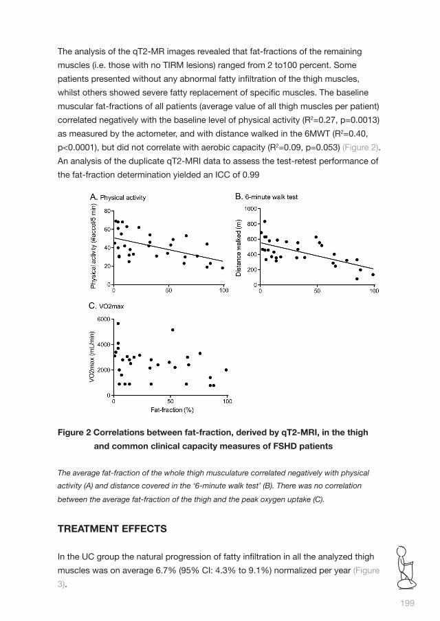
199
The analysis of the qT2-MR images revealed that fat-fractions of the remaining
muscles (i.e. those with no TIRM lesions) ranged from 2 to100 percent. Some
patients presented without any abnormal fatty infiltration of the thigh muscles,
whilst others showed severe fatty replacement of specific muscles. The baseline
muscular fat-fractions of all patients (average value of all thigh muscles per patient)
correlated negatively with the baseline level of physical activity (R2=0.27, p=0.0013)
as measured by the actometer, and with distance walked in the 6MWT (R2=0.40,
p<0.0001), but did not correlate with aerobic capacity (R2=0.09, p=0.053) (Figure 2).
An analysis of the duplicate qT2-MRI data to assess the test-retest performance of
the fat-fraction determination yielded an ICC of 0.99
Figure 2 Correlations between fat-fraction, derived by qT2-MRI, in the thigh
and common clinical capacity measures of FSHD patients
The average fat-fraction of the whole thigh musculature correlated negatively with physical
activity (A) and distance covered in the ‘6-minute walk test’ (B). There was no correlation
between the average fat-fraction of the thigh and the peak oxygen uptake (C).
TREATMENT EFFECTS
In the UC group the natural progression of fatty infiltration in all the analyzed thigh
muscles was on average 6.7% (95% CI: 4.3% to 9.1%) normalized per year (Figure
3).

Figure 3 Treatment effects of aerobic exercise training and cognitive
behavioral therapy on MRI derived fat-fractions of the individual thigh
muscles in FSHD patients
UC = usual care, AET = aerobic exercise training, CBT = cognitive behavioral therapy. Bars
indicate the average change in fat-fraction normalized per year for the thigh muscles of FSHD
patients receiving no treatment (Usual care), AET or CBT. Values are presented as means±SE.
The progression of fatty infiltration was lower for the two treatment groups compared to usual
care (* p< 0.05 and ** p< 0.01).
The linear mixed model analysis revealed that for individual muscles an increase in
fatty infiltration occurred on average in all muscles except in the G, S and VL. The
largest progression was found in the AM with 19% per year (95% CI: 12% to 26%)
(Table 3).
Cha
pte
r 8
Qua
ntita
tive
MR
I rev
eals
dec
eler
ated
fatt
y in
filtr
atio
n in
mus
cles
of a
ctiv
e FS
HD
pat
ient
s
200

201
Tabl
e 3
Inte
rven
tion
eff
ects
on
the
prog
ress
ion
of fa
tty
infi
ltra
tion
for
indi
vidu
al t
high
mus
cles
in F
SHD
pat
ient
sO
bse
rved
fat
-fr
acti
on
(%)
Est
imat
ed m
ean
fat-
frac
tio
n ex
trap
ola
ted
to
one
yea
r af
ter
bas
elin
e (%
)
Mus
cle
for
all g
roup
s at
b
asel
ine
UC
AE
T
CB
T
Mea
n (S
D)
Mea
n (9
5% C
I)M
ean
(95%
CI)
PM
ean
(95%
CI)
P
AM
38.1
(39.
2)57
.1(4
9.9
to 6
4.2)
42.7
(33.
2 to
52.
2)0.
0036
.5(2
6.4
to 4
6.6)
0.00
BFL
35.2
(38.
0)47
.9(4
2.7
to 5
3.2)
34.7
(27.
6 to
41.
7)0.
0034
.2(2
6.7
to 4
1.7)
0.00
BFS
33.1
(35.
7)42
.2(3
7.2
to 4
7.1)
36.4
(29.
9 to
43.
0)0.
0831
.7(2
4.5
to 3
8.8)
0.01
G27
.7(3
7.5)
29.4
(24.
5 to
34.
3)27
.1(2
0.7
to 3
3.6)
0.48
30.8
(23.
8 to
37.
8)0.
69
RF
36.2
(37.
2)45
.5(3
9.1
to 5
1.9)
37.5
(29.
2 to
45.
9)0.
0635
.2(2
6.0
to 4
4.4)
0.03
S19
.8(2
6.0)
22.0
(17.
1 to
26.
8)20
.6(1
4.3
to 2
7.0)
0.68
23.5
(16.
5 to
30.
5)0.
66
SM
54.5
(40.
2)65
.8(5
9.0
to 7
2.6)
59.7
(50.
7 to
68.
7)0.
1849
.9(4
0.2
to 5
9.7)
0.00
ST
41.8
(41.
6)52
.7(4
8.1
to 5
7.2)
43.1
(37.
1 to
49.
1)0.
0044
.1(3
7.4
to 5
0.8)
0.01
VI
14.7
(22.
8)23
.9(2
0.3
to 2
7.5)
15.7
(11.
0 to
20.
4)0.
0012
.6(7
.2 t
o 18
.0)
0.00
VL
13.3
(23.
3)15
.2(1
2.6
to 1
7.7)
13.9
(10.
6 to
17.
2)0.
4511
.5(7
.6 t
o 15
.4)
0.06
VM
19.3
(28.
9)35
.2(3
1.1
to 3
9.4)
19.1
(13.
7 to
24.
5)
0.
0017
.8(1
1.6
to 2
3.9)
0.00
UC
= u
sual
car
e, A
ET
= a
erob
ic e
xerc
ise
trai
ning
, CB
T =
cog
nitiv
e b
ehav
iora
l the
rap
y. A
M =
ad
duc
tor
mag
nus;
BFL
= b
icep
s fe
mor
is lo
ng h
ead
; BFS
= b
icep
s fe
mor
is s
hort
hea
d; G
= g
raci
lis; R
F =
rec
tus
fem
oris
; S =
sar
toriu
s; S
M =
sem
imem
bra
nosu
s; S
T =
sem
itend
inos
us; V
I = v
astu
s in
term
ediu
s; V
L =
vas
tus
late
ralis
; V
M =
vas
tus
med
ialis
. A li
near
mix
ed m
odel
was
use
d t
o ac
coun
t fo
r re
pea
ted
mea
sure
men
ts. P
val
ues
refe
r to
a c
omp
aris
on b
etw
een
ther
apy
grou
ps
and
UC
gr
oup
. Bas
elin
e fa
t-fr
actio
ns (%
) as
mea
n va
lues
(SD
) and
est
imat
ed fa
t-fr
actio
ns a
fter
one
yea
r as
mea
n va
lues
(95%
CI).

In the AET group the average increase in fat-fraction per year was 2.9% (95% CI:
0.7% to 5.2%), which was lower than observed in the UC group (p=0.03) (Figure 3,
Figure 4).
Figure 4 Baseline and follow-up fat-fractions of all individual muscles
AET = aerobic exercise therapy, CBT =cognitive behavioral therapy.
The solid line is the line of unity, if no change in fat fraction would occur all point would
be on this line. The dashed line signifies the 95% confidence interval of the fat-fraction
determination method. Dots above the unity and upper dashed line indicate muscles in which
significant progression has occurred. Note that this is more in the usual care group compared
to the intervention groups and that the baseline to follow-up period was shorter in the usual
care groups compared to both intervention groups (12 vs 28 weeks).
The individual muscle analyses by a linear mixed model showed significantly less
progression of fatty infiltration compared to the UC group in all but the G, S, SM
and VL muscles (Table 3). The deceleration of progression of fatty infiltration in the
AET group was largest for the VM muscle. In this muscle fatty infiltration did not
advance significantly whilst this was obvious in the UC group.
Compared to the UC fat-fraction values, we also computed a lower progression
rate for the CBT group: on average 1.7% per year (95% CI: -0.2% to 3.6%)
(p=0.0015) (Figure 3). The linear mixed model analyses showed that this
deceleration applied to all muscles except for the G, S and VL (Table 3). In this
intervention group the effect was largest for the AM muscle. FSHD patients
subjected to AET and CBT had fewer new hyper-intense lesions in their muscles on
TIRM images than those in the UC group, respectively 2 in 18 and 7 in 13 patients
(Table 2), indicating reduced edematous inflammation. Body weight, body mass
index and body fat percentage were unaffected by the interventions (Table 4). In
both intervention groups, mean registered physical activity increased compared to
the UC group. In the AET group, the average number of accelerations per 5-min
intervals increased with 2.8 and in the CBT group with 2.9 compared to a decrease
of -1.3 in the UC group, adjusted for pre-treatment values.
Cha
pte
r 8
Qua
ntita
tive
MR
I rev
eals
dec
eler
ated
fatt
y in
filtr
atio
n in
mus
cles
of a
ctiv
e FS
HD
pat
ient
s
202

203
Tabl
e 4
Body
wei
ght,
bod
y m
ass
inde
x an
d bo
dy fa
t pe
rcen
tage
are
not
aff
ecte
d by
life
sty
le in
terv
enti
on in
the
pat
ient
s e
xam
ined
by
MR
UC
A
ET
CB
T
Bas
elin
eFo
llow
-up
Bas
elin
eFo
llow
-up
Bas
elin
eFo
llow
-up
Mea
nS
DM
ean
SD
diff
pM
ean
SD
Mea
nS
Dd
iffp
Mea
nS
DM
ean
SD
diff
p
FFM
I17
.31.
317
.51.
30.
20.
619
.42.
518
.82.
2-0
.60.
218
.02.
417
.62.
2-0
.40.
2
BF%
28.4
9.4
27.5
10.0
-0.9
0.5
30.5
7.4
32.2
6.9
1.7
0.4
31.3
4.8
32.3
6.0
1.0
0.3
BM
I24
.53.
924
.54.
10.
00.
828
.64.
828
.24.
4-0
.40.
326
.63.
426
.43.
5-0
.20.
4
Wei
ght
(kg
)79
.012
.278
.612
.4-0
.40.
688
.613
.787
.613
.4-1
.00.
486
.716
.586
.316
.4-0
.40.
5
UC
= u
sual
car
e, A
ET
= a
erob
ic e
xerc
ise
trai
ning
, CB
T =
cog
nitiv
e b
ehav
iora
l the
rap
y. A
M =
ad
duc
tor
mag
nus;
BFL
= b
icep
s fe
mor
is lo
ng h
ead
; BFS
= b
icep
s fe
mor
is s
hort
hea
d; G
= g
raci
lis; R
F =
rec
tus
fem
oris
; S =
sar
toriu
s; S
M =
sem
imem
bra
nosu
s; S
T =
sem
itend
inos
us; V
I = v
astu
s in
term
ediu
s; V
L =
vas
tus
late
ralis
; VM
=
vas
tus
med
ialis
. A li
near
mix
ed m
odel
was
use
d t
o ac
coun
t fo
r re
pea
ted
mea
sure
men
ts. P
val
ues
refe
r to
a c
omp
aris
on b
etw
een
ther
apy
grou
ps
and
UC
gro
up.
Bas
elin
e fa
t-fr
actio
ns (%
) as
mea
n va
lues
(SD
) and
est
imat
ed fa
t-fr
actio
ns a
fter
one
yea
r as
mea
n va
lues
(95%
CI).

DISCUSSION
The main finding of this study is that aerobic exercise therapy (AET) and cognitive
behavioral therapy (CBT) directed at optimizing daily physical activity slow down
the progression of fatty replacement of muscle tissue in FSHD as evidenced by
quantitative T2-MR imaging, demonstrating that the fat-fraction derived by MRI
can serve as a sensitive and reproducible biomarker to reveal treatment effects in
FSHD. This is clinically relevant as we also demonstrate that fat-fractions correlate
with physical activity and distances walked in 6 minutes.
Previously, we proved that muscular fat-fraction determined by qT2-MRI is
a sensitive biomarker of disease state in FSHD and that it can detect natural
progression in individual muscles within the course of four months (15), whilst leg
muscle strength and MR-detected muscle fat infiltration were shown to be strongly
correlated (14, 15, 26). With the current qT2-MRI study we substantiate that
both AET and CBT interventions significantly decelerate disease progression as
reflected in the fatty infiltration of thigh muscles within a time span of four months.
Our muscle-specific linear mixed model analyses show that treatment effects are
not the same for all muscles. For instance, retardation of fatty infiltration is most
pronounced in the adductor magnus and vastus medialis but absent in the gracilis,
sartorius and vastus lateralis muscles. This difference can be explained by the
insignificant fatty progression rates for the latter muscles in the usual care group
across the trial period. A decreased fat infiltration rate was also detected in two leg
muscles of boys with Duchenne muscular dystrophy due to corticosteroid therapy
by determining fat-fractions with single voxel MR spectroscopy (27).
In the absence of a causal treatment for FSHD it is important to find approaches
that slow down disease progression (28). Of the studies exploring pharmacological
and non-pharmacological interventions (7, 8, 10, 28-35), until recently only some
physical therapy programs showed a beneficial effect, while their application
remained controversial due to the lack of controlled studies (28). We recently
reported in an RCT that both AET and CBT significantly reduce chronic fatigue in
FSHD patients as assessed with the Checklist Individual Strength (CIS-fatigue). In
another RCT from a different group, physical fitness, workload and walking speed
improved after 12 weeks of AET (8). However, a positive effect on the muscular
level has never been described before (7-10). In the present study, using the fat-
fractions derived from qT2-MR imaging, we were able to demonstrate significant
treatment effects, from which we infer that determining fatty infiltration volumes
in skeletal muscles is a more sensitive instrument than the mentioned physical
Cha
pte
r 8
Qua
ntita
tive
MR
I rev
eals
dec
eler
ated
fatt
y in
filtr
atio
n in
mus
cles
of a
ctiv
e FS
HD
pat
ient
s
204

capacity measures. The higher sensitivity and reproducibility of fat-fractions
obtained by qT2-MRI compared to muscle strength measurements in intervention
trials (36) can be explained by confounding practicalities of the latter method such
as influence of limb position, motivation and placebo effects (9) and also because
entire muscle groups are gauged, thus obscuring effects on individual muscles.
The similar positive effects of AET and CBT on the rate of muscle fat infiltration
is understandable as both interventions aimed to increase the level of physical
activity: AET by three-weekly cycling exercises and CBT by a daily physical activity
program consisting of graded increased cycling or walking exercises. Indeed,
both AET and CBT increased the level of daily activity, as objectively measured
by accelerometry (7). The particular physical activity programs may well explain
the positive effect of both therapies on fatty infiltration of the upper leg muscles
as these muscles are essential for walking and even more so for cycling. This
indicates that adopting an overall active lifestyle by an increased level of physical
activity of sufficient duration slows down the progression of fatty replacement of
muscle tissue in patients with FSHD by an underlying mechanism common for
both interventions. In future studies it will be of interest to include qT2-MRI in more
detailed investigations of the levels of physical activity required to obtain beneficial
effects in these patients.
Some aspects need to be taken into account in the interpretation of the results.
A selection bias may have occurred in the specific recruitment of the patients
for the MR study. However, as there were no significant differences in patient
characteristics and physical capacities between the 31 patients participating in
the MR study and the 26 non-participants, a selection bias is not expected to be
relevant. To correct for the timing difference between the baseline and follow-up
measurement of the control and intervention groups we normalized the change
in fat-fractions for all groups to one year. This assumes linear progression of fatty
infiltration in all groups, which might not reflect its true course. Because the interval
between the two MR measurements was longer in the intervention groups than in
the usual care group there is more time for a potential increase in fatty infiltration in
these intervention groups, in particular during the usual care period inserted before
the follow-up MR measurement. Hence, it is unlikely that the deceleration of fatty
infiltration by the interventions is positively biased by the normalization procedure.
Exercise enhances several capacities that will lead to stronger muscles and less
fatigue, but may also provide favorable conditions to slow down progression of
muscle affliction and to regenerate muscles in neuromuscular disorders (37, 38).
A primary role in the pathophysiology of FSHD has been suggested for a DUX4
205

induced immune response (39). This is in agreement with the observation of
hyper-intense signals in TIRM or STIR MR images of muscles in patients, which
are indicative of edematous inflammation (2, 14, 39). The MR signatures for these
processes are particularly relevant as they seem to indicate that edema may
precede the process of fatty infiltration (15-17). Therefore, it is of interest that both
intervention groups showed less new hyper-intense lesions at follow-up.
In this study we demonstrate that the rate of fatty infiltration as determined by qT2-
MRI in individual muscles of patients with FSHD is significantly decelerated after
AET and CBT compared to usual care. We also show that this MRI approach is
reproducible and can be used to obtain direct, sensitive and objective measures for
the muscular state in legs of FSHD patients.
These results reinforce the notion that quantitative MRI may become an
indispensible endpoint in clinical trials for muscular dystrophies (40).
Cha
pte
r 8
Qua
ntita
tive
MR
I rev
eals
dec
eler
ated
fatt
y in
filtr
atio
n in
mus
cles
of a
ctiv
e FS
HD
pat
ient
s
206

REFERENCES
1. Deenen JC, Arnts H, van der Maarel SM, et al. Population-based incidence and
prevalence of facioscapulohumeral dystrophy. Neurology 2014;83:1056-1059.
2. Tawil R, van der Maarel SM, Tapscott SJ. Facioscapulohumeral dystrophy: the
path to consensus on pathophysiology. Skeletal Muscle 2014;4:12.
3. Kalkman JS, Schillings ML, Zwarts MJ, van Engelen BG, Bleijenberg G. The
development of a model of fatigue in neuromuscular disorders: a longitudinal
study. Journal of Psychosomatic Research. 2007;62:571-579.
4. Kalkman JS, Schillings ML, van der Werf SP, et al. Experienced fatigue in
facioscapulohumeral dystrophy, myotonic dystrophy, and HMSN-I. Journal of
Neurology, Neurosurgery, and Psychiatry 2005;76:1406-1409.
5. Bortolanza S, Nonis A, Sanvito F, et al. AAV6-mediated systemic shRNA
delivery reverses disease in a mouse model of facioscapulohumeral muscular
dystrophy. Molecular Therapy 2011;19:2055-2064.
6. Wallace LM, Liu J, Domire JS, et al. RNA interference inhibits DUX4-induced
muscle toxicity in vivo: implications for a targeted FSHD therapy. Molecular
Therapy 2012;20:1417-1423.
7. Voet N, Bleijenberg G, Hendriks J, et al. Both aerobic exercise and cognitive-
behavioral therapy reduce chronic fatigue in FSHD: An RCT. Neurology
2014;18:1914-22.
8. Andersen G, Prahm KP, Dahlqvist JR, Citirak G, Vissing J. Aerobic training and
postexercise protein in facioscapulohumeral muscular dystrophy: RCT study.
Neurology 2015;85(5):396-403
9. A prospective, quantitative study of the natural history of facioscapulohumeral
muscular dystrophy (FSHD): implications for therapeutic trials. The FSH-DY
Group. Neurology 1997;48:38-46.
10. van der Kooi EL, Vogels OJ, van Asseldonk RJ, et al. Strength training and
albuterol in facioscapulohumeral muscular dystrophy. Neurology 2004;63:702-
708.
11. Phoenix J, Betal D, Roberts N, Helliwell TR, Edwards RH. Objective
quantification of muscle and fat in human dystrophic muscle by magnetic
resonance image analysis. Muscle & Nerve 1996;19:302-310.
12. McMahon CJ, Wu JS, Eisenberg RL. Muscle edema. American Journal of
Roentgenology 2010;194:W284-292.
13. Friedman SD, Poliachik SL, Carter GT, Budech CB, Bird TD, Shaw DW. The
magnetic resonance imaging spectrum of facioscapulohumeral muscular
dystrophy. Muscle & Nerve 2012;45:500-506.
207

14. Kan HE, Scheenen TW, Wohlgemuth M, et al. Quantitative MR imaging of
individual muscle involvement in facioscapulohumeral muscular dystrophy.
Neuromuscular Disorders 2009;19:357-362.
15. Janssen BH, Voet NB, Nabuurs CI, et al. Distinct disease phases in muscles
of facioscapulohumeral dystrophy patients identified by MR detected fat
infiltration. PloS one 2014;9:e85416.
16. Tasca G, Pescatori M, Monforte M, et al. Different molecular signatures in
magnetic resonance imaging-staged facioscapulohumeral muscular dystrophy
muscles. PloS one 2012;7:e38779.
17. Friedman SD, Poliachik SL, Otto RK, et al. Longitudinal features of stir bright
signal in FSHD. Muscle & Nerve 2014; 49:257-260.
18. Voet NB, Bleijenberg G, Padberg GW, van Engelen BG, Geurts AC. Effect of
aerobic exercise training and cognitive behavioural therapy on reduction of
chronic fatigue in patients with facioscapulohumeral dystrophy: protocol of the
FACTS-2-FSHD trial. BMC Neurology 2010;10:56.
19. Lemmers RJ, O’Shea S, Padberg GW, Lunt PW, van der Maarel SM. Best
practice guidelines on genetic diagnostics of Facioscapulohumeral muscular
dystrophy: workshop 9th June 2010, LUMC, Leiden, The Netherlands.
Neuromuscular Disorders 2012;22:463-470.
20. Ricci E, Galluzzi G, Deidda G, et al. Progress in the molecular diagnosis of
facioscapulohumeral muscular dystrophy and correlation between the number
of KpnI repeats at the 4q35 locus and clinical phenotype. Annals of Neurology
1999;45:751-757.
21. van der Werf SP, Prins JB, Vercoulen JH, van der Meer JW, Bleijenberg
G. Identifying physical activity patterns in chronic fatigue syndrome using
actigraphic assessment. Journal of Psychosomatic Research 2000;49:373-379.
22. Vercoulen JH, Bazelmans E, Swanink CM, et al. Physical activity in chronic
fatigue syndrome: assessment and its role in fatigue. Journal of Psychiatric
Research 1997;31:661-673.
23. Astrand PO, Ryhming I. A normogram for calculation for aerobic capacity
(physical fitness) from pulse rate during sub-maximal work. Journal of Applied
Physiology 1954:218-222.
24. Enright PL. The six-minute walk test. Respiratory Care 2003;48:783-785.
25. Shrout PE, Fleiss JL. Intraclass correlations: uses in assessing rater reliability.
Psychol Bull 1979;86:420-428.
26. Olsen DB, Gideon P, Jeppesen TD, Vissing J. Leg muscle involvement in
facioscapulohumeral muscular dystrophy assessed by MRI. Journal of
Neurology 2006;253:1437-1441.
Cha
pte
r 8
Qua
ntita
tive
MR
I rev
eals
dec
eler
ated
fatt
y in
filtr
atio
n in
mus
cles
of a
ctiv
e FS
HD
pat
ient
s
208

27. Arpan I, Willcocks RJ, Forbes SC, et al. Examination of effects of
corticosteroids on skeletal muscles of boys with DMD using MRI and MRS.
Neurology 2014;83:974-980.
28. Sacconi S, Salviati L, Desnuelle C. Facioscapulohumeral muscular dystrophy.
Biochimica et Biophysica Acta 2015;1852:607-614
29. Olsen DB, Orngreen MC, Vissing J. Aerobic training improves exercise
performance in facioscapulohumeral muscular dystrophy. Neurology
2005;64:1064-1066.
30. Pasotti S, Magnani B, Longa E, et al. An integrated approach in a case of
facioscapulohumeral dystrophy. BMC Musculoskelet Disord 2014;15:155.
31. Voet NB, van der Kooi EL, Riphagen, II, Lindeman E, van Engelen BG, Geurts
AC. Strength training and aerobic exercise training for muscle disease.
Cochrane Database of Systematic Reviews 2013;7:CD003907.
32. Tawil R, McDermott MP, Pandya S, et al. A pilot trial of prednisone in
facioscapulohumeral muscular dystrophy. FSH-DY Group. Neurology
1997;48:46-49.
33. Colson SS, Benchortane M, Tanant V, et al. Neuromuscular electrical
stimulation training: a safe and effective treatment for facioscapulohumeral
muscular dystrophy patients. Arch Phys Med Rehab 2010;91:697-702.
34. Tawil R. Facioscapulohumeral muscular dystrophy. Neurotherapeutics
2008;5:601-606.
35. Rose MR, Tawil R. Drug treatment for facioscapulohumeral muscular
dystrophy. Cochrane Database of Systematic Reviews 2004:CD002276.
36. Statland JM, McDermott MP, Heatwole C, et al. Reevaluating measures
of disease progression in facioscapulohumeral muscular dystrophy.
Neuromuscular Disorders 2013;23:306-312.
37. Pedersen BK. The diseasome of physical inactivity--and the role of myokines in
muscle--fat cross talk. Journal of Physiology 2009;587:5559-5568.
38. Anziska Y, Sternberg A. Exercise in neuromuscular disease. Muscle & Nerve
2013;48:3-20.
39. Frisullo G, Frusciante R, Nociti V, et al. CD8(+) T Cells in Facioscapulohumeral
Muscular Dystrophy Patients with Inflammatory Features at Muscle MRI.
Journal of Clinical Immunology 2010; 31:155-66.
40. Hollingsworth KG. Quantitative MRI in muscular dystrophy: An indispensable
trial endpoint? Neurology 2014;83:956-957.
209


CHAPTER 9SUMMARY AND GENERAL DISCUSSION

SUMMARYFacioscapulohumeral muscular dystrophy (FSHD) is the third most common
muscular dystrophy worldwide with an estimated prevalence of one in 8,000
persons. It is an autosomal dominant disorder affecting primarily the muscles of
the face and the shoulder girdle, but later in life many muscles of the trunk and
the extremities can become affected. In the last decade, substantial progress
has been made in the understanding of the molecular genetics of FSHD. More
than 95% of the cases of FSHD are associated with a partial deletion of a critical
number of repetitive elements (D4Z4) on chromosome 4q35 and the subsequent
toxic release of the DUX4 protein in muscle cells. It is, nevertheless, still unknown
why the weakening of different muscles and muscle groups occurs at different
rates and times. Moreover, there are no biomarkers for an objective assessment of
the severity and progression of FSHD.
To date, no curative treatment is available for FSHD. As a consequence, although
life expectancy is normal, 20% of the patients become wheelchair-bound due to
muscle weakness. In addition, more than 60% of the patients experience chronic
fatigue and with that, a lower level of social participation. Physical inactivity has
shown to be the most important perpetuating factor of fatigue in patients with
FSHD. However, being physically active is difficult for patients because of muscle
weakness, which may lead to a vicious circle of fatigue and inactivity.
The two main aims of this thesis were (1) to study the issues of fatigue and
(in)activity in neuromuscular disorders and (2) to evaluate the effect of model-based
interventions to optimize the activity level and reduce chronic fatigue in FSHD.
Chapter 1 is the general introduction and provided background information on
FSHD. Individuals with FSHD show a wide range of clinical manifestations. This
variability suggests that epigenetic mechanisms play an essential role. Epigenetics
is defined as heritable changes in gene activity and expression that occur without
alteration in DNA sequence. To objectively assess the severity and progression of
FSHD, magnetic resonance imaging (MRI), could provide an objective biomarker
for individual muscle involvement as it can easily visualize intramuscular fibrosis
and fatty infiltration. Fatigue is increasingly being recognized as a major clinical
problem in many conditions and evidence-based treatment programs are now
being developed for several patient groups. By using the subscale fatigue of the
Checklist Individual Strength, a multidimensional fatigue scale, it appeared that
more than 60% of the patients with FSHD were severely fatigued. Lack of physical
activity, sleep disturbances and pain contributed to experienced fatigue in FSHD.
Loss of muscle strength contributed to experienced fatigue through a lower level
of physical activity. This model of perpetuating factors served as a basis for the Cha
pte
r 9
Sum
mar
y an
d G
ener
al D
iscu
ssio
n
212

treatment protocol used in this thesis. This thesis reports the results of the FACTS-
2-FSHD study (acronym for Fitness And Cognitive behavioral TherapieS for Fatigue
and ACTivitieS in FSHD), which is the first model-based randomized clinical
trial that evaluated the effects of aerobic exercise training (AET) and cognitive
behavioral therapy (CBT) on chronic fatigue in patients with FSHD.
It was hypothesized that, in order to alleviate chronic fatigue, two different
therapeutic approaches could be followed: AET to promote physical activity and
CBT to stimulate an active lifestyle (yet avoiding excessive physical strain).
Hence, the primary objective of this study was to evaluate the effect of both
interventions on chronic fatigue in patients with FSHD as assessed with the
subscale fatigue of the Checklist Individual Strength. The secondary objective was
to evaluate the effects of each intervention on the known perpetuating factors of
chronic fatigue in FSHD.
The secondary outcome measures covered all domains of the International
Classification of Functioning, Disability and Health (ICF). In addition, it was aimed
to find clinically useful magnetic resonance imaging (MRI) biomarkers of disease
progression and response to therapy in patients with FSHD.
PART 1: PREVALENCE, ETIOLOGY, ASSESSMENT ANDMANAGEMENT OF FATIGUE
In part 1, an overview is provided of the prevalence, assessment, and treatment
of fatigue in neuromuscular disorders. The following research questions are
addressed:
1. What is the prevalence and relevance of fatigue in patients with muscular
dystrophy?
Chapter 2 shows that fatigue is a frequent and relevant symptom in patients
with muscular dystrophy. Distinguishing experienced fatigue from muscle
weakness, the key feature in muscular dystrophy, may be difficult. Although
experienced fatigue is difficult to define, it still is a valuable concept which
can be reliably measured by using questionnaires, like the Checklist Individual
Strength, subscale fatigue (CIS-fatigue). As fatigue in muscular dystrophy is a
multidimensional concept, it is important to understand factors that contribute
to fatigue. Based on such an analysis, preventive and therapeutic interventions
could be developed. Therefore, an overview of the pathophysiological
determinants of fatigue in muscular dystrophies was provided and the model of
perpetuating factors of experienced fatigue in FSHD was presented. A critical
overview of the possible treatment options with respect to fatigue in patients
213

with muscular dystrophy was presented including physical exercise training,
drug treatment and cognitive behavioral therapy.
2. How can we assess fatigue in patients with neuromuscular disorders?
Chapter 3 describes the results of a European Neuromuscular Centre (ENMC)
care Workshop regarding pain and fatigue in neuromuscular disorders (NMD).
This workshop aimed to achieve consensus on the definitions of pain and
fatigue in NMD and to define a core set of measurement instruments for pain
and fatigue in this group of disorders. Pain as well as fatigue are common
symptoms in NMD with a strong impact on many activities of daily life. Specific
types of pain and fatigue, related to the underlying (stages of) NMD could be
distinguished. These should be differentiated from aspecific pain and fatigue,
which are not primary related to the underlying disorder. Various methods were
being used to assess pain and fatigue in NMD. In order to develop effective
treatment approaches in NMD, both the definitions of pain and fatigue and their
assessment methods should be internationally agreed upon. Therefore, a core
set of measurement instruments for use in future research and clinical practice
in NMD was provided.
3. What is the evidence for exercise in muscle disease?
In chapter 4, an updated Cochrane review (most recent date of search 2 July
2012) is presented on the safety and efficacy of strength training and aerobic
exercise training in people with a muscle disease. Strength training or aerobic
exercise programs might optimize muscle and cardiorespiratory function and
prevent additional disuse atrophy and deconditioning in people with a muscle
disease. All randomized or quasi-randomized controlled trials comparing
strength training or aerobic exercise programs (or both) to no training, lasting
at least six weeks, in people with a well-described diagnosis of a muscle
disease were included. Five eligible trials with 170 participants fulfilled the
inclusion criteria: two trials of strength training in people with FSHD and
myotonic dystrophy (101 participants), two trials of strength training combined
with aerobic exercise in people with mitochondrial myopathy (18 participants)
and myotonic dystrophy type 1 (35 participants), and one trial of aerobic
exercise in people with polymyositis and dermatomyositis (14 participants).
These trials showed that moderate-intensity strength training in people with
myotonic dystrophy or FSHD, and aerobic exercise training in people with
dermatomyositis or polymyositis appeared not to harm muscles. Strength
training combined with aerobic exercise appeared to be safe in myotonic Cha
pte
r 9
Sum
mar
y an
d G
ener
al D
iscu
ssio
n
214

dystrophy type 1 and might be effective in increasing physical endurance
in people with mitochondrial myopathy. Finally, strength training was not
harmful in people with FSHD, myotonic dystrophy, mitochondrial disorders
or dermatomyositis / polymyositis, but there was insufficient evidence to
determine its potential benefit. Limitations in the design of studies in other
muscle diseases prevented more general conclusions.
PART 2: THE FACTS-2-FSHD STUDY
In part 2, the results of the FACTS-2-FSHD study are presented. The following
research questions are addressed:
4. What are the effects of aerobic exercise therapy and cognitive behavioral
therapy on chronic fatigue in patients with FSHD?
In chapter 5 the protocol of the FACTS-2-FSHD trial is described. The FACTS-
2-FSHD study was the first theory-based randomized clinical trial which
evaluated the effect and the maintenance of effects of AET and CBT on the
reduction of chronic fatigue in patients with FSHD. The interventions were
based on a theoretical model of perpetuating factors of chronic fatigue in
patients with FSHD. The primary objective of the FACTS-2-FSHD trial was to
study the efficacy of AET and CBT for decreasing chronic fatigue in patients
with FSHD type 1 compared to usual care. It was hypothesized that both
AET and CBT would be more effective in decreasing fatigue than usual care,
which is no therapy at all or occasional (conventional) physical therapy. The
improvement by AET might be obtained through enhancement of physical
(aerobic) capacity, whereas beneficial effects of CBT might be achieved
through changes in daily activities and behavior. Secondary objectives were to
evaluate the effects of AET and CBT on the known fatigue-perpetuating factors
in FSHD. The AET consisted of aerobic cycling exercise on a bicycle ergometer.
The training program had a duration of 16 weeks and comprised home training
twice a week and a supervised training once a week. CBT was focused on
all known perpetuating factors of fatigue and adapted to the needs of each
patient. Each session had a duration of one hour and was given by a registered
cognitive behavioral therapist. At baseline, patients were randomized to either
an AET group, a CBT group, or a control group receiving usual care. After an
intervention period of 16 weeks and a follow-up of 3 months, the control group
was as yet randomized to either AET or CBT (28 weeks after inclusion). It was
intended to eventually include 25 adult patients in each of the three groups:
AET, CBT, or usual care. Because patients in the control group received AET
215

or CBT in second instance, the minimal number of patients to be included
was 50 at baseline. Outcomes were assessed at baseline, immediately post
intervention (and control period), and at 12 en 24 weeks of follow-up. A linear
mixed model for repeated measurements was used to study the estimated
group differences.
Chapter 6 describes the results of the FACTS-2-FSHD randomized controlled
trial for which the protocol has been described in chapter 5. Fifty-seven
ambulant patients with FSHD type 1 and severe chronic fatigue were
randomly allocated to AET, CBT, or UC. Following treatment, both the AET (28
participants) and CBT (25 participants) intervention groups had significantly
less fatigue relative to the UC group (24 participants), with a difference of -9.1
for AET (95% CI: -12.4 to -5.8) and -13.3 for CBT (95% CI: -16.5 to -10.2)
on the CIS-fatigue. These beneficial effects lasted through follow-up, with a
difference of -8.2 for AET (95% CI: -12.4 to -5.8) and -10.2 for CBT (95% CI
-14.0 to -6.3). Post-treatment, 19 participants in the CBT group (76%) and 14
participants in the AET group (50%) no longer had scores indicative of severe
fatigue. The number needed to treat (NNT) for AET was 2.3 (95% CI 1.4– 3.1)
with an absolute risk reduction (ARR) of 50% (95% CI 32–69%). The NNT for
CBT was 1.3 (95% CI 1.1–1.7) with an ARR of 76% (95% CI 59–93%). In the
CBT group, all known fatigue-perpetuating factors (with the exception of pain)
were positively modified, including a higher level of social participation. The
patients who received AET showed an increase in registered physical activity
and maximal isometric quadriceps strength only. The increase in registered
physical activity in both groups and the improvement in social participation
following CBT were still present at follow-up. More than 70% of the AET and
almost 80% of the CBT participants continued their adjusted level of activity
once the study had ended. No improvement in aerobic capacity was found
in both intervention groups. Only mild adverse affects were found in the AET
group. The median number of therapy sessions was much lower in the CBT
group, i.e. five, than in the AET group (40 sessions). It was concluded that both
AET and CBT are able to ameliorate chronic fatigue in patients with FSHD.
5. Can we discover structural abnormalities in skeletal muscle of FSHD patients
that may serve as biomarkers for disease progression and response to therapy?
Even though the most important genetic event for the disease has been
identified, the underlying mechanisms causing FSHD are unknown.
Understanding these mechanisms first requires a better knowledge of the
process of fatty infiltration of the skeletal muscles.Cha
pte
r 9
Sum
mar
y an
d G
ener
al D
iscu
ssio
n
216
In the study described in chapter 7, fat fraction as assessed by quantitative
MRI (Q-MRI) appeared to be an objective and sensitive biomarker for muscular
affliction in FSHD, detecting even subclinical muscle involvement. An analysis
of the average fat fraction for all individual muscles uncovered an hourglass
pattern of many muscles with either a very low or high fat fraction, and few
muscles with an intermediate fat fraction. This quasi-binary distribution had
not been reported for other muscular dystrophies and may be FSHD specific.
The intramuscular fat fraction increased linearly from proximal to distal. Fat
replacement of entire muscles would, on the average, be completed within
approximately three and a half years. The steepest fat gradient occurred in the
intermediately affected muscles indicating that these muscles were quickly
progressing towards a complete fat infiltrated state. This can be relevant for
prognostication and monitoring therapy effectiveness in FSHD.
The primary goal of the study presented in chapter 8 was to examine the
effects of AET and CBT on the progression of fatty infiltration and edema in
individual leg muscles of FSHD type 1 patients by T2-MRI. Quantitative T2-MRI
(qT2-MRI) and fat-suppressed T2-MRI images of the thigh were obtained at
baseline and follow-up in 31 patients who were included in the FACTS-2-FSHD
study, of whom 13 received usual care (UC), nine AET, and nine CBT. In the
UC group the fatty infiltration in the affected muscles progressed on average
with 6.7% per year. Progression occurred on average in all muscles except
in the gastrocnemius, sartorius and vastus lateralis. Overall, the adductor
magnus showed the largest progression. This rate was significantly lowered
by both interventions to (on average) 2.9% per year in the AET group (AET –
UC, p<0.05) and 1.7% per year in the CBT group (CBT – UC, p<0.01). In both
intervention groups fewer muscles developed edema than was observed in the
UC group. The baseline muscular fat fractions of all patients (average value of
all thigh muscles per patient) were negatively correlated with the baseline level
of physical activity (R2=0.27, p=0.0013) as measured by the actometer and
with the maximum walking distance (R2=0.40, p<0.0001), but they were not
significantly correlated with aerobic capacity. It was concluded that qT2-MRI is
a reproducible and sensitive quantitative biomarker for monitoring the effects
of increased physical activity in individual muscles in FSHD and that both AET
and CBT slow down the progression of fatty replacement of muscle tissue in
FSHD.
217

GENERAL DISCUSSION
Mr. C, a 58-year old man with facioscapulohumeral dystrophy (FSHD) who was
briefly introduced in the general introduction of this thesis, was encouraged by
his wife to take part in the FACTS-2-FSHD study, hoping to achieve a reduction
of his fatigue. He hoped to be randomized to aerobic exercise training to resume
his cycling exercises. He had given up those exercises a couple of years ago for
fear of further deterioration of his muscle strength. However, in the study he was
randomized to cognitive behavioral therapy (CBT). Initially he was disappointed,
as he did not really want to talk to a psychologist about his muscle disease
and the burden of his disease. During the study, he received nine sessions of
CBT. In the beginning, he was skeptical about this treatment, and therefore did
not expect CBT to have any effect. He regarded his experienced fatigue as an
untreatable problem. Several measurements that were conducted during the first
treatment session with the psychologist showed that the CBT should be directed
at unhelpful thoughts and beliefs about fatigue, improper coping strategies,
sleep disturbances, physical inactivity and unhelpful social interactions. After
nine sessions of CBT and homework assignments, his physical activity had
increased substantially. Together with his partner, he went out on an electric bike
again to visit his family and friends. He was no longer afraid of muscle damage
from physical activity because he noticed that he felt more fit by being physically
active. He resumed his gardening activities, this time in the communal garden
of the apartment complex where he lived. He no longer slept during lunchtime,
so the quality of sleep at night became better. He was not seriously fatigued any
more. His mental and physical capacity increased, and there was room for new
activities. A few weeks after the end of the CBT, he started a new job.
A MODEL-BASED TRIAL
The Departments of Neurology, Rehabilitation and Pediatrics of the Radboud
University Medical Center together with the Expert Center for Chronic Fatigue
collaborate in the Center of Expertise for Muscular Dystrophy and have worked
together in research and patient care in muscular dystrophy for over 20 years.
One of the research successes is the result of cross-sectional and longitudinal
research on experienced fatigue in facioscapulohumeral dystrophy (FSHD). Cross-
sectional research showed that experienced fatigue is a frequent as well as a
relevant problem for patients with FSHD. Based on longitudinal data, a model of
perpetuating factors of experienced fatigue in patients with FSHD was developed
(1). Muscle weakness, the key feature of FSHD and the result of fatty infiltration Cha
pte
r 9
Sum
mar
y an
d G
ener
al D
iscu
ssio
n
218

of the skeletal muscles, appeared to contribute only indirectly to experienced
fatigue. However, muscle weakness leads to physical inactivity, the most important
perpetuating factor of experienced fatigue. Sleep disorders and pain are the other
proven perpetuating factors of experienced fatigue. This FSHD-specific model was
of major importance for the development of the evidence-based interventions in the
FACTS-2-FSHD study, namely aerobic exercise training (AET), aimed at increasing
aerobic capacity, and cognitive behavioral therapy (CBT), a psychological treatment
aimed at promoting a physically active lifestyle (1) (Figure 1).
Figure 1 Model of perpetuating factors of experienced fatigue, specifically for
patients with facioscapulohumeral dystrophy
Physical inactivity, sleep disturbances and pain are direct and muscle weakness is an indirect
perpetuating factor of experienced fatigue (1).
Both interventions in the study are directed at the aforementioned perpetuating
factors of experienced fatigue. AET focuses primarily on the perpetuating factor
of physical inactivity. This was expected to reduce the chronic experienced
fatigue and enhance the degree of social participation. It was hypothesized that
an increase in aerobic capacity could lead to a lower level of experienced fatigue
(Figure 2).
Cognitive behavioral therapy (CBT) is composed of six modules directed at the
proven and presumed perpetuating factors of experienced fatigue and their
related (unhelpful) cognitions in FSHD. The modules focus on: (1) unhelpful coping
strategies; (2) unhelpful cognitions about fatigue; activity, pain or other symptoms;
(3) catastrophic thoughts about fatigue; (4) sleep disturbances; (5) physical
inactivity or dysregulation of physical activity; and (6) a discrepancy between
expected and actual social support and interactions. Additionally, reducing
restrictions in social participation is an important objective of the CBT. In this way,
the final aim is to reduce chronic fatigue and social participation restrictions (Figure
3).
219

Figure 2 The expected working mechanism of aerobic exercise training (AET)
based on a model of perpetuating factors of experienced fatigue in
patients with facioscapulohumeral dystrophy (Figure 1)
AET focuses primarily on the perpetuating factor physical inactivity. Improved physical activity
would diminish the level of experienced fatigue and social participation restrictions.
Figure 3 The expected working mechanism of cognitive behavioral therapy
(CBT) based on a model of perpetuating factors of experienced
fatigue in patients with facioscapulohumeral dystrophy (Figure 1)
CBT focuses not only on the perpetuating factor physical inactivity, but also on sleep
disturbances, pain, unhelpful cognitions about fatigue and restrictions in social participation. Cha
pte
r 9
Sum
mar
y an
d G
ener
al D
iscu
ssio
n
220

The patient with FSHD described in the case study showed an improvement in all
domains of the International Classification of Functioning, Disability and Health
after only nine sessions of CBT (2). Not only did he experience a lower level of
fatigue and an improvement in sleep quality, he also became more physically
active. He performed gardening activities again, his social contacts increased
and he started a new job. CBT broke the downward spiral of physical inactivity,
experienced fatigue and social participation restrictions.
He became physically active in daily life, and social participation became possible
again.
The case study illustrates the general conclusion of this thesis: CBT is able to
reduce severe fatigue in patients with FSHD and improve social participation, by
increasing physical activity and changing all relevant fatigue perpetuating factors
(Figure 3).
In addition, the FACTS-2-FSHD study shows that AET can also achieve a reduction
of severe chronic fatigue and increase in physical activity in patients with FSHD
(Figure 2). Although AET, contrary to CBT, did not cause an improved quality of
sleep or social participation, it did cause an increase in muscle strength of the
quadriceps. Finally, a deceleration of fatty replacement of muscle tissue in the thigh
muscles was observed after both interventions.
POSSIBLE EXPLANATIONS FOR EFFECTS: A PHYSICALLY ACTIVE LIFESTYLE VERSUS PHYSICAL EXERCISE
Physical inactivity
After both CBT and AET, the level of physical activity in daily life increased. After
CBT, the level of physical activity remained high compared with the control group,
even after the follow-up period of 12 weeks. The increase in physical activity
in everyday life appears to play an important role in the positive effect of both
interventions on the level of fatigue. Based on the model of perpetuating factors of
fatigue and additional research, it is known that in FSHD the degree of fatigue is
not correlated with the severity of muscle weakness (1, 3). Apparently, this fatigue
seems more a result of unintentional unhelpful behavior associated with the disease
rather than the result of muscle weakness itself. Conversely, experienced fatigue
often leads to unhelpful cognitions and behavior that, in turn, further increase
the level of fatigue. A curative treatment for FSHD is not available yet; however, a
treatment aimed at the aforementioned unhelpful cognitions and behavior, namely
CBT and AET, now is.
221

A physically active lifestyle versus physical exercise: ‘Dutch standard for healthy exercise’ versus ‘ the standard for physical fitness’
During CBT, a reliable increase in physical activity is an important part of the
treatment: the module ‘physical-inactivity or a high dysregulation of activity’ was
applied in each participant. AET focused primarily on physical exercise on an
ergometer. At first sight, physical activity and physical exercise seem to be similar,
but on second thought they are substantially different. Physical activity is defined
as “any effort of skeletal muscles resulting in higher energy consumption than in
resting conditions (4).” Physical (aerobic) exercise is a form of physical activity
and is defined as “planned, structured and repetitive exercises with an increasing
magnitude and intensity in order to maintain or improve physical fitness or aerobic
capacity (4).”
The recommendations on physical activity for the healthy population have been
prescribed in the Dutch Standard for Healthy Exercise (Nederlandse Norm Gezond
Bewegen; NNGB). This standard aims at a physically active lifestyle and comprises
a total of 30 minutes of exercise of moderate intensity (at a slightly higher heart
and respiration rate than usual) of at least 4.0 MET a day, in blocks of at least 10
minutes at least five days a week.
The MET value or the metabolic equivalent is a unit of measurement within
physiology expressing the amount of energy for a certain physical effort compared
with the amount of energy required at rest. One MET corresponds to the resting
metabolic rate, the amount of energy consumed during inactivity. One MET is
equivalent to 3.5 ml of oxygen per kg of body weight per minute. The NNGB leads
to a total duration of 150 minutes of physical activity per week of 4.0 MET, which
implies a total increase of 450 MET per week compared to a physically inactive
lifestyle. Physical activity within the NNGB includes not only sports activities but
also daily-life activities such as household activities, cycling or walking the dog.
For physical exercise, the Dutch government has issued a standard for physical
fitness. This standard is aimed primarily at maintaining aerobic capacity through
physical exercise and requires intense physical activity of at least 6.0 MET for at
least 20 minutes and at least three times a week. Although the intensity is higher
than in the NNGB, the total length and the increase in MET per week is less, namely
300 MET. Thus, one can still have a physically inactive or sedentary lifestyle, in
spite of meeting the standard for physical fitness. In other words, the NNGB leads
to a higher level of physical activity than the Dutch standard for physical fitness.
Cha
pte
r 9
Sum
mar
y an
d G
ener
al D
iscu
ssio
n
222

The Dutch standard for physical fitness and the NNGB are defined only for healthy
adults and for healthy elderly. The minimum standard for patients with a chronic
disease, including FSHD, has not yet been defined. The NNGB not only leads to
a higher level of physical activity; this standard is probably also more feasible for
patients with FSHD, because daily-life activities are included. In other progressive
neurological diseases, such as Parkinson’s disease, there is already growing
evidence for a positive effect of decreasing the sedentary time (5). The question
now arises whether physical exercise of minimum intensity and an increase in
aerobic capacity are really necessary for the treatment of fatigue in patients with
FSHD. Would an increase in physical activity of moderate intensity and of sufficient
duration, i.e. a physically active lifestyle, not be much more relevant?
In order to answer this question, possible underlying mechanisms of the results
of the FACTS-2-FSHD study will be discusses. The results of the FACTS-2-
FSHD study will be compared with other exercise studies in FSHD and other
neuromuscular disorders (NMDs). The methodological limitations will be highlighted
and, finally, recommendations will be given for future research and clinical practice.
UNDERLYING MECHANISMS OF EFFECTS
Aerobic exercise: is an increase in aerobic capacity necessary?
In the FACTS-2-FSHD study, the primary purpose of AET was to treat fatigue in
patients with FSHD through physical exercise on an ergometer. The hypothesis
was that such exercise would improve aerobic capacity and, with that, break the
downward spiral of physical inactivity and fatigue (Figure 2). Indeed, the downward
spiral was broken: the level of physical activity increased after AET, and the level of
experienced fatigue decreased.
However, contrary to our expectations, we found no effect on aerobic capacity.
Unfortunately, with the results of the FACTS-2-FSHD study, a real absence of
effect cannot be determined with certainty. The Åstrand submaximal cycling
test appeared to be unfeasible for relatively severely affected patients. Further
considerations will be discusses in the Methodological Considerations paragraph
(see page 233).
The results of the FACTS-2-FSHD study suggest that an increase in aerobic
capacity is not essential for the reduction of chronic fatigue. Possibly promoting
physical activity through physical exercise is a more important mechanism.
223

Cognitive behavioral therapy: FSHD is more than impaired muscle function
Where AET aims at improving physical activity, CBT is aimed at all perpetuating
factors of the model of Kalkman (Figure 3). This could explain why CBT had a
lasting positive effect not only on physical activity but also on sleep quality and
social participation. The CBT intervention was more extensive than AET. The
treatment protocol of CBT consisted of six modules based on the proven and
presumed perpetuating factors of experienced fatigue in patients with FSHD. The
modules aimed to change unhelpful disease cognitions, to better regulate social
activities, to increase mental activities and to regulate social support. In the first
treatment session, the psychologist determined which modules were applicable
by performing an interview and specific tests. Thus, compared with AET, CBT was
more focused on the individual person with FSHD. In the longitudinal study in which
the model of perpetuating factors of experienced fatigue has been developed,
only a limited number of possible perpetuating factors could be explored (1). The
sample size was too small to reliably test more factors. Therefore, one or more
of the presumed perpetuating factors of experienced fatigue (discrepancy in the
level of perceived social support, unhelpful illness cognitions and limited social
and mental activities) could also be perpetuating factors of experienced fatigue
in patients with FSHD. For example, unhelpful illness cognitions are a known
perpetuating factor of experienced fatigue in multiple sclerosis and in chronic
fatigue syndrome (6). As an essential part of CBT, unhelpful cognitions can be
changed into helpful thoughts using Socratic dialogues to increase patients’
autonomy and self-efficacy.
Although increasing the amount of physical activity is an essential part of CBT
for Although increasing the amount of physical activity is an essential part of
CBT for fatigue in FSHD, both scientists and clinicians are astonished about
its beneficial effect. “Is fatigue all in the mind?” and “How can a psychological
treatment achieve an effect at a muscular level?” are frequently asked questions.
The answer to these questions is that a part of the solution is, in fact, literally “in
the mind.”Psychological factors, such as illness cognitions, coping style and level
of acceptance of the disease are known to be strongly correlated with the degree
of social participation in patients with a muscle disease, including FSHD (7). It
is noteworthy that these correlations, comparable with the level of experienced
fatigue, are relatively independent of the degree of physical impairments. This is
also called the “disability paradox:” having physical impairments has little influence
on the degree of social participation.
Cha
pte
r 9
Sum
mar
y an
d G
ener
al D
iscu
ssio
n
224
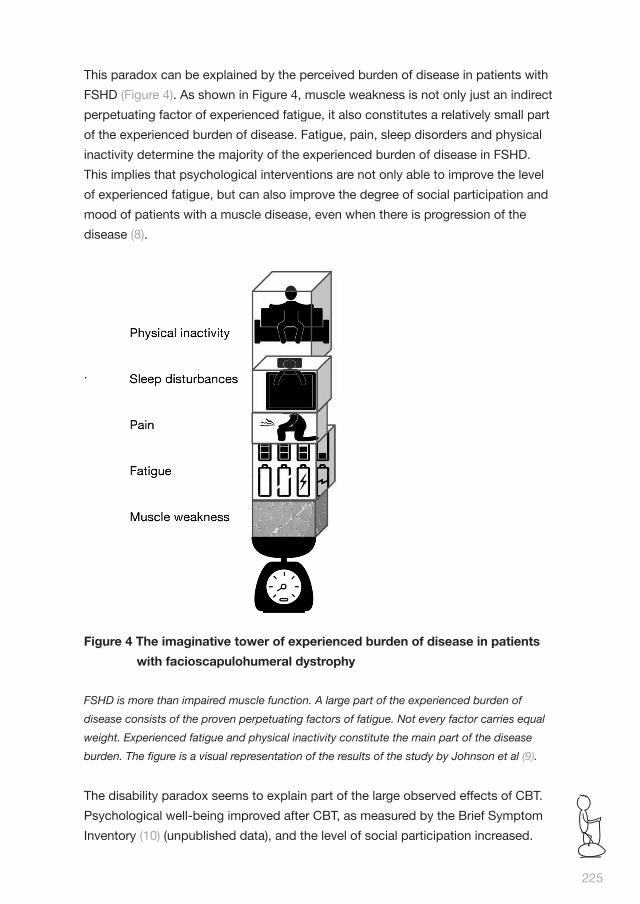
This paradox can be explained by the perceived burden of disease in patients with
FSHD (Figure 4). As shown in Figure 4, muscle weakness is not only just an indirect
perpetuating factor of experienced fatigue, it also constitutes a relatively small part
of the experienced burden of disease. Fatigue, pain, sleep disorders and physical
inactivity determine the majority of the experienced burden of disease in FSHD.
This implies that psychological interventions are not only able to improve the level
of experienced fatigue, but can also improve the degree of social participation and
mood of patients with a muscle disease, even when there is progression of the
disease (8).
.
Figure 4 The imaginative tower of experienced burden of disease in patients
with facioscapulohumeral dystrophy
FSHD is more than impaired muscle function. A large part of the experienced burden of
disease consists of the proven perpetuating factors of fatigue. Not every factor carries equal
weight. Experienced fatigue and physical inactivity constitute the main part of the disease
burden. The figure is a visual representation of the results of the study by Johnson et al (9).
The disability paradox seems to explain part of the large observed effects of CBT.
Psychological well-being improved after CBT, as measured by the Brief Symptom
Inventory (10) (unpublished data), and the level of social participation increased.
225

This occurred independent of a change in muscle strength, which is often used
as a measure of disease severity. For many patients, psychological distress is,
together with a decrease in muscle strength, a significant problem. Stress, fear for
the future, and fear for fatigue were frequently mentioned in the parallel qualitative
FACTS-2-PERSPECTIVES study during the interviews before the start of the
intervention (11, 12).
Effects at the muscular level: epigenetics and/or anti-inflammatory effects?
Both CBT and AET slow down the progression of fatty replacement of muscle
tissue in FSHD. This raises the question: “How is it possible that an increase in
physical activity causes a beneficial effect at the muscular level?” Epigenetics and
the inflammation theory can possibly offer an explanation.
Epigenetics
FSHD is a genetic disorder. More than 95% of cases of FSHD are associated
with the absence of certain pieces of DNA at the end of chromosome 4 (genetic
location: 4q35), the so-called D4Z4 deletion. This results in expression of the
harmful DUX4 gene and production of a toxic protein (DUX4) that causes dystrophy
(fatty replacement) of the skeletal muscles (13). The conversion of DNA into
functional products for the cell, such as proteins, is dependent on both the DNA
code itself (genetics) as well as on factors that may affect the activity of genes
(gene expression), so-called epigenetic factors (14, 15). Epigenetic phenomena
determine the “open” or “closed” state of parts of the genome and, thus, control
the “on” or “off” position of genes. This can take place by means of changes in
methylation, RNA molecules (intermediates between DNA and protein), or by the
so-called histone proteins that are involved in the packing (and hence access) of
the DNA in the chromosomes. FSHD is, therefore, an epigenetic disease (15). In
FSHD patients, the degree of methylation of the DNA influenced by epigenetic
factors plays an important role. Sometimes a small molecule group is added to the
DNA, a so-called methyl group, which carries additional information. FSHD patients
with a D4Z4 deletion (FSHD-1) show a decreased methylation of the D4Z4 region
on the chromosomes 4q and 10q (Figure 5).
However, the degree of methylation is not already determined at birth. It varies
between persons and may change under the influence of environmental factors
during one’s lifetime.
Cha
pte
r 9
Sum
mar
y an
d G
ener
al D
iscu
ssio
n
226

Figure 5 The role of epigenetic factors in facioscapulohumeral dystrophy
If the DUX4 gene is “readable” (i.e. can be transcripted), the DUX4 protein, which is toxic for
skeletal muscles, is produced. Epigenetic factors can make it an “unreadable” gene by an
increase in methylation. Figure courtesy of Sylvère van der Maarel.
Epigenetic factors ensure that the genetic defect in different people, even within
families, can be expressed differently (16). In recent research, the difference in
severity of the disease within families with FSHD is, among other phenomena,
attributed to epigenetic factors (17).
An increase in physical activity and/or physical exercise can cause changes in
the DNA methylation of healthy persons (18). It is possible that a physically active
lifestyle is an epigenetic factor for FSHD and can slow down the progression of
fatty replacement of muscle tissue by changes in DNA methylation. It is not a
coincidence that the perpetuating factors of fatigue, i.e. physical inactivity, sleep
disorders and pain, are known epigenetic factors (19). The degree of methylation
can be different for every individual cell under the influence of epigenetic factors.
This could be an explanation for the differences in effect on the fatty replacement
between different muscles of patients with FSHD after CBT and AET, as measured
by quantitative magnetic resonance imaging (MRI) (20). To conclude, the first
hypothesis is that AET as well as CBT influence the fatty replacement of muscle
tissue by modifying epigenetic mechanisms.
227
Theory of inflammation
A second explanation can perhaps be found in the beneficial effect of physical
activity on inflammation. Inflammatory reactions seem to play a role in the
increase in fatty replacement of muscle tissue in patients with FSHD and also in
the development of chronic experienced fatigue in various neurological disorders
(21). In approximately 5% of the muscles of patients with FSHD, edema has
been observed using MRI (20, 22). There is evidence that an increase in edema
is preceded by inflammation and is followed by fatty replacement of the muscle
tissues (23, 24). The inhibition of inflammatory reactions could, therefore,
theoretically slow down the progression of the disease. In healthy people, the
anti-inflammatory effect of physical activity has already been proven extensively
(25). Not only immune cells produce molecules that play a role in inflammatory
responses (cytokines). Skeletal contracting muscles also release significant
amounts of interleukin IL-6. IL-6 is a pro-inflammatory cytokine, in this situation
also called a myokine. IL-6 causes inflammation when it is excreted as a cytokine
by immune cells, but fights inflammation when it is released as a myokine by
muscle cells. This is most likely because other cytokines are not produced anymore
and another, “healthier” environment has been created (26, 27). In healthy adults,
the production of IL-6 during exercise is for at least 50% related to the intensity
and duration of the exercise (28, 29). To conclude, the second hypothesis is
that AET and CBT influence the fatty replacement of muscle tissue by positively
influencing inflammatory reactions. This again argues in favor of a physically active
lifestyle beyond physical exercise of limited duration.
COMPARISON WITH PREVIOUS RESEARCH
Aerobic exercise in muscle disease: from a discouraging to an encouraging approach
At the time of the design of the FACTS-2-FSHD study, clinicians were reluctant to
prescribe exercise to patients with FSHD. They often referred to possible muscle
damage due to overuse.
This assumption, however, was based on studies in which mdx mice with muscular
dystrophy had to run in a treadmill for days (30). Because mdx mice constitute a
model for Duchenne muscular dystrophy only, the results cannot be extrapolated
to other neuromuscular disorders, including FSHD. However, evidence from human
exercise studies in FSHD was scarce as well. Only one pilot study was conducted
with a healthy control group that described the beneficial effect of exercise in Cha
pte
r 9
Sum
mar
y an
d G
ener
al D
iscu
ssio
n
228

patients with FSHD (31). At the time, there was only limited evidence for a positive
effect of exercise in other muscle diseases too. The original Cochrane review
from 2005, which described the effect of AET and strength training in muscle
disease, contained only two randomized controlled trials (RCTs) and concluded:
“In myotonic dystrophy and facioscapulohumeral muscular dystrophy moderate-
intensity strength training appears not to do harm, but there is insufficient evidence
to establish that it offers benefit. Limitations in the design of studies in other muscle
diseases prevent general conclusions in these disorders (32). “The conclusions
of the other Cochrane reviews within the neuromuscular disorders group, which
described the effect of exercise in amyotrophic lateral sclerosis (ALS), McArdle and
peripheral neuropathies, were also limited because of the low number of exercise
studies of sufficientquality (33-35).
Throughout the duration of the FACTS-2- FSHD study, the Cochrane review on the
effect of AET and strength training in muscle diseases compared with no training
has been updated twice (36, 37). The third update is close to completion and is
expected to include nine studies including (in addition to the FACTS-2-FSHD study)
an RCT with a beneficial effect of AET in FSHD (38) and RCTs with positive effects
of AET in Duchenne muscular dystrophy (39), polymyositis and dermatomyositis
(40). This expected Cochrane update starts a new era in which exercise is
encouraged (rather than discouraged) in patients with FSHD and other muscular
disorders.
Aerobic exercise in FSHD
In the FACTS-2-FSHD study, as previously mentioned, no significant effect was
observed on aerobic capacity, in contrast to two other studies from Denmark,
which investigated the effect of cycling exercises in FSHD: the aforementioned
pilot study with a healthy control group (29) and a recently published RCT (38).
The absence of an increase in aerobic capacity as observed in the FACTS-2-FSHD
study could be explained by a limited adherence to the exercise sessions. Many
participants in the FACTS-2-FSHD study did not participate in all the 48 exercise
sessions. The participants in the Danish RCT (38) also had a limited adherence.
In that study, participants with a reduced adherence (6 of 41) were not included
in the analysis, which may explain the positive result. The FACTS-2-FSHD study
used an intention-to-treat analysis and had only one dropout, so the risk of bias is
smaller when it comes to estimating a realistic effect. Another explanation for the
differences between the studies can be found in the prescription of the exercise
and the assessment of the outcomes. In the Danish studies, the exercise intensity
and the aerobic capacity were determined with a maximum test. However, the
229

Danish patients were on average younger and less severely affected than the
patients in the FACTS-2-FSHD study.
In the FACTS-2-FSHD study, the exercise intensity was determined using the
Karvonen method, which uses the resting heart rate and the estimated maximum
heart rate (41). The aerobic capacity was measured using a (submaximal) Åstrand
cycling test (42). Both methods use an estimate of the maximal oxygen uptake.
As the study population in the FACTS-2-FSHD study consisted for a large part of
relatively severely affected elderly patients, a maximal test was not considered
appropriate. A possible explanation for the lack of increase in aerobic capacity
may be that an increase in aerobic capacity is not really possible in this population.
Moreover, the exercise prescription based on the Karvonen method might not
have been optimal. Finally, the Åstrand method may not be valid for estimating the
aerobic capacity in an older, relatively severely affected population of patients with
FSHD.
Another notable difference between the studies is that, unlike in the FACTS-2-
FSHD study, in the Danish study, in spite of an improvement of aerobic capacity,
no effect of AET was observed on physical activity (measured with accelerometry),
experienced fatigue (measured with a visual analogue scale (VAS) score) or muscle
strength (measured by dynamometry). A VAS-fatigue and dynamometry are less
valid and sensitive instruments than the Checklist Individual Strength (CIS) and
Quantitative Muscle Assessment, respectively. Again, the question arises whether,
in the FACTS-2-FSHD study, adhering to a more physically active lifestyle has
caused the observed increase in muscle strength and decrease in experienced
fatigue. Until now, an increase in physical activity or muscle strength has not been
found in any other exercise trial in FSHD, not even after strength training (31, 38,
43).
EXERCISE IS MEDICINE
What proved to be impossible in drug trials up to now did succeed in research
using behavioral interventions. AET as well as CBT did not only reduce the degree
of disease burden, but also established a beneficial effect at the muscular level
probably as a result of increased physical activity. Table 1 shows the history of
interventional research in FSHD. The question arises whether the “number needed
to treat” and the (minor) side effect profile based on the FACTS-2-FSHD trial can be
achieved with medication in future studies (44).
Cha
pte
r 9
Sum
mar
y an
d G
ener
al D
iscu
ssio
n
230

Nevertheless, the scientific acceptance of AET and CBT as medicine in FSHD
is still difficult. Although both interventions were examined “lege artis” and were
based on a theoretical model of perpetuating factors of experienced fatigue, there
is still uncertainty (and even scepsis) with regard to the underlying mechanisms.
Figure 6 is a nice illustration of the current situation with regard to believers and
non-believers.
Figure 6 The scientific acceptance of aerobic exercise and cognitive
behavioral therapy as medicine is still difficult
In English: Fokke and Sukke know what science is about: “very impressive, colleague, but
does it also work in theory?” Source: J. Reid, B. Geleijnse, JM van Tol.
231
Table 1: Demographics and baseline characteristics of FSHD patients
Intervention Effect NNT
Pilot studies
Prednison (45, 46) No n.a.
Albuterol (47) No n.a.
AET (31) Yes n.a.
Electrical stimulation and exercise (48) Yes n.a.
Randomized controlled trials
Albuterol (49) No n.a.
Albuterol and strength training (50) No n.a.
Salbutamol (51) No n.a.
AET and CBT (52) Yes 2.3 and 1.3
AET (38) Yes 2.1
There is no effect of medication on the primary outcome measure in contrast with a positive effect of
rehabilitation interventions (in bold letters). AET: aerobic exercise training, CBT: cognitive behavioral
therapy; NNT: number needed to treat. The NNT is the number of patients who need to be treated to
prevent one additional bad outcome.

The acceptance of functionally targeted interventions can possibly be accelerated
by providing more evidence for underlying mechanisms through basic research.
However, the biggest challenge is to get the scientific and clinical world moving
forward. This requires a societal change. A change in lifestyle requires a greater
effort from patients and practitioners than taking or prescribing a drug. And
even medication adherence is limited (45). In CBT, therapy sessions are usually
structured by a collaboratively agreed-on agenda. Homework sessions encourage
active participation. And during AET, patients exercise at home in addition to
supervised training. Research has shown that a patient-centered approach
improves treatment adherence in chronic patients and also improves job
satisfaction in health professionals (53).
Recently, a new definition of health has been introduced by Huber, in which
health is no longer described simply as the absence of disease (54). Policy
makers, researchers and clinicians have always had a rather narrow, biomedical
interpretation of health, paying particular attention to bodily functions, whereas
patients themselves often strived for a broader definition for the concept of
health. The new, more positive definition of health is “the ability to adapt and to
self-manage, in the face of social, mental and physical challenges of life.” This
means that a patient with FSHD, despite his or her muscle weakness, can still
be healthy if there is a balance in the demands and personal aims of everyday
life and if (s)he experiences sufficient self-control and meaning in life. Through
an increase in autonomy and active participation, which is the aim of both CBT
and (to a lesser extent) AET, the perceived health status can improve further.
Although most physicians still tend to adhere to a narrow, biomedical definition of
health, rehabilitation medicine has already embraced a more functionally oriented
definition of health since its existence. Therefore, Huber’s definition of health
corresponds well with the focus of rehabilitation medicine to promote autonomy
and independence in human beings independent of their disease status.
THE FACTS-2-NMD CONSORTIUM
The FACTS-2-FSHD study was part of a research consortium named FACTS-
2-NMD. Within this consortium, a comparable study evaluating AET and CBT
was conducted in post-polio syndrome (PPS) and ALS (the FACTS-2-PPS (55)
and FACTS-2-ALS study (56), respectively). In addition, the qualitative FACTS-2-
PERSPECTIVES study ran parallel to the three intervention trials, making use of
a responsive assessment methodology. Within the FACTS-2-FSHD study, patient
perspective data from 28 participants and five practitioners were gathered through
interviews and focus groups.Cha
pte
r 9
Sum
mar
y an
d G
ener
al D
iscu
ssio
n
232

In the meantime, the results of the FACTS-2-PPS study have been published (57).
This RCT did not demonstrate any change in fatigue, activities or quality of life after
AET or CBT, compared with usual care. A possible explanation for the discrepancy
with the FACTS-2-FSHD study results may lie in a difference in the level of physical
activity between the two populations. Patients with PPS seem to be on average just
as active as healthy subjects of the same age (58). They generally are not captured
in a negative spiral due to physical inactivity, in contrast to patients with FSHD (59).
For patients with PPS, it is not only difficult to bring about an increase in physical
activity, but there is also a realistic risk of overload. The results of the FACTS-2-ALS
and the FACTS-2-PERSPECTIVES studies are expected soon.
METHODOLOGICAL CONSIDERATIONS
The FACTS-2-FSHD study has some methodological shortcomings. Of these, the
limited adherence and the use of the Karvonen method for the determination of
exercise intensity have already been mentioned.
Validity and appropriateness of the Åstrand test
Unfortunately, the submaximal Åstrand test proved not to be suitable for the
determination of aerobic capacity in patients with FSHD. When performing the
Åstrand test, muscle weakness instead of aerobic capacity was a limiting factor in
10% of the participants, so the test results were invalid. Moreover, in 24% of the
participants, it was not possible to conduct a valid Åstrand test because of the
use of beta-blocker medication. The Åstrand test is, thus, less valid and has also
a limited applicability in patients with FSHD. A more reliable test to determine the
exercise intensity and/or aerobic capacity in patients with FSHD, particularly in
those who are relatively severely affected, is not yet available.
Blinding
Although physiotherapists who performed the physical tests were blinded, blinding
of participants was not possible. Especially when using a self-report primary
outcome measure (CIS-fatigue), this may have led to bias on the basis of a
Hawthorne or placebo effect. The Hawthorne effect is a psychological phenomenon
that produces an improvement in human behavior or performance as a result of
increased attention from therapists or outcome assessors (60). A placebo effect
can be due to specific expectations that patients may have about the efficacy of
the treatment they receive (61). This is why the FACTS-2-FSHD trial received only
a “Class III” label of evidence by the journal Neurology, whereas the results of the
233

MRI study received a “Class II” label by the same journal. Hawthorne or placebo
effects allow the possibility that the observed effects are not directly related to the
specific biological or psychological effects of the treatment. However, as the level
of experienced fatigue was still reduced after 12 weeks of CBT and AET compared
with the control group and we substantiated our results with MRI parameters,
it is very unlikely that the observed effects would be solely due to a placebo or
Hawthorne effect.
Randomization
To obtain sufficient power, the participants in the control group had to be
randomized again to one of both intervention groups. Fortunately, an analysis
revealed that there was no difference at the start of the intervention between the
groups with regard to the primary and secondary randomization. Performing a
cross-over trial was not a viable alternative, since an adequate wash-out period
could not be determined due to the possibility of carry-over effects (even after a
substantial wash-out period).
Selection bias
In comparison with other studies, the participants in the FACTS-2-FSHD study
indicated a lower pain score as measured with the VAS. On average, the VAS score
was 24 on a scale of 0 to 100. In other studies, higher mean VAS scores were
found: from 44 (62) to 45 (63). Only a small number of patients in the FACTS-2-
FSHD study had a high VAS score. At baseline, 63% of the patients had a VAS
score lower than 25 and only 14% had a VAS score greater than 50. This may
have been because the participants were a selected group of patients. When
patients experience great pain, they are probably less eager to participate in a
study that requires an increase in physical activity and that is focused primarily
on the treatment of experienced fatigue instead of pain. Moreover, during the
qualitative interviews in the FACTS-2-PERSPECTIVES study, participants did not
mention pain as an important theme. Interviews with the CBT therapists showed
that the module “unhelpful cognitions regarding pain” had been used infrequently
(unpublished data). The patient population in the FACTS-2-FSHD study did not
differ in demographic characteristics from the 139 patients included in the study by
Kalkman et al. (1). From this perspective, the participants included in the FACTS-2-
FSHD study seem to constitute a representative sample of the Dutch population of
patients with FSHD.
Cha
pte
r 9
Sum
mar
y an
d G
ener
al D
iscu
ssio
n
234

RECOMMENDATIONS
It seems that regular physical activity is at least essential to obtain a decrease in
fatigue in patients with FSHD. In those patients, the statement “I’m too fatigued
to be physically active” should therefore be changed into: “I’m fatigued, so I need
to be physically active.” Therefore, both interventions (AET and CBT) should be
made available to all patients with FSHD and severe experienced fatigue given the
positive results of the FACTS-2-FSHD study.
In addition, one can expect that both interventions are also beneficial to patients
with no or relatively minor fatigue for improving and maintaining their functional
capacities. The favorable effects of physical activity on the slowing down of fatty
muscular infiltration (CBT and AET) and on muscle strength (AET) will probably also
occur in patients with much lower levels of fatigue.
AET or CBT?
In the FACTS-2-FSHD study, the effect of AET and CBT was compared with a
waiting-list condition consisting of usual care. A logical question is whether one
should prescribe primarily CBT, AET or perhaps both interventions to patients
with FSHD and severe fatigue? A combination of both could possibly be even
more effective, but requires a larger investment of both practitioners and patients
with possibly a negative impact on compliance and costs. Both interventions
cannot be seen separately from each other. CBT contains a module directed at
the optimization of physical activities. And in order to maximize the impact of AET,
unhelpful cognitions regarding physical activity have to be discussed and possibly
changed. The results of the FACTS-2-FSHD study cannot definitively answer
the aforementioned question, nor can they answer the question whether AET is
more effective than CBT (or vice versa), because the study was not sufficiently
powered for this comparison. Given the small differences in effect size between
CBT and ART, such a comparison would require many more participants per
group and an international consortium, given the low prevalence of FSHD in the
Netherlands. For the time being, both treatments can be implemented in daily
practice depending on the local facilities. A combination treatment could be helpful
in individual cases, especially when, in addition to an effect on physical activity
and fatigue, an effect on muscle strength and/or aerobic capacity is aimed for. The
departments of Rehabilitation and Neurology of the Radboud University Nijmegen
Medical Center have developed the ENERGETIC program (64). This rehabilitation
program combines AET, supervised by a physiotherapist, and energy-conservation
235

strategies, supervised by an occupational therapist, in a self-management
group program to improve social participation, physical endurance and alleviate
fatigue. An RCT that evaluates the (cost-) effectiveness of the “ENERGETIC”
program has been conducted and the results are awaited soon. In all cases, the
recommendation for treatment should match with the individual goals, wishes and
needs of the patient and take into account relevant personal and environmental
factors.
AET: exercise as medicine
Currently, evidence-based exercise prescriptions do not exist for patients with
FSHD. Both patients and clinicians experience difficulties in preparing training
programs (65). The recommendations for an effective aerobic exercise program by
the American College of Sports Medicine are difficult to adhere to by many patients
with FSHD: 20 to 60 minutes AET, 3 to 5 days per week at an intensity of 40 to 85%
of the heart-rate reserve (66). The results of the present study will be used to set up
an AET standard for patients with neuromuscular disorders for use in rehabilitation
centers and physiotherapy practices. The results of the FACTS-2-FSHD study and
the experiences of the physiotherapists showed that the Borg scale, the talk test
(which means that one can carry on a light conversation while exercising), and the
rule that activities of daily life should not be negatively influenced by the exercise
program are useful indicators for a proper exercise intensity (67). Previous research
from Canada has shown that, in clinical practice, the exercise intensity is frequently
determined based on simple tests such as the response of participants to the
training, the Borg scale and/or the talk test (68).
In any case, to maintain the highest possible compliance, it is recommended
to prescribe exercise as medicine with a clear description of exercise duration,
frequency, intensity, location and supervision, and to search for a physical activity
that the patient prefers. The barriers that patients still experience when exercising
such as costs, shame for their limitations, and lack of facilities should be taken into
account (69). It is for a reason that the FSHD lifestyle guide (70) refers to “one has
to move, if possible. “ It is important to realize that a patient does not always have
to exercise. The results of the FACTS-2-FSHD study emphasize the relevance of
a physically active lifestyle. Ideally, an intervention for the improvement of chronic
fatigue would no longer be needed. When adherence to a physically active lifestyle
is already recommended shortly after the diagnosis, and physical activity and
exercises are maintained, a patient may not be caught in a downward spiral as a
result of physical inactivity.
Cha
pte
r 9
Sum
mar
y an
d G
ener
al D
iscu
ssio
n
236

CBT: part of rehabilitation
The beneficial effect of CBT was not only larger than expected, but also applicable
to more domains than expected. In the future, CBT should be implemented as
part of rehabilitation treatment for patients with FSHD and chronic fatigue. The
message that AET has a positive effect in FSHD has been expressed increasingly
in recent communications related to research and healthcare. However, in spite
of the positive and long-lasting effects of CBT as observed in the FACTS-2-FSHD
study, it will take more time before CBT will receive the same amount of attention.
The scientific evidence for the effect of AET in neuromuscular disorders is growing
steadily. Yet, until now, a positive effect of CBT in neuromuscular disorders has
been described only in the FACTS-2-FSHD study. Nevertheless, it is expected that
the promotion of CBT as an intervention for neuromuscular disorders can develop
equally to AET. Currently, an international study on the effect of CBT with and
without AET in patients with myotonic dystrophy type 1 is being conducted, the
OPTIMISTIC study (71).
Not every rehabilitation center or hospital will have a sufficient number of
psychologists qualified in CBT.
Implementing CBT as standard care in rehabilitation can therefore be difficult,
also because of the costs of a psychological treatment. To solve this problem, the
principle of “stepped care” could be applied. Stepped care means that the most
effective yet least resource-intensive treatment is delivered to patients first, only
“stepping up” to more intensive treatments when clinically required. Step one can
be the implementation of CBT techniques such as increasing physical activity
through a graded activity program by physical and/or occupational therapists,
and help to adhere to regular sleep–wake times and change unhelpful thoughts by
rehabilitation physicians (72). To further optimize the effect of CBT and in the case
of psychopathological symptoms, counseling by a psychologist specialized in CBT
can be administered as a second step. CBT has to be considered as medicine
too. The psychologist will have to prescribe which specific modules have to be
followed. Almost always, the dysregulation of physical activity module will be part
of the treatment. CBT can also be supported by e-health, for example by using an
interactive application, in order to save costs (73).
GENERALIZABILITY
During the FACTS-2-FSHD study, both interventions were performed at several
locations by multiple therapists according to the same protocol. This multi-
centeredness implies that the treatment protocols should be implementable in
other rehabilitation practices in the near future.
237

From the results of the FACTS-2-FSHD study, it cannot be concluded that AET and
CBT will also be effective to reduce fatigue in patients with other neuromuscular
disorders than FSHD. If epigenetics, inflammation and the downward spiral as
a result of physical inactivity are important explanatory mechanisms of effect in
FSHD, it is not justified to extrapolate the results to other neuromuscular disorders.
The negative results of the FACTS-2-PPS trial confirm this notion. Moreover, it is
unknown whether the results can be generalized to patients with FSHD without
severe fatigue. Of course, in daily rehabilitation practice, reducing cardiovascular
risk factors is often a secondary objective of exercise therapy. Previous studies
have shown that patients with neuromuscular disorders, including FSHD, are
at a greater risk of developing metabolic syndrome (74, 75). Thus, a physically
active lifestyle, taking into account the preventive effect on chronic disease and
the beneficial effects on fatty replacement in muscles (CBT and AET) and muscle
strength (AET), can be recommended also to FSHD patients without severe fatigue
with the aim to improve and maintain their functional capacities and health.
FUTURE PERSPECTIVES
To better interpret and implement the results of the FACTS-2-FSHD study, further
research is necessary. This research will have to focus both on the underlying
mechanisms of the observed effects and on improving the methodological
shortcomings as previously described.
Molecular signature
In order to better explain the positive effects on the fatty replacement of muscles,
the molecular signature of both interventions (CBT and AET) has yet to be found.
The aforementioned assumptions regarding the role of epigenetic factors and
inflammation could be confirmed or refuted by conducting a study with AET and/
or CBT including DNA analysis of muscle tissue and analysis of blood samples for
the determination of myokines and cytokines before and after the intervention. The
previously mentioned “OPTIMISTIC” trial already uses an unbiased approach to
determine the molecular signature of both CBT and AET. This is unusual. Usually,
an intervention starts with basic research, while in a later stage its impact on
daily functioning and social participation are measured. Here, basic research may
explain in a later phase the observed effects of AET and CBT on the functioning
and participation of patients with FSHD (‘reverse translational research’). The
long-term effect on fatty replacement of muscle tissue should be observed in a
longitudinal study. However, such a study will be labor- and cost-intensive because
of repeated MRI measurements. Cha
pte
r 9
Sum
mar
y an
d G
ener
al D
iscu
ssio
n
238

Optimizing life balance
Within the CBT module directed at dysregulation of physical activity, regaining a
balance in physical activities throughout the day and week is a key goal. However,
the concept of activity encompasses more than just physical activity. “Life balance”
refers to a highly personalized construct that includes balancing both the physical
demands and the more complex role demands (e.g. as employee, parent, or
volunteer), while preserving and creating meaning in life. Patients with FSHD often
experience their lives as “out of balance” due to the perceived burden of disease
as shown in Figure 4. It is a big challenge for them to balance all their current and
future activities with their physical and mental capacities. Too many and too heavy
activities can lead to overload, but having too few meaningful activities also leads
to an unsatisfactory situation. Of course, the degree of fatigue is an important
factor herein. The life balance of the patient in the reported case study was better
after CBT. He was able again to combine social activities with work. Within the field
of rehabilitation medicine, there is a growing interest in self-management programs
that are based on the use of energy-saving strategies (64). It is expected that an
optimal life balance is also of importance for the degree of social participation.
However, the concept ‘life balance’ is very difficult to measure because it concerns
the duration and intensity of selected activities as well as the personal meaning
of these activities. Until now, mainly measurement instruments at the ICF level
of impairments are available for patients with FSHD. Future research should,
therefore, focus on validating outcome measures at the ICF levels of activities
and participation with a focus on the construct of ‘life balance.’ Currently, a new
outcome measure is being validated to measure the degree of disease burden in
patients with FSHD: the FSHD Health Index (HI FSH) (76).
239
REFERENCES1. Kalkman JS, Schillings ML, van der Werf SP, Padberg GW, Zwarts MJ, van
Engelen BG, et al. Experienced fatigue in facioscapulohumeral dystrophy,
myotonic dystrophy, and HMSN-I. Journal of Neurology, Neurosurgery, and
Psychiatry. 2005;76(10):1406-9.
2. WHO. International Classification of Functioning, Disability and Health (ICF).
Geneva 2001.
3. Kalkman. Determinants of experienced fatigue and physiological fatigue
in patients with Facioscapiulohumeral Dystrophy, Myotonic Dystrophy and
HMSN-1. Unpublished data.
4. Caspersen CJ, Powell KE, Christenson GM. Physical activity, exercise, and
physical fitness: definitions and distinctions for health-related research. Public
Health Rep. 1985;100(2):126-31.
5. van der Kolk NM, van Nimwegen M, Speelman AD, Munneke M, Backx FJ,
Donders R, et al. A personalized coaching program increases outdoor activities
and physical fitness in sedentary Parkinson patients; a post-hoc analysis of the
ParkFit trial. Parkinsonism Relat Disord. 2014;20(12):1442-4.
6. Vercoulen JH, Swanink CM, Galama JM, Fennis JF, Jongen PJ, Hommes OR,
et al. The persistence of fatigue in chronic fatigue syndrome and multiple
sclerosis: development of a model. Journal of Psychosomatic Research.
1998;45(6):507-17.
7. Graham CD, Weinman J, Sadjadi R, Chalder T, Petty R, Hanna MG, et al. A
multicentre postal survey investigating the contribution of illness perceptions,
coping and optimism to quality of life and mood in adults with muscle disease.
Clinical Rehabilitation. 2014;28(5):508-19.
8. Albrecht GL, Devlieger PJ. The disability paradox: high quality of life against all
odds. Soc Sci Med. 1999;48(8):977-88.
9. Johnson NE, Quinn C, Eastwood E, Tawil R, Heatwole CR. Patient-identified
disease burden in facioscapulohumeral muscular dystrophy. Muscle & Nerve.
2012;46(6):951-3.
10. Derogatis LR, Melisaratos N. The Brief Symptom Inventory: an introductory
report. Psychological Medicine. 1983;13(3):595-605.
11. Bakker M, Schipper K, Geurts A, Abma T. The social context of illness
experiences of people with facioscapulohumeral dystrophy. Submitted.
12. Schipper K, Abma TA, Nollet F. Fitness And Cognitive behavioural TherapieS
for Fatigue and ACTivities in NeuroMuscular Diseases (FACTS-2-NMD):
Investigating patient perspectives. Revalidata 2009. p. 30.
Cha
pte
r 9
Sum
mar
y an
d G
ener
al D
iscu
ssio
n
240
13. Salani M, Morini E, Scionti I, Tupler R. Facioscapulohumeral Muscular
Dystrophy: From Clinical Data to Molecular Genetics and Return. In: Zaher A,
editor. Neuromuscular Disorders: Intech Open; 2012.
14. Jirtle RL, Skinner MK. Environmental epigenomics and disease susceptibility.
Nat Rev Genet. 2007;8(4):253-62.
15. Daxinger L, Tapscott SJ, van der Maarel SM. Genetic and epigenetic
contributors to FSHD. Current Opinion in Genetics and Development.
2015;33:56-61.
16. Neguembor MV, Gabellini D. In junk we trust: repetitive DNA, epigenetics and
facioscapulohumeral muscular dystrophy. Epigenomics. 2010;2(2):271-87.
17. Tawil R, van der Maarel SM, Tapscott SJ. Facioscapulohumeral dystrophy: the
path to consensus on pathophysiology. Skelet Muscle. 2014;4:12.
18. Barrès R, Yan J, Egan B, Treebak JT, Rasmussen M, Fritz T, et al. Acute
exercise remodels promoter methylation in human skeletal muscle. Cell Metab.
2012;15(3):405-11.
19. Skinner MK. Environmental stress and epigenetic transgenerational inheritance.
BMC Med. 2014;12(1):153.
20. Janssen BH, Voet NB, Nabuurs CI, Kan HE, de Rooy JW, Geurts AC, et al.
Distinct disease phases in muscles of facioscapulohumeral dystrophy patients
identified by MR detected fat infiltration. PLoS One. 2014;9(1):e85416.
21. Gandevia SC. Spinal and supraspinal factors in human muscle fatigue.
Physiological Reviews. 2001;81(4):1725-89.
22. Friedman SD, Poliachik SL, Carter GT, Budech CB, Bird TD, Shaw DW. The
magnetic resonance imaging spectrum of facioscapulohumeral muscular
dystrophy. Muscle & Nerve. 2012;45(4):500-6.
23. Friedman SD, Poliachik SL, Otto RK, Carter GT, Budech CB, Bird TD, et al.
Longitudinal features of stir bright signal in FSHD. Muscle & Nerve. 2013.
24. Frisullo G, Frusciante R, Nociti V, Tasca G, Renna R, Iorio R, et al. CD8(+) T
cells in facioscapulohumeral muscular dystrophy patients with inflammatory
features at muscle MRI. J Clin Immunol. 2011;31(2):155-66.
25. Gleeson M, Bishop NC, Stensel DJ, Lindley MR, Mastana SS, Nimmo MA. The
anti-inflammatory effects of exercise: mechanisms and implications for the
prevention and treatment of disease. Nat Rev Immunol. 2011;11(9):607-15.
26. Pedersen L, Hojman P. Muscle-to-organ cross talk mediated by myokines.
Adipocyte. 2012;1(3):164-7.
27. Petersen AM, Pedersen BK. The anti-inflammatory effect of exercise. Journal
of Applied Physiology. 2005;98(4):1154-62.
28. Pedersen BK, Steensberg A, Schjerling P. Muscle-derived interleukin-6:
possible biological effects. Journal of Physiology. 2001;536(Pt 2):329-37.
241

29. Pedersen BK, Febbraio MA. Muscle as an endocrine organ: focus on muscle-
derived interleukin-6. Physiological Reviews. 2008;88(4):1379-406.
30. Brussee V, Tardif F, Tremblay JP. Muscle fibers of mdx mice are more
vulnerable to exercise than those of normal mice. Neuromuscular Disorders.
1997;7(8):487-92.
31. Olsen DB, Orngreen MC, Vissing J. Aerobic training improves exercise
performance in facioscapulohumeral muscular dystrophy. Neurology.
2005;64(6):1064-6.
32. van der Kooi EL, Lindeman E, Riphagen I. Strength training and aerobic
exercise training for muscle disease. Cochrane Database SystRev.
2005(1):CD003907.
33. Bello-Haas VD, Florence JM, Kloos AD, Scheirbecker J, Lopate G, Hayes SM,
et al. A randomized controlled trial of resistance exercise in individuals with
ALS. Neurology. 2007;68(23):2003-7.
34. Quinlivan R, Vissing J, Hilton-Jones D, Buckley J. Physical training for McArdle
disease. Cochrane Database of Systematic Reviews. 2011(12).
35. White CM, Pritchard J, Turner-Stokes L. Exercise for people with peripheral
neuropathy. Cochrane Database of Systematic Reviews. 2004(4):CD003904.
36. Voet NB, van der Kooi EL, Riphagen, II, Lindeman E, van Engelen BG, Geurts
AC. Strength training and aerobic exercise training for muscle disease.
Cochrane Database of Systematic Reviews. 2013;7:CD003907.
37. Voet NB, van der Kooi EL, Riphagen II, Lindeman E, van Engelen BG, Geurts
AC. Strength training and aerobic exercise training for muscle disease.
Cochrane Database of Systematic Reviews. 2010(1):CD003907.
38. Andersen G, Prahm KP, Dahlqvist JR, Citirak G, Vissing J. Aerobic training and
postexercise protein in facioscapulohumeral muscular dystrophy: RCT study.
Neurology 2015;85(5):396-403
39. Jansen M, van Alfen N, Geurts AC, de Groot IJ. Assisted bicycle training
delays functional deterioration in boys with duchenne muscular dystrophy: the
randomized controlled trial “no use is disuse”. Neurorehabilitation and Neural
Repair. 2013;27(9):816-27.
40. Alexanderson H, Munters LA, Dastmalchi M, Loell I, Heimbürger M, Opava CH,
et al. Resistive home exercise in patients with recent-onset polymyositis and
dermatomyositis -- a randomized controlled single-blinded study with a 2-year
followup. Journal of Rheumatology. 2014;41(6):1124-32.
41. Durstine JL. ACSM’s exercise management for persons with chronic diseases
and disabilities. 3rd ed. ed. Leeds: Human Kinetics; 2009.
42. Astrand PO, Ryhming I. A normogram for calculation for aerobic capacity
(physical fitness) from pulse rate during sub-maximal work. Journal of Applied
Physiology. 1954(7):218-22.Cha
pte
r 9
Sum
mar
y an
d G
ener
al D
iscu
ssio
n
242

43. van der Kooi EL, Vogels OJM, van Asseldonk RJGP, Lindeman E, Hendriks
JCM, Padberg GW. The efficacy of strength training in facioscapulohumeral
muscular dystrophy. Neurology. 2000;54(Suppl 3):A435 (abstract).
44. Vina J, Sanchis-Gomar F, Martinez-Bello V, Gomez-Cabrera MC. Exercise
acts as a drug; the pharmacological benefits of exercise. British Journal of
Pharmacology. 2012;167(1):1-12.
45. Haynes RB, McDonald H, Garg AX, Montague P. Interventions for helping
patients to follow prescriptions for medications. Cochrane Database of
Systematic Reviews. 2002(2):CD000011.
46. Tawil R, McDermott MP, Pandya S, King W, Kissel J, Mendell JR, et al. A pilot
trial of prednisone in facioscapulohumeral muscular dystrophy. FSH-DY Group.
Neurology. 1997;48(1):46-9.
47. Kissel JT, McDermott MP, Natarajan R, Mendell JR, Pandya S, King WM, et
al. Pilot trial of albuterol in facioscapulohumeral muscular dystrophy. FSH-DY
Group. Neurology. 1998;50(5):1402-6.
48. Colson SS, Benchortane M, Tanant V, Faghan JP, Fournier-Mehouas M, Benaim
C, et al. Neuromuscular electrical stimulation training: a safe and effective
treatment for facioscapulohumeral muscular dystrophy patients. Archives of
Physical Medicine and Rehabilitation. 2010;91(5):697-702.
49. Kissel JT, McDermott MP, Mendell JR, King WM, Pandya S, Griggs RC,
et al. Randomized, double-blind, placebo-controlled trial of albuterol in
facioscapulohumeral dystrophy. Neurology. 2001;57(8):1434-40.
50. van der Kooi EL, Vogels OJ, van Asseldonk RJ, Lindeman E, Hendriks JC,
Wohlgemuth M, et al. Strength training and albuterol in facioscapulohumeral
muscular dystrophy. Neurology. 2004;63(4):702-8.
51. Payan CA, Hogrel JY, Hammouda EH, Lacomblez L, Ollivier G, Doppler V,
et al. Periodic salbutamol in facioscapulohumeral muscular dystrophy: a
randomized controlled trial. Archives of Physical Medicine and Rehabilitation.
2009;90(7):1094-101.
52. Voet N, Bleijenberg G, Hendriks J, de Groot I, Padberg G, van Engelen B, et al.
Both aerobic exercise and cognitive-behavioral therapy reduce chronic fatigue
in FSHD: An RCT. Neurology. 2014.
53. Michie S, Miles J, Weinman J. Patient-centredness in chronic illness: what is it
and does it matter? Patient Education and Counseling. 2003;51(3):197-206.
54. Huber MAS. Towards a new, dynamic concept of health : Its operationalisation
and use in public health and healthcare and in evaluating health effects of food:
MUMC; 2014.
243

55. Koopman FS, Beelen A, Gerrits KH, Bleijenberg G, Abma TA, de Visser M,
et al. Exercise therapy and cognitive behavioural therapy to improve fatigue,
daily activity performance and quality of life in postpoliomyelitis syndrome: the
protocol of the FACTS-2-PPS trial. BMC Neurology. 2010;10:8.
56. van Groenestijn AC, van de Port IG, Schröder CD, Post MW, Grupstra HF,
Kruitwagen ET, et al. Effects of aerobic exercise therapy and cognitive
behavioural therapy on functioning and quality of life in amyotrophic lateral
sclerosis: protocol of the FACTS-2-ALS trial. BMC Neurology. 2011;11:70.
57. Koopman FS, Voorn EL, Beelen A, Bleijenberg G, de Visser M, Brehm MA,
et al. No Reduction of Severe Fatigue in Patients With Postpolio Syndrome
by Exercise Therapy or Cognitive Behavioral Therapy: Results of an RCT.
Neurorehabilitation and Neural Repair. 2015.
58. Harris TJ, Owen CG, Victor CR, Adams R, Cook DG. What factors are
associated with physical activity in older people, assessed objectively by
accelerometry? British Journal of Sports Medicine. 2009;43(6):442-50.
59. van der Werf SP, Prins JB, Vercoulen JH, van der Meer JW, Bleijenberg G.
Identifying physical activity patterns in chronic fatigue syndrome using acti-
graphic assessment. Journal of Psychosomatic Research. 2000;49(5):373-9.
60. Parsons HM. What happened at Hawthorne? New evidence suggests the
Hawthorne effect resulted from operant reinforcement contingencies. Science.
1974;183(4128):922-32.
61. Finniss DG, Kaptchuk TJ, Miller F, Benedetti F. Biological, clinical, and ethical
advances of placebo effects. Lancet. 2010;375(9715):686-95.
62. Jensen MP, Abresch RT, Carter GT, McDonald CM. Chronic pain in persons
with neuromuscular disease. Archives of Physical Medicine and Rehabilitation.
2005;86(6):1155-63.
63. Miró J, Gertz KJ, Carter GT, Jensen MP. Pain location and intensity impacts
function in persons with myotonic dystrophy type 1 and facioscapulohumeral
dystrophy with chronic pain. Muscle & Nerve. 2014;49(6):900-5.
64. Veenhuizen Y, Cup EH, Groothuis JT, Hendriks JC, Adang EM, van Engelen
BG, et al. Effectiveness and cost-effectiveness of a self-management group
program to improve social participation in patients with neuromuscular
disease and chronic fatigue: protocol of the Energetic study. BMC Neurology.
2015;15(1):58.
65. Nierse CJ, Abma TA, Horemans AM, van Engelen BG. Research priorities
of patients with neuromuscular disease. Disability and Rehabilitation.
2013;35(5):405-12
Cha
pte
r 9
Sum
mar
y an
d G
ener
al D
iscu
ssio
n
244
66. Garber CE, Blissmer B, Deschenes MR, Franklin BA, Lamonte MJ, Lee IM, et
al. American College of Sports Medicine position stand. Quantity and quality
of exercise for developing and maintaining cardiorespiratory, musculoskeletal,
and neuromotor fitness in apparently healthy adults: guidance for prescribing
exercise. Medicine & Science in Sports & Exercise. 2011;43(7):1334-59.
67. Reed JL, Pipe AL. The talk test: a useful tool for prescribing and monitoring
exercise intensity. Current Opinion in Cardiology. 2014;29(5):475-80.
68. Doyle L, Mackay-Lyons M. Utilization of aerobic exercise in adult neurological
rehabilitation by physical therapists in Canada. Journal of Neurologic Physical
Therapy. 2013;37(1):20-6.
69. Phillips M, Flemming N, Tsintzas K. An exploratory study of physical activity
and perceived barriers to exercise in ambulant people with neuromuscular
disease compared with unaffected controls. Clinical Rehabilitation.
2009;23(8):746-55.
70. http://www.fshd.nl/images/leefwijzer.pdf
71. van Engelen B, Consortium O. Cognitive behaviour therapy plus aerobic
exercise training to increase activity in patients with myotonic dystrophy type
1 (DM1) compared to usual care (OPTIMISTIC): study protocol for randomised
controlled trial. Trials. 2015;16:224.
72. White CA. Cognitive behavioral principles in managing chronic disease.
Western Journal of Medicine. 2001;175(5):338-42.
73. Nijhof SL, Bleijenberg G, Uiterwaal CS, Kimpen JL, van de Putte EM.
Effectiveness of internet-based cognitive behavioural treatment for adolescents
with chronic fatigue syndrome (FITNET): a randomised controlled trial. Lancet.
2012;379(9824):1412-8.
74. Kilmer DD, Zhao HH. Obesity, physical activity, and the metabolic syndrome in
adult neuromuscular disease. Physical Medicine and Rehabilitation Clinics of
North America. 2005;16(4):1053-62, xi.
75. Aitkens S, Kilmer DD, Wright NC, McCrory MA. Metabolic syndrome in
neuromuscular disease. Archives of Physical Medicine and Rehabilitation.
2005;86(5):1030-6.
245

Glossary
of terms

GLOSSARY OF TERMS

1H proton
1RM one repetition maximum31P phosphorous
6MWT six-minute walk test
ACSM American college of sports medicine
AET aerobic exercise training
AFQ abbreviated fatigue questionnaire
AL adductor longus
ALS amyotrophic lateral sclerosis
AM adductor magnus
AP action potential
ARR absolute risk reduction
BDI Beck depression index
BDI-pc Beck depression inventory primary care
BF% body fat percentage
BFL biceps femoris long head
BFS biceps femoris short head
BMD Becker muscular dystrophy
BMI body mass index
CAF central activation failure
CBT cognitive behavioral therapy
CCMO centrale commissie mensgebonden onderzoek
CI confidence interval
CIS checklist individual strength
CIS-activity physical activity subscale of the checklist individual strength
CIS-fatigue fatigue subscale of the checklist individual strength
CK creatine phosphokinase
CMT Charcot Marie Tooth
CPEO chronic progressive external ophtalmoplegia
CRAMP computer registry of all myopathies and polyneuropathies
CSS clinical severity score
CT computer tomography
DM1 myotonic dystrophy type 1
DM2 myotonic dystrophy type 2
DMD Duchenne muscular dystrophy
DMPK gene dystrophia myotonica protein kinase gene
DN4 douleur neuropathic 4
EDL extensor digitorum longus
ENMC European neuromuscular centre
Glo
ssar
y of
ter
ms
248

ESS Epworth sleepiness scale
FACTS-2-FSHD acronym for Fitness And Cognitive behavioral TherapieS /
for Fatigue and ACTivitieS in facioscapulohumeral dystrophy
FACTS-2-NMD acronym for Fitness And Cognitive behavioral TherapieS /
for Fatigue and ACTivitieS in neuromuscular disorders
FFM fat-free mass
FFMI fat free mass index
FOV field of view
FSHD facioscapulohumeral dystrophy
FSS fatigue severity scale
FTI force time integral
G gracilis
HADS hospital anxiety and depression scale
HI FSH FSHD health index
HRR heart rate reserve
ICC intraclass correlation coefficient
ICF international classification of functioning, disability and health
IT inversion time
LGMD limb girdle muscular dystrophy
MDiff mean differences
MET metabolic equivalent
MIRS muscular impairment scale
MRI magnetic resonance imaging
MRS magnetic resonance spectroscopy
MVC maximal voluntary muscle force
MVIC Maximum voluntary isometric strength
NHP Nottingham health profile
NHP-sleep sleep subscale of the Nottingham health profile
NMD neuromuscular disorders
NMR magnetic resonance spectroscopy
NNGB Dutch standard for healthy exercise
NNT number needed to treat
NP/NA non-participant/ not available for MRI study
NPSI neuropathic pain symptom inventory
NRS numeric rating scale
PCr phosphocreatine
Pi inorganic phosphate
PPS post-polio syndrome
PROMM proximal myotonic myopathy
249

PSQI Pittsburgh sleep quality index
qT2-MRI quantitative T2-MRI
RCT randomized controlled trial
REE resting energy expenditure
REVMan review manager 5
RF rectus femoris
RM repetition maximum
ROI region of interest
S sartorius
SCL-90 symptom checklist-90
SF-36 short form 36
SFQ short fatigue questionnaire
SIP sickness impact profile
SIP68-sb social behavior subscale of the Sickness Impact Profile 68
SM semimembranosus
SMA spinal muscular atrophy
SNR signal to noise ratio
ST semitendinosus
TE echo time
TEE total daily energy expenditure
TIRM turbo inversion recovery magnitude
UC usual care
UNS Unnalinna narcolepsy scale
VAS visual analogue scale
VI vastus intermedius
VL vastus lateralis
VM vastus medialis
VO2max maximal oxygen uptake
VSN vereniging spierziekten Nederland
Glo
ssar
y of
ter
ms
250

251

NL
Vetfract ie in vastus sp ier
na AET & CGT
Kwal i te i t van leven
na AET & CGT
Ef fec t opz iekte l as t
af terCBT
af terAET
beforeAET & CBT

SAMENVATTING

SAMENVATTING
Facioscapulohumerale musculaire dystrofie (FSHD) is de op twee na meest
voorkomende spierdystrofie met een geschatte prevalentie van 1 op de 8.000
personen wereldwijd. Het is een erfelijke aandoening met autosomaal dominante
overerving, waarbij allereerst de gezichtsspieren en spieren van de schoudergordel
progressief verzwakken en later meestal ook de rompspieren en arm- en
beenspieren. In het afgelopen decennium is al veel inzicht verworven in de
onderliggende moleculair genetische oorzaak. De oorzaak van FSHD is in meer dan
95% van de gevallen te vinden in het ontbreken van bepaalde stukjes DNA (D4Z4)
op chromosoom 4q35. Hierdoor komt er een giftig eiwit, DUX4, in de spiercellen
vrij. Het is echter nog niet bekend waarom de mate, de verdeling en de progressie
van spierzwakte van patiënt tot patiënt zo verschillen. Bovendien zijn er geen
biomarkers beschikbaar om de ernst en progressie van FSHD objectief te kunnen
beoordelen.
Tot op heden is er geen curatieve behandeling beschikbaar voor patiënten met
FSHD. Hoewel de levensverwachting niet duidelijk beperkt is, wordt uiteindelijk
20% van de patiënten rolstoelafhankelijk door de spierzwakte. Bovendien ervaart
meer dan 60% van de patiënten ernstige en chronische vermoeidheid, wat de mate
van sociale participatie vermindert en de ziektelast vergroot. Fysieke inactiviteit
bleek uit eerder onderzoek de belangrijkste in stand houdende factor van deze
ervaren vermoeidheid bij patiënten met FSHD. Fysiek actief zijn én blijven is echter
door de spierzwakte moeilijk voor patiënten. Dit kan vervolgens leiden tot een
vicieuze cirkel van vermoeidheid, inactiviteit en afname van sociale participatie.
De twee belangrijkste doelstellingen van dit proefschrift waren:
De beschrijving van vermoeidheid en inactiviteit bij neuromusculaire
aandoeningen (deel 1).
Het bestuderen van het effect van twee, op een theoretisch model gebaseerde,
behandelingen die beogen de mate van activiteit te optimaliseren en de ervaren
vermoeidheid te verminderen bij patiënten met FSHD (deel 2).
Hoofdstuk 1 is de algemene inleiding van het proefschrift en verstrekt
achtergrondinformatie over FSHD. Het klinische beloop van FSHD is tussen
personen sterk variabel. Deze variabiliteit doet vermoeden dat epigenetische
mechanismen een rol spelen. Epigenetica wordt gedefinieerd als de erfelijke
veranderingen in genactiviteit en genexpressie die plaatsvinden, ook gedurende het
leven, onafhankelijk van veranderingen in de DNA volgorde.
Sam
enva
ttin
g
254

Vermoeidheid wordt in toenemende mate herkend én erkend als een belangrijk
klinisch probleem bij veel aandoeningen. Voor verscheidene patiëntpopulaties
worden nu ook evidence-based behandelprogramma’s ontwikkeld. Ook meer dan
60% van de patiënten met FSHD, gemeten met de subschaal vermoeidheid van
de Checklist Individuele Spankracht (CIS-vermoeidheid), een multidimensionale
vermoeidheidsvragenlijst, bleek ernstige vermoeidheid te ervaren. Fysieke
inactiviteit, slaapstoornissen en pijn bleken in volgorde van belangrijkheid
bij te dragen aan het in stand houden van vermoeidheid bij patiënten met
FSHD. Spierzwakte draagt bij aan fysieke inactiviteit, en daarmee indirect aan
(toename van) vermoeidheid. Dit model van in stand houdende factoren van
vermoeidheid vormde de basis voor het in dit proefschrift beschreven en gebruikte
behandelprotocol van de FACTS-2-FSHD studie (acroniem voor Fitness And
Cognitive behavioral TherapieS for Fatigue and ACTivitieS in FSHD). Dit betrof de
eerste, op een theoretisch model gebaseerde, klinische trial naar het effect van
aerobe training (AT) en cognitieve gedragstherapie (CGT) op ervaren vermoeidheid
bij patiënten met FSHD.
Er werd verondersteld dat, om ervaren vermoeidheid te verminderen, twee
verschillende behandelingen toegepast zouden kunnen worden: AT om de mate
van fysieke activiteit te vergroten en CGT om een actieve levensstijl te stimuleren
(maar tegelijkertijd overmatige fysieke belasting te voorkómen). Het primaire
doel van deze studie was dan ook om het effect van beide behandelingen op
ervaren vermoeidheid bij patiënten met FSHD te evalueren op basis van de CIS-
vermoeidheid. Een secundair doel was het effect van beide behandelingen op de
bekende in stand houdende factoren van vermoeidheid bij FSHD te evalueren.
De secundaire uitkomstmaten besloegen alle domeinen van de International
Classification of Functioning, Disability and Health (ICF). Tevens werd beoogd om
klinisch relevante biomarkers te vinden om de progressie van de aandoening en de
respons op de behandeling bij patiënten met FSHD te meten met behulp van MRI
(Magnetic Resonance Imaging). MRI is namelijk in staat om op relatief eenvoudige
wijze intramusculaire fibrose en vervetting van de spieren te visualiseren. Er werd
verondersteld dat het daarom een geschikte en objectieve biomarker zou kunnen
zijn om de betrokkenheid van individuele spieren vast te leggen en hiermee
objectief de ernst en progressie van de spieraantasting bij FSHD te meten.
255
DEEL 1: VERMOEIDHEID BIJ NEUROMUSCULAIRE AANDOENINGEN
In deel 1 wordt een overzicht gegeven van de prevalentie (het vóórkomen), de
meetinstrumenten en de behandeling van vermoeidheid bij neuromusculaire
aandoeningen (NMA).
De volgende onderzoeksvragen worden gesteld:
1. Wat is de prevalentie en de relevantie van vermoeidheid bij patiënten met
een spierdystrofie?
Uit hoofdstuk 2 blijkt dat vermoeidheid een veel voorkomend en ook
relevant symptoom is bij patiënten met een spierdystrofie. Het onderscheid
tussen ervaren vermoeidheid en spierzwakte, het kenmerkende symptoom
van spierdystrofie, kan lastig zijn. Hoewel ervaren vermoeidheid moeilijk te
definiëren is, kan het met vragenlijsten wel betrouwbaar gemeten worden,
zoals met de CIS-vermoeidheid. Omdat vermoeidheid bij spierdystrofieën
een multidimensioneel concept is, is het belangrijk om inzicht te hebben in
de onderliggende factoren die bijdragen aan deze vermoeidheid. Volgend
op een dergelijke analyse kunnen namelijk preventieve en therapeutische
behandelingen worden ontwikkeld. Daarom wordt een overzicht gegeven
van de onderliggende pathofysiologie van vermoeidheid bij verscheidene
spierdystrofieën en wordt het model van in stand houdende factoren van
ervaren vermoeidheid bij FSHD toegelicht. Tot slot wordt een systematisch
overzicht van de mogelijke behandelopties van vermoeidheid bij patiënten
met spierdystrofie gepresenteerd bestaande uit fysieke training, medicatie en
cognitieve gedragstherapie.
2. Hoe kan vermoeidheid bij patiënten met neuromusculaire aandoeningen
gemeten worden?
Hoofdstuk 3 beschrijft de resultaten van een workshop van het European
Neuromuscular Center (ENMC) over pijn en vermoeidheid bij patiënten met
NMA. Deze workshop had als doel overeenstemming te bereiken over de
definitie voor pijn en vermoeidheid bij NMA en om een eenduidige set van
meetinstrumenten voor pijn en vermoeidheid vast te leggen voor deze groep
aandoeningen. Zowel pijn als vermoeidheid komen veel voor bij NMA en
zijn van grote invloed op veel activiteiten van het dagelijkse leven. Er kan
onderscheid gemaakt worden tussen verschillende typen pijn en vermoeidheid, Sam
enva
ttin
g
256

gerelateerd aan de onderliggende (stadia van) NMA. Dit moet los gezien
worden van aspecifieke pijn en vermoeidheid die niet primair gerelateerd
zijn aan de onderliggende aandoening. Een verscheidenheid aan methoden
wordt gebruikt om pijn en vermoeidheid bij NMA te meten. Om effectieve
behandelingen voor NMA te kunnen ontwikkelen moet echter internationaal
overeenstemming bereikt worden over de definitie van en de meetinstrumenten
voor zowel pijn als vermoeidheid. Daarom wordt tot slot een eenduidige set
van meetinstrumenten voor gebruik in toekomstig onderzoek en in de klinische
praktijk beschreven.
3. Wat is het effect van training bij spierziekten?
Hoofdstuk 4 bestaat uit een Cochrane review waarin de veiligheid en het
effect van krachttraining en aerobe training bij mensen met een spierziekte
wordt beschreven. Krachttraining en aerobe training zouden spierfunctie
en cardiovasculaire functie kunnen optimaliseren en verdere spieratrofie
door inactiviteit en conditieverlies kunnen voorkómen bij mensen met een
spierziekte. Er werd in de bekende databases gezocht naar gerandomiseerde
of quasigerandomiseerde gecontroleerde onderzoeken die krachttraining en/
of aerobe training vergelijken met gebruikelijke zorg. De behandeling moest
minimaal 6 weken duren en de spierziekte moest op de juiste wijze vastgesteld
zijn bij de deelnemers. Vijf onderzoeken voldeden aan deze criteria, met
in totaal 170 deelnemers. Er waren twee onderzoeken naar het effect van
krachttraining bij FSHD en myotone dystrofie (totaal 101 deelnemers). Twee
onderzoeken beschreven het effect van krachttraining in combinatie met
aerobe training bij personen met mitochondriële myopathie (18 deelnemers)
en myotone dystrofie type I (35 deelnemers). Het laatste onderzoek betrof een
onderzoek naar het effect van aerobe training bij personen met polymyositis
en dermatomyositis (14 deelnemers). Deze onderzoeken toonden aan dat
krachttraining met een matige intensiteit bij mensen met myotone dystrofie of
FSHD, en aerobe training bij mensen met dermatomyositis of polymyositis niet
schadelijk is voor de spieren. Krachttraining in combinatie met aerobe training
bleek veilig te zijn bij myotone dystrofie type I en zou effectief kunnen zijn in
het verbeteren van de lichamelijke conditie bij mensen met een mitochondriële
myopathie. Tot slot bleek krachttraining niet schadelijk te zijn bij personen met
FSHD, myotone dystrofie, mitochondriële myopathie, of dermatomyositis/
polymyositis, maar er was onvoldoende bewijs om een positief effect aan te
tonen. Door beperkingen in de opzet van de studies konden meer algemene
conclusies niet getrokken worden.
257
DEEL 2: DE FACTS-2-FSHD STUDIE
In deel 2 worden de resultaten van de FACTS-2-FSHD studie beschreven. De
volgende onderzoeksvragen worden gesteld:
4. Wat is het effect van aerobe training en cognitieve gedragstherapie op ervaren
vermoeidheid bij patiënten met FSHD?
In hoofdstuk 5 wordt het protocol van de FACTS-2-FSHD studie beschreven.
De FACTS-2-FSHD studie is het eerste, op een theoretisch model gebaseerde,
gerandomiseerde klinische onderzoek dat het effect en behoud van effect
van aerobe training (AT) en cognitieve gedragstherapie (CGT) beschrijft
op het verminderen van ervaren vermoeidheid bij patiënten met FSHD.
De behandelingen zijn gebaseerd op een theoretisch model van in stand
houdende factoren van vermoeidheid bij patiënten met FSHD. Het primaire
doel van de FACTS-2-FSHD studie was om het effect van AT en CGT te
bestuderen op het verminderen van ervaren vermoeidheid bij patiënten met
FSHD type 1 in vergelijking met de gebruikelijke zorg. De verwachting was dat
zowel AT als CGT effectief zouden zijn in vergelijking met de gebruikelijke zorg.
De gebruikelijke zorg bestaat meestal uit geen enkele behandeling of soms
reguliere fysiotherapie. Er werd verwacht dat een eventuele verbetering door AT
verkregen zou worden door het verbeteren van de fysieke (aerobe) capaciteit,
terwijl de effecten door CGT veroorzaakt zouden worden door veranderingen
in de activiteiten van het dagelijks leven en in gedrag. Secundaire doelen
bestonden uit het evalueren van het effect van AT en CGT op de bekende in
stand houdende factoren van vermoeidheid bij FSHD.
De AT bestond uit fietstraining op een fietsergometer. Dit programma
duurde 16 weken en bevatte wekelijks één training onder supervisie van
een fysiotherapeut in een revalidatiecentrum en twee keer per week één
training thuis. CGT was gericht op alle bekende in stand houdende factoren
van vermoeidheid bij FSHD en werd aangepast aan de behoeften van de
individuele deelnemer. Elke sessie duurde een uur en werd begeleid door een
geregistreerde cognitieve gedragstherapeut. Na de eerste meting werden de
patiënten door middel van loting toegewezen aan de AT groep, CGT groep,
of een groep die gebruikelijke zorg kreeg. Na een behandelperiode van 16
weken en een follow-up van 3 maanden werd de groep met gebruikelijke
zorg opnieuw door middel van loting toegewezen aan ofwel AT ofwel CGT
(28 weken na inclusie). De bedoeling was om uiteindelijk 25 volwassen
patiënten in elk van de drie groepen in te sluiten: AT, CGT, of gebruikelijke
Sam
enva
ttin
g
258

zorg. Omdat patiënten in de controlegroep alsnog AT of CGT kregen, was het
minimum benodigde aantal patiënten 50. Uitkomstmaten werden verkregen
bij start van het onderzoek, onmiddellijk na beëindiging van de behandelingen
(en controleperiode) en na respectievelijk 12 en 24 weken van follow-up.
Een ‘linear mixed model for repeated measurements’ werd gebruikt om de
verschillen tussen de groepen te analyseren.
In hoofdstuk 6 worden de resultaten van de FACTS-2-FSHD onderzoek
beschreven waarvan het protocol in hoofdstuk 5 is toegelicht. In totaal 57
ambulante patiënten met FSHD type 1 en ernstige ervaren vermoeidheid
werden door middel van loting toegewezen aan AT, CGT, of gebruikelijke zorg.
Na de behandeling was de ervaren vermoeidheid voor zowel de deelnemers
in de AT groep (28 deelnemers) als de CGT groep (25 deelnemers) significant
afgenomen in vergelijking met de groep deelnemers die gebruikelijke zorg
kregen. Dit verschil, gemeten met de CIS-vermoeidheid, was -9,1 voor AT (95%
BI: -12,4 tot -5,8) en -13,3 voor CGT (95% BI -16,5 tot -10,2). Dit positieve
effect bleef voor beide groepen ook na de follow-up periode bestaan, met een
verschil van -8,2 voor AT (95% CI -12,4 tot -5,8) en -10,2 voor CGT (95% BI
-14,0 tot -6,3). Na de behandeling was er bij 19 deelnemers in de CGT groep
(75%) en 14 deelnemers in de AT groep (50%) geen sprake meer van ernstige
vermoeidheid. De ‘number needed tot treat’ (NNT), het aantal patiënten dat
behandeld moet worden om één patiënt te genezen van ernstige vermoeidheid,
kwam voor AT daarmee op 2.3 (95% BI 1,4 tot 3,1) met een absoluut risico
reductie (ARR) van 50% (95% BI 32 tot 69%). De NNT voor CGT was 1,3 (95%
BI 1,1 tot 1,7) met een ARR van 76% (95% BI 59 tot 93%). Bij de CGT groep
werden alle in stand houdende factoren van vermoeidheid positief beïnvloed,
met uitzondering van pijn. Ook nam de mate van sociale participatie toe. De
deelnemers aan de AT groep vertoonden een toename van objectief gemeten
fysieke activiteit en maximaal isometrische kracht van de quadriceps. De
toename van objectief gemeten fysieke activiteit bij beide groepen en de
verbetering van de mate van sociale participatie bij CGT was ook bij de follow-
up meting nog steeds aanwezig. Meer dan 70% van de deelnemers in de AT
groep en bijna 80% van de deelnemers in de CGT groep bleef fysiek actief, ook
na beëindiging van het onderzoek. Bij beide groepen werd geen verbetering in
aerobe capaciteit gevonden. Alleen milde bijwerkingen werden gerapporteerd
in de AT groep. Het mediaan aantal behandelsessies was veel lager in de CGT
groep, namelijk 5, dan in de AT groep (40 sessies). Er werd geconcludeerd dat
zowel AT als CGT in staat zijn ervaren vermoeidheid te genezen bij patiënten
met FSHD.
259
5. Zijn er structurele afwijkingen aantoonbaar in skeletspieren van patiënten met
FSHD die kunnen dienen als biomarkers voor het meten van ziekteprogressie
en behandeleffect?
Ondanks dat het meest relevante onderliggende genetische mechanisme
van de aandoening FSHD ontdekt is, is het exacte mechanisme nog niet
bekend. Dit is wel noodzakelijk om het proces dat leidt tot vervetting van de
skeletspieren beter te begrijpen.
Uit de studie beschreven in hoofdstuk 7 blijkt dat vetfractie, gemeten door
middel van kwantitatieve MRI (q-MRI), zowel een objectieve als een gevoelige
biomarker is om betrokkenheid van de spier, zelfs subklinisch, aan te tonen. Uit
een analyse van de gemiddelde vetfractie van alle gemeten individuele spieren
bleek dat er een zandloperpatroon bestaat van veel spieren: spieren zijn ofwel
geheel, ofwel vrijwel niet vervet; weinig spieren zijn gemiddeld vervet. Deze
quasibinaire verdeling is niet eerder beschreven voor andere spierdystrofieën,
en zou specifiek kunnen zijn voor FSHD. De intramusculaire vetfractie nam
lineair toe van proximaal naar distaal. Vervetting van een gehele spier zou,
gemiddeld gezien, ongeveer 3,5 jaar duren. De meest steile gradiënt van
vervetting werd gezien in de gemiddeld aangedane spieren, wat een aanwijzing
kan zijn voor het feit dat deze spieren in relatief korte tijd snel vervetten totdat
ze volledig aangedaan zijn. Deze bevinding zou relevant kunnen zijn voor het
stellen van de prognose en het bepalen van behandeleffecten bij FSHD.
Het belangrijkste doel van de studie die beschreven wordt in hoofdstuk 8
is het bepalen van het effect van AT en CGT op de toename van vervetting
en het voorkómen van oedeem in individuele beenspieren van patiënten met
FSHD type 1 door middel van T2-MRI. Kwantitatieve T2-MRI (qT2-MRI) en
vetsuppressie T2-MRI afbeeldingen van het bovenbeen werden verkregen bij
start en follow-up van 31 patiënten. Al deze patiënten waren geïncludeerd in
de FACTS-2-FSHD studie; 13 van hen kregen de gebruikelijke zorg, 9 AT en
9 CGT. In de groep met gebruikelijke zorg nam de vervetting in de aangedane
spieren gemiddeld met 6,7% per jaar toe. Toename van vervetting trad
gemiddeld gezien in alle spieren op behalve in de gastrocnemius, sartorius en
vastus lateralis. The adductor magnus spier vertoonde de grootste toename
van vervetting. Deze toename was significant lager na beide behandelingen.
Er was nog maar een toename van 2,9% van vervetting per jaar in de AT groep
(95% BI 0,7 tot 5,2; p< 0,05) en 1,7% per jaar in de CGT groep (95% BI -0,2
tot 3,6; p < 0,01). In beide behandelgroepen ontwikkelden een kleiner aantal
spieren oedeem in vergelijking met de groep die gebruikelijke zorg kreeg. De Sam
enva
ttin
g
260
vetfractie in de spieren van alle patiënten bij de eerste meting (gemiddelde
waarde voor alle bovenbeenspieren per patiënt) was negatief gecorreleerd
met de mate van fysieke activiteit bij de eerste meting (R2= 0,27; p = 0,0013),
gemeten met behulp van de actometer, en met de maximale loopafstand (R2=
0,40; p < 0,0001), maar niet significant gecorreleerd met aerobe capaciteit. Er
werd geconcludeerd dat qT2-MRI een reproduceerbare en gevoelige biomarker
is om het effect te meten van een toename van fysieke activiteit in individuele
spieren van patiënten met FSHD en dat zowel AT als CGT de toename van
vervetting van spierweefsel bij FSHD afremmen.
261

DANKWOORD

Bij het schrijven van het dankwoord besef ik ineens hoeveel mensen bij het
onderzoek betrokken zijn geweest. Het zou een tweede deel van het proefschrift
vergen om iedereen persoonlijk te bedanken. Het onderzoek was uniek door de
samenwerking op alle vlakken, wat mij ook heeft gevormd als wetenschapper, arts
én persoon. Dank allen hiervoor!
Allereerst wil ik alle 57 deelnemers aan het onderzoek hartelijk bedanken, zonder
u was dit proefschrift niet mogelijk geweest. Ik heb u allen als een zeer betrokken
groep ervaren. De studie vroeg veel inzet én inspanning die als vanzelfsprekend
werd geleverd. De sleutel tot het succes van dit proefschrift lag bij uw inspanning.
Door het thuis afleveren van de hometrainers samen met mijn vriend heb ik bij
een aantal van u letterlijk en figuurlijk een kijkje in huis mogen nemen. De ervaren
gastvrijheid was uniek. Dit heeft mij extra bagage als revalidatiearts en persoon
meegegeven en ik heb er heel wat adressen voor een kopje koffie bij gekregen.
Prof. Geurts, beste Sander, ik had mij geen betere eerste promotor kunnen
wensen. Je hebt mij de kans gegeven zelfstandig te groeien in mijn carrière als
wetenschappelijk onderzoeker, maar ook in de academische wereld. Je reactie
op mijn stukken was altijd snel en adequaat. Ik bewonder je kennis en analytisch
vermogen. Ik zal niet snel vergeten dat je, om de snelheid van indienen van
het manuscript bij Neurology te bevorderen, je commentaar op jouw initiatief
telefonisch met mij besprak, vijf minuten voordat je met de camper op vakantie
ging.
Prof. van Engelen, beste Baziel, elk gesprek begon allereerst met een persoonlijke
vraag, hoe gaat het met de mens achter de onderzoeker? Door als filosoof boven
de stof te staan heb je mij unieke inzichten gegeven en gestimuleerd het geheel te
overzien. Ik heb vaak driftig meegeschreven om je unieke oneliners te notuleren.
Je bent een mensenmens en een netwerker waar ik mij graag aan spiegel. De vele
mails en telefoontjes met de boodschap ‘ik wil je aan (...) voorstellen’ hebben mij
een uniek (inter)nationaal netwerk van betrokken onderzoekers bezorgd, een van de
meest plezierige arbeidsvoorwaarden van een promotietraject.
Prof. Bleijenberg, beste Gijs, dank voor je hulp bij de opzet, uitvoering en
verwerking van de resultaten van het onderzoek, wat ook nog doorging na
je emiraat. Met cognitieve gedragstherapie hebben we een unieke vorm van
behandeling in handen die de kwaliteit van leven van veel patiënten fors zal
verbeteren. Je enthousiasme hiervoor neem ik graag mee in verder onderzoek en
de implementatie hiervan. Ook in mijn klinisch werk bij mensen met hersenletsel
ervaar ik nu de grote verbeteringen in de mate van participatie door cognitieve Dan
kwoo
rd
264

gedragstherapie. Als het gevonden effect was bereikt door een medicijn was er
minder verbazing geweest, maar ooit zal de medisch-wetenschappelijke wereld
snappen waarom een psychologische behandeling effect heeft op het niveau van
de spier…
Jos, wij waren een uniek team! Ik vind het geweldig dat je, door te assisteren
bij mijn onderzoek, aangestoken bent met het onderzoeksvirus en nu zelf een
promotietraject bent begonnen. Mede door jou bleven patiënten betrokken bij
het onderzoek. Je hebt de unieke eigenschap goed om te kunnen gaan met
uiteenlopende persoonlijkheden, van jong tot oud, uit alle windstreken. Dit heeft er
zeker voor gezorgd dat er slechts één deelnemer is uitgevallen voor de metingen
tijdens de studie. Ik wens je alle geluk bij het afronden van je promotieonderzoek
en hoop nog vaak met je bij te kunnen kletsen, samen met Eva. Omdat ik me
tijdens het onderzoek altijd erg gesteund heb gevoeld door je medewerking op alle
vlakken, ben ik ook erg blij dat je mij ook tijdens de bijzondere dag van de promotie
bij wil staan als paranimf.
Jessica, ook jij dank voor je nauwgezette hulp en betrokkenheid bij de start van
het onderzoek.
Barbara, een groot deel van dit proefschrift is door onze samenwerking tot stand
gekomen. En die verliep zo soepel als een pas gesmeerde ketting, ook nog na de
publicatie van de artikelen, ondanks dat jouw promotie al was afgerond. Dank voor
al je hulp! Hetzelfde geldt voor Christine.
Prof. Heerschap, beste Arend, dank voor al je hulp bij het publiceren van de MRI
artikelen, die voor heel wat beroering in de medische wereld hebben gezorgd, maar
die je steeds moeiteloos wist te weerleggen.
Dank aan de leden van het FACTS-2-NMA consortium, met een speciaal woord
van dank voor Fieke Koopman, Annerieke Groenestijn, Eric Voorn, Minne
Bakker en Karen Schipper, collega junior onderzoekers.
Elly van der Kooi en Joke Kalkman, dit proefschrift is “model based”, het is
gebaseerd op jullie onderzoeksbevindingen. Dank voor al jullie steun tijdens het
traject. Jullie hebben mede de basis gelegd voor gedegen onderzoek en een goede
band met de patiënten en de patiëntenvereniging.
Alle therapeuten van het onderzoek wil ik hartelijk danken voor hun inzet en
enthousiasme: Hein, Hans, Marcel, Ralph, Sanne, Liesbeth, Ton, Kevin,
Jacques, Frank, Bea, Rob en Sandra.
265

Dank aan de revalidatieartsen van de betrokken centra die belangeloos alle
medewerking hebben verleend bij de inclusie van patiënten en faciliteren van de
trainingen: Reinout van Vliet, Dirk van Kuppevelt, Erwin Baars, Gerard van der
Leeuw, Esther Kruitwagen, Manon Janse en neuroloog Anneke van der Kooi.
Ron Meijer, een speciaal woord voor jou. Onze kennismaking ontstond
door de keyboardlessen aan je zoon. Je enthousiasme voor het vak van
Revalidatiegeneeskunde en de wetenschap inclusief de garantie dat ik het zou
kunnen combineren met een sportcarrière heeft mijn keuze voor een specialisatie
net op tijd weten te veranderen van de sport- naar de revalidatiegeneeskunde. Hier
ben ik je nog altijd dankbaar voor.
Mijn collega revalidatieartsen en arts-assistenten van Klimmendaal. Dankzij
jullie ga ik altijd met veel plezier naar mijn werk, wat ook een van de belangrijkste
redenen was om jullie collega te blijven. Dank voor alle ruimte die ik krijg om mij te
ontwikkelen én mijzelf te zijn. Zelfs wanneer ik onverhoopt, doordat mijn kleding
nog thuis lag, een presentatie geef in mijn fietskleding, waar jullie niets eens van
opkeken :)
De collega onderzoekers en revalidatieartsen van het RadboudUMC, in het
bijzonder Imelda de Groot. Dank voor alle gezelligheid en hulp!
De neuromusculaire neurologen van het Radboudumc, Joost Raaphorst, dank
voor het delen van je kennis tijdens mijn neuromusculaire etalagestage, Nens
van Alfen en Nicol Voermans, dank voor de samenwerking bij de opzet van
toekomstig onderzoek, wat ook geldt voor revalidatieartsen Jan Groothuis, Viola
Altmann, ergotherapeute Edith Cup en logopedist Bert de Swart.
Doordat het proefschrift raakvlakken had met veel medische subspecialismen
heb ik artikelen kunnen schrijven met auteurs vanuit de radiologie, neurologie,
revalidatiegeneeskunde, longgeneeskunde, metabolomics en psychologie. Dank
aan alle mede-auteurs van artikelen, voor zover niet eerder genoemd:
prof. Padberg, dr. Hendriks, dr. Van Alfen, dr. Wijkstra, prof. Kerstjens,
dr. Meissner, dr. Deelder, dr. Mayboroda, dr. Morello J, dr. Nevedomskaya,
dr. Pacchiarotta, dr. Pillen, dr de Rooy, dr. Kan, dr. Nabuurs, dr. Lindeman ƚ, dr Riphagen, dr. Van Middendorp, dr Rahbek, dr. Jansen, dr. Van den Engel-
Hoek, dr. Zwarts, dr. Brehm en dr. Beelen.
Dan
kwoo
rd
266

De subsidiegevers ZonMw, Prinses Beatrix Spierfonds, Revalidatiefonds,
Revalidatie Nederland en Global FSH hebben dit onderzoek financieel
ondersteund.
Dank aan de FSHD stichting en Spierziekten Nederland, inclusief de leden van
de FSHD werkgroep, met een speciaal woord van dank voor Anke Lanser, zij
hebben steeds alle mogelijke medewerking op alle vlakken verleend.
“Mijn” teamleden van team 4 en Zintens, ik vind het geweldig hoe we samen
aan het bouwen zijn aan goede behandelproducten. Ik bewonder jullie inzet, kennis
en betrokkenheid. Een speciaal woord van dank voor “mijn” secretaresse Marian
Bouwmeester, ik moet het eigenlijk veel vaker zeggen, maar ik ben enorm blij met
jou als secretaresse, alleen jammer van die frequente buikpijn (van het lachen). Trap
gerust nog geregeld op de rem, lach om mijn verstrooidheid, app me updates over
de Olympische Spelen, of stuur me na een lange dag weg. Ik ben ook trots op jou!
Danke Schön!
Mevrouw Jensma, beste Ytje, en nu ook mevrouw Agterdenbos, Esther: dank
voor de geboden mogelijkheid om na mijn opleiding bij Klimmendaal te blijven
en de ondersteuning op alle vlakken bij mijn promotie en het symposium. Ik vond
en vind op Klimmendaal een unieke combinatie van een prettige werksfeer en
een uitdagend werkklimaat, waar ik nog lang mijn steentje aan hoop bij te zullen
dragen.
Eveline en Erica, jullie zijn gouden meiden. Met zijn drieën hebben we heel wat
afgegiebeld tijdens de brainstormsessies. Het is uniek hoeveel uren tijd jullie aan
de opmaak van dit proefschrift besteed hebben. Erica, je wist altijd precies weer
te geven wat de kern van mijn boodschap was en hebt hierdoor ook inhoudelijk
een bijdrage geleverd. Hierdoor heeft mijn proefschrift een geheel eigen uiterlijk
gekregen. Waar Jos een grote rol had in de opzet en de uitvoering van het
onderzoek, heb jij dit gehad in de vormgeving van het proefschrift. Super dat je mij
ook bij staat als paranimf.
Lieve Tessa, een unieke vriendschap, ik weet nog goed hoe we vrienden werden
tijdens het maken van appelmoes in de kleuterklas! We zijn met elkaar opgegroeid,
hebben samen alle levensfasen doorlopen. Schreven elkaar wekelijks dikke
brieven, uren spellen spelen, spraken cassettebandjes vol, samen naar de Efteling
en Parijs, gingen studeren, koffie rondbrengen als vakantiewerk. Ik hoop hier straks
als twee omaatjes tevreden op terug te kijken!
267

Lieve Mebeline, grappig hoe een vriendschap begint door bij toeval naast elkaar te
zitten tijdens een eerste hoorcollege. Met jou deel ik een liefde voor geneeskunde
en sport en nu ook in de zorg voor patiënten met niet-aangeboren hersenletsel.
Jammer dat je niet bij de promotie aanwezig kunt zijn, maar ik weet dat je dat in
gedachten wel bent.
Edwin en José, met Joost kreeg ik er gelijk leuke vrienden bij. In die mate, dat
Joost een keer verbaasd zei dat ik jullie vaker spreek dan hijzelf ;) Ik hoop dat ik
samen met José nog vaak afspraken op verzoek van de heren mag maken!
Lieve Mireille, het is alsof we elkaar al ons hele leven kennen, terwijl onze
vriendschap pas een paar jaar bestaat. Het is zo fijn om geregeld over en weer
stoom af te blazen tijdens fysieke activiteiten en het wederzijdse begrip te voelen
en uiten. Ik hoop dit nog heel lang te kunnen blijven doen!
Lieve Debbie, we hebben elkaar leren kennen tijdens geneeskunde. Jammer dat
je niet meer in de buurt woont, maar als we elkaar bellen, halen we de schade
ruimschoots in!
Hanneke en Eric, dank voor jullie vriendschap, stiekem hoop ik jullie vaker weer
in Nederland te zien! Ik waardeer het zeer dat Hanneke een bijdrage levert aan het
symposium.
Kaori Muratsubaki and the Muratsubaki family, my forever friend and second
family. We know each other for already 17 years now since we first met in Izu,
Japan during a finswimming open water competition. I hope we can continue
visiting our houses and home countries again and again! Sayonara!
Zoals vermeld in de stellingen: ‘Het leven is als fietsen, om overeind te blijven moet
je blijven bewegen’. De sport heeft me tijdens dit zware traject overeind gehouden.
Maar zeker ook mijn medesporters:
Mede triatleten van KIJANI, dank voor jullie vriendschap en sportieve uitdagingen.
Mijn prestaties voor het team in het laatste seizoen werden sterk gedrukt door alle
life-events, inclusief de promotie, maar ik heb daarvoor vanuit jullie enorm veel
begrip ervaren.
Datzelfde geldt voor de leden van Pontos, ondanks mijn onregelmatige opkomst
bij de trainingen, met tijdens de dienst mijn waterdichte mobiele telefoon op
het startblok, was ik altijd welkom in het wedstrijdteam. Tot slot de RZC’ers.
Onvoorstelbaar hoe Joost en ik ons in zo’n korte tijd thuis zijn gaan voelen bij deze
zwemclub. Tegenwoordig zelfs met triatlonafdeling, dus we gaan nog lang niet weg!Dan
kwoo
rd
268

Lieve grote broers Patrick en Marc. Een klein meisje, maar ook een volwassen
vrouw, kan niet blijer zijn dan met twee grote broers die trots zijn op, en
beschermend naar hun kleine zusje zijn. Ik ben ook enorm trots op jullie! Ilonka,
Mabel, Siméon en Michon, “mede Voetjes” dank voor alle liefde, gezelligheid
en de onvoorwaardelijke familieband! Dat we nog maar snel en vaak op
voetenweekend gaan en dan veel onhandige ‘voetjes’ aan de eettafel mogen
maken!
Jan, Thea, Imke, Magiel, Mats, Jurre en Mare. Ze noemen het de koude kant,
maar ik voel alleen maar warmte, en dat komt niet alleen door de houtkachel van
Jan!
Lieve papa en mama, woorden zijn bij lange na niet voldoende om de liefde te
omschrijven die ik voor jullie voel. Jullie hebben mij zoveel gegeven, dat kan ik
nooit in gelijke mate teruggeven. Als nakomertje werd ik overstelpt met liefde en
tijd. Ik weet zeker dat dit in grote mate heeft bijgedragen aan mijn ‘prestaties’
in studie, werk en sport. Waar jullie enorm trots op zijn. Van twee kanten heb ik
leergierigheid, doorzettingsvermogen, het belang van fysieke activiteit maar ook
eigenwijsheid meegekregen. Dit proefschrift is daar een resultaat van, en daarom
aan jullie opgedragen.
Lieve Joost, onze relatie duurt net iets langer dan mijn promotietraject. Je kent
mij bijna niet anders dan als promovenda! Partner van een promovenda zijn is
als de stoker op een tandem, je hebt wel enig idee waar het naar toe gaat, maar
onderweg zie je niets en de koers kan ineens veranderen. Takken die de schouder
van de captain raken treffen de stoker vol in het gezicht. Je trapt maar mee om
de vaart er in te houden, maar kan zelf niet remmen en sturen. Je kunt alleen
mee- of tegenhangen. Soms lukt het daarmee de koers te wijzigen of onderuit te
gaan, en dan blijkt dat je toch nog meer invloed hebt dan je denkt, “achterop”.
Communiceren is daarbij van levensbelang. Ze zeggen altijd: “wherever your
relationship is going, a tandem will bring you there faster”. Ik ben ervan overtuigd
dat minimaal hetzelfde geldt voor een promotietraject. Dat wij dit nu beiden én
samen hebben doorstaan zegt mij meer dan voldoende! Mijn liefde voor het
onderzoek gaat verder maar mijn liefde voor jou is niet in woorden te omvatten.
Schat, we gaan weer naar het grote blad!
269

1983 2001
2005 2007
2008 2016

CURRICULUM VITAE

BIOGRAFIE
Nicole Voet is op 15 april 1983 in Nijmegen geboren. Ze groeit op in Wijchen
en sluit het Gymnasium op het Dominicus college in Nijmegen cum laude af. In
2001 start zij vervolgens haar studie geneeskunde in Nijmegen. In 2002 behaalt
zij haar propedeuse cum laude. In 2005 schrijft zij tijdens een keuzevak mee aan
een wetenschappelijk artikel waarbij haar interesse voor de wetenschap wordt
gewekt. Tijdens haar coschappen maakt zij per toeval kennis met de revalidatie
als zij keyboardles geeft aan de zoon van haar latere opleider. Het blijkt dat de
revalidatiegeneeskunde perfect aansluit bij haar, tijdens het doctoraal examen
geuite, wens om chronische aandoeningen te “genezen” met sport en bewegen.
Na het afronden van haar studie in 2007 werkt zij een half jaar als arts in
revalidatiecentrum Klimmendaal te Arnhem. In 2008 wordt zij aangenomen
als AIOSKO revalidatiegeneeskunde (arts in opleiding tot specialist en
klinisch onderzoeker) in het opleidingscircuit Radboud universitair medisch
centrum (Radboudumc) te Nijmegen en Klimmendaal. Zij start in 2008 met de
voorbereidingen van het FACTS-2-FSHD onderzoek.
Het FACTS-2-FSHD onderzoek (acroniem voor Fitness And Cognitive behavioural
TherapieS/for Fatigue and ACTivitieS in FSHD) werd gesubsidieerd door het
Prinses Beatrix Spierfonds, ZonMw, het Revalidatiefonds, Revalidatie Nederland
en Global FSH en was het eerste, op een theoretisch model gebaseerde,
gerandomiseerde klinische onderzoek naar het effect van aerobe training en
cognitieve gedragstherapie op ervaren vermoeidheid bij patiënten met FSHD. De
resultaten hiervan staan beschreven in dit proefschrift.
Het heeft geleid tot 16 wetenschappelijke publicaties en vele (inter)nationale
presentaties en deelname in meerdere (inter)nationale werkgroepen. In maart
2015 heeft zij haar opleiding tot revalidatiearts afgerond, waarna zij werkzaam is
op de afdeling Na Hersenletsel en Zintens arbeid en gezondheid in Klimmendaal.
Haar aandachtsgebieden zijn cognitieve revalidatie, bewegen en sport, en arbeid.
Ze participeert nog steeds, in nauwe samenwerking met het Radboudumc, aan
wetenschappelijk onderzoek bij neuromusculaire aandoeningen.
Nicole Voet woont samen met Joost Christiaans in Wageningen.
Cur
ricul
um v
itae
272
BIOGRAPHY
Nicoline (Nicole) Voet was born on April 15th 1983 in Nijmegen, the Netherlands.
She grew up in Wijchen. In 2001 she passed secondary school (Dutch Gymnasium)
summa cum laude after which she commenced her study medicine. In 2002, she
received her propaedeutics summa cum laude. During her study, she already
published an article in a peer reviewed journal. During her internships, she switched
her preference from sports medicine to rehabilitation medicine, because of her
wish, as she pronounced after receiving her Master’s degree, to “cure chronic
diseases with exercise”.
After her graduation in 2007, she worked for 6 months in rehabilitation center
Klimmendaal in Arnhem. In 2008 she started her PhD project as a primary
investigator of the multicenter, randomized clinical trial the FACTS-2-FSHD study.
The FACTS-2-FSHD study (acronym for Fitness And Cognitive behavioral
TherapieS/for Fatigue and ACTivitieS in FSHD) was funded by the Prinses
Beatrix Spierfonds (PBF), the Netherlands Organization for Health Research and
Development (ZonMw), and the FSHD Global Research Foundation and was
the first theory-based randomized clinical trial that evaluated the effect and the
maintenance of effects of aerobic exercise therapy and cognitive behavioral
therapy on the reduction of chronic fatigue in patients with FSHD. The results of
this study are described in the present thesis.
In 2009, she started her residency at the Radboud University Medical Center
in Nijmegen (Radboudumc). She alternately worked at the Radboudumc and
Klimmendaal in Arnhem. After finishing her residency in 2015, she started
working as a rehabilitation physician in Klimmendaal, at the in- and outpatient
brain injury department and, a couple of months later, at the department
“Zintens” for vocational rehabilitation. Her specialty is cognitive rehabilitation,
vocational rehabilitation, and exercise training. She is continuing her research in
neuromuscular disorders at Klimmendaal, in close co-operation with Radboudumc.
Nicole Voet lives together with Joost Christiaans in Wageningen.
273

SPORTCARRIERE
Vanaf haar 12e jaar beoefent Nicole topsport. Na het behalen van een 12e plaats
op de World Games tijdens de 400 meter vinzwemmen in Japan in 2001 krijgt
ze de Olympische B status bij het NOC*NSF waardoor het mogelijk was een
topsportcarrière te combineren met haar studie geneeskunde. In 2003 wordt ze
Europees kampioen met het onderwaterhockeyteam en in 2007 2e op het WK open
water vinzwemmen.
In 2010 start zij met het beoefenen van triatlons met vanaf haar eerste wedstrijd
een reeks podiumplaatsen tot gevolg tijdens zowel triatlons, loopwedstrijden als
tijdritwedstrijden. Tot haar beste prestaties behoren de winst tijdens het Nederlands
Kampioenschap tijdrijden voor rijders zonder licentie in 2012 en een top 10 plaats
tijdens het NK triatlon Olympische afstand in 2013.
Momenteel neemt ze met triatlonteam KIJANI deel aan de Eredivisie triatlon
wedstrijden.
Cur
ricul
um v
itae
274

SPORTS CAREER
From the age of 12 years, Nicoline is involved in top sport. Because she finished
12th at the 400 meter finswimming in the World Games in Japan, she received
support from the Dutch Olympic committee and was able to combine her
study with top sport. In 2003 she became European champion with the Dutch
underwaterhockey team and in 2007 she gained a second place at the open water
finswimming World Championships.
From 2010 she participates in triathlons and, since her first competition, she
frequently finishes on the podium during triathlons, running- as well as time
trial races. Her best results are a first place at the Dutch National time trial
championships for semi-professional cyclists in 2012 and a top 10 finish during the
Olympic Distance Triathlon National Championships in 2013.
Currently, she participates in the triathlon competitions of the primary league with
her triathlon team KIJANI.
275


LIST OF PUBLICATIONS

Voet NBM, Janssen BH, Geurts ACH, van Engelen BGM, Heerschap A.
Misunderstanding quantitative MRI and statistics in longitudinal muscle studies.
Neurology (in press)
Janssen BH*, Voet NBM*, Geurts ACH, van Engelen BGM, Heerschap A.
Quantitative MRI reveals decelerated fatty infiltration in muscles of active FSHD
patients. Neurology 2016;86(18):1700-1707 *: contributed equally
Voet NBM. Vermoeidheid als moeilijke of niet objectiveerbare klacht. Letsel &
Schade 2016;156:14-17
Voet NBM, Bleijenberg G, Hendriks JC, de Groot IJM, Padberg GW, van Engelen
BGM, Geurts ACH. [Both aerobic exercise and cognitive-behavioral therapy
reduce fatigue in FSHD: an RCT]. Nederlands Tijdschrift voor Geneeskunde
2015;159:A8806
Hazenberg A, van Alfen N, Voet NBM, Kerstjens HAM, Wijkstra PJ.
Facioscapulohumeral muscular dystrophy and respiratory failure; what about the
diaphragm? Respiratory Medicine Case Reports 2014;14:37-39
Morello J, Nevedomskaya E, Pacchiarotta T, Schoemaker B, Derks R, Voet NBM,
Meissner A, Deelder AM, van Engelen BGM, Mayboroda OA. Effect of Suboptimal
Sampling and Handling Conditions on Urinary Metabolic Profiles. Chromatographia
2014:1-6
Voet NBM, Bleijenberg G, Hendriks JC, Padberg GW, Van Engelen BGM, Geurts
ACH. Both aerobic exercise and cognitive behavior therapy reduce fatigue in
FSHD: a RCT. Neurology 2014;83(21):1914-1922
Janssen BH, Pillen S, Voet NBM, Heerschap A, van Engelen BGM, van Alfen N.
Quantitative muscle ultrasound versus quantitative MRI in facioscapulohumeral
dystrophy. Muscle & Nerve 2014;50(6):968-75
Janssen BH, Voet NBM, Nabuurs CI, Kan HE, de Rooy JWJ, Geurts ACH, Padberg
GW, van Engelen BGM, Heerschap A. Distinct Disease Phases in Muscles of
Facioscapulohumeral Dystrophy Patients Identified by MR Detected Fat Infiltration.
PLoS ONE 9(1): e85416.
List
of p
ublic
atio
ns
278

Voet NBM, van der Kooi EL, Riphagen II, Lindeman E, van Engelen BGM, Geurts
ACH. Strength training and aerobic exercise training for muscle disease. Cochrane
Database of Systematic Reviews 2013, Issue 6. Art. No.: CD003907
De Groot IJM, Voet NBM, van Middendorp H, Knoop JA, Rahbek J, van Engelen
BGM. 184th ENMC workshop report: Pain and fatigue in neuromuscular disorders.
Neuromuscular Disorders 2013;23(12): 1028-1032
Voet NBM, Bleijenberg G, Padberg GW, van Engelen BGM, Geurts ACH. Effect of
aerobic exercise training and cognitive behavioural therapy on reduction of chronic
fatigue in patients with facioscapulohumeral dystrophy: protocol of the FACTS-2-
FSHD trial. BMC Neurology 2010;30(10):56.
Voet NBM, van der Kooi EL, Riphagen II, Lindeman E, van Engelen BGM, Geurts
ACH. Strength training and aerobic exercise training for muscle disease. Cochrane
Database of Systematic Reviews 2010, Issue 1. Art. No.: CD003907.
Theelen T, Meulendijks CF, Geurts DE, van Leeuwen AM, Voet NBM, Deutman AF.
Impact factors on intraocular pressure measurements in healthy subjects. British
Journal of Ophtalmology 2004; 88(12):1510-1
BOOK CHAPTERS
Kan HE, Janssen BH, Voet NBM (2013). Chapter 23: Neuromuscular Imaging
in FSHD. In: Wattjes MP & Fischer D (Eds), Neuromuscular Imaging, Springer
Sciences & Business Media, 295-304
De Groot IJM, Voet NBM, Jansen M, van den Engel-Hoek L (2012). Rehabilitation
in Muscular Dystrophies: Changing Approach. In: Hegde (Ed.), Muscular Dystrophy,
InTech, DOI: 10.5772/32629. Available from: http://www.intechopen.com/books/
muscular-dystrophy/rehabilitation-in-muscular-dystrophy
Voet, NBM, van Engelen, BGM (2012). Hoofdstuk 15.2: Neuromusculaire
aandoeningen. In: Bleijenberg G., Van der Horst H, van der Meer J, Knoop H (Eds.).
Handboek chronische vermoeidheid, De Tijdstroom uitgeverij, 135-142
Voet NBM, Geurts ACH, Bleijenberg G, Zwarts MJ, Padberg GW, van Engelen
BGM (2009). Muscle fatigue in muscular dystrophies. In: Williams C., Ratel S (Eds.),
Human muscle fatigue, Oxon: Routledge, 285-312.
279

List
of p
ublic
atio
ns
280
SUBMITTED ARTICLES
Koopman S, Brehm M, Beelen A, Voet N, Bleijenberg G, Geurts A, Nollet F et al.
Cognitive behavioral therapy for reducing fatigue in post-polio syndrome compared
to facioscapulohumeral dystrophy: the role of fatigue related cognitions.

281

Bedankt lieve Joost


DONDERS GRADUATE School for Cognitive Neuroscience
For a successful research Institute, it is vital to train the next generation of young
scientists. To achieve this goal, the Donders Institute for Brain, Cognition and
Behaviour established the Donders Graduate School for Cognitive Neuroscience
(DGCN), which was officially recognised as a national graduate school in 2009.
The Graduate School covers training at both Master’s and PhD level and provides
an excellent educational context fully aligned with the research programme of the
Donders Institute.
The school successfully attracts highly talented national and international students
in biology, physics, psycholinguistics, psychology, behavioral science, medicine
and related disciplines. Selective admission and assessment centers guarantee the
enrolment of the best and most motivated students.
The DGCN tracks the career of PhD graduates carefully. More than 50% of PhD
alumni show a continuation in academia with postdoc positions at top institutes
worldwide, e.g. Stanford University, University of Oxford, University of Cambridge,
UCL London, MPI Leipzig, Hanyang University in South Korea, NTNU Norway,
University of Illinois, North Western University, Northeastern University in Boston,
ETH Zürich, University of Vienna etc.. Positions outside academia spread among
the following sectors: specialists in a medical environment, mainly in genetics,
geriatrics, psychiatry and neurology. Specialists in a psychological environment,
e.g. as specialist in neuropsychology, psychological diagnostics or therapy.
Positions in higher education as coordinators or lecturers. A smaller percentage
enters business as research consultants, analysts or head of research and
development. Fewer graduates stay in a research environment as lab coordinators,
technical support or policy advisors. Upcoming possibilities are positions in the
IT sector and management position in pharmaceutical industry. In general, the
PhDs graduates almost invariably continue with high-quality positions that play an
important role in our knowledge economy.
For more information on the DGCN as well as past and upcoming defenses please
visit: http://www.ru.nl/donders/graduate-school/donders-graduate/


