Aerobic exercise training for improving robustness of Atlantic salmon (Salmo salar) Aerobisk trening for å bedre robusthet hos atlantisk laks (Salmo salar) Philosophiae Doctor (PhD) Thesis Vicente Castro Department of Animal and Aquacultural Sciences Norwegian University of Life Sciences Ås 2012 Thesis number 2012:29 ISSN 1503-1667 ISBN 978-82-575-1065-7
Transcript
Aerobic exercise training for improving robustness of Atlantic salmon (Salmo salar)
Aerobisk trening for å bedre robusthet hos atlantisk laks (Salmo salar)
Philosophiae Doctor (PhD) Thesis
Vicente Castro
Department of Animal and Aquacultural Sciences
Norwegian University of Life Sciences
Ås 2012
Thesis number 2012:29 ISSN 1503-1667
ISBN 978-82-575-1065-7
2
3
Acknowledgements
The work here presented was performed at Nofima AS, Norway, during 2009-2012. The
funding bodies were the Research Council of Norway and The Fishery and Aquaculture
Industry Research Fund.
I’m extremely thankful of my supervisor, Harald Takle, for offering me a place in this
institute and in such an interesting and innovative project. It has been a great challenging-
demanding-productive-encouraging adventure working with him, but most specially, it has
been significantly fun! Thank you for having always managed to make me feel at ease during
my stay.
For his great support as co-supervisor, I’m thankful of Dr. Ståle Helland. Further, special
thanks for their kindness (and scientific advice of course) to all the participants in the project
from Nofima, Barb Grisdale-Helland, Sven Martin Jørgensen, Aleksei Krasnov and Jacob
Torgersen. Thanks to Hege Munck and Katrine Hånes for all your help in the lab.
Living abroad for a long time gets much easier with good friends. Though I would like to
thank a lot of people, both from within and outside the office, I am most especially grateful of
Gerrit, Thomas and Carlos.
To my parents Maria Teresa and Fernando, who have blindly supported me always with
my studies (well, and throughout my whole life!).
Finally, and most importantly, to Elvira and Marina. Marina, your restless-contagious
happiness makes everything just meaningful! And Elvira, your unlimited love, care, guidance,
patience and support has made all of this possible.
Vicente
4
5
List of contents
1 List of articles ................................................................................................................7
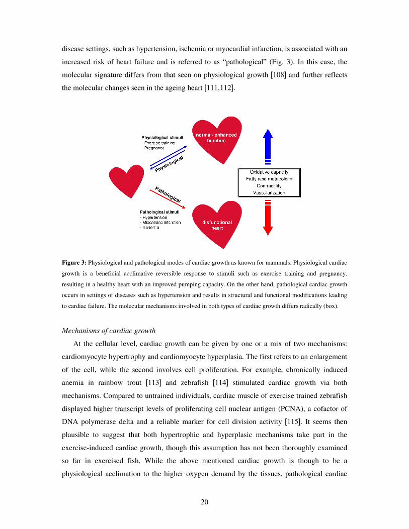
R) [160]. This type of inflammation is strongly associated with ageing, smoking and obesity,
and with the occurrence of cardiovascular failures, diabetes type 2 and muscle wasting.
Exercise training would reduce systemic low-level inflammation via production and release of
IL6 from the contracting muscles [161]. IL6 produced in this way appears to orchestrate the
formation of an anti-inflammatory environment by increasing the plasmatic levels of the anti-
inflammatory IL1ra, IL10 and sTNF-R at the same time of inhibiting the production of TNFα
[162–164]. IL6 also drives some of the metabolic effects of exercise training, as it will
activate muscle AMPK through the gp130/IL6Rα receptor, stimulating fatty acid oxidation
and glucose uptake. At the systemic level, IL6 stimulates fatty acid oxidation resulting in
visceral fat reduction [165] with the consequent reduction in visceral’s fat production of
TNFα [166].
28
Overall, engaging in physical activity seems to enhance the immune system and induce an
anti-inflammatory state coupled to a fatty acid oxidation dominant environment. All of the
above would be directly implicated in conferring the exercise-induced benefits in terms of
resistance to infections as well as in lower prevalence of lifestyle diseases. Nevertheless, none
of these effects have been so far examined in fish. Furthermore, and in addition to above
mentioned immune and anti-inflammatory potential effects exercise training may have on
fish, other mechanisms affected by exercise further argues for an improved robustness and
overall health status. Specially, an improved cardiac performance with an overall higher
oxygen distribution capacity (see section 3.3.2.2) is highly suggestive of an improved
resistance to metabolic and life-style associated diseases as seen for humans.
4.3.4 Inherent individual variability in robustness
An important factor affecting the robustness level of a fish population is the high inherent
or naturally occurring individual variation in physiological performance and responses to
environmental challenges [167–169] For example, Claireaux et al. [168] found that the
inherent differences in swimming capacity of juvenile rainbow trout (Oncorhynchus mykiss)
were maintained nine months later as reflected by differences in several cardiovascular and
performance parameters. In humans there is a significant individual variation in the
responsiveness to exercise training which can range from no gain to up to 100% improvement
in cardiorespiratory fitness. The level of pre-training and the heritability effect are the main
factors explaining these response [170,171].
A difficulty with interventional experiments like exercise training is the degree of inherent
individual diversity that exists in physiological performance traits, which can often be greater
than the change elicited by the experimental intervention. Hence such inherent trait variations
may be large enough to mask the actual effects of training. Further, it may be important to
identify the set of individuals in which exercise training has the potential to cause significant
changes. The latter would be of particular importance in an aquaculture facility where
minimizing the costs of the procedure is always desirable.
29
5 Aims of the study
Principal aim
Evaluate the potential of exercise training as a measure to improve robustness of farmed
Atlantic salmon smolts.
Specific aims
1- Evaluate the effects of exercise training on the resistance to infectious diseases.
2- Assess the impact of inherent swimming capacities on disease resistance, and the
interaction of these with exercise training.
3- Characterize the exercise-induced cardiac molecular signature underlying a robust heart.
4- Evaluate the effects of exercise training on growth.
30
6 Results and discussion
The effects of exercise training on robustness were investigated through a series of two
experiments in which Atlantic salmon pre-smolts were trained at different intensities (water
velocities), durations and modalities (continuous vs. interval training).
The overall effects of exercise training on robustness were assessed by measuring disease
resistance, the cardiac molecular acclimation response as well as growth performance. Growth
and disease resistance are the two most relevant operational parameters in the fish farming
industry. Linked to this, an improved cardiac capacity would further argue for higher
robustness given not only the heart’s role in efficiently pumping oxygen and nutrients through
the system, but also due to its potential association with reduced levels of life-style diseases as
seen in mammals.
6.1 Disease resistance
Given the widely known health effects exercise training produces in mammals as well as
the importance of improving health conditions in farmed fish, the potential effects of exercise
training on disease resistance were assessed for Atlantic salmon, hypothesizing a similar
beneficial response as in higher vertebrates. To evaluate disease resistance, trained fish were
transferred to seawater and challenged by co-habitation with IPNV infected fish to simulate a
natural-like infection transmission mechanism. IPN was the selected model given its position
as a principal viral disease in salmon aquaculture, especially affecting smolts after sea water
transfer [4].
6.1.1 Exercise effects on disease resistance
A striking result of this thesis was the finding of exercise training conferring higher
disease resistance capabilities to Atlantic salmon smolts. This was reflected by trained fish
displaying enhanced survival to IPN in comparison to untrained fish (paper 1) or fish trained
at sub-optimal regimes (paper 2). Such an effect was found to be highly dependent on the
intensity and modality of the regime, with higher survival tending to occur in response to
moderate training intensities.
31
In humans, the benefits gain of going from no exercise to moderate exercise are
potentially much larger than when just increasing the intensity of a previous exercise regime
[172]. Due to this, in trial I (paper 1) the main objective was to asses the sole effect of
exercise training the fish in comparison to individuals being held in, practically, standing
waters (control). In that trial, fish were trained for six weeks and then allowed a further six
weeks to detrain and smoltify at control water velocity. Two modes of training were
performed and compared against the control group. Intriguingly, improved survival was found
for fish being trained at an interval regime with an average water velocity of 0.85 bls-1 and
0.25-fold daily changes in water velocity (0.8 + 1 bls-1) compared to fish trained at a similar
average but constant velocity and to untrained fish. In good agreement with this, another trial
performed by our research group (unpublished results) showed that higher survival (~20%) to
a natural outbreak of winter ulcer was displayed by Atlantic salmon previously trained for
nine weeks at a similar interval regime (0.8 + 1.2 bls-1) as in trial I. In that case, survival was
higher compared to a continuous velocity regime (1 bls-1) and to the control group (0.5 bls-1).
With the knowledge gained in trial I, trial II (paper 2) was designed as to expand the
duration and the range of the exercise intensities. Two continuous (0.65 and 1.31 bls-1) and
two interval (0.3 + 1.31 bls-1; differing between them on the quantity of velocity changes
during a day) training regimes were performed for ten weeks, and disease resistance (IPN)
was compared against a control group trained at 0.3 bls-1 continuously. This control was
selected, instead of still water as in trial I, as to mimic a currently common fish farming
condition, hence allowing for the assessment of the potential impact that exercise could have
on the industry. Importantly, the best survival of this trial was achieved by fish being trained
at constant water velocity averaging 0.65 bls-1 throughout the trial, while the second best
group was trained constantly at 1.31 bls-1. The interval regimes with large magnitude fold
changes (3-fold) resulted in the worst survival even though their average water speeds were
also 0.65 bls-1.
Beside the different control group velocities as well as the training regimes used, trials I
and II further differed in the duration of the exercise stimulus (6 and 10 weeks, respectively)
and of the detraining period, i.e. time between end of the training regimes and beginning of
disease challenge test in sea water (6.5 and 2 week, respectively). Among other implications,
this means that fish from trial II were exercise trained during most of the smoltification
period, and it is currently unknown how this may affect the disease resistance performance of
fish after sea transfer.
32
It can be concluded that optimal exercise training of Atlantic salmon pre-smolts in the
fresh water rearing stage resulted in improved robustness reflected by a higher resistance to
infectious diseases after sea water transfer. The best results were obtained when interval
exercise was performed with mild daily changes in water velocity (<0.5-fold) with an average
velocity around 1 bls-1. On the contrary, the relatively large changes in water velocity (trial II)
appeared to worsen the disease resistance performance, probably due to higher stress levels
resulting in a poor capacity of the fish to reach allostasis. Future research should then include
trials focused on fine-tuning the training intensities, as well as in determining optimal
durations for both training and detraining periods. Importantly, such an optimal regime must
meet other requirements from the industry beside disease resistance, such as growth, overall
fitness and its technical applicability.
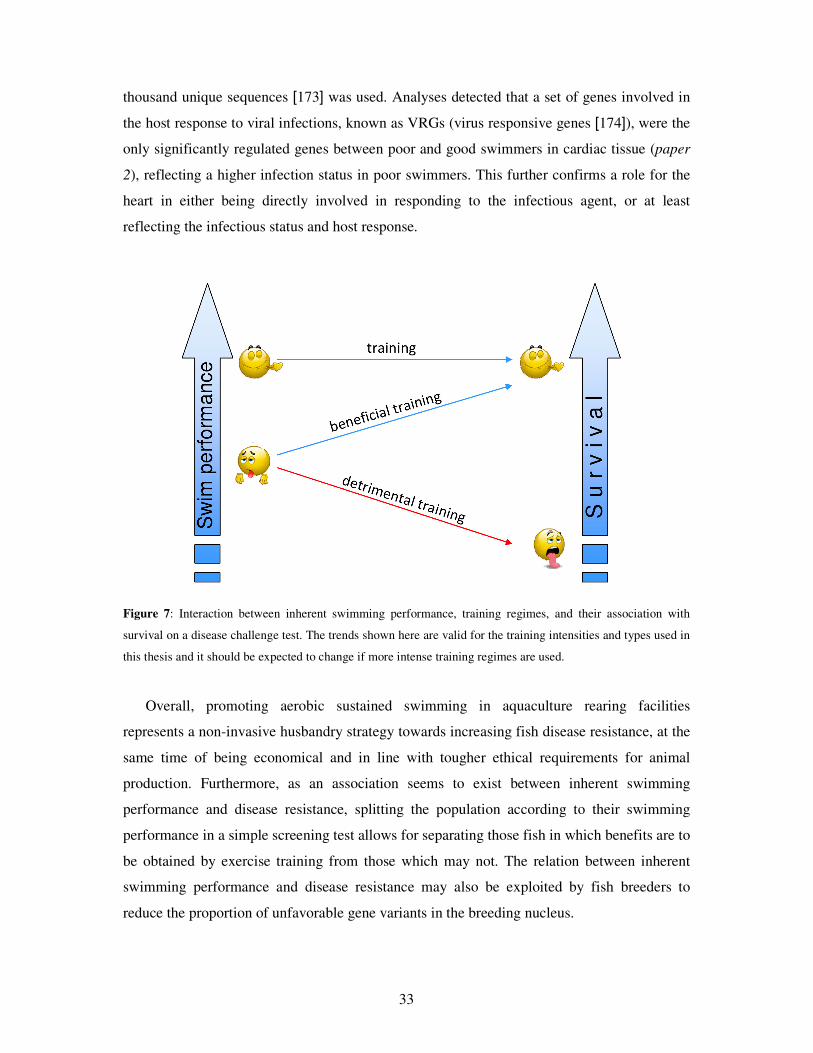
6.1.2 Disease resistance is dependent on inherent swimming capacity
In humans, exercise training differently affects individuals with different fitness
backgrounds given by pre-training fitness status and/or heritability [171]. Since fish have
shown to posses a wide degree of inherent swimming capacity which is related to their
cardiovascular performance [168], we evaluated if the effects of exercise training in disease
resistance were population-wide, or only a part of the population (i.e. either the inherently
poor or good swimmers) would result more benefited than the other.
Strikingly, fish that were categorized as good swimmers before commencement of
exercise training (paper 2) displayed a significantly higher resistance to IPN than those
initially categorized as poor swimmers. Another finding was that, though the inherent
swimming capacity appeared to predict disease resistance after sea transfer, exercise training
was sufficient to modify this (paper 2). While exercise training was not found to have a
considerable effect on disease resistance in the inherently good swimmers, disease resistance
of the inherently poor swimmers appeared to be highly affected by the exercise stimulus (Fig.
7). This was demonstrated by poor swimmers responding positively to an optimal training
regime and negatively to a non-optimal regime. This further suggests that the overall disease
resistance differences found between the different training regimes was mainly given by an
exercise training effect upon the inherently poor swimmers.
To search for gene expression correlates between inherent swimming capacity and disease
resistance at the end of the disease challenge test, a microarray platform containing 21
33
thousand unique sequences [173] was used. Analyses detected that a set of genes involved in
the host response to viral infections, known as VRGs (virus responsive genes [174]), were the
only significantly regulated genes between poor and good swimmers in cardiac tissue (paper
2), reflecting a higher infection status in poor swimmers. This further confirms a role for the
heart in either being directly involved in responding to the infectious agent, or at least
reflecting the infectious status and host response.
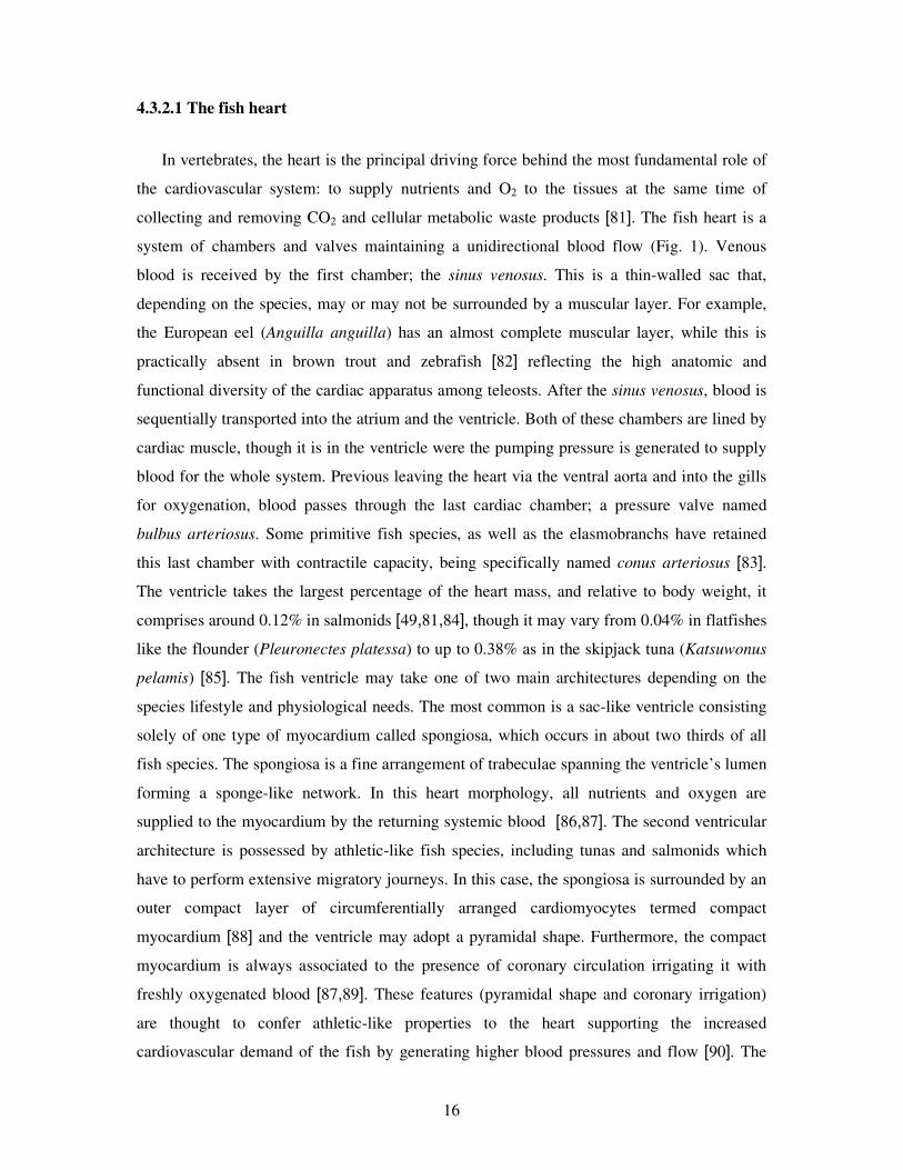
Figure 7: Interaction between inherent swimming performance, training regimes, and their association with
survival on a disease challenge test. The trends shown here are valid for the training intensities and types used in
this thesis and it should be expected to change if more intense training regimes are used.
Overall, promoting aerobic sustained swimming in aquaculture rearing facilities
represents a non-invasive husbandry strategy towards increasing fish disease resistance, at the
same time of being economical and in line with tougher ethical requirements for animal
production. Furthermore, as an association seems to exist between inherent swimming
performance and disease resistance, splitting the population according to their swimming
performance in a simple screening test allows for separating those fish in which benefits are to
be obtained by exercise training from those which may not. The relation between inherent
swimming performance and disease resistance may also be exploited by fish breeders to
reduce the proportion of unfavorable gene variants in the breeding nucleus.
34
6.2 Cardiac molecular acclimation response
Given that exercise training promoted higher disease resistance and that training
strengthens cardiac performance [80], the cardiac molecular acclimation response was
analyzed to gain a deeper insight into the driving mechanisms behind such beneficial
exercise-induced effects. In the first trial (paper 1), emphasis was set on finding sets of genes
whose regulation in response to training would correlate with the observed differences in
disease resistance seen after sea transfer. This was done by using real-time quantitative RT-
PCR (qPCR) as well as a microarray platform specially designed for studies involving the
immune response (SFA 2.0 immunochip [175]). In the second trial (paper 3), several key
cellular mechanisms known to participate in the physiological cardiac enlargement response
to exercise, as seen in mammals, were studied. In this case, only those training regimes
consisting on continuous water velocities were selected to further assess the effect of the
intensity, or cardiac workload, on the given changes.
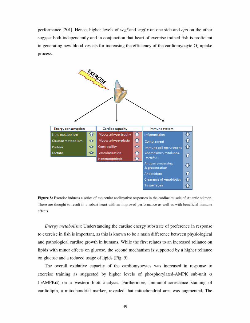
6.2.1 Effects on cardiac immune-related mechanisms
The emphasis on analyzing the cardiac molecular response was not only because of the
heart’s central position as a main exercise target organ, but also because of the fish heart
being an immune-relevant organ where many viral pathogens manifest themselves [175–178].
Hence, we looked for those underlying transcriptional changes which occurring in association
with disease resistance could explain the acclimation response to the exercise stimulus.
In trial I (paper 1) we found a significant modulation in the cardiac transcription of genes
directly involved in promoting protection against infectious agents as well as in conferring
tissue protection against both endogenous and exogenous toxic compounds. Among these,
down-regulation in the expression of genes involved in the inflammatory response was of
special interest given the known anti-inflammatory effects of training in mammals [155]. Both
continuous and interval training showed significantly lower transcript levels of tnfα and il1β
when compared to the untrained control group as evidenced by qPCR analyzes at the end of a
six week detraining period. Nevertheless, only those fish belonging to the interval training
regime showed further down-regulation of genes participating in other inflammatory
pathways, especially those related to the production of eicosanoids (microarray data). This
provided valuable information, as interval training showed higher disease resistance than the
35
continuous training regime. Expression of several other immune relevant groups of genes
demonstrated to correlate with disease resistance, including components of the complement
system and other immune effector molecules, cell adhesion and recruitment, chemokines and
cytokines (Fig. 8). The expression of genes not directly linked with the immune system was
also associated with higher survival. These included genes involved in protection against
different tissue damaging products such as oxidants and xenobiotics, as well as mRNA of
genes coding for tissue reparation and differentiation.
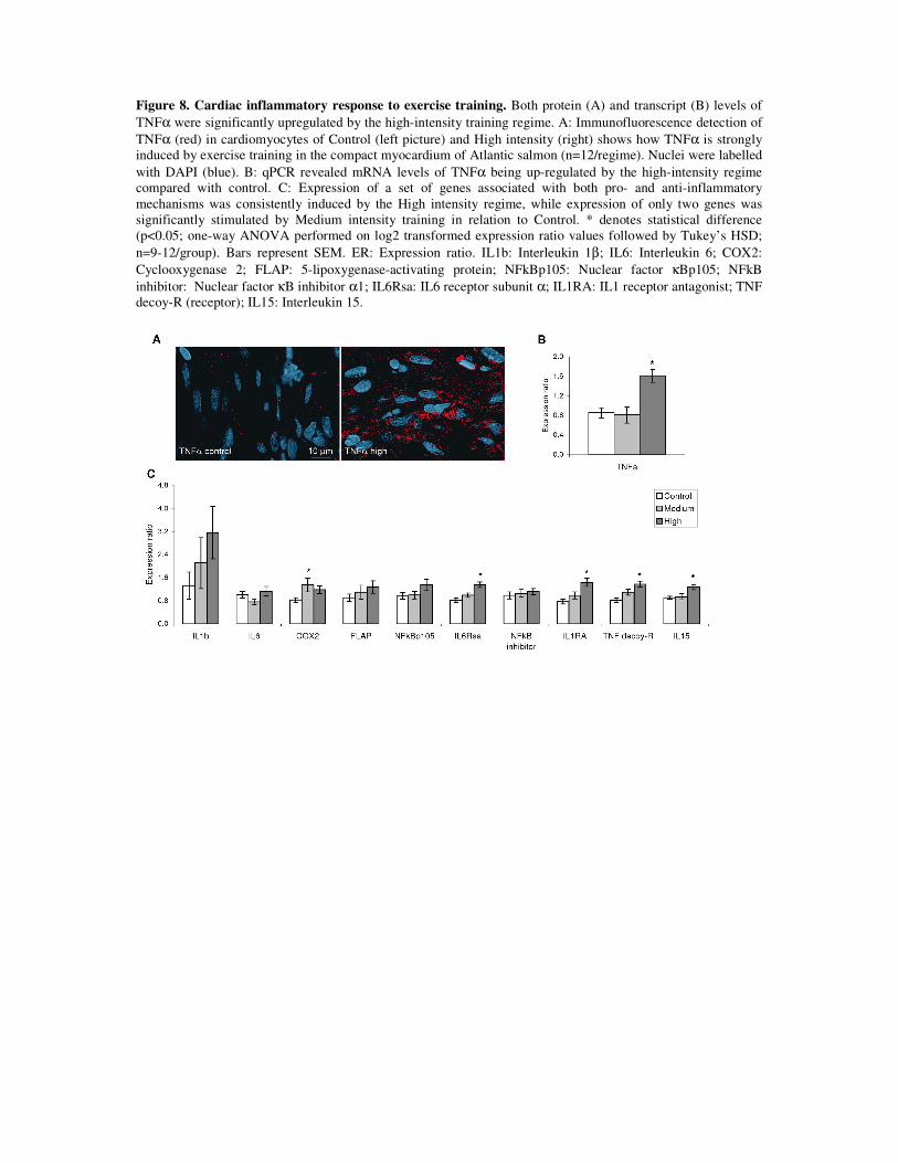
In paper 3, a significant up-regulation of TNFα was found at both the protein and mRNA
level when analyzed immediately at the end of the training period for the highest intensity
regime (1.3 bls-1) compared to the control. This was accompanied by higher mRNA levels of
proteins involved in both pro- and anti-inflammatory mechanisms such as cyclooxygenase2
(COX2), IL1ra and sTNF-R, suggesting a balanced cardiac inflammatory environment. Thus,
on the contrary to the moderate training regimes in trial I and the 0.65 bls-1 regime in trial II,
the higher intensity training of 1.3 bls-1 was sufficient to promote a significant pro- and anti-
inflammatory response in the heart at end of training. Still, due to the lowered expression of
inflammatory genes after six weeks of detraining in trial I, the results suggest that training
may have long term effects on the cardiac inflammatory status during detraining even without
a strong inflammatory response during training. Unfortunately, the cardiac inflammatory
status was not examined at the very beginning of the IPN challenge tests, which could have
revealed potential detraining effects also for the high intensity fish in trial II. The significant
effect of detraining on cardiac immune status may be important for future rearing strategies of
fish before the severe stressful sea transfer of smolts. Another intervening factor when
comparing trial I and II results was the difference in treatment of controls. Holding the control
fish in standing waters as in trial I may result in higher stress levels due to the formation of
social hierarchies and increased levels of aggression [7], potentially leading to higher
inflammation. In the contrary, the control group in trial II was continuously trained at a low
speed, but it may have been sufficient for dampening the above mentioned social effect. Thus,
to clarify the individual importance of factors as intensity, duration, modality and recovery of
training programs these must be isolated and tested independently.
Overall, exercise training modulates the cardiac expression of inflammation-related
molecules. Lower levels of these appear to positively associate with higher disease resistance
when further linked to transcription modulation of participants involved in other immune and
tissue protective mechanisms. Although the mechanisms are far from being acknowledged in
fish, it is possible to argue for the existence of a similar anti-inflammatory effect of exercise
36
training among vertebrates, which might be the driver of several of the long-term exercise-
induced health benefits as has been found for humans [161].
Future studies should include the immune response in other tissues, such as head kidney
and spleen, as these are the fish’s primary immune organs.
6.2.2 Effects on cardiac performance-related mechanisms
Given the well-known effects of exercise training in cardiac performance of salmonids
[25,43,49,101], one of the aims of this thesis was to characterize the underlying molecular
signature driving the exercise-induced cardiac acclimation. Further, the existence of an
intensity (cardiac workload) factor in conferring such effects was addressed by comparing
three regimes of similar style (continuous velocity), but of increasing intensity including the
low (control), medium and high intensity groups (0.32, 0.65 and 1.31 bls-1 , respectively). For
both RVM and gene expression, the effects displayed intensity dependence, with the higher
intensity regime inducing larger changes than the medium regime, compared to the controls.
A typical acclimation response to exercise training in mammals and fish, is an increment
in RVM. Larger hearts promote an improved cardiac output (stroke volume x heart rate),
facilitating the delivery of oxygen and nutrients to the contracting skeletal muscles. In this
thesis, we report that higher RVM was seen in response to exercise training under some
circumstances (paper 3) but not others (paper 1). A plausible explanation for this difference is
that in paper 1 fish were exercise trained for 6 weeks, while the duration was extended to 10
weeks in paper 3, allowing more time for acclimation changes to be seen. Furthermore, in
paper 3 the intensity of the group presenting higher RVM (1.3 bls-1) compared to the controls
was higher than that used in paper 1 (~0.85 bls-1). Thus, it could be suggested that increases
in RVM in response to exercise training are dependent on both duration and intensity of the
training regime. This is in line with previous studies of salmonids showing that longer and
more intense regimes result in higher RVM than shorter and less intense ones
[36,39,43,49,79,100]. Similarly, the 20% increase in RVM seen in paper 3 falls within the
range previously found for exercised salmonids. In fish, increments in RVM are seen in
response to different environmental and physiological factors involved in increasing the
cardiac functional demand, including acclimation to low temperatures [78,92,179–181],
induced chronic anemia [113,114,182] and sexual maturation [93,183,184].
37
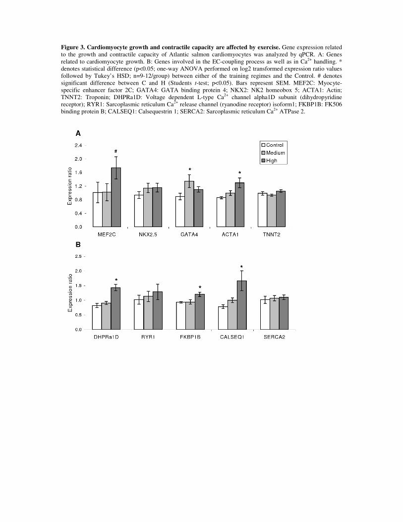
The expression of 38 molecular markers involved in mechanisms known to be associated
with physiological-beneficial cardiac growth in mammals was analyzed in paper 3. Exercise-
induced modulation of these markers reflected the molecular signature underlying cardiac
growth and, potentially, the exercise-induced cardiovascular effects in fish. Markers were
related to growth, cardiomyocyte contractility, blood supply (capillarization and
erythropoiesis) and energy metabolism.
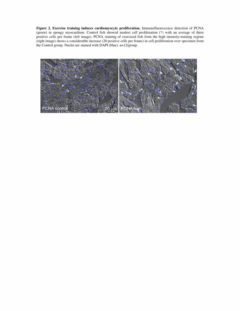
Cardiac growth: Since exercise training results in cardiac enlargement, the cellular
mechanisms potentially involved in such a response were examined. For this, protein and
gene expression levels of both cardiomyocyte hyperplasia and hypertrophy were analyzed.
The training regime of higher intensity (1.3 bls-1) showed to have elevated protein levels of
PCNA compared to control fish, which is strongly suggestive of cell division and, thus,
cardiomyocyte hyperplasia in response to training. Furthermore, increased expression of the
transcription factors mef2c and gata4 was suggestive of cardiomyocyte hypertrophy, as these
are known to govern the cardiac growth response to external stimuli in mammals [185].
Ideally this suggestion should be confirmed by direct cardiomyocyte size measurements, but
that is difficult in the intricate cardiac tissue. While for mammals cardiac growth is given
almost uniquely by cardiomyocyte hypertrophy after birth, the heart of fish seems to maintain
both types of cardiac growth mechanisms throughout life facilitating the remodelling process
in response to physiological cardiac growth inducers.
Contractility: An improvement in the pumping performance of the cardiac muscle is a key
feature for several of the beneficial cardiovascular effects elicited by training, including
cardiac output. Higher levels of molecules participating in the E-C coupling process reflect an
improved contractile capacity in the skeletal muscles of fish and in the skeletal and cardiac
muscles of mammals. This appears to be valid for both between and within vertebrate species
[105,106,117–121,125,186,187]. For example, the skeletal muscle of reared Atlantic salmon
possesses significantly lower levels of DHPR and RyR than its wild counterpart, reflecting the
higher swimming capacity of the latter [30]. Because of this, analyzing the expression levels
of such markers in the heart of exercise trained Atlantic salmon allowed us to indirectly assess
its contractile capacity. Results demonstrated that exercise training affected the mRNA
amount of proteins involved in the E-C coupling process, as suggested by significant
transcription up-regulation of dhpr, fkbp1b and calsequestrin1. DHPR is a voltage dependent
L-type Ca2+ channel present in the sarcolemmal membrane. FKBP1B and Calsequestrin1 are
present in the sarcoplasmic reticulum (SR) and modulate the release and recycling of Ca2+ in
association with RyR.
38
The rate of contraction-relaxation is regulated by Ca2+ cycling into and out of the
cardiomyocyte cytoplasm. In mammals and other higher vertebrates the SR functions as an
intracellular Ca2+ store, reducing the diffusional distances for Ca2+ movement and increasing
the efficiency of the system [188]. Not surprisingly, cardiomyocyte contraction in species
displaying high cardiac and swimming performance, such as the yellowfin and skipjack tunas,
do appear to rely on SR stored Ca2+ for contraction [189,190]. Other athletic-like fish species,
such as rainbow trout and mackerel (Scomber japonicus) have been also found to depend (to a
certain degree) on SR Ca2+ release for contraction, as suggested by a decreased force
production on cardiac strips whose RyR channels have been blocked with ryanodine
[78,191,192]. Altogether, it is plausible to argue that exercise training results in a better
cardiac contractile capacity in Atlantic salmon. An improved myocardium performance is
further suggested by an exercise-induced higher involvement of the SR Ca2+ stores in the
contractile process.
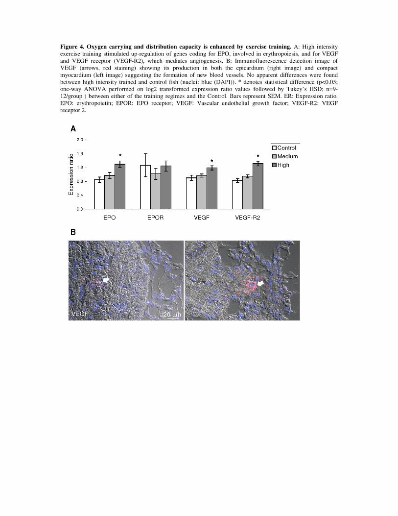
Blood supply: As mentioned in section 3.3.2.1, a specialized coronary irrigation system
delivering freshly oxygenated blood to the compact myocardium layer results fundamental for
its proper function in times of high cardiovascular demand. In the current work, exercise
training induced the cardiac transcription of genes with fundamental roles in oxygen
convection, such as vascular endothelial growth factor (vegf), its receptor (vegf-r) and
erythropoietin (epo). VEGF is an endothelial cell mitogenic factor and probably the most
important angiogenic factor in vertebrates [193]. In mammals, exercise training increases
coronary vascular supply in the healthy, the ageing and the recovering heart [194–198]. This
effect has been directly linked to higher gene and protein expression levels of VEGF, its
cardiac receptors, as well as the phosphorylation levels of VEGF’s downstream mediators
[199].
EPO is the main regulator of red blood cell production; hence, it is involved in
maintaining an adequate oxygen supply to the tissues. Interestingly, while kidney is the
primary EPO producing organ in humans, this role is appears to be played by the heart in fish
[200]. Since the spongy myocardium layer receives oxygen depleted blood returning from the
systemic circulation, it is perfectly arguable for a role of this in sensing O2 levels and
mounting a response in the form of EPO production and release. While EPO has been also
found to be produced in the heart of juvenile zebrafish, its transcript levels were not modified
by exercise training [64], contrary to what was found in this study.
Interestingly, EPO signaling through EPO receptor induces VEGF production and
myocardial endothelial proliferation in the mammalian heart resulting in improved cardiac
39
performance [201]. Hence, higher levels of vegf and vegf-r on one side and epo on the other
suggest both independently and in conjunction that heart of exercise trained fish is proficient
in generating new blood vessels for increasing the efficiency of the cardiomyocyte O2 uptake
process.
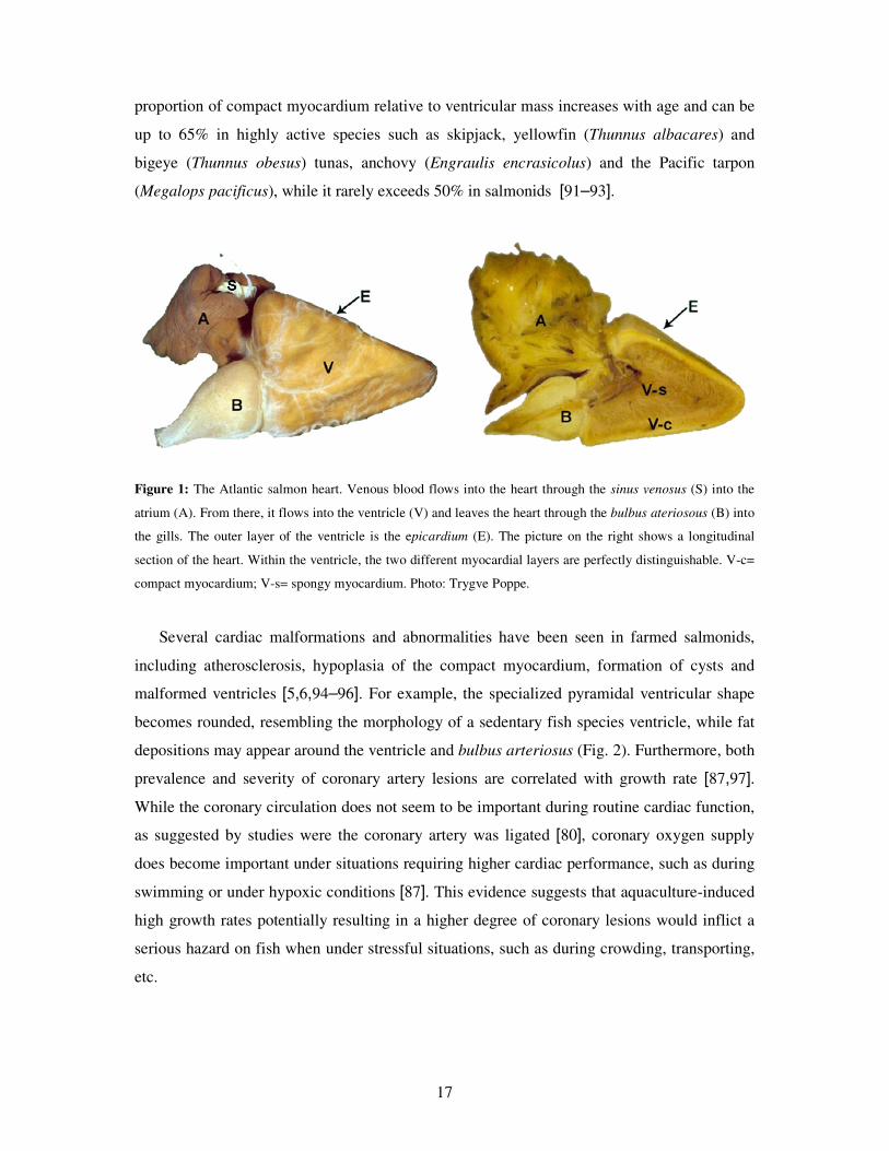
Figure 8: Exercise induces a series of molecular acclimative responses in the cardiac muscle of Atlantic salmon.
These are thought to result in a robust heart with an improved performance as well as with beneficial immune
effects.
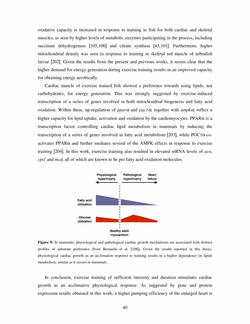
Energy metabolism: Understanding the cardiac energy substrate of preference in response
to exercise in fish is important, as this is known to be a main difference between physiological
and pathological cardiac growth in humans. While the first relates to an increased reliance on
lipids with minor effects on glucose, the second mechanism is supported by a higher reliance
on glucose and a reduced usage of lipids (Fig. 9).
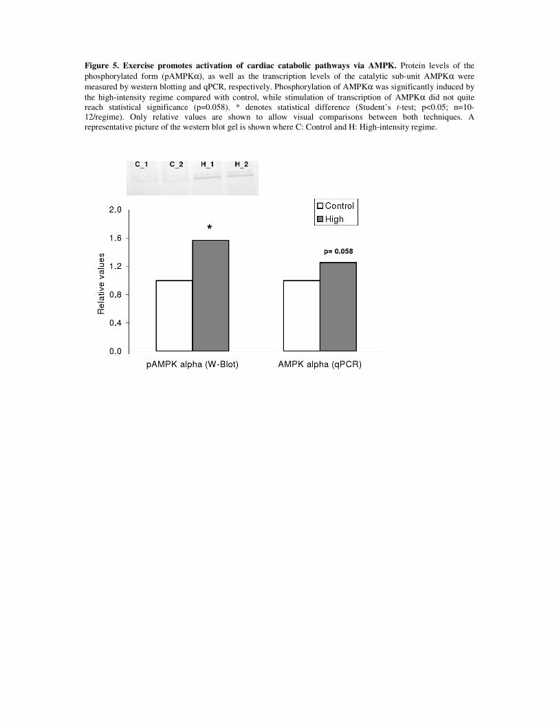
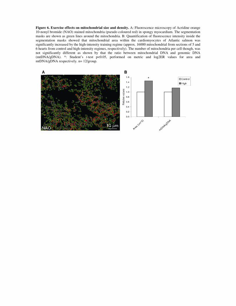
The overall oxidative capacity of the cardiomyocytes was increased in response to
exercise training as suggested by higher levels of phosphorylated-AMPK sub-unit α
(pAMPKα) on a western blott analysis. Furthermore, immunofluorescence staining of
cardiolipin, a mitochondrial marker, revealed that mitochondrial area was augmented. The
40
oxidative capacity is increased in response to training in fish for both cardiac and skeletal
muscles, as seen by higher levels of metabolic enzymes participating in the process, including
succinate dehydrogenase [105,106] and citrate synthase [43,101]. Furthermore, higher
mitochondrial density was seen in response to training in skeletal red muscle of zebrafish
larvae [202]. Given the results from the present and previous works, it seems clear that the
higher demand for energy generation during exercise training results in an improved capacity
for obtaining energy aerobically.
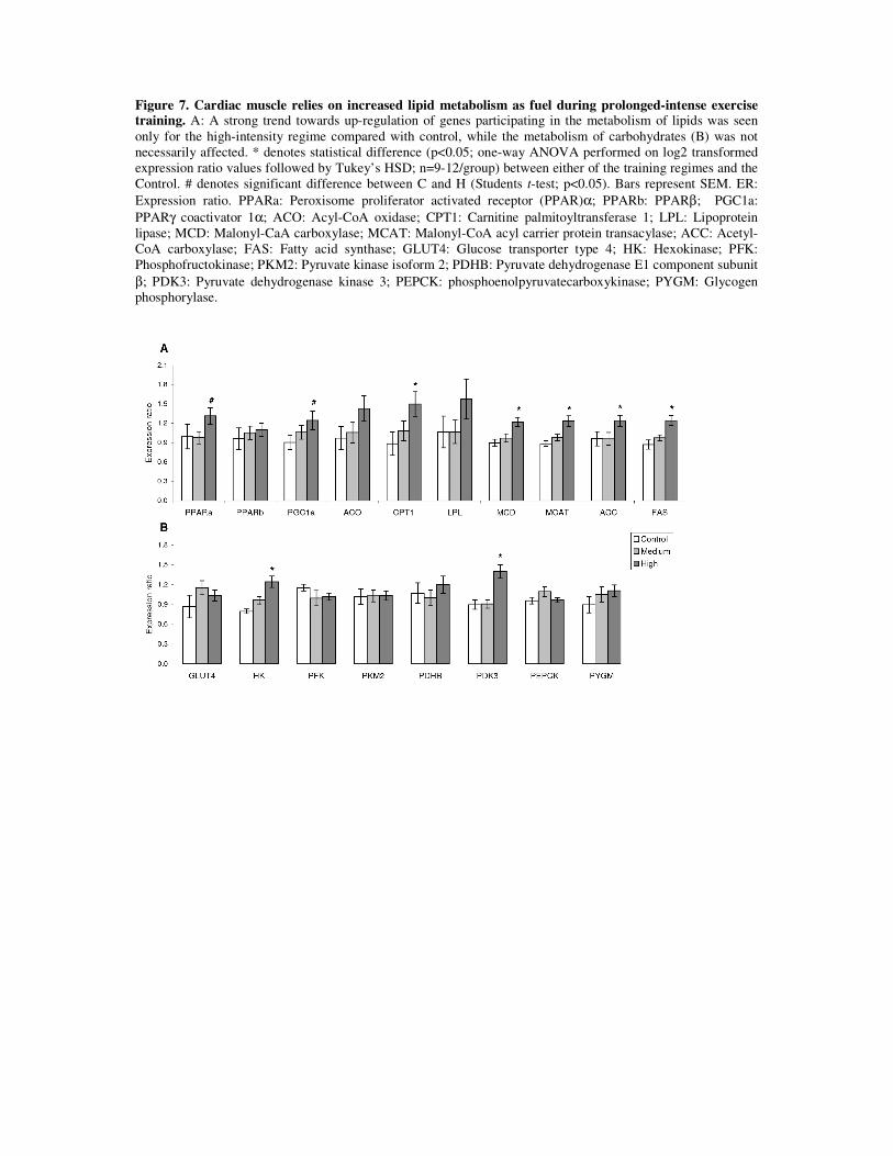
Cardiac muscle of exercise trained fish showed a preference towards using lipids, not
carbohydrates, for energy generation. This was strongly suggested by exercise-induced
transcription of a series of genes involved in both mitochondrial biogenesis and fatty acid
oxidation. Within these, up-regulation of pparα and pgc1α, together with ampkα, reflect a
higher capacity for lipid uptake, activation and oxidation by the cardiomyocytes. PPARα is a
transcription factor controlling cardiac lipid metabolism in mammals by inducing the
transcription of a series of genes involved in fatty acid metabolism [203], while PGC1α co-
activates PPARα and further mediates several of the AMPK effects in response to exercise
training [204]. In this work, exercise training also resulted in elevated mRNA levels of aco,
cpt1 and mcd, all of which are known to be pro fatty acid oxidation molecules.
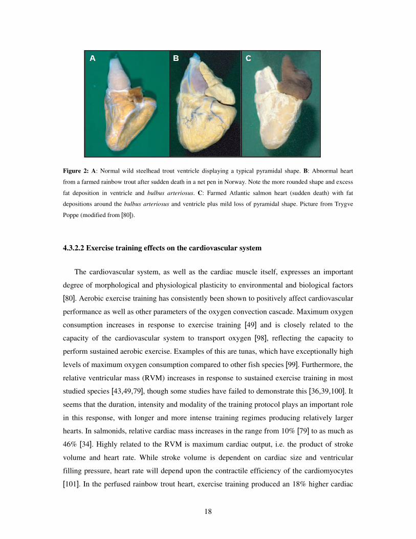
Figure 9: In mammals, physiological and pathological cardiac growth mechanisms are associated with distinct
profiles of substrate preference (from Bernardo et al. [108]). Given the results reported in this thesis,
physiological cardiac growth as an acclimation response to training results in a higher dependence on lipids
metabolism, similar as it occurs in mammals.
In conclusion, exercise training of sufficient intensity and duration stimulates cardiac
growth as an acclimative physiological response. As suggested by gene and protein
expression results obtained in this work, a higher pumping efficiency of the enlarged heart is
41
supported by an improved capacity for contractility as well as for oxygen supply to the
compact myocardium. The increased requirement of energy is satisfied by a higher reliance
on lipids over carbohydrates. All of these changes resemble the molecular signature behind
the larger and strengthened “athlete’s heart” of exercise trained humans and have, then, the
potential to be employed as molecular markers of cardiac status in fish.
6.3 Growth
Achieving optimum growth is probably the single most important parameter involved in
defining production strategies in the industry, at the same time of being a central feature of a
robust fish.
Increased growth is a well known effect of exercise training in salmonids, especially when
moderate swimming speeds between 0.5 and 1.5 bls-1 are promoted [7,25]. An increment in
somatic growth was a consistent result in these studies (papers 1 & 2). The effect was much
greater in paper 1, as gains in thermal growth coefficient (TGC) were in the range of 20% for
both trained groups compared to the untrained control fish. In paper 2 though, improved
growth was in the range of 6% for all groups, but only significant between low (control) and
highest intensity. The differences seen between the trials are most probably explained by the
velocity at which the control group was reared; the control group described in paper 1 was
held in water at velocities below 0.1 bls-1, while control group in paper 2 was held at 0.32 bls-
1. Thus, the magnitude of the growth effect would partly correspond to the intensity difference
between the control and target group.
Growth seen in the present study is comparable to that reported by Jørgensen and Jobling
[67] in Atlantic salmon, who found a 15% higher growth rate in pre-smolts trained at 1 bls-1
compared to control fish reared at 0.3 bls-1. Nevertheless, increments in growth has been
found to be as high as 38% [40] in Atlantic salmon and 76% [33] in brown trout. Different
explanations may be found for such large increments in growth in comparison to those
reported in this thesis. The work by Totland et al. [40] used fish averaging 2 kg and training
lasted for a long period (8 months). Further, trained and control fish were reared in different
facilities (trained on raceways and control on a standard cages with water velocities below 0.1
bls-1). In the work by Davison and Goldspink [33], though it lasted one month only, control
(static water) and trained fish were also reared differently and control fish did hardly grow at
all during the experiment (3% compared to 79% of fish trained at 1.5 bls-1). It is valid to argue
42
that the fish strains used in those studies were probably not as adapted as the current ones to
captivity due to increments in stress tolerance and the strong selective breeding for growth
performance has likely exploited much of the total growth capacity. The strong domestication
of Atlantic salmon may further be the main underlying reason explaining why we found
increased feed intake to be the key explanation for training-induced growth, while older
reports have documented also strong positive effects on the feed conversion efficiency [7].
Still, in paper 2 we found improved feed efficiency for fish trained continuously at moderate
intensity.
Altogether, this may suggest that COT is at its lowest between ~0.6-1.2 bls-1 and that the
optimum swimming speed (Uopt) for Atlantic salmon pre-smolts would be found in this range,
in good agreement with previous results seen for salmonids [7,25].
43
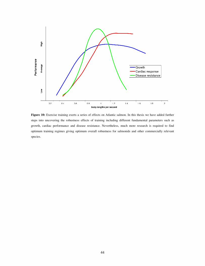
7 Conclusions
This thesis represents an important step forward into the acknowledgement of the
robustness effects that exercise training may exert upon farmed fish. Previous studies on
salmonids have found that intensities between 0.5 and 1.5 bls-1 appeared as optimal in terms
of growth and feeding efficiency, among others. Results from this thesis as well as from other
studies within our research group, have narrowed the intensity range which appears to be
optimal for overall robustness to be improved. Training Atlantic salmon pre-smolts around 1
bls-1 would result in sounder benefits including not only growth, but higher disease resistance
as well (Fig. 10). Further, training in intervals appeared as highly beneficial, but only if the
difference in magnitude between the high and low velocities is small. Large changes in
velocity probably cause stress with consequent deleterious effects on disease resistance.
Fish can be divided according to their inherent swimming (cardiovascular) capacity into
poor or good swimmers, which further associates with disease resistance. While good
swimmers performed better than poor irrespective of the exercise training regime used, poor
swimmers appeared to be more affected by training in either a positive or negative way. The
possibility to improve the disease resistance of unfit fish by optimal training programs is of
great interest for the aquaculture industry. Likewise, sorting fish based on swimming
performance can be utilized to improve the genetic material in breeding programs.
Several of the molecular mechanisms driving the cardiac acclimation response to exercise
training were uncovered. These included protective-related mechanisms (immune system,
inflammation, antioxidants and xenobiotics) as well as cardiac performance-related (growth,
contractility, vascularization and metabolism). Improved cardiac capacity as suggested by
molecular expression was seen in fish trained at high intensity (1.3 bls-1) when compared to
the low intensity group (0.3 bls-1). As fish trained in medium intensity regime (0.65 bls-1)
already showed signs of higher cardiac capacity than the low intensity group, it remains
unknown which is the intensity threshold for such effects to become evident.
Exercise training was proficient in promoting growth. The range at which this was found
goes from 0.65 to 1.31 bls-1, though relative differences appeared to be mostly dependent on
the control group training level.
Overall, optimal exercise training improves robustness of Atlantic salmon pre-smolts,
including better disease resistance, a strengthening of the cardiovascular system and a better
growth, all of which are though to be highly interesting in a fish farming industry context.
44
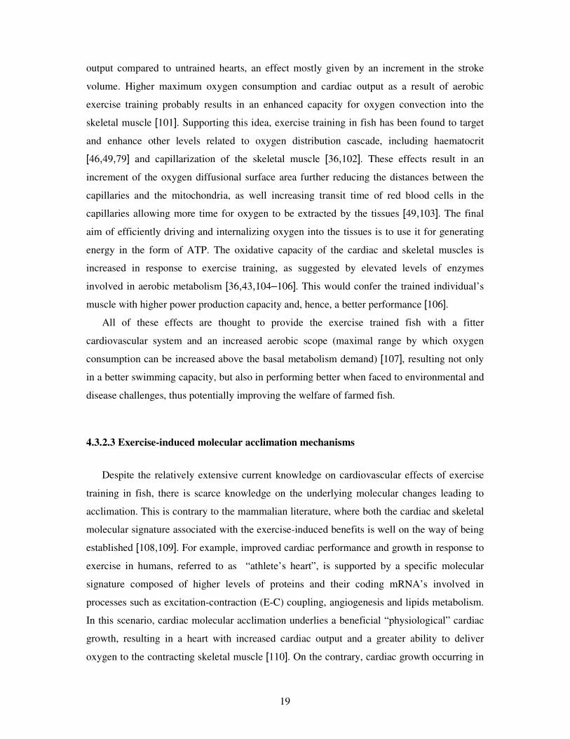
Figure 10: Exercise training exerts a series of effects on Atlantic salmon. In this thesis we have added further
steps into uncovering the robustness effects of training including different fundamental parameters such as
growth, cardiac performance and disease resistance. Nevertheless, much more research is required to find
optimum training regimes giving optimum overall robustness for salmonids and other commercially relevant
species.
45
8 Future perspectives
Even though the new knowledge generated with this thesis sets a promising future for
employing exercise training as a measure to increase overall robustness of farmed Atlantic
salmon, there is still a large scope for improvement as well as a wide range of areas that
should be further investigated.
Analyzing the potential effects of training on the earlier life stages becomes relevant, as it
could generate robust fast-growing individuals from early-on. For example, exercise training
improved the efficiency of the swimming muscle of free-swimming larvae of zebrafish,
suggesting that muscle fibers were already sufficiently plastic at such an early stage [202].
Similarly, the effects of exercise training on post-smolts and up to slaughter size should be
further investigated. In the former case, the use of closed systems for raising salmon up to 1
kg size would result highly favorable if promoting optimal swimming activity is made
possible.
From the results in this thesis, the existence of a detraining period was found to be
important in the immune acclimative response of cardiac gene expression, which was further
positively associated to improved disease resistance. The duration of such a period as well as
the necessary detraining time required for the transcriptional immune changes to appear
should be then acknowledged.
Other disease models must be performed as to expand on the protective effects of
exercise. The viral agents of highest interest are those currently affecting farmed salmonids
such as CMS, HSMI, ISA and PD. While we have also seen a positive response of trained fish
against a bacterial infection (Moritella viscosa) causing a natural winter ulcer outbreak
(unpublished), controlled trials for this and other relevant bacteria may become more
informative. Exercise training effects upon the sea lice (Lepeophtheirus salmonis) is also an
interesting area to be explored.
In terms of the driving molecular acclimations behind exercise-induced disease resistance,
future research should include the response of other immune relevant tissues such as the
spleen and head kidney as to investigate potential shifts in the cellular immune response.
Similarly, studies should in general be broadened to include effects in peripheral organs as
skin and gills to gain insight into the system effects of exercise training. The interaction of
exercise training with other factors such as nutrition and breeding, as well as in the fish’s
response to regular aquacultural handling procedures should be further evaluated.
46
Finally, each research group working with exercise in fish species has, of course, its own
questions and ways to tackle these. Nevertheless, given the numerous ways in which fish can
be exercise trained (e.g. rearing facility type, exercise intensity, exercise type, fish size,
duration, etc.), it becomes hard to make reliable between-studies comparisons, hence making
it difficult to achieve consensuses. Thus, standardization of methods would be highly helpful,
especially when trying to answer similar questions as, for example, exercise effects on disease
resistance.
47
9 References
1. The World Commission on environment and Development, Our Common Future.
(Oxford University Press, 1987).
2. Statistics Norway 2012. http://www.ssb.no.
3. Johnson SC, Treasurer JW, Bravo S, Nagasawa K, Kabata Z (2004) A review of the impact of parasitic copepods on marine aquaculture. Zoological Studies 43: 229-243.
4. Robertsen B (2011) Can we get the upper hand on viral diseases in aquaculture of Atlantic salmon? Aquaculture Research 42: 125-131.
5. Poppe TT, Taksdal T (2000) Ventricular hypoplasia in farmed Atlantic salmon Salmo
salar. Diseases of Aquatic Organisms 42: 35-40.
6. Brocklebank J, Raverty S (2002) Sudden mortality caused by cardiac deformities following seining of preharvest farmed Atlantic salmon (Salmo salar) and by cardiomyopathy of postintraperitoneally vaccinated Atlantic salmon parr in British Columbia. Canadian Veterinary Journal-Revue Veterinaire Canadienne 43: 129-130.
7. Jobling M, Baardvik BM, Christiansen JS, Jorgensen EH (1993) The effects of prolonged exercise training on growth performance and production parameters in fish. Aquaculture International 1: 95-111.
8. Baeverfjord G, Lein I, Hjelde K, Takle H, Helland S (2009) Recommendations for malformation control in Atlantic salmon juveniles. In: Baeverfjord G, Helland S, Houfg C, editors. Control of malformations in fish aquaculture; Science and practice. Luxembourg: Rapid Press.
9. Overland M, Sorensen M, Storebakken T, Penn M, Krogdahl A, Skrede A (2009) Pea protein concentrate substituting fish meal or soybean meal in diets for Atlantic salmon (Salmo salar)-Effect on growth performance, nutrient digestibility, carcass composition, gut health, and physical feed quality. Aquaculture 288: 305-311.
10. Gjedrem T (2000) Genetic improvement of cold-water fish species. Aquaculture Research 31: 25-33.
11. Grammes F, Rorvik KA, Takle H (2012) Tetradecylthioacetic acid modulates cardiac transcription in Atlantic salmon, Salmo salar L., suffering heart and skeletal muscle inflammation. Journal of Fish Diseases 35: 109-117.
12. Midtlyng PJ, Reitan LJ, Lillehaug A, Ramstad A (1996) Protection, immune responses and side effects in Atlantic salmon (Salmo salar L) vaccinated against furunculosis by different procedures. Fish & Shellfish Immunology 6: 599-613.
48
13. Lillehaug A, Lunestad BT, Grave K (2003) Epidemiology of bacterial diseases in Norwegian aquaculture - a description based on antibiotic prescription data for the ten-year period 1991 to 2000. Diseases of Aquatic Organisms 53: 115-125.
14. Markestad A, Grave K (1997) Reduction of antibacterial drug use in Norwegian fish farming due to vaccination. Fish Vaccinology 90: 365-369.
15. Håstein T, Gudding R, Evensen O (2005) Bacterial vaccines for fish an update of the current situation worldwide. Developmental Biology 121: 55-74.
16. Gudmundsdottir BK, Bjornsdottir B (2007) Vaccination against atypical furunculosis and winter ulcer disease of fish. Vaccine 25: 5512-5523.
17. Bjorge MH, Nordgreen J, Janczak AM, Poppe T, Ranheim B, Horsberg TE (2011) Behavioural changes following intraperitoneal vaccination in Atlantic salmon (Salmo salar). Applied Animal Behaviour Science 133: 127-135.
18. Sorum U, Damsgard B (2004) Effects of anaesthetisation and vaccination on feed intake and growth in Atlantic salmon (Salmo salar L.). Aquaculture 232: 333-341.
19. Poppe TT, Breck O (1997) Pathology of Atlantic salmon Salmo salar intraperitoneally immunized with oil-adjuvanted vaccine. A case report. Diseases of Aquatic Organisms 29: 219-226.
20. Aunsmo A, Larssen RB, Valle PS, Sandberg M, Evensen O, Midtlyng PJ, Ostvik A, Skjerve E (2008) Improved field trial methodology for quantifying vaccination side-effects in farmed Atlantic salmon (Salmo salar L.). Aquaculture 284: 19-24.
21. Aunsmo A, Guttvik A, Midtlyng PJ, Larssen RB, Evensen O, Skjerve E (2008) Association of spinal deformity and vaccine-induced abdominal lesions in harvest-sized Atlantic salmon, Salmo salar L. Journal of Fish Diseases 31: 515-524.
22. Børno G, Sviland C, Jensen BB, Tarpai A, Garseth ÅH, Skjelstad HR, Johansen R, Dale OB, Fritsvold C, Nilsen H, et al. (2009) The health situation in Norwegian aquaculture2009.
23. Maule AG, Tripp RA, Kaattari SL, Schreck CB (1989) Stress alters immune function and disease resistance in Chinook salmon (Oncorhynchus tshawytscha). Journal of Endocrinology 120: 135-142.
24. Schreck CB (1996) Immunomodulation: endogenous factors. In: Iwama G, Nakanishi T, editors. The Fish Immune system. San Diego, Ca, USA.: Academi Press. pp. 311-337.
25. Davison W (1997) The effects of exercise training on teleost fish, a review of recent literature. Comparative Biochemistry and Physiology A-Physiology 117: 67-75.
49
26. Palstra AP, Planas JV (2011) Fish under exercise. Fish physiology and biochemistry 37: 259-272.
27. Kristensen T, Atland A, Rosten T, Urke HA, Rosseland BO (2009) Important influent-water quality parameters at freshwater production sites in two salmon producing countries. Aquacultural Engineering 41: 53-59.
28. Rimmer DM, Saunders RL, Paim U (1985) Effects of temperature and season on the position holding performance of juvenile Atlantic salmon (Salmo salar). Canadian Journal of Zoology-Revue Canadienne de Zoologie 63: 92-96.
29. Duthie GG (1987) Observations of poor swimming performance among hatchery-reared rainbow trout, Salmo gairdneri. Environmental Biology of Fishes 18: 309-311.
30. Anttila K, Manttari S (2009) Ultrastructural differences and histochemical characteristics in swimming muscles between wild and reared Atlantic salmon. Acta Physiologica 196: 249-257.
31. Tørud B, Hillestad M (2004) Hjerte-rapporten. Rapport om hjertelidelser hos laks og regnbueørret.
32. Tucker VA (1970) Energetic cost of locomotion in animals. Comparative Biochemistry and Physiology 34: 841-846.
33. Davison W, Goldspink G (1977) Effect of prolonged exercise on lateral musculature of brown trout (Salmo trutta). Journal of Experimental Biology 70: 1-12.
34. Greer Walker M, Emerson L (1978) Sustained swimming speeds and myotomal muscle function in trout, Salmo gairdneri. Journal of Fish Biology 13: 475-481.
35. Nahhas R, Jones NV, Goldspink G (1982) Growth, training and swimming ability of young trout (Salmo gairdneri R) maintained under different salinity conditions. Journal of the Marine Biological Association of the United Kingdom 62: 699-708.
36. Davie PS, Wells RMG, Tetens V (1986) Effects of sustained swimming on rainbow trout muscle structure, blood-oxygen transport, and lactate-dehydrogenase isozymes: evidence for increased aerobic capacity of white muscle. Journal of Experimental Zoology 237: 159-171.
37. Leon KA (1986) Effect of exercise on feed consumption, growth, food conversion, and stamina of brook trout. Progressive Fish-Culturist 48: 43-46.
38. East P, Magnan P (1987) The effect of locomotor-activity on the growth of brook charr, Salvelinus fontinalis Mitchill. Canadian Journal of Zoology-Revue Canadienne de Zoologie 65: 843-846.
39. Houlihan DF, Laurent P (1987) Effects of exercise training on the performance, growth, and protein-turnover of rainbow trout (Salmo gairdneri). Canadian Journal of Fisheries and Aquatic Sciences 44: 1614-1621.
50
40. Totland GK, Kryvi H, Jodestol KA, Christiansen EN, Tangeras A, Slinde E (1987) Growth and composition of the swimming muscle of adult Atlantic salmon (Salmo salar L) during long-term sustained swimming. Aquaculture 66: 299-313.
41. Christiansen JS, Ringo E, Jobling M (1989) Effects of sustained exercise on growth and body-composition of 1st-feeding fry of Arctic charr, Salvelinus alpinus (L). Aquaculture 79: 329-335.
42. Christiansen JS, Jobling M (1990) The behavior and the relationship between food-intake and growth of juvenile Arctic charr, Salvelinus alpinus L, subjected to sustained exercise. Canadian Journal of Zoology-Revue Canadienne de Zoologie 68: 2185-2191.
43. Farrell AP, Johansen JA, Steffensen JF, Moyes CD, West TG, Suarez RK (1990) Effects of exercise training and coronary ablation on swimming performance, heart size, and cardiac enzymes in rainbow trout, Oncorhynchus mykiss. Canadian Journal of Zoology-Revue Canadienne de Zoologie 68: 1174-1179.
44. Jorgensen EH, Jobling M (1993) The effects of exercise on growth, food utilization and osmoregulatory capacity of juvenile Atlantic salmon, Salmo salar. Aquaculture 116: 233-246.
45. Grunbaum T, Cloutier R, Le Francois NR (2008) Positive effects of exposure to increased water velocity on growth of newly hatched Arctic charr, Salvelinus
alpinus L. Aquaculture Research 39: 106-110.
46. Thorarensen H, Gallaugher PE, Kiessling AK, Farrell AP (1993) Intestinal blood-flow in swimming Chinook salmon Oncorhynchus tshawytscha and the effects of hematocrit on blood-flow distribution. Journal of Experimental Biology 179: 115-129.
47. Dougan, M. C. R (1993) Growth and development of Chinook salmon, Oncorhynchus
tshawytscha: effects of exercise training and seawater transfer. PhD Thesis. [dissertation]. University of Canterbury, Christchurch, New Zealand.
48. Kiessling A, Higgs DA, Dosanjh BS, Eales JG (1994) Influence of sustained exercise at 2 ration levels on growth and thyroid-function of all-female Chinook salmon (Oncorhynchus tshawytscha) in Seawater. Canadian Journal of Fisheries and Aquatic Sciences 51: 1975-1984.
49. Gallaugher PE, Thorarensen H, Kiessling A, Farrell AP (2001) Effects of high intensity exercise training on cardiovascular function, oxygen uptake, internal oxygen transport and osmotic balance in Chinook salmon (Oncorhynchus
tshawytscha) during critical speed swimming. Journal of Experimental Biology 204: 2861-2872.
50. Nakagawa H, Nishino H, Nematipour GR, Ohya S, Shimizu T, Horikawa Y, Yamamoto S (1991) Effects of water velocities on lipid reserves in ayu. Nippon Suisan Gakkaishi 57: 1737-1741.
51
51. Hinterleitner S, Huber M, Lackner R, Wieser W (1992) Systemic and enzymatic responses to endurance training in 2 cyprinid species with different life-styles (Teleostei, Cyprinidae). Canadian Journal of Fisheries and Aquatic Sciences 49: 110-115.
52. Young PS, Cech JJ (1993) Improved growth, swimming performance, and muscular development in exercise-conditioned young-of-the-year striped bass (Morone
saxatilis). Canadian Journal of Fisheries and Aquatic Sciences 50: 703-707.
53. Young PS, Cech JJ (1994) Optimum exercise conditioning velocity for growth, muscular development, and swimming performance in young-of-the-year striped bass (Morone saxatilis). Canadian Journal of Fisheries and Aquatic Sciences 51: 1519-1527.
54. Hammer C (1994) Effects of endurance swimming on the growth of 0-age and 1-age group of whiting, Merlangius merlangus, Gadidae. Archive of Fishery and Marine Research 42: 105-122.
55. Yogata H, Oku H (2000) The effects of swimming exercise on growth and whole-body protein and fat contents of fed and unfed fingerling yellowtail. Fisheries Science 66: 1100-1105.
56. Palstra AP, Tudorache C, Rovira M, Brittijn SA, Burgerhout E, van den Thillart GEEJ, Spaink HP, Planas JV (2010) Establishing zebrafish as a novel exercise model: swimming economy, swimming-enhanced growth and muscle growth marker gene expression. Plos One 5: e14483.
57. Brown EJ, Bruce M, Pether S, Herbert NA (2011) Do swimming fish always grow fast? Investigating the magnitude and physiological basis of exercise-induced growth in juvenile New Zealand yellowtail kingfish, Seriola lalandi. Fish Physiology and Biochemistry 37: 327-336.
58. Ibarz A, Felip O, Fernandez-Borras J, Martin-Perez M, Blasco J, Torrella JR (2011) Sustained swimming improves muscle growth and cellularity in gilthead sea bream. Journal of Comparative Physiology B-Biochemical Systemic and Environmental Physiology 181: 209-217.
59. Davison W, Goldspink G (1978) Effect of training on swimming muscles of goldfish (Carassius auratus). Journal of Experimental Biology 74: 115-122.
60. Forster IP, Ogata H (1996) Growth and whole-body lipid content of juvenile red sea bream reared under different conditions of exercise training and dietary lipid. Fisheries Science 62: 404-409.
61. Bjornevik M, Karlsen O, Johnston IA, Kiessling A (2003) Effect of sustained exercise on white muscle structure and flesh quality in farmed cod (Gadus morhua L.). Aquaculture Research 34: 55-64.
62. Karlsen O, Norberg B, Kjesbu OS, Taranger GL (2006) Effects of photoperiod and exercise on growth, liver size, and age at puberty in farmed Atlantic cod (Gadus morhua L.). Ices Journal of Marine Science 63: 355-364.
52
63. McClelland GB, Craig PM, Dhekney K, Dipardo S (2006) Temperature- and exercise-induced gene expression and metabolic enzyme changes in skeletal muscle of adult zebrafish (Danio rerio). Journal of Physiology-London 577: 739-751.
64. van der Meulen T, Schipper H, van den Boogaart JGM, Huising MO, Kranenbarg S, van Leeuwen JL (2006) Endurance exercise differentially stimulates heart and axial muscle development in zebrafish (Danio rerio). American Journal of Physiology-Regulatory Integrative and Comparative Physiology 291: R1040-R1048.
65. Lemoine CMR, Craig PM, Dhekney K, Kim JJ, McClelland GB (2010) Temporal and spatial patterns of gene expression in skeletal muscles in response to swim training in adult zebrafish (Danio rerio). Journal of Comparative Physiology B-Biochemical Systemic and Environmental Physiology 180: 151-160.
66. Merino GE, Piedrahita RH, Conklin DE (2007) Effect of water velocity on the growth of California halibut (Paralichthys californicus) juveniles. Aquaculture 271: 206-215.
67. Jørgensen EH, Jobling M (1994) Feeding and growth of exercised and unexercised juvenile Atlantic salmon in freshwater, and performance after transfer to seawater. Aquaculture International 2: 154-164.
68. Woodward JJ, Smith LS (1985) Exercise training and the stress response in rainbow trout, Salmo gairdneri Richardson. Journal of Fish Biology 26: 435-447.
69. Boesgaard L, Nielsen ME, Rosenkilde P (1993) Moderate exercise decreases plasma-cortisol levels in Atlantic salmon (Salmo salar). Comparative Biochemistry and Physiology A-Molecular & Integrative Physiology 106: 641-643.
70. Christiansen JS, Jorgensen EH, Jobling M (1991) Oxygen-consumption in relation to sustained exercise and social stress in Arctic charr (Salvelinus alpinus L). Journal of Experimental Zoology 260: 149-156.
71. Muir BS, Kendall JI (1968) Structural modifications in gills of tunas and some other oceanic fishes. Copeia 388-398.
72. Farrell AP, Steffensen JF (1987) An analysis of the energetic cost of the branchial and cardiac pumps during sustained swimming in trout. Fish Physiology and Biochemistry 4: 73-79.
73. Steffensen JF (1985) The transition between branchial pumping and ram ventilation in fishes: energetic consequences and dependence on water oxygen-tension. Journal of Experimental Biology 114: 141-150.
74. Barrett BA, Mckeown BA (1988) Sustained exercise increases plasma growth-hormone concentrations in 2 anadromous salmonids. Canadian Journal of Fisheries and Aquatic Sciences 45: 747-749.
75. Johnston IA (1999) Muscle development and growth: potential implications for flesh quality in fish. Aquaculture 177: 99-115.
53
76. Bugeon J, Lefevre F, Fauconneau B (2003) Fillet texture and muscle structure in brown trout (Salmo trutta) subjected to long-term exercise. Aquaculture Research 34: 1287-1295.
77. Hughes GM, Lebraspennec Y, Pennec JP (1988) Relationships between swimming speed, oxygen-consumption, plasma-catecholamines and heart performance in rainbow trout (Salmo gairdneri R). Experimental Biology 48: 45-49.
78. Keen JE, Farrell AP (1994) Maximum prolonged swimming speed and maximum cardiac-performance of rainbow trout, Oncorhynchus mykiss, acclimated to 2 different water temperatures. Comparative Biochemistry and Physiology A-Physiology 108: 287-295.
79. Hochachka PW (1961) The effect of physical training on oxygen debt and glycogen reserves in trout. Can J Zool 39: 767-776.
80. Gamperl AK, Farrell AP (2004) Cardiac plasticity in fishes: environmental influences and intraspecific differences. Journal of Experimental Biology 207: 2539-2550.
81. Sandblom E, Clark TD, Hinch SG, Farrell AP (2009) Sex-specific differences in cardiac control and hematology of sockeye salmon (Oncorhynchus nerka) approaching their spawning grounds. American Journal of Physiology-Regulatory Integrative and Comparative Physiology 297: R1136-R1143.
82. Yamauchi A (1980) Fine structure of the fish heart. In: Bourne GH, editors. Hearts and heartlike organs. New York: Academic Press. pp. 119-148.
83. Satchell, G. H (1991) Physiology and form of fish circulation. Cambridge: Cambridge University Press.
84. Clark TD, Hinch SG, Taylor BD, Frappell PB, Farrell AP (2009) Sex differences in circulatory oxygen transport parameters of sockeye salmon (Oncorhynchus
nerka) on the spawning ground. Journal of Comparative Physiology B-Biochemical Systemic and Environmental Physiology 179: 663-671.
85. Farrell AP, Jones DR (1992) The heart. In: Hoar WS, Randall DJ, Farrell AP, editors. Fish Physiology. San Diego: Academic Press. pp. 1-88.
86. Santer RM, Walker MG, Emerson L, Witthames PR (1983) On the morphology of the heart ventricle in marine teleost fish (Teleostei). Comparative Biochemistry and Physiology A-Physiology 76: 453-457.
87. Farrell AP (2002) Coronary arteriosclerosis in salmon: growing old or growing fast? Comparative Biochemistry and Physiology A-Molecular & Integrative Physiology 132: 723-735.
88. Tota B (1983) The Importance of Comparative Cardiology. Comparative Biochemistry and Physiology A-Physiology 76: 401-403.
54
89. Davie PS, Farrell AP (1991) The coronary and luminal circulations of the myocardium of fishes. Canadian Journal of Zoology-Revue Canadienne de Zoologie 69: 1993-2001.
90. Farrell AP (1991) From hagfish to tuna: a perspective on cardiac-function in fish. Physiological Zoology 64: 1137-1164.
91. Santer RM, Walker MG (1980) Morphological studies on the ventricle of teleost and elasmobranch hearts. Journal of Zoology 190: 259-272.
92. Farrell AP, Hammons AM, Graham MS, Tibbits GF (1988) Cardiac growth in rainbow trout, Salmo gairdneri. Canadian Journal of Zoology-Revue Canadienne de Zoologie 66: 2368-2373.
93. Franklin CE, Davie PS (1992) Sexual maturity can double heart mass and cardiac power output in male rainbow trout. Journal of Experimental Biology 171: 139-148.
94. Baeverfjord G (1998) Feilutvikling og deformiteter hos laks (in Norwegian), Aas, Norway, p.22.
95. Poppe TT, Johansen R, Torud B (2002) Cardiac abnormality with associated hernia in farmed rainbow trout Oncorhynchus mykiss. Diseases of Aquatic Organisms 50: 153-155.
96. Poppe TT, Johansen R, Gunnes G, Torud B (2003) Heart morphology in wild and farmed Atlantic salmon Salmo salar and rainbow trout Oncorhynchus mykiss. Diseases of Aquatic Organisms 57: 103-108.
97. Saunders RL, Farrell AP, Knox DE (1992) Progression of coronary arterial lesions in Atlantic salmon (Salmo salar) as a function of growth-rate. Canadian Journal of Fisheries and Aquatic Sciences 49: 878-884.
98. Gallaugher P, Thorarensen H, Farrell AP (1995) Hematocrit in oxygen transport and swimming in rainbow trout (Oncorhynchus mykiss). Respiration Physiology 102: 279-292.
100. Davison W (1994) Exercise training in the banded wrasse Notolabrus Fucicola affects muscle fibre diameter, but not muscle mitochondrial morphology. New Zealand Natural Sciences 21: 11-16.
101. Farrell AP, Johansen JA, Suarez RK (1991) Effects of exercise-training on cardiac-performance and muscle enzymes in rainbow trout, Oncorhynchus mykiss. Fish Physiology and Biochemistry 9: 303-312.
102. Sanger AM, Potscher U (2000) Endurance exercise training affects fast white axial muscle in the cyprinid species Chalcalburnus chalcoides mento (Agassiz, 1832), cyprinidae, teleostei. Basic Applied Myology 10: 297-300.
55
103. Farrell AP, Clutterham SM (2003) On-line venous oxygen tensions in rainbow trout during graded exercise at two acclimation temperatures. Journal of Experimental Biology 206: 487-496.
104. Johnston IA, Moon TW (1980) Endurance exercise training in the fast and slow muscles of a teleost fish (Pollachius virens). Journal of Comparative Physiology 135: 147-156.
105. Anttila K, Manttari S, Jarvilehto M (2006) Effects of different training protocols on Ca2+ handling and oxidative capacity in skeletal muscle of Atlantic salmon (Salmo salar L.). Journal of Experimental Biology 209: 2971-2978.
106. Anttila K, Jaevilehto M, Manttari S (2008) The swimming performance of brown trout and whitefish: the effects of exercise on Ca2+ handling and oxidative capacity of swimming muscles. Journal of Comparative Physiology B-Biochemical Systemic and Environmental Physiology 178: 465-475.
107. Eliason EJ, Clark TD, Hague MJ, Hanson LM, Gallagher ZS, Jeffries KM, Gale MK, Patterson DA, Hinch SG, Farrell AP (2011) Differences in thermal tolerance among sockeye salmon populations. Science 332: 109-112.
108. Bernardo BC, Weeks KL, Pretorius L, McMullen JR (2010) Molecular distinction between physiological and pathological cardiac hypertrophy: Experimental findings and therapeutic strategies. Pharmacology & Therapeutics 128: 191-227.
109. Keller P, Vollaard NBJ, Gustafsson T, Gallagher IJ, Sundberg CJ, Rankinen T, Britton SL, Bouchard C, Koch LG, Timmons JA (2011) A transcriptional map of the impact of endurance exercise training on skeletal muscle phenotype. Journal of Applied Physiology 110: 46-59.
110. Pavlik G, Major Z, Varga-Pinter B, Jeserich M, Kneffel Z (2010) The athlete's heart Part I ( Review). Acta Physiologica Hungarica 97: 337-353.
111. Ritchie RH, Delbridge LMD (2006) Cardiac hypertrophy, substrate utilization and metabolic remodelling: Cause or effect? Clinical and Experimental Pharmacology and Physiology 33: 159-166.
112. Fares E, Howlett SE (2010) Effect of age on cardiac excitation-contraction coupling. Clinical and Experimental Pharmacology and Physiology 37: 1-7.
113. Simonot DL, Farrell AP (2009) Coronary vascular volume remodelling in rainbow trout Oncorhynchus mykiss. Journal of Fish Biology 75: 1762-1772.
114. Sun XJ, Hoage T, Bai P, Ding YH, Chen ZY, Zhang RL, Huang W, Jahangir A, Paw B, Li YG, Xu XL (2009) Cardiac hypertrophy involves both myocyte hypertrophy and hyperplasia in anemic zebrafish. Plos One 4: e6596.
115. Strzalka W, Ziemienowicz A (2011) Proliferating cell nuclear antigen (PCNA): a key factor in DNA replication and cell cycle regulation. Annals of Botany 107: 1127-1140.
56
116. Johansen IB, Lunde IG, Rosjo H, Christensen G, Nilsson GE, Bakken M, Overli O (2011) Cortisol response to stress is associated with myocardial remodeling in salmonid fishes. Journal of Experimental Biology 214: 1313-1321.
117. Moran M, Saborido A, Megias A (2003) Ca2+ regulatory systems in rat myocardium are altered by 24 weeks treadmill training. Pflugers Archiv-European Journal of Physiology 446: 161-168.
118. Saborido A, Molano F, Moro G, Megias A (1995) Regulation of dihydropyridine receptor levels in skeletal and cardiac-muscle by exercise training. Pflugers Archiv-European Journal of Physiology 429: 364-369.
119. Manttari S, Anttila K, Kaakinen M, Jarvilehto M (2006) Effects of low-intensity training on dihydropyridine and ryanodine receptor content in skeletal muscle of mouse. Journal of Physiology and Biochemistry 62: 293-301.
120. Rolim NPL, Medeiros A, Rosa KT, Mattos KC, Irigoyen MC, Krieger EM, Krieger JE, Negrao CE, Brum PC (2007) Exercise training improves the net balance of cardiac Ca2+ handling protein expression in heart failure. Physiological Genomics 29: 246-252.
121. Ferreira JCB, Bacurau AV, Bueno CR, Cunha TC, Tanaka LY, Jardim MA, Ramires PR, Brum PC (2010) Aerobic exercise training improves Ca2+ handling and redox status of skeletal muscle in mice. Experimental Biology and Medicine 235: 497-505.
122. Fill M, Copello JA (2002) Ryanodine receptor calcium release channels. Physiological Reviews 82: 893-922.
123. Tibbits GF, Moyes CD, Hove-Madsen L (1992) Excitation-contraction coupling in the teleost heart. In: Hoar WS, Randall DJ, Farrell AP, editors. The Cardiovascular System. San Diego: Academic. pp. 267-304.
124. Bassani JWM, Bassani RA, Bers DM (1994) Relaxation in rabbit and rat cardiac-cells: species-dependent differences in cellular mechanisms. Journal of Physiology-London 476: 279-293.
125. Landeira-Fernandez AM, Morrissette JM, Blank JM, Block BA (2004) Temperature dependence of the Ca2+-ATPase (SERCA2) in the ventricles of tuna and mackerel. American Journal of Physiology-Regulatory Integrative and Comparative Physiology 286: R398-R404.
126. Moyes CD (1996) Cardiac metabolism in high performance fish. Comparative Biochemistry and Physiology A-Physiology 113: 69-75.
127. Hardie DG (2007) AMP-activated/SNF1 protein kinases: conserved guardians of cellular energy. Nature Reviews Molecular Cell Biology 8: 774-785.
128. Jibb LA, Richards JG (2008) AMP-activated protein kinase activity during metabolic rate depression in the hypoxic goldfish, Carassius auratus. Journal of Experimental Biology 211: 3111-3122.
57
129. Polakof S, Panserat S, Craig PM, Martyres DJ, Plagnes-Juan E, Savari S, ris-Brosou S, Moon TW (2011) The metabolic consequences of hepatic AMP-kinase phosphorylation in rainbow trout. Plos One 6: e20228.
130. Magnoni LJ, Vraskou Y, Palstra AP, Planas JV (2012) AMP-activated protein kinase plays an important evolutionary conserved role in the regulation of glucose metabolism in fish skeletal muscle cells. Plos One 7: e31219.
131. Hardie DG, Sakamoto K (2006) AMPK: A key sensor of fuel and energy status in skeletal muscle. Physiology 21: 48-60.
132. Russell RR, Bergeron R, Shulman GI, Young LH (1999) Translocation of myocardial GLUT-4 and increased glucose uptake through activation of AMPK by AICAR. American Journal of Physiology-Heart and Circulatory Physiology 277: H643-H649.
133. Luiken JJFP, Coort SLM, Willems J, Coumans WA, Bonen A, van der Vusse GJ, Glatz JFC (2003) Contraction-induced fatty acid translocase/CD36 translocation in rat cardiac myocytes is mediated through AMP-activated protein kinase signaling. Diabetes 52: 1627-1634.
134. Hardie DG (2004) AMP-activated protein kinase: A master switch in glucose and lipid metabolism. Reviews in Endocrine & Metabolic Disorders 5: 119-125.
135. Lanctin HP, Mcmorran LE, Driedzic WR (1980) Rates of glucose and lactate oxidation by the perfused isolated trout (Salvelinus fontinalis) heart. Canadian Journal of Zoology-Revue Canadienne de Zoologie 58: 1708-1711.
136. Milligan CL, Farrell AP (1991) Lactate utilization by insitu perfused trout heart: effects of workload and blockers of lactate transport. Journal of Experimental Biology 155: 357-373.
137. Moyes CD, Mathieucostello OA, Brill RW, Hochachka PW (1992) Mitochondrial metabolism of cardiac and skeletal-muscles from a fast (Katsuwonus pelamis) and a slow (Cyprinus carpio) fish. Canadian Journal of Zoology-Revue Canadienne de Zoologie 70: 1246-1253.
138. West TG, Arthur PG, Suarez RK, Doll CJ, Hochachka PW (1993) Invivo utilization of glucose by heart and locomotory muscles of exercising rainbow trout (Oncorhynchus mykiss). Journal of Experimental Biology 177: 63-79.
139. Tocher DR (2003) Metabolism and functions of lipids and fatty acids in teleost fish. Reviews in Fisheries Science 11: 107-184.
140. Todorcevic M, Vegusdal A, Gjoen T, Sundvold H, Torstensen BE, Kjaer MA, Ruyter B (2008) Changes in fatty acids metabolism during differentiation of Atlantic salmon preadipocytes; Effects of n-3 and n-9 fatty acids. Biochimica et Biophysica Acta-Molecular and Cell Biology of Lipids 1781: 326-335.
141. Torstensen BE, Nanton DA, Olsvik PA, Sundvold H, Stubhaug I (2009) Gene expression of fatty acid-binding proteins, fatty acid transport proteins (cd36
58
and FATP) and beta-oxidation-related genes in Atlantic salmon (Salmo salar L.) fed fish oil or vegetable oil. Aquaculture Nutrition 15: 440-451.
142. Bilinski E, Jonas REE (1970) Utilization of lipids by fish .6. Effects of coenzyme-A and carnitine on fatty acid oxidation by rainbow trout mitochondria. Journal of the Fisheries Research Board of Canada 27: 857-&.
143. Froyland L, Lie O, Berge RK (2000) Mitochondrial and peroxisomal beta-oxidation capacities in various tissues from Atlantic salmon Salmo salar. Aquaculture Nutrition 6: 85-89.
144. Schulz H (1994) Regulation of fatty-acid oxidation in heart. Journal of Nutrition 124: 165-171.
145. Huss JM, Kelly DP (2004) Nuclear receptor signaling and cardiac energetics. Circulation Research 95: 568-578.
146. Schoonjans K, PeinadoOnsurbe J, Lefebvre AM, Heyman RA, Briggs M, Deeb S, Staels B, Auwerx J (1996) PPAR alpha and PPAR gamma activators direct a distinct tissue-specific transcriptional response via a PPRE in the lipoprotein lipase gene. Embo Journal 15: 5336-5348.
147. Alne H, Thomassen MS, Takle H, Terjesen BF, Grammes F, Oehme M, Refstie S, Sigholt T, Berge RK, Rorvik KA (2009) Increased survival by feeding tetradecylthioacetic acid during a natural outbreak of heart and skeletal muscle inflammation in S0 Atlantic salmon, Salmo salar L. Journal of Fish Diseases 32: 953-961.
148. Moon TW (2001) Glucose intolerance in teleost fish: fact or fiction? Comparative Biochemistry and Physiology B-Biochemistry & Molecular Biology 129: 243-249.
149. Planas JV, Capilla E, Gutierrez J (2000) Molecular identification of a glucose transporter from fish muscle. Febs Letters 481: 266-270.
150. Hemre GI, Mommsen TP, Krogdahl A (2002) Carbohydrates in fish nutrition: effects on growth, glucose metabolism and hepatic enzymes. Aquaculture Nutrition 8: 175-194.
151. Sugden MC, Holness MJ (2003) Recent advances in mechanisms regulating glucose oxidation at the level of the pyruvate dehydrogenase complex by PDKs. American Journal of Physiology-Endocrinology and Metabolism 284: E855-E862.
152. Lopaschuk GD, Ussher JR, Folmes CDL, Jaswal JS, Stanley WC (2010) Myocardial fatty acid metabolism in health and disease. Physiological Reviews 90: 207-258.
153. Hansford RG, Cohen L (1978) Relative importance of pyruvate-dehydrogenase interconversion and feedback inhibition in effect of fatty-acids on pyruvate oxidation by rat-heart mitochondria. Federation Proceedings 37: 1801.
59
154. Oleksiak MF, Roach JL, Crawford DL (2005) Natural variation in cardiac metabolism and gene expression in Fundulus heteroclitus. Nature Genetics 37: 67-72.
155. Claireaux G, McKenzie DJ, Genge AG, Chatelier A, Aubin J, Farrell AP (2005) Linking swimming performance, cardiac pumping ability and cardiac anatomy in rainbow trout. Journal of Experimental Biology 208: 1775-1784.
156. MacKenzie S, Ribas L, Pilarczyk M, Capdevila DM, Kadri S, Huntingford FA (2009) Screening for coping style increases the power of gene expression studies. Plos One 4: e5314.
157. Hautala AJ, Kiviniemi A, Makikallio T, Kinnunen H, Nissila S, Huikuri HV, Tulppo MP (2006) Individual differences in the responses to endurance and resistance training. European Journal of Applied Physiology 96: 535-542.
158. Bouchard C, Rankinen T (2001) Individual differences in response to regular physical activity. Medicine and Science in Sports and Exercise 33: S446-S451.
159. Ellis AE (2001) Innate host defense mechanisms of fish against viruses and bacteria. Developmental and Comparative Immunology 25: 827-839.
160. Pedersen BK, Steensberg A, Fischer C, Keller C, Keller P, Plomgaard P, Febbraio M, Saltin B (2003) Searching for the exercise factor: is IL-6 a candidate? Journal of Muscle Research and Cell Motility 24: 113-119.
161. Gleeson M (2007) Immune function in sport and exercise. Journal of Applied Physiology 103: 693-699.
162. Gleeson M (2000) Overview: Exercise immunology. Immunology and Cell Biology 78: 483-484.
163. Nieman DC (1997) Immune response to heavy exertion. Journal of Applied Physiology 82: 1385-1394.
164. Pedersen BK (2009) The diseasome of physical inactivity - and the role of myokines in muscle-fat cross talk. Journal of Physiology-London 587: 5559-5568.
165. Bruunsgaard H (2005) Physical activity and modulation of systemic low-level inflammation. Journal of Leukocyte Biology 78: 819-835.
166. Pedersen BK, Febbraio MA (2008) Muscle as an endocrine organ: Focus on muscle-derived interleukin-6. Physiological Reviews 88: 1379-1406.
167. Ostrowski K, Rohde T, Asp S, Schjerling P, Pedersen BK (1999) Pro- and anti-inflammatory cytokine balance in strenuous exercise in humans. Journal of Physiology-London 515: 287-291.
168. Starkie R, Ostrowski SR, Jauffred S, Febbraio M, Pedersen BK (2003) Exercise and IL-6 infusion inhibit endotoxin-induced TNF-alpha production in humans. Faseb Journal 17: 884-886.
60
169. Steensberg A, Fischer CP, Keller C, Moller K, Pedersen BK (2003) IL-6 enhances plasma IL-1ra, IL-10, and cortisol in humans. American Journal of Physiology-Endocrinology and Metabolism 285: E433-E437.
170. van Hall G, Steensberg A, Sacchetti M, Fischer C, Keller C, Schjerling P, Hiscock N, Moller K, Saltin B, Febbraio MA, Pedersen BK (2003) Interleukin-6 stimulates lipolysis and fat oxidation in humans. Journal of Clinical Endocrinology & Metabolism 88: 3005-3010.
171. Hotamisligil GS, Spiegelman BM (1994) Tumor-necrosis-factor-alpha - a key component of the obesity-diabetes link. Diabetes 43: 1271-1278.
172. Pate RR, Pratt M, Blair SN, Haskell WL, Macera CA, Bouchard C, Buchner D, Ettinger W, Heath GW, King AC, et al. (1995) Physical-activity and public-health - a recommendation from the Centers for Disease Control and Prevention and the American College of Sports Medicine. Jama-Journal of the American Medical Association 273: 402-407.
173. Krasnov A, Timmerhaus G, Afanasyev S, Jorgensen SM (2011) Development and assessment of oligonucleotide microarrays for Atlantic salmon (Salmo salar L.). Comparative Biochemistry and Physiology D-Genomics & Proteomics 6: 31-38.
174. Krasnov A, Timmerhaus G, Schiotz BL, Torgersen J, Alanasyev S, Iliev D, Jorgensen J, Takle H, Jorgensen SM (2011) Genomic survey of early responses to viruses in Atlantic salmon, Salmo salar L. Molecular Immunology 49: 163-174.
175. Jorgensen SM, Afanasyev S, Krasnov A (2008) Gene expression analyses in Atlantic salmon challenged with infectious salmon anemia virus reveal differences between individuals with early, intermediate and late mortality. BMC Genomics 9: 179.
176. Ellis AE, Cavaco A, Petrie A, Lockhart K, Snow M, Collet B (2010) Histology, immunocytochemistry and qRT-PCR analysis of Atlantic salmon, Salmo salar L., post-smolts following infection with infectious pancreatic necrosis virus (IPNV). Journal of Fish Diseases 33: 803-818.
177. Lovoll M, Wiik-Nielsen J, Grove S, Wiik-Nielsen CR, Kristoffersen AB, Faller R, Poppe T, Jung J, Pedamallu CS, Nederbragt AJ, et al. (2010) A novel totivirus and piscine reovirus (PRV) in Atlantic salmon (Salmo salar) with cardiomyopathy syndrome (CMS). Virology Journal 7: 309.
178. Palacios G, Lovoll M, Tengs T, Hornig M, Hutchison S, Hui J, Kongtorp RT, Savji N, Bussetti AV, Solovyov A, Kristoffersen AB, Celone C, Street C, Trifonov V, Hirschberg DL, Rabadan R, Egholm M, Rimstad E, Lipkin WI (2010) Heart and Skeletal Muscle Inflammation of Farmed Salmon Is Associated with Infection with a Novel Reovirus. Plos One 5: e11487.
179. Graham MS, Farrell AP (1989) The effect of temperature-acclimation and adrenaline on the performance of a perfused trout heart. Physiological Zoology 62: 38-61.
61
180. Sephton DH, Driedzic WR (1995) Low-temperature acclimation decreases rates of protein-synthesis in rainbow trout (Oncorhynchus mykiss) heart. Fish Physiology and Biochemistry 14: 63-69.
181. Aho E, Vornanen M (2001) Cold acclimation increases basal heart rate but decreases its thermal tolerance in rainbow trout (Oncorhynchus mykiss). Journal of Comparative Physiology B-Biochemical Systemic and Environmental Physiology 171: 173-179.
182. Simonot DL, Farrell AP (2007) Cardiac remodelling in rainbow trout Oncorhynchus
mykiss Walbaum in response to phenylhydrazine-induced anaemia. Journal of Experimental Biology 210: 2574-2584.
183. Bailey JR, West JL, Driedzic WR (1997) Heart growth associated with sexual maturity in male rainbow trout (Oncorhynchus mykiss) is hyperplastic. Comparative Biochemistry and Physiology B-Biochemistry & Molecular Biology 118: 607-611.
184. Clark RJ, Rodnick KJ (1998) Morphometric and biochemical characteristics of ventricular hypertrophy in male rainbow trout (Oncorhynchus mykiss). Journal of Experimental Biology 201: 1541-1552.
185. Akazawa H, Komuro I (2003) Roles of cardiac transcription factors in cardiac hypertrophy. Circulation Research 92: 1079-1088.
186. Anttila K, Jokikokko E, Erkinaro J, Jarvilehto M, Manttari S (2011) Effects of training on functional variables of muscles in reared Atlantic salmon Salmo salar smolts: connection to downstream migration pattern. Journal of Fish Biology 78: 552-566.
187. Anttila K, Jarvilehto M, Manttari S (2008) Ca2+ handling and oxidative capacity are greatly impaired in swimming muscles of hatchery-reared versus wild Atlantic salmon (Salmo salar). Canadian Journal of Fisheries and Aquatic Sciences 65: 10-16.
189. Keen JE, Farrell AP, Tibbits GF, Brill RW (1992) Cardiac physiology in tunas .2. Effect of ryanodine, calcium, and adrenaline on force frequency relationships in atrial strips from skipjack tuna, Katsuwonus pelamis. Canadian Journal of Zoology-Revue Canadienne de Zoologie 70: 1211-1217.
190. Shiels HA, Freund EV, Farrell AP, Block BA (1999) The sarcoplasmic reticulum plays a major role in isometric contraction in atrial muscle of yellowfin tuna. Journal of Experimental Biology 202: 881-890.
191. Shiels HA, Farrell AP (1997) The effect of temperature and adrenaline on the relative importance of the sarcoplasmic reticulum in contributing Ca2+ to force development in isolated ventricular trabeculae from rainbow trout. Journal of Experimental Biology 200: 1607-1621.
62
192. Shiels HA, Farrell AP (2000) The effect of ryanodine on isometric tension development in isolated ventricular trabeculae from Pacific mackerel (Scomber
japonicus). Comparative Biochemistry and Physiology A-Molecular and Integrative Physiology 125: 331-341.
193. Yancopoulos GD, Davis S, Gale NW, Rudge JS, Wiegand SJ, Holash J (2000) Vascular-specific growth factors and blood vessel formation. Nature 407: 242-248.
194. Tomanek RJ (1970) Effects of age and exercise on extent of myocardial capillary bed. Anatomical Record 167: 55-62.
195. Bloor CM, Leon AS (1970) Interaction of age and exercise on heart and its blood supply. Laboratory Investigation 22: 160-165.
196. Brown DA, Jew KN, Sparagna GC, Musch TI, Moore RL (2003) Exercise training preserves coronary flow and reduces infarct size after ischemia-reperfusion in rat heart. Journal of Applied Physiology 95: 2510-2518.
197. Brown MD (2003) Exercise and coronary vascular remodelling in the healthy heart. Experimental Physiology 88: 645-658.
198. Leosco D, Rengo G, Iaccarino G, Golino L, Marchese M, Fortunato F, Zincarelli C, Sanzari E, Ciccarelli M, Galasso G, et al. (2008) Exercise promotes angiogenesis and improves beta-adrenergic receptor signalling in the post-ischaemic failing rat heart. Cardiovascular Research 78: 385-394.
199. Iemitsu M, Maeda S, Jesmin S, Otsuki T, Miyauchi T (2006) Exercise training improves aging-induced downregulation of VEGF angiogenic signaling cascade in hearts. American Journal of Physiology-Heart and Circulatory Physiology 291: H1290-H1298.
200. Chou CF, Tohari S, Brenner S, Venkatesh B (2004) Erythropoietin gene from a teleost fish, Fugu rubripes. Blood 104: 1498-1503.
201. Westenbrink BD, Ruifrok WPT, Voors AA, Tilton RG, Van Veldhuisen DJ, Schoemaker RG, Van Gilst WH, De Boer RA (2010) Vascular endothelial growth factor is crucial for erythropoietin-induced improvement of cardiac function in heart failure. Cardiovascular Research 87: 30-39.
202. Pelster B, Sanger AM, Siegele M, Schwerte T (2003) Influence of swim training on cardiac activity, tissue capillarization, and mitochondrial density in muscle tissue of zebrafish larvae. American Journal of Physiology-Regulatory Integrative and Comparative Physiology 285: R339-R347.
203. Lehman JJ, Kelly DP (2002) Transcriptional activation of energy metabolic switches in the developing and hypertrophied heart. Clinical and Experimental Pharmacology and Physiology 29: 339-345.
204. Jager S, Handschin C, Pierre J, Spiegelman BM (2007) AMP-activated protein kinase (AMPK) action in skeletal muscle via direct phosphorylation of PGC-1 alpha.
63
Proceedings of the National Academy of Sciences of the United States of America 104: 12017-12022.
Paper 1
Paper 1
Aerobic training stimulates growth and promotes disease resistance in Atlanticsalmon (Salmo salar)
Vicente Castro a,b, Barbara Grisdale-Helland c,d, Ståle J. Helland b,c,d, Torstein Kristensen e,Sven Martin Jørgensen a, Jan Helgerud f, Guy Claireaux g, Anthony P. Farrell h,Aleksei Krasnov a, Harald Takle a,i,⁎a Nofima Marin, P.O. Box 5010, 1430 Ås, Norwayb Institute of Animal Sciences, Norwegian University of Life Sciences (UMB), P.O. Box 5003, 1432, Ås, Norwayc Aquaculture Protein Centre, CoE, 1430 Ås, Norwayd Nofima Marin, 6600 Sunndalsøra, Norwaye Norwegian Institute of Water Research, Bodø Office, Mørkvedbukta, 8020 Bodø, Norwayf Norwegian University of Science and Technology, Faculty of Medicine, P.O. Box 8905, N-7491 Trondheim, Norwayg Universite´ de Bretagne Occidentale, 29285 Brest, Franceh Faculty of Land and Food Systems, & Department of Zoology, 2357 Main Mall, UBC, Vancouver, BC, Canada V6T 1Z4i AVS Chile S.A. Casilla 300, Puerto Varas, Chile
a b s t r a c ta r t i c l e i n f o
Article history:Received 26 April 2011Received in revised form 20 June 2011Accepted 20 June 2011Available online 25 June 2011
Improving fish robustness is of utmost relevance to reducing fish losses in farming. Although not previouslyexamined, we hypothesized that aerobic training, as shown for human studies, could strengthen diseaseresistance in Atlantic salmon (Salmo salar). Thus, we exercised salmon pre-smolts for 6 weeks at two differentaerobic training regimes; a continuous intensity training (CT; 0.8 bl s−1) and an interval training (IT;0.8 bl s−1 16 h and 1.0 bl s−1 8 h) and compared themwith untrained controls (C; 0.05 bl s−1). The effects ofendurance training on disease resistance were evaluated using an IPN virus challenge test, while the cardiacimmune modulatory effects were characterized by qPCR and microarray gene expression analyses. Inaddition, swimming performance and growth parameters were investigated. Survival after the IPN challengewas higher for IT (74%) fish than for either CT (64%) or C (61%) fish. While both CT and IT groups showedlower cardiac transcription levels of TNF-α, IL-1β and IL-6 prior to the IPN challenge test, IT fish showed thestrongest regulation of genes involved in immune responses and other processes known to affect diseaseresistance. Both CT and IT regimes resulted in better growth compared with control fish, with CT fishdeveloping a better swimming efficiency during training. Overall, interval aerobic training improved growthand increased robustness of Atlantic salmon, manifested by better disease resistance, which we found wasassociated with a modulation of relevant gene classes on the cardiac transcriptome.
The Atlantic salmon (Salmo salar L.) farming industry has a long-term goal of improving the strength and robustness of the productionfish. A robust fish can be defined by a general strong diseaseresistance, an ability to cope with environmental challenges andcapability of combining fast growth with normal organ development.Importantly, improved robustness gives better growth, survival and
fish welfare, which accommodates profitability in harmony withconsumers increasing request for sustainable and ethical food. Thus,improving robustness has a strategic impact on the industry'seconomy and reputation. One of the main challenges of the salmonindustry is the unacceptable high mortality rates observed during thesea water period. In 2008, 34 out of 235 million fish (15%) diedafter sea transfer in Norway (Directorate of Fisheries, http://www.fiskeridir.no/english). The annual fish health report from theNorwegian Veterinary Institute noted that the situation was similarfor 2009. Associated with this critical phase of the rearing cycleare particular viral diseases such as infectious pancreas necrosis(IPN), infectious salmon anemia (ISA), pancreas disease (PD) andcardiomyopathy syndrome (CMS), as well as the salmon louse(Lepeophtheirus salmonis K). In addition, farmers experience signifi-cant unspecific losses that are not explained by the presence of anyknown pathogen. These mortalities have partly been associated with
Comparative Biochemistry and Physiology, Part A 160 (2011) 278–290
different cardiac failures such as atherosclerosis, hypoplasia of theouter compact layer of the ventricle, aberrant ventricle morphologyand malformed hearts with cysts (Baeverfjord, 1998; Poppe andTaksdal, 2000; Brocklebank and Raverty, 2002; Poppe et al., 2002,2003; Tørud and Hillestad, 2004).
An increasing body of evidence suggests that aerobic endurancetraining is an effective proactive preventive strategy to improverobustness of farmed fish. Exercise induced benefits, includingimproved growth performance and reduced stress, are generallyrestricted to active species like Atlantic salmon. These benefits mayalso be ascribed to the behavioral effects caused by increasing thewater speed in the rearing tanks, were a better distribution of both,feed and water quality, will dampen the formation of dominancehierarchies with the consequent reduction in aggressive behavior andlosses of energy due to spontaneous activity (Kalleberg, 1958; Totlandet al., 1987; Christiansen et al., 1991; Boesgaard et al., 1993; Joblinget al., 1993; Jørgensen and Jobling, 1993, 1994; Davison, 1997).
In agreement with mammalian studies, exercise may also improvecardiac capacity of fish. Sustained aerobic exercise has been found toalter various components of the salmonid cardiovascular system, e.g.inducing cardiac ventricular muscle growth (Hochachka, 1961; Farrellet al., 1990), and to increase mass-specific maximum cardiac output(Qmax), haematocrit, arterial O2 content, skeletal muscle capillarityand tissue O2 extraction (Hochachka, 1961; Davie et al., 1986; Farrellet al., 1990, 1991; Thorarensen et al., 1993. Cousins et al., 1997;Gallaugher et al., 2001; Gamperl and Farrell, 2004; Anttila et al., 2008).It is important to know how different aerobic training intensitiesinfluence adaptations in physiological parameters when selecting anoptimum training regimen. Both in healthy humans and in cardio-vascular disease patients superior cardiovascular effect of aerobicinterval training compared to continuous moderate and low intensitytraining has been documented (Helgerud et al., 2007; Helgerud et al.,2010).Which exercise intensity yields maximal beneficial adaptationsin Atlantic salmon is however still unknown.
In the absence of studies on the effects of exercise on immunesystem performance in fish, numerous studies with humans haveshown that moderate regular exercise strengthens disease resistance.In contrast to a sedentary way of living, exercise training results in areduction in the incidence of infections (Gleeson, 2007), and providesprotection against lifestyle diseases like cardiac diseases, diabetes II,obesity, colon and breast cancer and dementia (Boule et al., 2001;Piepoli et al., 2004; Petersen and Pedersen, 2005; Mathur andPedersen, 2008). In humans, many of the long-term benefits ofexercise have been suggested to come from its effects on loweringsystemic, low-grade inflammation levels by modulation of keycytokines like tumor necrosis factor (TNF)-α and interleukines IL-1βand IL-6 (Bruunsgaard, 2005; Mathur and Pedersen, 2008). Hence,exercise counteracts the increased systemic inflammation found inrelation to lifestyle factors such as inactivity, diet, obesity, as well asaging (Bruunsgaard, 2005). In spite of these reported benefits ofexercise, farmed juvenile Atlantic salmon are normally confined to atank environment where water velocity is dictated by low waterexchange rates (Kristensen et al., 2009) which are set according to therequirements for self-cleaning and oxygen distribution, and not forspecific biological reasons (Rosten et al., 2007).
To test the hypothesis that aerobic endurance training of pre-smoltsimproves robustness and disease resistance after sea-transfer, Atlanticsalmon were trained under two different swimming intensity regimesin freshwater. Their disease resistance was compared with untrainedfish by conducting a controlled IPN virus challenge test on post-smolts.As heart ventricle has proven to be an immune-relevant organ in whichmanyviral pathogensmanifest themselves (Smail et al., 2006; Jørgensenet al., 2007, 2008; Ellis et al., 2010; Lovoll et al., 2010; Palacios et al.,2010) and at the same time is one of the main target organs for theexercise induced benefits, we further investigated how trainingmodulates the cardiac expression of genes involved in inflammation
with the use of microarray and quantitative RT-PCR. In addition, wesearched for other exercise-responsive immune-related gene classesthat could be involved in the generation of robust fish. Swimmingefficiency and growth were also investigated.
2. Materials and methods
2.1. Fish and experimental design
The trial was conducted in agreement with the ‘EuropeanConvention for the Protection of Vertebrate Animals used forExperimental and other Scientific Purposes’ (EST 123). JuvenileAtlantic salmon (S. salar L.) of the Salmobreed strain were producedat Nofima Marin, Sunndalsøra, Norway. All fish were tagged (PassiveIntegrated Transponder (PIT), Glass tag Unique 2.12×12 mm, JojoAutomasjon AS, Sola, Norway) before the start of the trial. After 2 daysof fasting, fish were individually weighed (49.1±0.6 g), fork-lengthwas measured (16.0±0.2 cm) and 120 fish were randomly distrib-uted in each of 9 cylindro-conical tanks (500 l, 82 cm in diameter).The center of each tank was fitted with a plastic pipe (31.5 cmdiameter) which reduced the area in the tank with lowest waterspeed. Awiremesh fencewas attached between the pipe and the edgeof the tank to prevent the fish from drifting back over. Water velocitywas controlled by increasing the inlet water pressure and the numberand size of the holes in the inlet water pipe of each tank. Six-weekendurance aerobic training programs were tested in triplicate tanks(Fig. 1). Control (C) untrained fish swam at a constant average waterspeed of 0.05 body lengths/second (bl s−1). The continuous intensitytraining (CT) program exposed salmon to a constant average waterspeed of 0.8 bl s−1, while an interval training (IT) program exposedfish to an average speed of 0.8 bl s−1 for 16 h per day (including 4 hlight–12 h dark photoregime) and an increased water speed of1.0 bl s−1 for 8 h (light). The fish in the control tanks seemed toposition themselves randomly around the tank. At the higher speeds,the fish swam behind the fence in the lower 2/3 of the tank,distributing themselves back over. They were fairly evenly distributedalong the radius. Fish swimming speed (bl s−1) was estimated fromthe average water speed (measured by a Höntzsch HFA propeller(Waiblingen, Germany) with HLOG software) in the tank divided bythe average of the fish initial and final lengths during training. Duringthe 6-week training period fish were fed an experimental, extrudeddiet based on fish meal, fish oil and wheat (produced at NofimaIngrediens, Fyllingsdalen, Bergen, Norway). Tanks were supplied withfreshwater (11.3±0.1 °C) under a short-day light regime (12:12 LD).At the end of the training period, the PIT-tags were registered whenfish were individually weighed and fork length measured. An equalnumber of fish from each tank were distributed into two (2 m2) tankssupplied with freshwater (6.7±0.5 °C) and maintained undercontinuous light (24 L) for a further 6 weeks at a water speed of0.05 bl s−1 to smoltify and detrain. During this period, the fish werefed a commercial diet (Skretting Nutra Parr) to satiation usingautomatic feeders, after which the smolts were reweighed andremeasured. Fish from one tank were randomly distributed to 3(1 m3) tanks suppliedwith seawater (7.4±0.3 °C) and fed to satiationwith a commercial diet (Skretting Nutra Olympic) prior to swimtunnel respirometry tests after a total of 11 weeks of detraining. Thefish in the other tank were subjected to a disease challenge test. A 24 Lphotoregime was maintained until the end of the experiment.
2.2. Disease challenge test
After 6 weeks of detraining, the second group of smolts weretransferred to VESO research station, Vikan, Norway and acclimated forthree days in freshwater (12±0.2 °C). At that time water was changedto seawater (12±0.2 °C; 3.3±0.5% salinity and 0.5 l kg−1 min−1 openflow). Thenextday315fishequally representing the three experimental
279V. Castro et al. / Comparative Biochemistry and Physiology, Part A 160 (2011) 278–290
groups were challenged in a 1.5 m3 tank by cohabitation with IPN-infected challenger fish (n=60), previously marked and i.p. injectedwith a dose of ~107 TCID50 of IPNV. Challenger fish were of same originas test fish. The IPNV strain (V-1244) was cultured at the NorwegianSchool of Veterinary Sciences and delivered as a fresh supernatant.During the challenge test, fish were observed daily and mortality pergroup was recorded until termination five weeks after challenge.Bacteriological examination was performed on a representative selec-tion of dead fish using blood agar plates with 2% NaCl (VESO Vikan).
2.3. Growth and feed utilization
During training, the fish were fed in excess and the accumulation ofwaste feed was collected daily from the effluent water of each tank in awire mesh box, weighed and stored at −20 °C. The waste feedexpressed as dry matter (DM) content was used to recalculate dailyfeed intake in order to adjust ration level every second day (Hellandet al., 1996). Water temperature was measured daily and oxygensaturation (N85%) was measured weekly. Any dead fish were removedwith daily inspections and weighed.
Whole-body composition (DM, crude protein, crude lipid (withoutHCl hydrolysis), ash, minerals and energy, as described below) wasmeasured on 10 fish from each tank taken at the start and completionof training. The fish were anesthetized (tricaine methanesulfonate,MS 222, Argent Chemical Laboratories Inc., Redmont, WA, USA), killedwith a blow to the head and weighed. Feed and feces were removedfrom intestines and stomach before fish were stored at −20 °C untilanalysis.
The experimental diet was analyzed for DM (105 °C, until constantweight), crude lipid (Soxtec HT6 after hydrolysis with HCl, Tecator,Höganäs, Sweden), nitrogen (crude protein (CP)=nitrogen×6.25;Kjeltec Auto System, Tecator, Höganäs, Sweden), ash (550 °C, over-night) and minerals (inductively coupled plasma mass spectroscopy atBioforsk, Ås, Norway). Gross energy was measured using an adiabaticbomb calorimeter (Parr 1271, Parr Instrument Company, Moline, IL,USA). The 3 mm pellets contained 93.5% DM and on a DM basis, 52.6%crude protein, 23.3% crude lipid, 10.8% ash and 24.1 MJ kg−1. The dietcontained 14.43 g kg− 1 P, 21.39 g kg− 1 Ca, 21.20 g kg− 1 Mg,86.25 g kg−1 Na, 142 mg kg−1 Fe, 28.3 mg kg−1 Mn, 133.8 mg kg−1
Zn and 7.8 mg kg−1 Cu.
Measurement of gill ATPase was conducted by a commerciallaboratory (Havbruksinstituttet AS, Bergen, Norway). After killing 14–18 fish from each group after 6 weeks of detraining and prior toseawater transfer (as above), the second gill branch was dissectedfrom the right side of the fish and immediately frozen at −80 °Cbefore shipment on dry-ice.
2.4. Cardiac muscle gene expression
Heart ventricles for gene expression analyses were sampled at theend of the training period and after 6 weeks of detraining. Hearts weredissected and blood excess was removed by gently squeezing theventricle in paper towel. After cutting off the atrium and bulbusarteriosus, ventricles (n=12 per treatment) were individuallyweighed on an analytical scale and frozen in liquid nitrogen prior tostorage at−80 °C until analysis. Total RNA was extracted using TRIzoland purified with PureLink RNA Mini Kit (Invitrogen, Carlsbad, CA,USA) following manufacturer's guidelines and stored at −80 °C untiluse. RNA concentration was quantified using NanoDrop 1000spectrophotometer (Thermo Fischer Scientific, Waltham, MA, USA),and for microarrays, RNA integrity was tested by Bioanalyzer 2100(Agilent Technologies, Waldbronn, Germany).
2.4.1. Quantitative real-time RT-PCR (qPCR)Expression of the genes coding for the inflammation-related
cytokines TNF-α, IL-1β and IL-6 was analyzed by qPCR for fish sampledimmediately after 6 weeks of training and following 6 weeks ofdetraining. The cDNA synthesis was performed on 0.2 μg of DNAsetreated (DNA-free; Ambion, Austin, TX, USA) total RNA using TaqMan®reverse transcription reagents (Applied Biosystems, Foster City, CA,USA) and random hexamer priming. The qPCR primers were designedusing either the Vector NTI software (Invitrogen) or the ePrimersoftware (Rice et al., 2000) and synthesized by Invitrogen (Table 1).PCR efficiencies were assessed by tenfold serial dilution of pooledtemplates for each primer pair, and specificity of the amplified productwas confirmed by melting curve analysis (Tm calling; LightCycler 480,Roche Diagnostics, Mannheim, Germany). All qPCR assays wereperformed on 96-well optical plates on LightCycler® 480 using 2×SYBR Green Master Mix (Roche), by using 5 μl of 1:10 diluted cDNAtemplates in 12 μl reactions, where concentration of both primerstogether was 0.83 μM. PCR conditions were as follows: 5 min pre-
C
CT
IT
0.05 bl s-1
0.8 bl s-1
1.0 bl s-1
Start
24h
Exercise training Detraining
SW6w 6w 11w
12:12 h 24 h light
Fig. 1. Experimental design. Two aerobic endurance training treatments (CT: continuous training and IT: interval training) and an untrained control treatment (C) were performedfor 6 weeks (w) at different water speeds under a 12 h light (L)–12 h dark (D) (12:12 h) photoperiod. Thereafter, the treatment groups were held at control conditions for further6 weeks to detrain under a 24 L regime for smoltification. Smolts representing the three groups were then transferred to seawater (SW) and subjected to either a challenge testagainst IPNV or to respirometry and growth analysis until the end of the experiment after 11 weeks of detraining.
280 V. Castro et al. / Comparative Biochemistry and Physiology, Part A 160 (2011) 278–290
2.4.2. Microarray analysesThe salmonid fish microarray (SFA 2.0 immunochip), containing
1800 unique clones printed each on 6 spot replicates, was used. Theclones were selected based on functional roles, including immuneresponses, cell communication, response to oxidative stress and cellcycle (Jørgensen et al., 2008). The complete sequences and compo-sition of the array can be found at GEO depository GPL6154. Twelvetwo-color microarray analyses were performed on RNA samples(10 μg each) taken after 6 weeks of detraining from 6 individual fishper treatment (CT and IT) against 6 pooled untrained control fish.Control and test RNA were labeled with the fluorescent dyes Cy3-dUTP and Cy5-dUTP respectively (Amersham Pharmacia, LittleChalfont, UK). These were incorporated into cDNA using the Super-ScriptTM indirect cDNA labeling system (Invitrogen). Synthesis ofcDNA was performed at 46 °C for 3 h in a 23 μl reaction volume,followed by RNA degradation with 2.5 M NaOH at 37 °C for 15 minand alkaline neutralization with 2 M Hepes. Labeled cDNA wascombined and purified with Microcon YM30 (Millipore, Bedford,MA, USA). Microarray slides were pre-treated with 1% BSA fraction V,5× SSC and 0.1% SDS for 30 min at 50 °C and then washed with 2× SSC(3 min) followed by 0.2× SSC (3 min) at room temperature andhybridized overnight at 60 °C in a cocktail containing 1.3× Denhardt's,3× SSC, 0.3% SDS, 0.67 μg μl−1 polyadenylate and 1.4 μg μl−1 yeasttRNA. After hybridization, slides were washed at room temperature in0.5× SSC and 0.1% SDS (15 min), 0.5× SSC and 0.01% SDS (15 min),0.06× SSC (2 min) and 0.06× SSC (1 min). Scanning was performedwith GenePix 4100A and images were processed with GenePix 6.1
(Molecular Devices, Sunnyvale, CA, USA). The spots were filtered by thecriterion (I−B)/(SI+SB)≥0.6, where I and B are the mean signal andbackground intensities, and SI and SB are the standard deviations. Lowquality spots were excluded from analyses and genes with less thanthree high quality spots on a slide were discarded. After subtraction ofmedian background from median signal intensities, expression ratios(ER) were calculated. Lowess normalizationwas performed first for thewhole slide and next for twelve rows and four columns per slide.Differentially expressed genes were selected using a two-step proce-dure. First, technical accuracy was analyzed by difference of log2-ERfrom zero in six spot replicates (Student's t-test, pb0.05). Mean valueswere calculated and a single value per individual was used insubsequent analyses. Second, the genes with technically significantchanges in at least half of the samplesandmean fold changegreater than1.6were selected and difference fromcontrolwas assessed by biologicalreplicates (t-test, pb0.05). Complete microarray results are provided insupplementary data (Supplementary file 1).
2.5. Swimming respirometry
The effect of exercise on oxygen consumption (MO2) and the cost oftransport (COT) during swimmingwere examined using two Brett-typeintermittent flow-through swim-tunnel respirometers. Six groups offive fish from each treatmentwere tested during the last week of the 6-week training period, (freshwater; 11.2±0.1 °C). A further 6 groups oftwo fish from each treatment were tested after 11 weeks of detraining(seawater; 10.9±0.2 °C). The swimming respirometers had a totalvolume of 30 l with a swimming section of 215 cm2 cross-sectional area(length 47 cm). Each respirometer was submerged in an outer tankfilled with temperature-controlled water. Laminar-like flow wasobtained using a honeycomb grid placed upstream of the swimmingsection,while a propeller attached to a variable speedmotormaintainedthe water flow. A roller pump (Ismatec) continuously moved a smallvolume of water from the respirometer past an oxygen electrode(OxyGuard, Birkerød, Denmark) and back to the respirometer. In aclosed circulation mode, the decline in oxygen saturation in the swimtunnel, recorded using Labtech Notebook and stored in an Excelspreadsheet, was used to calculate oxygen consumption (MO2, mg O2-kg−1 min−1). The oxygen electrode was zero-calibrated using asaturated solution of sodium sulfite. At the start of each experiment,the electrode was calibrated using 100% air-saturated water. Withoutfasting, the fish were randomly taken from a tank and quickly placed in
Table 1List of primers used for RT-qPCR.
Genes Primers 5′ to 3′ Accession number PCR efficiency
281V. Castro et al. / Comparative Biochemistry and Physiology, Part A 160 (2011) 278–290
the respirometer chamber. Measurements of MO2 were startedimmediately. The speedwas initially set at 16 cm s−1 andwas increasedby 4 cm s−1 steps. In the experiments done during the last week oftraining, each trial was ended when any of the fish were unable toremove themselves from the back screen. For the experimentsperformed after 11 weeks of detraining, the maximum speed testedwas 28 cm s−1, or approximately 1.3 bl s−1. Thus, maximum swim-ming speed was not evaluated. MO2 was measured during a 5 minclosed recirculation period at each speed. Thiswas followed by a 15 minperiod of open water flow, when the respirometer was flushed withaerated water and the fish rested at a water speed of 6 cm s−1. At theend of the testfishwere removed from the respirometer and killedwitha blow to the head. The bodymass, fork length, width and height of thefish were measured. The solid blocking effect of the fish (Bell andTerhune, 1970) was accounted for (which ranged from 3.5 to 4.7%)when converting absolute speed to relative speed to permit compar-isons among fish of different length. MO2 was calculated based on theweight of the fish, the volume of the respirometer and oxygen solubilityin water at the temperature and salinity used in the trial. Only slopeswith r2N0.95 were used in the calculations. Cost of transport (COT)wascalculated for each swimming speed (U) as MO2/U.
2.6. Calculations and statistics for growth, feed, VSI, ATPase andrespirometry analyses
Relative feed intake: 100×(dry feed intake/mean body mass (BM)/days fed).Thermal growth coefficient (TGC): 1000×[(BM1
0.33−BM00.33) /
∑day−degrees], where BM1 andBM0 are final and initial body masses, respectively.Feedefficiency ratio: (Wetfishgain+deadfishmass)/dry feed intake.Condition factor: 100×BM (g)×fork length (cm)3.Ventricular-somatic index (VSI): 100×heart mass (g)/BM (g).Retention of nitrogen and energy in whole fish (%): 100×[(BM1×-final body nutrient content)−(BM0×initial body nutrient content)] /(total intake×nutrientcontent in diet).
For growth, feed, VSI and ATPase analyses, the individual fish datawere analyzed by analysis of variance in a hierarchical model includingthe fixed effect of trainingmethod and the random effect of tank withintreatment. Themeandata for each tankwere tested by variance analysis(means compared using the least-squares means procedure) (SASsoftware, version 9.1, SAS Institute, Inc., Cary, NC, USA). Percentage datawere transformed (arcsine square root) before being subjected toanalysis. Themeanweights of the fish in each tank at the end of trainingwere used as a covariate in the analysis of whole-body composition.Differences between treatments were considered significant at thepb0.05 level, and are presented as mean±SEM.
For respirometry the effect of one increment in swimming speed onMO2 and COT in each training protocol was tested using the GLMprocedure in SAS (version 9, SAS Institute Inc., Cary, NC, USA). Theoverall effect of swimming speed on MO2 and COT in each trainingprotocol was determined using a repeated-measures MIXED procedureand comparisons of MO2 and COT at each swimming speed betweentraining protocols were made using the GLM procedure in SAS.
3. Results
3.1. Improved disease resistance in interval-trained fish
We used a challenge model for IPN since this is one of the mostsignificant viral diseases in salmon aquaculture, mainly affecting post-smolts after transfer to seawater (Ariel and Olesen, 2002). Infection bycohabitation allowed us to study the effects of exercise training on
survival to an infectious disease that was transmitted mimicking anatural disease outbreak. Mortalities due to IPN started 11 days afterthe experimental fish had been mixed with challenger fish andcontinued until termination at 31 days post challenge, when cumu-lative mortality reached a plateau phase. Only four challengerfish died during the experiment. For the exercised groups, IT fishshowed better survival (74%; p=0.07) than CT and C (64 and 61%,respectively) (Fig. 2).
3.2. Aerobic endurance training induces beneficial changes in cardiacgene transcription
The improved performance of IT fish against IPN was furthersupported by examination of cardiac transcription of immune relatedgenes prior to the disease challenge. Expression of TNF-α, IL-1β and IL-6was examined immediately after 6 weeks of training and again after6 weeks of detraining. While transcription of the three genes wasunaffected immediately after training (results not shown), real-timeqPCR analysis showed lowered transcription levels of the cytokines inboth CI and IT groups compared to non-exercised controls after6 weeks of detraining (Fig. 3). The reductionwasmost pronounced forIT fish, which showed significantly lower TNF-α, IL-1β and IL-6expression (ER=0.35, 0.42 and 0.56, respectively; pb0.05); whereasCT fish only showed significantly lower expression for TNF-α and IL-1β(ER=0.42 and 0.25, respectively; pb0.05).
0 5 10 15 20 25 30 3550
60
70
80
90
100
C
IT
CT
Days
Per
cen
t su
rviv
al
Fig. 2. Effect of a 6 week aerobic endurance training regime followed by 7 weeks ofdetraining on smolt survival when they were challenged with IPNV. Interval trainedfish (IT) showed better survival (74%; p=0.07) than continuously trained (CT) (64%)and untrained controls (C) (61%) when 105 fish per group were challenged bycohabitation in one tank.
282 V. Castro et al. / Comparative Biochemistry and Physiology, Part A 160 (2011) 278–290
To further explore if other transcriptional changes could explainthe enhanced disease performance of endurance trained groups, weapplied gene expression profiling with the SFA 2.0 cDNA microarrayusing a hypothesis-free approach. Analysis of the cardiac ventriclefollowing 6 weeks of detraining identified a suite of genes involved inimmune responses (Table 2) and other processes that are known toaffect disease resistance (Table 3). These effects were pronounced for
the IT group, with only a small number of genes being differentiallyexpressed in CT.
Interval training decreased expression of several genes involved inbiosynthesis of eicosanoids, the inflammatory regulators of lipid origin.Arachidonate 5-lipoxygenase (5-LO) and 5-lipoxygenase activatingprotein (FLAP) have important roles initiating the transformation ofessential fatty acids to leukotrienes. Prostaglandin endoperoxidesynthase-2, also known as cyclooxygenase-2 (COX-2), convertsarachidonic acid to prostaglandin H2 (PGH2) (Funk, 2001). Up-regulated thromboxane A synthase-1 uses PGH2 for production ofthromboxanes; these eicosanoids are involved mainly in regulation ofvasoconstriction and platelet aggregation (De Caterina and Basta,2001). Chemokines and cytokines were up- and down-regulated in ITfish. Greatest increase (2.6-fold) was seen in chitinase-1, an enzymethat attracts eosinophils (Elias et al., 2005). A similar by magnitudedown-regulation was shown by leukocyte-cell-derived chemotaxin(LECT)-2, which is associated with acute conditions. Increasedexpression was observed in several receptors of chemokines andcytokines including receptors to IL-1. In parallel, up-regulation wasseen in a suite of genes that regulate recruitment and trafficking ofleukocytes by interactions between proteins on the surfaces of immunecells, endothelial cells and extracellular matrix. CD18 (integrin beta-2),CD97 and CD166 (activated leukocyte cell adhesion molecule) playimportant parts in cell adhesion and signaling. Selectins and leukocyteadhesion molecules are proteins of endothelial cells that directmovement of immune cells (Murphy et al., 2008).
Genes encoding signal transducers and transcription factors alsochanged expression in different directions in IT fish. Up-regulation
Table 2Effect of 6 weeks of aerobic endurance training on the expression of genes with animmune function. CT: continuous training; IT: interval training. Data are presented asfold difference in gene expression relative to untrained fish (±SE). All genes presentedin the table showed differential expression in IT relative to control (pb0.05).
IFN-dependentCA363490 Toll-like receptor 3-2 1.36±0.05 0.61±0.04GO062240 beta-2 microglobulin-1 BA1 1.34±0.16 0.47±0.04CU070775 MHC class I heavy chain-1 1.51a±0.18 0.31±0.04CA355799 MHC class I heavy chain-like 0.96±0.04 0.49±0.04CR943429 MHC class II invariant chain-like protein 1 1.22±0.05 0.55±0.04CA352734 Interferon regulatory factor 1-2 1.11±0.04 0.44±0.04CA369896 Interferon regulatory factor 4-1 0.96±0.06 0.66±0.03CA362766 Interferon inducible protein 1 1.80±0.45 0.47±0.03
Complement and other effectorsCA373548 C1rs-A protein 0.88±0.07 1.70±0.09CA388197 Complement factor H-4 0.97±0.08 1.68±0.12CA367288 Properdin 1.39a±0.04 0.35±0.04CA362385 Complement factor D 0.75±0.11 2.12±0.11CA365099 Complement C3-1 1.00±0.07 2.09±0.21CA365306 Complement component C3-3 0.71±0.06 1.78±0.15CA368571 Complement component C7 0.92±0.07 1.72±0.10CA349943 C-type mannose-binding lectin 0.89±0.11 2.13±0.19CA359451 C type lectin receptor C 0.64±0.06 1.63±0.08CA353501 C type lectin receptor B 0.71±0.05 2.42±0.23CA387966 Liver-expressed antimicrobial peptide 2B 0.89±0.11 2.08±0.20CA364112 N-acetylmuramoyl-L-alanine amidase 0.82±0.05 1.98±0.11CA372761 Pyrin-2 1.14±0.08 0.44±0.02
a Differentially expressed in CT (pb0.05).
Table 3Effects of 6 weeks of aerobic endurance training on the expression of genes involved inthe protection and reparation of tissues (results of microarray analyses). CT: continuoustraining; IT: interval training. Data are presented as fold difference in gene expressionrelative to untrained fish (±SE). All genes presented in the table showed differentialexpression in IT relative to control (pb0.05).
283V. Castro et al. / Comparative Biochemistry and Physiology, Part A 160 (2011) 278–290
was observed in tyrosine kinases SYK and FRK that controldifferentiation of immune cells (Turner et al., 2000). Down-regulatedCCAAT/enhancer binding protein delta (C/EBPδ) is involved ininflammatory pathways by inducing the transcription of acute phasereactants (Ali et al., 2010). Co-ordinated down-regulation was shownby genes associated with anti-viral responses including toll-likereceptor (TLR)-3, a receptor of double stranded RNA, components ofthe antigen presenting major histocompatibility complex (MHC)-Iand three interferon (IFN) dependent proteins. With respect to theimmune effectors, the greatest expression changes were seen in thecomplement system, which is an important part of the immunehumoral response playing key roles in bridging innate and adaptiveimmunity (Boshra et al., 2006). Only one gene (properdin) was down-regulated, while genes associated with the three complement path-ways were up-regulated: classical (C1rs-A), alternative (factor H) andlectin (C-type mannose-binding lectin). C3 protease is the effector, onwhich the three complement pathways converge, while the down-stream factors C6 and C7 form a part of the membrane attack complex(Murphy et al., 2008). The IT regime increased expression of severalantibacterial effectors, including lectin receptors, liver expressedantimicrobial peptide (LEAP)-2B and N-acetylmuramoyl-L-alanineamidase, an enzyme destroying cell wall. Down-regulation wasshown by pyrin-2, a component of the inflammasome (Bertin andDiStefano, 2000).
In addition to the immune genes, IT stimulated several functionalgroups that protect organisms from different hazards. Peroxiredoxinsand selenium-binding protein regulate the redox status by removingfree radicals and transaldolase has an essential role in responses tooxidative stress (Banki et al., 1998). Albumin and lipoproteins work asscavengers of oxidized lipids in blood plasma. Up-regulation was alsoshown by a group of genes involved in xenobiotics metabolismincluding a transporter (multidrug resistance protein) and severalenzymes that transform toxic compounds by increasing their solubility(UDP-glucuronosyltransferase (UGT)-2B15) and oxidation (aldehydedehydrogenase (ALDH)-9 and aldehyde oxidase (AOX), Cytochrome(CYP)-450 2 F1 and epoxide hydrolase (EPHX)-1). The role of alanine-glyoxylate aminotransferase (AGXT)-2 is not known precisely, however
this gene has shown response to various aquatic contaminants insalmonid fish (Krasnov et al., 2007). Aryl hydrocarbon receptor nucleartranslocator (ARNT) regulates expression of genes involved inbiotransformation. Interval training also stimulated genes taking partin the remodeling and reparation of heart. Beta-2-glycoprotein I(B2GP1) protects tissues by binding to phospholipids of damagedcells (Keeling et al., 1993). Notch 2 controls formation of various organsand structures including heart (Pedrazzini, 2007) and deltex protein-1is a regulator of notch (Matsuno et al., 1998). Midkine-related growthfactor (Mdk)-2 and periostin are involved in reparation of arterial andmyocardial injuries (Horiba et al., 2006; Stansfield et al., 2009).Cysteine-rich angiogenic inducer (CYR)-61, latent transforming growthfactor-β binding protein (LTBP)-4 and signal peptide CUB-EGF-likedomain containing protein (SCUBE)-1 regulate differentiation ofdiverse cell lines.
Microarray results were validated with qPCR analysis of five genespresent on the array, using the same RNA samples from the IT treat-ment after 6 weeks detraining (Fig. 4). Results showed a significantcorrelation (p=0.036) in expression levels produced with the twomethods.
3.3. Aerobic endurance training improves growth performance
The effect of aerobic endurance training on growth and nutrientutilization was assessed immediately after training and following11 weeks of detraining. After training, TGC was 20 and 22% higher inCT and IT fish, respectively, as compared with untrained controls(pb0.05; Fig. 5), a difference that was maintained through the11 weeks of detraining. Relative feed intake during the training periodwas 23 and 26% greater for the CT and IT groups, respectively,compared with C (pb0.05), but feed efficiency was unchanged.
The condition factor of trained fish (CT and IT) increasedsignificantly compared with controls (pb0.05) at end of training(Table 4), a difference that was lost during 6 and 11 weeks ofdetraining. Training had no effect on macronutrient and energycomposition of the fish, but the ash and sodium concentrations in theCT fish were significantly lower than in the other groups (Table 5). In
CTLR
Cathepsin C3 MHC I
MMP17
Comp.D
-0,8
-0,6
-0,4
-0,2
0,0
0,2
0,4
0,6
-0,12 -0,06 0,00 0,06 0,12
log-ER array
log
-ER
qP
CR
Fig. 4.Microarray versus real-time qPCR analyses. Five genes from interval trained (IT) group after 6 weeks of detraining were analyzed by qPCR for validation of microarray results.Data are logER (n=6 for both methods). Comp. D (complement factor D); MMP17 (metalloproteinase 17); CTLR (C-type lectin receptor C), MHC I (MHC class I heavy chain-1).Pearson correlation: r=0.91; p=0.036.
284 V. Castro et al. / Comparative Biochemistry and Physiology, Part A 160 (2011) 278–290
contrast, whole body phosphorus and calcium concentrations weresignificantly lower in CT and IT groups compared with C. Proteinretention in the body (Table 5) was lower in both CT (47.7±1.1%) andIT (48.2±0.5%) treatments in comparisonwith C (52.4±1.2%, pb0.05).No effect of treatment was found on energy retention. Training had noeffect on VSI (data not shown).
Endurance training was not found to affect the smoltificationprocess, as it had no effect on gill ATPase levels measured just prior toseawater transfer (data not shown).
3.4. Effects of aerobic endurance training on swimming respirometry
3.4.1. Respirometry during exercise trainingThe fish used for the respirometry experiments during the 6th week
of training had similar body masses and lengths (78.7±2.7, 85.7±3.1and 84.9±3.2 g, and 17.8±0.2, 18.2±0.2 and 18.0±0.2 cm, for C, CTand IT, respectively) (mean±SEM). Swim efficiency was comparedamong test groups at speeds thatwere performed by all three groups upto 1.6 bl s−1. MO2 increased significantly with increasing speed in
control fish but did not in any of the two trained groups (Fig. 6a).Importantly, there was an overall effect of swimming speed on COT incontrol and CT groups, but not in IT fish. COT decreased for CT fish atspeeds between 0.9 and 1.1 bl s−1 (pb0.05) (Fig. 6b). Similarly, COTtended to decrease (pb0.1) in control fish over the same swimmingspeeds. As a result, and for all swimming speeds tested below1.6 bl s−1,there was no significant effect of training on either MO2 or COT.
0
0,5
1
1,5
2
W 1-17W 1-12W 1-6Weeks in trial
TG
C CCTIT
a a
aaa a
b
b
b
Fig. 5. Thermal growth coefficient (TGC) for the 6-week period of aerobic endurancetraining (Weeks 1–6), including 6 weeks of subsequent detraining (Weeks 1–12) and11 weeks of subsequent detraining (Weeks 1–17). C: untrained fish; CT: continuoustraining; IT: interval training. Different superscripts indicate significant differencesbased on one-way ANOVA (pb0.05).
Table 4Growth of the aerobic endurance trained and untrained Atlantic salmon (mean±SEM).C: untrained fish; CT: continuous training; IT: interval training. Week (W) 6 = end of6 weeks of training; W 12 = end of 6 weeks of detraining; W 17 = end of 11 weeks ofdetraining.
a,b: Significance probability associated with the F statistic. Means in the same row withdifferent superscripts are significantly different based on one-way ANOVA (pb0.05).
Table 5Whole body composition and protein and energy retention of salmon at the end of6 weeks of aerobic endurance training (means±SEM; n=3). C: untrained fish; CT:continuous training; IT: interval training.
a,bSignificance probability associated with the F statistic. Means in the same row withdifferent superscripts are significantly different based on one-way ANOVA (pb0.05).
0.9 1.1 1.3 1.5 1.7
3
4
5
6C
IT
CT
a
ab
b
Swimming speed (bl s-1)
Swimming speed (bl s-1)0.9 1.1 1.3 1.5 1.7
30
40
50
60
70
80
90
100C
IT
CT
aa
b
a
b
MO
2 (m
g O
2 kg
-1 m
in-1
)C
OT
(µg
O2
kg-1
bl-1
)
Fig. 6. Relationships between (a) oxygen consumption (MO2) and (b) cost of transport(COT) and swimming speed (in body lengths per second; bl s−1) for Atlantic salmon after6 weeks of aerobic endurance training (N=4–6). C: untrained fish; CT: continuoustraining; IT: interval training. abDifferent superscripts indicate significant differences inMO2 or COT between training protocols at 1.6 bl s−1 (pb0.05).
285V. Castro et al. / Comparative Biochemistry and Physiology, Part A 160 (2011) 278–290
However, at 1.6 bl s−1 MO2 was higher in control fish than after the CTregimeandCOTwashigher in both control and ITfish comparedwithCTfish (pb0.05). Therefore, continuous training, but not interval training,resulted in more efficient swimming at the highest test speed(1.6 bl s−1) when compared with untrained fish.
3.4.2. Respirometry after detraining for 11 weeksThe fish used for the respirometry experiments after 11 weeks of
detraining had similar body masses and lengths (146.8±5.2, 152.1±5.4 and 149.2±6.2 g, and 23.0±0.3, 23.2±0.2 and 22.7±0.3 cm,respectively, for C, CT and IT fish). An overall effect of swimming speedon MO2 was evident in for CT fish (pb0.05; Fig. 7a), but not for C or ITfish. COT tended (pb0.1) to change overall with increases in swimmingspeed (Fig. 7b) and specifically decreased significantly for IT fish whenthe swimming speedwas increased from 0.7 to 0.9 bl s−1. Therewas noeffect of training protocol on MO2 or COT at any speed (pb0.05).Therefore, the benefit of continuous training on the efficiency ofswimming at the fastest speed was lost during detraining.
4. Discussion
Reducingmortalities caused by infectious pathogens is an importantchallenge for the salmon aquaculture industry, and improving thegeneral robustness of fishmay be an efficient strategy to achieve this. Toour knowledge, this is the first report to show positive effects of aerobicendurance training on survival after a disease challenge. Cardiac genetranscription analysis in exercised fish revealed that improved survivalcould be explained by modulation of the immune system. Further,endurance training for a period of just six weeks increased growthperformance that lasted for at least eleven weeks after detraining.
Sustained aerobic exercise training in the range of 1–1.5 bl s−1 injuvenile Atlantic salmon has been found to elicit a number of positive
effects. Care should be taken as for ascribing some of these effects toeither the physical activity per se, to the increased water speed in therearing tanks or to a mix of both. Most of the cardiovascular effects likeimproved cardiac capacity and growth, ability of oxygen extraction inthe tissues, higher haematocrit and capillarization are logicallyattributable to exercise, while others like a decrease in agonisticinteractionsdue to a betterwater quality and feeddistribution, resultingin a more homogeneous population growth are more obviouslyassigned to the increased water current (reviewed by Jobling et al.,1993;Davison, 1997).Nevertheless, beneficial effects likehigher growthand reduced stress can be though as a result of both behavioral andexercise-induced molecular/physiological responses.
In this study we introduced aerobic interval training to salmonssince this type of training regime is documented to have superiorbeneficial effects in humans (Helgerud et al., 2007; Helgerud et al.,2010). The interval-trained salmon had much better survival in theIPN challenge test compared to both the continuously trained and theuntrained control fish. The IT fish trained at a 25% higher swimmingspeed than the CT group during 8 h of the 12 h of daylight. Thus, thebeneficial effect of the interval regime could be equally well explainedby either the higher intensity of swimming exercise that these fishperformed during the six weeks of training or by the potential benefitof reduced swimming speed during nighttime, or by a combinationof both. The IT regime may have also benefited by having a richerenvironment, as the changes in water speed would mimic a morenatural situation.
In order to gain a better understanding of underlying differencesthat could explain the better survival of the interval trained fish, weinvestigated cardiac gene expression. The functional relation betweenincreased cardiac workload through exercise and modulation ofimmune function has not been addressed previously in fish. Theconsensus from humanmedicine is that regular exercise reduces low-grade systemic inflammation, an effect driven by myokines. These arecytokines produced and released by the active skeletal muscle, withIL-6 playing a central metabolic and anti-inflammatory role at bothlocal and systemic levels (Pedersen et al., 2007; Brandt and Pedersen,2010). However, the 6-week training regimes used in the presentstudy did not induce up-regulation of IL-6. Perhaps the trainingregimeswere not long or strenuous enough to trigger such a response.Even so, both training regimes significantly reduced the mRNAexpression of TNF-α and ΙL-1β after 6 weeks of detraining. Thus,aerobic endurance training showed a similar long-term anti-inflammatory effect in the cardiac muscle of Atlantic salmon as it does at thesystemic level in mammals. One could then argue that this effect oncardiac muscle would also extend to skeletal muscle. If so, this willlikely provide a systemic anti-inflammatory effect as has been foundin humans (Gleeson, 2007; Mathur and Pedersen, 2008).
The similar cardiac myokine response produced by both CT and ITregimes clearly argues for other exercise-induced changes accountingfor the improved survival of interval trained fish against IPN. Indeed,transcriptional changes in IT fish were greater than in the CT group asrevealed by microarray and we suggest that interval trainingimproved protection of salmon against pathogens by suppression ofpotentially harmful processes, while stimulating favorable ones.
Beside the down-regulation of inflammatory cytokines, expressionof IL-1 receptors increased suggesting that heart was better prepared toperceive noxious stimuli. In addition to TNF-α, IL-1β, and LECT-2,endurance IT decreased expression of key genes that control productionof eicosanoids. These molecules are autocrine and paracrine mediatorswhich have been reported to be involved in the inflammatory response(Funk, 2001). Leukotrienes, synthesized after oxidation of arachidonicacid by 5-LO, have chemotactic and activating properties in leukocytes(De Caterina and Basta, 2001). Prostaglandins and thromboxanes areproduced after the oxidation of arachidonic acid by the enzyme COX-2,which was down-regulated in trained fish. The pivotal role of COX-2 iswell illustrated with the fact that this enzyme is the molecular target of
0.7 0.8 0.9 1.0 1.1 1.2 1.3 1.4
2
3
4
5
6C
IT
CT
0.7 0.8 0.9 1.0 1.1 1.2 1.3 1.4
40
50
60
70
80
90
100
110C
IT
CT
b
Swimming speed (bl s-1)
Swimming speed (bl s-1)
CO
T (
µg O
2 kg
-1 b
l-1)
MO
2 (m
g O
2 kg
-1 m
in-1
)
a
Fig. 7. Relationships between (a) oxygen consumption (MO2) and (b) cost of transport(COT) and swimming speed (in body lengths per second; bl s−1) for Atlantic salmonafter 11 weeks of detraining (N=6). C: untrained fish; CT: continuous training;IT: interval training.
286 V. Castro et al. / Comparative Biochemistry and Physiology, Part A 160 (2011) 278–290
non-steroidal anti-inflammatory pharmaceuticals including aspirin andibuprofen (Cryer and Dubois, 1998). In endothelial cells, prostaglandinsact as vasodilators and participate in endothelium activation favoringleukocyte extravasation, while thromboxanes are potent coagulatingfactors released by platelets (De Caterina and Basta, 2001). COX-2, aswell as CEBP/δ, is induced by cytokines such as TNF-α and IL-1β viaNuclear Factor-κB (NF-κB) activation; a transcription factor withfundamental roles in the immediate early pathogen response and inpromoting inflammation (Steer and Corbett, 2003; Ali et al., 2010).Down-regulation of virus-responsive genes probably can be interpretedin the context of the anti-inflammatory status since the IFN axisresponds to the same stimuli as TNF-α and IL-1β.
The up-regulation of genes implicated in the recruitment andtrafficking of immune cells suggests an increased influx of leukocytesor an enhanced capability for their rapid mobilization. However, thiswas not associated with activation of tissue damaging processes,which is commonly recognized by a distinct transcription signaturethat includes up-regulation of TNF related genes, matrix metallopro-teases and the components of respiratory burst complex (Iliev et al.,2010). Overall, interval training stimulated the effector mechanismsthat might improve resistance, like antibacterial and complementproteins, while maintaining minimum risk for the host.
The components of the complement systemare present in plasma asinactive proteinswhose activation is triggered by pathogen recognition.It acts as oneof thefirst lines of host defense and is immediately ready totarget and eliminate bacteria and viruses (Lachmann and Davies, 1997)mainly by amplifying the immune response, labeling for destruction(opsonization) and directly killing pathogens by the formation of themembrane-attack complex (Murphy et al., 2008). There are severalstudies showing the importance of the complement system inprovidingresistance against infectious diseases in fish. Complement moleculeswere the first to appear in sea bass (Dicentrarchus labrax) in response toa parasite (Henry et al., 2009), while. Peatman et al. (2008) found up-regulation of several complement factors in catfish (Ictalurus furcatus)challenged with a gram-negative bacteria. Zhang et al. (2011)demonstrated that elevated levels of complement activity prior to aninfection challenge with Aeromonas salmonicidawas decisive for havinga higher survival rate in Atlantic salmon, similarly to what we havefound in this study for up-regulated complement transcripts and IPNVresistance. Information regarding the effects of complement factors onIPN resistance in salmonids is limited and contradictory. While someauthors (Ellis, 2001) state that complement is not required forantibodies to neutralize non-enveloped viruses like IPNV, others havereported that the presence of complement in serum of chum salmon(Oncorhynchus keta) made the fish less susceptible to IPNV andinfectious hematopoietic necrosis virus (IHNV) (Boshra et al., 2006).Moreover, the components of the complement system were the onlyimmune genes in the liver of Atlantic salmon that showed a strongrelationship with survival when challenged with furunculosis (Skugoret al., 2009). That study used the samemicroarray platform (SFA2.0) forcomparison of fish with high and low resistance to the pathogen andrevealed a suite of genes associated with survival. Altogether, it isevident that elevated levels of complement mediators found in IT fishcould strengthen the general pathogen resistance.
Outcome of infectious diseases is not determined exclusively withimmunity and may depend greatly on the ability to protect againstvarious impacts including free radicals and toxic compounds and torepair damaged tissues (Skugor et al., 2009). In this respect, stimulationof genes involved in xenobiotics metabolism and scavengers of reactiveoxygen species mightmean better protection of IT salmon. A significantregulation of genes related to oxidative stress was of particular interest,as it has been shown that aerobic training increases production ofreactive oxygen species (ROS) due to higher levels of oxygen required tosatisfy the increased ATP demands (Urso and Clarkson, 2003). Severalreports have demonstrated a beneficial effect of exercise training interms of decreased oxidative stress in species such as mice (Navarro
et al., 2004; Silva et al., 2009), rats (Itoh et al., 1998; Hollander et al.,2001), horses (Avellini et al., 1999) and humans (Kojda andHambrecht,2005; Dane et al., 2008). In the present study, IT salmon hearts showedstronger antioxidant capacity than salmon hearts exposed to CT or Ctreatments. Mild increases in ROS produced during moderate exercisegenerate an adaptation process where ROS themselves can act asintracellular signaling molecules (Sachdev and Davies, 2008). On theother hand, high levels of ROS produced during intense to heavyexercise overwhelms the endogenous cellular antioxidant system,causing tissue damage and inducing an inflammatory response viaactivation of transcription factors such as NF-κB (Conner and Grisham,1996; Allen and Tresini, 2000). It has been demonstrated that severalantioxidants inhibit the activation of NF-κB not only in response tooxidants such as H2O2, but also in response to different inducers,including TNF-α and IL-1 (Müller et al., 1997). Hence, differentmechanisms might be involved in lowering the inflammatory geneexpression in heart of endurance trained salmon. Further, the resultsindicate that interval trained fish had good tissue reparation capacities.Collectively, a combined improvement in antioxidant capacity, removalof toxic compounds and tissue repair will strengthen the capacity of theheart to copewith any physiological or disease challenge as seen in thisstudy.
While a few reports show lower growth in some fish species afterexercise, the general consensus is that moderate aerobic trainingstimulates growth in fish, especially in salmonid species at waterspeeds between 1 and 2 bl s−1 (Jobling et al., 1993; Davison, 1997).The present work extends this general finding by showing a trainingeffect that lasted at least until 11 weeks beyond the training regime.Enhanced growth in salmonids is often achieved mainly due to animproved efficiency of feed conversion (Davison, 1997). Yet here, andwhen Atlantic salmon were trained at 1 bl s−1 for 122 days beforeseawater transfer (Jørgensen and Jobling, 1994), training had nosignificant effect on feed conversion efficiency. Hence increasedgrowth is explained mainly with an increase in feed intake during thetraining period. Previous authors have emphasized the need for morestandardized protocols in fish-training studies (Davison, 1989, 1997;Jobling et al., 1993) to account for dissimilar results with highlyvariable types of exercise training regimes.
Macronutrient composition in the fish was unaffected by training inthe present study. Yet untrained fish retained significantlymore proteinthan trained fish, while both trained fish had significantly highercondition factors after the training period. Similar trends in conditionfactors were obtained for trained Atlantic salmon (Jørgensen andJobling, 1994) and brown trout (Bugeon et al., 2003). A training effect ofincreased development of muscle fibers in the cross-section, asproposed by Azuma et al. (2002) who found higher condition factorin 1-year-old Masou salmon continuously exercised for 11 months atan average water speed above 0.5 bl s−1, would represent a paradox-ical situation because we would expect to see higher protein levels aswell, which was not the case. Higher condition factors could also beexplained by a relative slower growth rate of the vertebral columncompared to the skeletal muscle growth. It is well established thatincreased mechanical load imposed by sustained exercise trainingalters vertebrae architecture by inducing bone mineralization incombination with lowering the total bone area in teleosts (Deschampset al., 2009). The amount of mineralized extracellular matrix dependsmainly on calcium and phosphorus availability, as they are needed toform hydroxyapatite crystals (Francillon-Vieillot et al., 1990). However,these minerals are also required for other physiological processesincluding osmoregulation and skeletal muscle growth. Thus, thesignificantly lower calcium and phosphorus level in the trained Atlanticsalmon smolts suggests that they may have had a mineral deficiencydue to an increased demand. This deficiencymay also have contributedin reducing the vertebral bone area (for a review, see Sugiura et al.,2004), hence increasing the condition factors of the trained fish. Suchdeficiencies might be ameliorated in seawater, where there is calcium
287V. Castro et al. / Comparative Biochemistry and Physiology, Part A 160 (2011) 278–290
and phosphorus available in the surrounding water as well as in thefood.
The results of the swimming respirometry are predicated by thelimitation that fish were challenged to only 1.6 bl s−1 and not theirmaximum capacity. McKenzie et al. (1998) reported that Atlanticsalmon in seawater fed a diet containing fish oil had critical swimmingspeeds the same as the highest speed attained by the present fish. Incontrast, salmon studied byWagner et al. (2004) and Lijalad and Powell(2009) attained critical swimming speeds of 2.6–3.0 bl s−1. MaximumMO2 determined during the exercise tests was almost 8 mg kg−1 m−1
in the studyofMcKenzie et al. (1998);whereas itwasonly a third of thatlevel in the salmon tested by Lijalad and Powell (2009). Thus, thereseems to be a high level of variation in the swimming capacity of salmonand in their maximumMO2. Nevertheless, continuous training resultedin more efficient swimming of juvenile Atlantic salmon at the highest,but not lower swimming speeds, a benefit that was quickly lost duringdetraining, unlike the changes in growth and gene expression. A similardetraining effect was previously reported for striped bass (Young andCech, 1994). The limited increases in MO2 with increasing speed isperhaps not surprising because of the relatively low test speeds and anexpected reduction in COT at these speeds (e.g. Lee et al., 2003a), eventhough the cost of moving through water increases exponentially withswimming speed. Different confounding effects could have bluntedpotential effects of training. One factor is that the fish were not starvedprior to the swim trials becausewe did not wish to interrupt the growthstudy. AsdiscussedbyEliason et al. (2007), routineMO2maybedoubledas a result of specific dynamic action (SDA). Furthermore, digestivefunction is reduced progressively, but not completely, as swimmingspeed increases in Chinook salmon (Thorarensen et al., 1993). Thus,some of the oxygen costs of SDA could be switched off and apportionedto swimming with no net change in overall MO2 or COT. In addition,given the different food intake for the control and trained fish this couldthen result in differences among fish groups. Another confoundingfactor could be an anaerobic contribution to swimming. While notassessed here, the effect of anaerobic swimming with increasingswimming speed in salmonids (Burgetz et al., 1998; Lee et al., 2003a)must be added to COT to obtain the true cost of swimming and this canbe done by measuring excess post-exercise oxygen consumption (Leeet al., 2003b). Lee et al. (2003b) estimated that COT measured duringhigh swimming speeds underestimates the true cost of swimming byabout 18–22%. Since CT resulted in a lower COT, this could have been aresult ofmore anaerobic swimming aswell as better aerobic swimming,but the present results cannot distinguish between the two possibilities.If the formerpossibility is true, however, the fact that ITfishdidnot havethe same benefit could reflect a training-imposed depletion of muscleglycogen, which is known to decrease subsequent swimming perfor-mance (Scarabello et al., 1991; McFarlane et al., 2001).
5. Conclusions
Both aerobic endurance training regimes tested here significantlystimulated growth performance of juvenile Atlantic salmon byincreasing the feed intake. Interval training had a higher impact onimproving robustness in the form of higher disease resistance (achallenge against IPN after sea transfer) compared to continuousintensity training at a lower intensity. Improved robustness wasassociated by a stronger activation of protective mechanisms in thecardiac transcriptome, including up-regulation of immune effectors,antioxidant capacity, removal of xenobiotics, tissue reparation andlowered inflammatory status.
Acknowledgements
This study was funded by The Fishery and Aquaculture IndustryResearch Fund (FHF) and The Research Council of Norway (grantnumber: 190067/S40).
Appendix A. Supplementary data
Supplementary data to this article can be found online at doi:10.1016/j.cbpa.2011.06.013.
References
Ali, S., Singh, N.N., Yildirim, H., Ramji, D.P., 2010. Requirement for nuclear factor kappa Bin the interleukin-1 induced expression of the CAAT/enhancer binding protein-δ inhepatocytes. Int. J. Biochem. Cell. B 42, 113–119.
Allen, R.G., Tresini, M., 2000. Oxidative stress and gene regulation. Free Radic. Bio. Med.28, 463–499.
Andersen, C.L., Jensen, J.L., Orntoft, T.F., 2004. Normalization of real-time quantitativereverse transcription-PCR data: a model-based variance estimation approach toidentify genes suited for normalization, applied to bladder and colon cancer datasets. Cancer Res. 64, 5245–5250.
Anttila, K., Jarvilehto, M., Manttari, S., 2008. The swimming performance of brown troutand whitefish: the effects of exercise on Ca2+ handling and oxidative capacity ofswimming muscles. J. Comp. Physiol. B. 178, 465–475.
Ariel, E., Olesen, N.J., 2002. Finfish in aquaculture and their diseases—a retrospectiveview on the European Community. Bull. Eur. Assn. Fish. P. 22, 72–85.
Avellini, L., Chiaradia, E., Gaiti, A., 1999. Effect of exercise training, selenium and vitaminE on some free radical scavengers in horses (Equus caballus). Comp. Biochem. Phys.B 123, 147–154.
Azuma, T., Noda, S., Yada, T., Otokake, M., Nagoya, H., Moriyama, S., Yamada, H.,Nakanishi, T., Iwata, M., 2002. Profiles in growth, smoltification, immune functionand swimming performance of 1-year-old masu salmon Oncorhynchus masoumasou reared in water flow. Fish. Sci. 68, 1282–1294.
Baeverfjord, G., 1998. Feilutvikling og deformiteter hos laks (in Norwegian), Aas,Norway, p. 22.
Banki, K., Hutter, E., Gonchoroff, N.J., Perl, A., 1998. Molecular ordering in HIV-inducedapoptosis. Oxidative stress, activation of caspases, and cell survival are regulated bytransaldolase. J. Biol. Chem. 273, 11944–11953.
Bell, W.H., Terhune, L.D.B., 1970. Water tunnel design for fisheries research. J. Fish. Res.Board Can. Technical Report 195, 1–69.
Bertin, J., DiStefano, P.S., 2000. The PYRIN domain: a novel motif found in apoptosis andinflammation proteins. Cell Death Differ. 7, 1273–1274.
Boshra, H., Li, J., Sunyer, J.O., 2006. Recent advances on the complement system ofteleost fish. Fish Shellfish Immunol. 20, 239–262.
Boule, N.G., Haddad, E., Kenny, G.P., Wells, G.A., Sigal, R.J., 2001. Effects of exercise onglycemic control and body mass in type 2 diabetes mellitus: a meta-analysis ofcontrolled clinical trials. JAMA 286, 1218–1227.
Brandt, C., Pedersen, B.K., 2010. The role of exercise-induced myokines in musclehomeostasis and the defense against chronic diseases. J. Biomed. Biotechnol. ArticleID. 520258, 6 pp. doi:10.1155/2010/520258.
Brocklebank, J., Raverty, S., 2002. Sudden mortality caused by cardiac deformitiesfollowing seining of preharvest farmed Atlantic salmon (Salmo salar) and bycardiomyopathy of postintraperitoneally vaccinated Atlantic salmon parr in BritishColumbia. Can. Vet. J. 43, 129–130.
Bruunsgaard, H., 2005. Physical activity and modulation of systemic low-levelinflammation. J. Leukoc. Biol. 78, 819–835.
Burgetz, I., Rojas-Vargas, A., Hinch, S.G., Randall, D.J., 1998. Initial recruitment of anaerobicmetabolism during sub-maximal swimming in rainbow trout (Oncorhynchus mykiss).J. Exp. Biol. 201, 2711–2721.
Christiansen, J.S., Jørgensen, E.H., Jobling, M., 1991. Oxygen consumption in relation tosustained exercise and social stress in Arctic charr (Salvelinus alpinus L.). J. Exp.Zool. 260, 149–156.
Cousins, K.L., Farrell, A.P., Sweeting, R.M., Vesely, D.L., Keen, J.E., 1997. Release of atrialnatriuretic factor prohormone peptides 1–30, 31–67 and 99–126 from freshwater-and seawater-acclimated perfused trout (Oncorhynchus mykiss) hearts. J. Exp. Biol.200, 1351–1362.
Cryer, B., Dubois, A., 1998. The advent of highly selective inhibitors of cyclooxygenase—a review. Prostag. Oth. Lipid M. 56, 341–361.
Dane, S., Taysi, S., Gul, M., Akcay, F., Gunal, A., 2008. Acute exercise induced oxidativestress is prevented in erythrocytes of male long distance athletes. Biol. Sport 25,115–124.
Davie, P.S., Wells, R.M.G., Tetens, V., 1986. Effects of sustained swimming on rainbow-trout muscle structure, blood oxygen transport, and lactate dehydrogenaseisozymes: evidence for increased aerobic capacity of white muscle. J. Exp. Zool.237, 159–171.
Davison, W., 1989. Training and its effects on teleost fish. Comp. Biochem. Physiol. 94,1–10.
Davison, W., 1997. The effects of exercise training on teleost fish, a review of recentliterature. Comp. Biochem. Phys. 117A, 67–75.
De Caterina, R., Basta, G., 2001. n−3 fatty acids and the inflammatory response—biological background. Eur. Heart. J. Suppl. 3D, D42–D49.
288 V. Castro et al. / Comparative Biochemistry and Physiology, Part A 160 (2011) 278–290
Deschamps, M.H., Labbé, L., Baloche, S., Fouchereau-Péron, M., Dufour, S., Sire, J.Y., 2009.Sustained exercise improves vertebral histomorphometry and modulates hormon-al levels in rainbow trout. Aquaculture 296, 337–346.
Elias, J.A., Homer, R.J., Hamid, Q., Lee, C.G., 2005. Chitinases and chitinase-like proteinsin T(H)2 inflammation and asthma. J. Allergy Clin. Immunol. 116, 497–500.
Eliason, E.J., Higgs, D.A., Farrell, A.P., 2007. Effect of isoenergetic diets with differentprotein and lipid content on the growth performance and heat increment ofrainbow trout. Aquaculture 272, 723–736.
Ellis, A.E., 2001. Innate host defense mechanisms of fish against viruses and bacteria.Dev. Comp. Immunol. 25, 827–839.
Ellis, A.E., Cavaco, A., Petrie, A., Lockhart, K., Snow, M., Collet, B., 2010. Histology,immunocytochemistry and qRT-PCR analysis of Atlantic salmon, Salmo salar L.,post-smolts following infection with infectious pancreatic necrosis virus (IPNV).J. Fish Dis. 33, 803–818.
Farrell, A.P., Johansen, J.A., Steffensen, J.F., Moyes, C.D., West, T.G., Suarez, R.K., 1990.Effects of exercise training and coronary ablation on swimming performance, heartsize, and cardiac enzymes in Rainbow trout, Oncorhynchus mykiss. Can. J. Zool. 68,1174–1179.
Farrell, A.P., Johansen, J.A., Suarez, R.K., 1991. Effects of exercise-training on cardiacperformance and muscle enzymes in rainbow trout, Onchorhynchus mykiss. FishPhysiol. Biochem. 9, 303–312.
Francillon-Vieillot, H., de Buffrenil, V., Castanet, J., Geraudie, J., Meunier, F.J., Sire, J.Y.,Zylberberg, L., De Ricqles, A., 1990. Microstructure and mineralization of vertebrateskeletal tissues. In: Carter, J.G. (Ed.), Skeletal Biomineralization: Patterns Processesand Evolutionary Trends. Van Nostrand Reinhold, New York, pp. 471–530.
Funk, C.D., 2001. Prostaglandins and leukotrienes: advances in eicosanoids biology.Science 294, 1871–1875.
Gallaugher, P.E., Thorarensen, H., Kiessling, A., Farrell, A.P., 2001. Effects of highintensity exercise training on cardiovascular function, oxygen uptake, internaloxygen transport and osmotic balance in Chinook salmon (Oncorhynchustshawytscha) during critical speed swimming. J. Exp. Biol. 204, 2861–2872.
Gamperl, A.K., Farrell, A.P., 2004. Cardiac plasticity in fishes: environmental influencesand intraspecific differences. J. Exp. Biol. 207, 2539–2550.
Gleeson, M., 2007. Immune function in sport and exercise. J. Appl. Physiol. 103,693–699.
Helgerud, J., Hoydal, K., Wang, E., Karlsen, T., Berg, P., Bjerkaas, M., Simonsen, T., Helgesen,C., Hjorth, N., Bach, R., Hoff, J., 2007. Aerobic high-intensity intervals improve VO2maxmore than moderate training. Med. Sci. Sports Exerc. 39, 665–671.
Helgerud, J., Karlsen, T., Kim, W.Y., Høydahl, K.L., Støylen, A., Pedersen, H., Brix, L.,Ringgaard, S., Kværness, J., Hoff, J., 2010. Interval and strength training in CADpatients. Int. J. Sports Med. 32, 54–59.
Helland, S.J., Grisdale-Helland, B., Nerland, S., 1996. A simple method for the mea-surement of daily feed intake of groups of fish in tanks. Aquaculture 139, 157–163.
Henry, M.A., Alexis, M.N., Fountoulaki, E., Nengas, I., Rigos, G., 2009. Effects of a naturalparasitical infection (Lernanthropus kroyeri) on the immune system of Europeansea bass, Dicentrarchus labrax. Parasite. Immunol. 31, 729–740.
Hochachka, P., 1961. Effects of physical training on oxygen debt and glycogen reservesin trout. Can. J. Zool. 39, 767–776.
Hollander, J., Fiebig, R., Gore, M., Ookawara, T., Ohno, H., Ji, L.L., 2001. Superoxidedismutase gene expression is activated by a single bout of exercise in rat skeletalmuscle. Pflug. Arch. Eur. J. Phy. 442, 426–434.
Horiba, M., Kadomatsu, K., Yasui, K., Lee, J.K., Takenaka, H., Sumida, A., Kamiya, K., Chen,S., Sakuma, S., Muramatsu, T., Kodama, I., 2006. Midkine plays a protective roleagainst cardiac ischemia/reperfusion injury through a reduction of apoptoticreaction. Circulation 114, 1713–1720.
Iliev, D.B., Jørgensen, S.M., Rode, M., Krasnov, A., Harneshaug, I., Jørgensen, J.B., 2010.CpG-induced secretion of MHCIIbeta and exosomes from salmon (Salmo salar)APCs. Dev. Comp. Immunol. 34, 29–41.
Itoh, H., Ohkuwa, T., Yamamoto, T., Sato, Y., Miyamura, M., Naoi, M., 1998. Effects ofendurance physical training on hydroxyl radical generation in rat tissues. Life Sci.63, 1921–1929.
Jobling, M., Baardvik, B.M., Christiansen, J.S., Jørgensen, E.H., 1993. The effects ofprolonged exercise training on growth performance and production parameters infish. Aquacult. Int. 1, 95–111.
Jørgensen, E.H., Jobling, M., 1993. The effects of exercise on growth, food utilization andosmoregulatory capacity of juvenile Atlantic salmon, Salmo salar. Aquaculture 116,233–246.
Jørgensen, E.H., Jobling, M., 1994. Feeding and growth of exercised and unexercisedjuvenile Atlantic salmon in freshwater, and performance after transfer to seawater.Aquacult. Int. 2, 154–164.
Jørgensen, S.M., Hetland, D.L., Press, C.M., Grimholt, U., Gjøen, T., 2007. Effect of earlyinfectious salmon anaemia virus (ISAV) infection on expression of MHC pathwaygenes and type I and II interferon in Atlantic salmon (Salmo salar L.) tissues. FishShellfish Immunol. 23, 576–588.
Jørgensen, S.M., Afanasyev, S., Krasnov, A., 2008. Gene expression analyses in Atlanticsalmon challenged with infectious salmon anemia virus reveal differences betweenindividuals with early, intermediate and late mortality. BMC Genomics 9, 179.
Kalleberg, H., 1958. Observations in a stream tank of territoriality and competition injuvenile salmon and trout (Salmo salar and Salmo trutta). Report of the Institute forFreshwater Research, Drottningholm, 39, pp. 55–98.
Keeling, D.M., Wilson, A.J., Mackie, I.J., Isenberg, D.A., Machin, S.J., 1993. Role of beta 2-glycoprotein I and anti-phospholipid antibodies in activation of protein C in vitro.J. Clin. Pathol. 46, 908–911.
Kojda, G., Hambrecht, R., 2005. Molecular mechanisms of vascular adaptations toexercise. Physical activity as an effective antioxidant therapy? Cardiovasc. Res. 67,187–197.
Krasnov, A., Afanasyev, S., Oikari, A., 2007. Hepatic responses of gene expression injuvenile brown trot (Salmo trutta lacustris) exposed to three model contaminantsapplied singly and in combination. Environ. Toxicol. Chem. 26, 100–109.
Kristensen, T., Åtland, Å., Rosten, T., Urke, H.A., Rosseland, B.O., 2009. Importantinfluent-water quality parameters at freshwater production sites in two salmonproducing countries. Aquacult. Eng. 41, 53–59.
Lachmann, P.J., Davies, A., 1997. Complement and immunity to viruses. Immunol. Rev.159, 69–77.
Lee, C.G., Farrell, A.P., Lotto, A., MacNutt, M.J., Hinch, S.G., Healey, M.C., 2003a. The effect oftemperature on swimming performance and oxygen consumption in adult sockeye(Oncorhynchus nerka) and coho (O. kisutch) salmon stocks. J. Exp. Biol. 206, 3239–3251.
Lee, C.G., Farrell, A.P., Lotto, A., Hinch, S.G., Healey, M.C., 2003b. Excess post-exerciseoxygen consumption in adult sockeye (Onchorhynchus nerka) and coho (O. kisutch)salmon following critical speed swimming. J. Exp. Biol. 206, 3253–3260.
Lijalad, M., Powell, M.D., 2009. Effects of lower jaw deformity on swimmingperformance and recovery from exhaustive exercise in triploid and diploid Atlanticsalmon Salmo salar L. Aquaculture 290, 145–154.
Lovoll, M., Wiik-Nielsen, J., Grove, S., Wiik-Nielsen, C.R., Kristoffersen, A.B., Faller, R.,Poppe, T., Jung, J., Pedamallu, C.S., Nederbragt, A.J., Meyerson, M., Rimstad, E., Tengs,T., 2010. A novel totivirus and piscine reovirus (PRV) in Atlantic salmon (Salmosalar) with cardiomyopathy syndrome (CMS). Virology J. 7, 309.
Mathur, N., Pedersen, B.K., 2008. Exercise as a mean to control low-grade systemicinflammation. Mediat. Inflamm. Art ID. 109502, 6 pp.
Matsuno, K., Eastman, D., Mitsiades, T., Quinn, A.M., Carcanciu, M.L., Ordentlich, P.,Kedesch, T., Artavanis-Tsakonas, S., 1998. Human deltex is a conserved regulator ofnotch signalling. Nat. Genet. 19, 74–78.
McFarlane, W.J., Heigenhauser, G.J.F., McDonald, D.G., 2001. Creatine supplementationaffects sprint endurance in juvenile rainbow trout. Comp. Biochem. Phys. A 130,857–866.
Müller, J.M., Rupec, R.A., Baeuerle, P.A., 1997. Study of gene regulation by NF-κB and AP-1 in response to reactive oxygen intermediates. Methods 11, 301–312.
Murphy, K., Travers, P., Walport, M., 2008. Janeway's Immunology, 7th ed. GarlandScience, New York.
Navarro, A., Gomez, C., Lopez-Cepero, J.M., Boveris, A., 2004. Beneficial effects of moderateexercise on mice ageing: survival, behavior, oxidative stress, and mitochondrialelectron transfer. Am. J. Physiol. Regul. Integr. Comp. Physiol. 286, R505–R511.
Palacios, G., Lovoll, M., Tengs, T., Hornig, M., Hutchison, S., Hui, J., Kongtorp, R.T., Savji,N., Bussetti, A.V., Solovyov, A., Kristoffersen, A.B., Celone, C., Street, C., Trifonov, V.,Hirschberg, D.L., Rabadan, R., Egholm, M., Rimstad, E., Lipkin, W.I., 2010. Heart andskeletal muscle inflammation of farmed salmon is associated with infection with anovel reovirus. PLoS ONE 5, e11487.
Peatman, E., Terhune, J., Baoprasertkul, P., Xu, P., Nandi, S., Wang, S., Somridhivej, B.,Kucuktas, H., Li, P., Dunham, R., Liu, Z., 2008. Microarray analysis of gene expressionin the blue catfish liver reveals early activation of the MHC class I pathway afterinfection with Edwardsiella ictaluri. Mol. Immunol. 45, 553–566.
Pedersen, B.K., Åkerström, T.C.A., Nielsen, A.R., Fischer, C.P., 2007. Role of myokines inexercise and metabolism. J. Appl. Physiol. 103, 1093–1098.
Pedrazzini, T., 2007. Control of cardiogenesis by the notch pathway. Trends Cardiovas.Med. 17, 83–90.
Petersen, A.M.W., Pedersen, B.K., 2005. The anti-inflammatory effect of exercise. J. Appl.Physiol. 98, 1154–1162.
Smail, D.A., Bain, N., Bruno, D.W., King, J.A., Thompson, F., Pendrey, D.J., Morrice, S.,Cunningham, C.O., 2006. Infectious pancreatic necrosis virus in Atlantic salmon,Salmo salar L., post-smolts in the Shetland Isles, Scotland: virus identification,histopathology, immunohistochemistry and genetic comparison with Scottishmainland isolates. J. Fish. Dis. 29, 31–41.
289V. Castro et al. / Comparative Biochemistry and Physiology, Part A 160 (2011) 278–290
Stansfield, W.E., Andersen, N.M., Tang, R.H., Selzman, C.H., 2009. Periostin is a novelfactor in cardiac remodeling after experimental and clinical unloading of the failingheart. Ann. Thorac. Surg. 88, 1916–1921.
Steer, S.A., Corbett, J.A., 2003. The role and regulation of COX-2 during viral infection.Viral Immunology 16, 447–460.
Sugiura, S.H., Hardy, R.W., Roberts, R.J., 2004. The pathology of phosphorus deficiency infish—a review. J. Fish Dis. 27, 255–265.
Thorarensen, H., Gallaugher, P.E., Kiessling, A.K., Farrell, A.P., 1993. Intestinal blood flowin swimming Chinook salmon Oncorhynchus tshawytscha and the effects ofhaematocrit on blood flow distribution. J. Exp. Biol. 179, 115–129.
Tørud, B., Hillestad, M., 2004. Hjerte-rapporten. Rapport om hjertelidelser hos laks ogregnbueørret. http://www.fiskerifond.no/files/projects/attach/hjerterapporten.pdf.
Totland, G.K., Kryvi, H., Jødestøl, K.A., Christiansen, E.N., Tangerås, A., Slinde, E., 1987.Growth and composition of the swimming muscle of adult Atlantic salmon (Salmosalar L.) during long-term sustained swimming. Aquaculture 66, 299–313.
Turner, M., Schweighoffer, E., Colucci, F., Di Santo, J.P., Tybulewicz, V.L., 2000. Tyrosinekinase SYK: essential functions for immunoreceptor signalling. Immunol. Today 21,148–154.
Young, P.S., Cech Jr., J.J., 1994. Optimum exercise conditioning velocity for growth,muscular development, and swimming performance in young-of-the-year stripedbass (Morone saxatilis). Can. J. Fish. Aquat. Sci. 51, 1519–1527.
Zhang, Z., Niu, C., Storset, A., Bøgwald, J., Dalmo, R.A., 2011. Comparisson of Aeromonassalmonicida resistance and susceptible salmon families: a high immune response isbeneficial for the survival against Aeromonas salmonicida challenge. Fish ShellfishImmunol. 31, 1–9.
290 V. Castro et al. / Comparative Biochemistry and Physiology, Part A 160 (2011) 278–290
Exercise training effects on disease resistance are
dependent on training regimes and inherent swimming
performance in Atlantic salmon
Vicente Castro1,2, Barbara Grisdale-Helland3,4, Sven M Jørgensen1, Jan Helgerud5, Guy
Claireaux6, Anthony P Farrell7, Aleksei Krasnov1, Ståle J Helland2,3,4 and Harald
Takle1,8*
1Nofima, Ås, Norway, 2Institute of Animal Sciences, Norwegian University of Life Sciences (UMB), Ås, Norway, 3Aquaculture Protein Centre, CoE, Norway, 4Nofima, Sunndalsøra, Norway, 5Norwegian University of Science and Technology, Faculty of Medicine, Trondheim, Norway, 6Université de Bretagne Occidentale, LEMAR, Unité de Physiologie Fonctionnelle des Organismes Marins, Ifremer, Plouzané, France, 7Faculty of Land and Food Systems, & Department of Zoology, University of British Columbia, Vancouver, BC, Canada, 8AVS Chile S.A., Puerto Varas, Chile *Corresponding author Email addresses: HT [email protected]
Abstract
Background: Fish, just like humans, can be classified according to their athletic performance. Further, fish improve their growth and fitness by aerobic exercise training, and recent results have documented improved disease resistance in exercised Atlantic salmon. In this study we investigated to what extent disease resistance of fish is dependent on the interaction between inherent swimming capacity and the response to an exercise regime. Atlantic salmon were first classified as either poor or good according to their swimming performance in a screening test and then exercise trained for 10 weeks using one of two constant-velocity or two interval-velocity training regimes for comparison against untrained fish. Disease resistance was assessed by a viral challenge test and gene expression analyses identified among-group immune correlates of disease response. Results: An inherently good swimming performance was strongly associated with disease resistance, as good swimmers out-performed poor swimmers in the viral challenge test. Furthermore, the constant-velocity training regimes conferred higher protection compared to interval-velocity training. However, training-induced effects on disease resistance were significant only for poor swimmers. While a medium-intensity, constant-velocity regime boosted resistance to infections in poor swimmers, an interval-training regime produced the opposite effect. Apparently, the poor swimmers had a reduced capacity to cope with the higher allostatic load imposed by the repeated changes in swimming speed. Differences in mortalities were correlated by mRNA expression of virus responsive genes reflecting the infection status. Finally, all training regimes had a positive effect on growth. Conclusions: In conclusion, disease resistance in Atlantic salmon is enhanced by an inherent good swimming capacity, while low disease resistance associated with an inherent poor swimming capacity results improved by exercise training. Furthermore, balanced levels of feed intake and efficiency seem to be a requirement for an association between exercise-induced growth and disease resistance. Background
Diseases represent the main constrain for the success of an expanding aquaculture industry. Atlantic salmon (Salmo salar) farmers can experience severe fish losses due to both infectious and non-infectious diseases, usually during the
seawater growth stage (Directorate of fisheries, http://www.fiskeridir.no/english).
Infectious pancreatic necrosis (IPN), pancreas disease (PD), infectious salmon anemia (ISA), as well as the sea lice parasite (Lepeophtheirus salmonis K) represent some of the most hazardous diseases [1,2], but losses have also been associated with life-style
diseases such as cardiac failures [3,4]. Biosecurity countermeasures to control the disease situation include vaccines and pharmaceuticals, as well as improvements of the genetic material, feeds and husbandry practices. The aim of current and future countermeasures is to strengthen the fish robustness, which is the capability to combine fast growth and normal organ development with improved resistance to both disease and physiological challenges.
Aerobic exercise training has been documented to impose a range of robustness skills for cultured fish, including increased somatic and cardiac growth, cardiac performance, aerobic capacity of the muscle, oxygen carrying and extraction capacity and improved bone quality [5-10]. Khovanskiy et al. [11] found that exercised chum salmon (Oncorhynchus keta) displayed lower mortality associated to an improved osmoregulatory capacity after seawater transfer. Going further, we have recently demonstrated direct effects of aerobic exercise on disease resistance, showing that survival of Atlantic salmon challenged with infectious pancreatic necrosis virus (IPNV) was 13% higher for fish subjected to a moderate interval-training regime for six weeks prior to smoltification when compared with fish held at a low, constant swimming speed [12]. Thus, aerobic exercise in fish can induce a similar robustness effect as in humans, where a moderate aerobic training is also known to decrease the risk of infections and chronic life-style diseases [13,14].
It has been observed that exercised salmonids [12,15] and non-salmonids, (Plecoglossus altivelis [16]; Chalcarburnus chalcoides mento [17]; Morone saxatilis [18]; Sparus aurata [19]; Danio
rerio [20]) exhibit improved growth due to improved feed efficiency, higher feed intake or a mix of both. Several studies have reported on the relationship between improved growth performance and disease resistance in fish (see reviews by Merrifield et al. [21,22]). For example, Gjedrem [23] suggested that a breeding program selecting for growth also induced a positive genetic response for disease resistance, although conflicting results exist [24]. Recently, an association between exercise-induced growth and improved disease resistance was shown in Atlantic salmon [12]. The possibility of a linkage between these two factors, growth and disease resistance, is of obvious importance for the fish farming sector.
Migratory fish such as salmonids have a great inherent capacity for sustained aerobic swimming. Benefits from exercise seem to be maximized at speeds close to the optimal swimming speed (Uopt), where energy use is more efficient and the cost of transport is minimized [25]. Exercising fish at speeds other than Uopt results in additional energy usage for locomotion, even at low speeds due to behavioral changes (e.g. increased aggression and
spontaneous activity). Further, the highest speeds may prove stressful and unsustainable compared with Uopt [20]. Because of their natural swimming behavior and high aerobic capacity, salmonids are naturally amenable to long-term continuous exercise training, provided sustainable water velocities are used. This is in contrast to terrestrial animals that more typically require resting periods between bouts of exercise training. In humans, where most exercise training research has been performed, the intensity seems to be a fundamental factor affecting the individual’s systemic immunity. While engaging in regular moderate exercise activity seems to enhance immune functions [13], high intensity aerobic training results in acute and chronic states of impaired immunity [26].
On top of training effects, there seems to be an equally large inherent variation in fitness among fish and humans. For example, juvenile rainbow trout (Oncorhynchis mykiss) can be classified according to their inherent swimming performance as either poor or good swimmers. Interestingly, such classification was associated with several cardiac and metabolic capacities after 9 months of common rearing [27].
This study aimed to identify the effects of four exercise-training regimes on robustness of Atlantic salmon smolts. Juvenile fish were identified according to their inherent swimming performance by pre-screening them on a swim challenge test. Fish classified as either poor or good swimmers were then trained to investigate if training differentially affected them. After smoltification, a controlled disease challenge with IPNV allowed us to assess differences in disease resistance among and within the four training regimes and two performance groups. This was further confirmed by analyzing expression levels of sensitive virus responsive genes (VRGs) in head kidney and cardiac tissues. In addition, exercise-induced effects on robustness were evaluated by growth performance and feed efficiency. Results Growth is stimulated by different training regimes
After six weeks of exercise training in freshwater, no significant differences in thermal growth coefficient (TGC) were found among training regimes and control fish. At the end of the freshwater phase of the experiment (10 weeks of training plus 1 week detraining), TGC was significantly higher (p<0.05) in the high intensity (H) training regime (1.61) compared with the control (C) group (1.50) (Table 1). The tendency of the other training regimes to have higher TGC than the control did not reach statistical significance (p<0.1). At the end of exercise training, medium intensity (M) trained fish showed a significantly higher condition factor (CF) than C. Feed intake showed significant differences among training regimes, with the higher values belonging to the H and short interval (Sint) regimes at the end the first six
training weeks, while H and long interval (Lint) had the highest feed intake in the second part of the training experiment (Table 1). After six weeks of training, the only significant differences for feed efficiency ratio (FER) among training groups were H and Sint groups being lower than both C and M (Table 1). No effects of training were found for whole-body concentrations of protein, lipid and energy at the end of the training period. Phosphorus, calcium and sodium concentrations were highest in Lint, but not significantly greater than in the C group (Table 2). Protein retention was also affected, being significantly lower in the Sint and H training regimes when compared with control. No differences for any of the above parameters were found when grouping the fish according to swimming performance.
Disease resistance is dependent on both the training regime and inherent swimming performance
Mortality following the IPN challenge started 18 days after the introduction of virus-shedding fish and reached a plateau around day 38 post-challenge. When survival was examined independent of the inherent swimming performance, differences among training regimes did not reach statistical significance (p=0.21). The continuous-velocity training regimes tended to improved survival compared with untrained control group (87.1, 84.2 and 82.2% survival for respectively M, H and C), whereas interval-velocity training regimes tended to negatively impact survival (78.2 and 75.3% survival for Sint and Lint, respectively; Figure 1A).
In contrast, inherent swimming performance showed a strong association with survival after the IPN challenge test. Fish initially categorized as good swimmers had significantly better survival than poor swimmers (86.1 and 77.6%, respectively; p=0.02) when analyzed across all groups (Figure 1B).
Moreover, exercise training seemed to have a stronger impact on disease resistance of poor swimmers, with minimum effect on good swimmers (Figure 2). Within the poor swimmers (p=0.2), M training reduced the mortality by 6.4% compared to untrained fish (C), while Lint training increased fish mortality by 7.9%.
In comparison, exercised and untrained good swimmers showed similar levels of mortality (p=0.9) that was overall reduced compared with poor swimmers (Figure 2). Virus responsive gene expression reflects and supports mortality data
To verify mortality data from IPNV, infection level and immune status were analyzed in challenged fish at termination of the disease trial. Quantification of IPNV (by real-time qPCR in
head-kidney) in surviving fish showed low prevalence of virus-positive fish (33%) and low level of viral transcripts in positive fish. Heart tissue was also tested and found negative for all fish. Thus, sampled fish were in a late stage of infection with either low levels or no virus replication.
Gene expression analysis was performed to investigate among-groups differences in host immune correlates of disease response. For initial screening of correlates, transcriptome analysis by microarray of poor and good swimmers was assessed, since the strongest contrast in mortality was associated with swimming performance. This resulted in 21 genes with significantly higher expression ratios in poor versus good swimmers (t-test, p<0.05) (Figure 3). By function, all genes were previously identified as virus-responsive genes (VRGs) having a common activation to most of the known viruses infecting Atlantic salmon, being sensitive antiviral markers reflecting the infection status and the level of viral transcripts in cells [28]. Thus, poor swimmers seemed to have higher activation of antiviral immune genes and sustained infection status compared to good swimmers at the end of the infection trial.
To further substantiate these results and to evaluate effects of the different training regimes with sufficient biological replication, expression of six VRGs was analyzed in heart ventricle tissue of poor and good swimmers from the C, M and Lint exercise-trained groups using qPCR. Results showed that induced levels of VRGs in poor swimmers were mainly explained by a strong expression in Lint exercised fish (Figure 4A). Untrained controls and M trained poor swimmers had equal expression level of VRGs. Within the good swimmers, VRG expression levels were higher in M and Lint trained compared to untrained fish.
We further analyzed expression of eight VRGs in head kidney, where IPNV replication was observed. Expression levels within exercised good swimmers showed a similar trend as observed for heart tissue (Figure 4B). Within poor swimmers, VRG expression levels were significantly lower in M compared to both untrained and Lint trained fish (p<0.05). In contrast to heart tissue, VRG expression levels in head-kidney of untrained good swimmers were significantly lower compared to untrained poor swimmers (p=0.03). Thus, the increased mortality observed for poor swimmers was reflected by stronger expression levels of antiviral immune genes and sustained infection in Lint trained (heart) and untrained C (kidney) fish as compared to good swimmers. While no evidence for any positive effects of exercise training on mortality and infection status was observed for good swimmers, results implied beneficial effects in terms of reduced infection status (lower VRG expression) from M trained poor swimmers.
Results produced for 8 genes by microarray and qPCR were in close concordance (Figure 5, Pearson r = 0.92; p=0.001).
Discussion In this study we found that constant-velocity and interval-velocity training had opposite effects on survival against IPN. Furthermore, we showed that inherent swimming performance of juvenile Atlantic salmon is associated with differences in survival to an infectious disease challenge after seawater transfer. Interestingly, exercise affected resistance to IPN in fish initially classified as poor swimmers but not in good swimmers. Mortalities were supported by mRNA expression levels of a subset of VRGs reflecting the infection status. All tested training regimes had a positive effect on growth, but feed efficiency was regime dependent. Swimming performance did not affect growth performance. The impact of training regimes on disease resistance Continuous and interval training had opposite effects on survival against IPNV in this study. Overall, the three continuous training regimes (including C) resulted in better survival than the two interval regimes. The continuous 0.65 bodylengths (BL)s-1 M regime gave the best results, which is in agreement with our previous finding where Atlantic salmon pre-smolts trained at a similar intensity for six weeks (0.8 BLs-1 for 16 h and 1 BLs-1 for 8 h per day) showed 13% higher survival following an IPN challenge test when compared to untrained fish [12]. Such improvements are very important in an industry context. In contrast to the improvement in disease resistance from utilizing an interval training regime with mild speed changes as in the previously mentioned study, the ~3-fold daily changes in swimming speed applied for the Sint and Lint regimes reduced disease resistance against IPN compared to controls kept continuously at 0.32 BLs-1. Since Sint and Lint trained fish had theoretically swum the same distance as the M trained fish, it could be argued that the relatively strong daily changes in water speed for both interval regimes may be the cause of the negative impact on disease resistance of these fish. A possible explanation would be a detrimental effect of the potential stress enforced on the fish by the repeatedly strong changes in water speed between 0.32 and 1.31 BLs-1, which certainly increases the allostatic load. An increased cost of the allostatic load is possibly indicated by the lower FER of the two interval groups as well as for H trained fish. Furthermore, the inherent capacity to maintain allostasis in changing environments is also highly variable within a population of fish, as further discussed below.
Exercise-induced effects on disease resistance are dependent on inherent swimming performance The inherent swimming performance of individual juvenile salmon was positively associated with disease resistance. Fish classified as good swimmers showed 8.5% better survival against IPN virus than poor swimmers when challenged 3 months after the swimming performance classification. Claireaux et al. [27] demonstrated that good swimmers of a cohort of rainbow trout, classified by a similar methodological approach as used in this work, retained this advantage nearly 9 months later, despite a common rearing environment and similar growth performance, and displayed a significantly better cardiac capacity and morphology. This and the present study collectively suggest that a simple screening test for swimming performance can efficiently distinguish between fish with low and high robustness, with the latter possessing better cardiac capacity and disease resistance. It must be noted that none of these studies could find differences in growth performance between poor and good swimmers. In addition to the effects of the inherent swimming performance of fish on robustness, exercise training was able to modulate disease resistance, particularly for the poor swimmers. While the M training regime improved the survival rate of the poor swimmers, the Lint regime, which resulted in the overall lowest survival rate, clearly worsened the survival of the poor swimmers. The observed interaction effect between inherent poor swimming performance and training regime on survival was supported by expression analysis of VRGs in surviving fish from the different training regimes and performance groups at the end of the IPN challenge. Results showed that improved survival was associated with lower expression levels of virus responsive genes, probably reflecting an overall lower level of infection pressure, a more rapid or efficient viral clearance and/or a reduced antiviral status to recover and regain homeostasis. Thus, the ability to rapidly clear or reduce virus replication and antiviral immunity at the end of a viral infection might be important for survival. In a previous study, we demonstrated that the improved survival, induced by aerobic training of juvenile Atlantic salmon, was related to a specific cardiac transcriptome signature, suggesting lower levels of inflammation and higher levels of immune effector molecules, antioxidant enzymes and xenobiotics clearance capacity prior to an IPN challenge [12]. The reduced disease resistance of the inherently poor swimmers trained with the Lint regime is probably due to lower adaptation capacity of these fish to the relatively strong changes in swimming velocity compared to the good swimmers. There might be several different reasons explaining the weaker plasticity of the poor swimmers, including an inherently lower aerobic scope or stress tolerance,
unknown physical disadvantages related to locomotive power possibly related to skeletal muscle composition or vertebral flexibility, and an undisclosed behavioral context. Independent of the causality, it seems that the allostatic cost by following the Lint regime was too high for the poor swimmers, thus their immune defense system was impaired when challenged with IPN. Inherently good swimmers, however, had enough plasticity to avoid significant impairment in immune competence. Intriguingly, the poor swimmers obtained a better effect from the M training regime than the good performance group. An explanation for this is that the intensity of M training promoted an adaptive response in the poor swimmers, boosting their disease resistance to the level of the good performance group. Thus, the overall profit of conducting M regime training appears to be the achievement of a more homogenous population when it comes to disease resistance. This would imply an indisputable benefit for salmon producers.
The impact of training regimes on growth and feed utilization By definition, a robust fish must possess a good combination of both high growth performance and disease resistance. Jobling et al. [29] and Davison [15] stated that higher growth may be achieved for fish when training intensity lies between 0.75 and 1.5 BLs-1. Our results are in agreement with this; the H training regime (1.31 BLs-1) had significantly higher TGC than the control group. Interestingly, the other three regimes (M, Sint and Lint), which had an average water speed of 0.65 BLs-1, showed a strong tendency towards improved growth compared to control fish, suggesting the existence of a correlation between growth and total work load of the swimming-induced exercise. Here we suggest two scenarios to explain this effect. The first scenario applies for the three most intense training regimes (H, Sint and Lint) where higher growth was mainly due to increased feed intake associated with a lower feed efficiency and protein retention. This suggests that fish subjected to those exercise regimes required more energy to satisfy the increased demand. Despite a lowered feed efficiency, increases in feed intake were sufficient to overcompensate the needs of simultaneous swimming and growth. It could be argued, that training at these intensities stimulated the regulation of neuroendocrine factors involved in controlling feeding, resulting in an anabolic dominant state. It is logical to think though, that growth will be compromised at higher water speeds than those tested here, as has been found in salmonids [30,31] and other species, such as striped bass Morone saxatilis [32]. Indeed, routine gut blood flow, which is a basic requirement for effective digestion, is reduced in salmon as they
swim progressively faster and can stop with abrupt stresses [33,34]. The M-trained fish represent the second scenario, where higher growth might be explained by modest improvements (though not statistically different from the control) in both appetite and feed efficiency during the last six weeks of training, which coincided with the energy-demanding smoltification period. The combination of high CF, feed intake and efficiency, coupled to the higher disease resistance, suggests that the M regime increased the proportion of robust individuals. Another effect that may contribute to exercise-induced growth is the possibility of salmon juveniles changing from active to passive (ram) ventilation. Ram ventilation is the capacity of some fish species to ventilate passively by opening their mouths when swimming or facing high water currents, allowing water to pass through the gills with enough pressure for gas exchange to occur without the need for active branchial pumping [35,36]. The energy sparing effect of ram ventilation ranged from 8.4 to 13.3% in adult rainbow trout, which shifted ventilation mode when swimming above 0.5-1 BLs-1 [37]. Nevertheless, we cannot know for certain if this is also the case in this study. Gross composition of fish was unaffected by training in the present study, while training reduced the retention of protein, which is consistent with our previous findings [12]. Although not addressed in this experiment, studies specifically directed towards investigating energy substrate use during sustained exercise in salmonids, have found that the main fuel burned is lipid, followed by carbohydrates, and proteins playing a minor role [38-40]. Castro et al. [12] found that continuous or interval training had a negative effect on whole-body P and Ca concentrations compared with untrained salmon. In this study, lowered P and Ca levels in M and Sint suggest that fish subjected to those regimes could have had an increased mineral demand, resulting in mineral deficiency. Nevertheless, the mineral content of all groups was within normal range for salmon [41].
Conclusions This study provides the first evidence demonstrating that inherent swimming performance in juvenile fish is predetermining disease resistance later in life. In contrast to fish classified as good swimmers, which show low mortality when challenged with IPN, the inherently poor swimmers are more affected by training regimes of different intensities and design. We show that continuous-velocity aerobic exercise training tends to improve disease resistance; while interval-training regimes with high daily variation in swimming speeds have an opposite effect, especially on the survival of poor swimmers. Furthermore, our results confirm that aerobic exercise independent of
training regime stimulates growth performance of Atlantic salmon. Exercise-induced growth produced by a balanced effect of feed intake and feed efficiency might show a positive association with improved disease resistance as found for the continuous M-trained fish. The results of this study reveal that stabilizing the water current conditions in land-based salmon production sites may significantly reduce disease losses. The great variability in swimming performance within populations of fish opens up novel possibilities for phenotype or marker-assisted selection in breeding programs and as a discrimination tool to sort out poor juvenile fish when it is still cost-effective. Furthermore, early screening of fish would identify subgroups that might benefit more from exercise training of the type used here (the poor swimmers) than others (the good swimmers) for which few gains are to be made. Materials and Methods Swimming performance screening and training experimental setup
Juvenile Atlantic salmon belonging to the Salmobreed strain were produced and reared at Nofima AS, Sunndalsøra, Norway. The trial was conducted in agreement with the Norwegian Animal Research Authority (NARA) regulations.
A total of 1355 fish were individually tagged (Passive Integrated Transponder (PIT), Glass tag Unique 2.12 x 12 mm, Jojo Automasjon AS, Sola, Norway) and measured (mass = 40.7±0.2 g and fork lengths = 15.0±0.3 cm) before being graded according to their swimming performance. Groups of approximately 100 fish were placed in a pre-conditioned 1.5 m diameter circular tank with an inner ring to reduce the swimming area to a 40 cm radius (Figure 6). The water inlet to the swimming arena was tangentially situated on the side of the outer tank so that it generated the maximum water velocity. The inner ring was placed on four pieces of PVC (1 cm high) that allowed the water to drain freely to the center of the tank, while preventing the fish from leaving the swimming arena. Maximum water inflow generated water velocities of 42-20 cm s-1 nearest the center, 73-58 cm s-1 in the middle of the stream and 97-81 cm s-1 furthest from the center. A grid (painted metal meshing) was secured downstream of the water inlet to prevent fish from drifting backwards, and a floodlight placed above the grid encouraged fish to remain upstream. Water velocity and height (10-15 cm) were controlled by adjusting the water supply valve and the position of the draining stand pipe. After being introduced in the swimming arena, fish were left undisturbed for 15 min at the lowest speed to acclimatize. Water speed was then increased gradually every 1-2 min
until half of a fish group had been removed from the arena. Fish that were unable to swim against the increasing water current typically laid against the back-mesh grid. They were removed with a dip-net, identified (PIT tag reading) and placed back in their rearing tank. During a trial, fish were regularly and gently repositioned in the arena to ensure that they would all be exposed to exercising conditions and would not evade from the high speed outer portion of the swimming ring. Based on their swimming performance, fish were then allocated to one of two groups. The first 50% that stopped swimming were categorized as “poor swimmers”, and the last 50% still swimming were the “good swimmers”. Both poor and good swimmers were randomly mixed among 16 cylindro-conical tanks (500 l, 82 cm in diameter, 77-86 fish tank-1) and left undisturbed for one week before the start of the training regimes. The center of each experimental tank was fitted with a plastic pipe (31.5 cm diameter), which reduced the area in the tank with lowest water speed. A frequency-controlled pump (Hanning Elektro Werke, PS 18-300; Oerlinghausen, Germany) directed the water current and a wire mesh fence, attached between the pipe and the edge of the tank, prevented the fish from drifting backwards. The water speeds were calibrated by using the average speed measured at twelve points in the tank (four horizontal locations and three depths at each location (Höntzsch HFA propeller, Waiblingen, Germany with HLOG software)). Five different aerobic exercise-training regimes were tested; the control regime in quadruplicate tanks and the other regimes in triplicate tanks. Three of the training regimes were continuous velocity: the control (C; 5.7 cm s-1), medium intensity (M; 11.5 cm s-1) and high intensity (H; 23 cm s-1) regimes. At start of the 10-week training experiment, these speeds were equivalent to 0.38, 0.77 and 1.53 BLs-1 for C, M and H, respectively. As fish grew during the trial, average relative water speeds were reduced to 0.32 (C), 0.65 (M) and 1.31 (H) BLs-1. The two remaining training regimes used interval training, with daily increments in the relative water velocity from 0.32 to 1.31 BLs-1 for either a single 8 h period (Long interval; Lint) or four 2-h periods for a total training period of 8 h (short interval; Sint). Theoretically, both interval groups swam the same distance as the M group. The 10 weeks of training were followed by a one-week recovery at control speed prior transferring the fish to seawater. To stimulate smoltification during the experiment, fish were exposed to a short daylight regime (12-12 Light-Dark) for the first six weeks, followed by continuous light for the remaining five weeks. Water temperature was measured daily (10.5±0.8°C) while oxygen saturation was measured weekly and was maintained over 85% with oxygen supplementation. Dead fish were removed with daily inspections and weighed.
Growth, feed intake and body composition
During the training experiment, fish were fed to excess an extruded diet based on fish meal, ground wheat and fish oil (produced at Nofima AS, Fyllingsdalen, Bergen, Norway), using automatic feeders. The effluent water of each tank was led into a wire mesh box to enable sieving of waste feed. To minimize feed leaching, the effluent water was directed to two different areas of the wire box using pinch valves on the water pipes, dependent on whether feeding was occurring. The waste feed, expressed as dry matter (DM) content, was used to recalculate daily feed intake in order to adjust ration level every second day (Helland et al., 1996). After the change in the photoperiod at the sixth week of training, feeding regime was increased from 12 to 24 times per day.
Fish were weighed in bulk after six weeks of training and a two-day fast. At the end of the trial, fish were individually re-weighed and re-measured. Whole-body composition (DM, crude protein, crude lipid (without HCl hydrolysis), ash, minerals and energy, as described below) was determined on 15 fish at the start of the trial and 10 fish from each tank at the completion of 10 weeks of training. The fish were anaesthetized (tricaine methanesulfonate, MS 222, Argent Chemical Laboratories Inc., Redmont, WA, USA), killed with a blow to the head and weighed. Feed and feces were removed from intestines and stomach before fish were stored at -20°C until analysis.
The experimental diet was analyzed for DM (105°C, until constant weight), crude lipid (Soxtec HT6 after hydrolysis with HCl, Tecator, Höganäs, Sweden), nitrogen (crude protein (CP) = nitrogen x 6.25; Kjeltec Auto System, Tecator, Höganäs, Sweden), ash (550°C, overnight) and minerals (inductively coupled plasma mass spectroscopy at Bioforsk, Ås, Norway). Gross energy was measured using an adiabatic bomb calorimeter (Parr 6300, Parr Instrument Company, Moline, IL, USA). The 2.5 mm pellets contained 92.7% DM and on a DM basis, 52.5% crude protein, 23.4% crude lipid, 9.2% ash and 24.1 MJ kg-1. The diet contained 14.77 g kg-1 P, 16.88 g kg-1 Ca, 2.21 g kg-1 Mg, 9.72 g kg-1 Na, 230 mg kg-1 Fe, 38 mg kg-1 Mn, 147 mg kg-1 Zn and 9.6 mg kg-1 Cu. Viral challenge test Following the 1-week detraining period, approx. 110 fish per training regime from all four performance groups were pooled and transferred to seawater at VESO research station (Vikan, Norway) for an IPN challenge test. An additional 9 fish per training regime were similarly pooled and transferred to act as controls (unchallenged fish). The two groups of fish were acclimatized for one week in a separate 1.5 and 1 m3 tank (11 ± 0.2°C and 0.5 l kg-1 min-1). A cohabitation challenge test started when 20% of IPN-infected challenger fish were added to the experimental tank. A similar
proportion of uninfected smolts were added to the unchallenged tank, keeping similar densities in both tanks. Challenger fish were previously marked by a fin clip and injected with 5 ml of ~3 x 10⁶ TCID50/ml of IPNV (strain V-1244 cultured at the Norwegian School of Veterinary Science). Throughout the challenge test, fish were observed and mortalities were recorded daily for 45 days, when the trial was terminated. A representative selection of dead fish was bacteriologically examined on 2% NaCl blood agar plates at VESO. Gene expression analyzes Fish for gene expression analyses were sampled at the end of the IPN challenge test (day 45), when mortality of all groups had leveled off. Challenged fish belonging to both swimming performance categories (poor and good swimmers) were sampled for each of the treatments C, M and Lint. While nine unchallenged untrained (C) fish were used as hybridization controls for microarray analyses. Cardiac ventricle and head kidney tissues were immediately dissected from fish killed by a blow to the head and stored in RNAlater (Ambion, Austin, TX, USA). Total RNA was extracted using TRIzol and purified with PureLink RNA Mini Kit (Invitrogen, Carlsbad, CA, USA) following manufacturers guidelines. RNA concentration was measured using NanoDrop 1000 spectrophotometer (Thermo Fischer Scientific, Waltham, MA, USA), and RNA integrity was assessed with an Agilent 2100 Bioanalyzer (Agilent Technologies, Waldbronn, Germany). All samples had RIN values above 9. Microarray analyses were performed with the salmonid oligonucleotide microarray (SIQ2.0, NCBI GEO platform GPL10679), consisting of 21K features printed in duplicate on 4 x 44K arrays from Agilent Technologies [42]. Eighteen two-color microarray hybridizations were performed. Individual heart ventricle samples of challenged fish from poor and good swimming performers (n=9 each; 3 from each training regime) were competitively hybridized against a pooled sample consisting of equal amounts of RNA from the controls (n=9) per array. Unless specified otherwise, all reagents and equipment were from Agilent Technologies and used according to manufacturer’s protocol. Amplification and labeling of RNA (200 ng) were done using the LowInput-QuickAmp Labeling kit. Cy5 (test) and Cy3 (control) labeled RNA was purified with RNeasy Mini Kit (Qiagen, MD, USA), and the Gene Expression Hybridization Kit was used for RNA fragmentation. Hybridizations were performed for 17 h at 65°C and rotation speed of 10 rpm in a hybridization oven. Arrays were washed twice (Gene Expression wash buffers 1 & 2) and immediately scanned with a GenePix 4100A (Molecular Devices, Sunnyvale, CA, USA) at 5 µm resolution and with manually adjusted laser power to ensure an overall intensity ratio close to unity between Cy3 and Cy5 channels and with
minimal saturation of features. The GenePix Pro 6.1 software was used for spot-grid alignment, feature extraction of fluorescence intensity values and spot quality assessment. Low quality spots were filtered with aid of GenePix flags as well as by the criterion (I-B)/(SI-SB) ≥0.6 where I and B are the mean signal and background intensities, respectively, and SI and SB are the respective standard deviations. Data were exported into the STARS platform [42] for data transformation and Lowess normalization of log2-expression ratios (ER). Differentially expressed genes were selected based on spot signal threshold, log2-ER average >|0.6| in at least two individuals and significant difference between groups (swimming performers) p<0.05, n=9 (Student’s t-test). Recording of microarray experiment metadata was in compliance with the Minimum Information About a Microarray Experiment (MIAME) guidelines (Brazma et al., 2001). Data was submitted to GEO (submission number will be given). Expression of single genes (VRGs) was assessed by quantitative real-time RT-PCR (qPCR) on heart ventricle and head kidney samples from swim performers of the three groups C, M and Lint at day 45 post-challenge. Expression levels were compared against the same 9 control samples used for microarrays. A total of six and eight VRGs were analyzed for heart (n=16 per regime; 8 good and 8 poor swimmers) and head kidney (n=10 per regime; 5 good and 5 poor swimmers), respectively. The same samples from both tissues were further scanned for viral presence by using a set of specific IPNV primers (Table 3). Confirmation of array results by qPCR was based on eight genes (4 up- and 4 down-regulated) in the same individual samples as for microarray. All qPCR primers were designed using the ePrimer3 from the EMBOSS online package [43], except for the IPNV primers [44] and synthesized by Invitrogen (Table 3). Synthesis of cDNA was performed on 0.5 µg of DNAse treated (DNA-free; Ambion) RNA samples using TaqMan@ Reverse Transcription reagents (Applied Biosystems, Foster City, CA, USA) and primed with an equal mix of oligo dT and random hexamers. PCR reactions were prepared manually and run in duplicates in 96-well optical plates on a LightCycler® 480 (Roche Diagnostics, Mainheim, Germany) using 2X SYBR Green Master mix (Roche), 5 µl of cDNA samples and a primer concentration of 0.42 µΜ each in a final volume of 12 µl. For all genes, cDNA was previously diluted 1:10 (1:1000 for 18S). qPCR thermal cycling was as follows: 5 min pre-incubation at 95°C, followed by 45 amplification cycles consisting of 95°C for 10 s, 60°C for 15 s and 72°C for 15 s, followed by a melting curve protocol (95°C for 5 s, continuous increase from 65°C to 97°C) to assess specificity of the amplicon. Fluorescence was measured at the end of every
extension step and throughout the melting curve step. Cycle threshold (CT) values were calculated using the second derivative method. Duplicate reactions differing more than 0.5 CT values were discarded, and values were averaged for relative quantification. PCR efficiency was assessed by six 10-fold serial dilutions of pooled sample templates for each primer pair. Relative expression ratios were calculated by the Pfaffl method [45] with the averaged CT values of two genes as normalization factor (18S and Elongation
factor 1α). An index value of VRG expression was calculated for each group (training regime or swimming performance) by averaging the relative expression ratio of the single genes. Calculation and statistics Relative feed intake: 100 x (dry feed intake/mean body mass (BM)/days fed). TGC: 1000 x [(BM1
0.33 – BM00.33)/∑day-degrees],
where BM1 and BM0 are final and initial body masses, respectively. FER: (Wet fish gain + dead fish mass)/dry feed intake. CF: 100 x BM (g) x fork length (cm)3. Retention of nitrogen and energy in whole fish (%): 100 x [(BM1 x final body nutrient content) – (BM0 x initial body nutrient content)]/ (total intake x nutrient content in diet).
For growth and CF analyses, the individual fish data were analyzed by analysis of variance in a hierarchical model including the fixed effect of training regime and the random effect of tank within regime. The mean data for each tank were tested by variance analysis (means compared using the least-squares means procedure) (SAS software, version 9.1, SAS Institute, Inc., Cary, NC, USA). Percentage data were transformed (arcsine square root) before being subjected to analysis. The mean weights of the fish in each tank at the end of the trial were used as a covariate in the analysis of whole-body composition. Differences between training regimes were considered significant at the p<0.05 level, and are presented as mean±SEM.
Differences in survival during the IPN challenge test were evaluated using the Mantel-Cox test in GraphPad Prism (version 5.01, GraphPad Software, Inc., San Diego, CA, USA). For the microarray analyses, expression differences between the groups where assessed by Student’s t-test; p<0.05, and data are presented as log2ER±SEM. Difference in expression levels for the indexed values of pooled VRGs, was assessed by paired Student’s t-test, p<0.05; between target and control groups in qPCR. Correlation between microarray and qPCR results for selected genes was assessed by Pearsons’r. Authors’ contributions
VC carried out the molecular studies, participated in samplings, data interpretation and drafted the manuscript. BG performed the growth and nutrition
studies analyses. SMJ interpreted the microarray data and draft the corresponding section. JH participated in the design of the study and revised the manuscript. GC designed the screening test facilities, performed the classification of fish according to swimming capacities, and revised the manuscript. APF participated in designing the study and critically revised the manuscript. AK analyzed the microarray data and revised the manuscript. SJH designed the fish training facilities and performed and participated in nutrition analyzes. HT obtained the funding, conceived and designed the study, coordinated and participated in samplings, data interpretation and drafting of the manuscript. All authors read and approved the final manuscript. Acknowledgements We would like to thank the technical staff at Nofima for feed production, caring of the fish and laboratory analyses. This study was funded by The Fishery and Aquaculture Industry Research Fund (FHF) and The Research Council of Norway (Grant number: 190067). References
1. Johnson SC, Treasurer JW, Bravo S, Nagasawa K, Kabata Z: A review of the
impact of parasitic copepods on marine aquaculture. Zool Stud 2004, 43:229-243.
2. Robertsen B: Can we get the upper hand on viral diseases in aquaculture of Atlantic salmon? Aquac Res 2011, 42:125-131.
3. Poppe TT, Taksdal T: Ventricular hypoplasia in farmed Atlantic salmon Salmo salar. Dis Aquat Organ 2000, 42:35-40.
4. Brocklebank J, Raverty S: Sudden mortality caused by cardiac deformities following seining of preharvest farmed Atlantic salmon (Salmo salar) and by cardiomyopathy of postintraperitoneally vaccinated Atlantic salmon parr in British Columbia. Can Vet J 2002, 43:129-130.
5. Hochachka PW: The effects of physical training on oxygen debt and glycogen reserves in trout. Can J Zool 1961, 39:767-776.
6. Davie PS, Wells RMG, Tetens V: Effects of sustained swimming on rainbow-trout muscle structure, blood-oxygen transport, and lactate-dehydrogenase isozymes -
evidence for increased aerobic capacity of white muscle. J Exp Zool 1986, 237:159-171.
7. Farrell AP, Johansen JA, Steffensen JF, Moyes CD, West TG, Suarez RK: Effects of Exercise Training and Coronary Ablation on Swimming Performance, Heart Size, and Cardiac Enzymes in Rainbow-Trout, Oncorhynchus-Mykiss. Can J Zool 1990, 68:1174-1179.
8. Gallaugher PE, Thorarensen H, Kiessling A, Farrell AP: Effects of high intensity exercise training on cardiovascular function, oxygen uptake, internal oxygen transport and osmotic balance in chinook salmon (Oncorhynchus tshawytscha) during critical speed swimming. J Exp Biol 2001, 204:2861-2872.
9. Anttila K, Jaevilehto M, Manttari S: The
swimming performance of brown trout and whitefish: the effects of exercise on Ca2+ handling and oxidative capacity of swimming muscles. J Comp Physiol B 2008, 178:465-475.
10. Totland GK, Fjelldal PG, Kryvi H, Lokka G, Wargelius A, Sagstad A, Hansen T, Grotmol S: Sustained swimming increases the mineral content and osteocyte density of salmon vertebral bone. J Anat 2011, 219:490-501.
11. Khovanskiy IY, Natochin YV, Shakhmatova YI: Effect of physical exercise on
12. Castro V, Grisdale-Helland B, Helland SJ, Kristensen T, Jorgensen SM, Helgerud J, Claireaux G, Farrell AP, Krasnov A, Takle H: Aerobic training stimulates growth and promotes disease resistance in Atlantic salmon (Salmo salar). Comp Biochem Phys A 2011, 160:278-290.
13. Gleeson M: Immune function in sport and exercise. J Appl Physiol 2007, 103:693-699.
14. Mathur N, Pedersen BK: Exercise as a mean to control low-grade systemic inflammation. Mediat Inflamm 2008: doi:10.1155/2008/109502.
15. Davison W: The effects of exercise training on teleost fish, a review of recent literature. Comp Biochem Physiol A 1997, 117:67-75.
16. Nakagawa H, Nishino H, Nematipour GR, Ohya S, Shimizu T, Horikawa Y, Yamamoto S: Effects of Water Velocities on Lipid Reserves in Ayu. Nippon Suisan Gakk 1991, 57:1737-1741.
17. Hinterleitner S, Huber M, Lackner R, Wieser W: Systemic and enzymatic responses to endurance training in 2 Cyprinid species with different life-styles (Teleostei, Cyprinidae). Can J Fish Aquat Sci 1992, 49:110-115.
18. Young PS, Cech JJ: Improved growth, swimming performance, and muscular development in exercise-conditioned young-of-the-year striped bass (Morone saxatilis). Can J Fish Aquat Sci 1993, 50:703-707.
19. Ibarz A, Felip O, Fernandez-Borras J, Martin-Perez M, Blasco J, Torrella JR: Sustained swimming improves muscle growth and cellularity in gilthead sea bream. J Comp Physiol B 2011, 181:209-217.
20. Palstra AP, Tudorache C, Rovira M, Brittijn SA, Burgerhout E, van den Thillart GEEJ, Spaink HP, Planas JV: Establishing zebrafish as a novel exercise model: swimming economy, swimming-enhanced growth and muscle growth marker gene expression. PLoS ONE 2010, 5: doi:10.1371/journal.pone.0014483.
21. Merrifield DL, Dimitroglou A, Foey A, Davies SJ, Baker RTM, Bogwald J, Castex M, Ringo E: The current status and future focus of probiotic and prebiotic applications for salmonids. Aquaculture 2010, 302:1-18.
22. Sweetman JW, Torrecillas S, Dimitroglou A, Rider S, Davies SJ, Izquierdo MS: Enhancing the natural defences and barrier protection of aquaculture species. Aquac Res 2010, 41:345-355.
23. Gjedrem T: Genetic improvement of cold-water fish species. Aquac Res 2000, 31:25-33.
24. Overturf K, LaPatra S, Towner R, Campbell N, Narum S: Relationships between growth and disease resistance in rainbow trout, Oncorhynchus mykiss (Walbaum). J Fish
Dis 2010, 33:321-329.
25. Tucker VA: Energetic cost of locomotion in animals. Comp Biochem Physiol 1970, 34:841-846.
27. Claireaux G, McKenzie DJ, Genge AG, Chatelier A, Aubin J, Farrell AP: Linking swimming performance, cardiac pumping ability and cardiac anatomy in rainbow trout. J Exp Biol 2005, 208:1775-1784.
28. Krasnov A, Timmerhaus G, Schiotz BL, Torgersen J, Alanasyev S, Iliev D, Jorgensen J, Takle H, Jorgensen SM: Genomic survey of early responses to viruses in Atlantic salmon, Salmo salar L. Mol Immunol 2011, 49:163-174.
29. Jobling M, Baardvik BM, Christiansen JS, Jorgensen EH: The effects of prolonged exercise training on growth performance and production parameters in fish. Aquacult
Int 1993, 1:95-111.
30. Greer Walker M, Emerson L: Sustained swimming speeds and myotomal muscle function in trout, Salmo gairdneri. J Fish Biol 1978, 13:475-481.
31. East P, Magnan P: The effect of locomotor-activity on the growth of brook charr, Salvelinus fontinalis Mitchill. Can J Zool 1987, 65:843-846.
32. Young PS, Cech JJ: Optimum exercise conditioning velocity for growth, muscular development, and swimming performance in young-of-the-year striped bass (Morone saxatilis). Can J Fish Aquat Sci 1994, 51:1519-1527.
tshawytscha and the effects of hematocrit on blood-flow distribution. J Exp Biol 1993, 179:115-129.
34. Farrell AP, Thorarensen H, Axelsson M, Crocker CE, Gamperl AK, Cech JJ: Gut blood flow in fish during exercise and severe hypercapnia. Comp Biochem Physiol A 2001, 128:551-563.
35. Muir BS, Kendall JI: Structural modifications in gills of tunas and some other oceanic fishes. Copeia 1968, 2:388-398.
36. Farrell AP, Steffensen JF: An analysis of the energetic cost of the branchial and cardiac pumps during sustained swimming in trout. Fish Physiol Biochem 1987, 4:73-79.
37. Steffensen JF: The transition between branchial pumping and ram ventilation in fishes - energetic consequences and dependence on water oxygen-tension. J
Exp Biol 1985, 114:141-150.
38. Lauff RF, Wood CH: Respiratory gas exchange, nitrogenous waste excretion, and fuel usage during aerobic swimming in juvenile rainbow trout. J Comp Physiol
B 1996, 166:501-509.
39. Richards JG, Mercado AJ, Clayton CA, Heigenhauser GJF, Wood CM: Substrate utilization during graded aerobic exercise in rainbow trout. J Exp Biol 2002, 205:2067-2077.
40. Lauff RF, Wood CM: Effects of training on respiratory gas exchange, nitrogenous waste excretion, and fuel usage during aerobic swimming in juvenile rainbow trout (Oncorhynchus mykiss). Can J Fish
Aquat Sci 1997, 54:566-571.
41. Shearer KD, Hardy RW: Phosphorus deficiency in rainbow-trout fed a diet containing deboned fillet scrap. Prog Fish
Cult 1987, 49:192-197. 42. Krasnov A, Timmerhaus G, Afanasyev S,
Jørgensen SM: Development and
assessment of oligonucleotide microarrays for Atlantic salmon (Salmo salar L.). Comp
Biochem Phys D 2011, 6:31-38. 43. Rice P, Longden I, Bleasby A: EMBOSS: The
European molecular biology open software suite. Trends Genet 2000, 16:276-277.
44. Skjesol A, Skjæveland I, Elnæs M,
Timmerhaus G, Fredriksen BN, Jørgensen SM, Krasnov A, Jørgensen JB: IPNV with high and low virulence: host immune responses and viral mutations during infection. Virol J 2011, 8:396.
Figure 1 - Atlantic salmon survival in IPN challenge test A: Fish exercised at constant speeds for 10 weeks showed a trend towards higher disease resistance than those subjected to interval training regimes (Medium intensity (M)=87.1%; High intensity (H)= 84.2%; Control (C)= 82.2%; Short interval (Sint)= 78.2% and Long interval (Lint)= 75.3%) (p=0.21). B: The inherent swimming performance of the fish had a significant effect on disease resistance, with the Good swimmers showing a higher survival (86.1%) than the Poor swimmers (77.6%). *indicates significant difference, Mantel-Cox test; p< 0.05.
Figure 2 - Cumulative mortality reflects different effects of exercise and swimming performance to an IPN challenge test. Only those regimes analyzed by gene expression are displayed. These reflect the lower (Medium= 12.9%), middle (Control= 17.8%) and higher (Long interval= 24.7%). Data is shown independently of swimming performance (Poor & Good, p=0.09); within Poor swimmers (p=0.2) and within Good swimmers (p=0.87) for the different training regimes or independent of training regime (Poor vs. Good swimmers, red bars, p=0.08). Difference in mortality was larger between poor and good swimmers for the Long interval (12.9%, p=0.12), smaller for Control (6.6%, p=0.39) and minimum for the Medium intensity regime (2%, p=0.74; Mantel-Cox test). Initial number of fish on the challenge ranged from 100 to 112/regime.
Figure 3 – Microarray results. Microarray analyses resulted in 21 genes being significantly up-regulated in poor swimmers compared to good swimmers. By function, all of these genes have been previously identified as virus responsive genes (VRGs). Data is log2ER±SEM (n=9). ER= Expression ratio.
Figure 4 – Expression of VRGs showed that effect of training on survival depended on swimming performance. Improved survival to an IPN challenge test associates with the level of virus responsive genes (VRGs). Higher expression levels of VRGs in cardiac tissue of poor swimmers from the Lint regime (A), and in head kidney from the control fish (B) reflects the overall higher mortality of poor swimmers in comparison to the good swimmers. Further, VRGs expression in both tissues was in concordance with differences in survival between interval (Lint) and constant speed (Control and Medium). abc: Denotes significant difference (p<0.05; paired t-test) between treatments (Poor & Good), only between the poor swimmers (Poor) and only between the good swimmers (Good) from each treatment. Similar symbol (*#§) denotes significant difference (p<0.05; paired t-test) between Poor and Good swimmers within –or independent of- training regime. For A and B, each bar is a composed index of 6 (n=8 fish/swim-performance/regime) and 8 (n=5 fish/swim-performance/regime) genes, respectively. ER: Expression ratio. Genes included: RSAD2 (radical S-adenosyl methionine domain containing
protein2), IFIT5 (interferon-induced protein with tetratricopeptide repeats 5), STAT1 (signal transducer and
polypeptide), MDA5 (interferon induced with helicase C domain 1).
Figure 5 - Microarray results validation by qPCR. Eight genes were chosen for confirmation of microarray results with qPCR. Expression levels of 9 challenged fish (poor swimmers) were compared by both methods. BNP: B-type natriuretic peptide, LEI: leukocyte elastase
inhibitor, put_collA1: putative collagen alpha1, IFIT5: interferon-induced protein with tetratricopeptide repeats
Figure 6 – Swimming performance screening test. Groups of approx. 100 fish were placed in the screening tank each time and allowed to acclimate for 15 minutes at minimum water speed before start of the water speed increments. A plastic mesh (green, left side) prevented fish from drifting backwards. Inner ring was lifted one centimetre with the help of PVC pieces (not on diagram) to allow water drainage into the centre of the tank where the main outflow was located. Blue arrows indicate direction of water flow (clockwise).
Tables
Table 1 - Growth parameters and dry matter intake of exercise trained Atlantic salmon. C M H Sint Lint Body weight (BW) (g) Start 40.9±0.2 40.7±0.2 40.6±0.2 40.2±0.4 41.0±0.4 W 6 70.5±1.3 70.9±0.7 72.4±2.4 72.3±0.6 71.4±0.4 W 11 95.4±0.3 99.9±1.1 99.4±2.4 98.2±2.2 100.4±2.2 Length (cm) Start 15.1±0.03 15.0±0.04 15.0±0.01 15.0±0.05 15.1±0.03 W 11 20.0±0.04 20.2±0.07 20.2±0.16 20.2±0.14 20.4±0.14 CF Start 1.18±0.003 1.18±0.002 1.18±0.005 1.18±0.002 1.18±0.001 W 11 1.18±0.004b 1.20±0.004a 1.19±0.004ab 1.18±0.002b 1.18±0.008b TGC W 1-6 1.56±0.05 1.59±0.04 1.66±0.09 1.68±0.02 1.59±0.01 W 6-11 1.44±0.08 1.64±0.06 1.53±0.05 1.47±0.12 1.64±0.10 W 1-11 1.50±0.01b 1.58±0.03ab 1.61±0.02a 1.59±0.05ab 1.59±0.03ab Relative feed intake (% BW d-1)
W 1-6 0.87±0.02c 0.88±0.02c 0.98±0.03ab 0.99±0.01a 0.92±0.01bc
W 6-11 0.65±0.02c 0.69±0.01bc 0.74±0.01a 0.70±0.02ab 0.74±0.01a FER W 1-6 1.43±0.04a 1.42±0.02a 1.32±0.00b 1.34±0.00b 1.37±0.02ab W 6-11 1.66±0.08 1.70±0.03 1.52±0.06 1.56±0.11 1.58±0.04
C: Control; M: medium intensity; H: high intensity; Sint: short interval; Lint: long interval. CF: condition factor; TGC: thermal growth coefficient; week (W) 6: End of first six weeks of training under a short day light photoperiod. W 11: End of 10 weeks of training and one week of detraining. a,b,c: Significance probability associated with the F statistic. Means in the same row with different superscripts are significantly different based on one-way ANOVA (p<0.05). Data are means±SEM.
Table 2 - Body composition and protein and energy retention of Atlantic salmon subjected to five different training regimes. Initial C M H Sint Lint Moisture, % 69.3 68.8±0.3 68.8±0.1 68.8±0.3 68.3±0.2 68.4±0.2 Crude protein, % 17.0 17.6±0.1 17.2±0.1 17.7±0.3 17.5±0.3 17.4±0.5 Crude lipid, % 11.3 11.1±0.2 11.5±0.0 11.5±0.5 11.7±0.2 11.8±0.2 Ash, % 2.4 2.2±0.0 2.2±0.0 2.2±0.1 2.2±0.1 2.2±0.0 Gross energy, MJ kg-1 8.71 8.60±0.04 8.70±0.06 8.60±0.26 8.86±0.09 8.88±0.15
C: Control; M: medium intensity; H: high intensity; Sint: short interval; Lint: long interval. a,b: Significance probability associated with the F statistic. Means in the same row with different superscripts are significantly different based on one-way ANOVA (p<0.05). Data are means±SEM.
Table 3 - List of primers used for the qPCR reactions.
Genes Short name Sequence 5' to 3' Accession
radical S-adenosyl methionine domain-containing protein 2 Rsad2 F-GTACCGCAGATGCACAACAC AF076620
R-TTGACACTGCTTGGAGTTGC
interferon inducible protein Gig1 Gig1 F-GGCAACCTGAATCCAGAAGA DW569595
R-GTCTGGACGCAGACTGATGA
VHSV-inducible protein Vhsv2 F-GGTGAAGACCTGGACCTGAA BT072288
R-TGACCCCTGTTGACCTTCTC
interferon-induced protein with tetratricopeptide repeats 5 ifit5 F-CAGAGAGGTGCCAGGCTAAC BT046021
Cardiac Molecular-Acclimation Mechanisms in Response to
Swimming-Induced Exercise in Atlantic Salmon
Vicente Castro1,2, Barbara Grisdale-Helland3,4, Ståle J. Helland2,3,4, Jacob Torgersen1,
Torstein Kristensen5, Guy Claireaux6, Anthony P. Farrell7 and Harald Takle1,8*
1 Nofima AS, Ås, Norway, 2 Institute of Animal Sciences, Norwegian University of Life Sciences (UMB), Ås, Norway, 3 Aquaculture Protein Centre, CoE, Norway, 4 Nofima AS, Sunndalsøra, Norway, 5 Norwegian Institute of Water Research, Bodø, Norway, 6 Université de Bretagne Occidentale, LEMAR, Unité de Physiologie Fonctionnelle des Organismes Marins, Ifremer, Plouzané, France, 7 Faculty of Land and Food Systems, & Department of Zoology, University of British Columbia, Vancouver, BC, Canada, 8 AVS Chile S.A., Puerto Varas, Chile
Cardiac muscle is a principal target organ for exercise-induced acclimation mechanisms in vertebrates, given that sustained aerobic exercise training improves the cardiac output in both fish and mammals. Nevertheless, the molecular mechanisms underlying exercise-induced cardiac acclimation have been scarcely investigated in teleosts. Consequently, we studied mechanisms related to cardiac growth, contractility, vascularization, energy metabolism and myokine production in Atlantic salmon pre-smolts that had been exercise-trained for 10 weeks at three different swimming intensities: 0.32 (control), 0.65 (medium intensity) and 1.31 (high intensity) body lengths s-1. Cardiac effects were examined by means of somatic index, immunofluorescence, western-blotting and qPCR analysis of a large number of target genes encoding proteins with significant and well-characterized function. Exercise had an overall stimulatory effect on the cardiac muscle that was dependent on training intensity. High intensity training produced changes of greater magnitude than either medium or control intensity. Cardiac growth involved a mixture of both cardiomyocyte hypertrophic and hyperplasic mechanisms as suggested by elevated mRNA levels of MEF2C, GATA4 and ACTA1, as well as protein levels of PCNA. The larger heart in high intensity-trained fish was supported by improvements to the contractile machinery (as suggested by an up-regulation of EC coupling transcripts) and the oxygen supply network (as suggested by elevated mRNA levels of VEGF and EPO). Also, increased cardiac lipid oxidation to service enhanced energetic requirements was suggested by higher levels of phosphorylated AMPKα as well as mRNA copies of PPARα, PGC1α and CPT1 among others. Training also elevated transcription of a set of myokines and other gene products related to the inflammatory process, such as TNFα, NFκB, COX2, IL1RA and TNF decoy receptor. This study provides the first characterization of the underlying molecular acclimation mechanisms in the heart of exercise-trained fish, which resemble those reported for mammalian cardiac growth. Introduction
Aerobic exercise training imposes an intensity-
dependent cardiac load in vertebrates as a result of the increased need for internal oxygen transport. In humans, aerobic training results in physiological cardiac growth which is typically associated with improved physical capacity [1–3]. On the other hand, pathological cardiac growth occurs in response to volume overload in disease settings (e.g. hypertension), to myocardial infarctions and ischemia, within others [4]. Similar to humans, the cardiovascular system in fish is an important target
for the training-induced effects. This is a plastic system [5] where training intensity plays an important modulatory role influencing cardiac performance and other physiological systems, such as osmoregulation [6,7]. Cardiovascular variables known to improve with exercise in fish include maximum oxygen consumption [7], maximum cardiac output [8,9], capillarization [10,11], oxygen extraction by tissues [12], haematocrit [7,13,14], tissue oxidative capacity [8] and relative cardiac size [5,15]. Thus, many levels of the oxygen cascade are targeted with exercise training in fish. Cardiac stroke volume is highly dependent on cardiac growth, which has proven to be
highly plastic to environmental and physiological stressors in fish, e.g. sexual maturation, cold temperature acclimation and anemia [5,16–18]. During sexual maturation [19,20] or under anemic conditions [21], cardiac muscle growth involves both cardiomyocyte enlargement (hypertrophy) and proliferation (hyperplasia), while in response to exercise training, the heart of zebrafish (Danio rerio) grows by hyperplasia as evidenced by elevated transcription of proliferating cell nuclear antigen (PCNA) [22]. In mammals, cardiomyocytes practically lose their ability to proliferate after birth, hence cardiac growth thereafter involves mainly cellular hypertrophy [23], a response which is governed by the expression of transcription factors such as myocyte enhancer factor (MEF)2C, GATA4 and the homeobox protein NKX2.5 [24].
In mammals, exercise training improves cardiac muscle contractile capacity by elevating the transcript and protein levels of a suite of proteins involved in the excitation–contraction (EC) coupling process, as well as in the handling of Ca2+ fluxes required for contraction activation [25–27]. Within these, a special role is played by the voltage dependent L-type Ca2+ channel (dihydropyridine receptor; DHPR), the sarcoplasmic reticulum (SR) Ca2+ release channel (ryanodine receptor; RYR) and the SR Ca2+ ATPase (SERCA) which re-sequesters Ca2+ back into the SR allowing muscle relaxation. In fish, a direct relation has been found between DHPR or RYR and skeletal muscle contractile performance [28–30]. However, to our knowledge, no information is published on the exercise training regulation of these molecules in cardiac muscle of fish.
An adequate oxygen supply would support a robust myocardium. Exercise training improves capillarity in fish skeletal muscles increasing the oxygen diffusional surface area and reducing the distance between the capillary and mitochondria [7,10,31,32]. Such an effect is probably driven by vascular endothelial growth factor (VEGF), the most important angiogenic factor in vertebrates [33]. Furthermore, exercise training stimulates erythropoiesis in fish, as evidenced by increased haematocrit levels [14]. The principal regulator of erythropoiesis is the hormone erythropoietin (EPO), which was recently cloned in fish and was found to be mainly produced in the heart and not in the kidney, as for mammals [34].
In high performance fish, as well as in humans, cardiac muscle prefers lipids as a fuel under normal conditions [35,36]. When tunas and salmonids face a higher energetic demand, as during aerobic swimming-induced exercise, the reliance of cardiac muscle on lipids increases while the use of glucose remains limited [37]. AMP activated protein kinase (AMPK) is a master-switch energy sensor, which under activation by upstream kinases, promotes catabolic and inhibits some anabolic processes [38]. An increase in the ratio of AMP/ATP, as during
exercise training, is the main signal promoting the activation of AMPK by phosphorylation. Recently, several AMPK effects have been found to be replicated in fish [39,40] suggesting a well conserved role of this protein in vertebrates. Some of the metabolic effects of AMPK, are decreases in glycogen synthesis by inactivation of glycogen synthase, a decrease in fatty acid synthesis by phosphorylation of acetyl CoA carboxylase (ACC), as well as higher glucose intake by increased expression and membrane translocation levels of glucose transporter type 4 (GLUT4) [40,41]. Peroxisome proliferator activated receptor (PPAR)α is a key cardiac transcription factor regulating lipid catabolism pathways by inducing the transcription of genes such as carnitine
palmitoyltransferase 1 (CPT1) and others involved in fatty acid transport, esterification, mitochondrial import as well as mitochondrial and peroxisomal fatty acid oxidation [42]. PPARγ co-activator (PGC)1α is a cardiac-enriched PPAR coactivator that directly activates PPARα, boosting its effects at the same time of co-activating other transcription factors involved in mitochondrial biogenesis.
The contracting skeletal muscle produces and releases a set of cytokines, termed myokines, which are involved in both short and long term beneficial acclimation processes induced by regular exercise training in mammals [43]. Furthermore, a modulation in cardiac myokines production in response to training was recently shown for Atlantic salmon (Salmo salar) [44]. Thus, exercise-induced myokine regulation may be an important molecular acclimation mechanism affecting cardiac performance.
Since sustained exercise training strengthens the cardiovascular capacity of athletic fish such as Atlantic salmon, we hypothesized that the pathways involved are conserved among fishes and terrestrial vertebrates. Further, we hypothesized that exercise-induced activation of underlying gene transcription mechanisms must be dependent on the cardiac workload. To test our hypotheses, we trained Atlantic salmon pre-smolts at three different exercise intensities for 10 weeks and then analyzed key markers of pathways affecting the traits known to be involved in physiological cardiac growth in mammals. Hence, processes such as cardiomyocyte growth and proliferation, contractility, capillarization, oxygen transport, myokine production, energy metabolism and fuel preference were examined. Results Cardiac growth and contractile capacity After 10 weeks of training, average mass and length of sampled fish were 99.6±5.7 g and 19.9±0.33 cm for the control (C); 91.7±1.7 g and 19.2±0.16 cm for the medium intensity (M), and 92.8±5.2 g and 19.6±0.13 cm for the high intensity (H) groups, respectively (p>0.05). Relative to body mass, cardiac ventricles of trained fish grew larger than control fish
by 11.1% for M and 19.4% for H, with relative ventricular mass (RVM) values of 0.087±0.004%; 0.097±0.007% and 0.104±0.007% for C, M and H, respectively. Hence, there was a clear tendency (p=0.06, Student’s t-test) in cardiac growth response to be intensity-dependent between C and H. To evaluate if cardiac growth was dependent on hyperplasia, cardiomyocyte proliferation was examined by PCNA immunofluorescence which was ∼7 fold higher in cardiac ventricle of H relative to C (p=0.05; Figure 2). Gene expression of growth and contractility markers were assessed to further define the molecular signature behind cardiac growth. Ten genes directly related with the processes of cardiac muscle growth, development, contraction machinery and EC coupling were studied by qPCR (Figure 3). Cardiac growth markers showed a trend towards higher transcription levels in the trained than in the control fish suggesting hypertrophic cardiomyocyte growth (Figure 3A). The H regime showed significant differences when compared to C for the genes encoding MEF2C and actin alpha 1 (ACTA1). Conversely, only M had up-regulated levels of GATA4 in comparison to C. Examination of key marker genes for evaluation of the contractile machinery suggested improved performance of exercised hearts. Significant higher transcription was found between H and C for the genes encoding DHPR, FK-506 binding protein (FKBP1B) and calsequestrin 1 (CALSEQ1) (Figure 3B). Capillarization and oxygen carrying capacity An improved oxygen carrying capacity together with a larger bed of coronary vessels would result in better cardiac oxygen and nutrient delivery. Five genes involved in oxygen transport and blood diffusion were studied by qPCR (Figure 4A; inducible nitric oxide synthase –iNOS- was undetectable). Transcription levels of EPO were significantly higher in ventricle of H-trained fish, while the EPO receptor (EPOR) showed no variation. Training induced a significant increase in the transcription levels of VEGF and its cell surface receptor VEGF-R2 in the H group compared to C. The spatial expression of VEGF was localized in the epi- and myocard of both H and C cardiac ventricles, while difference in protein levels was not found by IF (Figure 4B). Energy metabolism
To address if cardiac ventricle fuel handling was affected in exercise-trained fish, we analyzed the levels of phosphorylated (active) AMPK subunit alpha (pAMPKα) by western blotting due to its fundamental role in energy sensing. Protein levels of pAMPKα were 57% higher in H than in C (p=0.005), although at the mRNA level, the elevated AMPKα in H compared with C did not quite reach statistical significance (p=0.058; Figure 5). A greater oxidative
capacity was confirmed by average area of mitochondria being significantly larger (46%) in H compared with C (1.23±0.015 µm2; n=15620 and 0.84±0.09 µm2; n=17780, respectively). The mitochondrial to genomic DNA ratio (mtDNA/gDNA) was unaffected by exercise (Figure 6).
To gain insight into the fuel preference of the cardiac muscle performing aerobic exercise, we examined the expression of 18 genes involved in the metabolism of lipids and glucose by qPCR. Exercise training had a profound effect on the cardiac capacity for lipid metabolism in Atlantic salmon. A set of 7 out of 10 genes involved in the control of lipid metabolism showed consistent up-regulation in H-trained salmon compared with C (Figure 7A). These included genes encoding proteins promoting mitochondrial biogenesis and fatty acid oxidation such as PPARα, PGC1α, CPT1, and malonyl-CoA decarboxylase (MCD). Furthermore, mRNA of proteins participating in lipogenetic pathways were also up-regulated by H, including malonyl CoA-acyl carrier protein transacylase (MCAT), ACC and fatty acid synthase (FAS). Despite showing a tendency towards up-regulation in M, no genes were differentially expressed between M and C.
In contrast, two out of eight genes involved in glucose metabolism were significantly induced by training (Figure 7B). Expression of hexokinase HK and pyruvate dehydrogenase kinase (PDK3) was significantly up-regulated in the H regime compared to C.
Cardiomyokine expression Aerobic exercise increased cardiac concentrations of pro- and anti-inflammatory cytokines in an intensity-dependent manner. Protein and mRNA levels of tumor necrosis factor (TNF)α were significantly up-regulated in H compared with C, as shown by IF and qPCR, respectively (Figures 8A & 8B). Further, IF analysis allocated TNFα production to the cardiomyocytes and not to potential resident leukocytes. Other significantly up-regulated pro-inflammatory-related gene in H was the interleukin
(IL)6 receptor subunit alpha (IL6Rsα), while M promoted elevated transcription of cyclooxygenase (COX)2 only. Within the genes with anti-inflammatory properties, H significantly induced the transcription of TNF decoy receptor and IL1 receptor
antagonist (IL1RA). Further, IL15, which has been linked to muscle growth [45], was up-regulated by H (Figure 8C). Exercise Intensity Overall, intensity of the training regime was directly related to the magnitude in the gene expression response (Pearson correlation; r=0.99; p=0.032). Furthermore, significant differences were found between the three groups (p<0.0001), with M and H displaying a 10.9 and 37.8% higher gene
expression than C, respectively. Correlation between exercise intensity and RVM, though high (r= 0.96), was not statistically significant (p=0.18). Discussion In this study, we demonstrate how known genes and proteins that are a key components of the molecular signature for the physiological cardiac growth stimulated by exercise training in mammals, also underlie the exercise-induced responses of the Atlantic salmon heart. Aerobic exercise training stimulated cardiac hypertrophy mechanisms, and the larger heart was apparently supported by a more efficient contractile machinery, as well as an improved capillarization and oxygen transport capacity. An increased metabolic capacity was orchestrated by AMPK activation and enhanced lipolysis. A similar molecular signature lies behind the enlarged “athlete’s heart” produced as an acclimation to chronic exercise training in humans [4,42]. Training intensity influenced the cardiac response at several levels, stimulating cardiac growth and gene expression, with lower values in C, a low-to-moderate effect in M and higher magnitude in H. Previous work on this subject has proven that intense training results in more clear cardiac improvements, while programs that are not intense enough or too short may not elicit cardiovascular changes in mammals and fish [7,46,47]. Previous works on salmonids have shown that optimal effects of exercise training, from a sound perspective including improved growth, reduced stress and higher disease resistance, are achieved at swimming speeds between 1 and 1.5 body lengths (BL) s-1 [15,44,48]. The present study is in agreement with this, as fish subjected to the H training regime (1.31 BL s-1) reflected the most significant effects. There is growing consensus regarding increased RVM in response to exercise training in fish, although some studies have found opposite results [49]. The RVM increment of 19.4% found for the H group is generally higher than that reported previously for salmonids. Hochachka [13] found a 10% increase in trained rainbow trout (Oncorhynchus
mykiss) ventricle, while Gallaugher et al. [7] observed a 12% increase in Chinook salmon (Oncorhynchus
tshawytscha), though a gain as high as 46% was reported for rainbow trout trained at high speeds [50]. Overall fish robustness would be enhanced by a relatively larger cardiac mass, with the resulting improved cardiac capacity playing an important role in conferring the individuals with a well-equipped cardiovascular system to satisfy the energetic and nutritional demands for both swimming and growth simultaneously. In the present work, we found that exercise-induced cardiac enlargement was given by a mixture of cardiomyocyte hyperplasic and hypertrophic
mechanisms, as suggested by higher expression levels of PCNA, as well as of the morphogenic and hypertrophic related genes MEF2C, GATA4 and ACTA1. This indicates a similar cardiac molecular acclimation mechanism in fish in response to a series of factors affecting the oxygen supply demand, such as under induced anemia or during sexual maturation in salmonids [19–21]. We also demonstrated that exercise induced an improved cardiac contractile capacity, as suggested by higher transcription activity of genes involved in calcium handling during EC coupling. Improved contractile capacity is a feature of physiological cardiac growth in mammals, and is reflected by higher expression of Ca2+ channels and a better Ca2+
homeostasis [4,51]. Conversely, ageing-induced repression of cardiac contractility is related to a disrupted Ca2+ homeostasis [52]. In mammals, after sarcolemmal depolarization, the influx of a small extracellular Ca2+ amount via the DHPR voltage sensitive channel is necessary to stimulate a larger Ca2+ release from the SR. This takes place through RYR channels present in the SR membrane in a calcium-induced calcium release CICR mode [53–55]. In most fish, however, extracellular Ca2+ entry via DHPR is sufficient to stimulate cardiomyocyte contraction, suggesting a minimum reliance on intracellular Ca2+ stores in the process [56]. The exception to this would be the highly athletic tuna species such as yellowfin (Thunnus albacares) and skipjack (Katsuwonus pelamis), where strong evidence supports the dependence on SR Ca2+ release and recycling for contraction [57–59]. In other athletic fish species, such as rainbow trout and the Pacific mackerel (Scomber japonicus), reliance on SR Ca2+ has only been found under suboptimal conditions [59–61]. The present work showed induced transcription of FKBP1B and CALSEQ1, which suggests an increased reliance on SR-stored Ca2+ to support the higher contractility requirements of salmon swimming at elevated speeds. FKBP1B is a RYR modulator in cardiac muscle, while CALSEQ1 is the most important Ca2+ binding protein inside the SR and aggregates in regions where RYR is present, suggesting a modulatory effect [55]. We suggest that, as an acclimation process to the increased contractility requirements imposed by exercise, cardiac muscle of Atlantic salmon stimulates the transcription of relevant EC coupling genes. We further provide molecular evidence for an increased reliance on SR stored Ca2+ to support the cardiomyocyte contractile machinery. Our results suggest increased capillarization in the exercise-trained Atlantic salmon heart, as reflected by up-regulated mRNA levels of both VEGF and its receptor. In salmonids, as well as in other athletic fish species, the ventricle is powered by a compact external myocardium layer, which preferentially receives freshly oxygenated blood from a coronary circulatory system [62]. A proper function of the
compact myocardium under high workloads is highly dependent on such a coronary circulation, as ligation of the coronary artery resulted in reduced compact myocardium size and respiratory capacity in trained rainbow trout [8]. Furthermore, exercise training is known to ameliorate the ageing-related decrease in cardiac capillarization and blood supply in mammals [63,64]; an effect that has been directly associated with increased amounts of VEGF and its receptor at the mRNA and protein levels [65]. Hence, exercise training would alter cardiac vascular tissue, resulting in improved capacity to deliver oxygen and nutrients to contracting cardiomyocytes. This conclusion is further supported by the increased transcription levels of EPO in the H-trained group, suggesting increased production of red blood cells for oxygen delivery into the heart and swimming muscles. Even though the mechanisms behind EPO production have not been completely uncovered in fish, there is evidence for a conserved biological role of this within vertebrates [66,67]. Increased EPO levels in response to training, as seen in this study, partly explains the molecular mechanism behind the consensus that aerobic exercise training increases haematocrit levels, haemoglobin concentration and the oxygen carrying capacity in teleosts [15]. Intriguingly, EPO is also produced and released by the human skeletal muscle having potent metabolic effects, including weight reduction related to increased lipid and reduced glucose metabolism [68,69]. Furthermore, EPO over-expression resulted in muscle hypertrophy and higher capillarization, confirming previous evidence in support of EPO as an angiogenic factor with EPO receptors being expressed in endothelial cells [68,70,71].
An enlarged heart, with better contractile machinery and a more efficient system for delivering oxygen to the cardiomyocytes, necessarily requires an improved metabolic capacity. Here, the respiratory capacity was increased in response to exercise training, as suggested by the relatively larger mitochondria in the H-trained group at the end of the training experiment. Furthermore, the exercise-induced increases in energetic demand were intensity dependent and supported largely by a higher reliance on lipid oxidation, as indicated by increased transcription of PPARα and its co-factor PGC1α in the H group, as well as the exercise-induced transcription and activation of AMPKα. The first two are key drivers of mitochondrial biogenesis as well as the β-oxidation processes [72,73]. AMPK has a central role in maintaining cellular energetic homeostasis and activates and induces the transcription of PGC1α, which has been suggested to mediate some of the beneficial effects of exercise in mammals [74]. It should be noted that pathological cardiac growth is associated with repressed PPARα and PGC1α transcription [42]. Training up-regulated the transcription of CPT1, whose transcription is stimulated by PPARα and is involved in the transport
of long-chain fatty acids into the mitochondria [72]. MCD, which is directly stimulated by AMPK [75], converts malonyl-CoA into acetyl-CoA, favoring the presence of acetyl-CoA which can be further oxidized for energy in the citric acid cycle, at the same time as reducing the inhibitory effects that malonyl-CoA imposes on β-oxidation. Intriguingly, expression of a set of genes involved in lipogenesis was also up-regulated in response to training, including MCAT, ACC and FAS. These are involved in the synthesis of fatty acids from acetyl-CoA and malonyl-CoA and their up-regulation may be interpreted as a compensatory mechanism for the increased accumulation of lipolytic enzymes and further argues for cardiac muscle of trained fish potentially building-up lipid stores, suggesting an optimized feeding efficiency in response to training. In agreement with our results, molecular signs of increased lipid β-oxidation in response to exercise have been found in heart of rainbow trout [8,9]. Similarly, a shift towards a cardiac lipolytic dominant state also occurs in the enlarged heart of rainbow trout during sexual maturation [20] and cold acclimation [76]. Exercise training did not affect the transcription of enzymes involved in glucose metabolism as heavily as those for lipid metabolism. Only two genes were significantly up-regulated by training (HK and PDK) and these do not necessarily operate in the same metabolic direction. HK transforms glucose into glucose 6-phosphate (G6P), making it available to be further metabolized for energy generation via glycolysis. Farrell et al. [8] found that training induced elevated levels of enzymes involved in both lipid and glucose oxidation in heart of trained rainbow trout, suggesting an increased dependence on both fuels to cope with the higher demand. In this study, though, we found that together with HK, exercise induced higher levels of PDK. This enzyme inactivates the pyruvate dehydrogenase complex by phosphorylation, hence reducing the transformation of the glycolysis end product, pyruvate, into acetyl-CoA. This information, added to the lack of exercise-induced regulation in other proteins involved in glucose metabolism, argues for maintenance, but not an increase, in the myocardium use of glucose as energy source during exercise training. It must be mentioned that the fish heart is also proficient in the use of other substrates, such as proteins and lactate [9,77,78], though assessing their participation in energy generation was not in the scope of this study. The expression of myokines, as well as other inflammation-related factors, was further modulated by the intense H training regime, suggesting participation of these molecules in providing a cardiac acclimation response to exercise. The modulation in expression of cardiac myokines in response to exercise training was previously demonstrated in fish [44]. In that study, cardiac down-regulation of TNFα, IL1β and IL6 after six week of de-training was associated with higher survival of Atlantic salmon in a
viral disease challenge test. In mammals, the contracting skeletal muscle is heavily involved in the production and release of myokines, which seem to further drive the exercise-induced local and systemic acclimation process to exercise. In this scenario, one of the main actors is IL6, which, after being induced by exercise, plays a fundamental role in both metabolic and anti-inflammatory mechanisms [43,79]. IL6 receptor was significantly up-regulated by training in this study. After binding and signaling through its receptor complex gp130Rβ/IL6Rα, IL6 activates AMPK in the myocytes resulting, within others, in an increased lipid oxidation capacity [43,80]. Accordingly, IL15 expression in response to training has been linked to muscle growth as well as reduced adipose tissue mass [43]. While in the mammalian skeletal muscle model IL6 up-regulation is not associated with higher levels of pro-inflammatory mediators, in the present work we did find higher levels of cardiac TNFα and COX2. COX2 has a pivotal role in the production of prostaglandins, inflammatory mediators of lipid origin [81,82]. Possible inducers of inflammatory mediators are reactive oxygen species (ROS), as it is well known that aerobic exercise results in increased leakage of ROS from mitochondria [83]. ROS can induce the activation of NFκB [84], resulting in increased transcription of inflammation-related mediators including TNFα and COX2 [82,84]. Though it was not addressed in this study, regular aerobic exercise stimulates the production of endogenous antioxidant enzymes, which not only counteract the oxidative stress imbalance, but further create a beneficial antioxidant environment [83]. Interestingly, such an exercise-induced acclimation effect has already been seen in exercised Atlantic salmon [44]. Following the same concept, exercise-induced expression of anti-inflammatory genes such as IL1RA and TNF decoy receptor in this study, probably responds to the presence of pro-inflammatory molecules. Both molecules block the inflammatory effects of IL1β and TNFα, respectively. While IL1RA antagonistically binds the IL1 receptor in the cell surface inhibiting its activation by IL1 [85], TNF decoy receptor binds TNFα in solution with a similar effect. Thus, the overall net result of myokine activation in the exercised heart may be induced muscle growth and lipid oxidation. In light of the present results, as well as others studies showing the production of myokines in the cardiac muscle in response to different stimuli [86–90], we suggest that the term “cardiomyokine” should be used when referring to cytokines/myokines which are directly expressed and produced by the cardiac muscle cells. Further research is needed though, to establish if these molecules are actually being released to the extracellular space. In conclusion, we document that aerobic exercise training induces a wide range of cardiac molecular responses. These correspond to those underlying
physiological cardiac growth in mammals, validating our hypothesis of a conserved exercise-induced cardiac response mechanism within lower and higher vertebrates. Further, this response most probably represents the molecular driving force behind the exercise-induced cardiovascular effects in fish. An increased cardiac mass, as an acclimation response to the higher cardiac output required by the skeletal muscle, was given by a mixture of cardiomyocytes enlargement and proliferation. This was further associated with a more efficient contractile machinery, as well as higher capacity for oxygen carrying and distribution. The exercise-associated increases in energetic requirements were satisfied by a higher reliance on lipid oxidation in enlarged mitochondria, while the use of glucose as energy substrate was not affected. Interestingly, the swimming-induced production of cardiomyokines may suggest an important role of these in the cardiac acclimation process to exercise. The reported molecular cardiac signature strongly suggests an exercise-induced strengthening of the heart, with improved capacity for supplying the necessary oxygen and nutrients to the swimming muscle. Furthermore, and in line with our second hypothesis, most of the exercise-induced responses were manifested in an intensity-dependent fashion. Materials and methods Experimental fish
Juvenile Atlantic salmon belonging to the Salmobreed strain were produced and reared at Nofima AS, Sunndalsøra, Norway. Freshwater stage experimental procedures took place on the same research station, which is an approved facility under the Norwegian Animal Research Authority (NARA). Stunning and sampling of fish was done in agreement with the Norwegian regulations. As fish were exposed to different sustainable water velocities that did not induce an un-physiological state, no specific NARA approval was required according to Dr. G. Baeverfjord, member of the national NARA board and local NARA officer at Nofima AS.
Exercise training regimes
Before the start of the trial, all fish were tagged (Passive Integrated Transponder (PIT), Glass tag Unique 2.12 x 12 mm, Jojo Automasjon AS, Sola, Norway). After individual measurements of body mass (40.7±0.2 g) and length (15.0±0.3 cm), 77-86 fish were set in each of 9 cylindro-conical tanks (500 l; 82 cm in diameter) and allowed to acclimate for one week under minimum disturbance prior beginning of the experimental trial. The center of each experimental tank was fitted with a plastic pipe (31.5 cm diameter), which reduced the area in the tank with lowest water speed. A frequency-controlled pump (Hanning Elektro Werke, PS 18-300; Oerlinghausen, Germany) directed the water current and a wire mesh
fence, attached between the pipe and the edge of the tank, prevented the fish from drifting backwards. The water speeds were calibrated by using the average speed measured at 12 points in the tank (four horizontal locations and three depths at each location (Höntzsch HFA propeller, Waiblingen, Germany with HLOG software)). Fish were subjected for 10 weeks to one of three swimming-induced training regimes. Each regime was run in triplicate tanks with constant water speeds of either 5.7 (C), 11.5 (M) and 23 (H) cm s-1. At start of the experiment, these speeds were equivalent to 0.38, 0.77 and 1.53 BL s-1 for C, M and H, respectively. Due to increases in fish length during the trial, relative water speeds were slightly reduced towards the end of training. Because fish had a similar length across the three groups, this reduction in relative water speed was similar among groups (75% of initial speed in BL s-1). Using average fish length during the trial, relative water speeds averaged 0.32 (C), 0.65 (M) and 1.31 (H) BL s-1 (Figure 1). To stimulate smoltification, fish were exposed to a short daylight regime (12-12 Light-Dark) the first 6 weeks, followed by continuous light (24 L) the remaining 4 weeks. Tanks were supplied with fresh water and temperature was measured daily (10.5±0.8 °C). Oxygen saturation was measured weekly and was maintained over 85% with oxygen supplementation. Measurement of ATPase in gills (n=10) sampled from each group was conducted in a commercial laboratory, Havbruksinstituttet AS, Bergen, Norway, and confirmed that all fish were sampled within the smolt-window (data not shown). Relative ventricular mass (RVM)
Foster City, CA, USA) and primed with a mix of oligo dT and random hexamers. Reactions took place on 96well optical plates on a LightCycler® 480 (Roche Diagnostics, Mainheim, Germany) using 2X SYBR Green Master mix (Roche), 5 µl of cDNA and primer concentrations of 0.42 µm each (final reaction volume was 12 µl). Thermal cycling protocol was as follows: 5 min pre-incubation at 95°C, followed by 45 amplification cycles of 95°C for 10 s, 60°C for 15 s and 72°C for 15 s, followed by a melting curve protocol (95°C for 5 s, continuous increase from 65°C to 97°C) for amplicon specificity assessment. Difference in gene expression ratio (ER) between the groups was assessed by the method described by Pfaffl et al. [91], normalized using an averaged value from two genes, elongation factor 1α and 18S rRNA. PCR efficiency was assessed by six 10-fold serial dilutions of pooled sample templates for each primer pair. The cDNA was diluted 10-fold before use for all the genes, except for the abundantly expressed 18S, which was diluted 1000-fold. All primers were designed using ePrimer3 from the EMBOSS online package [92] and synthesized by Invitrogen (Table S1). Mitochondrial quantification The ratio of mitochondrial DNA to genomic DNA (mtDNA/gDNA) was calculated for the C and H regimes by qPCR, as an estimate of the number of mitochondria per cardiac cell. The same individual fish used for the gene expression study were analyzed (n=12/regime). Total DNA was isolated using Dneasy kit (Qiagen) according to manufacturer instructions and diluted 200-fold before use. Concentration was measured on NanoDrop 1000 spectrophotometer (Thermo Fischer Scientific) and quality was checked on a 1% agarose gel. The relative difference in the abundance of mitochondria per cell was calculated after qPCR amplification of specific mitochondrial (D-loop) and nuclear (myoD) DNA sequences. For the latter, primers spanned an intron-exon region (Table S1). Comparisons among groups, as well as the amplification reactions and primer efficiency calculations were performed similarly to the gene expression analyses described above, but using myoD as reference gene against which the levels of D-loop were normalized and quantified. Immunofluorescence and mitochondrial staining
Hearts sampled for immunofluorescence (IF) were embedded in paraffin and 7 µm sections were prepared. After paraffin removal and dehydration, microwave facilitated antigen retrieval was carried out for 20 min in 10 mM Tris-Hcl pH 10. After rinsing in PBST (phosphate buffered saline with 0.02% Tween 20), the tissue sections were permeabilized for 20 min in 1 x PBST with 1% Triton X100. Blocking was carried out for 2 h in 1x PBST with 5% skimmed dry milk. Polyclonal TNFα (100-fold dilution; obtained according to the method described in Bethke et al. [93]
and provided by L. Mercado at Pontificia Universidad Catolica de Valparaiso, Chile) PCNA (70-fold dilution; PMID 17349083; Zymed Laboratories Inc., CA, USA) and VEGF (50-fold dilution; PMID 15177948; Santa Cruz Biotechnology, CA, USA) primary antibodies were diluted in 1 x PBST with 2% dry milk and 0.01% Triton X 100 and applied to the sections over night at 4oC. After extensive washing in 1 x PBST, the sections were incubated for 2 h with Alexa conjugated secondary antibodies diluted 200-fold (Invitrogen, Carlsbad, CA, USA). Finally, the sections were mounted after several washes in 1 x PBST and nuclear staining with DAPI. All images were captured using a Zeiss Axioplan Z1 microscope and post processed using the Zeiss Axiovison software. Identical exposure and image manipulation settings were applied to the images to enable comparison between swimming regimes. For visualization of TNFα, image stacks were deconvolved using an iterative algorithm in the Axiovision software.
Staining of cardiolipin in the mitochondrial membranes was carried out using 10-nonyl acridine orange (NAO; PMID 16172211). Briefly, paraffin was removed from 3 µm sections before rehydration and permeabilization with 1% Triton X100 in 1 x PBST. Quenching of auto fluorescence was achieved by incubating the sections for 2 min in 0.1% Sudan black dissolved in 70% ethanol. After washing in 1 x PBST, the sections were incubated for 10 min in 10 µM NAO diluted in 1 x PBST, before washing and mounting. A total of 17700 and 15600 mitochondria from the control (n=5) and exercised fish (n=6), respectively, were analyzed for fluorescence intensity and size using a semiautomatic script which isolated the mitochondria from the background using fluorescence intensity and size segmentation. SDS-PAGE and western blot
Following ventricle dissection and storage, approx. 50 mg of tissue spanning the whole vertical axis of the ventricle including both compact and spongy myocardium layers, (n=10/regime; only C and H) was homogenized in 500 µl of a 50 mM Tris-HCl, 10 mM EDTA buffer at pH 8.3, containing 5 µl of a protease inhibitor cocktail (Halt™, Thermo Scientific, Rockford, IL, USA). Protein concentration was assessed by the method developed by Bradford [94] using Bradford reagent (Sigma-Aldrich, St.Louis, MO, USA). Samples were denatured for 7 min at 75°C and 25 µg of protein per sample were used for SDS-PAGE [95] on 8% polyacrylamide gels (Novex® Tris-Glycine Gels, Invitrogen). Electrophoresis was performed for 80 min at 80 V. Precision Plus Protein™ Standards (Bio-Rad, Hercules, CA, USA) was used as molecular marker on each gel, and a random sample was loaded on each gel for normalization, allowing between gel comparisons. Dry-blotting of the separated proteins in the gel into a nitrocellulose membrane was done with
the iBlot® system (Invitrogen). Membranes were blocked for 1 h in 5% skimmed milk powder in TBS and immediately set for overnight incubation at 4°C in a shaker with primary antibody (rabbit monoclonal anti-phospho-AMPKα, Cell Signaling Technology, Danvers, MA, USA) 1000 x in TBST (TBS plus 0.1% Tween 20) with 5% BSA. On the next morning, membranes were washed with TBST and incubated at room temperature for 2 h with an AP-conjugated secondary antibody (polyclonal goat anti-rabbit IgG, Cell Signaling Technology). Detection was done using a buffer containing BCIP/NBT (Bio-Rad) substrate for 30 min in the dark, and band intensities were measured with the ImageLab™ software (Bio-Rad). Statistics Statistical analyses among the different groups were assessed by analysis of variance (ANOVA) and comparison between groups with Tukey’s HSD post-hoc test. Student’s t-test was performed between C and H groups for those analyses on which only these two groups were of interest (a priori comparisons for mtDNA/gDNA ratio, mitochondrial area, immunofluorescence, western blot and RVM). We have further used Student’s t-test between C and H groups for those highly interesting genes p-value after ANOVA was close to significance (0.05<p<0.08). Correlation between training intensity, RVM and gene expression was assessed by Pearsons’r (SAS 9.1; SAS Institute Inc., NC, USA). Differences and correlations were considered significant at p<0.05. References 1. Ellison GM, Waring CD, Vicinanza C,
Torella D (2011) Physiological cardiac remodeling in response to endurance exercise training: cellular and molecular mechanisms. Heart 98: 5-10.
2. Heggelund J, Nilsberg GE, Hoff J, Morken G, Helgerud J (2011) Effects of high aerobic intensity training in patients with schizophrenia-A controlled trial. Nord J Psychiat 65: 269-275.
3. Helgerud J, Rodas G, Kemi OJ, Hoff J (2011) Strength and endurance in elite football players. Int J Sports Med 32: 677-682.
4. Bernardo BC, Weeks KL, Pretorius L, McMullen JR (2010) Molecular distinction between physiological and pathological cardiac hypertrophy: Experimental findings and therapeutic strategies. Pharmacol Therapeut 128: 191-227.
5. Gamperl AK, Farrell AP (2004) Cardiac plasticity in fishes: environmental influences and intraspecific differences. J Exp Biol 207: 2539-2550.
6. Khovanskiy IY, Natochin YV, Shakhmatova YI (1993) Effect of physical exercise on osmoregulatory capability in hatchery-reared juvenile chum salmon, Oncorhynchus keta. J Ichthyol 33: 36-43.
7. Gallaugher PE, Thorarensen H, Kiessling A, Farrell AP (2001) Effects of high intensity exercise training on cardiovascular function, oxygen uptake, internal oxygen transport and osmotic balance in chinook salmon (Oncorhynchus tshawytscha) during critical speed swimming. J Exp Biol 204: 2861-2872.
8. Farrell AP, Johansen JA, Steffensen JF, Moyes CD, West TG, et al. (1990) Effects of exercise training and coronary ablation on swimming performance, heart size, and cardiac enzymes in rainbow trout, Oncorhynchus mykiss. Can J Zool 68: 1174-1179.
9. Farrell AP, Johansen JA, Suarez RK (1991) Effects of exercise-training on cardiac-performance and muscle enzymes in rainbow trout, Oncorhynchus mykiss. Fish Physiol Biochem 9: 303-312.
10. Davie PS, Wells RMG, Tetens V (1986) Effects of sustained swimming on rainbow trout muscle structure, blood oxygen transport, and lactate dehydrogenase isozymes: Evidence for increased aerobic capacity of white muscle. J Exp Zool 237: 159-171.
11. Sanger AM, Potscher U (2000) Endurance exercise training affects fast white axial muscle in the cyprinid species Chalcalburnus chalcoides mento (Agassiz, 1832), cyprinidae, teleostei. Basic Appl Myol 10: 297-300.
12. Farrell AP, Clutterham SM (2003) On-line venous oxygen tensions in rainbow trout during graded exercise at two acclimation temperatures. J Exp Biol 206: 487-496.
13. Hochachka PW (1961) The effect of physical training on oxygen debt and glycogen reserves in trout. Can J Zool 39: 767-776.
14. Thorarensen H, Gallaugher PE, Kiessling AK, Farrell AP (1993) Intestinal blood-flow in swimming chinook salmon Oncorhynchus
tshawytscha and the effects of hematocrit on blood-flow distribution. J Exp Biol 179: 115-129.
15. Davison W (1997) The effects of exercise training on teleost fish, a review of recent literature. Comp Biochem Phys A 117: 67-75.
16. Simonot DL, Farrell AP (2007) Cardiac remodelling in rainbow trout Oncorhynchus
mykiss Walbaum in response to phenylhydrazine-induced anaemia. J Exp Biol 210: 2574-2584.
17. Simonot DL, Farrell AP (2009) Coronary vascular volume remodelling in rainbow trout Oncorhynchus mykiss. J Fish Biol 75: 1762-1772.
18. Sun X, Hoage T, Bai P, Ding Y, Chen Z, et al. (2009) Cardiac hypertrophy involves both myocyte hypertrophy and hyperplasia in anemic zebrafish. PLoS ONE 4: e6596. doi:10.1371/journal/pone.0006596.
19. Bailey JR, West JL, Driedzic WR (1997) Heart growth associated with sexual maturity in male rainbow trout (Oncorhynchus
mykiss) is hyperplastic. Comp Biochem Phys B 118: 607-611.
20. Clark RJ, Rodnick KJ (1998) Morphometric and biochemical characteristics of ventricular hypertrophy in male rainbow trout (Oncorhynchus mykiss). J Exp Biol 201: 1541-1552.
21. McClelland GB, Dalziel AC, Fragoso NM, Moyes CD (2005) Muscle remodeling in relation to blood supply: implications for seasonal changes in mitochondrial enzymes. J Exp Biol 208: 515-522.
22. van der Meulen T, Schipper H, van den Boogaart JGM, Huising MO, Kranenbarg S, et al. (2006) Endurance exercise differentially stimulates heart and axial muscle development in zebrafish (Danio
rerio). Am J Physiol-Reg I 291: R1040-R1048.
23. Soonpaa MH, Kim KK, Pajak L, Franklin M, Field LJ (1996) Cardiomyocyte DNA synthesis and binucleation during murine development. Am J Physiol-Heart C 271: H2183-H2189.
24. Akazawa H, Komuro I (2003) Roles of cardiac transcription factors in cardiac hypertrophy. Circ Res 92: 1079-1088.
25. Saborido A, Molano F, Moro G, Megias A (1995) Regulation of dihydropyridine receptor levels in skeletal and cardiac-muscle by exercise training. Pflug Arch Eur J Phy 429: 364-369.
26. Moran M, Saborido A, Megias A (2003) Ca(2+) regulatory systems in rat myocardium are altered by 24 weeks treadmill training. Pflug Arch Eur J Phy 446: 161-168.
27. Rolim NPL, Medeiros A, Rosa KT, Mattos KC, Irigoyen MC, et al. (2007) Exercise training improves the net balance of cardiac Ca2+ handling protein expression in heart failure. Physiol Genomics 29: 246-252.
28. Anttila K, Manttari S, Jarvilehto M (2006) Effects of different training protocols on Ca2+ handling and oxidative capacity in skeletal muscle of Atlantic salmon (Salmo
salar L.). J Exp Biol 209: 2971-2978.
29. Anttila K, Jaevilehto M, Manttari S (2008) The swimming performance of brown trout and whitefish: the effects of exercise on Ca2+ handling and oxidative capacity of swimming muscles. J Comp Physiol B 178: 465-475.
30. Anttila K, Jokikokko E, Erkinaro J, Jarvilehto M, Manttari S (2011) Effects of training on functional variables of muscles in reared Atlantic salmon Salmo salar smolts: connection to downstream migration pattern. J Fish Biol 78: 552-566.
31. Sanger AM (1992) Quantitative fine-structural diversification of red and white muscle-fibers in cyprinids. Environ Biol Fish 33: 97-104.
32. Pelster B, Sanger AM, Siegele M, Schwerte T (2003) Influence of swim training on cardiac activity, tissue capillarization, and mitochondrial density in muscle tissue of zebrafish larvae. Am J Physiol-Reg I 285: R339-R347.
33. Yancopoulos GD, Davis S, Gale NW, Rudge JS, Wiegand SJ, et al. (2000) Vascular-specific growth factors and blood vessel formation. Nature 407: 242-248.
34. Chou CF, Tohari S, Brenner S, Venkatesh B (2004) Erythropoietin gene from a teleost fish, Fugu rubripes. Blood 104: 1498-1503.
35. Moyes CD, Mathieucostello OA, Brill RW, Hochachka PW (1992) Mitochondrial metabolism of cardiac and skeletal-muscles from a fast (Katsuwonus pelamis) and a slow (Cyprinus carpio) fish. Can J Zool 70: 1246-1253.
36. West TG, Arthur PG, Suarez RK, Doll CJ, Hochachka PW (1993) In vivo utilization of glucose by heart and locomotory muscles of exercising rainbow trout (Oncorhynchus
mykiss). J Exp Biol 177: 63-79.
37. Moyes CD (1996) Cardiac metabolism in high performance fish. Comp Biochem Phys A 113: 69-75.
38. Hardie DG, Hawley SA, Scott J (2006) AMP-activated protein kinase - development of the energy sensor concept. J Physiol-London 574: 7-15.
39. Polakof S, Panserat S, Craig PM, Martyres DJ, Plagnes-Juan E, et al. (2011) The metabolic consequences of hepatic AMP-Kinase phosphorylation in rainbow trout. PLoS ONE 6: e20228. doi:10.1371/journal.pone.0020228.
40. Magnoni LJ, Vraskou Y, Palstra, AP, Planas JV (2012) AMP-activated protein kinase plays an important evolutionary conserved role in the regulation of glucose metabolism in fish skeletal muscle cells. PLoS ONE 7: e31219. doi:10.1371/journal.pone.0031219.
41. Jibb LA, Richards JG (2008) AMP-activated protein kinase activity during metabolic rate depression in the hypoxic goldfish, Carassius
auratus. J Exp Biol 211: 3111-3122.
42. Lehman JJ, Kelly DP (2002) Transcriptional activation of energy metabolic switches in the developing and hypertrophied heart. Clin Exp Pharmacol P 29: 339-345.
43. Pedersen BK, Febbraio MA (2008) Muscle as an endocrine organ: Focus on muscle-derived interleukin-6. Physiol Rev 88: 1379-1406.
44. Castro V, Grisdale-Helland B, Helland SJ, Kristensen T, Jorgensen SM, et al. (2011) Aerobic training stimulates growth and promotes disease resistance in Atlantic
salmon (Salmo salar). Comp Biochem Phys A 160: 278-290.
45. Pedersen BK, Akerstrom TCA (2007) Role of myokines in exercise and metabolism. J Appl Physiol 103: 1093-1098.
46. Kemi OJ, Haram PM, Loennechen JP, Osnes JB, Skomedal T, et al. (2005) Moderate vs. high exercise intensity: Differential effects on aerobic fitness, cardiomyocyte contractility, and endothelial function. Cardiovasc Res 67: 161-172.
47. Tjonna AE, Lee SJ, Rognmo O, Stolen TO, Bye A, et al. (2008) Aerobic interval training versus continuous moderate exercise as a treatment for the metabolic syndrome - A pilot study. Circulation 118: 346-354.
48. Jobling M, Baardvik BM, Christiansen JS, Jorgensen EH (1993) The effects of prolonged exercise training on growth performance and production parameters in fish. Aquacult Int 1: 95-111.
49. Houlihan DF, Laurent P (1987) Effects of exercise training on the performance, growth, and protein-turnover of rainbow trout (Salmo gairdneri). Can J Fish Aquat Sci 44: 1614-1621.
50. Greer Walker M, Emerson L (1978) Sustained swimming speeds and myotomal muscle function in trout, Salmo gairdneri. J Fish Biol 13: 475-481.
51. Ritchie RH, Delbridge LMD (2006) Cardiac hypertrophy, substrate utilization and metabolic remodelling: Cause or effect? Clin Exp Pharmacol P 33: 159-166.
52. Fares E, Howlett SE (2010) Effect of age on cardiac excitation-contraction coupling. Clin Exp Pharmacol P 37: 1-7.
53. Fabiato A (1983) Calcium-induced release of calcium from the cardiac sarcoplasmic-reticulum. Am J Physiol 245: C1-C14.
54. Fill M, Copello JA (2002) Ryanodine receptor calcium release channels. Physiol Rev 82: 893-922.
55. Schredelseker J, Shrivastav M, Dayal A, Grabner M (2010) Non-Ca2+-conducting Ca2+ channels in fish skeletal muscle excitation-contraction coupling. P Natl Acad Sci USA 107: 5658-5663.
56. Tibbits GF, Moyes CD, Hove-Madsen L (1992) Excitation-contraction coupling in the teleost heart. In: Hoar WS, Randall DJ, Farrell AP, editors. The Cardiovascular System. San Diego: Academic. pp. 267-304.
57. Keen JE, Farrell AP, Tibbits GF, Brill RW (1992) Cardiac physiology in tunas .II. Effect of ryanodine, calcium, and adrenaline on force frequency relationships in atrial strips from skipjack tuna, Katsuwonus pelamis. Can J Zool 70: 1211-1217.
58. Shiels HA, Freund EV, Farrell AP, Block BA (1999) The sarcoplasmic reticulum plays a major role in isometric contraction in atrial muscle of yellowfin tuna. J Exp Biol 202: 881-890.
59. Landeira-Fernandez AM, Morrissette JM, Blank JM, Block BA (2004) Temperature dependence of the Ca2+-ATPase (SERCA2) in the ventricles of tuna and mackerel. Am J Physiol-Reg I 286: R398-R404.
60. Vornanen M (1998) L-type Ca2+ current in fish cardiac myocytes: Effects of thermal acclimation and beta-adrenergic stimulation. J Exp Biol 201: 533-547.
61. Hove-Madsen L, Llach A, Tort L (1998) Quantification of Ca2+ uptake in the sarcoplasmic reticulum of trout ventricular myocytes. Am J Physiol-Reg I 275: R2070-R2080.
62. Davie PS, Farrell AP (1991) The coronary and luminal circulations of the myocardium of fishes. Can J Zool 69: 1993-2001.
63. Tomanek RJ (1970) Effects of age and exercise on extent of myocardial capillary bed. Anat Rec 167: 55-62.
64. Bloor CM, Leon AS (1970) Interaction of age and exercise on heart and its blood supply. Lab Invest 22: 160-165.
65. Iemitsu M, Maeda S, Jesmin S, Otsuki T, Miyauchi T (2006) Exercise training improves aging-induced downregulation of VEGF angiogenic signaling cascade in hearts. Am J Physiol-Heart C 291: H1290-H1298.
66. Chu CY, Cheng CH, Yang CH, Huang CJ (2008) Erythropoietins from teleosts. Cell Mol Life Sci 65: 3545-3552.
67. Nikinmaa M, Rees BB (2005) Oxygen-dependent gene expression in fishes. Am J Physiol-Reg I 288: R1079-R1090.
68. Hojman P, Brolin C, Gissel H, Brandt C, Zerahn B, et al. (2009) Erythropoietin over-expression protects against diet-induced obesity in mice through increased fat oxidation in muscles. PloS ONE 4: e5894. doi:10.1371/journal.pone.0005894.
69. Rundqvist H, Rullman E, Sundberg CJ, Fischer H, Eisleitner K, et al. (2009) Activation of the erythropoietin receptor in human skeletal muscle. Eur J Endocrinol 161: 427-434.
70. Anagnostou A, Liu ZY, Steiner M, Chin K, Lee ES, et al. (1994) Erythropoietin receptor messenger-rna expression in human endothelial-cells. P Natl Acad Sci USA 91: 3974-3978.
71. Brines M, Cerami A (2005) Emerging biological roles for erythropoietin in the nervous system. Nature Rev Neurosci 6: 484-494.
72. Chinetti G, Fruchart JC, Staels B (2000) Peroxisome proliferator-activated receptors (PPARs): Nuclear receptors at the crossroads between lipid metabolism and inflammation. Inflamm Res 49: 497-505.
73. Handschin C, Spiegelman BM (2008) The role of exercise and PGC1 alpha in inflammation and chronic disease. Nature 454: 463-469.
74. Jager S, Handschin C, Pierre J, Spiegelman BM (2007) AMP-activated protein kinase (AMPK) action in skeletal muscle via direct phosphorylation of PGC-1 alpha. P Natl Acad Sci USA 104: 12017-12022.
75. Kuhl JE, Ruderman NB, Musi N, Goodyear LJ, Patti ME, et al. (2006) Exercise training decreases the concentration of malonyl-CoA and increases the expression and activity of malonyl-CoA decarboxylase in human muscle. Am J Physiol-Endoc M 290: E1296-E1303.
76. Patey CP, Driedzic WR (1997) Cold acclimation increases activities of mitochondrial long-chain acyl-CoA synthetase and carnitine acyl-CoA transferase I in heart of rainbow trout (Oncorhynchus mykiss). Can J Zool 75: 324-331.
77. Lanctin HP, Mcmorran LE, Driedzic WR (1980) Rates of glucose and lactate oxidation by the perfused isolated trout (Salvelinus
fontinalis) heart. Can J Zool 58: 1708-1711.
78. Milligan CL, Farrell AP (1991) Lactate utilization by in situ perfused trout heart: effects of workload and blockers of lactate transport. J Exp Biol 155: 357-373.
79. Pedersen BK (2009) The diseasome of physical inactivity - and the role of myokines in muscle-fat cross talk. J Physiol-London 587: 5559-5568.
80. Rose-John S, Scheller J, Elson G, Jones SA (2006) Interleukin-6 biology is coordinated by membrane-bound and soluble receptors: role in inflammation and cancer. J Leukocyte Biol 80: 227-236.
81. Funk CD (2001) Prostaglandins and leukotrienes: Advances in eicosanoid biology. Science 294: 1871-1875.
82. Tak PP, Firestein GS (2001) NF-kappa B: a key role in inflammatory diseases. J Clin Invest 107: 7-11.
83. Sachdev S, Davies KJA (2008) Production, detection, and adaptive responses to free radicals in exercise. Free Radical Biol Med 44: 215-223.
84. Allen RG, Tresini M (2000) Oxidative stress and gene regulation. Free Radical Bio Med 28: 463-499.
85. Arend WP, Malyak M, Guthridge CJ, Gabay C (1998) Interleukin-1 receptor antagonist: Role in biology. Annu Rev Immunol 16: 27-55.
86. Kapadia S, Lee J, Torreamione G, Birdsall HH, Ma TS, et al. (1995) Tumor-necrosis-factor-alpha gene and protein expression in adult feline myocardium after endotoxin administration. J Clin Invest 96: 1042-1052.
87. Yamauchitakihara K, Ihara Y, Ogata A, Yoshizaki K, Azuma J, et al. (1995) Hypoxic stress induces cardiac myocyte-derived interleukin-6. Circulation 91: 1520-1524.
88. Doyama K, Fujiwara H, Fukumoto M, Tanaka M, Fujiwara Y, et al. (1996) Tumour necrosis factor is expressed in cardiac tissues of patients with heart failure. Int J Cardiol 54: 217-225.
89. Gwechenberger M, Mendoza LH, Youker KA, Frangogiannis NG, Smith CW, et al. (1999) Cardiac myocytes produce interleukin-6 in culture and in viable border zone of reperfused infarctions. Circulation 99: 546-551.
90. Craig R, Larkin A, Mingo AM, Thuerauf DJ, Andrews C, et al. (2000) p38 MAPK and NF-kappa B collaborate to induce interleukin-6 gene expression and release - Evidence for a cytoprotective autocrine signaling pathway in a cardiac myocyte model system. J Biol Chem 275: 23814-23824.
92. Rice P, Longden I, Bleasby A (2000) EMBOSS: The European molecular biology open software suite. Trends Genet 16: 276-277.
93. Bethke J, Rojas V, Berendsen J, Cardenas C, Guzman F, et al. (2012) Development of a new antibody for detecting natural killer enhancing factor (NKEF)-like in infected salmonids. J Fish Dis. In press.
94. Bradford MM (1976) Rapid and sensitive method for quantitation of microgram quantities of protein utilizing principle of protein-dye binding. Anal Biochem 72: 248-254.
95. Laemmli UK (1970) Cleavage of structural proteins during assembly of head of bacteriophage-T4. Nature 227: 680-685.
Figures Captions
Figure 1. Exercise training experimental design. Atlantic salmon pre-smolts were exercise trained at different relative water speeds (body lengths per second (BL s-1)) for ten weeks (w). Throughout the trial, swimming speeds experienced a constant decrease due to fish length increase. Speeds shown in the middle of the figure (bold) are average, while start and end speeds are shown in the left and right, respectively (brackets). The first six weeks took place under a short day-light photoregime (12L-12D), while the last 4 weeks were on a continuous light photoregime (24L) to induce the smoltification process.
Figure 2. Exercise training induces cardiomyocyte proliferation. Immunofluorescence detection of PCNA (green) in spongy myocardium. Control fish showed modest cell proliferation (*) with an average of three positive cells per frame (left image). PCNA staining of exercised fish from the high intensity-training regime (right image) shows a considerable increase (20 positive cells per frame) in cell proliferation over specimen from the Control group. Nuclei are stained with DAPI (blue). n=12/group.
Figure 3. Cardiomyocyte growth and contractile capacity are affected by exercise. Gene expression related to the growth and contractile capacity of Atlantic salmon cardiomyocytes was analyzed by qPCR. A: Genes related to cardiomyocyte growth. B: Genes involved in the EC-coupling process as well as in Ca2+ handling. * denotes statistical difference (p<0.05; one-way ANOVA performed on log2 transformed expression ratio values followed by Tukey’s HSD; n=9-12/group) between either of the training regimes and the Control. # denotes significant difference between C and H (Students t-test; p<0.05). Bars represent SEM. MEF2C: Myocyte-specific enhancer factor 2C; GATA4: GATA binding protein 4; NKX2: NK2 homeobox 5; ACTA1: Actin; TNNT2: Troponin; DHPRa1D: Voltage dependent L-type Ca2+ channel alpha1D subunit (dihydropyridine receptor); RYR1: Sarcoplasmic reticulum Ca2+ release channel (ryanodine receptor) isoform1; FKBP1B: FK506 binding protein B; CALSEQ1: Calsequestrin 1; SERCA2: Sarcoplasmic reticulum Ca2+ ATPase 2.
Figure 4. Oxygen carrying and distribution capacity is enhanced by exercise training. A: High intensity exercise training stimulated up-regulation of genes coding for EPO, involved in erythropoiesis, and for VEGF and VEGF receptor (VEGF-R2), which mediates angiogenesis. B: Immunofluorescence detection image of VEGF (arrows, red staining) showing its production in both the epicardium (right image) and compact myocardium (left image) suggesting the formation of new blood vessels. No apparent differences were found between high intensity trained and control fish (nuclei: blue (DAPI)). * denotes statistical difference (p<0.05; one-way ANOVA performed on log2 transformed expression ratio values followed by Tukey’s HSD; n=9-12/group ) between either of the training regimes and the Control. Bars represent SEM. ER: Expression ratio. EPO: erythropoietin; EPOR: EPO receptor; VEGF: Vascular endothelial growth factor; VEGF-R2: VEGF receptor 2.
Figure 5. Exercise promotes activation of cardiac catabolic pathways via AMPK. Protein levels of the phosphorylated form (pAMPKα), as well as the transcription levels of the catalytic sub-unit AMPKα were measured by western blotting and qPCR, respectively. Phosphorylation of AMPKα was significantly induced by the high-intensity regime compared with control, while stimulation of transcription of AMPKα did not quite reach statistical significance (p=0.058). * denotes statistical difference (Student’s t-test; p<0.05; n=10-12/regime). Only relative values are shown to allow visual comparisons between both techniques. A representative picture of the western blot gel is shown where C: Control and H: High-intensity regime.
Figure 6. Exercise effects on mitochondrial size and density. A: Fluorescence microscopy of Acridine orange 10-nonyl bromide (NAO) stained mitochondria (pseudo coloured red) in spongy myocardium. The segmentation masks are shown as green lines around the mitochondria. B: Quantification of fluorescence intensity inside the segmentation masks showed that mitochondrial area within the cardiomyocytes of Atlantic salmon was significantly increased by the high-intensity training regime (approx. 16000 mitochondrial from sections of 5 and 6 hearts from control and high-intensity regimes, respectively). The number of mitochondria per cell though, was not significantly different as shown by that the ratio between mitochondrial DNA and genomic DNA (mtDNA/gDNA). *: Student’s t-test p<0.05, performed on metric and log2ER values for area and mtDNA/gDNA respectively. n= 12/group.
Figure 7. Cardiac muscle relies on increased lipid metabolism as fuel during prolonged-intense exercise training. A: A strong trend towards up-regulation of genes participating in the metabolism of lipids was seen only for the high-intensity regime compared with control, while the metabolism of carbohydrates (B) was not necessarily affected. * denotes statistical difference (p<0.05; one-way ANOVA performed on log2 transformed expression ratio values followed by Tukey’s HSD; n=9-12/group) between either of the training regimes and the Control. # denotes significant difference between C and H (Students t-test; p<0.05). Bars represent SEM. ER: Expression ratio. PPARa: Peroxisome proliferator activated receptor (PPAR)α; PPARb: PPARβ; PGC1a: PPARγ coactivator 1α; ACO: Acyl-CoA oxidase; CPT1: Carnitine palmitoyltransferase 1; LPL: Lipoprotein lipase; MCD: Malonyl-CaA carboxylase; MCAT: Malonyl-CoA acyl carrier protein transacylase; ACC: Acetyl-CoA carboxylase; FAS: Fatty acid synthase; GLUT4: Glucose transporter type 4; HK: Hexokinase; PFK: Phosphofructokinase; PKM2: Pyruvate kinase isoform 2; PDHB: Pyruvate dehydrogenase E1 component subunit β; PDK3: Pyruvate dehydrogenase kinase 3; PEPCK: phosphoenolpyruvatecarboxykinase; PYGM: Glycogen phosphorylase.
Figure 8. Cardiac inflammatory response to exercise training. Both protein (A) and transcript (B) levels of TNFα were significantly upregulated by the high-intensity training regime. A: Immunofluorescence detection of TNFα (red) in cardiomyocytes of Control (left picture) and High intensity (right) shows how TNFα is strongly induced by exercise training in the compact myocardium of Atlantic salmon (n=12/regime). Nuclei were labelled with DAPI (blue). B: qPCR revealed mRNA levels of TNFα being up-regulated by the high-intensity regime compared with control. C: Expression of a set of genes associated with both pro- and anti-inflammatory mechanisms was consistently induced by the High intensity regime, while expression of only two genes was significantly stimulated by Medium intensity training in relation to Control. * denotes statistical difference (p<0.05; one-way ANOVA performed on log2 transformed expression ratio values followed by Tukey’s HSD; n=9-12/group). Bars represent SEM. ER: Expression ratio. IL1b: Interleukin 1β; IL6: Interleukin 6; COX2: Cyclooxygenase 2; FLAP: 5-lipoxygenase-activating protein; NFkBp105: Nuclear factor κBp105; NFkB inhibitor: Nuclear factor κB inhibitor α1; IL6Rsa: IL6 receptor subunit α; IL1RA: IL1 receptor antagonist; TNF decoy-R (receptor); IL15: Interleukin 15.
Supplementary file
Table 1. Genes and primer sequences used for the qPCR analyzes.