Agenda 11/28/11 • Do DNA modeling – get through all the building parts and then each person answers question sheet (questions can be done for homework) • I check Ch. 17 & 18 Notes and SQ while you do this • 5 min left- break down and put away except I keep one good group for demo tomorrow • Homework – Finish activity questions tonight!!! Quiz Fri on Ch. 16 and 17 Ch. 19 Notes and SQ due next Monday Me- pour plates for transformation
Transcript
Agenda 11/28/11
• Do DNA modeling – get through all the building parts and then each person answers question sheet (questions can be done for homework)
• I check Ch. 17 & 18 Notes and SQ while you do this
• 5 min left- break down and put away except I keep one good group for demo tomorrow
• Homework – Finish activity questions tonight!!!Quiz Fri on Ch. 16 and 17 Ch. 19 Notes and SQ due next Monday
Me- pour plates for transformation
Agenda 11/29/11• Start with next slide as Intro• Ch. 16 and 17 highlights with guided notes – focus on pics and
especially new material, use models to demo and go over yesterday’s answers between 16&17 – will likely discuss mutations another day
• Ch. 17 is one of the top 5 chapters you must know to perform well on the AP exam!!!
• Know these: !!!!– DNA DNA=– DNA RNA =– RNA protein = Homework – •Quiz Fri on Ch. 16, 17
•Ch. 19 Notes and SQ due next Monday•Read Transformation Lab 6A and do prelab worksheet to discuss tomorrow!!! Me-streak starter plates at lunch – 2 per class
• Transcription, RNA processing, and translation are the processes that link DNA sequences to the synthesis of a specific polypeptide chain.
Fig. 17.25
•A gene is a region of DNA whose final product is either a polypeptide or an RNA molecule.
Agenda 11/30/11
• Finish guided notes from yesterday
• Discuss plasmids and bacterial genomes
• Intro Transformation lab (go over prelab worksheet) and assign roles for tomorrow
Homework – Quiz Fri on Ch. 16, 17
•Ch. 19 Notes and SQ due next Monday•Go over 6A in manual and quickguide and know procedure well, and study for quiz!
Me- set up lab group stuff
• Transcription, RNA processing, and translation are the processes that link DNA sequences to the synthesis of a specific polypeptide chain.
Fig. 17.25
•A gene is a region of DNA whose final product is either a polypeptide or an RNA molecule.
• A tRNA molecule consists of a strand of about 80 nucleotides that folds back on itself to form a three-dimensional structure.
• 45 tRNA’s exist (not 61) because of wobble
Fig. 17.13
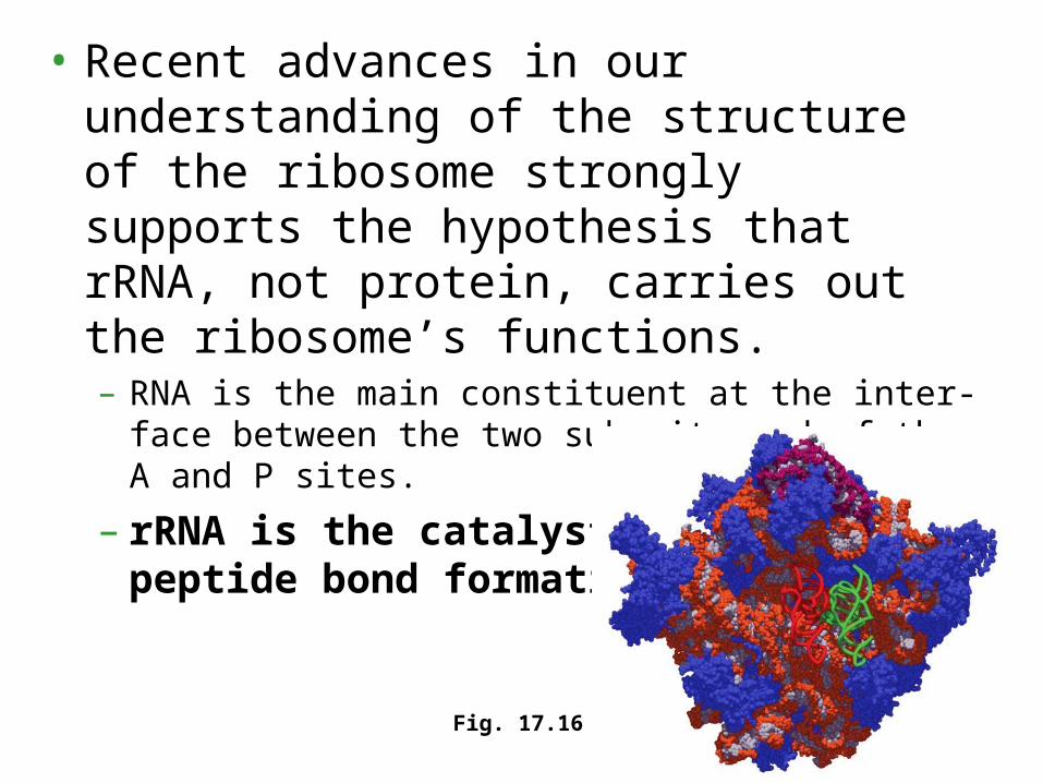
• Recent advances in our understanding of the structure of the ribosome strongly supports the hypothesis that rRNA, not protein, carries out the ribosome’s functions.– RNA is the main constituent at the inter-face between
the two subunits and of the A and P sites.
– rRNA is the catalyst for peptide bond formation
Fig. 17.16
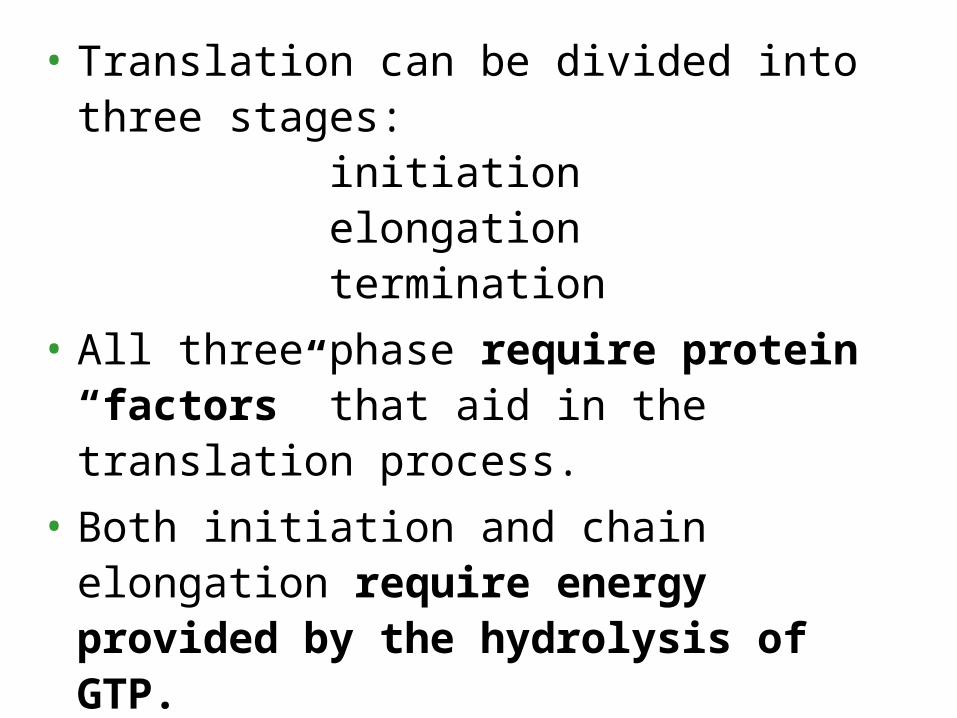
• Translation can be divided into three stages: initiation elongation termination
• All three phase require protein “factors” that aid in the translation process.
• Both initiation and chain elongation require energy provided by the hydrolysis of GTP.
• Initiation brings together mRNA, a tRNA with the first amino acid, and the two ribosomal subunits.– First, a small ribosomal subunit binds with mRNA and a
special initiator tRNA, which carries methionine and attaches to the start codon.
– Initiation factors bring in the large subunit such that the initiator tRNA occupies the P site.
Fig. 17.17
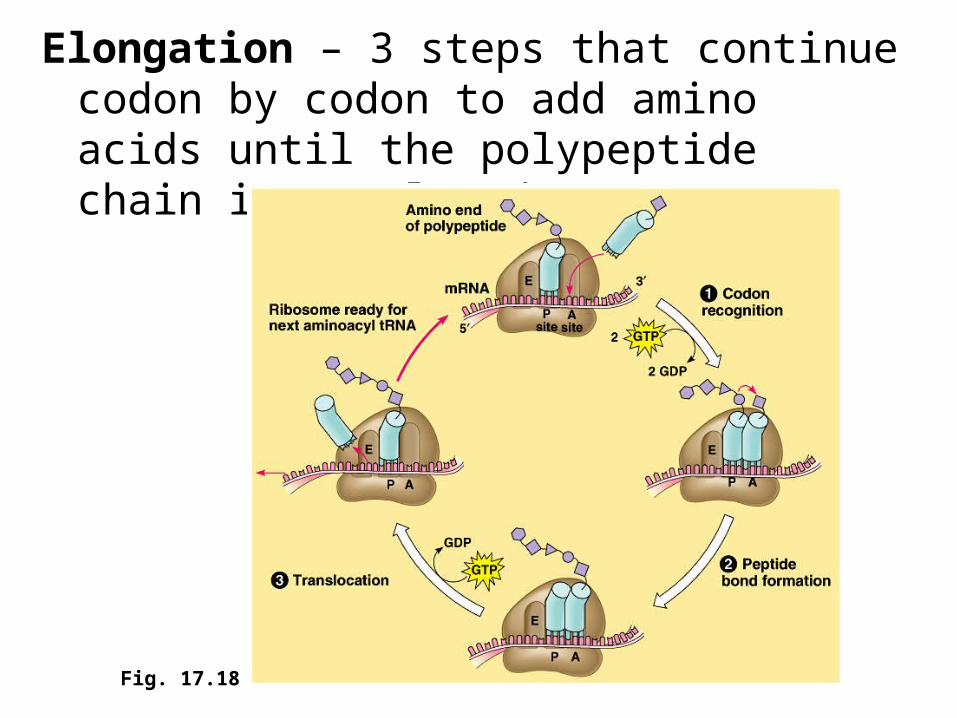
Elongation – 3 steps that continue codon by codon to add amino acids until the polypeptide chain is completed.
Fig. 17.18
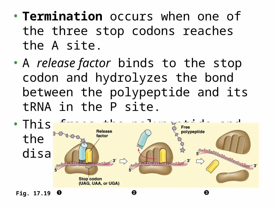
• Termination occurs when one of the three stop codons reaches the A site.
• A release factor binds to the stop codon and hydrolyzes the bond between the polypeptide and its tRNA in the P site.
• This frees the polypeptide and the translation complex disassembles.
Fig. 17.19
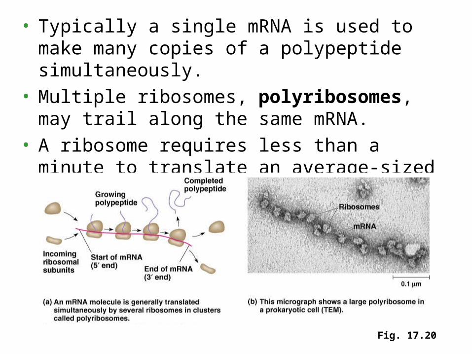
• Typically a single mRNA is used to make many copies of a polypeptide simultaneously.
• Multiple ribosomes, polyribosomes, may trail along the same mRNA.
• A ribosome requires less than a minute to translate an average-sized mRNA into a polypeptide.
Fig. 17.20
• While bound and free ribosomes are identical in structure, their location depends on the type of protein that they are synthesizing.
• Translation in all ribosomes begins in the cytosol, but a polypeptide destined for the endomembrane system or for export has a specific signal peptide region at or near the leading end.– This consists of a sequence of about 20 amino acids.
• A signal recognition particle (SRP) binds to the signal peptide and attaches it and its ribosome to a receptor protein in the ER membrane.– The SRP consists of a protein-RNA complex.
Fig. 17.21
• The diverse functions of RNA range from structural to informational to catalytic.
• E – Compartmentalized with Transcription in nucleus, Translation in Cytoplasm and extensive RNA processing in between, also complicated mechanisms for targeting proteins to the appropriate organelle.
• P- no nuclei so transcription and translation can occur simultaneously - Ribosomes attach to the leading end of a mRNA molecule while transcription is still in progress, protein diffuses to where needed
Fig. 17.2a
INITIATION AND ELONGATION
E - The promoter also includes a binding site for RNA polymerase several dozen nucleotides upstream of the start point.
P - In prokaryotes, RNA polymerase can recognize and bind directly to the promoter region.
E - eukaryotes have three RNA polymerases (I, II, and III) in their nuclei.– RNA polymerase II is used for mRNA synthesis.
P - Bacteria have a single type of RNA polymerase that synthesizes all RNA molecules.
• STEP 3 - TERMINATION– E - RNA polymerase continues for hundreds
of nucleotides past the terminator sequence, AAUAAA.
– P - RNA polymerase stops transcription right at the end of the terminator.• Both the RNA and DNA are then released.
RIBOSOMES
• While very similar in structure and function, prokaryotic and eukaryotic ribosomes have enough differences that certain antibiotic drugs (like tetracycline) can paralyze prokaryotic ribosomes without inhibiting eukaryotic ribosomes.
• More on Bacteria…
1) Let’s draw a bacteria cell with nucleoid and plasmid
2) How do bacterial cells divide?
• Bacterial cells divide by binary fission.
• This is preceded by replication of the bacterial chromosome from a single origin of replication.
Fig. 18.11
• Bacteria proliferate very rapidly- In a lab, one cell divides after 20 min producing a colony of 107 to 108 bacteria in as little as 12 hours.– In the human colon, E. coli reproduces rapidly
enough to replace the 2 x 10¹⁰ bacteria lost each day in feces.
• Through binary fission, most of the bacteria in a colony are genetically identical to the parent cell.
• New mutations, though individually rare, can have a significant impact on genetic diversity when reproductive rates are very high because of short generation spans.
• Individual bacteria that are genetically well equipped for the local environment clone themselves more prolifically than do less fit individuals.
• In contrast, organisms with slower reproduction rates (like humans) create most genetic variation not by novel alleles produced through mutation, but by sexual recombination of existing alleles.
• In addition to mutations, genetic recombination generates diversity within bacterial populations.
• Recombination occurs through three
processes:
transformation
transduction
conjugation
2. Genetic recombination produces new bacterial strains
• Transformation is the alteration of a bacterial cell’s genotype by the uptake of naked, foreign DNA from the surrounding environment.– For example, harmless Streptococcus pneumoniae
bacteria can be transformed to pneumonia-causing cells. (Remember Griffith’s experiments?)
– This occurs when a live nonpathogenic cell takes up a piece of DNA with allele for pathogenicity from dead, broken-open pathogenic cells.
– The foreign allele replaces the native allele & resulting cell is now recombinant with DNA derived from two different cells.
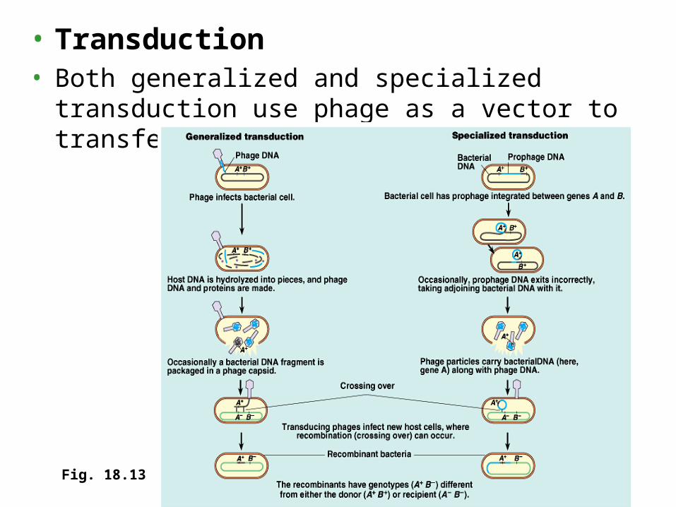
• Transduction• Both generalized and specialized transduction use
phage as a vector to transfer genes between bacteria.
Fig. 18.13
• Conjugation = “bacterial sex”
• One cell (“male”) donates DNA and its “mate” (“female”) receives the genes.
• A sex pilus from the male initially joins the two cells and creates a cytoplasmic bridge between cells.
• “Maleness”, the ability to form a sex pilus and donate DNA, results from an F factor as a section of the bacterial chromosome or as a plasmid.
Fig. 18.14
• Plasmids, – small, circular, self-replicating DNA molecules.– usually have only a few genes that are not
required for normal survival and reproduction.– generally, benefit the bacterial cell by providing
genes that are advantageous in stressful conditions.• The F plasmid facilitates genetic recombination when
environmental conditions no longer favor existing strains.
• R plasmid has genes for antibiotic resistance
• Episomes, like the F plasmid, can undergo reversible incorporation into the cell’s chromosome.– Temperate viruses also qualify as episomes.
• The F factor or its F plasmid consists of about 25 genes, most required for the production of sex pili.– Cells with either the F factor or the F plasmid are
called F+ and they pass this condition to their offspring.
– Cells lacking either form of the F factor, are called F-, and they function as DNA recipients.
• When an F+ and F- cell meet, the F+ cell passes a copy of the F plasmid to the F- cell, converting it.
Fig. 18.15a
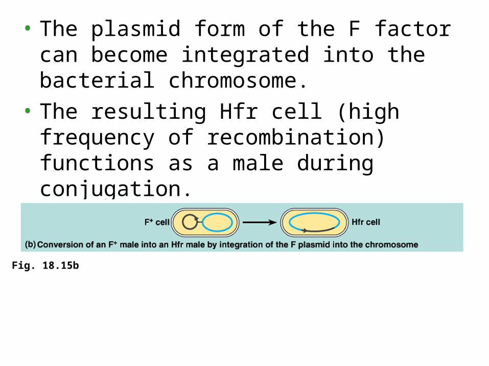
• The plasmid form of the F factor can become integrated into the bacterial chromosome.
• The resulting Hfr cell (high frequency of recombination) functions as a male during conjugation.
Fig. 18.15b
• In the 1950s, Japanese physicians began to notice that some bacterial strains had evolved antibiotic resistance.– The genes conferring resistance are carried by
plasmids, specifically the R plasmid (R for resistance).
– Some of these genes code for enzymes that specifically destroy certain antibiotics, like tetracycline or ampicillin.
• When a bacterial population is exposed to an antibiotic, individuals with the R plasmid will survive and increase in the overall population.
• Because R plasmids also have genes that encode for sex pili, they can be transferred from one cell to another by conjugation.
• A transposon is a piece of DNA that can move from one location to another in a cell’s genome.
• Transposon movement occurs as a type of recombination between the transposon and another DNA site, a target site.– In bacteria, the target site may be within the
chromosome, from a plasmid to chromosome (or vice versa), or between plasmids.
• Transposons can bring multiple copies for antibiotic resistance into a single R plasmid by moving genes to that location from different plasmids.– This explains why some R plasmids convey
resistance to many antibiotics.
• The transposase enzyme recognizes the inverted repeats as the edges of the transposon.
• Transposase cuts the transposon from its initial site and inserts it into the target site.
• The simplest bacterial transposon, an insertion sequence, consists only of the transposase gene
Fig. 18.17
• Composite transposons (complex transposons) include extra genes sandwiched between two insertion sequences.– It is as though two insertion sequences
happened to land relatively close together and now travel together, along with all the DNA between them, as a single transposon.
Fig. 18.18
• While insertion sequences may not benefit bacteria in any specific way, composite transposons may help bacteria adapt to new environments.– For example, repeated movements of resistance
genes by composite transposition may concentrate several genes for antibiotic resistance onto a single R plasmid.
– In an antibiotic-rich environment, natural selection favors bacterial clones that have built up composite R plasmids through a series of transpositions.
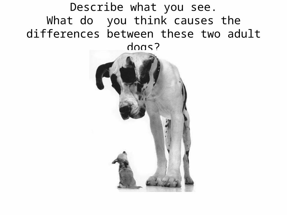
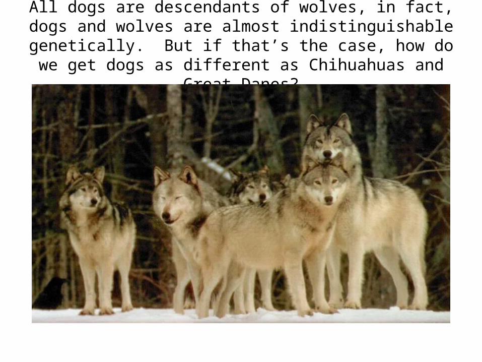
Describe what you see.What do you think causes the differences
between these two adult dogs?
All dogs are descendants of wolves, in fact, dogs and wolves are almost indistinguishable genetically. But if
that’s the case, how do we get dogs as different as Chihuahuas and Great Danes?
Describe what you see (1)
Describe what you see (2)
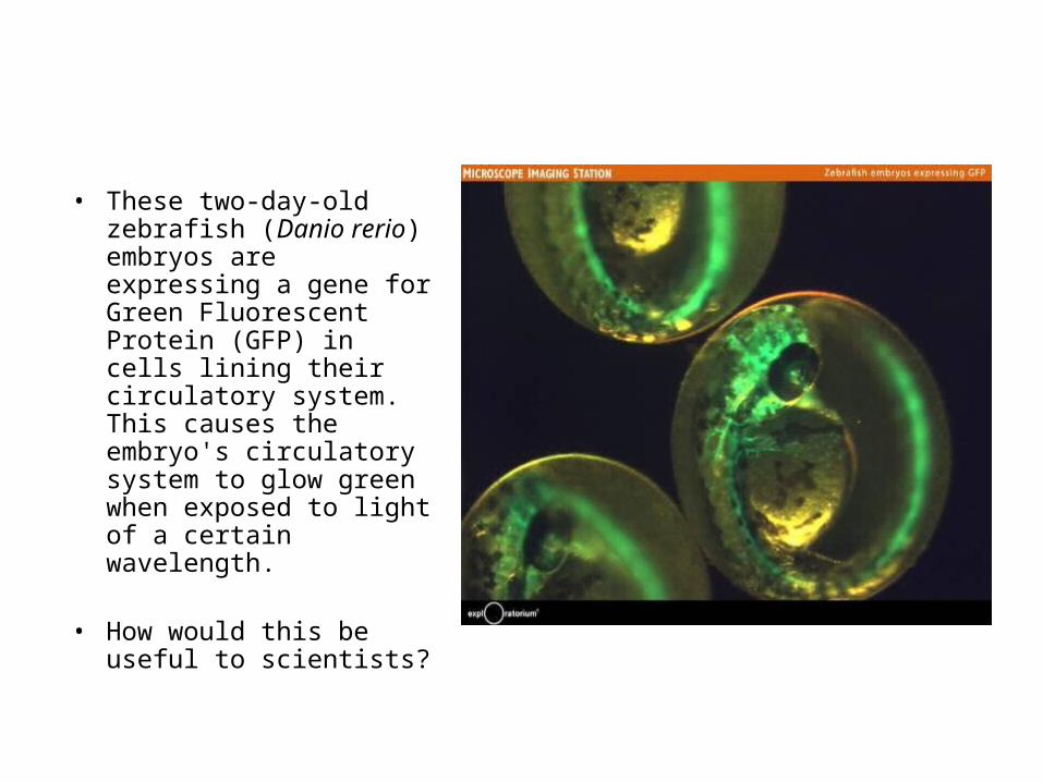
• These two-day-old zebrafish (Danio rerio) embryos are expressing a gene for Green Fluorescent Protein (GFP) in cells lining their circulatory system. This causes the embryo's circulatory system to glow green when exposed to light of a certain wavelength.
• How would this be useful to scientists?
Links to Real-world
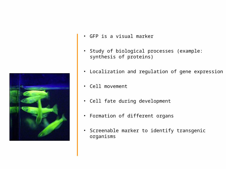
• GFP is a visual marker
• Study of biological processes (example: synthesis of proteins)
• Localization and regulation of gene expression
• Cell movement
• Cell fate during development
• Formation of different organs
• Screenable marker to identify transgenic organisms
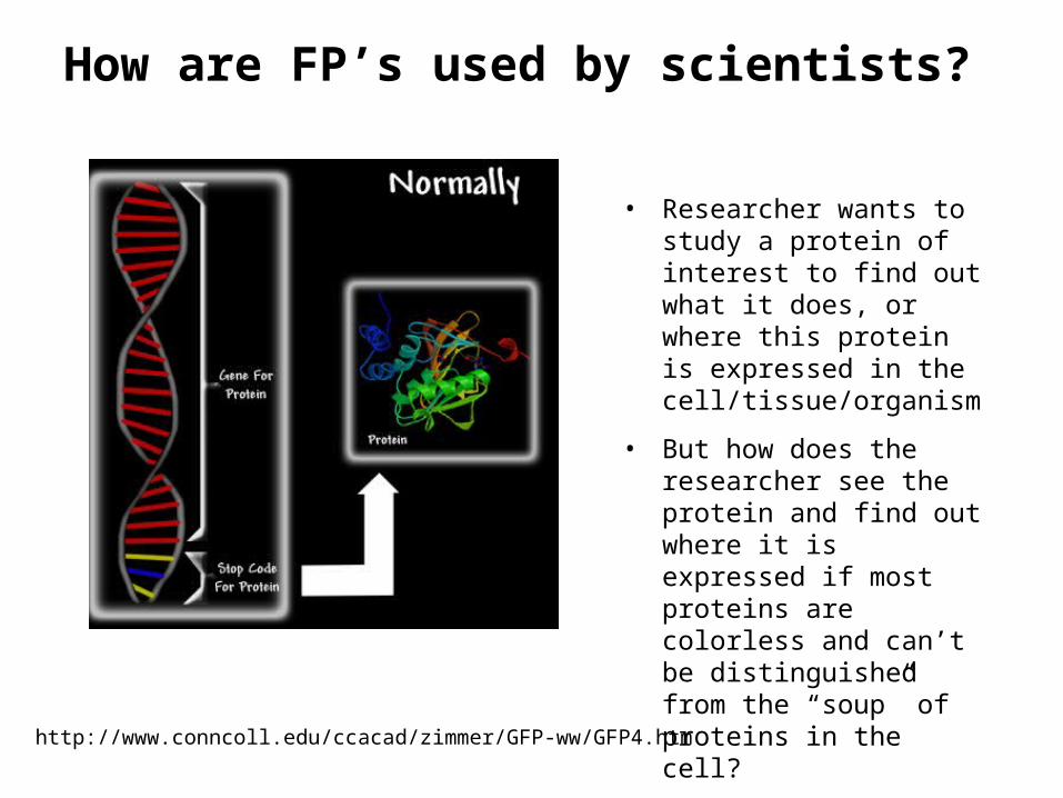
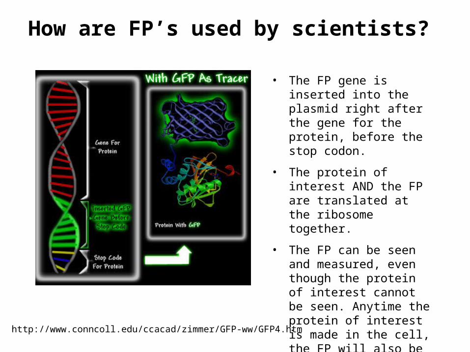
How are FP’s used by scientists?
• Researcher wants to study a protein of interest to find out what it does, or where this protein is expressed in the cell/tissue/organism
• But how does the researcher see the protein and find out where it is expressed if most proteins are colorless and can’t be distinguished from the “soup” of proteins in the cell?
• The FP gene is inserted into the plasmid right after the gene for the protein, before the stop codon.
• The protein of interest AND the FP are translated at the ribosome together.
• The FP can be seen and measured, even though the protein of interest cannot be seen. Anytime the protein of interest is made in the cell, the FP will also be made.
Human cell stained with two different fluorescent proteins to visulalize cytoskeletal components. Transfected with GFP-tubulin / mCherry actin (Ben Giepmans)
Cellular organelles targeted with FPs
C Elegans transfected with GFP tubulin construct (Susan Kline)
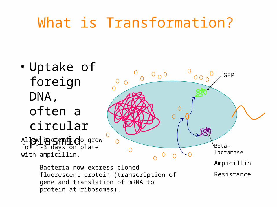
What is Transformation?
• Uptake of foreign DNA, often a circular plasmid
GFP
Beta-lactamase
Ampicillin
Resistance
Allow bacteria to grow for 1-3 days on plate with ampicillin.
Bacteria now express cloned fluorescent protein (transcription of gene and translation of mRNA to protein at ribosomes).
What is a plasmid?
• A small circular piece of DNA that replicates separately from the main bacterial chromosome
• Originated in bacteria to allow survival in specific environmental conditions
• May express antibiotic resistance gene or be modified in the lab to express proteins of interest
How are plasmids engineered?
Host DNA fragments (i.e. coral or jellyfish FP coding DNA)
DNA Plasmid Vector
Cut plasmids open with restriction enzymes
Cut genomic DNA into fragments
+
Ligate (paste) fragments into cut DNA vector
End result: Plasmid containing FP gene
Transformation procedure
1. Suspend bacterial colonies in cold CaCl2
2. Add plasmid DNA
3. Place tubes on ice for 10 min
4. Heat-shock at 42°C for 45 seconds & place on ice again for 2 min
5. Plate out bacteria
PLEASE READ AND FOLLOW LAB INSTRUCTIONS CAREFULLY!
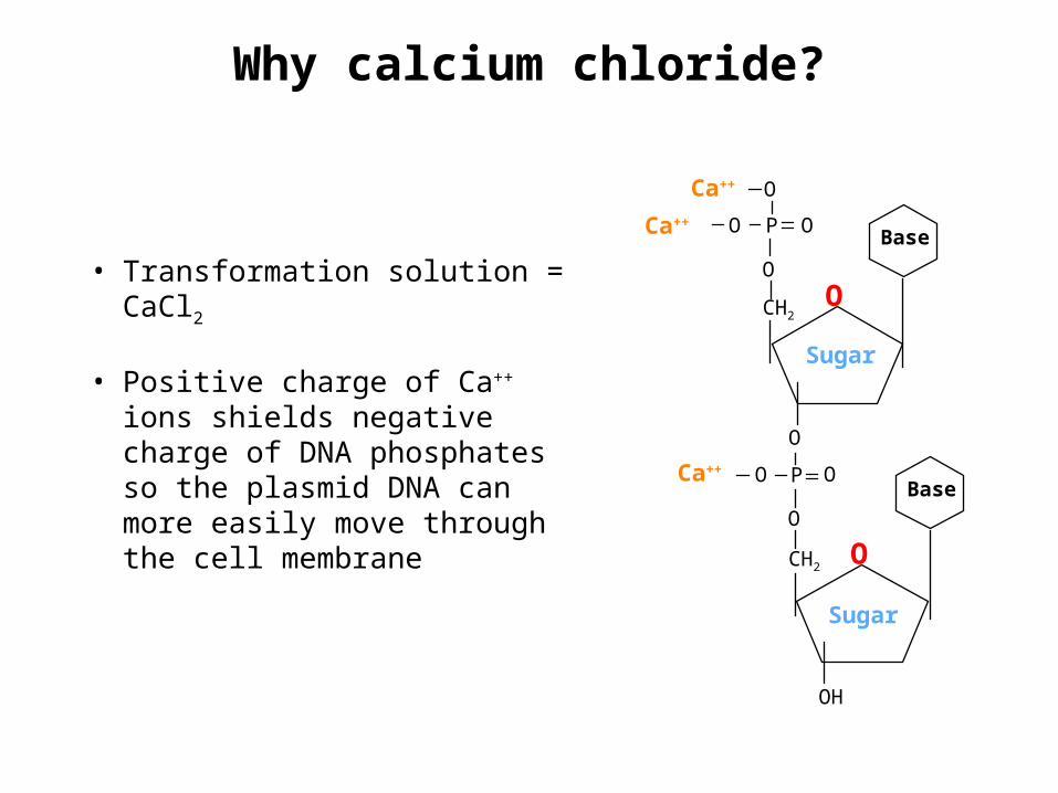
Why calcium chloride?
Ca++
Ca++
OCH2
O
P O
O
OBase
CH2
O
P
O
O
O
Base
OH
Sugar
Sugar
OCa++
• Transformation solution = CaCl2
• Positive charge of Ca++ ions shields negative charge of DNA phosphates so the plasmid DNA can more easily move through the cell membrane
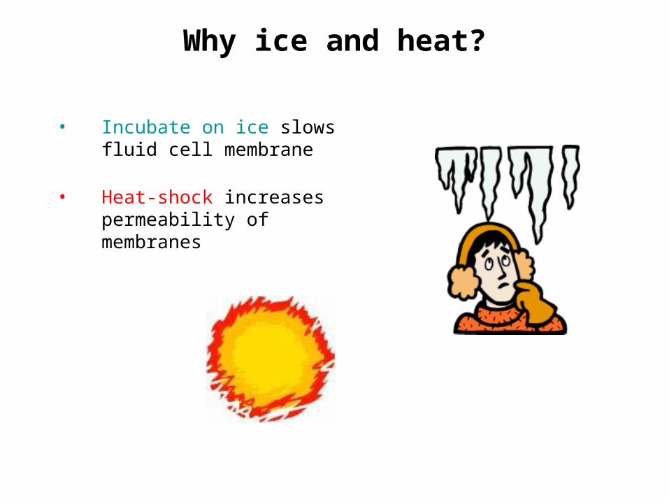
Why ice and heat?
• Incubate on ice slows fluid cell membrane
• Heat-shock increases permeability of membranes
Why Ampicillin?
• Ampicillin inhibits cell growth. Only cells that can deactivate the ampicillin around them will grow.
• Ampicillin resistance is tied to (expressed with) the fluorescent protein gene
• The ampicillin is the selection mechanism that allows only the transformed bacteria to grow on the plate
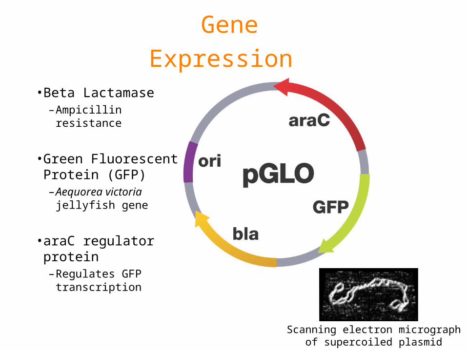
Gene
Expression • Beta Lactamase
–Ampicillin resistance
• Green Fluorescent Protein (GFP)–Aequorea victoria
jellyfish gene
• araC regulator protein–Regulates GFP
transcription
Scanning electron micrograph of supercoiled plasmid
Grow? Glow?
• Follow protocol
• On which plates will colonies grow?
• Which colonies will glow?
Laboratory Quick Guide
Agenda 12/1/11
• Transformation lab – 6A
Homework – •Ch. 19 Notes and SQ due next Monday•Do any analysis questions for transformation lab that you can•Study for quiz tomorrow on Ch. 16, 17
Agenda 12/2/11
• Quiz Ch. 16, 17 and correct• Analyze Transformation data and wrap up/ discuss
Analysis • If extra time, could do mutation slides (Ch. 17, slides 74-
82) – otherwise will do it next Tuesday
Homework – • Ch. 19 Notes and SQ due next Monday, short
Transformation quiz so be comfortable with lab• Mutation practice due next Tuesday