May 2015 Page 1 of 12 Alert for Klebsiella pneumoniae septicaemia in piglets APHA Disease Surveillance Report May 2015 Texel microphthalmia Botulism in sheep Porcine reproductive and respiratory virus: multiple outbreaks, multiple presentations CATTLE Musculoskeletal disease ‘White muscle disease’ (WMD) was diagnosed on four occasions at Shrewsbury in suckler calves. Although reports of suspected WMD (or ‘nutritional myopathy’) are received at this time each year, samples are not often received for diagnosis. In each calf there were typical signs reported including reluctance to stand and walking with a stiff gait. A summary of the results is tabulated below which shows the value of testing blood for evidence of muscle damage, as the creatine kinase (CK) activity rises massively in WMD. Vitamin E concentrations were consistently low and three of the four animals were also identified with low selenium status (measured by glutathione peroxidase, GSH-Px, activity). Case number/age CK (APHA reference interval 0-200 U/l @ 37 o C) GSH-Px (> 30 U/ml RBCs) Vitamin E (> 2.3 μmol/l) 1: 6 weeks 175,531 17.4 1.0 2: 2 months 278,750 10.3 < 1.0 3: 3 months 242,552 106.4 2.2 4: 3 months 84,839 8.5 1.0 Systemic disease Bovine neonatal pancytopaenia: Cases of bovine neonatal pancytopenia (BNP) continue to be diagnosed in VI Centres. At Thirsk a 16-day-old Aberdeen Angus calf was submitted for postmortem examination from a suckler herd of 40 cows. It had become anaemic and exhibited melaena,
Transcript
May 2015 Page 1 of 12
Alert for Klebsiella pneumoniae septicaemia in piglets
APHA Disease Surveillance Report May 2015
Texel microphthalmia
Botulism in sheep
Porcine reproductive and respiratory virus: multiple outbreaks, multiple presentations
CATTLE Musculoskeletal disease ‘White muscle disease’ (WMD) was diagnosed on four occasions at Shrewsbury in suckler calves. Although reports of suspected WMD (or ‘nutritional myopathy’) are received at this time each year, samples are not often received for diagnosis. In each calf there were typical signs reported including reluctance to stand and walking with a stiff gait. A summary of the results is tabulated below which shows the value of testing blood for evidence of muscle damage, as the creatine kinase (CK) activity rises massively in WMD. Vitamin E concentrations were consistently low and three of the four animals were also identified with low selenium status (measured by glutathione peroxidase, GSH-Px, activity).
Case number/age CK (APHA reference interval 0-200 U/l @ 37oC)
GSH-Px (> 30 U/ml RBCs)
Vitamin E (> 2.3 µmol/l)
1: 6 weeks 175,531 17.4 1.0
2: 2 months 278,750 10.3 < 1.0
3: 3 months 242,552 106.4 2.2
4: 3 months 84,839 8.5 1.0
Systemic disease Bovine neonatal pancytopaenia: Cases of bovine neonatal pancytopenia (BNP) continue to be diagnosed in VI Centres. At Thirsk a 16-day-old Aberdeen Angus calf was submitted for postmortem examination from a suckler herd of 40 cows. It had become anaemic and exhibited melaena,
May 2015 Page 2 of 12
scleral haemorrhages and dyspnoea prior to its death. The carcase was pale and jaundiced, and there were haemorrhages present in the oesophagus, trachea, lungs, myocardium, spleen and kidneys. A colisepticaemia was identified by bacterial culture and histopathology confirmed trilineage hypoplasia which is the characteristic feature of BNP. The disease was also suspected at Shrewsbury in a 10-day-old Simmental bull calf which was reported to be bleeding from both ear tags, had haematomas at injection sites and was very pale. Haematology confirmed a white cell count of only 1.2 x10⁹/l (reference interval 4-12 x109/l) with only 74 x109/l platelets (reference interval 100-800 x109/l) and no detectable neutrophils: the haematological features of BNP were reported by Bell and others (2010). Nervous disease An early gestational genetic defect was diagnosed in a Longhorn calf which was born dead and had mandibular and maxillary brachygnathia, a ‘hare lip’ and cleft palate, suspected hydranencephaly, arthrogryposis and a frontal bone deficit. The calf was the only affected animal in a herd of 55 Longhorn cattle. Testing for Schmallenberg virus infection by PCR was negative and there was no evidence of an inflammatory component. The frontal bone lesion was considered likely to be due to a neural tube closure deficit which can lead to the formation of an encephalocoele or meningocoele, these changes causing dysplasia in the brain and spinal cord. Histopathology confirmed hydrocephalus, cerebellar hypoplasia and brainstem dysplasia with myelodysplasia present in the spinal cord. The potential causes of neural tube defects include genetic conditions and exposure to environmental teratogens early in gestation. Respiratory disease Severe necrotising pneumonia with abscessation was found postmortem in three dairy cows examined at the Starcross VI Centre (Fig 1). The animals were received from a 550 cow dairy herd where chronic, ongoing respiratory disease was reported. Ten cows had developed respiratory disease, five of which had died. Joint swelling and lameness was also reported in some animals. Each cow had severe consolidation and abscessation affecting more than 70% of the lung lobes. One cow also had mastitis affecting two quarters. Trueperella pyogenes was isolated from the lungs of two of the animals. DGGE examination of lung samples detected Mycoplasma bovis in two of the three cows and also in the mastitic milk. Following on from these initial diagnoses a further four milk samples were submitted from mastitic cows which had failed to respond to routine treatments and a joint fluid sample from a lame cow. Mycoplasma bovis was detected by DGGE from the joint fluid and three out of the four mastitis milk samples. In the last 18 months there has been increased awareness in the profession of Mycoplasma bovis-associated disease in adult cattle, presenting as either arthritis, mastitis or pneumonia.
May 2015 Page 3 of 12
Fig 1: Necrotising pneumonia in a dairy cow SMALL RUMINANTS Systemic disease Tick-borne fever (TBF) was confirmed in a lamb submitted to the Wales Veterinary Science Centre from a flock of about 300 ewes and lambs where fifteen lambs had been found dead (Daniel and others 2015). Reproductive disease Q fever: Initial investigations into abortions in a newly established goat herd gave findings indicative of Listeria spp involvement but subsequent submissions were positive for the presence of Coxiella burnetii, the causative agent of Q-fever. The zoonotic aspects of this infection were stressed to the submitting veterinary practice and in turn to the owner, including the propensity of this organism to spread by aerosol when dried in bedding contaminated by the products of parturition. Bury investigated a flock of 145 Texel ewes where seven lambs were reported to have been born with no eyes. This was a closed organic flock with three rams recently introduced. A live lamb submitted for examination appeared bright and responsive to acoustic stimuli but with permanently shut eyes. On postmortem examination the apertures of both orbits were narrow and the cavities shallow; the eyelids of both eyes were not fully open whilst the eyes were rudimentary and of small size. Bilateral microphthalmia has been reported as an inherited genetic defect in Texel sheep linked to an autosomal recessive trait and is reported to be of low incidence. The histological features indicated a very early gestational malformation within the spectrum described for Texel microphthalmia (van der Linde-Sipman and others 2003). This genetic defect has also been recorded in crossbred Texel sheep (where Texel breeding is present in both the dam and the sire) and is now known to be associated with a mutation in the PITX3 gene (Becker and others 2010). Border disease and Schmallenberg viruses and trace element deficiency were ruled out in this case.
May 2015 Page 4 of 12
Enteric disease
Black disease: Deaths of three ewes from a flock of 150 Swaledale ewes that had lambed about two weeks previously were investigated by Thirsk. Twin lamb disease and/or hypocalcaemia had been suspected a few weeks prior to lambing. The ewes were treated for gut worms and liver fluke just prior to lambing and had received supplementation throughout the winter. The condition of the ewes was good and no scouring had been detected. Two ewes were submitted for postmortem examination which revealed numerous haemorrhagic tracks and areas of black discoloured emphysematous tissue in the livers. There were also thickened bile ducts and dead adult flukes in one of the two ewes. Further testing revealed positive results for Clostridium novyi on FAT on liver tissue from both ewes. It is possible that the fluke treatment provided prior to lambing was effective but that the damage caused in the livers led to the development of Black disease causing the deaths of these individuals.
Nervous disease
Botulism: A live ewe was submitted to Thirsk from a flock of 83 broken mouthed mule ewes with lambs. The ewes had lambed two months previously and had been in the same field for the entire period. Five affected ewes were either seen to walk slightly oddly a week before submission, progressing to recumbency or death. Three recumbent ewes were described as bright but with a certain degree of paresis/weakness. There was no response to calcium injections and none of the lambs had been affected. Poultry manure from a broiler house had been spread on an adjacent stubble field two weeks earlier and ploughed in within 24 hours. The submitted ewe was bright and in sternal recumbency. There was a good menace reflex but the head drooped when the ewe was left undisturbed. When the front legs were placed knuckled over there was no attempt at correction (figure 2). Withdrawal reflex in the hind limbs was fair and there was no obvious tongue or anal paresis. The history of spreading poultry manure, the clinical signs and the lack of significant postmortem findings strongly suggested the possibility of botulism. The farmer could not move the sheep and lambs from that particular field, but an electric fence was installed to increase the distance between the sheep and the field where the poultry manure was spread. No further mortalities were reported and the case was reported as a food safety incident.
May 2015 Page 5 of 12
Fig 2. Recumbent ewe with botulism. The ewe was unable to correct the knuckled front
limb. PIGS OUTBREAKS OF PIGLET SEPTICAEMIA DUE TO KLEBSIELLA PNEUMONIAE:
ALERT FOR FURTHER OUTBREAKS THIS SUMMER
Each summer since 2011 APHA (formerly AHVLA) has diagnosed outbreaks of septicaemia in piglets due Klebsiella pneumoniae ssp pneumoniae (Kpp). All but one of these cases has occurred in East Anglian herds, one outbreak in 2014 was diagnosed in the South West of England. The seasonal nature of disease occurring between May and September is illustrated in the figure below.
The most common features of the outbreaks are
Sudden or very rapid deaths Preweaned pigs affected from 10-days-old to point of weaning Non-specific lesions (septicaemic) at post-mortem examination Outdoor pig units Mortality variable 1- 6 % of pigs born (one herd 16%) Appears self-limiting but has recurred in two herds in subsequent years
May 2015 Page 6 of 12
One septicaemia outbreak in East Anglia in 2014 involved concurrent severe mastitis in lactating sows. Post-mortem examination and bacteriology are required to confirm a diagnosis. The same emerging strain of Kpp (sequence type 25) has been involved in all outbreaks to date. The website link below includes details of the clinical presentation and pathology. Veterinary surgeons wishing to submit or discuss diagnosis of possible cases which may arise during the coming months, should contact an APHA Veterinary Investigation Officer. http://webarchive.nationalarchives.gov.uk/20140707141417/http://www.defra.gov.uk/ahvla-en/publication/klebsiella-septicaemia/
Alimentary Disease Post-weaning mortality associated with PRRS challenge and enteric disease: Live pigs were submitted to Bury St Edmunds as part of a series from a continuous nursery-finisher site with a 24-month history of elevated mortality (varying from 8-15%) associated with respiratory disease and wasting in each three-weekly batch. The pigs were sourced from a single breeding herd and were vaccinated for porcine circovirus 2 (PCV2), Mycoplasma hyopneumoniae and porcine reproductive and respiratory syndrome virus (PRRSv). All three submitted pigs were in poor body condition with the heaviest being 5.4kg at five-weeks-old; they had watery small intestinal contents from which monophasic Salmonella 4,12:i:- phage type 120 was isolated. No rotavirus, porcine epidemic diarrhoea (PED) or enteropathogenic E. coli was found. PRRSv was detected in pooled sera from the pigs and sequencing revealed it was a field strain which showed a high degree of similarity to the viruses isolated in two submissions to Bury St Edmunds in February and October 2014, and to the PRRSv strain detected on the breeding unit supplying this farm. Lung histopathology was suggestive of PRRS; the poor body condition of the piglets and detection of PRRSv early post-weaning suggest that the PRRSv was playing a role in the clinical disease prior to weaning, possibly prior to vaccination, although PRRSv was not detected by immunohistochemistry in the lungs. Subsequently, PRRSv infection was also diagnosed in another submission from this unit, this time with immunohistochemistry confirming involvement of PRRSv in the pneumonia, with sequencing identifying the same virus strain as in the earlier submissions from this unit and the source breeding unit. On this occasion, all three pigs submitted had polyarthritis but no bacteria were isolated, probably due to prior antimicrobial treatment. The small intestine of one pig was thin-walled and distended with watery fluid and F4 antigen-positive E. coli was isolated in heavy pure growth from this pig, consistent with enteric colibacillosis. As in the previous submission, the five-week-old pigs submitted were significantly underweight with the heaviest being 4.4kg. Reproductive disease Active swine influenza detected on farm experiencing an abortion outbreak: An abortion outbreak was investigated by the University of Bristol in which no infectious cause was established but serology on aborting sows showed high antibody titres to swine influenza. Previous outbreaks of acute swine influenza in pregnant sows have occasionally caused small outbreaks of late stage abortion and stillbirths due to the maternal effect of the viral
infection and two sows were seen to be dyspnoeic in the dry sow yards at the time of the abortions. It was agreed that if respiratory disease continued, that further samples would be submitted. Nasal swabs were subsequently submitted for Defra-funded swine influenza testing from pigs showing acute respiratory disease in the first two weeks after weaning. Swabs from two pigs were PCR-positive for both for influenza M gene and the pandemic H1N1 2009 strain of the virus confirming active swine influenza infection on the unit. Reproductive failure and weak live-born piglets due to PRRS: PRRSv was detected in all three fetuses tested from one of two litters sampled to investigate infertility, abortions mummification, stillbirths and poor viability piglets occurring in a weaner-producer unit in the Bury St Edmunds region. Sows were vaccinated against PRRSv and those of parity 1 to 3 were particularly affected. The virus showed very close similarity to that detected on a nursery-finisher unit receiving piglets from this breeding unit on which there was increased mortality as described below. An earlier submission of fetuses from this unit did not identify PRRSv but, on that occasion, only two fetuses were submitted, one of which was severely autolysed. This case demonstrates the value of submitting multiple freshly aborted litters to increase the chances of detecting an infectious cause, if present. Respiratory Disease Complex disease with underlying PRRS and mixed infections: Multiple diseases with underlying PRRS were diagnosed in five-week-old pigs on a single-source indoor nursery-finisher receiving pigs weekly. Sneezing, weight loss and diarrhoea had developed within a week of arrival in the previous three batches of weaners and, in the batch from which pigs were submitted to Bury St Edmunds, 10% were affected and nine pigs had died from a group of 600. Pigs were vaccinated for PCV2 and M. hyopneumoniae but not for PRRSv which was detected by PCR and confirmed as causing respiratory disease by lung immunohistochemistry. In addition, Streptococcus suis 8 was isolated from the lung, a likely secondary pathogen in one pig and Staphylococcus aureus was involved in long-standing joint infections in two pigs, likely secondary to trauma while on the breeding unit. Interestingly, although not detected by PCR in the three submitted pigs, swine influenza was detected in an oral fluid sample from the affected pigs tested elsewhere, demonstrating the value of the wider sampling enabled through oral fluid collection. The continuous nature of this large unit, and the weekly introduction of significant numbers of weaners, both favour the persistence of viral and other infections from batch to batch. Swine influenza outbreak in late finishers: Swine influenza was diagnosed when samples were submitted to Bury St Edmunds from 20-week-old housed finishers with respiratory disease and increasing mortality, in which PRRS was suspected. No PRRS was detected but histopathology revealed a bronchiolitis and immunohistochemistry confirmed swine influenza. The cause of death following swine influenza infection is usually a secondary bacterial infection, rather than the influenza virus itself; histopathology revealed an interstitial pneumonia which may have been due to systemic disease but no samples were submitted for culture.
May 2015 Page 8 of 12
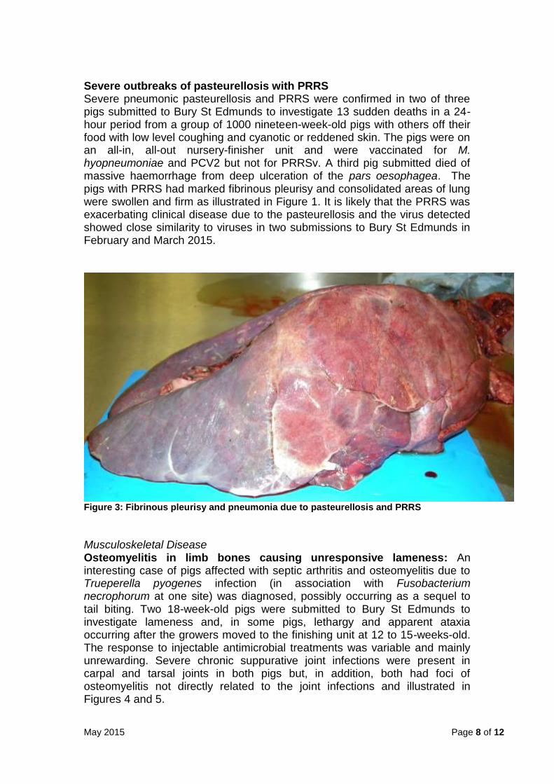
Severe outbreaks of pasteurellosis with PRRS Severe pneumonic pasteurellosis and PRRS were confirmed in two of three pigs submitted to Bury St Edmunds to investigate 13 sudden deaths in a 24-hour period from a group of 1000 nineteen-week-old pigs with others off their food with low level coughing and cyanotic or reddened skin. The pigs were on an all-in, all-out nursery-finisher unit and were vaccinated for M. hyopneumoniae and PCV2 but not for PRRSv. A third pig submitted died of massive haemorrhage from deep ulceration of the pars oesophagea. The pigs with PRRS had marked fibrinous pleurisy and consolidated areas of lung were swollen and firm as illustrated in Figure 1. It is likely that the PRRS was exacerbating clinical disease due to the pasteurellosis and the virus detected showed close similarity to viruses in two submissions to Bury St Edmunds in February and March 2015.
Figure 3: Fibrinous pleurisy and pneumonia due to pasteurellosis and PRRS
Musculoskeletal Disease Osteomyelitis in limb bones causing unresponsive lameness: An interesting case of pigs affected with septic arthritis and osteomyelitis due to Trueperella pyogenes infection (in association with Fusobacterium necrophorum at one site) was diagnosed, possibly occurring as a sequel to tail biting. Two 18-week-old pigs were submitted to Bury St Edmunds to investigate lameness and, in some pigs, lethargy and apparent ataxia occurring after the growers moved to the finishing unit at 12 to 15-weeks-old. The response to injectable antimicrobial treatments was variable and mainly unrewarding. Severe chronic suppurative joint infections were present in carpal and tarsal joints in both pigs but, in addition, both had foci of osteomyelitis not directly related to the joint infections and illustrated in Figures 4 and 5.
May 2015 Page 9 of 12
Figures 4 and 5 showing osteomyelitis lesions in long bones of forelimbs of pigs (arrowed)
Interestingly, both pigs had bitten tails and although the length of the tails made them appear that they had been docked, it was subsequently reported that they had not been docked on-farm, confirming that significant tail biting had occurred. The tail lesions may well have been the origin of infection through haematogenous spread of bacteria and this was highlighted to the attending practitioner. There were no other gross lesions to indicate that underlying metabolic bone disease was playing a predisposing role. Antimicrobial treatment of osteomyelitis is not usually feasible and affected pigs should be culled and the predisposing factors, in this case, likely tail biting, controlled or prevented. BIRDS Broilers
Inclusion body hepatitis (IBH) was diagnosed in submission of 11-day-old broiler chicks with a history of high mortality. Enlarged livers with an enhanced reticular pattern, pallor or with small red foci were the main postmortem findings. Histological examination of liver tissue revealed parenchymal necrosis accompanied by numerous degenerate hepatocytes containing intranuclear inclusion bodies, consistent with adenovirus infection. IBH can be associated with infection with a range of avian adenoviruses.
E coli septic arthritis and septicaemia were diagnosed in a submission of 37-day-old broilers with a history of increased mortality and lameness. Postmortem examination revealed purulent exudate in the leg joints and occasional swelling of the liver and spleen and abdominal cellulitis. Routine
May 2015 Page 10 of 12
cultures of the affected leg joints and the spleens yielded heavy pure growths of E. coli.
Commercial Layers
Avian intestinal spirochaetosis: Brachyspira pilosicoli was isolated from the caecal contents of 30-week-old free range layers submitted to investigate loss of body condition, abnormal caramel coloured frothy droppings and a slight drop in egg production and egg quality. Postmortem examination revealed marked variation in carcase weight; however, all the birds examined were currently in lay with unremarkable intestinal contents. Selective culture of the caecal contents revealed B. pilosicoli, one of the pathogenic Brachyspira species associated with avian intestinal spirochaetosis and causing drops in egg quality and production and loss of body condition. Although the birds examined were in lay, the loss of body condition together with the bacteriology results and the history on the farm were strongly suggestive of a current Brachyspira challenge.
Egg peritonitis: E coli septicaemia and egg peritonitis were seen in a submission of 65-week-old free range layers with a history of increased mortality in a flock of 22,000 birds. The postmortem findings included swollen livers and spleens, salpingitis and fibrinous peritonitis. Routine cultures of liver, spleen, peritoneum and oviduct yielded heavy growths of E. coli, as is typical of egg peritonitis. Game birds
Salmonella Typhimurium infection was diagnosed in five-day-old pheasant chicks on a unit of 20,000 birds. Postmortem examination showed evidence of systemic spread and the findings also included chronic typhlitis with caecal cores. S. Typhimurium Copenhagen was isolated on cultures of liver, spleen and caeca. Further postmortem examination of birds from the same site at fifteen days of age revealed similar findings and S. Typhimurium was again isolated indicating continued mortality associated with salmonellosis despite a course of antibiotic treatment having been administered.
Rotavirus infection was suspected in three submissions of seven- to eight- day-old pheasant chicks with a history of increased mortality and poor growth and body condition. The findings at postmortem examination included
distended caeca with yellow-creamy coloured and occasionally gassy contents. Possible rotavirus involvement was confirmed by polyacrylamide gel electrophoresis (PAGE) testing on samples of caecal contents from only one of these submissions even though the gross appearance of the carcases and intestinal tracts from all three submissions was highly suspicious of rotavirus involvement.
May 2015 Page 11 of 12
Backyard Flocks Suspect rodenticide poisoning: Out of a small group of peafowl, one bird had been unexpectedly found dead ten days previously and a second was missing but found dead two days later in the building where they roosted. A third bird was alive but obviously unwell. The birds were free to range but always roosted in a particular building at night and were fed mixed corn in the building. Post mortem examination was undertaken on the second bird. It was in good condition with food material in the crop and several bright blue soft granules measuring 2-3 mm diameter were also present. There was marked subcutaneous haemorrhage and damage to the muscles around the keel, with haemorrhage between the muscle layers and blood in the body cavity. Discussion with the owner suggested that blue material was rodenticide which had been used on the premises. It was not clear how the bird had access to the poison and the owner was to re-assess its use on the premises to ensure that the recommendations that come with the product
were closely followed. No specific human food safety risk was identified. References
BECKER, D., TETENS, J., BRUNNER, A., BÜRSTEL, D., GANTER, M., KIJAS, J., DRÖGEMÜLLER, C. & FOR THE INTERNATIONAL SHEEP GENOMICS, C. (2010) Microphthalmia in Texel Sheep Is Associated with a Missense Mutation in the Paired-Like Homeodomain 3 (<italic>PITX3</italic>) Gene. PLoS ONE 5, e8689 Bell, C.R., Scott, P.R., Sargison, N.D., Wilson, D.J., Morrison, L., Howie, F., Willoughby, K. and Penny, C.D. (2010) Idiopathic bovine neonatal pancytopenia in a Scottish beef herd. Veterinary Record 167, 938-940
DANIEL, R., PUGH, K., TORRENS, N., CARSON, A. & WESSELS, M. (2015) Intercurrent tickborne fever infection and Bibersteinia trehalosi septicaemia in a five-week-old lamb. Veterinary Record 177, 24
VAN DER LINDE-SIPMAN, J. S., VAN DEN INGH, T. S. G. A. M. & VELLEMA, P. (2003) Morphology and Morphogenesis of Hereditary Microphthalmia in Texel Sheep. Journal of Comparative Pathology 128, 269-275
This summary is produced by the APHA and is drawn from reports provided at the time of reporting by the APHA laboratories at Bury St Edmunds, Carmarthen, Lasswade, Penrith, Shrewsbury, Starcross and Thirsk, and partner external postmortem providers to APHA (University of Bristol School of Veterinary Sciences, Royal Veterinary College, SAC Consulting Veterinary Services St Boswells, University of Surrey). APHA laboratory services at
May 2015 Page 12 of 12
Weybridge provide diagnostic testing for surveillance. These providers contribute to the VIDA diagnoses recorded on the APHA FarmFile database and comply with standardised diagnostic criteria and laboratory testing requirements. APHA monthly reports are available online at https://www.gov.uk/government/publications/disease-surveillance-reports-2015